Traumatic Diaphragmatic Injuries, A Hospital Based Study at ...
9
Medical Journal of Babylon-Vol. 12- No. 2 -2015 ٢٠١٥ - اﻟﻌﺪد اﻟﺜﺎﻧﻲ- اﻟﻤﺠﻠﺪ اﻟﺜﺎﻧﻲ ﻋﺸﺮ- ﻣﺠﻠﺔ ﺑﺎﺑﻞ اﻟﻄﺒﯿﺔ428 Traumatic Diaphragmatic Injuries, A Hospital Based Study at Al-Yarmouk Teaching Hospital Raid Jasim Ahmed 1 Haitham Abid Hammoud 2 1,2 Al-Yarmouk Teaching Hospital, Baghdad 4 201 December 22 Accepted 4 201 September 7 Received Abstract Traumatic Diaphragmatic injury [TDI] is not uncommon accompaniment to blunt or penetrating trauma to the abdomen or thorax; it may present acutely with hemodynamic and respiratory compromise and be associated with significant injury to other organs or may not be diagnosed at the initial trauma at all and later present as a diaphragmatic hernia. This study examines the incidence, site of injury, time of diagnosis, available diagnostic tools and surgical approaches used to treat these patients. A retrospective study of 67 patients proved to have TDI between February 2009 to February 2012 at Al-Yarmouk Teaching Hospital who have sustained blunt or penetrating trauma to chest and or abdomen. Out of 67 patients identified to have TDI, 51 patients [76 %] have sustained penetrating type of trauma, while only 16 patients [24%] have history of blunt trauma. Patients' age ranges between 5 – 67 years, median age is 32 years. Penetrating trauma is most common in males 39 patients [58.3%] in comparison to 12 females [17.9%] while in blunt trauma it was equal [11.9%]. Most penetrating TDI were on right side 23 patients [34%] while in blunt TDI left side 13 patients [19%]was most common. Abdominal visceral injuries were the most common, 52 patients [77.6%] followed by associated thoracic injuries 48 patients [71.6%]. Most cases of TDI were diagnosed preoperatively 49 patients [73.1%] while only 12 patients [17.9%] were diagnosed postoperatively and six patients [8.9%] were diagnosed later. Computerized Tomography was most helpful diagnostic radiological test in 12 patients [17.9%] while focused assessment with sonography in trauma [FAST] was the least helpful in diagnosing TDI 2 patients [3%]. Most cases 57 patients [85%] of TDI were repaired via abdominal approach. Most common morbidity was atelectasis in 16 patients [23.8%]. During whole study, five patients died [7.4%]. Morbidity was due to other associated injuries and hemorrhagic shock Preoperative diagnosis of TDI was made in a small number of patients by the surgical team by radiological studies.A better radiological interpretive skills by the surgical team might improve preoperative detection of TDI. Due to high incidence of associated abdominal injuries, most cases of TDI can be managed through abdominal approach. TDI alone is not a predictor of mortality rate; it is the severity of associated injuries that predict higher mortality. Key words: Diaphragm, Blunt, Penetrating, trauma, hernia, Abdominal, thoracic. اﺻﺎﺑﺎت اﻟﺣﺟﺎب اﻟﺣﺎﺟز اﻟﺷدي اﻟﺨﻼﺻﺔ ﻻ ﯾﻌﺗﺑر ﻓﺣص اﻟﺣﺟﺎب اﻟﺣﺎﺟز اﻟﺷدي ﻣﺻﺎﺣﺑﺎ ﻏﯾر ﺷﺎﺋﻊ ﻻﺻﺎﺑﺎت اﻟﺷدة اﻟﺧﺎرﺟﯾﺔ ﻟﻣﻧطﻘﺗﻲ اﻟﺑطن واﻟﺻدر ﻓﻬو ﯾﻣﻛن ان ﯾظﻬر ﻋﻠﻰ ﺷﻛل اﺑﺎت ﻓﻲ اﻟﺗﻧﻔس و اﻟدورة اﻟدﻣوﯾﺔوﺑﺻورة ﺣﺎدة ﻛﻣﺎ ﯾﻣﻛن ان ﯾﺻﺎﺣب ﺑﺎﺻﺎﺑﺎت ﺑﻠﯾﻐﺔ ﻻﻋﺿﺎء اﺧرى ﻓﻲ اﻟﺟﺳم وﯾﻣﻛن اﯾﺿﺎ ان ﻻ ﯾﺗم ﺗﺷﺧﯾ اﺿطر ﺻﻪ ﻋﻧد اﻟﻔﺣص اﻻوﻟﻲ ﻟﻼﺻﺎﺑﺔ وﻣن ﺛم ﯾظﻬر ﻋﻠﻰ ﺷﻛل ﻓﺗق ﺣﺟﺎﺑﻲ ﺑﻌد ﻓﺗرة. اﺣﻲ اﻟﻣﺳﺗﺧدﻣﺔ اﺳﺔ ﻣدى ﺷﯾوع اﻣﺎﻛن اﻻﺻﺎﺑﺔ ووﻗت اﻟﺗﺷﺧﯾص وﻣدى ﺗوﻓر ادوات اﻟﻔﺣص اﻟﻼزﻣﺔ ﻟﻠﺗﺷﺧﯾص وطرق اﻟﺗداﺧل اﻟﺟر ﺗﻔﺣص ﻫذﻩ اﻟدر ﻟﻌﻼج ﻫؤﻻء اﻟﻣرﺿﻰ.
-
Upload
khangminh22 -
Category
Documents
-
view
1 -
download
0
Transcript of Traumatic Diaphragmatic Injuries, A Hospital Based Study at ...
Medical Journal of Babylon-Vol. 12- No. 2 -2015 مجلة بابل الطبیة- المجلد الثاني عشر-العدد الثاني- ٢٠١٥
428
Traumatic Diaphragmatic Injuries, A Hospital Based Study at Al-Yarmouk Teaching Hospital
Raid Jasim Ahmed1 Haitham Abid Hammoud2
1,2Al-Yarmouk Teaching Hospital, Baghdad
4201 December 22Accepted 4201 September 7Received Abstract Traumatic Diaphragmatic injury [TDI] is not uncommon accompaniment to blunt or penetrating trauma to the abdomen or thorax; it may present acutely with hemodynamic and respiratory compromise and be associated with significant injury to other organs or may not be diagnosed at the initial trauma at all and later present as a diaphragmatic hernia. This study examines the incidence, site of injury, time of diagnosis, available diagnostic tools and surgical approaches used to treat these patients. A retrospective study of 67 patients proved to have TDI between February 2009 to February 2012 at Al-Yarmouk Teaching Hospital who have sustained blunt or penetrating trauma to chest and or abdomen. Out of 67 patients identified to have TDI, 51 patients [76 %] have sustained penetrating type of trauma, while only 16 patients [24%] have history of blunt trauma. Patients' age ranges between 5 – 67 years, median age is 32 years. Penetrating trauma is most common in males 39 patients [58.3%] in comparison to 12 females [17.9%] while in blunt trauma it was equal [11.9%]. Most penetrating TDI were on right side 23 patients [34%] while in blunt TDI left side 13 patients [19%]was most common. Abdominal visceral injuries were the most common, 52 patients [77.6%] followed by associated thoracic injuries 48 patients [71.6%]. Most cases of TDI were diagnosed preoperatively 49 patients [73.1%] while only 12 patients [17.9%] were diagnosed postoperatively and six patients [8.9%] were diagnosed later. Computerized Tomography was most helpful diagnostic radiological test in 12 patients [17.9%] while focused assessment with sonography in trauma [FAST] was the least helpful in diagnosing TDI 2 patients [3%]. Most cases 57 patients [85%] of TDI were repaired via abdominal approach. Most common morbidity was atelectasis in 16 patients [23.8%]. During whole study, five patients died [7.4%]. Morbidity was due to other associated injuries and hemorrhagic shock Preoperative diagnosis of TDI was made in a small number of patients by the surgical team by radiological studies.A better radiological interpretive skills by the surgical team might improve preoperative detection of TDI. Due to high incidence of associated abdominal injuries, most cases of TDI can be managed through abdominal approach. TDI alone is not a predictor of mortality rate; it is the severity of associated injuries that predict higher mortality. Key words: Diaphragm, Blunt, Penetrating, trauma, hernia, Abdominal, thoracic.
اصابات الحجاب الحاجز الشدي الخالصة
ال یعتبر فحص الحجاب الحاجز الشدي مصاحبا غیر شائع الصابات الشدة الخارجیة لمنطقتي البطن والصدر فهو یمكن ان یظهر على شكل صه اضطرابات في التنفس و الدورة الدمویةوبصورة حادة كما یمكن ان یصاحب باصابات بلیغة العضاء اخرى في الجسم ویمكن ایضا ان ال یتم تشخی
.االولي لالصابة ومن ثم یظهر على شكل فتق حجابي بعد فترةعند الفحص تفحص هذه الدراسة مدى شیوع اماكن االصابة ووقت التشخیص ومدى توفر ادوات الفحص الالزمة للتشخیص وطرق التداخل الجراحي المستخدمة
.لعالج هؤالء المرضى
Medical Journal of Babylon-Vol. 12- No. 2 -2015 مجلة بابل الطبیة- المجلد الثاني عشر-العدد الثاني- ٢٠١٥
429
في مستشفى الیرموك التعلیمي ٢٠١٢ولغایة شباط ٢٠٠٩دهم للفترة من شباط مریضا اثبتت اصابة الحجاب الحاجز الشدي عن ٦٧دراسة تتابعیة ل .والذین تعرضوا الصابات شدة خارجیة سطحیة او نافذة للبطن او الصدر
١٦اصیب بینما نافذةبشدة خارجیة ااصیبو كانوا قد %) ٧٦(مریضا ٥١فان ،اصابة الحجاب الحاجز الشدي لدیهم مریضا الذین ثبت ٦٧مجموع من كانت اصابات الشدة الخارجیة النافذة . سنة ٣٢سنة وبمعدل ٦٧سنوات الى ٥تراوحت اعمار المرضى من . بشدة خارجیة سطحیة%) ٢٤(مریضا
ویة بین یة متسابینما كانت نسبة اصابة الشدة الخارجیة السطح%) ١٧,٩(انثى اثني عشربمقارنة %) ٥٨,٣( مریضا ٣٩اكثر شیوعا بین الذكور هيبینما كانت اصابات الشدة السطحیة اكثر %) ٣٤(مریضا ٢٣كانت معظم اصابات الحجاب الحاجز النافذة على الجاني االیمن . %) ١١.٩(الجنسین
لصدر تبعها اصابات احشاء ا%) ٧٧.٦(مریضا ٥٢كانت اصابات االحشاء البطنیة هي االكثر شیوعا بواقع %) . ١٩(مریضا ١٣في الجانب االیسر %).٧١.٦(مریضا ٤٨
تم تشخیصهم قبل العملیة %) ١٧.٩(مریضا ١٢بینما فقط %) ٧٣.١(مریضا ٤٩معظم حاالت اصابات الحجاب الحاجز تم تشخیصها اثناء العملیة كان فحص بینما%) ١٧.٩(مریضا ١٢كان المفراس الطبقي اكثر الفحوصات الشعاعیة مساعدة في . الحقا %) ٨.٩(مرضى ٦كما تم تشخیص
عن طریق عملیة فتح البطن %) ٨٥(مریضا ٥٧تم اصالح معطم الحاالت . فقط%) ٣(مرضى ٢السونار االقل مساعدة في التشخیص في كانت . وفاة خالل مدة الدراسة %) ٧.٤(حاالت ٥حدثت %) . ٢٣.٨( مریضا ١٦االعتالالت االكثرظهورا بین المرضى كانت انكماش الرئة في .
.صابات المصاحبة والصدمة النزفیةتالالت هي االاسباب االع لقراءة الالزمة المهارة تحسبن تم لو فیما المرضى من كثیر في یتم ان یمكن التشخیص المبكر الصابات الحجاب الحاجز الشدیة قبل اجراء العملیة
جرائها عن طریق البطن فان اكثر حاالت اصابة الحجاب الحاجز الشدیة یمكن ا بسبب كثرة االصابات المصاحبة الحشاء. الجراحي للفریق االشعة .المرضى بل شدة االصابات الملحقة ال تعتبر اصابات الحجاب الحاجز لوحدها مؤشر دال لنسبة الوفیات بین. عملیات فتح البطن
ـــــــــــــــــــــــــــــــــــــــــــــــــــــــــــــــــــــــــــــــــــــــــــــــــــــــــــــــــــــــــــــــــــــــــــــــــــــــــــــــــــــــــــــــــــــــــــــــــــــــــــــــــــــــــــــــــIntroduction
raumatic Diaphragmatic injury, a frequent accompaniment of thoracoabdominal trauma, remains a
diagnostic challenge to trauma physicians[1]. However, it is important to recognize diaphragmatic injury early because of the serious morbidity and mortality associated with missed diaphragmatic injury [2]. Traumatic Diaphragmatic injury reflects transfer of energy across the diaphragm which may be a penetrating fragment, stab wound or a pressure gradient from compressive blunt trauma and hence represents the possibility of visceral injury in either cavity. It is the likely associated visceral injury that will dictate the diagnostic and therapeutic approach to the casualty and contribute to morbidity and mortality.T.D.I. occurs in 1-7 % of major blunt trauma victims and in 10-15 % of patients with penetrating trauma [3]. Its presentation varies from gross hemodynamic instability and cardiac arrest to gastrointestinal obstruction and respiratory insufficiency weeks or even many years later [4]. The involvement of two body cavities may cause problems in
identifying the path of ballistic injury, deciding on the priority of intervention and limitation of contamination. Diagnosis is complicated by the lack of a single reliable radiological diagnostic modality and the frequent presence of concomitant multisystem injuries, which direct attention away from the diaphragm [5]. Patients and Methods A retrospective study of 67 patients with T.D.I who havebeen operated upon in three years period between Feb. 2009- Feb. 2012, at Al-Yarmouk teaching hospital thoracic and general surgical wards, (47 males [70.1%] and 20 females [29.9%]). Fifty one patients [76%] sustained penetrating T.D.I., while 16 patients [24%] led a blunt trauma).We document the age, sex, site of injury, type of trauma and associated injuries. Time of diagnosis in relation to operation, investigations used in diagnosis and cavity used for repair are also stated. Repair of diaphragm was accomplished by direct suture of non-absorbable interrupted type of stitches.
T
Medical Journal of Babylon-Vol. 12- No. 2 -2015 مجلة بابل الطبیة- المجلد الثاني عشر-العدد الثاني- ٢٠١٥
430
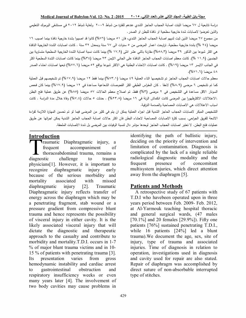
Results Age of patients ranged between 5 to 67 years, with a median age of 32 yr. penetrating type of trauma was predominant in 51 patients [76 % ] of whom 23 patients [34.3%] had right sided TDI, 19 patients [28.3%] had left sided TDI, and 9 patients (13.4%) had bilateral TDI in comparison to 16 patients [24%] that sustained blunt
trauma, of whom only 1 patient [1.4 %] had right sided TDI while left sided TDI was seen in 13 patient [19.6%] and only 2 patients [3%] had bilateral TDI of this patient group and the age group 21-40 was commonly affected by trauma in which 43.28% [ 29/67] as demonstrated in table (1).
Table 1: Distribution of cases due to type of mechanism according to age group
Total Blunt mechanism Penetrating mechanism Age (year) % No. Both Left Right Both Left Right
% No. % No.
% No. % No. % No. % No.
16.42 11 0 0 3 2 0 0 3 2 4.4 3 5.9 4 1-20 43.28 29 0 0 5.9 4 1.4 1 7.4 5 11.9 8 16.4 11 21-40 34.33 23 3 2 7.4 5 0 0 3 2 10.4 7 10.4 7 41-60 5.97 4 0 0 3 2 0 0 0 0 1.4 1 1.4 1 60˃ 100% 67 3 2 19.4 13 1.4 1 13.4 9 28.3 19 34.3 23 total
From table (2) it was founded that Male was affected by both types of injuries of diaphragm more than in female in which
70.15% of cases were among men also right side injury was more than the left side for both sex
Table 2: Distribution of cases due to mechanism of injuries according to sex .
Total Blunt mechanism Penetrating mechanism Sex % No. Both Left Right Both Left Right
% No. % No. % No. % No. % No. % No. 70.15 47 1.4 1 8.9 6 1.4 1 11.9 8 19.4 13 26.8 18 Male 29.85 20 1.4 1 10.4 7 0 0 1.4 1 8.9 6 7.4 5 Female 100% 67 3 2 19.4 13 1.4 1 13.4 9 28.3 19 34.3 23 Total
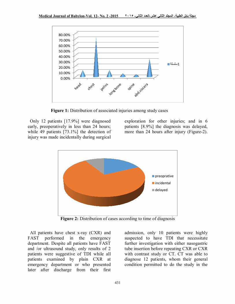
Other injuries were accompanying those patients included trauma to head 8 patients , chest 48 patients , pelvis 4 patients, long bones 22 patients, spine 3 patients and abdominal viscera 52 patients. The associated injury severity in blunt TDI patients was greater than in penetrating TDI
patients. Head injuries and pelvic fractures were associated mainly with blunt TDI, whereas cardiac injuries were mainly associated with penetrating TDI. Lung,chest wallor thoracic organs injury was more common in blunt trauma (Figure -1).
Medical Journal of Babylon-Vol. 12- No. 2 -2015 مجلة بابل الطبیة- المجلد الثاني عشر-العدد الثاني- ٢٠١٥
431
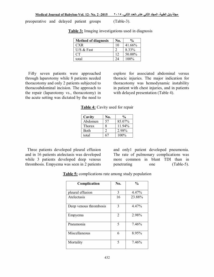
Figure 1: Distribution of associated injuries among study cases Only 12 patients [17.9%] were diagnosed early, preoperatively in less than 24 hours; while 49 patients [73.1%] the detection of injury was made incidentally during surgical
exploration for other injuries; and in 6 patients [8.9%] the diagnosis was delayed, more than 24 hours after injury (Figure-2).
Figure 2: Distribution of cases according to time of diagnosis
All patients have chest x-ray (CXR) and FAST performed in the emergency department. Despite all patients have FAST and /or ultrasound study, only results of 2 patients were suggestive of TDI while all patients examined by plain CXR at emergency department or who presented later after discharge from their first
admission, only 10 patients were highly suspected to have TDI that necessitate further investigation with either nasogastric tube insertion before repeating CXR or CXR with contrast study or CT. CT was able to diagnose 12 patients, whom their general condition permitted to do the study in the
0.00%10.00%20.00%30.00%40.00%50.00%60.00%70.00%80.00%
1سلسلة
preoprative
incidental
delayed
Medical Journal of Babylon-Vol. 12- No. 2 -2015 مجلة بابل الطبیة- المجلد الثاني عشر-العدد الثاني- ٢٠١٥
432
preoperative and delayed patient groups (Table-3). Table 3: Imaging investigations used in diagnosis
Method of diagnosis No. % CXR 10 41.66% U/S & Fast 2 8.33% CT 12 50.00% total 24 100%
Fifty seven patients were approached through laparotomy while 8 patients needed thoracotomy and only 2 patients subjected to thoracoabdominal incision. The approach to the repair (laparotomy vs., thoracotomy) in the acute setting was dictated by the need to
explore for associated abdominal versus thoracic injuries. The major indication for thoracotomy was hemodynamic instability in patient with chest injuries, and in patients with delayed presentation (Table 4).
Table 4: Cavity used for repair
Cavity No. % Abdomen 57 85.07% Thorax 8 11.94% Both 2 2.98% total 67 100%
Three patients developed pleural effusion and in 16 patients atelectasis was developed while 3 patients developed deep venous thrombosis. Empyema was seen in 2 patients
and only1 patient developed pneumonia. The rate of pulmonary complications was more common in blunt TDI than in penetrating one (Table-5).
Table 5: complications rate among study population
Complication
No. %
pleural effusion 3 4.47% Atelectasis
16 23.88%
Deep venous thrombosis
3 4.47%
Empyema
2 2.98%
Pneumonia
5 7.46%
Miscellaneous
6 8.95%
Mortality
5 7.46%
Medical Journal of Babylon-Vol. 12- No. 2 -2015 مجلة بابل الطبیة- المجلد الثاني عشر-العدد الثاني- ٢٠١٥
433
Traumatic diaphragmatic hernia was present in 22 of 67 patients (33%) and was localized to the left hemidiaphragm in 17 [77%]. The incidence of hernia in blunt trauma [12 of 16, %75] was higher than in penetrating trauma [10 of 51, %19.5]. The stomach was the organ most frequently found herniating into the chest [12 of 22, %54.5], followed by the spleen [6 of 22, %27], and small bowel [4 of 22, %18]. The overall mortality from TDI was 7 %[ 5 of 67]. The blunt 3% [2 of 67] and penetrating 4% [3 of 67]. The 5 patients who died had associated injuries requiring operative intervention and the main causes of death were traumatic brain injury hemorrhagic shock. Discussion In previously published series, the incidence of blunt injuries to the diaphragm was almost threefold that of penetrating injuries [6-8]. In the present study, we have shown the exact opposite, with penetrating trauma accounting for 76.2% of TDI vs. 23.8% for blunt, which is similar of study by Hanna et al. [9]
The plain chest x-ray has been reported to be useful for the diagnosis of diaphragmatic injury, with sensitivities ranging of 30 % to 62% [10] in the absence of a diaphragmatic hernia and up to 94% in the presence of a hernia [11]. Most of these studies used chest x-ray’s interpretations by a radiologist. On the other hand we found that chest x-ray is much less accurate in diagnosing a TDI [10 patients [15%] of 67 patients] when interpreted by surgical team. Intraoperative identification remains the gold standard for the diagnosis and treatment of TDI. Exploration of the abdomen has traditionally been advocated because it allows concurrent examination of the often–injured abdominal organs [12-14]. The data we have provided support this approach. For penetrating TDI, we found that intra-abdominal organs were injured in
most cases. In some cases [8 (%12) of patients] a thoracotomy was required to repair intrathoracic organs. Despite the greater incidence of life threatening injuries in blunt TDI, we do not identify increased mortality rate for this subset [4%] compared with penetrating [3%] TDI. This differs from the results of other studies that almost uniformly cite an increased mortality rate for blunt [2-5, 7, 12, 14] Penetrating injury of the diaphragm occurs with almost equal frequency to both sides [15]. In our patients, left to right TDI was [34% to 27%]. However, blunt injury of left hemidiaphragm has been observed more often (19 % to 1%) [16-18]. The treatment of traumatic diaphragmatic hernia is surgical repair and it should be performed shortly after the diagnosis is made .CT scanning with axial, sagittal and coronal reformations is reported to reach a sensitivity of 50% and 78% in diagnosing right and left diaphragmatic injuries respectively [19-21]. However this technique cannot be performed in emergency situations or multitrauma patients [22, 23]. Laparotomy is necessarily used when other abdominal injuries are associated. Thoracotomy is performed when there are thoracic injuries, a large tear, a large herniation and an empyema is developed [24-27]. Strong intrathoracic adhesions, due to old herniation are easily treated with intrathoracic approach [28]. Conclusion A preoperative diagnosis of TDI should be made in large number of patients; however, the surgical team must improve their x- ray interpretation skills. The high association of intraabdominal injuries irrespective of the location of penetrating wounds mandates that TDI be approached from the abdomen in patients who require exploration.
Medical Journal of Babylon-Vol. 12- No. 2 -2015 مجلة بابل الطبیة- المجلد الثاني عشر-العدد الثاني- ٢٠١٥
434
Blunt trauma by itself is no longer a predictor of death from TDI. Rather, severe associated injuries are associated with a higher mortality rate. References (1) Rubikas R. Diaphragmatic injuries. Eur. J. Cardiothorac. Surg. 2001; 20(1): 53-57. (2) Degiannis E, Levy RD, Sofianos C, Potokar T, Florizoone MG, Saadia R. Diaphragmatic herniation after penetrating trauma. Br.J.Surg. 1996 Jan; 83(1):88-91. (3) Mihos P, Potaris K, Gakidis, et al. Traumatic rupture of the diaphragm: experience with 65 patients.Injury 2003; 3: 169- 72. (4) Clarke DL, Greatorex B, Oosthuizen GV, Muckart DJ. The spectrum of diaphragmatic injury in a busy metropolitan surgical service. Injury 2009; 40:932-7 (5) Christiansen L A, Stage P, Bille Brahe E, Bertelsen S. Rupture of the diaphragmThorax 1974; 29,:559- 563. (6) Shah R, Sabaratnam S, Mearns A. Traumatic rupture of diaphragm. Ann Thorac Surg 1995;60: 1444 –9. (7) Rubikas R. Diaphragmatic injuries. Eur J CardiothoracSurg 2001; 20:53–57. (8) Beauchamp G, Khalfallah A, Girard R, Dube S, Laurendeau F, Legros G. Blunt diaphragmatic rupture. Am. J Surg., 1984; 148:292–5. (9) Waël C. Hanna, Lorenzo E. Ferri, Paola Fata, Tarek Razek, and David S. Mulder.The Current Status of Traumatic DiaphragmaticInjury: Lessons Learned From 105 Patients Over 13 Years. Ann ThoracSurg 2008;85:1044–8 (10) Payne JH Jr, Yellin AE. Traumatic diaphragmatic hernia.ArchSurg 1982; 117: 18 –24. (11) Waldschmidt ML, Laws HL. Injuries of the diaphragm.J Trauma 1980; 20: 587–91.
(12) Grimes OF. Traumatic injuries of the diaphragm. Diaphragmatichernia. Am J Surg 1974; 128:175- 181. (13). Broos PLO, Rommens PM, Carlier H, Van LeeuwenJE,Somville FJ, Gruwez JA. Traumatic Rupture of the diaphragm. Review of 62 successive cases. Int Surg 1989; 74:88–92. (14) Demetriades D, Kakoyiannis S, Parekh D, Hatzitheofilou C. Penetrating injuries of the diaphragm. Br J Surg 1988; 75:824–6. (15) Miller LW, Bennett EV Jr, Root HD, et al: Management ofpenetrating and blunt diaphragmatic injury. J Trauma 24: 403, 1984. (16) Hood RM: Traumatic diaphragmatic hernia (collective review).Ann ThoracSurg 12:311, 1971 (17) Mansour KA, Clements JL, Hatcher CR, et al: Diaphragmatichernia caused by trauma: experience with 35 cases.Am Surg 41:97, 1975 (18) Brown GL, Richardson JD: Traumatic diaphragmatic hernia:a continuing challenge. Ann ThoracSurg39:170, 1985. (19). Reber PU, Schmied B, Seiler CA, et al. Missed diaphragmatic injuries and their long termsequelae. J Trauma 1998; 44 (1): 183-8.) (20) Murray JG, Caoili E, Gruden JF, et al. Acute rupture of the diaphragm due to blunt trauma: diagnostic sensitivity and specifity of CT. AJR 1996. (21) Rosati C. Acute traumatic injury of the diaphragm. ChestSurgClin North Am 1998; 8 (2): 371-9. (22) Shanmuganathan K, Mirvis SE, White CS, et al. MR Imaging evaluation of hemi diaphragms in acute blunt trauma: experience with 16 patients. AJR 1996; 167: 397-402. (23) Killeen KL, Mirvis SE, Shanmuganathan K. Helical CT of diaphragmatic rupture caused by blunt trauma. AJR 1999; 173 (6): 1611-6. (24) Van Vugt AB, Schoots FJ. Acute diaphragmatic rupture due to blunt trauma:

Medical Journal of Babylon-Vol. 12- No. 2 -2015 مجلة بابل الطبیة- المجلد الثاني عشر-العدد الثاني- ٢٠١٥
435
a retrospective analysis. J Trauma 1989; 29: 683. (25) Miller LW, Bennet EV, Root HD, et al. Management of penetrating and blunt diaphragmatic injury. J Trauma 1984; 24: 403. (26) Liu DW, Liu HP, Lin PJ, et al. Video-assisted thoracic surgeryin treatment of chest trauma. J Trauma 1997; 42: 670. (27) Ochsner MG, Rozycki GS, Lucente F, et al. Prospective evaluation of
thoracoscopy for diagnosing diaphragmatic injury in thoraco-abdominal trauma: a preliminary report. J Trauma 1993; 34: 704. (28) Beltrami V, Mucilli M, Di Nuzzo D. Post-traumatic diaphragmatic hernia. Second world week of professional updatingin surgery and in surgical and oncological disciplines of the University of Milan. Lecture Book, Monduzzi Editore, Milano1990; 138-40
Left diaphragmatic tear in a 24-year-old woman who was injured in a motor vehicle accident. Initial chest radiograph shows intrathoracic herniation of the stomach (S), a pleural effusion, a pulmonary contusion, and contralateral mediastinal shift .
Medical Journal of Babylon-Vol. 12- No. 2 -2015 مجلة بابل الطبیة- المجلد الثاني عشر-العدد الثاني- ٢٠١٥
436
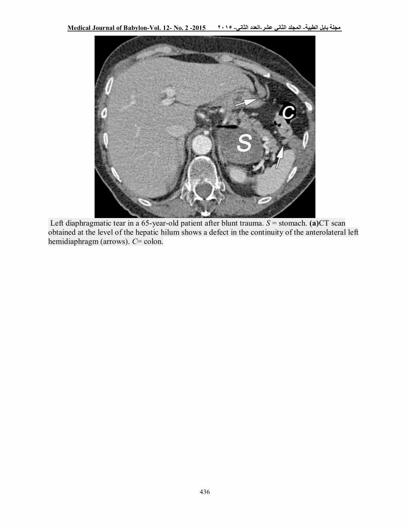
Left diaphragmatic tear in a 65-year-old patient after blunt trauma. S = stomach. (a)CT scan obtained at the level of the hepatic hilum shows a defect in the continuity of the anterolateral left hemidiaphragm (arrows). C= colon.


![[Treating frostbite injuries]](https://static.fdokumen.com/doc/165x107/633ff39332b09e4bae09a1b5/treating-frostbite-injuries.jpg)