![[Treating frostbite injuries]](https://static.fdokumen.com/doc/165x107/633ff39332b09e4bae09a1b5/treating-frostbite-injuries.jpg)
Football Injuries
9
Football Injuries: Current Concepts David E. Olson, MD 1,2 , Robby Singh Sikka, MD 3 , Abigail Hamilton, MD 4 , and Austin Krohn, MD 2 Abstract Football is one of the most popular sports in the United States and is the leading cause of sports-related injury. A large focus in recent years has been on concussions, sudden cardiac death, and heat illness, all thought to be largely preventable health issues in the young athlete. Injury prevention through better understanding of injury mechanisms, education, proper equipment, and practice techniques and preseason screening may aid in reducing the number of injuries. Proper man- agement of on-field injuries and health emergencies can reduce the morbidity associated with these injuries and may lead to faster return to play and reduced risk of future injury. This article reviews current concepts surrounding frequently seen football-related injuries. Introduction In 2005, nearly two million high school, collegiate, and professional athletes played football (58,76). Participation has increased steadily, with more than 100,000 additional participants each of the last 10 years (59,76). Football is the leading cause of sports-related injury, with an injury rate twice that of basketball, the nation’s second most popular sport. An estimated 300,000 to 1.2 million high school athletes sustain football-related injuries annually (1,76). Studies examining the epidemiology of football-related injuries often focus on injuries to specific body parts (e.g., knee or ankle) or on a particular diagnosis (e.g., concussion) rather than considering all injuries. Variations in injury diagnosis and reporting also limit evaluation of such data. Guskiewicz et al. (33) reported that concussions made up a greater proportion of injuries among high school players than among college players (5.5% vs 4.4%). Shankar et al. (76) reported that the rate of injury was five times higher in games than in practice at both high school and collegiate levels. The most commonly injured sites were the knee and ankle. The overall rate of injury was higher in the National Collegiate Athletic Association (NCAA) (8.61 per 1,000 athlete exposures) than in high school (4.36 per 1,000 athlete exposures), but high school players sustained a greater proportion of fractures and concussions. Tackling or being tackled resulted in the greatest number of injuries. Running plays were the leading cause of injury, with running backs and linebackers as the most likely to be injured. Running plays also accounted for the majority of concussions and season-ending injuries. Career-ending injuries constituted fewer than 0.1% of all injuries. Collegiate athletes were more likely to sustain overuse noncontact injuries than high school athletes (76). Certain injuries are more prevalent in football, and recent media attention has focused on the topics of concussions, heat illness, and sickle trait, among others, in this population. Other musculoskeletal injuries frequently associated with football may include anterior cruciate ligament (ACL) in- juries, femoroacetabular impingement (FAI), shoulder dislo- cations, and stingers. This article reviews these injuries and illnesses associated with football. Preparticipation Physical Examination Current recommendations for preparticipation physical examination (PPE) screening are based largely on consensus opinion from experts, as current research has failed to link the PPE with improved outcomes regarding morbidity and mortality of athletes (6,16). Key components of any PPE are a detailed medical history and a focused physical examina- tion, with emphasis on cardiovascular examination. In the United States, screening with further cardiovascular testing (i.e., electrocardiography, echocardiography, or exercise stress testing) is not recommended by the American Heart Association (AHA) (74). In addition, laboratory testing in asymptomatic athletes has failed to meet the standards expected for medical screening tests (i.e., sensitivity, specif- icity, predictive value, or improved outcomes) (5,27,42,57). In a review of 2,574 athletes, history alone elicited 88% of all abnormal findings and 58% of the reasons cited activity restriction (69). Special attention should be paid to personal and family histories of cardiovascular health issues because cardiac causes are the leading cause of sudden death in athletes (56). The incidence of sudden cardiac death (SCD) in U.S. high school athletes is estimated to be 1 in every 200,000 per year (46). Hypertrophic cardiomyopathy has Sport-Specific Illness and Injury 290 Volume 10 c Number 5 c September/October 2011 Football Injuries 1 Minnesota Vikings, Minneapolis, MN; 2 Department of Family Medicine, University of Minnesota, Minneapolis, MN; 3 Department of Anaesthesiology, University of Minneapolis, Minneapolis, MN; and 4 TRIA Orthopaedic Center, Minneapolis, MN Address for correspondence: Robby Singh Sikka, MD, TRIA Orthopaedic Center, 8100 Northland Drive, Minneapolis, MN 55431 (E-mail: [email protected]). 1537-890x/1005/290Y298 Current Sports Medicine Reports Copyright * 2011 by the American College of Sports Medicine Copyright © 2011 by the American College of Sports Medicine. Unauthorized reproduction of this article is prohibited.
-
Upload
independent -
Category
Documents
-
view
1 -
download
0
Transcript of Football Injuries

Football Injuries: Current ConceptsDavid E. Olson, MD1,2, Robby Singh Sikka, MD3, Abigail Hamilton, MD4, and Austin Krohn, MD2
AbstractFootball is one of the most popular sports in the United States and isthe leading cause of sports-related injury. A large focus in recent yearshas been on concussions, sudden cardiac death, and heat illness, allthought to be largely preventable health issues in the young athlete.Injury prevention through better understanding of injury mechanisms,education, proper equipment, and practice techniques and preseasonscreening may aid in reducing the number of injuries. Proper man-agement of on-field injuries and health emergencies can reduce themorbidity associated with these injuries and may lead to faster return toplay and reduced risk of future injury. This article reviews currentconcepts surrounding frequently seen football-related injuries.
IntroductionIn 2005, nearly two million high school, collegiate, and
professional athletes played football (58,76). Participationhas increased steadily, with more than 100,000 additionalparticipants each of the last 10 years (59,76). Football is theleading cause of sports-related injury, with an injury ratetwice that of basketball, the nation’s second most popularsport. An estimated 300,000 to 1.2 million high schoolathletes sustain football-related injuries annually (1,76).
Studies examining the epidemiology of football-relatedinjuries often focus on injuries to specific body parts (e.g.,knee or ankle) or on a particular diagnosis (e.g., concussion)rather than considering all injuries. Variations in injurydiagnosis and reporting also limit evaluation of such data.Guskiewicz et al. (33) reported that concussions made up agreater proportion of injuries among high school playersthan among college players (5.5% vs 4.4%). Shankar et al.(76) reported that the rate of injury was five times higher ingames than in practice at both high school and collegiatelevels. The most commonly injured sites were the knee andankle. The overall rate of injury was higher in the NationalCollegiate Athletic Association (NCAA) (8.61 per 1,000
athlete exposures) than in high school(4.36 per 1,000 athlete exposures), buthigh school players sustained a greaterproportion of fractures and concussions.Tackling or being tackled resulted in thegreatest number of injuries. Runningplays were the leading cause of injury,with running backs and linebackers asthe most likely to be injured. Runningplays also accounted for the majority ofconcussions and season-ending injuries.Career-ending injuries constituted fewerthan 0.1% of all injuries. Collegiateathletes were more likely to sustain
overuse noncontact injuries than high school athletes (76).Certain injuries are more prevalent in football, and recent
media attention has focused on the topics of concussions,heat illness, and sickle trait, among others, in this population.Other musculoskeletal injuries frequently associated withfootball may include anterior cruciate ligament (ACL) in-juries, femoroacetabular impingement (FAI), shoulder dislo-cations, and stingers. This article reviews these injuries andillnesses associated with football.
Preparticipation Physical ExaminationCurrent recommendations for preparticipation physical
examination (PPE) screening are based largely on consensusopinion from experts, as current research has failed to linkthe PPE with improved outcomes regarding morbidity andmortality of athletes (6,16). Key components of any PPE area detailed medical history and a focused physical examina-tion, with emphasis on cardiovascular examination. In theUnited States, screening with further cardiovascular testing(i.e., electrocardiography, echocardiography, or exercisestress testing) is not recommended by the American HeartAssociation (AHA) (74). In addition, laboratory testing inasymptomatic athletes has failed to meet the standardsexpected for medical screening tests (i.e., sensitivity, specif-icity, predictive value, or improved outcomes) (5,27,42,57).
In a review of 2,574 athletes, history alone elicited 88%of all abnormal findings and 58% of the reasons citedactivity restriction (69). Special attention should be paid topersonal and family histories of cardiovascular health issuesbecause cardiac causes are the leading cause of sudden deathin athletes (56). The incidence of sudden cardiac death (SCD)in U.S. high school athletes is estimated to be 1 in every200,000 per year (46). Hypertrophic cardiomyopathy has
Sport-Specific Illness and Injury
290 Volume 10 c Number 5 c September/October 2011 Football Injuries
1Minnesota Vikings, Minneapolis, MN; 2Department of FamilyMedicine, University of Minnesota, Minneapolis, MN; 3Departmentof Anaesthesiology, University of Minneapolis, Minneapolis, MN;and 4TRIA Orthopaedic Center, Minneapolis, MN
Address for correspondence: Robby Singh Sikka, MD, TRIA OrthopaedicCenter, 8100 Northland Drive, Minneapolis, MN 55431(E-mail: [email protected]).
1537-890x/1005/290Y298Current Sports Medicine ReportsCopyright * 2011 by the American College of Sports Medicine
Copyright © 2011 by the American College of Sports Medicine. Unauthorized reproduction of this article is prohibited.

been reported as the most common cause of SCD at 36%,followed by congenital coronary anomalies at 17% (45,47).The AHA recommends 12 critical points of the cardio-vascular examination, with 8 of these points coming fromthe history (Table 1) (74). Pertinent examinations shouldfocus also on history of asthma, infectious skin or systemicdisease, seizures, previous heat illness, single bilateral organs,previous injuries/restrictions, concussion history, and pre-ventive health (i.e., sexual history, alcohol use, nicotineuse) (Table 2) (68).
A focused approach is more efficient and practical tothe PPE than a complete and thorough physical examina-tion. The AHA recommends four specific points to cover incardiovascular examination (Table 1) (74). Any athlete withcardiovascular symptoms or signs such as those outlined by theAHA requires a careful and thorough evaluation and referralto a cardiologist for electrocardiography, echocardiography,or stress testing. The remainder of the physical examinationshould include vital signs; general appearance; ear/nose/throat,visual acuity, pulmonary, abdominal, skin, and hernia checks;and musculoskeletal screening. If the athlete has a previousinjury or other signs or symptoms detected by the generalscreening or history, then supplemental site-specific examina-tion and imaging are appropriate.
ConcussionThe incidence of concussion is difficult to assess and likely
is underreported because of the rapid onset and sometimes
spontaneous resolution of neurologic impairments. Furthercomplicating the issue is the lack of standardization amongcoaches, athletic trainers, and other providers in assessing
Table 1.AHA key points in cardiovascular portion of PPE (74).
Personal history
1. Any chest pain/discomfort with activity
2. Syncope/near-syncope with activity (not vasovagal)
3. Excessive dyspnea/fatigue with activity
4. History of heart murmur
5. History of hypertension
Family history
6. Premature death (sudden/unexpected or otherwise)before age 50 yr due to heart disease
7. Disability from heart disease before age 50 yr
8. History of hypertrophic cardiomyopathy, long-QTsyndrome, Marfan syndrome, or other arrhythmias
Physical examination
9. Noninnocent heart murmur (at least III/VI in quality,increases with Valsalva maneuver, or diastolic)
10. Palpation of femoral and brachial pulses to excludecoarctation of the aorta
11. Physical stigmata of Marfan syndrome (kyphoscoliosis,pectus excavatum, arachnodactyly, hyperlaxity, armspan greater than height, myopia, aorticinsufficiency, MVP)
12. Blood pressure measurement
Table 2.Medical conditions and sports participation (68).
Cardiovascular V suspicion of any of the following shouldlead to further evaluation, most often with a cardiologist
Structural/acquired heart disease: hypertrophiccardiomyopathy, coronary artery anomalies,arrhythmogenic right ventricular cardiomyopathy,acute rheumatic fever with carditis, Ehlers-Danlossyndrome, vascular-form Marfan syndrome, mitralvalve prolapse, anthracycline use
Vasculitis/vascular disease: Kawasaki disease,pulmonary hypertension
Others: Myocarditis, hypertension (995% for age),congenital heart defects, dysrhythmias,murmurs (unless innocent in quality)
Musculoskeletal
Atlantoaxial instability V needs further evaluation
Previous injuries/instability V needs further evaluation
Obesity V clear to participate
Hematologic
Bleeding disorder V needs further evaluation
Sickle cell disease V needs further evaluation
Sickle cell trait V clear to participate
General medical
Asthma V clear to participate with proper medication/education
Diabetes V clear to participate
Acute illness (diarrhea, fever, respiratory infection) V needsfurther evaluation
Eating disorders V needs further evaluation
Cerebral palsy V needs further evaluation
Malabsorption or short bowel V needs further evaluation
Previous heat illness V needs further evaluation
Skin infections V during contagious period, participation insports with mats or physical contact is not allowed
Unilateral organ
Kidney V needs further evaluation for contact, collision, andlimited-contact sports
Eye V needs further evaluation
Ovary V clear to participate
Testicle V clear to participate (may need protective cup)
Others
Hepatitis V clear to participate
Human immunodeficiency virus V clear to participate
Seizure disorder V base clearance on quality of control andindividual sport
www.acsm-csmr.org Current Sports Medicine Reports 291
Copyright © 2011 by the American College of Sports Medicine. Unauthorized reproduction of this article is prohibited.

and managing concussion. With increased efforts towardeducation and raising awareness of concussions, the NCAA’sInjury Surveillance System showed an average annualincrease of 7.0% from the 1988 to 1989 through the 2003 to2004 seasons (P G 0.01) (35). Table 3 displays the concussionrate in both high school and collegiate athletes in practiceand in competition from 2005 to 2006 (23).
Studies examining the incidence of concussions and mea-sures to decrease the risk of concussions have used variable
definitions of concussions over time; thus, comparisons aredifficult. In addition, conflicts of interest including industry-sponsored studies have made interpretation challenging. Ina study of 2,141 high school athletes, Collins et al. (23) didshow a 31% decreased relative risk and 2.3% (P = 0.027)decreased absolute risk for sustaining a concussion in ath-letes wearing the Revolution helmet manufactured byRiddell, Inc. (Chicago, IL). However, this study was spon-sored by Riddell, and helmets were of different ages, com-plicating the interpretation of such data. Overall, researchregarding helmets, mouth guards, and other personal pro-tective equipment is limited in demonstrating prevention ofconcussion. Improvements in equipment provide potential,but more immediate benefits in concussion severity andincidence will stem from rule changes, improved education,and proper coaching/training (25).
The Zurich Conference endorses the Pocket Sport Con-cussion Assessment Tool (SCAT2), which is a condensedversion of its guidelines and is appropriate for use on the fieldof play (Fig.). The National Football League (NFL) Head,Neck, and Spine Medical Committee also has endorsedusing a modified SCAT2 for sideline evaluation of playerswith possible concussions. The SCAT2 has many of theworrisome signs and symptoms of concussion listed andprovides a tool for sports medicine professionals to assessconcussion rapidly. On-the-field assessment should startwith completing the CABs (compressions-airway-breath-ing), if needed, and clearing the cervical spine. After this, theremainder of the examination should be completed in aquiet location. Evidence has shown that standard orientationquestions (e.g., time, place, person) have been unreliable in thesporting situation when compared with memory assessment
Table 3.Concussion rates in U.S. high school and collegiate athletes inpractice and competition, 2005 to 2006 V rates more than 1,000athlete exposuresa (23).
Sport Division Practice Competition Overall
Football High school 0.21 1.55 0.47
Collegiate 0.39 3.02 0.61
Men’s soccer High school 0.04 0.59 0.22
Collegiate 0.24 1.38 0.49
Men’s basketball High school 0.06 0.11 0.07
Collegiate 0.22 0.45 0.27
Men’s sport total High school 0.13 0.61 0.25
Collegiate 0.30 1.26 0.45
aExposure defined as one athlete playing in one game or practice.
[Adapted from Armstrong LE. Exertional heat stroke in American
football: persistent battles, research frontiers. Curr. Sports Med. Rep.2010; 9:125Y7.]
Figure: Version of the SCAT2 concussion assessment.
292 Volume 10 c Number 5 c September/October 2011 Football Injuries
Copyright © 2011 by the American College of Sports Medicine. Unauthorized reproduction of this article is prohibited.
(44). In addition, the SCAT2, CogSport, HeadMinder, andother abbreviated assessment methods are meant for rapidconcussion screening on the sidelines and are not meant toreplace comprehensive neuropsychological testing (25).
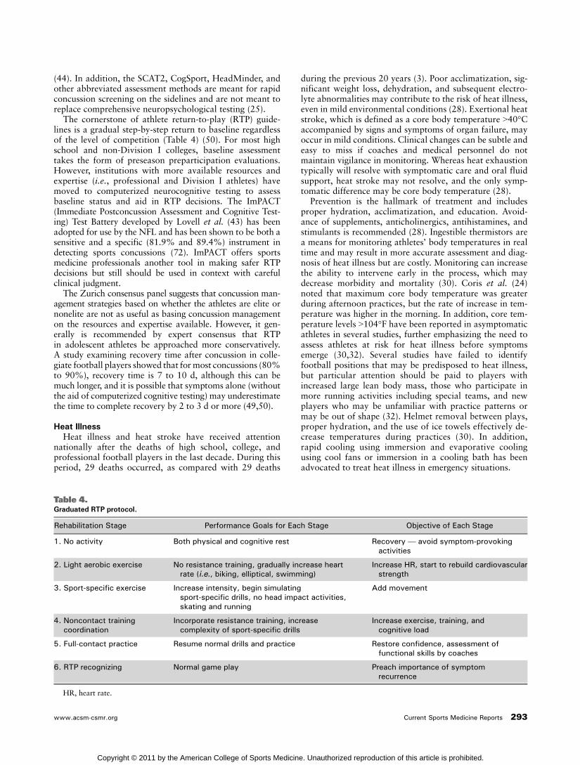
The cornerstone of athlete return-to-play (RTP) guide-lines is a gradual step-by-step return to baseline regardlessof the level of competition (Table 4) (50). For most highschool and non-Division I colleges, baseline assessmenttakes the form of preseason preparticipation evaluations.However, institutions with more available resources andexpertise (i.e., professional and Division I athletes) havemoved to computerized neurocognitive testing to assessbaseline status and aid in RTP decisions. The ImPACT(Immediate Postconcussion Assessment and Cognitive Test-ing) Test Battery developed by Lovell et al. (43) has beenadopted for use by the NFL and has been shown to be both asensitive and a specific (81.9% and 89.4%) instrument indetecting sports concussions (72). ImPACT offers sportsmedicine professionals another tool in making safer RTPdecisions but still should be used in context with carefulclinical judgment.
The Zurich consensus panel suggests that concussion man-agement strategies based on whether the athletes are elite ornonelite are not as useful as basing concussion managementon the resources and expertise available. However, it gen-erally is recommended by expert consensus that RTPin adolescent athletes be approached more conservatively.A study examining recovery time after concussion in colle-giate football players showed that for most concussions (80%to 90%), recovery time is 7 to 10 d, although this can bemuch longer, and it is possible that symptoms alone (withoutthe aid of computerized cognitive testing) may underestimatethe time to complete recovery by 2 to 3 d or more (49,50).
Heat IllnessHeat illness and heat stroke have received attention
nationally after the deaths of high school, college, andprofessional football players in the last decade. During thisperiod, 29 deaths occurred, as compared with 29 deaths
during the previous 20 years (3). Poor acclimatization, sig-nificant weight loss, dehydration, and subsequent electro-lyte abnormalities may contribute to the risk of heat illness,even in mild environmental conditions (28). Exertional heatstroke, which is defined as a core body temperature 940-Caccompanied by signs and symptoms of organ failure, mayoccur in mild conditions. Clinical changes can be subtle andeasy to miss if coaches and medical personnel do notmaintain vigilance in monitoring. Whereas heat exhaustiontypically will resolve with symptomatic care and oral fluidsupport, heat stroke may not resolve, and the only symp-tomatic difference may be core body temperature (28).
Prevention is the hallmark of treatment and includesproper hydration, acclimatization, and education. Avoid-ance of supplements, anticholinergics, antihistamines, andstimulants is recommended (28). Ingestible thermistors area means for monitoring athletes’ body temperatures in realtime and may result in more accurate assessment and diag-nosis of heat illness but are costly. Monitoring can increasethe ability to intervene early in the process, which maydecrease morbidity and mortality (30). Coris et al. (24)noted that maximum core body temperature was greaterduring afternoon practices, but the rate of increase in tem-perature was higher in the morning. In addition, core tem-perature levels 9104-F have been reported in asymptomaticathletes in several studies, further emphasizing the need toassess athletes at risk for heat illness before symptomsemerge (30,32). Several studies have failed to identifyfootball positions that may be predisposed to heat illness,but particular attention should be paid to players withincreased large lean body mass, those who participate inmore running activities including special teams, and newplayers who may be unfamiliar with practice patterns ormay be out of shape (32). Helmet removal between plays,proper hydration, and the use of ice towels effectively de-crease temperatures during practices (30). In addition,rapid cooling using immersion and evaporative coolingusing cool fans or immersion in a cooling bath has beenadvocated to treat heat illness in emergency situations.
Table 4.Graduated RTP protocol.
Rehabilitation Stage Performance Goals for Each Stage Objective of Each Stage
1. No activity Both physical and cognitive rest Recovery V avoid symptom-provokingactivities
2. Light aerobic exercise No resistance training, gradually increase heartrate (i.e., biking, elliptical, swimming)
Increase HR, start to rebuild cardiovascularstrength
3. Sport-specific exercise Increase intensity, begin simulatingsport-specific drills, no head impact activities,skating and running
Add movement
4. Noncontact trainingcoordination
Incorporate resistance training, increasecomplexity of sport-specific drills
Increase exercise, training, andcognitive load
5. Full-contact practice Resume normal drills and practice Restore confidence, assessment offunctional skills by coaches
6. RTP recognizing Normal game play Preach importance of symptomrecurrence
HR, heart rate.
www.acsm-csmr.org Current Sports Medicine Reports 293
Copyright © 2011 by the American College of Sports Medicine. Unauthorized reproduction of this article is prohibited.

Casa and Csillan (18) reported on recommendationsfrom the American College of Sports Medicine using a 14-dheat acclimatization period for secondary school athletes.Participation in only one practice was recommended forthe first 5 d of practice, and total practice time should notexceed more than 3 h in 1 d. A 3-h recovery period shouldbe included if the player participates in a walk-through for asecond practice (11). Temperatures may increase up to 1-Fevery 10 min in the absence of thermoregulation in thebody, which may occur during exertional heat stroke (24,30).Significant physiologic adaptations occur during days 8 to 14of exercise-induced heat exposure. Limited uniform itemsduring initial workouts may be beneficial in allowing athletesto acclimatize (18).
Recently, the NCAA recommended that all incomingDivision I student athletes be tested for sickle cell trait,show proof of a previous test, or sign a waiver releasing aninstitution from liability if they decline testing (8). Theprevalence of sickle cell trait in the African-American pop-ulation is 8% (2). At least 15 college football players withsickle cell trait have died from complications of exertionalsickling in the last two decades (2). In sickle cell trait,maximal sustained exercise may cause hypoxemia, acidosis,hyperthermia, and red cell dehydration, all of which maycontribute to sickling, potentially causing fulminant rhab-domyolysis. Simple screening and precautions may preventsevere complications and allow for timely and effectivemanagement of acute rhabdomyolysis. Acute rhabdomyol-ysis should be differentiated from exertional heat illnesson the basis of history, core body temperature, and responseto physical training (2).
Tibiofibular Syndesmotic InjuriesHigh ankle sprains or syndesmotic injuries are more
likely to produce prolonged disability compared with lateralankle sprains (86,87). Boytim et al. (9) reviewed a series of98 ankle injuries in professional football players and notedthat 18 were syndesmotic sprains. They described the typi-cal mechanism of injury as an external rotation force ap-plied to the foot with respect to the tibia/fibula. In a seriesof three professional football teams with 36 total injuries,Sikka et al. (78) reported a correlation between an increasednumber of missed practices and games with higher grades ofinjury according to MRI as well as with a positive squeezetest. Grade III injuries included injury to the posteriortibiofibular ligament, and nearly all athletes with grade IIIinjuries missed games. Subjective complaints and functionalsymptoms were the primary factors in determining whena player could return to competition. MRI was useful inclearly defining the diagnosis and associated injuries andcould be used in helping determine prognosis after a syn-desmotic sprain in the absence of mortise widening on stressradiographs (78). Results from plain radiographs are oftennormal, and stress films may show widening of the mortisein grade IV injuries, even in the absence of fracture, but maybe normal in grade I to III injuries.
Brown et al. (14) reported anterior talofibular injuries in74% of athletes with syndesmotic injuries. Combined lat-eral ligament injuries also were evaluated in the series bySikka et al. (78) and were predictive of increased swellingand pain, as well as increased missed practice time; how-
ever, these did not result in an increase in missed games.Boytim et al. (9) reported that lateral ankle injuries causedan average of 1.1 missed practices and 0.04 missed games,whereas syndesmotic injuries resulted in an average of 6.3missed practices and 1.4 missed games. It is likely that theseverity of the syndesmotic injury is a stronger predictor ofmissed games than the associated lateral ligament injury incombined injuries (78). Taping and limiting of dorsiflexionmay reduce pain. Early range-of-motion exercises and pro-gressive strengthening also may be of benefit.
Sports HerniaGroin injuries are not uncommon among football players
and often are difficult to accurately diagnose because of alarge number of potential sources of groin pain, the com-plexity of surrounding anatomy, and an overlapping ofsymptoms described by athletes with different pathologicdiagnoses. Sports hernias represent 0.5% to 6.2% of allgroin injuries sustained by athletes (61). When evaluating afootball player with groin pain, one must keep a broad dif-ferential and perform a thorough assessment for pathologicabnormalities, including but not limited to sports hernia orathletic pubalgia, various muscular strains, osteitis pubis(abnormal pubic symphysis inflammation), FAI with possibleresulting hip labral pathologic disease, and intra-abdominalvisceral causes unrelated to sports activity.
‘‘Sports hernia’’ remains an oft-used term because of mediaand layman use; however, it may be misleading. There hasbeen a shift to using the term ‘‘athletic pubalgia’’ instead as amore descriptive term that does not denote a specific cause ofpain often not present. Experts in treating athletic pubalgiafeel that it may be unrelated to inguinal herniation (61).Many studies propose rather that the injury is to the commonaponeurosis of the rectus abdominis and adductor longustendons located along the anterior aspect of the pubic sym-physis (54,55,81). Findings from examinations may includedeep groin pain, radiation to the back, lack of a palpablebulge or hernia, pain with bearing down, and tendernessalong the conjoined tendon. First-line treatment usually isconservative and includes initial rest followed by rehabil-itation with focus on sport-specific eccentric exercises tostabilize the anterior pelvis. Corticosteroid injections in theregion proved helpful for transient relief of symptoms, butathletes may experience recurrence of symptoms (73). Often,diagnosis is made at the time of surgery, and MRI of the hipand pelvis may be useful to rule out other causes includingintra-articular pathologic abnormalities. Surgical manage-ment has variable results and has included inguinal herniarepair, adductor tenotomy, and repair of the common apo-neurosis and stabilization of the anterior pelvis. Timingof surgical treatment during the season can be challengingbecause, depending on the procedure performed, RTP canvary (54,55).
FAIFootball players may experience loading of their hip up to
12 times their body weight during competition (34). Hipinjuries represent a small but substantial percentage ofinjuries that occur in the NFL. Intra-articular injuries aremore serious and may result in a significant loss of playingtime. The ‘‘sports hip triad’’ (labral tear, adductor strain,
294 Volume 10 c Number 5 c September/October 2011 Football Injuries
Copyright © 2011 by the American College of Sports Medicine. Unauthorized reproduction of this article is prohibited.

and rectus strain) is described as a common injury patternin the elite athlete (29). Most labral tears are associatedwith FAI (34). History and examination findings consistentwith FAI may include deep-seated groin pain, positiveFABER (flexion-abduction-external rotation) test, mechan-ical symptoms in the hip, and pain with flexion, internalrotation, and adduction. Philippon et al. (66) reportedresults on 45 athletes who underwent hip arthroscopy anddecompression for FAI, with 93% able to RTP and 78%still active at 1.6 years after the procedure. In football,repetitive loading in flexion, abduction, and external rota-tion in combination with the presence of any abnormality ofthe femoral head-neck junction is potentially detrimentalfor the labrum and the acetabular rim. Morphologicalalterations of the femur (cam) or acetabulum (pincer) canlead to FAI (41). Beck et al. (4) reported that 86% ofpatients with FAI had elements of both cam and pincerimpingement.
Larson et al. (41) noted that radiographic signs of FAIwere seen frequently in collegiate-level NFL prospects, with90% having radiographic evidence of cam or pincer impinge-ment. The majority were secondary to either acetabular ret-roversion or focal anterior overcoverage. Increasing > angleand mixed cam/pincer impingement have been correlatedwith increasing risk of groin and hip pain. The presence ofgroin-related hip symptoms and associated FAI, body massindex, and player position were not predictive of beingdrafted in the NFL.
Intra-articular lesions may produce symptoms such asgroin pain, anterior thigh pain, or mechanical symptoms.Labral pathologic tear usually arises secondary to impinge-ment but may result rarely from traumatic subluxation ordislocation in contact sports. The presence of a labral teardoes not require immediate surgical management. In-seasonmanagement of the injury may include intra-articular in-jection, which may confirm the source of the pain and mayalso improve symptoms. If symptoms persist, definitivemanagement of labral tears often involves debridement orrepair of the labrum and may involve treatment of theunderlying impingement if present (77).
Acromioclavicular Joint SeparationsThe acromioclavicular (AC) joint is a common site of
injury in football athletes. It is injured most commonly aftera direct blow to the superior or lateral shoulder with eitherplayer contact or contact with the playing surface. In aseries of elite collegiate athletes, AC joint separation was themost commonly sustained shoulder injury (41%) (36). Areview of shoulder injuries sustained by quarterbacks in theNFL revealed AC joint sprains to be the most common(40%) (37). Injury types are classified by degree and direc-tion of clavicle displacement as Type I to VI (70). Types I toII are managed nonoperatively. Types IV to VI generallymerit acute surgical management. Treatment of Type III ACjoint separations is more controversial. Published meta-analysis data show comparable results between operativeand nonoperative treatments (88% vs 87% satisfactoryoutcomes) and no decrease in return-to-normal activities ordecrease in chronic discomfort after surgery (67). However,acute surgical correction may be considered in a high-leveloverhead or throwing athlete, as trends have been seen for
better results and return to sport with early surgical repair(51,82,85). In a review of NFL Combine injuries, previousAC joint injury was not prognostic of playing NFL except indefensive ends; however, this study did not delineatebetween types of separations or treatment (11,13).
Shoulder InstabilityGlenohumeral dislocation and subluxation events are
common in contact sports. Glenohumeral dislocation is themost common dislocation/separation injury sustained byhigh school athletes (39). In male collegiate athletes, gle-nohumeral instability events, which occur most commonlyduring spring football, are sustained predominantly duringcompetitive play and are caused by player contact (62).Most of the traumatic glenohumeral instability events sus-tained in contact athletics are anterior in nature; however, athorough history and examination are required to rule outposterior or multidirectional instability. Acute traumaticanterior glenohumeral dislocations historically have beenknown to be associated with anterior labral injuries (Bankartlesion); however, traumatic transient luxation events alsohave been recently shown to be associated with a high rate oflabral pathology and Hill-Sachs lesions (cortical depressionin the posterior superior head of the humerus after contactwith the anterior glenoid rim), which may place players atrisk for recurrent instability (62,63). Anteroposterior, lat-eral, and axillary radiographs of the shoulder are helpful inthe immediate phase to diagnose dislocation and, if patientspresent in a delayed fashion after reduction, may show aHill-Sachs lesion or bony Bankart lesion. An MR arthrogrammay delineate further any labral pathologic abnormality.
Treatment planning of anterior shoulder instability re-quires a detailed discussion with patients regarding thenatural history of the disorder, the treatment options, andthe timed planning of treatment given the goal of return tosport with the least time missed as possible. Natural historystudies have found a 90% to 95% recurrence rate in patientsyounger than 20 yr, a 60% to 80% recurrence rate in patientsaged 20 to 40 yr, and a G10% recurrence in patients olderthan 40 yr (52,71). These rates may be even higher in contactathletes (26,80). These data clearly suggest that young foot-ball athletes are at high risk for recurrent instability, andthere has been a paradigm shift to offer surgical stabilizationafter only a single instability event.
Nonsurgical management of in-season athletes remainscommon. In a study of 30 high school and collegiate athletes(nine football athletes) treated nonoperatively for ante-rior dislocation or subluxation, 26 were able to RTP for thecomplete season (nine of nine football athletes) with imme-diate rehabilitation and brace fitting (15). Five (56%) of thenine football athletes experienced at least one additionalepisode of instability during the season after return comparedwith 6 (35%) of the 17 athletes in other sports.
In athletes who ultimately require surgical stabilization,there remains controversy whether arthroscopic or openrepair should be performed. Although the literature in gen-eral reports comparable recurrent instability rates betweenarthroscopic and open techniques, there have been somestudies suggesting a higher rate of failure after arthroscopicstabilization in football athletes (22,48,65). Open repair infootball athletes has resulted in excellent range of motion
www.acsm-csmr.org Current Sports Medicine Reports 295
Copyright © 2011 by the American College of Sports Medicine. Unauthorized reproduction of this article is prohibited.

and overall outcomes when compared to arthroscopic repair(64). The definitive indication for open repair is any gleno-humeral bone defect not amenable to arthroscopic treatment.However, open repair also should be considered with anyrevision procedure and ligamentous hyperlaxity and in ath-letes involved in collision (7,40).
Stingers‘‘Stingers’’ or ‘‘burners’’ are manifestations of traction,
compression, or direct-blow injury to the upper roots of thebrachial plexus. Incidence rates as high as 65% have beenreported in collegiate football players (53). Symptoms areusually transient and resolve with supportive on-field man-agement. Athletes may RTP when symptoms resolve andthey regain full strength, sensation, and range of motion ofthe upper extremity and cervical spine, although strict RTPguidelines do not exist (83,84). Treatment should include acomplete cervical spine rehabilitation protocol, as well as amodification of the tackling technique and the protectivegear as appropriate. Recurrence remains unpredictable andmay occur in athletes without underlying anatomic abnor-malities; however, cervical canal stenosis is associated withrecurrence and should be considered in any athlete whosustains subsequent injury (19,20,37,38). Absolute indica-tions for imaging remain controversial. Further workupwith either electromyography or cervical spine imagingshould be considered in patients with persistent symptomslasting longer than 6 wk or with recurrent episodes (60).
ACL InjuriesDuring the last 20 years, surgical and rehabilitative tech-
niques have improved outcomes after ACL injury (21,31,79).It is thought now that ACL reconstruction often can result inRTP for football players of all levels. A variety of graft typesare available, and RTP often can be expected within 1 yr.Consequently, more college and professional football play-ers are entering the NFL with prior ACL reconstructions.From 1987 to 2001, ACL reconstruction was the third mostcommon procedure among participants noted at the NFLScouting Combine (11). Brophy et al. (13) noted that pre-vious ACL reconstruction reduced the chances of makingthe transition from collegiate to professional level, partic-ularly for linemen and linebackers. However, among kickersand quarterbacks, athletes with a history of ACL injury hadthe same percentage or higher of going on to play in theNFL as athletes without such an injury, suggesting thattreatment for these injuries can be very effective in playersin these positions.
During a 5-year span in the NFL from 1994 to 1998, 209ACL injuries were reported. A survey of all NFL teamorthopedic surgeons demonstrated that the vast majorityfavored early reconstruction with patellar tendon autograftfor most players, although in recent years, an increasingnumber of players have undergone hamstring or allograftACL reconstruction. Although 90% of NFL team physi-cians gave the answer to the question, ‘‘What percentage ofplayers actually return to play in the NFL following ACLreconstruction?’’ as ‘‘90% to 100%’’ (10), recent researchby Shah et al. (75) suggests that RTP rates after ACL re-construction are lower than previously perceived. In theirstudy, only 63% (31 of 49) of NFL athletes returned to NFL
game play at an average of 10.8 months after surgery. Theaverage number of games before surgery was 51 for thosewho did RTP and 28 for those who did not (P = 0.039).More experienced and established athletes were morelikely to return to competition at the same level than thosewith less professional experience. Selection in the first fourrounds of the NFL draft was highly predictive of RTP;however, no correlation was seen with age at the time ofsurgery or position and RTP (75). Carey et al. (17) exam-ined the effect of ACL injury on running backs and widereceivers already playing in the NFL and noted that 80%returned to play; however, performance for those return-ing was reduced by one-third on the basis of statisticalparameters. Isolated ACL surgery did not reduce significantlythe length of career in years or games played. Comparingthe athletes with meniscectomy or ACL reconstruction toathletes with combined ACL reconstruction and meniscec-tomy, a history of both surgeries resulted in a shorter careerin games started (7.9 vs 35.1, P G 0.01), games played (41 vs63, P = 0.07), and years (4.0 vs 5.8, P = 0.08) than a historyof either surgery alone (12).
ConclusionsFootball is the leading cause of sports-related injury. Re-
search into the prevention of concussions, heat-related ill-ness, and SCD will help reduce the incidence of injury infootball. Improved management of on-field injuries includingnew surgical techniques and better diagnostic capabilitiesmay reduce the morbidity associated with these injuries. Thismay lead to faster RTP and improved performance in foot-ball athletes, with reduced risk of future injury.
AcknowledgmentThe authors have no funding disclosures.
References1. Adickes MS, Stuart MJ. Youth football injuries. Sports Med. 2004;
34:201Y7.
2. Anzalone ML, Green VS, et al. Sickle cell trait and fatal rhabdomyolysisin football training: a case study. Med. Sci. Sport Exerc. 2010; 42:3Y7.
3. Armstrong LE. Exertional heat stroke in American football: persistentbattles, research frontiers. Curr. Sports Med. Rep. 2010; 9:125Y7.
4. Beck M, Kalhor M, Leunig M, et al. Hip morphology influences the pat-tern of damage to the acetabular cartilage: femoroacetabular impinge-ment as a cause of early osteoarthritis of the hip. J. Bone Joint Surg. Br.2005; 87:1012Y8.
5. Bernhardt DT, Roberts WO. Preparticipation Physical Evaluation. 4thed. American Academy of Family Physicians; 2010. p. 1Y80. Jointlypublished by the American Academy of Pediatrics, American College ofSports Medicine, American Medical Society for Sports Medicine, AmericanOrthopaedic Society for Sports Medicine, and American Osteopathic Acad-emy of Sports Medicine.
6. Best TM. The preparticipation evaluation: an opportunity for change andconsensus. Clin. J. Sport Med. 2004; 14:107Y8.
7. Boileau P, Villalba M, Hery J, et al. Risk factors for recurrence of shoulderinstability after arthroscopic Bankart repair. J. Bone Joint Surg. Am.2006; 88:1755Y63.
8. Bonham VL, Dover GJ, Brody LC. Screening student athletes for sicklecell trait V a social and clinical experiment. N. Engl. J. Med. 2010;363:997Y9.
9. Boytim MJ, Fischer DA, Neumann L. Syndesmotic ankle sprains. Am. J.Sports Med. 1991; 19:294Y8.
10. Bradley JP, Klimkiewicz JJ, Rytel MJ, et al. Anterior cruciate ligamentinjuries in the National Football League: epidemiology and current treat-ment trends among team physicians. Arthroscopy. 2002; 18:502Y9.
296 Volume 10 c Number 5 c September/October 2011 Football Injuries
Copyright © 2011 by the American College of Sports Medicine. Unauthorized reproduction of this article is prohibited.

11. Brophy RH, Barnes R, Rodeo SA, Warren RF. Prevalence of muscu-loskeletal disorders at the NFL Combine: trends from 1987 to 2000.Med. Sci. Sports Exerc. 2007; 39:22Y7.
12. Brophy RH, Gill CS, Lyman S, et al. Effect of anterior cruciate ligamentreconstruction and meniscectomy on length of career in National Foot-ball League athletes: a case control study. Am. J. Sports Med. 2009;37:2102Y7.
13. Brophy RH, Lyman S, Chehab EL, et al. Predictive value of prior injury oncareer in professional American football is affected by player position.Am. J. Sports Med. 2009; 37:768Y75.
14. Brown KW, Morrison WB, Schweitzer ME, et al. MRI findings associatedwith distal tibiofibular syndesmosis injury. AJR Am. J. Roentgenol.2004; 182:131Y6.
15. Buss DD, Lynch GP, Meyer CP, et al. Nonoperative management for in-season athletes with anterior shoulder instability. Am. J. Sports Med.2004; 32:1430Y3.
16. Carek P. Evidence-based preparticipation physical examination. In:MacAuley D, editor. Evidence-Based Sports Medicine. Malden (MA):Blackwell Publishing; 2007. p. 18Y35.
17. Carey JL, Huffman GR, Parekh SH, et al. Outcomes of anterior cruciateligament injuries to running backs and wide receivers in the NationalFootball League. Am. J. Sports Med. 2006; 34:1911Y7.
18. Casa D, Csillan D. Preseason heat-acclimatization guidelines for secon-dary school athletics. J. Athl. Train. 2009; 44:332Y3.
19. Castro FP Jr. Stingers, cervical cord neurapraxia, and stenosis. Clin.Sports Med. 2003; 22:483Y92.
20. Castro FP Jr., Ricciardi J, Brunet ME, et al. Stingers, the Torg ratio, andthe cervical spine. Am. J. Sports Med. 1997; 25:603Y8.
21. Chang SK, Egami DK, Shaieb MD, et al. Anterior cruciate ligamentreconstruction: allograft vs autograft. Arthroscopy. 2003; 19:453Y62.
22. Cole BJ, L’Insalata J, Irrgang J, Warner JJ. Comparison of arthroscopicand open anterior shoulder stabilization. A two to six-year follow-upstudy. J. Bone Joint Surg. Am. 2000; 82-A:1108Y14.
23. Collins M, Lovell MR, Iverson GL, et al. Examining concussion rates andreturn to play in high school football players wearing newer helmettechnology: a three-year prospective cohort study. Neurosurgery. 2006;58:275Y86.
24. Coris EE, Mehra S, Walz SM, et al. Gastrointestinal temperature trendsin football lineman during physical exertion under heat stress. SouthMed. J. 2009; 102:569Y74.
25. Daneshvar DH, Baugh CM, Nowinski CJ, et al. Helmets and mouthguards: the role of personal equipment in preventing sport-related con-cussions. Clin. Sports Med. 2011; 30:145Y63.
26. DeBerardino TM, Arciero RA, Taylor DC. Arthroscopic stabilization ofacute initial anterior shoulder dislocation: the West Point experience.J. South Orthop. Assoc. 1996; 5:263Y71.
27. Dodge WF, West EF, Smith EH, et al. Proteinuria and hematuria inschoolchildren: epidemiology and early natural history. J. Pediatr. 1976;88:327Y47.
28. Armstrog LE, Casa DJ, Millard-Stafford M, et al. Exertional heat illnessduring training and competition. Med. Sci. Sport Exerc. 2007; 556Y72.
29. Feeley BT, Powell JW, Muller MS, et al. Hip injuries and labral tears inthe National Football League. Am. J. Sports Med. 2008; 36:2187Y95.
30. Fischer DA, Barta C, Sikka RS. Observations on core body temperature.Paper presented at: NFL Scouting Combine: NFL Physicians Meeting;2006; Indianapolis, IN.
31. Freedman KB, D’Amato MJ, Nedeff DD, et al. Arthroscopic anteriorcruciate ligament reconstruction: a metaanalysis comparing patellartendon and hamstring tendon autografts. Am. J. Sports Med. 2003;31:2Y11.
32. Godek SF, Bartolozzi AR, Burkholder R, et al. Core temperature andpercentage of dehydration in professional football lineman and backsduring preseason practices. J. Athl. Train. 2006; 41:8Y14.
33. Guskiewicz KM, Weaver L, et al. Epidemiology of concussion in colle-giate and high school football players. Am. J. Sports Med. 2000; 28:643Y50.
34. Heyworth BE, Shindle MK, et al. Radiologic and intraoperative findingsin revision hip arthroscopy. Arthroscopy. 2007; 23:1295Y302.
35. Hootman JM, Dick R, Agel J. Epidemiology of collegiate injuries for 15sports: summary and recommendations for injury prevention initiatives.J. Athl. Train. 2007; 42:311Y9.
36. Kaplan LD, Flanigan DC, Norwig J, et al. Prevalence and variance ofshoulder injuries in elite collegiate football players. Am. J. Sports Med.2005; 33:1142Y6.
37. Kelly BT, Barnes RP, Powell JW, Warren RF. Shoulder injuries to quarter-backs in the national football league. Am. J. Sports Med. 2004; 32:328Y31.
38. Kelly JD 4th, Aliquo D, Sitler MR, et al. Association of burners withcervical canal and foraminal stenosis. Am. J. Sports Med. 2000; 28:214Y7.
39. Kerr ZY, Collins CL, Pommering TL, et al. Dislocation/separation injuriesamong US high school athletes in 9 selected sports: 2005Y2009. Clin.J. Sport Med. 2011; 21:101Y8.
40. Kropf EJ, Tjoumakaris FP, Sekiya JK. Arthroscopic shoulder stabiliza-tion: is there ever a need to open? Arthroscopy. 2007; 23:779Y84.
41. Larson CM, Sikka RS, Sardelli M, et al. Prevalence of radiographicabnormalities and femoroacetabular impingement and the relationshipto athletic related groin pain in NFL prospects. NFL Scouting Combine:NFL Physicians Meeting; 2011; Indianapolis, IN.
42. Lombardo JA, Robinson JB, Smith DM. The preparticipation evaluation.Prim. Care. 1991; 18:777Y807.
43. Lovell MR, Collins MW, Podell K, et al. Impact: Immediate Post-Concussion Assessment and Cognitive Testing. Pittsburgh (PA):NeuroHealth Systems, LLC; 2000.
44. Maddocks DL, Dicker GD, Saling MM. The assessment of orientationfollowing concussion in athletes. Clin. J. Sport Med. 1995; 5:32Y5.
45. Maron BJ, Shirani J, Poliac LC, et al. Sudden death in young competitiveathletes. Clinical, demographic and pathological profiles. JAMA. 1996;276:199Y204.
46. Maron BJ, Thompson PD, Puffer JC, et al. Cardiovascular prepar-ticipation screening of competitive athletes. A statement for healthprofessionals from the Sudden Death Committee (clinical cardiology)and Congenital Cardiac Defects Committee (cardiovascular diseasein the young), American Heart Association. Circulation. 1996;94:850Y6.
47. Maron BJ, Zipes DP. Introduction: eligibility recommendations forcompetitive athletes with cardiovascular abnormalities V general con-siderations. J. Am. Coll. Cardiol. 2005; 45:1318Y21.
48. Mazzocca AD, Brown FM Jr, Carreira DS, et al. Arthroscopic anteriorshoulder stabilization of collision and contact athletes. Am. J. SportsMed. 2005; 33:52Y60.
49. McCrea M, Guskiewicz KM, Marshall SW. Acute effects and recoverytime following concussion in collegiate football players: the NCAAConcussion Study. JAMA. 2003; 290:2556Y63.
50. McCrory P, Meeuwisse W, Johnston K, et al. Consensus statement onconcussion in sport: the 3rd International Conference on Concussion inSport held in Zurich, November 2008. J. Athl. Train. 2009; 44:434Y48.
51. McFarland EG, Blivin SJ, Doehring CB, et al. Treatment of grade IIIacromioclavicular separations in professional throwing athletes: resultsof a survey. Am. J. Orthop. (Belle Mead N.J.). 1997; 26:771Y4.
52. McLaughlin HL, CavallaroWU. Primary anterior dislocation of the shoulder.Am. J. Surg. 1950; 80:615Y21.
53. Meyer SA, Shulte KR, Callaghan JJ, et al. Cervical spinal stenosis andstingers in collegiate football players. Am. J. Sports Med. 1994; 22:158Y66.
54. Meyers WC, Foley DP, Garrett WE, et al. Management of severe lowerabdominal or inguinal pain in high-performance athletes. PAIN (Per-forming Athletes with Abdominal or Inguinal Neuromuscular Pain StudyGroup). Am. J. Sports Med. 2000; 28:2Y8.
55. Meyers WC, Lanfranco A, Castellanos A. Surgical management ofchronic lower abdominal and groin pain in high-performance athletes.Curr. Sports Med. Rep. 2002; 1:301Y5.
56. Maron BJ, Doerer JJ, Haas TS, et al. Sudden deaths in young competitiveathletes. Circulation 2009; 119:1085Y1092.
57. National Athletic Trainers Association. Consensus Statement: SickleCell Trait and the Athlete [Internet]. [cited 2011 Mar 12]. Availablefrom: http://www.nata.org/statements/consensus/sicklecell.pdf.
58. National Collegiate Athletic Association. 1981-82-2004-05 NCAA SportsSponsorship and Participation Rates Report. Indianapolis (IN): TheNational Collegiate Athletic Association; 2005.
59. National Federation of State High School Associations. 2005Y2006 highschool athletic participation summary [Internet]. [Cited March 12, 2011.]Available from: Available from:http://www.nfhs.org/content.aspx?id=3282.
60. Olson DE, McBroom SA, Nelson BD, et al. Unilateral cervical nerveinjuries: brachial plexopathies. Curr. Sports Med. Rep. 2007; 6:43Y9.
61. Omar IM, Zoga AC, Kavanagh EC, et al. Athletic pubalgia and ‘‘sportshernia’’: optimal MR imaging technique and findings. Radiographics.2008; 28:1415Y38.
www.acsm-csmr.org Current Sports Medicine Reports 297
Copyright © 2011 by the American College of Sports Medicine. Unauthorized reproduction of this article is prohibited.
62. Owens BD, Agel J, Mountcastle SB, et al. Incidence of glenohumeralinstability in collegiate athletics. Am. J. Sports Med. 2009; 37:1750Y4.
63. Owens BD, Nelson BJ, Duffey ML, et al. Pathoanatomy of first-time,traumatic, anterior glenohumeral subluxation events. J. Bone Joint Surg.2010; 92:1605Y11.
64. Pagnani MJ, Dome DC. Surgical treatment of traumatic anterior shoul-der instability in American football players. J. Bone Joint Surg. Am.2002; 84:711Y5.
65. Pagnani MJ, Warren RF, Altchek DW, et al. Arthroscopic shoulder sta-bilization using transglenoid sutures. A four-year minimum followup.Am. J. Sports Med. 1996; 24:459Y67.
66. Philippon MJ, Schenker ML, Briggs KK, et al. Femoroacetabular impinge-ment in 45 professional athletes: associated pathologies and return to sportfollowing arthroscopic decompression. Knee Surg. Sports Traumatol.Arthrosc. 2007; 15:908Y14.
67. Phillips AM, Smart C, Groom AFG. Acromioclavicular dislocations.Conservative of surgical therapy. Clin. Orthop. Relat. Res. 1998; 353:10Y7.
68. Rice SG, American Academy of Pediatrics Council on Sports Medicineand Fitness. Medical conditions affecting sports participation. Pedia-trics. 2008; 121:841Y8.
69. Rifat SF, Ruffin MT 4th, Gorenflo DW. Disqualifying criteria in pre-participation sports evaluation. J. Fam. Pract. 1995; 41:42Y50.
70. Rockwood CA Jr. Injuries to the acromioclavicular joint. In: RockwoodCS Jr, Green DP, editors. Fractures in Adults. Saunders; Philadelphia:Lippincott Williams and Wilkins, 1984. p. 860Y910.
71. Rowe CR. Acute and recurrent anterior dislocations of the shoulder.Orthop. Clin. North Am. 1980; 11:253Y70.
72. Schatz P, Pardini JE, Lovell MR, et al. Sensitivity and specificity of theImPACT Test Battery for concussion in athletes. Arch. Clin. Neuro-psychol. 2006; 21:91Y9.
73. Schilders E, Talbot JC, Robinson P, et al. Adductor-related groin pain inrecreational athletes: role of the adductor enthesis, magnetic resonanceimaging, and entheseal pubic cleft injections. J. Bone Joint Surg. Am.2009; 91:2455Y60.
74. Seto CK, Pendleton ME. Preparticipation cardiovascular screening inyoung athletes: current guidelines and dilemmas. Curr. Sports Med.Rep. 2009; 8:59Y64.
75. Shah VM, Andrews JR, Fleisig GS. Return to play after anterior cruciateligament reconstruction in National Football League athletes. Am. J.Sports Med. 2010; 38:2233Y9.
76. Shankar PR, Fields SK, et al. Epidemiology of high school and collegiatefootball injuries in the United States, 2005Y2006. Am. J. Sports Med.2007; 35:1295Y303.
77. Shindle MK, Voos JE, Heyworth BE, et al. Hip arthroscopy in the athleticpatient: current techniques and spectrum of disease. J. Bone Joint Surg.2007; 89:29Y43.
78. Sikka RS, Fetzer GB, Maurer J, et al. Syndesmotic ankle sprains in pro-fessional football players: correlating MRI appearance with time of dis-ability. Paper presented at: AOSSM Annual Meeting; July 9Y11, 2010;Providence, RI.
79. Spindler KP, Kuhn JE, Freedman KB, et al. Anterior cruciate ligamentreconstruction autograft choice: bone-tendon-bone versus hamstring:does it really matter? A systematic review. Am. J. Sports Med. 2004;32:1986Y95.
80. Taylor DC, Arciero RA. Pathologic changes associated with shoulderdislocations. Arthroscopic and physical examination findings in first-time, traumatic anterior dislocations. Am. J. Sports Med. 1997; 25:306Y11.
81. Taylor DC, Meyers WC, Moylan JA, et al. Abdominal musculatureabnormalities as a cause of groin pain in athletes. Inguinal hernias andpubalgia. Am. J. Sports Med. 1991; 19:239Y42.
82. Trainer G, Arciero RA, Mazzocca AD. Practical management of grade IIIacromioclavicular separations. Clin. J. Sport Med. 2008; 18:162Y6.
83. Vaccaro AR, Klein GR, Ciccoti M, et al. Return to play criteria for theathlete with cervical spine injuries resulting in stinger and transientquadriplegia/paresis. Spine J. 2002; 2:351Y6.
84. Weinberg J, Rokito S, Silber JS. Etiology, treatment and prevention ofathletic ‘‘stingers.’’ Clin. Sports Med. 2003; 22:493Y500.
85. Weinstein DM, McCann PD, McIlveen SJ, et al. Surgical treatment ofcomplete acromioclavicular dislocations. Am. J. Sports Med. 1995; 23:324Y31.
86. Williams GN, Jones MH, Amendola A. Syndesmotic ankle sprains inathletes. Am. J. Sports Med. 2007; 35:1197Y207.
87. Wright RW, Barile RJ, Surprenant DA, et al. Ankle syndesmosis sprainsin national hockey league players. Am. J. Sports Med. 2004; 32:1941Y5.
298 Volume 10 c Number 5 c September/October 2011 Football Injuries
Copyright © 2011 by the American College of Sports Medicine. Unauthorized reproduction of this article is prohibited.