
TUOMO THESLEFF Cervical Spine Injuries - Trepo
161
TUOMO THESLEFF Cervical Spine Injuries Epidemiology and diagnostic challenges Acta Universitatis Tamperensis 2333
-
Upload
khangminh22 -
Category
Documents
-
view
3 -
download
0
Transcript of TUOMO THESLEFF Cervical Spine Injuries - Trepo

TUOMO THESLEFF
Cervical Spine InjuriesEpidemiology and diagnostic challenges
Acta Universitatis Tamperensis 2333
TUO
MO
THE
SLE
FF Cervical S
pine Injuries A
UT 2333

TUOMO THESLEFF
Cervical Spine Injuries
Epidemiology and diagnostic challenges
ACADEMIC DISSERTATIONTo be presented, with the permission of
the Faculty Council of the Faculty of Medicine and Life Sciences of the University of Tampere,
for public discussion in the Lecture room F025 of the Arvo building, Arvo Ylpön katu 34, Tampere,
on 1 December 2017, at 12 o’clock.
UNIVERSITY OF TAMPERE
TUOMO THESLEFF
Cervical Spine Injuries
Epidemiology and diagnostic challenges
Acta Universi tati s Tamperensi s 2333Tampere Universi ty Pres s
Tampere 2017
ACADEMIC DISSERTATIONUniversity of Tampere, Faculty of Medicine and Life Sciences Tampere University Hospital, Department of NeurosurgeryFinland
Reviewed by Docent Leena KivipeltoUniversity of HelsinkiFinlandDocent Ville VuorinenUniversity of TurkuFinland
Supervised by Docent Antti RonkainenUniversity of TampereFinlandDocent Teemu LuotoUniversity of TampereFinlandProfessor Juha ÖhmanUniversity of TampereFinland
Copyright ©2017 Tampere University Press and the author
Cover design byMikko Reinikka
Acta Universitatis Tamperensis 2333 Acta Electronica Universitatis Tamperensis 1838ISBN 978-952-03-0593-2 (print) ISBN 978-952-03-0594-9 (pdf )ISSN-L 1455-1616 ISSN 1456-954XISSN 1455-1616 http://tampub.uta.fi
Suomen Yliopistopaino Oy – Juvenes PrintTampere 2017 441 729
Painotuote
The originality of this thesis has been checked using the Turnitin OriginalityCheck service in accordance with the quality management system of the University of Tampere.

To Sanna, Pihla and Arno

ABSTRACT
Cervical spine injury (CSI) is a serious condition which may cause permanent
disability or even death. CSI occurs in 2-7% of blunt trauma admissions. In
countries with similar population demographics to Finland, the annual incidence
of CSI is approximately 9-17/100,000 inhabitants. The number of patients with
CSI, who succumb prior to hospitalization, is not well known. The most common
injury mechanisms leading to CSIs are traffic accidents and falls, and the most
commonly injured vertebra is the axis (C2). CSI diagnostics are based on prompt
clinical assessment and utilization of radiological imaging. Certain patient
groups, such as the elderly and patients with traumatic brain injury (TBI), are
considered to be highly susceptible to CSI. CSI diagnostics remain challenging
for clinical practitioners and failure to diagnose CSI in acute care may have
serious consequences.
The aims of this thesis are 1) to define the trends in incidence and
characteristics of fatally cervically injured patients in Finland; 2) to define the
rate of errors in cervical spine injury diagnostics and to characterize patients who
are most at risk of diagnostic errors and preventable adverse events (PAE); 3) to
study the comorbidity of TBI and cervical spine fractures among head-injured
patients in an emergency department (ED) setting; and 4) to study risk factors for
cervical spine fractures and fracture distribution among head-injured patients
treated in an ED.
Epidemiological data, and data on diagnostic errors and other adverse events
(AE) were obtained from the death certificates of cervical spine-injured patients
(n=2,041), that were issued in Finland between 1987 and 2010. Risk factors for
cervical spine fractures in head-injured patients were evaluated from patients
treated at the Tampere University Hospital´s ED between August 2010 and July
2012. The original ED sample included 3,023 consecutive patients who
underwent head CT due to an acute head injury (HI).
In the nationwide, death certificate-based study, we found that the incidence
of CSI increased between 1987 and 2010 from 16/million/year to 19/million/year.
The mean age of patients with fatal CSI increased dramatically. Falls exceeded
traffic accidents as the predominant cause of CSIs in 1998. Moreover, alcohol
contributed to a considerable number of CSIs. The frequency of diagnostic errors
and PAEs increased slightly during the study period despite improvements in
radiological services and advancements in medical care in general. Diagnostic
errors were most commonly associated with high patient age and ground-level
falls. In the ED setting (among patients with HI), those with CT positive TBI,
high energy injury mechanism, high age, and facial fractures, had an increased
risk of concurrent cervical spine fractures. The axis (C2) was the most commonly
injured vertebra.
In conclusion, this thesis confirms the changing demographics in cervical
spine trauma. The incidence of fatal CSI is not decreasing despite improved traffic
safety and general health of people. Low-energy falls by elderly men have
contributed most to the number of fatal CSIs in recent years. Alcohol is a major
risk factor for fatal CSI especially among young men. CSI diagnostics continue
to be challenging despite the wide availability of CT and magnetic resonance
imaging. CT positive TBI and high-energy injury mechanism, as well as high age
are risk factors for cervical spine fractures among HI patients. However, special
consideration in cervical spine evaluation should be given to elderly patients who
sustain a low energy trauma as most of the errors in CSI diagnostics occur to this
subgroup.
TIIVISTELMÄ
Kaularankavamma voi johtaa pysyvään vammautumiseen tai kuolemaan.
Väestöltään Suomen kaltaisissa maissa kaularankavamman vuosittainen
ilmaantuvuus on noin 9-17/100,000 asukasta. Tarkasti ei tiedetä, kuinka moni
kaularankavamman saaneista menehtyy ennen hoitoon pääsyä. Yleisimmät
vammamekanismit ovat liikenneonnettomuudet ja kaatumisvammat. Yleisimmin
vaurioituu kiertonikama (C2). Kaularankavamman diagnostiikka perustuu
huolelliseen kliiniseen arvioon ja kuvantamistutkimuksiin. Kaularankavamman
riski on suurentunut esimerkiksi iäkkäillä sekä potilailla, joilla todetaan
samanaikainen tapaturmainen aivovamma. Kaularankavammojen diagnostiikka
on edelleen haastavaa, ja voi epäonnistuessaan johtaa vakaviin seurauksiin.
Tämän tutkimuksen tavoitteena oli 1) määrittää kuolemaan johtavien
kaularankavammojen ilmaantuvuus Suomessa sekä vammautuneiden
erityispiirteet 2) selvittää diagnostisten virheiden ja ennaltaehkäistävien
haittatapahtumien määrää ja kehitystä sekä määrittää minkä tyyppiset potilaat
ovat suurimmassa riskissä näille tapahtumille 3) selvittää tapaturmaisen
aivovamman ja kaularankamurtumien yhteyttä 4) selvittää kaularankamurtumille
altistavia tekijöitä päävammapotilailla sekä määrittää tyypillisimmät
kaularankamurtumat.
Epidemiologiset sekä diagnostisiin virheisiin ja haittatapahtumiin liittyvät
tiedot kerättiin Suomessa laadituista kuolintodistuksista, joissa oli merkintä
kaularankavammasta (2,041 kuolintapausta) vuosilta 1987–2010.
Kaularankamurtumien riskitekijöitä päävammapotilailla tutkittiin potilaista
(alkuperäinen potilasmäärä 3,023), joille oli tehty akuutin pään vamman vuoksi
tietokonetomografia (TT) Tampereen yliopistollisen sairaalan ensiavussa
elokuun 2010 ja heinäkuun 2012 välisenä aikana.
Koko maan kattavassa, kuolintodistuksiin perustuvassa tutkimuksessa oli
löydöksenä kaularankavamman vuosittaisen ilmaantuvuuden nousu vuosien 1987
ja 2010 välillä arvosta 16, arvoon 19/miljoona henkilöä. Potilaiden keski-ikä
nousi samoin huomattavasti. Vuoteen 1998 asti liikenneonnettomuudet olivat
suurin kuolemaan johtavan kaularankavamman aiheuttaja, mutta siitä eteenpäin
kaatumiset olivat suurin näiden vammojen aiheuttaja. Alkoholi oli mukana
monessa kuolemaan johtavassa kaularankavammassa. Diagnostisia virheitä ja
estettävissä olevia haittatapahtumia sattui enemmän tarkastelujakson
loppupuolella, huolimatta siitä, että radiologisten tutkimusten saatavuus ja
yleinen terveydenhuollon taso on parantunut. Diagnostisia virheitä sattui eniten
lääkäreille vanhuspotilaiden kohdalla, sekä niiden, joilla vammamekanismina oli
vähäenerginen kaatuminen. Suurentunut kaularankamurtuman riski todettiin
niillä ensiavussa pään vamman vuoksi tutkituilla potilailla, joilla oli
korkeaenerginen vammamekanismi, korkea ikä, löydöksiä pään TT
tutkimuksessa tai kasvomurtuma. Yleisimmin murtui kiertonikama (C2).
Tämä tutkimus vahvistaa käsitystä siitä, että kaularankavamman saaneiden
potilaiden demografia on muuttunut ja että kaularankavamman ilmaantuvuus on
nousussa huolimatta esimerkiksi liikenneturvallisuuden parantumisesta.
Iäkkäiden miesten kaatumistapaturmat aiheuttavat nykyään suurimman osan
kuolemaan johtavista kaularankavammoista. Alkoholi on erityisesti nuorten
miesten keskuudessa yleinen vammautumiseen myötävaikuttava tekijä.
Kaularankavammojen diagnosointi on vaikeaa huolimatta
kuvantamistutkimusten (TT ja magneettikuvaus) parantuneesta saatavuudesta.
Päävammapotilaiden TT-tutkimuksessa havaittava akuutti aivovamma,
korkeaenerginen vammamekanismi ja potilaan korkea ikä lisäävät
kaularankamurtuman todennäköisyyttä. Erityistä huolellisuutta tulisi noudattaa
tutkittaessa iäkkäitä kaatumavammapotilaita, sillä suurin osa diagnostisista
virheistä tapahtuu heidän kohdalla.
CONTENTS
Abstract ............................................................................................................................ 5
Tiivistelmä........................................................................................................................ 7
List of original publications ........................................................................................... 12
Abbreviations ................................................................................................................. 13
1 Introduction ......................................................................................................... 15
2 Literature review ................................................................................................. 17
2.1 Anatomy of the cervical spine .................................................................. 17
2.2 Cervical spine injury (CSI) classification ................................................. 20 2.2.1 Craniovertebral junction (CVJ) injuries ................................... 21 2.2.2 Occipital condyle (C0) fractures ............................................... 21 2.2.3 Atlas (C1) fractures .................................................................. 22 2.2.4 Axis (C2) fractures ................................................................... 23 2.2.5 C3-C7 fractures ......................................................................... 24 2.2.6 Spinal cord injury (SCI)............................................................ 25 2.2.7 Vertebral Artery Injury (VAI) .................................................. 26
2.3 Epidemiology and incidence of CSI ......................................................... 27
2.4 Risk factors for CSI .................................................................................. 32 2.4.1 Gender, age and injury mechanism........................................... 32 2.4.2 Alcohol and drugs ..................................................................... 32 2.4.3 Head injury ............................................................................... 33 2.4.4 Ankylosing spinal disorders ..................................................... 33 2.4.5 Other risk factors ...................................................................... 34
2.5 CSI diagnostics ......................................................................................... 34 2.5.1 Clinical evaluation .................................................................... 35 2.5.2 Cervical spine imaging ............................................................. 36
2.6 Consequences of CSI ................................................................................ 38
2.7 Treatment of CSI ...................................................................................... 39
2.8 Adverse events .......................................................................................... 40 2.8.1 Diagnostic errors ....................................................................... 41
3 Aims of the study................................................................................................. 43
4 Materials and methods ........................................................................................ 44
4.1 Study design and ethical aspects .............................................................. 44
4.2 Subjects in Studies I and II ....................................................................... 44 4.2.1 The official cause of death register .......................................... 44 4.2.2 Death certification and medicolegal autopsies ......................... 45
4.3 Subjects in Studies III and IV .................................................................. 45 4.3.1 Tampere Traumatic Head and Brain Injury Study ................... 45
4.4 Methods .................................................................................................... 46 4.4.1 Data collection from death certificates (Studies I and II) ........ 46 4.4.2 Data collection from death certificates for diagnostic
error study (Study II) ................................................................ 46 4.4.3 Data collection from Tampere Traumatic Head and
Brain Injury Study registry (Studies III and IV) ...................... 47 4.4.4 Statistical methods ................................................................... 48
5 Results ................................................................................................................. 50
5.1 Incidence of fatal CSI in Finland (Study I) .............................................. 50
5.2 Patient - and injury characteristics of fatal CSI victims ........................... 51 5.2.1 Gender, age and alcohol ........................................................... 54 5.2.2 Injury mechanism and place of death ....................................... 55 5.2.3 Day and month of injury and Interval between injury
and death .................................................................................. 58 5.2.4 Causes of death ........................................................................ 59 5.2.5 Level of injury .......................................................................... 59 5.2.6 Incidence and share of spinal cord injury in relation to
patient´s age ............................................................................. 60 5.2.7 Method of death certification (Study I and II) ......................... 60
5.3 Preventable adverse events (Study II) ...................................................... 60 5.3.1 Comparison between patients who died during the day
of injury and patients who survived longer than the day
of injury (Study II) ................................................................... 60 5.3.2 Incidence of preventable adverse events .................................. 61 5.3.3 Characteristics of patients with preventable diagnostic
error .......................................................................................... 64
5.4 Concurrence of head injury and cervical spine fracture (Study III
and IV) ..................................................................................................... 68 5.4.1 Cervical spine fractures in patients with CT-positive
versus CT-negative head injuries ............................................. 68 5.4.2 Cervical spine CT findings....................................................... 70
5.5 Risk factors for cervical spine fractures in head-injured patients
(Study IV) ................................................................................................ 73 5.5.1 Primary diseases and the risk of cervical spine fractures
(Study IV) ................................................................................ 74

6 Discussion ........................................................................................................... 75
6.1 Increasing trend in CSI incidence and change in patient profile .............. 75
6.2 Survival after a CSI................................................................................... 77
6.3 Challenges of CSI diagnostics .................................................................. 78
6.4 Cervical spine fracture characteristics ...................................................... 81
6.5 CSI in head-injured patients; comorbidity and risk factors ...................... 82
6.6 Study strengths and limitations ................................................................. 84
6.7 Future prospects ........................................................................................ 85
7 Conclusions and main findings............................................................................ 87
8 Acknowledgements ............................................................................................. 88
9 References ........................................................................................................... 90
10 Appendix ........................................................................................................... 116

12
LIST OF ORIGINAL PUBLICATIONS
This thesis is based on the following four original publications which are referred
to in the text using Roman numerals I-IV. The original publications have been
reprinted with the permission of the copyright holders. In addition, some
previously unpublished data are included in the thesis.
I Thesleff, T., Niskakangas, T., Luoto T. M., Öhman, J., Ronkainen,
A. (2016). Fatal cervical spine injuries: a Finnish nationwide register-
based epidemiologic study on data from 1987 to 2010. Spine J.
Aug;16(8):918-26.
II Thesleff, T., Niskakangas, T., Luoto, T. M., Iverson, G., Öhman, J.,
Ronkainen, A. (2017). Preventable Diagnostic Errors in Fatal
Cervical Spine Injuries: A Nationwide Register-Based Study
from 1987 to 2010. Spine J. in Press
III Thesleff, T., Kataja, A., Öhman, J., Luoto, T. M., (2017). Head
Injuries and the Risk of Concurrent Cervical Spine Fractures. Acta
Neurochirurgica. May;159(5):907-914.
IV Thesleff, T., Niskakangas, T., Öhman, J., Ronkainen, A., Luoto, T.
M. Concurrent Cervical Spine Fractures in Patients with Acute Head
Injuries – Risk Factors and Clinicoradiological Findings. Submitted
for publication.

13
ABBREVIATIONS
AE Adverse Event
AIS ASIA Impairment Scale
ALL Anterior Longitudinal Ligament
AS Ankylosing Spondylitis
ASIA American Spinal Injury Association
CCR Canadian C-Spine Rule
CSI Cervical Spine Injury
CT Computed Tomography
CTA Computed Tomography Angiography
CVJ Craniovertebral junction
CI Confidence Interval
C0 Occipital condyle
C1 First cervical vertebra (atlas)
C2 Second cervical vertebra (axis)
C3 Third cervical vertebra
C4 Fourth cervical vertebra
C5 Fifth cervical vertebra
C6 Sixth cervical vertebra
C7 Seventh cervical vertebra
DISH Diffuse Idiopathic Skeletal Hyperostosis
DSA Digital Subtraction Angiography
ED Emergency Department
GCS Glasgow Coma Scale
HI Head Injury
ISS Injury Severity Score
ICD-10 International Classification of Diseases 10th revision
MRA Magnetic Resonance Angiography
MRI Magnetic Resonance Imaging
NEXUS The National Emergency X-ray Utilization Study
14
OSF Official Statistics of Finland
PAE Preventable Adverse Event
PLL Posterior longitudinal ligament
SCI Spinal Cord Injury
SCIWORA Spinal Cord Injury Without Radiological Abnormality
SD Standard Deviation
SLIC Subaxial Injury Classification
TBI Traumatic Brain Injury
U.S. United States
VAI Vertebral Artery Injury
WHO World Health Organization

15
1 INTRODUCTION
From diagnostics to definitive treatment, the management of cervical spine
injuries (CSIs) continues to be a clinical challenge. Injury to cervical spine may
be a minor distension or major injury leading to tetraplegia (impairment of
function in all four limbs, trunk, and pelvic organs) or even death. In addition to
the often detrimental impact on the individual patient and his/her family and
surroundings, CSI causes substantial economic consequences in the form of direct
and indirect costs (Baaj, Uribe et al. 2010, Krueger, Noonan et al. 2013).
Approximately 2-7% of blunt trauma patients suffer a CSI (Milby, Halpern et al.
2008, Hasler, Exadaktylos et al. 2012, Sanchez, Waxman et al. 2005). The
estimated whole population incidence of CSI, in countries with similar population
demographics to Finland (e.g., Norway, Sweden, Canada), is about 9-17/100,000
(Fredø, Bakken et al. 2014, Brolin, von Holst 2002, Hu, Mustard et al. 1996). The
reported incidence numbers do not include patients who succumbed prior to
hospitalization.
As CSI is potentially preventable, it is of utmost importance to understand its
epidemiological features in order to allocate preventive measures to high-risk
groups. CSI occurs in all demographic categories, but incidence rates and other
epidemiological features differ considerably depending on geographical and
cultural differences (Yang, Ding et al. 2013, Gupta, Reeves 2009). The most
typical trauma mechanisms in CSI are traffic accidents and falls (Leucht, Fischer
et al. 2009, Clayton, Harris et al. 2012). Falls are common in the elderly, and they
may sustain a CSI after a seemingly low-energy trauma, such as a ground-level
fall (Wang, Coppola et al. 2013, Kannus, Palvanen et al. 2007). It is estimated
that 30% of people aged 65 or older fall every year (Nevitt, Cummings et al. 1989,
Hoidrup, Sorensen et al. 2003). The incidence of fall-related CSIs among elderly
patients has increased during the past decades (Kannus, Palvanen et al. 2007).
Patients aged 65 years or older have a relative risk for CSI twice that of younger
trauma patients (Lowery, Wald et al. 2001, Goode, Young et al. 2014).
Failure to diagnose a CSI at the time of presentation may have disastrous
consequences, with a high risk of neurological deterioration (Morris, McCoy
2004). Clinical examination is an essential component in CSI diagnostics,

16
however, clinical prediction rules are not operable in certain circumstances such
as among patients with decreased level of consciousness, for example. (Hoffman,
Mower et al. 2000, Stiell, Wells et al. 2001).
Head injuries (HI) are one of the most common reasons for emergency
department (ED) admissions (Thurman, Alverson et al. 1999, Corrigan, Selassie
et al. 2010), and patients with HI and/or traumatic brain injury (TBI) comprise
the largest group of patients seen in EDs where clinical examination alone is not
sufficient to rule out CSI. To what extent head trauma severity is associated with
concomitant CSIs is controversial (Gbaanador, Fruin et al. 1986, Hasler,
Exadaktylos et al. 2012, Soicher, Demetriades 1991, Vahldiek, Thieme et al.
2017, Williams, Jehle et al. 1992). Furthermore, CSI diagnostics are especially
challenging among elderly patients and it has been shown that clinical prediction
rules as applied to elderly patients, have failed to predict injury (Denver, Shetty
et al. 2015, Healey, Spilman et al. 2017, Lieberman, Webb 1994).
Assessment of spinal stability is essential, as the choice of treatment in each
specific type of CSI is based on whether the injury is considered stable or not.
The analysis of fractures is important in treatment planning. The axis (C2) is the
most commonly injured cervical vertebra, followed by the C6 and C7 vertebrae
(Pryputniewicz, Hadley 2010, Goldberg, Mueller et al. 2001). Among head-
injured patients, the patterns and distribution of cervical spine fractures is not well
known.
17
2 LITERATURE REVIEW
2.1 Anatomy of the cervical spine
In this chapter, a brief description of the anatomical features of the cervical spine
is presented according to Williams and Warwick (Williams, Warwick 1980).
The cervical spine consists of seven vertebrae C1 – C7 and is a relatively
complex anatomical structure. The atlas (C1) and the axis (C2) together with the
occiput (C0) comprise the upper cervical spine whereas vertebrae C3 to C7
comprise the subaxial or lower cervical spine. The atlas, the first cervical vertebra
supports the head (hence its name) by two ellipsoid shaped facet joints which are
seated in two bulky lateral masses. It is a solid bone ring and differs from all other
vertebrae in lacking a body. The two lateral masses are connected at the front by
an anterior arch and posteriorly by a longer posterior arch. Transverse processes
of the atlas are unusually long making them adequate levers for the muscles which
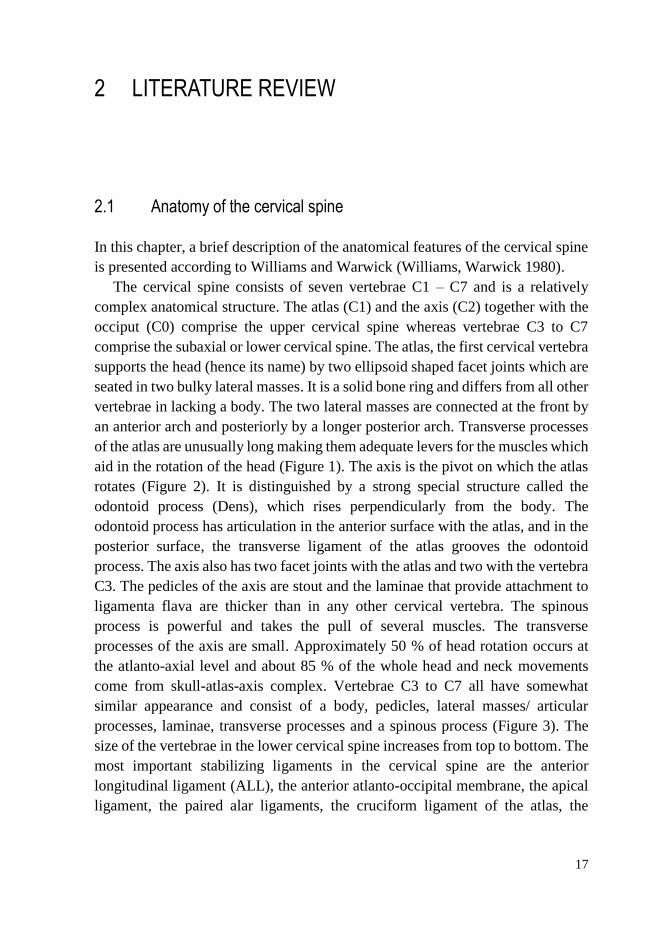
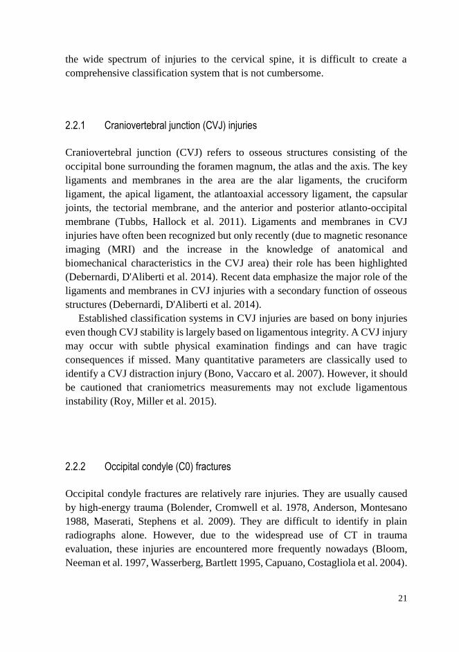
aid in the rotation of the head (Figure 1). The axis is the pivot on which the atlas
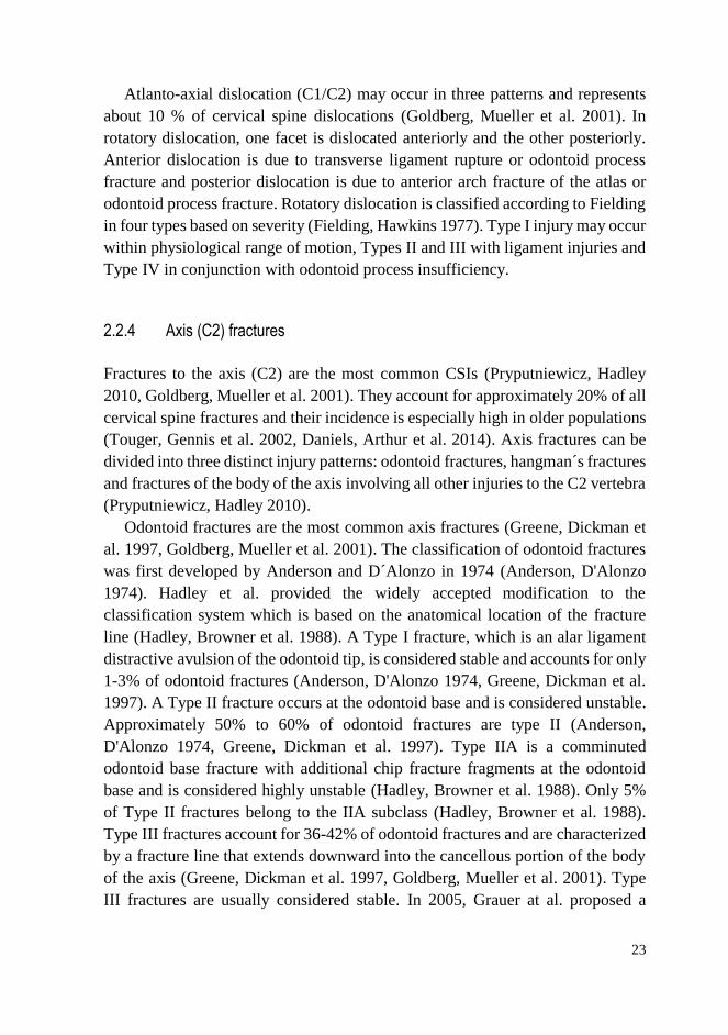
rotates (Figure 2). It is distinguished by a strong special structure called the
odontoid process (Dens), which rises perpendicularly from the body. The
odontoid process has articulation in the anterior surface with the atlas, and in the
posterior surface, the transverse ligament of the atlas grooves the odontoid
process. The axis also has two facet joints with the atlas and two with the vertebra
C3. The pedicles of the axis are stout and the laminae that provide attachment to
ligamenta flava are thicker than in any other cervical vertebra. The spinous
process is powerful and takes the pull of several muscles. The transverse
processes of the axis are small. Approximately 50 % of head rotation occurs at
the atlanto-axial level and about 85 % of the whole head and neck movements
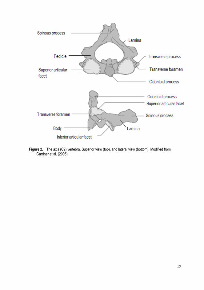
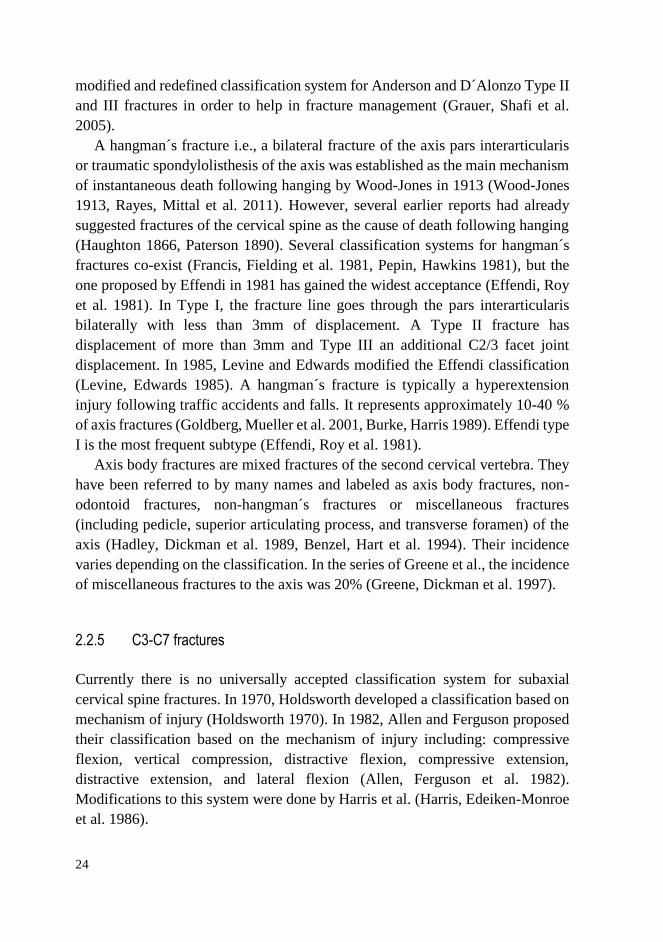
come from skull-atlas-axis complex. Vertebrae C3 to C7 all have somewhat
similar appearance and consist of a body, pedicles, lateral masses/ articular
processes, laminae, transverse processes and a spinous process (Figure 3). The
size of the vertebrae in the lower cervical spine increases from top to bottom. The
most important stabilizing ligaments in the cervical spine are the anterior
longitudinal ligament (ALL), the anterior atlanto-occipital membrane, the apical
ligament, the paired alar ligaments, the cruciform ligament of the atlas, the

18
posterior longitudinal ligament (PLL), the tectorial membrane (an upward
extension of the PLL), the ligamentum flavum, the posterior atlanto-occipital
membrane, the ligamentum nuchae, the interspinous ligaments, the
intertransverse ligaments, and the articular capsules. Altogether, the cervical
spine has 23 articulations: two C0/C1 facet joints, two C1/C2 facet joints and the
odontoid process articulation with the C1 arch, and two facet joints plus an
intervertebral disc in each of the six segments between C2/3 and C7/Th1. The
spinal cord is situated in the vertebral canal and continues as the medulla
oblongata at the level of the odontoid process. The spinal nerve roots exit the
spinal canal via the intervertebral foramina except the first and second roots
which exit the spinal canal posterior to the pedicles. The vertebral arteries arise
from the subclavian arteries and supply blood to the posterior portion of the brain.
They run upward through the foramina in the transverse processes of C6
(occasionally C7) to C1 to enter the skull through the foramen magnum.
Figure 1. The atlas (C1) vertebra. Superior view (top) and lateral view (bottom). Modified from Gardner et al. (2005).
19
Figure 2. The axis (C2) vertebra. Superior view (top), and lateral view (bottom). Modified from Gardner et al. (2005).
20
Figure 3. Typical subaxial vertebra. Superior view (top) and lateral view (bottom). Modified from Gardner et al. 2005.
2.2 Cervical spine injury (CSI) classification
In clinical practice, accurate and efficient diagnosis and management of CSIs is
necessary to avoid further neurological deterioration. Assessment of spinal
stability is essential as the choice of treatment in each specific type of CSI is based
on whether the injury is considered stable or not. Since the classification system
of cervical injuries by Böhler in 1951 (Hernigou 2016), many systems have been
developed to categorize CSIs, but none of them has gained uniform acceptance
among researchers or clinicians (Aebi, Nazarian 1987, Harris, Edeiken-Monroe
et al. 1986, Allen, Ferguson et al. 1982). CSIs may be classified according to the
level of injury (C0 –C7), mechanism of trauma (Allen, Ferguson et al. 1982,
Harris, Edeiken-Monroe et al. 1986), morphology (Bohlman 1979), instability of
the injury (Vaccaro, Koerner et al. 2016, Vaccaro, Hulbert et al. 2007), or
neurological status (Vaccaro, Koerner et al. 2016, Vaccaro, Hulbert et al. 2007).
An ideal classification system would be simple, reproducible and highlight the
injury characteristics that are relevant for the care of the patient. However, due to
21
the wide spectrum of injuries to the cervical spine, it is difficult to create a
comprehensive classification system that is not cumbersome.
2.2.1 Craniovertebral junction (CVJ) injuries
Craniovertebral junction (CVJ) refers to osseous structures consisting of the
occipital bone surrounding the foramen magnum, the atlas and the axis. The key
ligaments and membranes in the area are the alar ligaments, the cruciform
ligament, the apical ligament, the atlantoaxial accessory ligament, the capsular
joints, the tectorial membrane, and the anterior and posterior atlanto-occipital
membrane (Tubbs, Hallock et al. 2011). Ligaments and membranes in CVJ
injuries have often been recognized but only recently (due to magnetic resonance
imaging (MRI) and the increase in the knowledge of anatomical and
biomechanical characteristics in the CVJ area) their role has been highlighted
(Debernardi, D'Aliberti et al. 2014). Recent data emphasize the major role of the
ligaments and membranes in CVJ injuries with a secondary function of osseous
structures (Debernardi, D'Aliberti et al. 2014).
Established classification systems in CVJ injuries are based on bony injuries
even though CVJ stability is largely based on ligamentous integrity. A CVJ injury
may occur with subtle physical examination findings and can have tragic
consequences if missed. Many quantitative parameters are classically used to
identify a CVJ distraction injury (Bono, Vaccaro et al. 2007). However, it should
be cautioned that craniometrics measurements may not exclude ligamentous
instability (Roy, Miller et al. 2015).
2.2.2 Occipital condyle (C0) fractures
Occipital condyle fractures are relatively rare injuries. They are usually caused
by high-energy trauma (Bolender, Cromwell et al. 1978, Anderson, Montesano
1988, Maserati, Stephens et al. 2009). They are difficult to identify in plain
radiographs alone. However, due to the widespread use of CT in trauma
evaluation, these injuries are encountered more frequently nowadays (Bloom,
Neeman et al. 1997, Wasserberg, Bartlett 1995, Capuano, Costagliola et al. 2004).

22
They occur in 0.4-0.7% of all major trauma patients who survive to the emergency
department and represent less than 2% of all cervical spine fractures (Goldberg,
Mueller et al. 2001). In autopsy series, the incidence of C0 fractures has been
reported to be as high as 4 % in fatal head injuries (Tuli, Tator et al. 1997).
Anderson and Montesano (1988) were the first to classify occipital condyle
fractures in three categories (Anderson, Montesano 1988). Types I and II are
considered clinically stable. In Type III there is a fracture-avulsion of the occipital
condyle by the alar ligament and it is considered potentially unstable (Anderson,
Montesano 1988). The classification scheme by Tuli et al. (1997) broadened the
definition of stability to include also the atlantoaxial joint. In the presence of
atlanto-occipital misalignment, surgical stabilization is recommended (Maserati,
Stephens et al. 2009).
2.2.3 Atlas (C1) fractures
Atlas fractures account for about 9-11% of all cervical fractures and they often
occur in combination with axis (C2) fractures (Goldberg, Mueller et al. 2001,
Matthiessen, Robinson 2015, Kakarla, Chang et al. 2010, Hadley, Dickman et al.
1988). Atlas fractures were first described by Jefferson in 1920 (Jefferson 1927,
Jefferson 1919). The management of atlas fractures is largely dependent on the
integrity of the transverse atlantal ligament and whether the fracture occurs in
isolation or in combination with other cervical spine fractures (Dickman, Greene
et al. 1996, Spence, Decker et al. 1970). The fracture may involve the anterior
arch, the posterior arch, the lateral masses or a combination of these. The classic
Jefferson fracture is a burst fracture with lateral displacement of the lateral masses
(Kakarla, Chang et al. 2010). The most typical fracture type seen in clinical
practice involves either the anterior or the posterior arch alone or a combination
of these (Landells, Van Peteghem 1988, Goldberg, Mueller et al. 2001).
There is no single classification system to accommodate all fracture types seen
in clinical situations. The stability of atlas fractures has been based on the integrity
of the transverse atlantal ligament. Based on the results by Spence et al. it has
been suggested that if the sum of lateral displacement of the lateral masses is 7
mm or more, the transverse ligament is probably torn (Spence, Decker et al.
1970). According to the classification by Dickman et al., Type I involves
intraligamentous disruption and Type II involves avulsion of the ligament´s bony
insertion (Dickman, Greene et al. 1996).
23
Atlanto-axial dislocation (C1/C2) may occur in three patterns and represents
about 10 % of cervical spine dislocations (Goldberg, Mueller et al. 2001). In
rotatory dislocation, one facet is dislocated anteriorly and the other posteriorly.
Anterior dislocation is due to transverse ligament rupture or odontoid process
fracture and posterior dislocation is due to anterior arch fracture of the atlas or
odontoid process fracture. Rotatory dislocation is classified according to Fielding
in four types based on severity (Fielding, Hawkins 1977). Type I injury may occur
within physiological range of motion, Types II and III with ligament injuries and
Type IV in conjunction with odontoid process insufficiency.
2.2.4 Axis (C2) fractures
Fractures to the axis (C2) are the most common CSIs (Pryputniewicz, Hadley
2010, Goldberg, Mueller et al. 2001). They account for approximately 20% of all
cervical spine fractures and their incidence is especially high in older populations
(Touger, Gennis et al. 2002, Daniels, Arthur et al. 2014). Axis fractures can be
divided into three distinct injury patterns: odontoid fractures, hangman´s fractures
and fractures of the body of the axis involving all other injuries to the C2 vertebra
(Pryputniewicz, Hadley 2010).
Odontoid fractures are the most common axis fractures (Greene, Dickman et
al. 1997, Goldberg, Mueller et al. 2001). The classification of odontoid fractures
was first developed by Anderson and D´Alonzo in 1974 (Anderson, D'Alonzo
1974). Hadley et al. provided the widely accepted modification to the
classification system which is based on the anatomical location of the fracture
line (Hadley, Browner et al. 1988). A Type I fracture, which is an alar ligament
distractive avulsion of the odontoid tip, is considered stable and accounts for only
1-3% of odontoid fractures (Anderson, D'Alonzo 1974, Greene, Dickman et al.
1997). A Type II fracture occurs at the odontoid base and is considered unstable.
Approximately 50% to 60% of odontoid fractures are type II (Anderson,
D'Alonzo 1974, Greene, Dickman et al. 1997). Type IIA is a comminuted
odontoid base fracture with additional chip fracture fragments at the odontoid
base and is considered highly unstable (Hadley, Browner et al. 1988). Only 5%
of Type II fractures belong to the IIA subclass (Hadley, Browner et al. 1988).
Type III fractures account for 36-42% of odontoid fractures and are characterized
by a fracture line that extends downward into the cancellous portion of the body
of the axis (Greene, Dickman et al. 1997, Goldberg, Mueller et al. 2001). Type
III fractures are usually considered stable. In 2005, Grauer at al. proposed a
24
modified and redefined classification system for Anderson and D´Alonzo Type II
and III fractures in order to help in fracture management (Grauer, Shafi et al.
2005).
A hangman´s fracture i.e., a bilateral fracture of the axis pars interarticularis
or traumatic spondylolisthesis of the axis was established as the main mechanism
of instantaneous death following hanging by Wood-Jones in 1913 (Wood-Jones
1913, Rayes, Mittal et al. 2011). However, several earlier reports had already
suggested fractures of the cervical spine as the cause of death following hanging
(Haughton 1866, Paterson 1890). Several classification systems for hangman´s
fractures co-exist (Francis, Fielding et al. 1981, Pepin, Hawkins 1981), but the
one proposed by Effendi in 1981 has gained the widest acceptance (Effendi, Roy
et al. 1981). In Type I, the fracture line goes through the pars interarticularis
bilaterally with less than 3mm of displacement. A Type II fracture has
displacement of more than 3mm and Type III an additional C2/3 facet joint
displacement. In 1985, Levine and Edwards modified the Effendi classification
(Levine, Edwards 1985). A hangman´s fracture is typically a hyperextension
injury following traffic accidents and falls. It represents approximately 10-40 %
of axis fractures (Goldberg, Mueller et al. 2001, Burke, Harris 1989). Effendi type
I is the most frequent subtype (Effendi, Roy et al. 1981).
Axis body fractures are mixed fractures of the second cervical vertebra. They
have been referred to by many names and labeled as axis body fractures, non-
odontoid fractures, non-hangman´s fractures or miscellaneous fractures
(including pedicle, superior articulating process, and transverse foramen) of the
axis (Hadley, Dickman et al. 1989, Benzel, Hart et al. 1994). Their incidence
varies depending on the classification. In the series of Greene et al., the incidence
of miscellaneous fractures to the axis was 20% (Greene, Dickman et al. 1997).
2.2.5 C3-C7 fractures
Currently there is no universally accepted classification system for subaxial
cervical spine fractures. In 1970, Holdsworth developed a classification based on
mechanism of injury (Holdsworth 1970). In 1982, Allen and Ferguson proposed
their classification based on the mechanism of injury including: compressive
flexion, vertical compression, distractive flexion, compressive extension,
distractive extension, and lateral flexion (Allen, Ferguson et al. 1982).
Modifications to this system were done by Harris et al. (Harris, Edeiken-Monroe
et al. 1986).
25
In the SLIC-system (Subaxial Injury Classification), developed by The Spine
Trauma Study Group in 2007, injuries are characterized based on three main
categories: injury morphology, disco-ligamentous complex integrity, and
neurologic status (Vaccaro, Hulbert et al. 2007). Each of the categories is
individually analyzed and given a score. The sum of the scores for all three
categories is used for prognostication and management decision-making.
However, among surgeons, the SLIC system has shown low reproducibility in
treatment decision-making (Middendorp, Audige et al. 2013). In order to produce
a classification system with higher interobserver and intraobserver reliability, the
AOSpine subaxial cervical spine injury classification system has been developed
(Vaccaro, Koerner et al. 2016), In the AOspine system, the following four
classification criteria are used: injury morphology, facet injury, neurological
status, and the presence of specific modifiers (posterior capsuloligamentous
complex injury without complete disruption, critical disk herniation,
stiffening/metabolic bone disease, and signs of vertebral artery injury).
CSI occurs most often in the subaxial spine, while the axis is the most
commonly injured individual vertebra. C6 and C7 are the most frequently affected
vertebrae (about 50 % of CSIs occur in the C5/6 and C6/7 area) and C3 or C4 get
injured only rarely (Goldberg, Mueller et al. 2001). The dislocations or
subluxations in the subaxial spine occur most often in the C5/6 and C6/7
interspaces (Goldberg, Mueller et al. 2001). The distribution of fractures by
anatomical structure in blunt trauma patients by Goldberg et al. is as follows:
vertebral body 29.9%, pedicle 5.9%, lateral mass / articular process 14.9%,
lamina 16.4%, transverse process 9.2%, spinous process 20.8%, and other 2.9%.
2.2.6 Spinal cord injury (SCI)
About 10%–50% of CSI patients suffer a concomitant spinal cord injury (SCI)
(Leucht, Fischer et al. 2009, Bohlman 1979), and a small proportion of patients
suffer only a spinal nerve injury (Fredø, Bakken et al. 2014). The clinical severity
of SCI depends on the spinal level and completeness of the injury. By definition,
in complete injury, sacral sparing (either preservation of sensation in S4-5
dermatome or awareness of deep anal pressure or voluntary anal sphincter
contraction) is lost. The American Spinal Injury Association (ASIA) Impairment
Scale (AIS) designation is commonly applied for grading the degree of
impairment (Kirshblum, Burns et al. 2011). An AIS grade A refers to a complete
injury, an AIS B is a motor complete-sensory incomplete injury, AIS C and D are
26
incomplete motor and sensory injuries and an AIS E represents normal motor and
sensory functions at the time of examination.
MRI may show spinal cord edema, cord contusion, intramedullary
hemorrhage, cord transection, soft tissue injury, spinal canal stenosis or disk
herniation. Furthermore, the maximum spinal cord compression, maximum canal
compromise and length of spinal cord lesion may be assessed. (Miyanji, Furlan
et al. 2007). MRI findings of the spinal cord provide prognostic information
regarding long-term outcomes in SCI patients (Flanders, Schaefer et al. 1990,
Miyanji, Furlan et al. 2007). In addition to conventional MRI sequences, diffusion
tensor imaging has been shown to be a quantitative and objective tool for
assessing the state of the cervical spinal cord in patients with chronic SCI
(Koskinen, Brander et al. 2013).
SCI may occur without the presence of a bony injury or dislocation in
conventional three-view radiographic series or CT. The NEXUS-study reported
a 3% incidence of SCI after the absence of traumatic findings in plain radiographs
(Hendey, Wolfson et al. 2002). Kato et al. reported the incidence of cervical SCI
without bony injury or dislocation (both plain radiographs and CTs were
assessed) in up to 32% of all cervical SCI patients in Japan (Kato, Kimura et al.
2008). Cervical spondylosis, developmental narrowing of the spinal canal and
disc herniation are well known risk factors for SCI without bony injury (Epstein,
Epstein et al. 1980, Koyanagi, Iwasaki et al. 2000). SCIWORA (SCI without
radiographic abnormality) is a term used for a blunt injury to the spinal cord
without radiological findings (Pang 2004). The term was invented in the pre-MRI
era and is nowadays seldom used, except for pediatric patients (Pang, Wilberger
Jr 1982). The pediatric spine is physiologically hypermobile and therefore more
at risk for this type of injury (typically children less than 8 years of age) (Pang
2004).
2.2.7 Vertebral Artery Injury (VAI)
A vertebral artery injury (VAI) may occur in conjunction with a CSI and cause
additional morbidity and mortality. VAI is traditionally considered infrequent
among CSI patients, but due to the heightened awareness of the condition and
frequent use of CT angiography, the rate has increased dramatically. The
incidence of VAI is reported from 24 to 48% among patients with a cervical
fracture extending into the transverse foramen. (Parbhoo, Govender et al. 2001,
Giacobetti, Vaccaro et al. 1997, Friedman, Flanders et al. 1995). Facet joint
27
dislocations are also frequently associated with VAI with an average frequency
of 35% (Inamasu, Guiot 2006). VAI types include dissection with or without an
intimal flap or mural thrombus, pseudoaneurysm, occlusion, transection, and
arterio-venous fistula (Inamasu, Guiot 2006). Dissection and occlusion are the
two most frequent injury patterns.
2.3 Epidemiology and incidence of CSI
The reported incidence rates and other epidemiological features regarding CSI
differ considerably depending on the population characteristics, geographical and
cultural differences, and inclusion criteria and differences in data collection in
individual studies. However, CSI occurs in patients in all demographic categories.
The incidence of CSI in a whole population setting is not well known. There
are only a few studies on CSI incidence in the general population (Brolin, von
Holst 2002, Hu, Mustard et al. 1996, Fredø, Bakken et al. 2014). A study from
Sweden reported the incidence of cervical spine fractures to be 9.2/100,000/year
in 1999 (Brolin, von Holst 2002). In the Canadian population between 1981 and
1984 Hu et al. found the incidence of all spine fractures to be 64/100,000/year. In
that study, subgrouping into cervical, thoracic or lumbar fractures was performed
for only 45% of the patients that were admitted to hospitals. The estimated
incidence of cervical fractures was 12/100.000/year (Hu, Mustard et al. 1996). A
recent study from Norway (2009-2012) reported the incidence of severe CSI to
be 16.5/100,000/year and the incidence of traumatic cervical spine fractures
15.0/100,000/year (Fredø, Bakken et al. 2014).
CSI incidence in various subpopulations, such as trauma center patients,
specific age groups, head injury patients, and patients with a specific injury
mechanism has been studied widely (Brown, Brunn et al. 2001, Hills, Deane
1993, Michael, Guyot et al. 1989, Lowery, Wald et al. 2001, Thompson, Stiell et
al. 2009). For example, Schoenfeld et al. studied cervical spine fractures in the
U.S. military personnel and found an incidence of 29/100,000/year (Schoenfeld,
Sielski et al. 2012).
In blunt trauma populations, the overall incidence of CSI has been reported to
range from approximately 2 to 7%. Yanar et al. studied 8,401 pedestrians struck
by an automobile in Los Angeles County and found the incidence of CSI to be
2.1% (Yanar, Demetriades et al. 2007). However, there was a substantial variation
with age, ranging from 0.3% in the pediatric age group to 4.4% in the age group

28
older than 65 years. A prospective cohort study conducted from October 1996 to
April 1999 in Canada, involving almost nine thousand adults who presented to
the emergency department with a blunt trauma to the head/neck, had stable vital
signs and a Glasgow Coma Scale (GCS) score of 15, found the incidence of
clinically significant CSI to be 1.7% (Stiell, Wells et al. 2001). In an emergency
department sample from the U.S. involving blunt trauma patients, 7% had a CSI
(Sanchez, Waxman et al. 2005). A meta-analysis by Milby et al. found that 3.7%
of all trauma patients had a CSI (Milby, Halpern et al. 2008).
The reported incidence of CSI among patients with HI varies approximately
from 4-8% (Holly, Kelly et al. 2002, Hills, Deane 1993, Williams, Jehle et al.
1992, Mulligan, Friedman et al. 2010, Michael, Guyot et al. 1989). The incidence
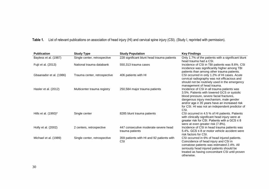
depends on the population studied and classification of both HI and CSI. Table 1
shows a list of relevant publications on the association of HI and CSI.
The proportion of cervical fractures among all patients with a spine fracture
also varies considerably. Nelson et al. conducted a national (U.S.) data bank study
of more than 80,000 blunt trauma patients with at least one spine fracture. The
relative incidences of cervical, thoracic and lumbar fractures were 41%, 37% and
43%, respectively (Nelson, Martin et al. 2013). In a trauma center study by Leucht
et al., cervical fractures represented only 21% and lumbar fractures 50% of all
spine fractures (Leucht, Fischer et al. 2009). According to Lenehan et al. 51% of
spine injury patients had a cervical injury (Lenehan, Boran et al. 2009). A
noncontiguous spinal injury is identified in 10-20% of patients with CSI (Miller,
Brubacher et al. 2011, Sharma, Oswanski et al. 2007).
The published incidence of traumatic spinal cord injury (SCI) ranges between
10 and 83 per million/year in the developed world (Wyndaele, Wyndaele 2006,
Sekhon, Fehlings 2001, Pickett, Campos-Benitez et al. 2006, Dahlberg, Kotila et
al. 2005). The incidence of SCI in the U.S. is approximately 40 per million
inhabitants per year, and in Finland according to a recent study by Koskinen et al.
the incidence is 25 to 38 per million per year depending on the catchment area
(Koskinen, Alen et al. 2014). The majority of SCIs occur in the cervical region.
In the study by Koskinen et al., 70% of the traumatic SCI patients were tetraplegic
and the incidence of traumatic cervical SCI would be 18 to 27 per million per year
accordingly (Koskinen, Alen et al. 2014). In a Chinese study, 72% of SCIs were
cervical (Ning, Yu et al. 2011), however, only 50% of the SCI patients in a
Canadian study were cervical (Lenehan, Street et al. 2012). According to Sekhon
and Fehlings, approximately 55% of acute SCI occurs in the cervical region
(Sekhon, Fehlings 2001). In a study from Finland by Ahoniemi et al., 57% of the
29
patients treated in the biggest national rehabilitation center between 1996 and
2005 were tetraplegic (Ahoniemi, Alaranta et al. 2008).
The number of patients with a CSI who succumb prior to hospitalization and
hence remain out of most of the incidence studies is not well known. Previous
reports have suggested that 21-24% of victims who die immediately or soon after
a traffic accident have a serious injury to the cervical spine of which the majority
affect the craniocervical junction (Alker, Oh et al. 1975, Bucholz, Burkhead et al.
1979).
30
Table 1. List of relevant publications on association of head injury (HI) and cervical spine injury (CSI). (Study I, reprinted with permission).
Publication Study Type Study Population Key Findings
Bayless et al. (1987) Single center, retrospective 228 significant blunt head trauma patients Only 1.7% of the patients with a significant blunt head trauma had a CSI.
Fujii et al. (2013) National trauma databank 550,313 trauma cases Incidence of CSI in TBI patients was 8.6%. CSI incidence was significantly higher among TBI patients than among other trauma patients.
Gbaanador et al. (1986) Trauma center, retrospective 406 patients with HI CSI occurred in only 1.2% of HI cases. Acute cervical radiography was not efficacious and should not be routinely used in the emergency management of head trauma.
Hasler et al. (2012) Multicenter trauma registry 250,584 major trauma patients Incidence of CSI in all trauma patients was 3.5%. Patients with lowered GCS or systolic blood pressure, severe facial fractures, dangerous injury mechanism, male gender and/or age ≥ 35 years have an increased risk for CSI. HI was not an independent predictor of CSI.
Hills et al. (1993)* Single center 8285 blunt trauma patients CSI occurred in 4.5 % of HI patients. Patients with clinically significant head injury were at greater risk for CSI. Patients with a GCS ≤ 8 were at even greater risk (7.8%).
Holly et al. (2002) 2 centers, retrospective 447 consecutive moderate-severe head trauma patients
Incidence of CSI in head trauma patients was 5.4%. GCS ≤ 8 or motor vehicle accident were risk factors for CSI.
Michael et al. (1989) Single center, retrospective 359 patients with HI and 92 patients with CSI
CSI occurred in 6% of head injured patients. Coincidence of head injury and CSI in comatose patients was estimated 2.4%. All seriously head injured patients should be treated as having concomitant CSI until proven otherwise.
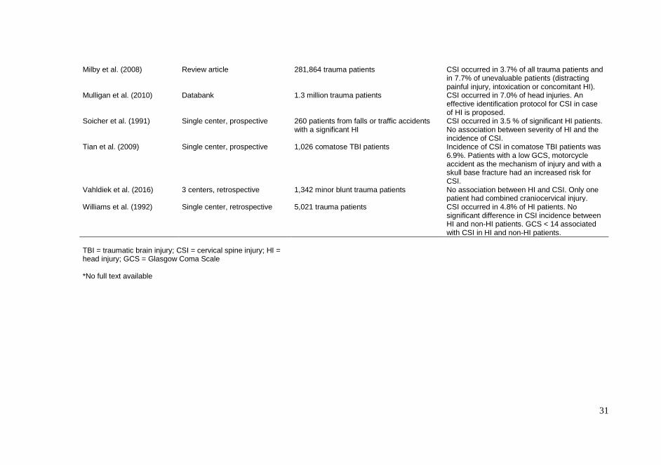
31
Milby et al. (2008) Review article 281,864 trauma patients CSI occurred in 3.7% of all trauma patients and in 7.7% of unevaluable patients (distracting painful injury, intoxication or concomitant HI).
Mulligan et al. (2010) Databank 1.3 million trauma patients CSI occurred in 7.0% of head injuries. An effective identification protocol for CSI in case of HI is proposed.
Soicher et al. (1991) Single center, prospective 260 patients from falls or traffic accidents with a significant HI
CSI occurred in 3.5 % of significant HI patients. No association between severity of HI and the incidence of CSI.
Tian et al. (2009) Single center, prospective 1,026 comatose TBI patients Incidence of CSI in comatose TBI patients was 6.9%. Patients with a low GCS, motorcycle accident as the mechanism of injury and with a skull base fracture had an increased risk for CSI.
Vahldiek et al. (2016) 3 centers, retrospective 1,342 minor blunt trauma patients No association between HI and CSI. Only one patient had combined craniocervical injury.
Williams et al. (1992) Single center, retrospective 5,021 trauma patients CSI occurred in 4.8% of HI patients. No significant difference in CSI incidence between HI and non-HI patients. GCS < 14 associated with CSI in HI and non-HI patients.
TBI = traumatic brain injury; CSI = cervical spine injury; HI = head injury; GCS = Glasgow Coma Scale *No full text available
32
2.4 Risk factors for CSI
2.4.1 Gender, age and injury mechanism
Male gender is a known risk factor for injuries in general and also for CSIs. The
proportion of male patients is reported around 60-80% in many CSI studies
(Hasler, Exadaktylos et al. 2012, Lowery, Wald et al. 2001, Clayton, Harris et al.
2012, Hoffman, Mower et al. 2000, Yang, Ding et al. 2013).
The number of patients with a CSI varies with age in bimodal fashion. Young
adults and elderly people have the highest CSI incidence (Lowery, Wald et al.
2001). The former is mostly due to road traffic accidents by young males and the
latter to ground level falls.
The causes of injury vary between countries, between regions within a country,
and between urban and rural locations (Yang, Ding et al. 2013). Sports injuries,
motor vehicle accidents and falls from a height have been described as risk factors
for CSI by many authors (Thompson, Stiell et al. 2009, Leucht, Fischer et al.
2009, Hasler, Exadaktylos et al. 2012, Clayton, Harris et al. 2012, Lenehan, Boran
et al. 2009). In recent years, the age distribution has shifted towards elderly people
and the mechanism of injury from motor vehicle injuries to ground level falls.
2.4.2 Alcohol and drugs
Alcohol is a major risk factor for injuries in general and CSI is not an exception.
In Finland, every third fatal injury happens under the influence of alcohol
(Tiirikainen 2009). The rate of alcohol intoxicated patients in trauma centers
worldwide ranges from approximately 20 to over 40% (Jurkovich, Rivara et al.
1992). Alcohol use at the time of injury associates especially with cervical SCI as
compared to lower spinal levels (Garrison, Clifford et al. 2004).
Non-prescription drugs increase the risk for traumatic injuries, though in
Finland, they are not as commonly used as alcohol. However, in recent years their
use has increased. For example, a Finnish study showed that between 1977 and
2007, driving under influence of non-prescription drugs increased manifold
33
(Ojaniemi, Lintonen et al. 2009). In addition to increasing the probability of an
accident, alcohol and other drugs can decrease the patient’s ability to feel pain.
Intoxicated patients with a CSI may report no tenderness in the neck even with a
significant injury.
2.4.3 Head injury
Sir Geoffrey Jefferson is considered to be the first person to report the coincidence
of head trauma and CSI (Jefferson 1927). He observed that any vertical force
directed to the vertex of the skull may result in the fracture of the atlas. Since
then, several investigators have studied the relationship between HI and CSI with
varying results. Table 1 summarizes relevant publications on the association
between HI and CSI. This association appears logical assuming that forces
applied to the face or head will be transmitted to the cervical spine and result in
injury. Foster et al. suggested that “all head and neck trauma patients should be
considered to have a cervical spine injury until proven otherwise” (Foster, Maisel
et al. 1981). However, there are multiple studies that did not find this association
between HI and CSI (Table 1). One theory is that the head and face may act as a
cushion and buffer, dissipating the energy that would otherwise be transferred to
the cervical spine, resulting in a lower risk of CSI. Moreover, CSIs may nowadays
be more commonly associated with inertial differences in the head and torso, as
opposed to transmitted compression forces from head or facial trauma. Increased
use of safety features such as seat belts and airbags may have influenced the risk
of HI-related CSI.
2.4.4 Ankylosing spinal disorders
The most common ankylosing spinal disorders are ankylosing spondylitis (AS,
also known as Bechterew disease) and diffuse idiopathic skeletal hyperostosis
(DISH, also known as Forestier disease) (Hartmann, Tschugg et al. 2017). AS is
a chronic systemic and inflammatory rheumatic disease with a reported
prevalence of up to 1.4 % (Braun, Sieper 2007). It mainly affects males. The
etiology of DISH is still unknown but there is strong association with obesity,
type 2 diabetes and high age (Weinfeld, Olson et al. 1997, Denko, Malemud
2006). The prevalence is estimated at between 3 and 25% (Hartmann, Tschugg et
al. 2017). The condition is more common in men and prevalence peaks in the 60-

34
to 69 - year old age group (Kim, Choi et al. 2004). Both of the disorders lead to
progressive ossification of the spinal column which makes the spine inflexible
and highly susceptible to trauma even after low-energy impacts (Caron, Bransford
et al. 2010). The spinal level most often injured in these patients is cervical. The
diagnosis of cervical spine fracture in patients with ankylosing spinal disorders is
often delayed and secondary deterioration after misdiagnosis of a fracture is a
frequent problem with these conditions (Westerveld, Verlaan et al. 2009,
Westerveld, van Bemmel et al. 2014).
2.4.5 Other risk factors
Several studies support a relationship between facial injuries and cervical spine
trauma, with some reporting an incidence as high as 19% (Lewis, Manson et al.
1985, Mukherjee, Abhinav et al. 2015). Pelvic fracture especially when in
conjunction with HI associates with CSI and probably reflects the high-energy
injury mechanism in general (Clayton, Harris et al. 2012). High Injury Severity
Score (ISS) and multiple extremity fractures are also reported to associate with
CSI as is a decreased GCS score (Hasler, Exadaktylos et al. 2012, Clayton, Harris
et al. 2012, Holly, Kelly et al. 2002, Hanson, Blackmore et al. 2000, Hills, Deane
1993). Moreover, clavicular injury has been found to associate with a CSI
(Williams, Jehle et al. 1992). Degenerative changes and osteoporosis predispose
to CSIs (typically odontoid process fractures), which are common among the
elderly after low energy injuries (Kaesmacher, Schweizer et al. 2017, Watanabe,
Sakai et al. 2014).
2.5 CSI diagnostics
Cervical spine clearance after blunt trauma is defined as accurately confirming
the absence of a cervical spine injury (Anderson, Gugala et al. 2010, Richards
2005). The clearance of the cervical spine in trauma patients is difficult, time-
consuming, and costly (Anderson, Gugala et al. 2010). The objective of cervical
spine clearance is to establish that an injury does not exist. Failure to diagnose a
CSI at the time of presentation can have disastrous consequences, with a high risk
for neurological deterioration (Morris, McCoy 2004). Immobilization in a

35
cervical collar should be initiated at the scene of injury and maintained until a
directed examination is performed during the secondary evaluation (Schmidt,
Gahr et al. 2009). However, cervical spine immobilization is not without
consequences and should be kept in minimum (Greenbaum, Walters et al. 2009,
Karason, Reynisson et al. 2014).
2.5.1 Clinical evaluation
Clinical examination is an essential component of the cervical spine clearance
process. It includes a review of the history with regard to the injury mechanism
and other relevant information (e.g., transient motor or sensory changes may
indicate significant spinal pathology, and when noted requires radiographic
assessment), identification of pain or tenderness in the head, neck or
thoracolumbar spine or any neurologic changes of sensation or muscle strength
in the trunk or extremities (Anderson, Gugala et al. 2010). Published, Level I
evidence shows that asymptomatic, alert, neurologically intact patients do not
need further imaging to declare the cervical spine clear (Hoffman, Mower et al.
2000, Stiell, Wells et al. 2001, Anderson, Muchow et al. 2010). The NEXUS
(National Emergency X-Radiography Utilization Study Group) method uses
specific criteria to identify the low-risk patient who can be cleared clinically
without imaging. All of the five following criteria must be met for a patient to be
considered low-risk: (i) an awake, alert patient; (ii) no history, signs, or laboratory
evidence of intoxication; (iii) no distracting injury; (iv) no cervical spine pain or
midline tenderness; and (v) no neurologic signs or symptoms (Hoffman, Mower
et al. 2000). The sensitivity of the NEXUS method is excellent – 99.0% for all
cervical injuries and 99.6% for significant CSI. Due to low specificity (12.9%),
many potentially unnecessary radiographs are taken.
An alternative to the NEXUS protocol is the Canadian C-Spine Rule (Stiell,
Wells et al. 2001). This rule applies to awake, non-intoxicated patients with a
GCS score of 15 and identifies those who require radiographs by answering three
questions. First, is the patient high-risk enough that radiographs are required?
(Risk factors include: age >65 years, reports of paresthesia, and a dangerous
mechanism of injury, for example, a fall from a height >1 m or five stairs, axial
load to the head, and a high-speed [>100 km/h] automobile, motorcycle,
recreational vehicle, or bicycle accident). Second, is there a low risk factor that
would allow the safe assessment of range of motion? Examples of such a factor
are a simple rear-end motor vehicle crash, a patient who has already sat upright
36
in the emergency department or was ambulatory at any time, a delay in the onset
of pain, and an absence of tenderness. Third, can the patient actively rotate the
head 45° to the right and left without pain? A patient who is not at high risk and
can safely perform the rotation test can be cleared clinically without radiographs.
The sensitivity of the Canadian C-Spine rule is reported to be 100% and the
specificity to be 42.5% (Stiell, Wells et al. 2001).
In a separate study, Stiell at al. found that in applying the Canadian C-Spine
rule instead of NEXUS criteria, 10 % fewer cases would have required
radiographs (Stiell, Clement et al. 2003). In a meta-analysis by Tontz et al.
totaling more than 63,000 patients, including three NEXUS, two Canadian C-
Spine Rule, and nine institutional protocols, the overall sensitivity based on a
random effects model was 98.1%, with specificity being 35.0%. Of 28 missed
injuries, only 2 were deemed significant but none was associated with
neurological deterioration (Tontz, Anderson et al. 2006).
2.5.2 Cervical spine imaging
Cervical spine imaging is a key element in addition to history and physical
examination in trauma patients who are suspected to have a CSI. A patient who
has neck pain, midline tenderness, or neurological symptoms requires
radiographic imaging. Imaging options are plain radiography, flexion-extension
radiography, CT and MRI. If vascular injury is suspected, angiographic studies
are needed.
Plain radiographs are usually not recommended in the acute phase evaluation
of CSI, because even with the best possible technique, they underestimate the
amount of traumatic spine injury and detect only 52-85% of fractures, even when
three views are obtained (Gale, Gracias et al. 2005, Holmes, Akkinepalli 2005,
Hadley, Walters 2013). However, plain radiographs are often used in the follow-
up evaluation of possible unstable injuries. The use of flexion-extension
radiographs in the acute setting is also controversial and carries a risk of causing
additional neurological damage, hence its use is best left for the subacute
evaluation when there is a specific clinical concern. (Anglen, Metzler et al. 2002,
Pollack, Hendey et al. 2001, Knopp, Parker et al. 2001).
Computed tomography (CT) has supplemented plain radiography in CSI
screening and is the primary imaging modality for evaluating patients with a blunt
CSI. It detects 97-100 % of fractures to the cervical spine (Shah, Ross 2016,
Hadley, Walters 2013, Brown, Antevil et al. 2005). The imaging must include

37
axial scans from the occiput to the first thoracic vertebra with coronal and sagittal
reconstructions.
MRI is superior to CT for the detection of neural, ligamentous, and disc
injuries and is primarily employed for the patient who presents with a
neurological deficit, or when ligamentous injury is suspected (Pourtaheri, Emami
et al. 2014, Schoenfeld, Bono et al. 2010, Muchow, Resnick et al. 2008).
Nevertheless, there is significant heterogeneity in the literature regarding the use
of MRI after a negative CT to rule out ligament injury (Malhotra, Wu et al. 2017).
The drawbacks of MRI are that it requires extensive time to perform, it interferes
with patient´s monitoring equipment, the inability to use it in hemodynamically
unstable patients, and its high cost (Dunham, Brocker et al. 2008).
Angiographic studies; computed tomography angiography (CTA), magnetic
resonance angiography (MRA), and digital subtraction angiography (DSA) are
utilized to detect vessel injuries in CSI patients. DSA is the gold standard for
detecting VAIs and is the primary imaging modality particularly when
endovascular treatment is considered. According to level I evidence, CTA is an
alternative to DSA and is usually the primary imaging modality, not least because
it is readily available (Utter, Hollingworth et al. 2006). The advantage of MRA is
that it does not use contrast agents and it may be obtained in conjunction with
MRI (Hadley, Walters 2013).
Patients with a decreased level of consciousness remain a group in which the
clearance of the cervical spine remains controversial and unresolved. The risks of
an occult CSI must be weighed against the potential harm caused by prolonged
cervical immobilization. In addition to general comfort issues, prolonged
immobilization may lead to complications such as increased intracranial pressure
for those with closed head injury, predisposition to pressure sore development,
and ventilator-associated pneumonias (Morris, McCoy 2004, Greenbaum,
Walters et al. 2009). Trauma centers show marked variation in spine clearance
protocols among patients with decreased level of consciousness. It is not clear to
what extent CT alone can direct clearance of the cervical spine. Several
investigations have advocated CT as a single modality capable of detecting all
significant CSIs (Tomycz, Chew et al. 2008, Schuster, Waxman et al. 2005,
Como, Thompson et al. 2007). However, a huge body of research suggest that
MRI of the cervical spine is a necessary adjunct in the evaluation of patients with
decreased levels of consciousness. (Menaker, Philp et al. 2008, Stassen, Williams
et al. 2006, Pourtaheri, Emami et al. 2014, Muchow, Resnick et al. 2008).
38
2.6 Consequences of CSI
Injury to the cervical spine may be a minor distension or major injury leading to
tetraplegia (impairment of function in all four limbs, trunk, and pelvic organs) or
even death. SCI, in addition to disturbing motor and sensory functions below the
level of injury, impairs somatic and autonomic nervous system, control of blood
vessels, heart, respiratory tract, sweat glands, bowel, urinary bladder, and sexual
organs (Krassioukov, Biering-Sorensen et al. 2012). Early death following
cervical SCI is usually due to respiratory failure and/or cardiovascular
dysfunction (Lemons, Wagner 1994). Complete SCI at or above the C3 spinal
level causes immediate death (if mechanical ventilation is not started
immediately) due to disruption of innervation to the diaphragm and intercostal
muscles. In the acute phase of SCI, patients are prone to spinal shock (flaccid
paralysis and areflexia) and neurogenic shock (i.e., bradyarrhythmias,
atrioventricular conduction block and hypotension) (Ditunno, Little et al. 2004,
Piepmeier, Lehmann et al. 1985). The incidence of neurogenic shock, depending
on the level and severity of injury, is reported up to 100% among patients with
cervical SCI and contributes to poor outcomes (Piepmeier, Lehmann et al. 1985).
SCI predisposes patients to various secondary complications throughout life.
In the past, renal failure and other urinary tract complications were the primary
causes of death of patients with long-standing SCI. Due to advances in medical
practice, the causes of death of patients with chronic SCI are approaching those
of the general population. However, increased mortality in this patient group is
still present (Hagen, Eide et al. 2010). Cardiovascular, and respiratory diseases
together with infections are the leading causes of death of the chronic SCI
population (DeVivo, Krause et al. 1999, Branco, Cardenas et al. 2007). Dysphagia
and aspiration are common in patients with cervical SCI and contribute to the
development of respiratory dysfunction and pneumonia (Shin, Yoo et al. 2011,
Jackson, Groomes 1994, Ihalainen, Rinta-Kiikka et al. 2017b, Ihalainen, Rinta-
Kiikka et al. 2017a).
In addition to often detrimental consequences for the individual patient, CSI is
also a major economic burden for society (Kukreja, Kalakoti et al. 2015, Daniels,
Arthur et al. 2014, Baaj, Uribe et al. 2010). In Canada, the estimated economic
life-time burden of a patient with a complete tetraplegia was three million dollars
(Krueger, Noonan et al. 2013). It is estimated that the increase in the economic
burden today is attributable to improved life expectancy in the SCI population
and increase in costs of care after SCI (Cao, Chen et al. 2011). Moreover, the

39
costs of patients with CSI who do not have a combined SCI have increased during
the last few years (Baaj, Uribe et al. 2010). This is probably due to an increase in
the incidence of these injuries especially among elderly patients. Due to more
complicated hospital stays, longer hospitalizations, and higher rates of inpatient
facility care after hospital admission, older patients seem to have a higher
propensity for greater health care resource utilization (Kukreja, Kalakoti et al.
2015, Baaj, Uribe et al. 2010).
Even though injuries to vertebral arteries often remain clinically occult, a small
percentage of patients may suffer devastating neurological complications due to
posterior circulation infracts (Inamasu, Guiot 2006).
2.7 Treatment of CSI
The goal in CSI treatment is to provide a stable and painless spine together with
the best possible neurological recovery (Lauweryns 2010). The chosen treatment
strategy of an individual patient is affected by multiple factors. For example, the
type of injury, neurological status of the patient, probability of vertebra
dislocation, the patient’s body habitus and compliance to the treatment should all
be taken into account. Although a multitude of guidelines for CSI treatment are
available, there are still several controversies in how to treat CSI patients with or
without SCI. The choice of one modality over another should be made on an
individual basis. After the diagnosis of CSI, the short and long-term management
should be determined. Long-term management is dependent on the location and
pattern of the injury. In the short-term, continued immobilization is usually
necessary to prevent further injury (Gardner, Grannum et al. 2005).
Operative treatment was given for 18-27% of patients with a CSI in Norway
(Fredo, Rizvi et al. 2012). Injury to the cervical spine increases mortality and
morbidity even without the presence of an SCI (Golob, Claridge et al. 2008,
Bohlman 1979, Harris, Reichmann et al. 2010, Fredø, Bakken et al. 2014). The
risk of complications in CSI treatment depends on the injury itself, the pre-injury
characteristics of the patient and the chosen treatment method. Operative
treatment of CSIs carries well documented risks (Fredø, Rizvi et al. 2016, Leckie,
Yoon et al. 2016), but conservative treatment with cervical collars or halovest
devices are not without complications either (Longo, Denaro et al. 2010, Butler,
Dolan et al. 2010). Conservative treatment can be initially administered and can
serve as an adjunct to surgery, or even be the definitive treatment. Supine skull
40
traction is seldom used, but in some cases, such as facet subluxation or dislocation
and burst-type fractures, it may be employed in the initial phase.
Surgical treatment of unstable CSIs usually allows earlier mobilization of the
patient and shortens the primary hospital stay. According to the individual patient
and injury type, surgery can be performed in numerous ways. Common upper
cervical spine procedures include for example anterior odontoid screw fixation,
posterior C1-C2 fixation, and occipito-cervical fixation. In the subaxial spine,
various methods exist also for anterior and posterior fixation with different kinds
of screws, rods, plates and wires. In patients with ankylosing spinal disorders,
fractures typically involve the anterior, middle, and posterior columns with high
dislocation probability and therefore surgical fixation is often mandatory. In these
cases, a posterior or circumferential approach is recommended due to the high
failure rate with anterior-only surgeries(Ma, Wang et al. 2015, Hartmann,
Tschugg et al. 2017).
2.8 Adverse events
An adverse event (AE) is usually defined as an unintended injury or complication,
caused by health care management rather than the patient’s underlying disease
(Brennan, Leape et al. 1991). Regarding in-hospital AEs, health care management
consists of actions of individual hospital staff as well as the broader systems and
care processes, and includes both acts of omission (failure to diagnose or treat)
and acts of commission (incorrect diagnosis or treatment, or poor performance)
(Baker, Norton et al. 2004). AEs result in prolonged hospital stays, disability at
the time of discharge, or death (Thomas, Studdert et al. 2000, Wilson, Runciman
et al. 1995). In general, in-hospital AEs occur in approximately 10% of admitted
patients, and about half of the AEs are considered preventable (preventable
adverse event = PAE) (de Vries, Ramrattan et al. 2008, Soop, Fryksmark et al.
2009). In surgical and orthopedic care, the rate of AEs is considered even higher
(up to 30%) and the majority are considered preventable (Merten, Johannesma et
al. 2015, Rutberg, Borgstedt-Risberg et al. 2016). In-hospital AEs are lethal in
about 7% of cases (de Vries, Ramrattan et al. 2008). Aside from the direct harm
for the patient, AEs are a massive financial burden on society (Thomas, Studdert
et al. 1999).
41
2.8.1 Diagnostic errors
Diagnostic AEs are mostly preventable and occur due to human failure (the main
causes being mistakes due to a lack of knowledge and information transfer
problems) in the majority of cases (Zwaan, de Bruijne et al. 2010). The mortality
rate for diagnostic AEs is higher than for other AEs (Zwaan, de Bruijne et al.
2010). Diagnostic errors occur in every medical specialty and some of these errors
lead to patient harm, that is, diagnostic AEs (Graber 2013). The error rate in
clinical medicine is estimated at 5-15% (Berner, Graber 2008, Schwartz, Elstein
2008). The ED is an arena which requires complex decision-making in settings
of above-average uncertainty and stress. Therefore, ED physicians are especially
prone to diagnostic errors (Guly 2001). The patient groups most at risk of
diagnostic errors are those admitted to a hospital in an emergency setting (Zwaan,
de Bruijne et al. 2010). The error rate in perceptual specialties such as radiology
or pathology is considered to be substantially lower than in clinical medicine
(FitzGerald 2005, Kronz, Westra et al. 1999).
Historically, in the era of plain roentgenograms, misdiagnosis of a CSI was
estimated in up to one-third of cases (Reid, Henderson et al. 1987, Davis,
Phreaner et al. 1993, Platzer, Hauswirth et al. 2006). Currently, in the CT era, the
rate of misdiagnosis is reported to range from 0 to 5% (Platzer, Hauswirth et al.
2006, Patel, Humble et al. 2015). A failure to identify a CSI is considered a
diagnostic error. The reasons for diagnostic errors in CSIs are various. A
successful cervical spine clearance is highly dependent on the appropriate
utilization of radiographic studies (Greenbaum, Walters et al. 2009, Zakrison,
Williams 2016). The ordering of inadequate, improper or non-sufficient
radiographs is one issue (Davis, Phreaner et al. 1993, Lekovic, Harrington 2007),
while scan misinterpretation (or non-reading) is another. It has been shown that
if radiological images are interpreted by ED physicians instead of trained
radiologists, clinically important CSIs are not sufficiently identified. (Berner,
Graber 2008, Van Zyl, Bilbey et al. 2014).
Failure to consider the correct diagnosis as a possibility plays a significant
role in diagnostics in general (Ely, Graber et al. 2011) as well as among CSI
patients (Lieberman, Webb 1994). Clinical decision rules (e.g. NEXUS and CCR
rules) have been developed to assist with deciding which patients require cervical
spine imaging among alert and examinable patients (Hoffman, Wolfson et al.
1998, Stiell, Wells et al. 2001, Stiell, Clement et al. 2003). However, these
criteria, as applied to elderly patients, have been reported to fail to predict injury

42
in some cases (Denver, Shetty et al. 2015, Healey, Spilman et al. 2017).
Furthermore, it is difficult to select patients who need additional imaging (e.g.
MRI) after CT to rule out significant ligament injuries (Malhotra, Wu et al. 2017).

43
3 AIMS OF THE STUDY
1. CSI EPIDEMIOLOGY
To determine the trends in the incidence and the characteristics of fatally
cervical spine injured victims in Finland (Study I).
2. DIAGNOSTIC ERRORS
To assess the rate and trend in incidence of errors in CSI diagnostics in
Finland and to define factors predisposing to diagnostic errors (Study II).
3. COMORBIDITY OF HEAD INJURY AND CSI
To study the comorbidity of TBI and cervical spine fractures among head-
injured patients in an ED setting (Study III).
4. RISK FACTORS FOR CSI
To study risk factors for cervical spine fractures, and fracture distribution
among head-injured patients treated in an ED (Study IV).
44
4 MATERIALS AND METHODS
4.1 Study design and ethical aspects
The study design of Studies I and II is retrospective and is based on the data of
death certificates issued in Finland between 1987 and 2010. The death certificates
for the study were obtained from the Official-Cause-of Death register, which is
coordinated by Statistics Finland (Official Statistics of Finland).
Studies III and IV are a part of the broader Tampere Traumatic Head and Brain
Injury Study which includes all consecutive patients with HI who underwent a
head CT at Tampere University Hospital ED between August 2010 and July 2012.
All the data are retrospectively recorded in a separate HI registry. Ethical approval
for the studies was obtained from the Ethical Committee of Pirkanmaa Hospital
District, Finland (codes: R12215 and R10027).
4.2 Subjects in Studies I and II
4.2.1 The official cause of death register
Subjects for Studies I and II were obtained from the official Cause-of-Death
register which is coordinated by Statistics Finland (Official Statistics of Finland).
Statistics Finland maintains an archive of death certificates which includes all
death certificates issued in Finland since 1936. The annual register covers all
persons whose domicile is in Finland and who die during the calendar year in
Finland or abroad. In Finland, the way to determine the cause of death of the
deceased is defined in the law (1973/459) (Official Statistics of Finland 2015 b).
Death certificates are issued by the physician establishing the death. If an autopsy
is needed for determining the cause of death, a forensic pathologist will issue the
death certificate. The death certificate is sent to the National Institute for Health
and Welfare where a forensic pathologist verifies the correctness of each
certificate, which is then sent on to Statistics Finland (Official Statistics of
Finland 2015 b). The Finnish official Cause-of-Death statistics are, in practice,

45
100% complete and therefore a very reliable source of research data (Official
Statistics of Finland 2015 b).
4.2.2 Death certification and medicolegal autopsies
The death certificate form is confirmed by the Ministry of Social Affairs and
Health (Appendix). It is a document that among other information includes: (i)
name, (ii) date of birth, (iii) date of death, (iv) municipality, (v) immediate cause
of death, (vi) intermediate cause of death, (vii) main cause of death, (viii) related
cause of death, (ix) manner of death, (x) place of the possible accident, (xi) place
of death, (xii) narrative of the path to death, and (xiii) type of death certification.
According to Finnish legislation, a medicolegal autopsy should be performed
in the following circumstances: when death is caused or suspected to be caused
by (i) a crime, (ii) a suicide, (iii) an accident, (iv) poisoning, (v) an occupational
disease, or (vi) medical treatment, or when death has (vii) not been caused by a
disease, or when (viii) during the last illness, the deceased had not been treated
by a doctor within 3 months, or when (ix) the death was otherwise unexpected.
In Finland, medicolegal autopsies are performed in up to 87.2% of all
unintentional injury-related deaths, 98.3% of homicides, and 99.5% of suicides,
which in part results in highly controlled and comprehensive national mortality
statistics. In Finland, the number of performed medicolegal autopsies has been
considerably higher than in many other developed countries (Lunetta, Lounamaa
et al. 2007).
4.3 Subjects in Studies III and IV
4.3.1 Tampere Traumatic Head and Brain Injury Study
Subjects for Studies III and IV are derived from the Tampere Traumatic Head and
Brain Injury Study that includes all consecutive patients (n=3,023) with HI who
underwent head CT at Tampere University Hospital’s ED between August 2010
and July 2012. The patients in the registry are prospectively enrolled from the ED
and the data are retrospectively recorded.
In the ED, an emergency non-contrast head CT scan was performed as per
Scandinavian guidelines for all patients (Ingebrigtsen, Romner et al. 2000), using
46
a 64-row CT scanner (Lightspeed VCT; GE, Wisconsin, USA). In a non-on-call
setting, all head CT scans were analyzed and systematically coded by two
neuroradiologists using a structured data collection form.
Cervical CT was performed primarily according to NEXUS
recommendations (Hoffman, Wolfson et al. 1998). On arrival, polytrauma
patients underwent whole-body CT (comprising cervical spine) according to
international recommendations (Blackmore, Emerson et al. 1999, Hessmann,
Hofmann et al. 2006, Linsenmaier, Krotz et al. 2002, Schmidt, Gahr et al.
2009)
4.4 Methods
4.4.1 Data collection from death certificates (Studies I and II)
From Statistics Finland, all death certificates (n=2,041) between 1987 and 2010
containing CSI as immediate, intermediate, main, or related cause of death were
obtained. During the study period, both the International Classification of
Diseases (ICD)-9 and the ICD-10 codes were in use. In our study, CSI was
defined as an injury to the cervical spine, including (i) fracture, (ii) dislocation,
(iii) fracture with spinal cord injury, (iv) isolated spinal cord injury, or (v) a
combination of the aforementioned.
The death certificates were thoroughly reviewed by the author (T.T.) to gather
information on diagnosis (ICD codes), gender, age, time of injury, place of injury,
time between injury and death, cause of death, type of injury, alcohol and drug
consumption at the time of injury, type of death certification, presence of spinal
cord injury, and rough level of cervical injury. Injuries at cervical levels C0-C2
were regarded as high injuries and injuries at levels C3-C7 as low injuries. In
cases where the level was undetermined or the injury consisted of multiple levels,
they were considered as low injuries. Substance abuse was considered positive if
it was mentioned in the death certificate regardless of the method of affirmation.
4.4.2 Data collection from death certificates for diagnostic error study (Study II)
For Study II, all death certificates in which the date of death was later than the
date of injury (n=744, 36.5%), were selected for further analysis. By doing this
47
selection, we excluded people who, in the majority of cases, succumbed at the
accident scene and therefore had a minor chance for PAE. The death reports of
those who survived at least until the next day were thoroughly re-examined to
detect reports with a suspicion of any kind of PAE. Preventability was defined as
care that fell below the level of expected performance for practitioners or systems
at the time of the PAE. If the death report did not provide enough information for
a solid conclusion, it was considered as non-PAE or PAE undetermined. PAEs
were categorized into two groups, namely diagnostic errors and other errors.
Errors were further divided in six subcategories. Place of PAE occurrence was
collected. If more than one PAE was detected for an individual patient, only the
most severe one was coded.
4.4.3 Data collection from Tampere Traumatic Head and Brain Injury Study registry (Studies III and IV)
The medical records of all patients in the Tampere Traumatic Head and Brain
Injury Study registry (n=3,023) were reviewed in detail to select those individuals
whose cervical spine was CT-imaged due to a clinical suspicion of a CSI within
one week after primary head CT. A total of 1,091 (36%) cervical spine CT-
imaged patients were identified and included in the study. The majority of the
patients (97%, n=1,053) were cervically CT-imaged within 24 hours after primary
head CT.
Data collected from the registry includes subject and injury-related data,
clinical information from the ED, and data on neurosurgical interventions.
Moreover, it includes the mechanisms of injury and time intervals (injury – ED
admission – head CT – ED discharge). The destination after the ED was
categorized into four groups: home, hospital ward, local health center, or death.
Health history was reviewed for pre-injury diseases which were grouped
according to ICD-10 to the following groups (yes / no): (1) Certain infectious and
parasitic diseases; (2) neoplasms; (3) diseases of the blood and blood-forming
organs and certain disorders involving the immune mechanism; (4) endocrine,
nutritional and metabolic diseases; (5) mental and behavioral disorders; (6)
diseases of the nervous system; (7) diseases of the eye and adnexa; (8) diseases
of the ear and mastoid process; (9) diseases of the circulatory system; (10)
diseases of the respiratory system; (11) diseases of the digestive system; (12)
diseases of the skin and subcutaneous tissue; (13) diseases of the genitourinary
system; (14) pregnancy, childbirth and the puerperium; (15) certain conditions
48
originating in the perinatal period; and (16) Congenital malformations,
deformations and chromosomal abnormalities.
Cervical CT imaging was performed with the same scanner as for the head
CTs. CSI was defined as a fracture or subluxation of any of the cervical vertebrae.
Whiplash injuries without radiological findings were not included in the analysis.
The injured cervical spine level, including occipital condyle (C0) fracture,
together with a detailed anatomic description of each vertebra and CT-detectable
ligament injury, were recorded systematically. On clinical basis, MRI was
performed on the spinal cord injured patients, but their results were not analyzed
in this study.
4.4.4 Statistical methods
In Study I, the data were drawn from the entire population of Finland and
therefore the numbers and incidences of cases are true descriptions of the entire
Finnish population and not cohort-based estimates. The epidemiological data of
the CSI patients were analyzed as a whole but also grouped according to age in
patients under 60 years, and patients 60 or older. The frequencies and percentage
of the analyzed variables were formed using descriptive statistics.
In Study II, III, and IV continuous variables were analyzed with the Pearson
(normal distribution) and Spearman (skewed distribution) correlation
coefficients. Group comparisons were tested with the Student’s t-test (normal
distribution) and the Mann-Whitney U-test (skewed distribution). Statistical
significance was set at p<0.05. In Study IV, in order to take the multiplicity of
comparisons into account (possibility of type I error), the Bonferroni inequality
correction was applied for testing the differences in pre-injury disease incidence
between different groups. The corrected significance level was set at p<0.0031
(0.05/16).
Continuous variables are presented as the mean, standard deviation (SD),
median, 95% confidence interval (CI) and range. The normality of the variable
distributions was tested using the Kolmogorov-Smirnov and Shapiro-Wilk tests.
IBM SPSS Statistics 20.0 (IBM Corp. Armonk, NY, USA) was used to perform
the statistical analyses.

49
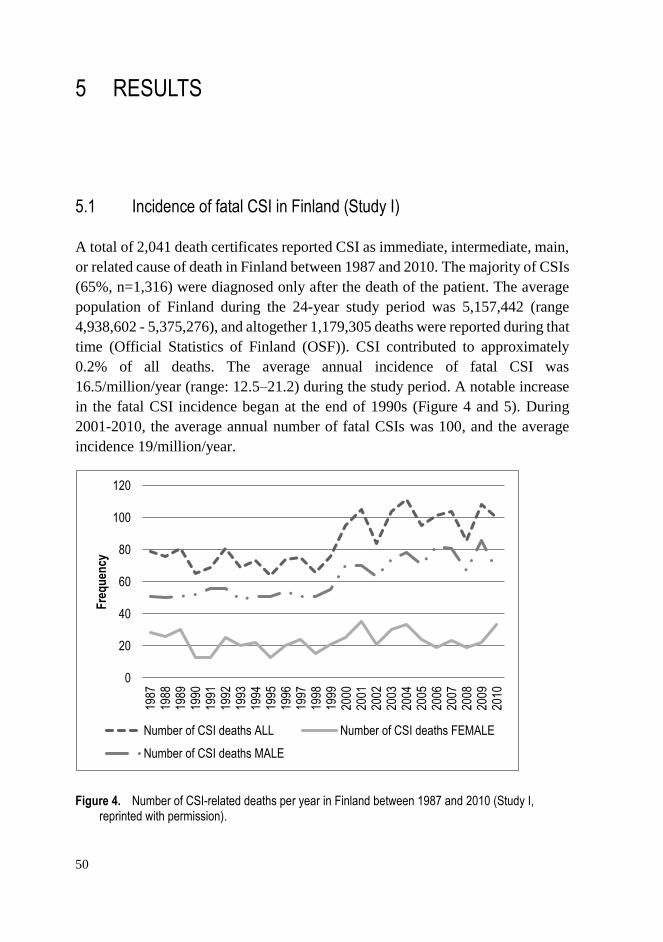
50
0
20
40
60
80
100
120
1987
1988
1989
1990
1991
1992
1993
1994
1995
1996
1997
1998
1999
2000
2001
2002
2003
2004
2005
2006
2007
2008
2009
2010
Fre
qu
ency
Number of CSI deaths ALL Number of CSI deaths FEMALE
Number of CSI deaths MALE
5 RESULTS
5.1 Incidence of fatal CSI in Finland (Study I)
A total of 2,041 death certificates reported CSI as immediate, intermediate, main,
or related cause of death in Finland between 1987 and 2010. The majority of CSIs
(65%, n=1,316) were diagnosed only after the death of the patient. The average
population of Finland during the 24-year study period was 5,157,442 (range
4,938,602 - 5,375,276), and altogether 1,179,305 deaths were reported during that
time (Official Statistics of Finland (OSF)). CSI contributed to approximately
0.2% of all deaths. The average annual incidence of fatal CSI was
16.5/million/year (range: 12.5–21.2) during the study period. A notable increase
in the fatal CSI incidence began at the end of 1990s (Figure 4 and 5). During
2001-2010, the average annual number of fatal CSIs was 100, and the average
incidence 19/million/year.
Figure 4. Number of CSI-related deaths per year in Finland between 1987 and 2010 (Study I, reprinted with permission).
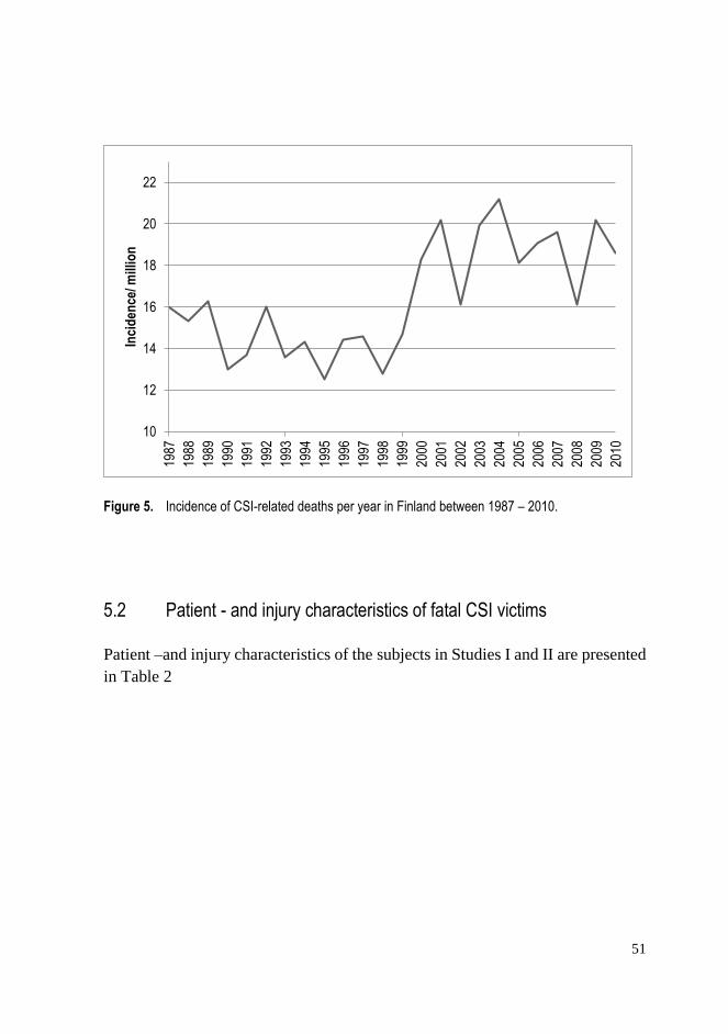
51
Figure 5. Incidence of CSI-related deaths per year in Finland between 1987 – 2010.
5.2 Patient - and injury characteristics of fatal CSI victims
Patient –and injury characteristics of the subjects in Studies I and II are presented
in Table 2
10
12
14
16
18
20
2219
87
1988
1989
1990
1991
1992
1993
1994
1995
1996
1997
1998
1999
2000
2001
2002
2003
2004
2005
2006
2007
2008
2009
2010
Inci
den
ce/ m
illio
n
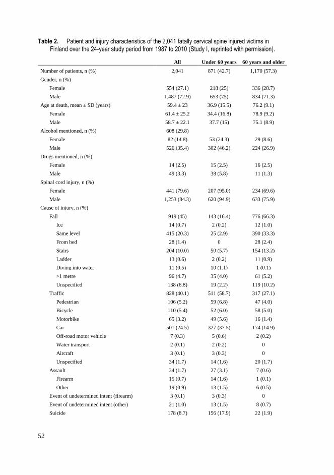
52
Table 2. Patient and injury characteristics of the 2,041 fatally cervical spine injured victims in Finland over the 24-year study period from 1987 to 2010 (Study I, reprinted with permission).
All Under 60 years 60 years and older
Number of patients, n (%) 2,041 871 (42.7) 1,170 (57.3)
Gender, n (%)
Female 554 (27.1) 218 (25) 336 (28.7)
Male 1,487 (72.9) 653 (75) 834 (71.3)
Age at death, mean ± SD (years) 59.4 ± 23 36.9 (15.5) 76.2 (9.1)
Female 61.4 ± 25.2 34.4 (16.8) 78.9 (9.2)
Male 58.7 ± 22.1 37.7 (15) 75.1 (8.9)
Alcohol mentioned, n (%) 608 (29.8)
Female 82 (14.8) 53 (24.3) 29 (8.6)
Male 526 (35.4) 302 (46.2) 224 (26.9)
Drugs mentioned, n (%)
Female 14 (2.5) 15 (2.5) 16 (2.5)
Male 49 (3.3) 38 (5.8) 11 (1.3)
Spinal cord injury, n (%)
Female 441 (79.6) 207 (95.0) 234 (69.6)
Male 1,253 (84.3) 620 (94.9) 633 (75.9)
Cause of injury, n (%)
Fall 919 (45) 143 (16.4) 776 (66.3)
Ice 14 (0.7) 2 (0.2) 12 (1.0)
Same level 415 (20.3) 25 (2.9) 390 (33.3)
From bed 28 (1.4) 0 28 (2.4)
Stairs 204 (10.0) 50 (5.7) 154 (13.2)
Ladder 13 (0.6) 2 (0.2) 11 (0.9)
Diving into water 11 (0.5) 10 (1.1) 1 (0.1)
>1 metre 96 (4.7) 35 (4.0) 61 (5.2)
Unspecified 138 (6.8) 19 (2.2) 119 (10.2)
Traffic 828 (40.1) 511 (58.7) 317 (27.1)
Pedestrian 106 (5.2) 59 (6.8) 47 (4.0)
Bicycle 110 (5.4) 52 (6.0) 58 (5.0)
Motorbike 65 (3.2) 49 (5.6) 16 (1.4)
Car 501 (24.5) 327 (37.5) 174 (14.9)
Off-road motor vehicle 7 (0.3) 5 (0.6) 2 (0.2)
Water transport 2 (0.1) 2 (0.2) 0
Aircraft 3 (0.1) 3 (0.3) 0
Unspecified 34 (1.7) 14 (1.6) 20 (1.7)
Assault 34 (1.7) 27 (3.1) 7 (0.6)
Firearm 15 (0.7) 14 (1.6) 1 (0.1)
Other 19 (0.9) 13 (1.5) 6 (0.5)
Event of undetermined intent (firearm) 3 (0.1) 3 (0.3) 0
Event of undetermined intent (other) 21 (1.0) 13 (1.5) 8 (0.7)
Suicide 178 (8.7) 156 (17.9) 22 (1.9)
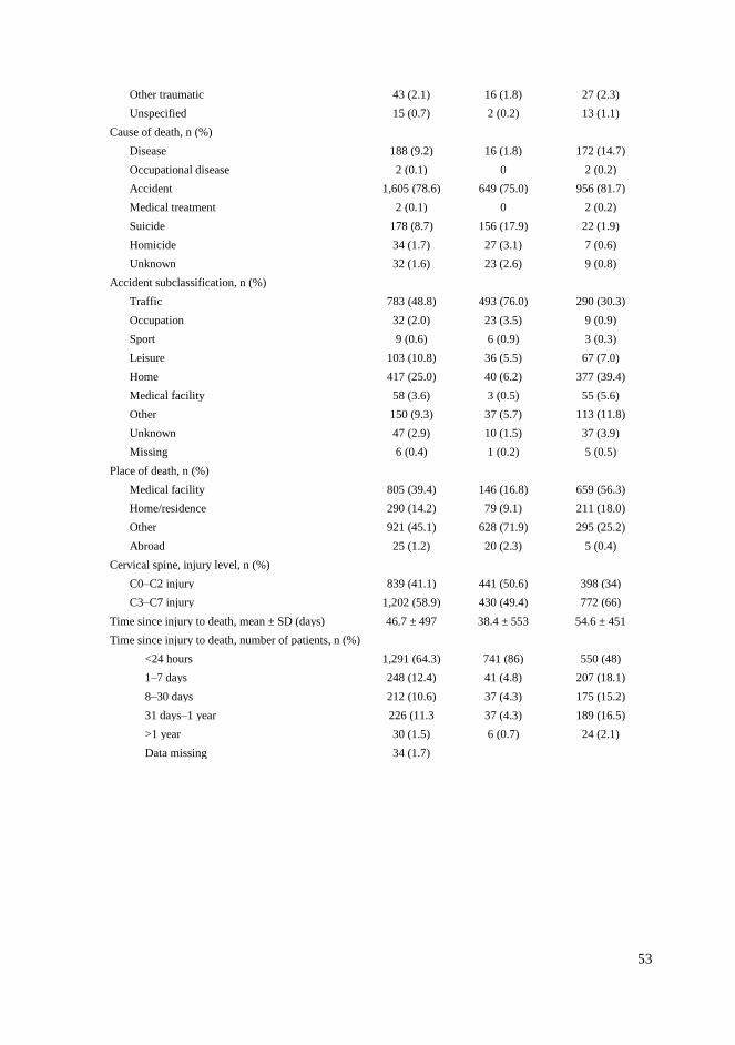
53
Other traumatic 43 (2.1) 16 (1.8) 27 (2.3)
Unspecified 15 (0.7) 2 (0.2) 13 (1.1)
Cause of death, n (%)
Disease 188 (9.2) 16 (1.8) 172 (14.7)
Occupational disease 2 (0.1) 0 2 (0.2)
Accident 1,605 (78.6) 649 (75.0) 956 (81.7)
Medical treatment 2 (0.1) 0 2 (0.2)
Suicide 178 (8.7) 156 (17.9) 22 (1.9)
Homicide 34 (1.7) 27 (3.1) 7 (0.6)
Unknown 32 (1.6) 23 (2.6) 9 (0.8)
Accident subclassification, n (%)
Traffic 783 (48.8) 493 (76.0) 290 (30.3)
Occupation 32 (2.0) 23 (3.5) 9 (0.9)
Sport 9 (0.6) 6 (0.9) 3 (0.3)
Leisure 103 (10.8) 36 (5.5) 67 (7.0)
Home 417 (25.0) 40 (6.2) 377 (39.4)
Medical facility 58 (3.6) 3 (0.5) 55 (5.6)
Other 150 (9.3) 37 (5.7) 113 (11.8)
Unknown 47 (2.9) 10 (1.5) 37 (3.9)
Missing 6 (0.4) 1 (0.2) 5 (0.5)
Place of death, n (%)
Medical facility 805 (39.4) 146 (16.8) 659 (56.3)
Home/residence 290 (14.2) 79 (9.1) 211 (18.0)
Other 921 (45.1) 628 (71.9) 295 (25.2)
Abroad 25 (1.2) 20 (2.3) 5 (0.4)
Cervical spine, injury level, n (%)
C0–C2 injury 839 (41.1) 441 (50.6) 398 (34)
C3–C7 injury 1,202 (58.9) 430 (49.4) 772 (66)
Time since injury to death, mean ± SD (days) 46.7 ± 497 38.4 ± 553 54.6 ± 451
Time since injury to death, number of patients, n (%)
<24 hours 1,291 (64.3) 741 (86) 550 (48)
1–7 days 248 (12.4) 41 (4.8) 207 (18.1)
8–30 days 212 (10.6) 37 (4.3) 175 (15.2)
31 days–1 year 226 (11.3 37 (4.3) 189 (16.5)
>1 year 30 (1.5) 6 (0.7) 24 (2.1)
Data missing 34 (1.7)

54
5.2.1 Gender, age and alcohol
Of all of the 2,041 CSI victims, 72.9% were male and the overall male to female
ratio was 2.6:1. The mean age at death was 59 years and 57% of the victims were
over 60 years old. Ninety-one (4.5%) patients were adolescent or children (˂18
years). There was a clear increase in the mean age at the time of death – from 54
to 68 years – during the study period (see Figure 6). Table 3 describes the share
of CSI patients in each ten-year age group and their change in incidence between
the first and last nine years of the study. The largest groups were patients aged
60-70 years (17%, n= 355) and 70-80 years (19%, n=395). Towards the end of
the study, the share of older patients increased notably.
Alcohol was mentioned in 608 (29.8%) and non-prescription drugs in 63
(3.1%) of the death certificates. Among < 60 years old male victims, alcohol was
present in 46.2% (302) of the cases. The share of alcohol intoxicated patients
remained more or less the same during the whole study period.
Figure 6. Mean age at death of fatally cervical spine injured patients in Finland between 1987 and 2010 (Study I, reprinted with permission).
40
45
50
55
60
65
70
1987
1988
1989
1990
1991
1992
1993
1994
1995
1996
1997
1998
1999
2000
2001
2002
2003
2004
2005
2006
2007
2008
2009
2010
Ag
e at
dea
th, y
ears
Year

55
Table 3. Total number of CSI cases per ten year age group, proportion of spinal cord injury victims, and comparison of the number of cases in years 1987–1995 and 2002–2010 (the first and last nine years of the study period) (Study I, reprinted with permission).
5.2.2 Injury mechanism and place of death
The most common injury mechanisms were falls (n=919, 45%) and traffic
accidents (n=828, n=40%). The mechanism of injury varied significantly by age.
Young and middle-aged victims (<60 years) were often injured in fatal traffic
accidents (n=511, 59%) or in suicides (n=156, 18%). Older (≥60 years) victims
were injured most often in falls (n=776, 66%) which were mostly regarded as
low-energy injuries. There were only nine sports related injuries and 11 injuries
resulting from diving into water (Table 2).
There were significant differences in the place of death for different age
groups. Of the older victims, 56% (n=659) died in a medical facility and 18%
Age group (years)
Total number of
CSI victims,
n (%)
Proportion of spinal
cord-injured victims,
n (%)
Number of CSI victims
between 1987–1995,
n (%)
Number of CSI victims
between 2002–2010,
n (%)
Change between
1987–1995 and 2002–
2010 n (%)
0–9.9 41 (2.0) 40 (97.6) 26 (4.0) 5 (0.6) -21 (-80.8)
10–19.9 100 (4.9) 99 (99) 47 (7.2) 25 (2.8) -22 (-46.8)
20–29.9 183 (9.0) 178 (97.3) 75 (11.4) 70 (7.8) -5 (-6.7)
30–39.9 142 (7.0) 141 (99.3) 67 (10.2) 41 (4.6) -26 (-38.8)
40–49.9 170 (8.3) 155 (91.2) 57 (8.7) 66 (7.4) 9 (+15.8)
50–59.9 235 (11.5) 214 (91.2) 65 (9.9) 98 (11.0) 33 (+50.8)
60–69.9 355 (17.4) 299 (84.2) 118 (18.0) 152 (17.0) 34 (+28.8)
70–79.9 395 (19.4) 313 (79.2) 115 (17.5) 187 (20.9) 72 (+62.6)
80–89.9 338 (16.6) 217 (62.2) 81 (12.3) 188 (21.1) 107 (+132.1)
90–99.9 82 (4.0) 38 (46.3) 6 (0.9) 61 (6.8) 55 (+916.7)
Total 2,041 (100) 1,694 (83.0) 657 (100) 893 (100) 236(+35.9)
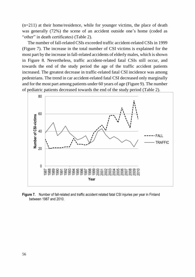
56
(n=211) at their home/residence, while for younger victims, the place of death
was generally (72%) the scene of an accident outside one’s home (coded as
“other” in death certificates) (Table 2).
The number of fall-related CSIs exceeded traffic accident-related CSIs in 1999
(Figure 7). The increase in the total number of CSI victims is explained for the
most part by the increase in fall-related accidents of elderly males, which is shown
in Figure 8. Nevertheless, traffic accident-related fatal CSIs still occur, and
towards the end of the study period the age of the traffic accident patients
increased. The greatest decrease in traffic-related fatal CSI incidence was among
pedestrians. The trend in car accident-related fatal CSI decreased only marginally
and for the most part among patients under 60 years of age (Figure 9). The number
of pediatric patients decreased towards the end of the study period (Table 2).
Figure 7. Number of fall-related and traffic accident related fatal CSI injuries per year in Finland between 1987 and 2010.
0
20
40
60
80
1987
1988
1989
1990
1991
1992
1993
1994
1995
1996
1997
1998
1999
2000
2001
2002
2003
2004
2005
2006
2007
2008
2009
2010
Nu
mb
er o
f C
SI-
vict
ims
Year
FALL
TRAFFIC
57
Figure 8. Number of cervical spine injury (CSI) - related deaths caused by fall-induced accidents per year in < 60 and ≥ 60-year-old patients (Study I, reprinted with permission).
Figure 9. Number of cervical spine injury (CSI) – related deaths by car accident per year in < 60 and ≥ 60-year-old patients in Finland between 1987 and 2010.
0
5
10
15
20
25
30
35
40
45
50
1987 1990 1993 1996 1999 2002 2005 2008
Nu
mb
er o
f C
SI-
vict
ims
Year
Female <60 FALL Male <60 FALL
Female ≥60 FALL Male ≥60 FALL
0
5
10
15
20
25
19
87
19
88
19
89
19
90
19
91
19
92
19
93
19
94
19
95
19
96
19
97
19
98
19
99
20
00
20
01
20
02
20
03
20
04
20
05
20
06
20
07
20
08
20
09
20
10
Num
ber
of C
SI-
vict
ims
Under 60 years 60 years and older

58
5.2.3 Day and month of injury and Interval between injury and death
The most common day of CSI was Saturday (18%, n=368). As shown in Figure
10, younger patients (<60 years) especially tended to get injured towards the end
of the week (Friday and Saturday, 39%). For elderly patients, there were no big
variations regarding the day of injury. Regarding the month of injury, there was
some preponderance in the summer months in the CSI frequency. The most
frequent month of injury was July (10%, n=206) (Figure 11).
The mean time between injury and death was 46.7 days (median: 0 days, min
0, and max 37.9 years). There were considerable variations in time intervals
between young (<60 years) and old (≥ 60 years) patients. Younger patients died
during the day of injury in the majority of cases (86%, n=741), whereas older
patients did so in less than half of the time (48%, n=550). A substantial number
(33.2%, n=388) of older patients lived more than one week post-injury (Table 2).
To summarize, young patients mainly died immediately due to high-energy traffic
accidents and old patients due to the sequalae of falling accidents.
Figure 10. Day of injury in < 60 - and ≥ 60-year-old fatally cervical spine injured patients in Finland between 1987 and 2010.
0
5
10
15
20
25
Per
cen
tag
e o
f in
juri
es
Day of injury
< 60 years ≥ 60 years
59
Figure 11. Month of injury in < 60 - and ≥ 60-year-old fatally cervical spine injured patients in Finland between 1987 and 2010.
5.2.4 Causes of death
CSI was the main cause of death in 90.5% (n=1,847) and related cause of death
in 9.5% (n=193) of the fatally cervical spine injured patients. Among those who
died during the day of injury (n=1,297) (most often at the scene of the accident),
CSI was the main cause of death 95.8% (1,243) of the time. Among elderly
patients (≥60 years, n=1170) the main cause of death was relatively often (n=179,
15.3%) other than a CSI. Among elderly patients, who died more than one week
after the CSI, the immediate causes of death were mostly respiratory (n=234,
60%) or circulatory (n=54, 13.9%) diseases.
5.2.5 Level of injury
CSI occurred most often in the C3-C7 region (58.9%, n=1,202), (including
combined upper and lower injuries as well as miscellaneous injuries). Young
patients (<60 years) had an upper CSI (C0-C2) in about half of the cases (51%,
n=441). Of those, SCI was present 96.1% (n=424) of the time and the majority
(91.8, n=402) of these patients died during the day of injury (mostly immediately
0
2
4
6
8
10
12
Per
cen
tag
e o
f in
juri
es
Month of injury
< 60 years ≥ 60 years
60
at the scene of the accident). However, in the older patient group (≥ 60 years),
34.0% (n=398) had an upper CSI and only 65.3% (n=260) of them had a SCI.
Only about half (49.6%, n=194) of the older patients with upper injury died during
the day of injury.
5.2.6 Incidence and share of spinal cord injury in relation to patient´s age
Spinal cord injury was found in 1,694 (83.0%) cases and in 91.2% (n=1,177) of
the patients who died during the day of injury. The incidence of spinal cord injury
decreased with increasing age so that in the oldest age group (90-100 years) less
than half (46.3%) had an SCI whereas among patients less than 40 years, almost
all (98%) had an SCI (Table 3).
5.2.7 Method of death certification (Study I and II)
In the study, a medicolegal autopsy was performed for 1,907 (93.4%) patients and
clinical autopsy for 5 (0.2%) patients. In 121 (5.9%) cases, the death certificate
was written by the treating physician without an autopsy, and in eight (0.4%)
cases the type of death certification was unknown. Among those who died during
the day of injury, medicolegal autopsy was performed in 1,291 (99.5%) cases,
and among those who died later than the day of injury in 616 cases (82.8%).
5.3 Preventable adverse events (Study II)
5.3.1 Comparison between patients who died during the day of injury and patients who survived longer than the day of injury (Study II)
Out of the 2,041 CSI-related deaths, 36.5% (n = 744) happened later than the date
of injury. The patients who survived at least until the next day were significantly
older (76 years vs. 55 years, p<0.001), the injury mechanism was more often a
fall (72%, n = 536 vs. 30%, n = 383, p<0.001), and the place of death was more
often a medical facility (94%, n = 698 vs. 8%, n = 107, p<0.001). Alcohol
consumption (37%, n = 482 vs. 17%, n = 126, p<0.001), the incidence of spinal
cord injury (91%, n = 1,182 vs. 69%, n = 512, p<0.001), and the rate of upper
61
CSIs (46%, n = 598 vs. 32%, n = 241, p<0.001) were higher among those who
died on the day of injury.
5.3.2 Incidence of preventable adverse events
Of those CSI patients who survived at least until the next day, a PAE was
identified in 27.7% (n = 206), of which half (n = 103) were considered diagnostic
errors. In 5% (n = 37) of those who survived at least until the next day, a PAE
could not be determined as being present or not. The number of PAEs and
diagnostic errors increased during the study period, but so did the number of all
CSI patients. The ratio of PAEs or diagnostic errors in relation to the number of
all CSI patients increased slightly during the study years (Figure 12). The PAEs
(n = 206) were classified into six different categories: diagnostic errors, health
care facility-related events, treatment-related events, secondary complications,
surgical treatment-related events and other events (Table 4). The most frequent
recorded PAE types were related to CSI diagnostics and health care facility-
related events.
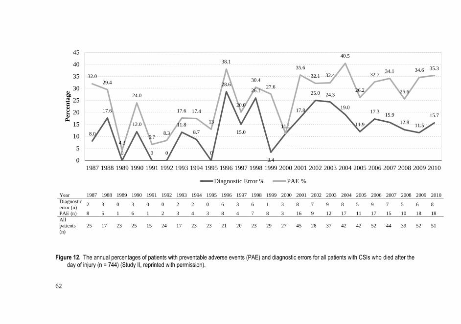
62
Year 1987 1988 1989 1990 1991 1992 1993 1994 1995 1996 1997 1998 1999 2000 2001 2002 2003 2004 2005 2006 2007 2008 2009 2010
Diagnostic
error (n) 2 3 0 3 0 0 2 2 0 6 3 6 1 3 8 7 9 8 5 9 7 5 6 8
PAE (n) 8 5 1 6 1 2 3 4 3 8 4 7 8 3 16 9 12 17 11 17 15 10 18 18
All
patients
(n)
25 17 23 25 15 24 17 23 23 21 20 23 29 27 45 28 37 42 42 52 44 39 52 51
Figure 12. The annual percentages of patients with preventable adverse events (PAE) and diagnostic errors for all patients with CSIs who died after the day of injury (n = 744) (Study II, reprinted with permission).
8.0
17.6
0
12.0
0 0
11.8
8.7
0
28.6
15.0
26.1
3.4
11.1
17.8
25.0 24.3
19.0
11.9
17.315.9
12.811.5
15.7
32.0
29.4
4.3
24.0
6.78.3
17.6 17.4
13
38.1
20.0
30.4
27.6
11.1
35.6
32.1 32.4
40.5
26.2
32.734.1
25.6
34.6 35.3
0
5
10
15
20
25
30
35
40
45
1987 1988 1989 1990 1991 1992 1993 1994 1995 1996 1997 1998 1999 2000 2001 2002 2003 2004 2005 2006 2007 2008 2009 2010
Per
cen
tag
e
Diagnostic Error % PAE %

63
Table 4. Preventable adverse events (n = 206) in fatal CSI patients who died after the date of injury stratified into six categories (Study II, reprinted with permission).
Preventable adverse event category N %
Total 206 100.0
Diagnostic errors 103 50.0
CSI diagnosis missed during lifetime 59 28.6
CSI diagnosis delayed 41 20.0
Diagnostic other 3 1.5
Health care facility related events (fall at the ward, fall from bed, wrong place of treatment, or fell from wheelchair)
36 17.5
Treatment related events (wrong treatment, CSI operation not performed, supine skull traction for elderly,
medication related, related to halo for stabilizing head and neck, iatrogenic aspiration of blood, too early extubation, or post-operative bleeding related to anticoagulant treatment)
31 15.0
Secondary complications (pressure ulcer, pulmonary embolism, or preventable aspiration) 20 9.7
Surgical treatment related events (peri-operative death, surgical hardware error, suboptimal operation, or
post-operative infection/hematoma)
12 5.8
Other (post-surgery brain infarction, removal of collar by patient, or
NORO-diarrhea)
3 1.5
64
5.3.3 Characteristics of patients with preventable diagnostic error
Patient characteristics, injury-related information, and diagnostic error data are
presented in Table 5. Diagnostic error occurred most of the time in specialized
health care (55%). Patients with diagnostic errors were significantly older (79.4
vs. 74.9 years, p = 0.001), and the injury mechanism was significantly more often
a fall compared to CSI patients who did not have a diagnostic error. Median time
from injury to death was significantly shorter in the diagnostic error group (seven
days compared to 17 days, p < 0.001). Examples of typical cases with a diagnostic
error are presented in Table 6. One of the most typical diagnostic errors was the
lack of suspicion of a CSI (Table 6, examples 1 and 4).

65
Table 5. Characteristics of patients with CSIs who died after the day of injury stratified by whether or not the patients experienced a diagnostic error (Study II, reprinted with permission).
Diagnostic error
n=103
No diagnostic error
n=641
Total
n=744
n % n % p n %
Gender 0.873
Female 24 23.3 154 24 178 23.9
Male 79 76.7 487 76 566 76.1
Age at death, years, median (95% CI) 79.4 (75.9-80.1) 74.9 (70.2-72.9) 0.001 75.7 (71.3-73.6)
Alcohol mentioned 20 19.4 106 16.5 0.469 126 16.9
Drugs mentioned 0 0 4 0.6 0.421 4 0.5
CSI as main cause of death 79 76.7 525 81.9 0.21 604 81.2
C0-C2 injury 28 27.2 213 33.2 0.224 241 32.4
Spinal cord injury 66 64.1 446 69.6 0.263 512 68.8
Cause of injury 0.002
Fall 89 86.4 447 69.7 536 72
Traffic accident 8 7.8 150 23.4 158 21.2
Suicide 0 0 5 0.8 5 0.7
Other traumatic or unspecified 6 5.8 39 6.1 45 6
Place of death 0.647
Medical facility 95 92.2 603 94.1 698 93.8
Home or residence 6 5.8 23 3.6 29 3.9
Other 2 1.9 12 1.9 14 1.9
Abroad 0 0 3 0.5 3 0.4
Time since injury to death, Median, Days (min-max) 7 (1-1,442) 17 (1-13,849) <0.001 16 (1-13,849)
Time since injury to death 0.005
1-7 days 49 47.6 198 30.9 247 33.2

66
8-30 days 27 26.2 183 28.5 210 28.2
31 days-1 year 19 18.4 206 32.1 225 30.2
> year 2 1.9 28 4.4 30 4.0
Data missing 6 5.8 26 4.1 32 4.3
Death certification 0.023
No autopsy 7 6.8 113 17.6 120 16.1
Hospital autopsy 0 0 5 0.8 5 0.7
Medicolegal autopsy 95 92.2 521 81.3 616 82.8
Other 1 1 2 0.3 3 0.4
Place of diagnostic error
Primary health care 41 39.8 - - 41 5.5
Spezialized health care 57 55.3 - - 57 7.7
Undetermined 5 4.9 - - 5 0.7
Table 6. Examples of typical cases with preventable diagnostic errors. (Modified from Study II, reprinted with permission).
1 79-year-old male with a history of Parkinson’s disease, coronary artery disease with a prior heart infarct and periodic heart arrhythmia.
Moving with aid of a walker. Frequent ground level falls. He was brought to a central hospital because of a ground level fall and
subsequent difficulties with getting up and moving. Traumatic intracranial changes were not detected. He was hospitalized and he died
nine days later. According to the medicolegal autopsy, the main cause of death was a fracture of the C5 vertebra and medullary
contusion. The immediate cause of death was pneumonia. Contributing causes of death were Parkinson’s disease and coronary artery
disease. Diagnostic error: lack of suspicion of a CSI.
2 80-year-old male. No information about chronic diseases. He fell from a rocking chair and hyperflexed his neck. Primarily neck
roentgenogram and neck CT were considered negative. After two weeks he developed tetraparesis and C5/C6 listhesis was detected.
An anterior decompression and fixation procedure were performed. After operation, excessive mucous secretion. He died during the
insertion of a feeding tube 29 days after injury. According to the medicolegal autopsy the main cause of death was a C5/C6 level
luxation with tetraplegia. Immediate cause of death was pneumonia. Diagnostic error: Image misinterpretation or inadequate imaging.
3 82-year-old male with coronary artery disease. Heart insufficiency, hypertension, chronic obstructive pulmonary disease and diabetes.
In 2004, he fell from bed at home. On the next day, he was taken in to a health care center ward due to pain in the shoulder and chest.
67
Heart infarct was not diagnosed. Afterwards pain localized more to the neck and due to unclear symptoms he was referred to a central
hospital five days later. He was tired and had shortness of breath and excess mucous secretion was observed. Cervical spine
roentgenogram and head CT were negative. He was found dead in a hospital ward bed eight days after the fall. According to the
medicolegal autopsy the main cause of death was fracture of the C6 with a medullary lesion. Intermediate cause of death was
bronchopneumonia incipiens. Immediate cause of death was bronchial aspiration. Contributing causes of death were fulminant
coronary artery disease and pulmonary emphysema. Diagnostic error: inadequate imaging.
4 90-year-old male. No information about chronic diseases. Walking frame assisted moving. In 1998, he fell at home. He was
transferred to a local health care center where he complained of pain in his left shoulder. He was taken in to the healthcare center ward
where increasing spasticity was observed in the left shoulder area. He was transferred to a central hospital 17 days after the injury
where a cervical spine fracture was detected and an external fixation device was inserted. He was transferred back to the health care
center where he died 36 days after injury. According to the medicolegal autopsy, the main and immediate causes of death was a C7
fracture with a medullary contusion. Diagnostic error: lack of suspicion of a CSI and delayed imaging.
68
5.4 Concurrence of head injury and cervical spine fracture (Study III and IV)
Of the whole cranio-cervically CT-imaged group of patients (n=1,091), 65.4%
(n=714) were male. Falls (47.8%, n=521) and car accidents (22.4%, n=244) were
the most frequent injury mechanisms. Of all the patients (n=1,091), 607 (55.6%)
fulfilled the clinical criteria for mild TBI, 201 (18.4%) had a moderate or severe
TBI and the rest (n=283, 25.9%) had a head trauma without clear signs of TBI.
Cervical spinal cord injury was found in 13 (16.9%) of the CSI patients. In one
patient, the existence of a possible spinal cord injury remained unknown due to
early death caused by a severe TBI.
5.4.1 Cervical spine fractures in patients with CT-positive versus CT-negative head injuries
Patients who had acute traumatic intracranial lesions in head CT (9.3%, n=25)
had significantly more cervical spine fractures (Pearson chi-square, p=0.04;
OR=1.689, 95% CI=1.019-2.802) compared to those who did not have
intracranial lesions visible in head CT (5.7%, n=47). However, patients with
positive head CT scans had fewer spinal cord injuries (0.4%, n=1 versus 1.5%,
n=12). Moreover, positive head CT findings had a significant association with C6
vertebra fractures (3.0%, n=8 versus 1.1%, n=9; p=0.031, OR=2.769, 95%
CI=1.057-7.250), but not with other cervical vertebra fractures (C0-5 and C7) or
dislocations/subluxations alone. Patients with positive head CT scans (2.2%, n=6)
did not have more neurosurgical operations within one year post-HI due to
cervical spine fractures compared with patients with negative head CT scans
(3.2%, n=26; p=0.431). Comparisons between head CT-positive and head CT-
negative patients are presented in Table 7.
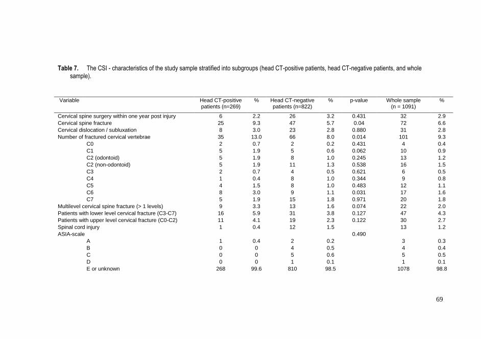
69
Table 7. The CSI - characteristics of the study sample stratified into subgroups (head CT-positive patients, head CT-negative patients, and whole sample).
Variable Head CT-positive patients (n=269)
% Head CT-negative patients (n=822)
% p-value Whole sample (n = 1091)
%
Cervical spine surgery within one year post injury 6 2.2 26 3.2 0.431 32 2.9
Cervical spine fracture 25 9.3 47 5.7 0.04 72 6.6
Cervical dislocation / subluxation 8 3.0 23 2.8 0.880 31 2.8
Number of fractured cervical vertebrae 35 13.0 66 8.0 0.014 101 9.3
C0 2 0.7 2 0.2 0.431 4 0.4
C1 5 1.9 5 0.6 0.062 10 0.9
C2 (odontoid) 5 1.9 8 1.0 0.245 13 1.2
C2 (non-odontoid) 5 1.9 11 1.3 0.538 16 1.5
C3 2 0.7 4 0.5 0.621 6 0.5
C4 1 0.4 8 1.0 0.344 9 0.8
C5 4 1.5 8 1.0 0.483 12 1.1
C6 8 3.0 9 1.1 0.031 17 1.6
C7 5 1.9 15 1.8 0.971 20 1.8
Multilevel cervical spine fracture (> 1 levels) 9 3.3 13 1.6 0.074 22 2.0
Patients with lower level cervical fracture (C3-C7) 16 5.9 31 3.8 0.127 47 4.3
Patients with upper level cervical fracture (C0-C2) 11 4.1 19 2.3 0.122 30 2.7
Spinal cord injury 1 0.4 12 1.5 13 1.2
ASIA-scale 0.490
A 1 0.4 2 0.2 3 0.3
B 0 0 4 0.5 4 0.4
C 0 0 5 0.6 5 0.5
D 0 0 1 0.1 1 0.1
E or unknown 268 99.6 810 98.5 1078 98.8

70
5.4.2 Cervical spine CT findings
On cervical CT, CSI was found in 7.1% (n=77) and cervical spine fracture in
6.6% (n=72) of the patients with HI (n=1,091). In total, these patients sustained
101 fractured vertebrae. Five patients had only a ligament injury without a
fracture in CT imaging. Of the patients with CSI, 31 (40.3%) had dislocations
and/or subluxations. Of the patients with fractures, 27 (37.5%) had dislocations
and/or subluxations. The median dislocation between cervical vertebrae was three
millimeters (range 1 - 10mm). The distribution of fractured vertebrae (n=101) is
presented in Figure 13. The distribution of dislocations/subluxations is presented
in Figure 14. Dislocations/subluxations occurred most often in the atlanto-axial
(n=9) and C4/C5 (n=7) levels. The distribution of different fracture locations
(n=161) within individual vertebrae in the upper cervical spine is presented in
Figures 15, 16 and 17) and in the lower cervical spine in Table 8. In the atlas (C1),
the fracture type was most often comminuted. In the axis (C2), odontoid process
types 2 and 3 and lateral mass/articular process fractures prevailed. In the subaxial
cervical spine, fractures involved most often the lateral masses/articular
processes.
Figure 13. Level of cervical spine fractures (Study III, reprinted with permission).
4
10
23
6
9
12
17
20
0
5
10
15
20
25
Nu
mb
er o
f fr
actu
red
ver
teb
rae(
n=
101)
Level of fractures

71
Figure 14. Level of dislocations/subluxations (Study III, reprinted with permission)
Figure 15. Location of fractures to C0-C1 vertebrae (Study IV).
1
9
3
1
7
5
3
2
012345678910
Nu
mb
er o
f d
islo
cati
on
s/su
blu
xa
tio
ns
(n=
31
)
Level of dislocations/subluxations
28.6%
14.3%
0
14.3%
42.9%
0
1
2
3
4
5
6
7
Occipital condyle C1 Isolatedposterior element
C1 Isolatedanterior element
C1 Lateralmass/articular
process
C1 Comminutedarc fracture
Nu
mb
er o
f fr
actu
res
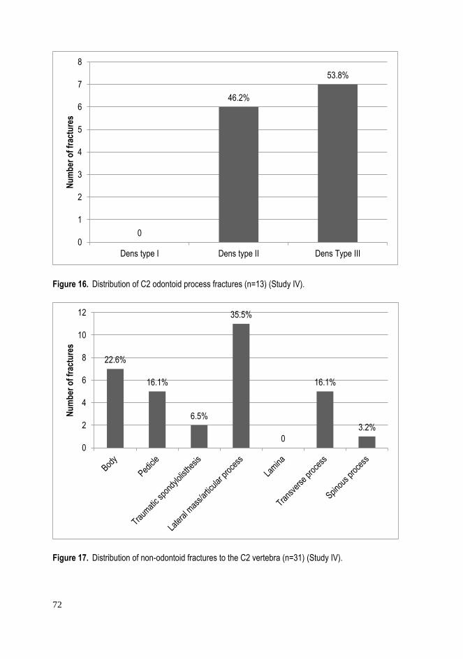
72
Figure 16. Distribution of C2 odontoid process fractures (n=13) (Study IV).
Figure 17. Distribution of non-odontoid fractures to the C2 vertebra (n=31) (Study IV).
0
46.2%
53.8%
0
1
2
3
4
5
6
7
8
Dens type I Dens type II Dens Type III
Nu
mb
er o
f fr
actu
res
22.6%
16.1%
6.5%
35.5%
0
16.1%
3.2%
0
2
4
6
8
10
12
Nu
mb
er o
f fr
actu
res

73
Table 8. The distribution of subaxial fracture locations (Study IV).
Location C3 C4 C5 C6 C7 Total %
Body 3 4 4 4 3 18 17.5
Pedicle - 3 2 - 3 8 7.8
Lateral mass/ articular process 1 7 8 5 12 33 32.0
Lamina 1 6 4 4 3 18 17.5
Transverse process 1 3 2 2 6 14 13.6
Spinous process - 1 2 4 3 10 9.7
Other 1 - - 1 - 2 1.9
Total 7 24 22 20 30 103 100.0
5.5 Risk factors for cervical spine fractures in head-injured patients (Study IV)
High age was a risk factor of cervical spine fractures among HI patients. Patients
who had a cervical spine fracture were almost ten years older than patients who
did not have a cervical spine fracture (median 55.5 versus 46.7 years, p=0.027).
In addition, facial bone fractures (13.9%, n=10 versus 6.9%, n=70, p=0.027) and
epidural hematomas (4.2%, n=3 versus 1.2%, n=12, p=0.035) were significantly
more common in the fracture group and therefore may be considered as risk
factors for cervical spine fractures. However, the total number of epidural
hematomas was low to allow solid conclusions to be formulated. In addition,
patients with a high-energy injury mechanism had more cervical spine fractures
compared to patients with a low-energy injury (59.7%, n=43 versus 40.3%, n=29,
p=0.021). Surprisingly, there were no statistical differences in the gender
distribution, clinical TBI severity, GCS level or presence of alcohol intoxication
between patients with or without a spine fracture. Patients with a cervical spine
fracture were hospitalized longer (96.4 hours versus 21.3 hours, p<0.001) and
more neurosurgical procedures (19.4%, n=37 versus 7.9%, n=81, p<0.001) were
performed on them.
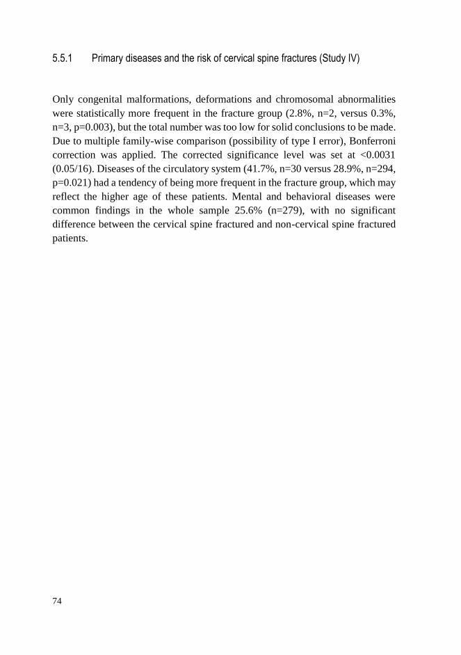
74
5.5.1 Primary diseases and the risk of cervical spine fractures (Study IV)
Only congenital malformations, deformations and chromosomal abnormalities
were statistically more frequent in the fracture group (2.8%, n=2, versus 0.3%,
n=3, p=0.003), but the total number was too low for solid conclusions to be made.
Due to multiple family-wise comparison (possibility of type I error), Bonferroni
correction was applied. The corrected significance level was set at <0.0031
(0.05/16). Diseases of the circulatory system (41.7%, n=30 versus 28.9%, n=294,
p=0.021) had a tendency of being more frequent in the fracture group, which may
reflect the higher age of these patients. Mental and behavioral diseases were
common findings in the whole sample 25.6% (n=279), with no significant
difference between the cervical spine fractured and non-cervical spine fractured
patients.
75
6 DISCUSSION
6.1 Increasing trend in CSI incidence and change in patient profile
To the best of my knowledge, this is the first whole nation-wide study to report
the incidence of fatal CSI. The epidemiological data published by most authors is
based on hospitalized patients and therefore underestimates the incidence of
certain injuries when leaving out casualties that died at the scene of an accident
or patients who are erroneously diagnosed. We found that the majority (65%) of
all fatal CSIs were diagnosed after the death of the patient. The death certificate-
based incidence of CSI increased from 16/million/year to 19/million/year
between 1987 and 2010. The most notable increase in the incidence has been after
the year 1999, which is mainly explained by the increase in ground-level falls
among elderly males (Figures 4, 5 and 8). This finding corroborates previous
studies. The age of those deceased changed notably during the observation period
(Figure 6). The mean age at the time of death increased from 54 years to 67 years,
and the number of pediatric patients decreased significantly. This is probably
partly due to improved road and in-car safety, and also the general safety
precautions related to children’s living environment (i.e., school, home, hobbies
etc.). Falls exceeded traffic accidents as the most common cause of CSI in 1998.
The number of fall-induced accidents among elderly individuals started to
increase at the end of the 1990s. It has been shown previously that even though
in general, falling accidents are more frequent in the female population, most of
the fall-induced severe CSIs happen to males (Kannus, Palvanen et al. 2007,
Kannus, Parkkari et al. 2005). The reason for this phenomenon is unknown. It has
been proposed that men are at greater risk of severe falls than women (Kannus,
Palvanen et al. 2007). Moreover, ankylosing spinal disorders (AS and DISH),
which increase the risk of cervical spine fractures, are more common in males.
Fall prevention measures for the elderly are crucial since it is estimated that in
Finland the number of people aged 65 or older is going to rise from the present

76
19.9% to 26% by the year 2030 (Official Statistics of Finland 2015 c). This puts
1.5 million Finnish seniors above the age of 65 years at risk of CSI with the
current safety measures.
Traffic accidents continue to cause fatal CSI especially among men despite the
decreasing general trend in all traffic accident-related fatal CSIs. Towards the end
of the study, older patients especially contributed most to the number of traffic
accident-related fatal CSIs. It has been shown previously that seatbelts and
airbags reduce mortality after motor vehicle accidents, (Cummings, McKnight et
al. 2002) and protect the cervical spine (Claytor, MacLennan et al. 2004). The use
of seatbelts and airbags increased in Finland during the study years, but this did
not influence much the incidence of fatal CSIs (Figure 5). In Finland, general road
traffic mortality decreased from 581 per year to 272 per year between 1987 and
2010 (Official Statistics of Finland (OSF)). During the same time, fatal CSIs
induced by traffic accidents decreased only marginally (Figure 7). In 1987, there
were 1.9 million registered vehicles whereas in 2010 this had increased to 3.4
million. The strongest decline in traffic accident-related fatal CSIs was among
pedestrians. Previously, Lieutaud et al. studied traffic-related SCIs in France
between two observation periods (1996–2001 and 2003–2008). They showed that
despite the significant drop in road trauma in general, there was no change in the
incidence and mortality of SCIs. There were actually more cervical SCIs seen in
the more recent period. Cervical spine injuries increased significantly among
pedestrians, cyclists and motorcyclists whereas among car users they dropped.
This is contrary to our study since we found that the fatal CSI number decreased
among pedestrians and remained quite stable among cyclists. The total number of
fatal CSI accidents with motorbikes is quite low (n=65) and its incidence
remained stable throughout the study. Only 16 motorbike accidents happened to
elderly people. Lieutaud et al. also found that those sustaining traffic-related SCI
were older than in the past (Lieutaud, Ndiaye et al. 2012). This is corroborated by
our study.
Primary prevention policies in traffic that target children and young adults
have significantly reduced traffic accidents with CSIs. However, elderly car users
and cyclists (especially male) are those who are especially at risk for traffic-
related fatal CSIs in Finland. The proportion of elderly people in traffic in general
has increased, which partly explains the non-declining trend in the total number
of fatal CSIs. Our findings confirm that although the incidence of traffic accidents
is in general tending to fall in developed countries, the situation needs to be
monitored continuously. Traffic-related primary prevention measures should be

77
also targeted at elderly people and infrastructure planning should be senior-
friendly.
We found only a few fatal sports-related CSIs which is contradictory to most
of the studies on CSI incidence (Sekhon, Fehlings 2001, Leucht, Fischer et al.
2009, Hasler, Exadaktylos et al. 2012). Even though sports-related CSIs
sometimes cause neurological symptoms, they are seldom fatal (Rihn, Anderson
et al. 2009), which explains why they are not shown in this study. Moreover,
certain sports that may be considered high risk for fatal CSI (such as mountain
climbing or base jumping for example) are relatively uncommon in Finland. An
autopsy-based retrospective study of all 48,335 fatalities in Hamburg between
1997 and 2006 found 176 sports-related deaths. Of those, 38.1 % were attributed
to traumatic causes. The most common traumatic causes were drowning, head
trauma and polytrauma. Only one patient died of post-CSI sequalae (Turk, Riedel
et al. 2008).
In Finland, jumping head first into shallow water is generally considered as a
rather common injury mechanism of CSI in the summer time. Surprisingly, only
11 cases with this type of injury were found during the 24-year study period. As
patients with the aforementioned injury mechanism are typically young people,
they seem to survive the CSI if they are rescued from water before drowning.
Alcohol is a major risk factor for fatal injuries in general (Tiirikainen 2009).
Alcohol played a role in about one third of the cases in our series which is in line
with previous series. The share of alcohol intoxicated patients remained more or
less the same during the whole study. Young male victims were most often
intoxicated. The number of patients who were intoxicated by non-prescription
drugs was relatively low (3%) among fatal CSI patients. Unfortunately, non-
prescription drugs are getting more common in general in society and therefore
the number of accidents related to their abuse may be expected to increase.
6.2 Survival after a CSI
Patients who survive the initial accident and die of sequalae of CSI are general
old adults. Due to improved medical care, young and presumably healthier
patients often survive the CSI if they manage to reach medical care. Lower
survival rates in hospitalized SCI and CSI patients have been described for
example among those with old age, high ISS, high level of injury, neurological
deficit, and presence of cardiopulmonary resuscitation (Van Den Berg, Castellote

78
et al. 2010, Patel, Smith et al. 2012, Morita, Takebayashi et al. 2017). It is well
accepted that recovery from a neurological deficit and survival after SCI is good
for young patients. However, patients with high-energy injury mechanisms that
cause CSI often die at the scene of accident. Many publications have described
how the survival of patients with SCI in developed countries has improved and
mortality is not far from the general population (DeVivo, Krause et al. 1999, Van
Den Berg, Castellote et al. 2010). However, prehospital deaths are seldom
counted in such studies. Like Lieutaud et al. we found that a substantial proportion
of patients who die due to sequalae of CSI do so before hospital admission (64.5%
of the fatal CSI were not diagnosed before death). Therefore, the survival rates
reported by many studies are artificially too high. Epidemiological studies are
often based on hospital diagnoses, leaving out patients who die at the scene of the
accident or whose injury is missed during hospitalization.
In our study, most of the CSI-related deaths among the young population took
place outside of medical facilities (84%, n=727) within the day of injury.
Whereas, in the older population, only 44% (n=511) of the deaths occurred
outside of medical facilities. Injuries among the young population occurred most
often during week-ends whereas for elderly people, injuries were more evenly
distributed for each week-day. One third (33%, n=388) of patients in the senior
population (60 years and older) died more than seven days post injury. In the
majority of these cases (74%), the immediate cause of death was due to
respiratory or circulatory diseases (e.g. pneumonia, pulmonary embolism or heart
attack). This is an important finding as those are potentially preventable
conditions if recognized early. In particular, as the share of elderly patients is
increasing among CSI patients. Diminished pulmonary function and
cardiovascular diseases have been previously reported with increased mortality
among SCI patients (Lemons, Wagner 1994, Van Den Berg, Castellote et al.
2010). As most of the CSI-related deaths in the young population (<60 years)
occur immediately, and outside of medical facilities, accident prevention and
behaviour modification are crucial to prevent these deaths.
6.3 Challenges of CSI diagnostics
Imaging is a key element in CSI diagnostics. Errors in CSI diagnostics result
typically from a lack of suspicion of CSI, inadequate imaging or image
misreading. The presence of neck pain or tenderness to palpation of the neck are
79
symptoms commonly present with cervical spine fractures and key components
of several clinical decision rules that guide clinical clearance of the cervical spine
(Hoffman, Mower et al. 2000, Stiell, Wells et al. 2001). In most cases the selection
of patients for cervical spine imaging is easy. However, in clinical practice, we
encounter every now and then patients who have experienced failed cervical spine
clearance. In retrospect, these diagnostic errors could have been prevented most
of the time. While the aforementioned protocols may work well with younger or
minimally-injured patients, they can fail for example with older adults. Several
recent studies suggest that clinical decision rules are not applicable in geriatric
patients (Healey, Spilman et al. 2017, Denver, Shetty et al. 2015, Schrag 2008).
According to Shrag et al. (2008) more than half of patients aged 65 years and
older who sustained a cervical spine fracture after a fall from a standing or sitting
height had no tenderness on clinical examination (Schrag 2008). Healey at al.
showed that one-fifth of patients aged 55 years and older with a cervical spine
fracture reported no pain on initial presentation and denied tenderness to palpation
on examination (Healey, Spilman et al. 2017). While CCR recommends
radiological studies on alert and stable patients aged 65 or older where CSI is a
concern, Healey et al. suggest that the age limit may be too high to capture
patients who require special attention (Healey, Spilman et al. 2017).
We found a considerably high rate of diagnostic errors (14%, n=103) among
fatal CSI patients who survived the day of injury. Those who experienced an error
were significantly older and the mechanism of injury was more often a fall as
compared to patients who did not experience diagnostic errors. Many times, the
error resulted from a lack of suspicion of a geriatric CSI (Table 6). Moreover,
image misinterpretation and inadequate imaging were seen. Often, only plain
radiographs were taken. The limitations of plain radiographs should be
acknowledged, and they are meaningful only if they happen to show a CSI.
Otherwise, they do not sufficiently rule out a CSI. Some diagnostic errors were
due to undiagnosed ligament injuries that possibly could have been diagnosed
with MRI. Based on our results, the liberal use of CT in CSI diagnostics is
recommended especially among elderly patients (however, the age limit remains
elusive). This is also supported by the fact that irradiation side-effects are not a
real concern for the elderly.
Surprisingly, the incidence of diagnostic errors did not decrease during the
study period from 1987 to 2010 despite advancements in radiological services
and increased availability of CT and MRI for CSI diagnostics. Many factors might
have contributed to this. First, the number of elderly and presumably more frail
80
people (who are prone to AEs) in society has increased. Also in this study, patients
were older towards the end of the study period. Second, a considerable number
of the diagnostic errors resulted from a lack of suspicion of CSI (throughout the
study period), thus not even plain roentgenograms of the neck were performed.
Therefore, the improvement in the CT availability over time could not influence
the error rate of the CSI patients. Many times, the trauma (often a fall) has
happened a few days or even weeks prior to a physician’s appointment and is not
acknowledged or asked about by the physician. Third, many death reports from
the late 1980’s and early 1990’s were shorter and less informative than more
recent ones, which made it impossible to draw solid conclusions on possible AEs.
Therefore, the number of cases in the early years of the study may be artificially
lower. Furthermore, an important contributing factor may be the increase in the
number of medicolegal autopsies in Finland until 2010 (Official Statistics of
Finland 2015 a) (Figure 18). Diagnostic accuracy actually has probably improved
over time, but is not seen in this death certificate-based study where medicolegal
autopsy was performed on 82.8% of all CSI patients who died after the day of
injury. A longitudinal study of autopsies in Switzerland (constant 90% autopsy
rate between 1972 and 1992) supports the assertion that the absolute rate of
diagnostic errors in general is decreasing over time (Sonderegger-lseli, Burger et
al. 2000).
Autopsies are important since there is a high probability that the autopsy will
change the perception of the cause of death (Shojania, Burton et al. 2002,
Lindsberg, Karjalainen-Lindsberg 2003). Finland has long been known for its
high rate of autopsies (especially medicolegal). However, the rate of hospital
autopsies has been declining in Finland since the end of the 1970s, and since 2010,
the rate of medicolegal autopsies has declined as well (Figure 18). In 2015,
medicolegal autopsy was performed for 16% and medical autopsy for 5 % of dead
persons (Official Statistics of Finland 2015 a). Feedback from autopsies is an
important component of physician training, but the declining number of autopsies
performed limits this opportunity. Even though diagnostic errors are inevitable,
continuing medical education and quality improvement initiatives might reduce
the frequency of some of errors.
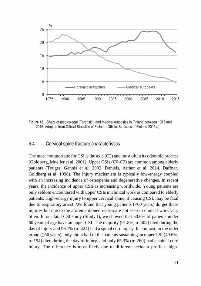
81
Figure 18. Share of medicolegal (Forensic), and medical autopsies in Finland between 1975 and 2015. Adopted from Official Statistics of Finland (Official Statistics of Finland 2015 a).
6.4 Cervical spine fracture characteristics
The most common site for CSI is the axis (C2) and most often its odontoid process
(Goldberg, Mueller et al. 2001). Upper CSIs (C0-C2) are common among elderly
patients (Touger, Gennis et al. 2002, Daniels, Arthur et al. 2014, Daffner,
Goldberg et al. 1998). The Injury mechanism is typically low-energy coupled
with an increasing incidence of osteopenia and degenerative changes. In recent
years, the incidence of upper CSIs is increasing worldwide. Young patients are
only seldom encountered with upper CSIs in clinical work as compared to elderly
patients. High-energy injury to upper cervical spine, if causing CSI, may be fatal
due to respiratory arrest. We found that young patients (<60 years) do get these
injuries but due to the aforementioned reason are not seen in clinical work very
often. In our fatal CSI study (Study I), we showed that 50.6% of patients under
60 years of age have an upper CSI. The majority (91.8%, n=402) died during the
day of injury and 96.1% (n=424) had a spinal cord injury. In contrast, in the older
group (≥60 years), only about half of the patients sustaining an upper CSI (49.6%,
n=194) died during the day of injury, and only 65.3% (n=260) had a spinal cord
injury. The difference is most likely due to different accident profiles: high-

82
energy traumas often causing occipitocervical dissociation in young patients and
low energy traumas often causing odontoid process fractures of the axis (often
not immediately fatal) in older and more fragile patients.
After axis fractures, C6 followed by C7 are the most common sites of cervical
spine fractures (Goldberg, Mueller et al. 2001). Vertebral body in the subaxial
spine, odontoid process in the axis and arch fracture in the atlas are reported to be
the most typical fracture sites within individual vertebrae (Goldberg, Mueller et
al. 2001). We studied cervical spine fracture patterns and distributions among HI
patients and found for the most part comparable results. In our series C7 was the
most commonly injured subaxial vertebra (20%) followed by C6 (17%). The
Lateral mass/articular process (32%), lamina (18%) and vertebral body (18%)
were the most common fracture locations in the subaxial spine and odontoid
process in the axis. In the atlas, comminuted fractures were the most common.
Even though it is interesting to know the distribution and types of different kind
of fractures, it has only little clinical relevance. More important than to know the
precise anatomical location of the fracture is to know the injury stability. Stability
along with neurologic function is a major determinant of treatment and prognosis
(Anderson, Moore et al. 2007). Several treatment algorithms have been developed
for upper and lower CSIs, but few have proven to be both reliable and user
friendly (Vaccaro, Koerner et al. 2016). The treatment should be planned on an
individual basis taking into account all injury and patient related variables.
6.5 CSI in head-injured patients; comorbidity and risk factors
In general practice, the possibility of cervical spine fracture among CT-positive
TBI patients is usually intuitively acknowledged, especially with regard to high-
energy injury mechanisms. Our results from Studies III and IV provide further
proof for this concept. In the case of HI patients with decreased levels of
consciousness (when clinical clearance rules are not operable), head CT
combined with cervical spine CT is recommended in the initial phase. In our
study, 9.3% of the HI patients who had CT-positive TBI had a cervical spine
fracture, whereas in the head CT-negative group 5.7 % of the patients had a
fracture. The risk difference, even though clinically significant (p=0.04), is too
small to make real difference in clinical work. Cervical spine clearance cannot be
done based on head CT status alone and clinical clearance rules still have to be
applied whenever possible. As found by the multitude of prior studies, we also
83
found older age to associate with cervical spine fractures among HI patients
(Fujii, Faul et al. 2013, Goode, Young et al. 2014).
Even though the association of head injury and CSI is reported by many
authors, several studies have published contradictory results (see Table 1). For
example, among low-energy injury patients (mean age 65.6 years), Vahldiek et
al. (2017) did not find an association between head CT findings and the incidence
of CSI (Vahldiek, Thieme et al. 2017). Of the 1,342 patients with blunt minor
trauma, only one had a combined injury to the head and cervical spine. In contrast
to the sample of Vahldiek et al. our ER sample consisted of HI patients only (HIs
ranging in severity from minimal to severe). In our study, patients with cervical
spine fractures had more often a high-energy injury mechanism compared to those
without such a fracture. However, we did not study the association of CSI and
head-CT findings in the low-energy injury population only, as Vahldiek et al. did.
Our results indirectly suggest the same as those of Vahldiek et al. since there were
actually more spinal cord injuries in the head CT-negative group than in the head
CT-positive group (1.5% vs 0.4%, i.e. 12 patients versus one single patient). That
is probably due to central cord syndromes after minor falls in the elderly. It seems
reasonable to think that low-energy injuries more seldom produce polytrauma
than high-energy injuries, and in the former, the association of CT-positive HI
and CSI seems to be less prominent. Moreover, the propensity of degenerative
spine to fracture due to minimal force certainly has its role. Studies showing that
elderly patients often have a narrow spinal canal and a stiff spinal column—
making them more prone for cervical SCI—support our results. When it comes
to clinical TBI severity, we did not find a difference between patients with or
without a spine fracture. Facial bone fractures were significantly associated with
cervical spine fractures which is also in line with prior literature (Hackl,
Hausberger et al. 2001, Lewis, Manson et al. 1985, Mukherjee, Abhinav et al.
2015, Mulligan, Friedman et al. 2010), and reflects a common injury mechanism
in both injuries.
Even though it is generally accepted that certain diseases such as osteoporosis
or AS increase the risk of fractures in general, we could not identify any
associations between cervical fractures and primary diseases as categorized
according to the ICD-10. Our relatively small sample size (n=72) of patients with
cervical fractures, is probably the main reason why associations could not be
identified.
84
6.6 Study strengths and limitations
The strength of this study is that its first part (Studies I and II) is based on Finnish
official cause-of-death statistics which are in practice, 100% complete. In
addition, the data was drawn from the entire population of Finland and therefore
the numbers and incidences of cases are true descriptions of the entire Finnish
population and not cohort-based estimates. Furthermore, the autopsy rates in
Finland are considerably higher than in many other western countries which
strengthens the results. The study period of almost a quarter century is long and
makes it possible to draw conclusions about the changing epidemiology of CSI.
Studies III and IV were based on a relatively large ED sample (n=3,023) of
CT-imaged HI patients. The original sample was non-selected and consists of HIs
ranging in severity from minimal to severe which is an advantage when applying
the results to clinical practice.
This study has some obvious limitations. First, data extraction in Studies I and
II was restricted to death certificates that were selected by diagnosis codes. The
risk of missing cases is obvious because the failure of some clinicians to take into
account the contribution of trauma to subsequent death means that some of the
deaths may have been erroneously diagnosed and may therefore remain outside
the study sample. This especially applies to fall-induced accidents among the
elderly, where the main cause (injury) behind the death may be difficult to infer.
We tried to avoid this by including also patients whose related cause of death was
a CSI. We also did not include ICD-10 codes T01.0–T06.0 (including multiple
trauma to head and neck), so some of the cases (mainly prehospital deaths due to
traffic accidents) may have been missed in the data acquisition. Due to these
reasons, the number of CSIs reported in Studies I and II may be underestimated.
The discrepancy between Studies I and II on the number of patients who died on
the day of injury (1,291 in Study I and 1,297 in Study II) is due to a failure in data
interpretation. However, it does not influence the conclusions.
Information available from death certificates is limited, which, in some cases,
complicated drawing solid conclusions about the death circumstances and
possible AEs. This is why the number of PAEs in Study II might be a conservative
estimate. Second, the identification and classification of PAEs was susceptible to
subjective considerations, and as such, could have introduced bias. Inter-rater
reliability was not measured. However, identification and classification of
diagnostic errors (that was the main objective of study II) was straightforward in
most cases.
85
In Studies III and VI, the general data in the HI registry had been
retrospectively derived from the patient records. There was a relatively high
amount of missing relevant information on, for example, alcohol intoxication and
GCS scores. This influenced the significance of the results. Second, head CT
criteria in minimal, mild and moderate HIs in the ED were based on the
Scandinavian guidelines. Apparently, some patients in the sample did not fulfill
the Scandinavian CT criteria and were CT-imaged without solid indications. In
contrast, possibly some HI patients were missed because they did not undergo CT
imaging although they met the criteria. Third, cervical spine fractures were
classified by only one researcher, thus, intra-observer bias was not eliminated.
Fourth, cervical spine ligament injuries were not systematically assessed with
MRI. Most probably, clinically significant CSIs were not missed since our study
included patients whose cervical spine was CT-imaged up to one week post-injury
and the care of CSIs in the study catchment area is centered on the study hospital.
It must be emphasized that data analysis in Studies III and IV concentrated only
on fractures and therefore conclusions cannot be drawn about ligament injuries.
6.7 Future prospects
It is expected that the incidence of CSIs will increases in Finland due to the
growing number of elderly people. Fall prevention measures should be actively
implemented to prevent some of these injuries. However, with advancements in
medical care, the incidence of some of the predisposing conditions, such as DISH,
AS and degenerative changes may begin to decrease in time, which may decrease
the number of geriatric CSIs. Nevertheless, to date, the frequency of these
predisposing conditions is not declining. The total alcohol consumption in
Finland has slightly decreased in recent years, which hopefully will reduce the
number of CSIs. The continuously improving safety features in driving also acts
similarly. Future acts to improve traffic safety with regard to CSIs should focus
on young people’s week-end driving as most of their injuries happen on Fridays
and Saturdays. As the share of elderly people driving is increasing, infrastructure
planning should be senior-friendly and driving skills of seniors should be
regularly checked.
In CSI diagnostics, education should be targeted especially at medical
students, ED – physicians and neurosurgeons to highlight the pitfalls in cervical
spine evaluation. Although diagnostic errors are inevitable to some extent,
86
adherence to evidence based guidelines and understanding their limitations, might
reduce the number of some of these events. It should be emphasized that the
diagnostics of geriatric CSI is challenging, and liberal use of CT is recommended
when injury is suspected. Furthermore, the comorbidity of CT-positive HI and
cervical spine fracture should be acknowledged.
To date, there is only limited data available on whole population incidence of
CSI. In order to investigate the true incidence of CSI in Finland a study that
combines death register data and data from the Finnish Hospital Discharge
Register should be performed. To study the change in the CSI incidence and the
impact of possible preventive measures, further, continuous research is needed.
Research should also focus on the prevention of falls among the elderly and on
finding pitfalls in CSI diagnostics. In order to identify diagnostic errors and AEs
from patient records, artificial intelligence could be increasingly utilized.
Asymptomatic CSIs in the elderly trauma population should be studied in a
prospective setting.
87
7 CONCLUSIONS AND MAIN FINDINGS
Based on the present study, the following conclusion can be made.
1. In Finland, the incidence of fatal CSIs increased between 1987 and 2010
and was approximately 19/million/year in the first decade of the 21st
century. In recent years, fatal CSIs are most often caused by falling
accidents among elderly males. The mean age of patients has dramatically
increased during the study period from about 50 to almost 70 years.
Alcohol is continuously a major risk factor for CSI.
2. Errors in CSI diagnostics occur despite advances in radiological
techniques and in medical care in general. The rate of diagnostic errors is
not declining. Hence, CSI diagnostics remain problematic and diagnostic
errors may lead to excess harm for the patient. Those who experience
diagnostic errors are elderly and the injury mechanism is most often a
ground-level fall.
3. Head injury and CSI largely co-exist. When evaluating patients with an
acute HI, special consideration should be given to the cervical spine in
the case of a positive head CT scan.
4. In head-injured patients, cervical spine fracture should be acknowledged
especially among the elderly, patients with facial fractures, and all
patients with high-energy injuries. The axis is the most commonly injured
cervical vertebra among HI patients.

88
8 ACKNOWLEDGEMENTS
This study was carried out at the Department of Neurosurgery, Tampere
University Hospital. It was financially supported by the Maire Taponen
Foundation and funds from Operational Unit 3 of the Tampere University
Hospital.
I want to express my deepest gratitude to my principal supervisors Professor
Juha Öhman, Docent Antti Ronkainen and Docent Teemu Luoto. Professor Juha
Öhman gave me the opportunity to carry out this thesis. I appreciate his
encouraging attitude and support towards this work. I wish to express my sincere
gratitude to Docent Antti Ronkainen for his positive and constructive comments
and support in both scientific and clinical work. I owe a special debt of gratitude
to Docent Teemu Luoto whose contribution was indispensable for me to complete
this work. I am especially grateful for his generous help in revising the
manuscripts and practical guidance through every step of the process.
I wish to extend special thanks to M.D. Tero Niskakangas, who originally
suggested to study CSIs from death reports and gave expert comments throughout
the process. The concept and design of publications I and II was his. I warmly
thank my co-author Professor Grant Iverson for his comments and contribution
in the diagnostic error-study (Study III).
I sincerely thank the external reviewers Docent Leena Kivipelto and Docent
Ville Vuorinen for their prompt and constructive feedback which helped me to
improve this thesis.
I wish to extend special thanks to Docent Pauli Helén and Docent Heikki
Numminen for their valuable advice while being in the follow-up group of this
work. While in charge of the Neurosurgical Department, Docent Pauli Helén
allowed me to combine both research and clinical work, for which I am
particularly grateful.
My deep gratitude goes to all staff members at the Department of
Neurosurgery for teaching me most of what I know about treating patients with
CSIs.
I wish to thank my fellow thesis workers, especially MA Tiina Ihalainen and
members of the SCI research group for their cooperation. I thank MSc Mika

89
Helminen for his help in statistical issues and Ms. Anne Simi for her assistance
in data collection.
My warmest thanks are owed to my mother Irma and father Jarl for their
endless love, support and encouragement. I am grateful to my sisters Henna and
Paula and their families for their help in various issues in life. I wish to thank my
parents-in-law Birgit and Markku for their continuous help in taking care of our
children. I am also thankful to all of my friends for helping me find the right
balance between my work and free time.
Finally, the dearest thanks go to my wife Sanna. Thank you for your love and
patience and for taking care of everything possible. Our sweet children Pihla and
Arno are thanked for making life meaningful.
90
9 REFERENCES
AEBI, M. and NAZARIAN, S., 1987. Classification of injuries of the cervical
spine. Der Orthopade, 16(1), pp. 27-36.
AHONIEMI, E., ALARANTA, H., HOKKINEN, E.M., VALTONEN, K. and
KAUTIAINEN, H., 2008. Incidence of traumatic spinal cord injuries in Finland
over a 30-year period. Spinal cord, 46(12), pp. 781-784.
ALKER, G.J., OH, Y.S., LESLIE, E.V., LEHOTAY, J., PANARO, V.A. and
ESCHNER, E.G., 1975. Postmortem radiology of head neck injuries in fatal
traffic accidents. Radiology, 114(3), pp. 611-617.
ALLEN, B.L.,Jr, FERGUSON, R.L., LEHMANN, T.R. and O'BRIEN, R.P.,
1982. A mechanistic classification of closed, indirect fractures and dislocations
of the lower cervical spine. Spine, 7(1), pp. 1-27.
ANDERSON, L.D. and D'ALONZO, R.T., 1974. Fractures of the odontoid
process of the axis. The Journal of bone and joint surgery.American volume,
56(8), pp. 1663-1674.
ANDERSON, P.A., GUGALA, Z., LINDSEY, R.W., SCHOENFELD, A.J. and
HARRIS, M.B., 2010. Clearing the cervical spine in the blunt trauma patient.
The Journal of the American Academy of Orthopaedic Surgeons, 18(3), pp. 149-
159.
ANDERSON, P.A. and MONTESANO, P.X., 1988. Morphology and treatment
of occipital condyle fractures. Spine, 13(7), pp. 731-736.
ANDERSON, P.A., MUCHOW, R.D., MUNOZ, A., TONTZ, W.L. and
RESNICK, D.K., 2010. Clearance of the asymptomatic cervical spine: a meta-
analysis. Journal of orthopaedic trauma, 24(2), pp. 100-106.
ANDERSON, P.A., MOORE, T.A., DAVIS, K.W., MOLINARI, R.W.,
RESNICK, D.K., VACCARO, A.R., BONO, C.M., DIMAR II, J.R., AARABI,
B., LEVERSON, G. and SPINAL TRAUMA STUDY GRP, 2007. Cervical
91
spine injury severity score assessment of reliability. Journal of Bone and Joint
Surgery - Series A, 89(5), pp. 1057-1065.
ANGLEN, J., METZLER, M., BUNN, P. and GRIFFITHS, H., 2002. Flexion
and Extension Views Are Not Cost-Effective in a Cervical Spine Clearance
Protocol for Obtunded Trauma Patients. The Journal of Trauma: Injury,
Infection, and Critical Care, 52(1), pp. 54-59.
BAAJ, A.A., URIBE, J.S., NICHOLS, T.A., THEODORE, N., CRAWFORD,
N.R., SONNTAG, V.K. and VALE, F.L., 2010. Health care burden of cervical
spine fractures in the United States: analysis of a nationwide database over a 10-
year period. Journal of neurosurgery.Spine, 13(1), pp. 61-66.
BAKER, G.R., NORTON, P.G., FLINTOFT, V., BLAIS, R., BROWN, A.,
COX, J., ETCHELLS, E., GHALI, W.A., HEBERT, P., MAJUMDAR, S.R.,
O'BEIRNE, M., PALACIOS-DERFLINGHER, L., REID, R.J., SHEPS, S. and
TAMBLYN, R., 2004. The Canadian Adverse Events Study: the incidence of
adverse events among hospital patients in Canada. CMAJ : Canadian Medical
Association journal = journal de l'Association medicale canadienne, 170(11),
pp. 1678-1686.
BENZEL, E.C., HART, B.L., BALL, P.A., BALDWIN, N.G., ORRISON,
W.W. and ESPINOSA, M., 1994. Fractures of the C-2 vertebral body. Journal
of neurosurgery, 81(2), pp. 206-212.
BERNER, E.S. and GRABER, M.L., 2008. Overconfidence as a cause of
diagnostic error in medicine. The American Journal of Medicine, 121(5 Suppl),
pp. S2-23.
BLACKMORE, C.C., EMERSON, S.S., MANN, F.A. and KOEPSELL, T.D.,
1999. Cervical spine imaging in patients with trauma: determination of fracture
risk to optimize use. Radiology, 211(3), pp. 759-765.
BLOOM, A.I., NEEMAN, Z., SLASKY, B.S., FLOMAN, Y., MILGROM, M.,
RIVKIND, A. and BAR-ZIV, J., 1997. Fracture of the occipital condyles and
associated craniocervical ligament injury: incidence, CT imaging and
implications. Clinical radiology, 52(3), pp. 198-202.
BOHLMAN, H.H., 1979. Acute fractures and dislocations of the cervical spine.
An analysis of three hundred hospitalized patients and review of the literature.
The Journal of bone and joint surgery.American volume, 61(8), pp. 1119-1142.
92
BOLENDER, N., CROMWELL, L.D. and WENDLING, L., 1978. Fracture of
the occipital condyle. AJR.American journal of roentgenology, 131(4), pp. 729-
731.
BONO, C.M., VACCARO, A.R., FEHLINGS, M., FISHER, C., DVORAK, M.,
LUDWIG, S., HARROP, J. and SPINE TRAUMA STUDY GROUP, 2007.
Measurement techniques for upper cervical spine injuries: consensus statement
of the Spine Trauma Study Group. Spine, 32(5), pp. 593-600.
BRANCO, F., CARDENAS, D.D. and SVIRCEV, J.N., 2007. Spinal cord
injury: a comprehensive review. Physical Medicine and Rehabilitation Clinics
of North America, 18(4), pp. 651-79, v.
BRAUN, J. and SIEPER, J., 2007. Ankylosing spondylitis. Lancet (London,
England), 369(9570), pp. 1379-1390.
BRENNAN, T.A., LEAPE, L.L., LAIRD, N.M., HEBERT, L., LOCALIO,
A.R., LAWTHERS, A.G., NEWHOUSE, J.P., WEILER, P.C. and HIATT,
H.H., 1991. Incidence of adverse events and negligence in hospitalized patients.
Results of the Harvard Medical Practice Study I. The New England journal of
medicine, 324(6), pp. 370-376.
BROLIN, K. and VON HOLST, H., 2002. Cervical injuries in Sweden, a
national survey of patient data from 1987 to 1999. Injury Control and Safety
Promotion, 9(1), pp. 40-52.
BROWN, C.V.R., ANTEVIL, J.L., SISE, M.J. and SACK, D.I., 2005. Spiral
Computed Tomography for the Diagnosis of Cervical, Thoracic, and Lumbar
Spine Fractures: Its Time has Come. The Journal of Trauma: Injury, Infection,
and Critical Care, 58(5), pp. 890-896.
BROWN, R.L., BRUNN, M.A. and GARCIA, V.F., 2001. Cervical spine
injuries in children: A review of 103 patients treated consecutively at a level 1
pediatric trauma center. Journal of pediatric surgery, 36(8), pp. 1107-1114.
BUCHOLZ, R.W., BURKHEAD, W.Z., GRAHAM, W. and PETTY, C., 1979.
Occult cervical spine injuries in fatal traffic accidents. The Journal of trauma,
19(10), pp. 768-771.
BURKE, J.T. and HARRIS, J.H.,Jr, 1989. Acute injuries of the axis vertebra.
Skeletal radiology, 18(5), pp. 335-346.

93
BUTLER, J.S., DOLAN, R.T., BURBRIDGE, M., HURSON, C.J., O’BYRNE,
J.M., MCCORMACK, D., SYNNOTT, K. and POYNTON, A.R., 2010. The
long-term functional outcome of type II odontoid fractures managed non-
operatively. European Spine Journal, 19(10), pp. 1635-1642.
CAO, Y., CHEN, Y. and DEVIVO, M.J.D.H., 2011. Lifetime Direct Costs
After Spinal Cord Injury. Topics in Spinal Cord Injury Rehabilitation, 16(4), pp.
10-16.
CAPUANO, C., COSTAGLIOLA, C., SHAMSALDIN, M., MALECI, A. and
DI LORENZO, N., 2004. Occipital condyle fractures: a hidden nosologic entity.
An experience with 10 cases. Acta Neurochirurgica, 146(8), pp. 779-784.
CARON, T., BRANSFORD, R., NGUYEN, Q., AGEL, J., CHAPMAN, J. and
BELLABARBA, C., 2010. Spine fractures in patients with ankylosing spinal
disorders. Spine, 35(11), pp. E458-64.
CLAYTON, J.L., HARRIS, M.B., WEINTRAUB, S.L., MARR, A.B.,
TIMMER, J., STUKE, L.E., MCSWAIN, N.E., DUCHESNE, J.C. and HUNT,
J.P., 2012. Risk factors for cervical spine injury. Injury, 43(4), pp. 431-435.
CLAYTOR, B., MACLENNAN, P.A., MCGWIN JR, G., RUE III, L.W. and
KIRKPATRICK, J.S., 2004. Cervical Spine Injury and Restraint System Use in
Motor Vehicle Collisions. Spine, 29(4), pp. 386-389.
COMO, J.J., THOMPSON, M.A., ANDERSON, J.S., SHAH, R.R.,
CLARIDGE, J.A., YOWLER, C.J. and MALANGONI, M.A., 2007. Is
magnetic resonance imaging essential in clearing the cervical spine in obtunded
patients with blunt trauma? The Journal of trauma, 63(3), pp. 544-549.
CORRIGAN, J.D., SELASSIE, A.W. and ORMAN, J.A., 2010. The
epidemiology of traumatic brain injury. The Journal of head trauma
rehabilitation, 25(2), pp. 72-80.
CUMMINGS, P., MCKNIGHT, B., RIVARA, F.P. and GROSSMAN, D.C.,
2002. Association Of Driver Air Bags With Driver Fatality: A Matched Cohort
Study. BMJ: British Medical Journal, 324(7346), pp. 1119-1122.
DAFFNER, R.H., GOLDBERG, A.L., EVANS, T.C., HANLON, D.P. and
LEVY, D.B., 1998. Cervical vertebral injuries in the elderly: A 10-year study.
Emergency Radiology, 5(1), pp. 38-42.

94
DAHLBERG, A., KOTILA, M., LEPPÄNEN, P., KAUTIAINEN, H. and
ALARANTA, H., 2005. Prevalence of spinal cord injury in Helsinki. Spinal
Cord, 43(1), pp. 47-50.
DANIELS, A.H., ARTHUR, M., ESMENDE, S.M., VIGNESWARAN, H. and
PALUMBO, M.A., 2014. Incidence and cost of treating axis fractures in the
United States from 2000 to 2010. Spine, 39(18), pp. 1498-1505.
DAVIS, J.W., PHREANER, D.L., HOYT, D.B. and MACKERSIE, R.C., 1993.
The etiology of missed cervical spine injuries. The Journal of trauma, 34(3), pp.
342-346.
DE VRIES, E.N., RAMRATTAN, M.A., SMORENBURG, S.M., GOUMA,
D.J. and BOERMEESTER, M.A., 2008. The incidence and nature of in-hospital
adverse events: a systematic review. Quality & safety in health care, 17(3), pp.
216-223.
DEBERNARDI, A., D'ALIBERTI, G., TALAMONTI, G., VILLA, F.,
PIPARO, M., LIGAROTTI, G. and CENZATO, M., 2014. Traumatic injuries to
the craniovertebral junction: a review of rare events. Neurosurgical review,
37(2), pp. 203-216.
DENKO, C.W. and MALEMUD, C.J., 2006. Body mass index and blood
glucose: correlations with serum insulin, growth hormone, and insulin-like
growth factor-1 levels in patients with diffuse idiopathic skeletal hyperostosis
(DISH). Rheumatology international, 26(4), pp. 292-297.
DENVER, D., SHETTY, A. and UNWIN, D., 2015. Falls and Implementation
of NEXUS in the Elderly (The FINE Study). The Journal of emergency
medicine, 49(3), pp. 294-300.
DEVIVO, M.J., KRAUSE, J.S. and LAMMERTSE, D.P., 1999. Recent trends
in mortality and causes of death among persons with spinal cord injury.
Archives of Physical Medicine and Rehabilitation, 80(11), pp. 1411-1419.
DICKMAN, C.A., GREENE, K.A. and SONNTAG, V.K., 1996. Injuries
involving the transverse atlantal ligament: classification and treatment
guidelines based upon experience with 39 injuries. Neurosurgery, 38(1), pp. 44-
50.
DITUNNO, J.F., LITTLE, J.W., TESSLER, A. and BURNS, A.S., 2004. Spinal
shock revisited: a four-phase model. Spinal cord, 42(7), pp. 383-395.
95
DUNHAM, C.M., BROCKER, B.P., COLLIER, B.D. and GEMMEL, D.J.,
2008. Risks associated with magnetic resonance imaging and cervical collar in
comatose, blunt trauma patients with negative comprehensive cervical spine
computed tomography and no apparent spinal deficit. Critical Care (London,
England), 12(4), pp. R89-R89.
EFFENDI, B., ROY, D., CORNISH, B., DUSSAULT, R.G. and LAURIN,
C.A., 1981. Fractures of the ring of the axis. A classification based on the
analysis of 131 cases. The Journal of bone and joint surgery.British volume, 63-
B(3), pp. 319-327.
ELY, J.W., GRABER, M.L. and CROSKERRY, P., 2011. Checklists to reduce
diagnostic errors. Academic medicine : journal of the Association of American
Medical Colleges, 86(3), pp. 307.
EPSTEIN, N., EPSTEIN, J.A., BENJAMIN, V. and RANSOHOFF, J., 1980.
Traumatic myelopathy in patients with cervical spinal stenosis without fracture
or dislocation: Methods of diagnosis, management, and prognosis. Spine, 5(6),
pp. 489-496.
FIELDING, J.W. and HAWKINS, R.J., 1977. Atlanto-axial rotatory fixation.
(Fixed rotatory subluxation of the atlanto-axial joint). The Journal of bone and
joint surgery.American volume, 59(1), pp. 37-44.
FITZGERALD, R., 2005. Radiological error: analysis, standard setting, targeted
instruction and teamworking. European radiology, 15(8), pp. 1760-1767.
FLANDERS, A.E., SCHAEFER, D.M., DOAN, H.T., MISHKIN,
GONZALEZ, C.F. and NORTHRUP, B.E., 1990. Acute cervical spine trauma:
Correlation of MR imaging findings with degree of neurologic deficit.
Radiology, 177(1), pp. 25-33.
FOSTER, C.A., MAISEL, R.H. and MEYERHOFF, W.L., 1981. Head and neck
trauma: initial evaluation, diagnosis and management. Minnesota Medicine,
64(2), pp. 85-90.
FRANCIS, W.R., FIELDING, J.W., HAWKINS, R.J., PEPIN, J. and
HENSINGER, R., 1981. Traumatic spondylolisthesis of the axis. The Journal of
bone and joint surgery.British volume, 63-B(3), pp. 313-318.
FREDO, H.L., RIZVI, S.A., LIED, B., RONNING, P. and HELSETH, E., 2012.
The epidemiology of traumatic cervical spine fractures: a prospective

96
population study from Norway. Scandinavian journal of trauma, resuscitation
and emergency medicine, 20, pp. 85-7241-20-85.
FREDØ, H.L., BAKKEN, I.J., LIED, B., RØNNING, P. and HELSETH, E.,
2014. Incidence of traumatic cervical spine fractures in the Norwegian
population: a national registry study. Scandinavian journal of trauma,
resuscitation and emergency medicine, 22(1), pp. 78.
FREDØ, H.L., RIZVI, S.A.M., REZAI, M., RØNNING, P., LIED, B. and
HELSETH, E., 2016. Complications and long-term outcomes after open surgery
for traumatic subaxial cervical spine fractures: a consecutive series of 303
patients. BMC surgery, 16(1), pp. 56.
FUJII, T., FAUL, M. and SASSER, S., 2013. Risk factors for cervical spine
injury among patients with traumatic brain injury. Journal of emergencies,
trauma, and shock, 6(4), pp. 252-258.
GALE, S.C., GRACIAS, V.H., REILLY, P.M. and SCHWAB, C.W., 2005. The
Inefficiency of Plain Radiography to Evaluate the Cervical Spine After Blunt
Trauma. The Journal of Trauma: Injury, Infection, and Critical Care, 59(5), pp.
1121-1125.
GARDNER, A., GRANNUM, S. and PORTER, K., 2005. Cervical spine
trauma. Trauma, 7(3), pp. 109-121.
GARRISON, A., CLIFFORD, K., GLEASON, S.F., TUN, C.G., BROWN, R.
and GARSHICK, E., 2004. Alcohol use associated with cervical spinal cord
injury. Journal of Spinal Cord Medicine, 27(2), pp. 111-115.
GBAANADOR, G.B., FRUIN, A.H. and TAYLON, C., 1986. Role of routine
emergency cervical radiography in head trauma. American Journal of Surgery,
152(6), pp. 643-648.
GOLDBERG, W., MUELLER, C., PANACEK, E., TIGGES, S., HOFFMAN,
J.R., MOWER, W.R. and NEXUS GROUP, 2001. Distribution and patterns of
blunt traumatic cervical spine injury. Annals of Emergency Medicine, 38(1), pp.
17-21.
GOLOB, J.F.,Jr, CLARIDGE, J.A., YOWLER, C.J., COMO, J.J. and
PEERLESS, J.R., 2008. Isolated cervical spine fractures in the elderly: a deadly
injury. The Journal of trauma, 64(2), pp. 311-315.
97
GOODE, T., YOUNG, A., WILSON, S.P., KATZEN, J., WOLFE, L.G. and
DUANE, T.M., 2014. Evaluation of cervical spine fracture in the elderly: can
we trust our physical examination? The American Surgeon, 80(2), pp. 182-184.
GRABER, M.L., 2013. The incidence of diagnostic error in medicine. BMJ
quality & safety, 22 Suppl 2, pp. ii21-ii27.
GRAUER, J.N., SHAFI, B., HILIBRAND, A.S., HARROP, J.S., KWON, B.K.,
BEINER, J.M., ALBERT, T.J., FEHLINGS, M.G. and VACCARO, A.R., 2005.
Proposal of a modified, treatment-oriented classification of odontoid fractures.
The spine journal : official journal of the North American Spine Society, 5(2),
pp. 123-129.
GREENBAUM, J., WALTERS, N. and LEVY, P.D., 2009. An Evidenced-
Based Approach to Radiographic Assessment of Cervical Spine Injuries in the
Emergency Department. Journal of Emergency Medicine, 36(1), pp. 64-71.
GREENE, K.A., DICKMAN, C.A., MARCIANO, F.F., DRABIER, J.B.,
HADLEY, M.N. and SONNTAG, V.K., 1997. Acute axis fractures. Analysis of
management and outcome in 340 consecutive cases. Spine, 22(16), pp. 1843-
1852.
GULY, H.R., 2001. Diagnostic errors in an accident and emergency department.
Emergency medicine journal : EMJ, 18(4), pp. 263-269.
GUPTA, A. and REEVES, B., 2009. Fijian seasonal scourge of mango tree
falls. ANZ Journal of Surgery, 79(12), pp. 898-900.
HACKL, W., HAUSBERGER, K., SAILER, R., ULMER, H. and GASSNER,
R., 2001. Prevalence of cervical spine injuries in patients with facial trauma.
Oral surgery, oral medicine, oral pathology, oral radiology, and endodontics,
92(4), pp. 370-376.
HADLEY, M.N., BROWNER, C.M., LIU, S.S. and SONNTAG, V.K., 1988.
New subtype of acute odontoid fractures (type IIA). Neurosurgery, 22(1 Pt 1),
pp. 67-71.
HADLEY, M.N., DICKMAN, C.A., BROWNER, C.M. and SONNTAG, V.K.,
1989. Acute axis fractures: a review of 229 cases. Journal of neurosurgery, 71(5
Pt 1), pp. 642-647.
98
HADLEY, M.N., DICKMAN, C.A., BROWNER, C.M. and SONNTAG, V.K.,
1988. Acute traumatic atlas fractures: management and long term outcome.
Neurosurgery, 23(1), pp. 31-35.
HADLEY, M.N. and WALTERS, B.C., 2013. Introduction to the Guidelines for
the Management of Acute Cervical Spine and Spinal Cord Injuries.
Neurosurgery, 72 Suppl 2, pp. 5-16.
HAGEN, E.M., EIDE, G.E., REKAND, T., GILHUS, N.E. and GRONNING,
M., 2010. A 50-year follow-up of the incidence of traumatic spinal cord injuries
in Western Norway. Spinal cord, 48(4), pp. 313-318.
HANSON, J.A., BLACKMORE, C.C., MANN, F.A. and WILSON, A.J., 2000.
Cervical Spine Injury: A Clinical Decision Rule to Identify High-Risk Patients
for Helical CT Screening. American Journal of Roentgenology, 174(3), pp. 713.
HARRIS, J.H.,Jr, EDEIKEN-MONROE, B. and KOPANIKY, D.R., 1986. A
practical classification of acute cervical spine injuries. The Orthopedic clinics of
North America, 17(1), pp. 15-30.
HARRIS, M.B., REICHMANN, W.M., BONO, C.M., BOUCHARD, K.,
CORBETT, K.L., WARHOLIC, N., SIMON, J.B., SCHOENFELD, A.J.,
MACIOLEK, L., CORSELLO, P., LOSINA, E. and KATZ, J.N., 2010.
Mortality in elderly patients after cervical spine fractures. The Journal of bone
and joint surgery.American volume, 92(3), pp. 567-574.
HARTMANN, S., TSCHUGG, A., WIPPLINGER, C. and THOME, C., 2017.
Analysis of the Literature on Cervical Spine Fractures in Ankylosing Spinal
Disorders. Global spine journal, 7(5), pp. 469-481.
HASLER, R.M., EXADAKTYLOS, A.K., BOUAMRA, O., BENNEKER,
L.M., CLANCY, M., SIEBER, R., ZIMMERMANN, H. and LECKY, F., 2012.
Epidemiology and predictors of cervical spine injury in adult major trauma
patients: a multicenter cohort study. The journal of trauma and acute care
surgery, 72(4), pp. 975-981.
HAUGHTON, S., 1866. On hanging, considered from a mechanical and
physiological point of view. London Edinburgh Dublin Philosophical Magazine
and Journal of Science, 32, pp. 23-34.
HEALEY, C.D., SPILMAN, S.K., KING, B.D., SHERRILL, J.E.,2nd and
PELAEZ, C.A., 2017. Asymptomatic cervical spine fractures: Current

99
guidelines can fail older patients. The journal of trauma and acute care surgery,
83(1), pp. 119-125.
HENDEY, G.W., WOLFSON, A.B., MOWER, W.R., HOFFMAN, J.R. and
NATIONAL EMERGENCY X-RADIOGRAPHY UTILIZATION STUDY
GROUP, 2002. Spinal Cord Injury without Radiographic Abnormality: Results
of the National Emergency X-Radiography Utilization Study in Blunt Cervical
Trauma. The Journal of Trauma: Injury, Infection, and Critical Care, 53(1), pp.
1-4.
HERNIGOU, P., 2016. The Vienna and German heritage of orthopaedics from
the first half of the twentieth century: Adolf Lorenz, Lorenz Bohler, Friedrich
Pauwels, Gerhard Kuntscher. International orthopaedics, 40(5), pp. 1049-1058.
HESSMANN, M.H., HOFMANN, A., KREITNER, K.F., LOTT, C. and
ROMMENS, P.M., 2006. The benefit of multislice CT in the emergency room
management of polytraumatized patients. Acta Chirurgica Belgica, 106(5), pp.
500-507.
HILLS, M.W. and DEANE, S.A., 1993. Head injury and facial injury: is there
an increased risk of cervical spine injury? The Journal of trauma, 34(4), pp.
549-53; discussion 553-4.
HOFFMAN, J.R., WOLFSON, A.B., TODD, K. and MOWER, W.R., 1998.
Selective cervical spine radiography in blunt trauma: methodology of the
National Emergency X-Radiography Utilization Study (NEXUS). Annals of
Emergency Medicine, 32(4), pp. 461-469.
HOFFMAN, J.R., MOWER, W.R., WOLFSON, A.B., TODD, K.H. and
ZUCKER, M.I., 2000. Validity of a Set of Clinical Criteria to Rule Out Injury to
the Cervical Spine in Patients with Blunt Trauma. The New England journal of
medicine, 343(2), pp. 94-99.
HOIDRUP, S., SORENSEN, T.I., GRONBAEK, M. and SCHROLL, M., 2003.
Incidence and characteristics of falls leading to hospital treatment: a one-year
population surveillance study of the Danish population aged 45 years and over.
Scandinavian Journal of Public Health, 31(1), pp. 24-30.
HOLDSWORTH, F., 1970. Fractures, dislocations, and fracture-dislocations of
the spine. The Journal of bone and joint surgery.American volume, 52(8), pp.
1534-1551.
100
HOLLY, L.T., KELLY, D.F., COUNELIS, G.J., BLINMAN, T., MCARTHUR,
D.L. and CRYER, H.G., 2002. Cervical spine trauma associated with moderate
and severe head injury: incidence, risk factors, and injury characteristics.
Journal of neurosurgery, 96(3 Suppl), pp. 285-291.
HOLMES, J.F. and AKKINEPALLI, R., 2005. Computed Tomography Versus
Plain Radiography to Screen for Cervical Spine Injury: A Meta-Analysis. The
Journal of Trauma: Injury, Infection, and Critical Care, 58(5), pp. 902-905.
HU, R., MUSTARD, C.A. and BURNS, C., 1996. Epidemiology of incident
spinal fracture in a complete population. Spine, 21(4), pp. 492-499.
IHALAINEN, T., RINTA-KIIKKA, I., LUOTO, T.M., KOSKINEN, E.A.,
KORPIJAAKKO-HUUHKA, A.M. and RONKAINEN, A., 2017a. Traumatic
cervical spinal cord injury: a prospective clinical study of laryngeal penetration
and aspiration. Spinal cord, (Epub ahead of print)
IHALAINEN, T., RINTA-KIIKKA, I., LUOTO, T.M., THESLEFF, T.,
HELMINEN, M., KORPIJAAKKO-HUUHKA, A.M. and RONKAINEN, A.,
2017b. Risk factors for laryngeal penetration-aspiration in patients with acute
traumatic cervical spinal cord injury. The spine journal : official journal of the
North American Spine Society, (Epub ahead of print)
INAMASU, J. and GUIOT, B.H., 2006. Vertebral artery injury after blunt
cervical trauma: an update. Surgical neurology, 65(3), pp. 238-45; discussion
245-6.
INGEBRIGTSEN, T., ROMNER, B. and KOCK-JENSEN, C., 2000.
Scandinavian guidelines for initial management of minimal, mild, and moderate
head injuries. The Scandinavian Neurotrauma Committee. The Journal of
trauma, 48(4), pp. 760-766.
JACKSON, A.B. and GROOMES, T.E., 1994. Incidence of respiratory
complications following spinal cord injury. Archives of Physical Medicine and
Rehabilitation, 75(3), pp. 270.
JEFFERSON, G., 1927. Remarks on fractures of the first cervical vertebra.
British Medical Journal, 2, pp. 153-157.
JEFFERSON, G., 1919. Fracture of the atlas vertebra. Report of four cases, and
a review of those previously recorded. British Journal of Surgery, 7(27), pp.
407-422.
101
JURKOVICH, G.J., RIVARA, F.P., GURNEY, J.G., SEGUIN, D., FLIGNER,
C.L. and COPASS, M., 1992. Effects of alcohol intoxication on the initial
assessment of trauma patients. Annals of Emergency Medicine, 21(6), pp. 704-
708.
KAESMACHER, J., SCHWEIZER, C., VALENTINITSCH, A., BAUM, T.,
RIENMULLER, A., MEYER, B., KIRSCHKE, J.S. and RYANG, Y.M., 2017.
Osteoporosis Is the Most Important Risk Factor for Odontoid Fractures in the
Elderly. Journal of bone and mineral research : the official journal of the
American Society for Bone and Mineral Research, 32, pp. 1582-1588.
KAKARLA, U.K., CHANG, S.W., THEODORE, N. and SONNTAG, V.K.,
2010. Atlas fractures. Neurosurgery, 66(3 Suppl), pp. 60-67.
KANNUS, P., PALVANEN, M., NIEMI, S. and PARKKARI, J., 2007.
Alarming rise in the number and incidence of fall-induced cervical spine
injuries among older adults. The journals of gerontology.Series A, Biological
sciences and medical sciences, 62(2), pp. 180-183.
KANNUS, P., PARKKARI, J., NIEMI, S. and PALVANEN, M., 2005. Fall-
induced deaths among elderly people. American Journal of Public Health,
95(3), pp. 422-424.
KARASON, S., REYNISSON, K., SIGVALDASON, K. and SIGURDSSON,
G.H., 2014. Evaluation of clinical efficacy and safety of cervical trauma collars:
differences in immobilization, effect on jugular venous pressure and patient
comfort. Scandinavian journal of trauma, resuscitation and emergency
medicine, 22, pp. 37.
KATO, H., KIMURA, A., SASAKI, R., KANEKO, N., TAKEDA, M.,
HAGIWARA, A., OGURA, S., MIZOGUCHI, T., MATSUOKA, T., ONO, H.,
MATSUURA, K., MATSUSHIMA, K., KUSHIMOTO, S., FUSE, A.,
NAKATANI, T., IWASE, M., FUDOJI, J. and KASAI, T., 2008. Cervical
Spinal Cord Injury Without Bony Injury: A Multicenter Retrospective Study of
Emergency and Critical Care Centers in Japan. The Journal of Trauma: Injury,
Infection, and Critical Care, 65(2), pp. 373-379.
KIM, S.K., CHOI, B.R., KIM, C.G., CHUNG, S.H., CHOE, J.Y., JOO, K.B.,
BAE, S.C., YOO, D.H. and JUN, J.B., 2004. The prevalence of diffuse
idiopathic skeletal hyperostosis in Korea. The Journal of rheumatology, 31(10),
pp. 2032-2035.
102
KIRSHBLUM, S.C., BURNS, S.P., BIERING-SORENSEN, F., DONOVAN,
W., GRAVES, D.E., JHA, A., JOHANSEN, M., JONES, L., KRASSIOUKOV,
A., MULCAHEY, M.J., SCHMIDT-READ, M. and WARING, W., 2011.
International standards for neurological classification of spinal cord injury
(revised 2011). The journal of spinal cord medicine, 34(6), pp. 535-546.
KNOPP, R., PARKER, J., TASHJIAN, J. and GANZ, W., 2001. Defining
radiographic criteria for flexion-extension studies of the cervical spine. Annals
of Emergency Medicine, 38(1), pp. 31-35.
KOSKINEN, E.A., ALEN, M., VAARALA, E.M., RELLMAN, J.,
KALLINEN, M. and VAINIONPAA, A., 2014. Centralized spinal cord injury
care in Finland: unveiling the hidden incidence of traumatic injuries. Spinal
cord, 52, pp. 779-784.
KOSKINEN, E., BRANDER, A., HAKULINEN, U., LUOTO, T.,
HELMINEN, M., YLINEN, A. and ÖHMAN, J., 2013. Assessing the State of
Chronic Spinal Cord Injury Using Diffusion Tensor Imaging. Journal of
neurotrauma, 30(18), pp. 1587-1595.
KOYANAGI, I., IWASAKI, Y., HIDA, K., AKINO, M., IMAMURA, H. and
ABE, H., 2000. Acute cervical cord injury without fracture or dislocation of the
spinal column. Journal of neurosurgery, 93(1), pp. 15-20.
KRASSIOUKOV, A., BIERING-SORENSEN, C.F., DONOVAN, W.,
KENNELLY, M., KIRSHBLUM, S., KROGH, K., ALEXANDER, M.S.,
VOGEL, L., AND WECHT, J. and CONTRIBUTORS, 2012. International
Standards to document remaining Autonomic Function after Spinal Cord Injury
(ISAFSCI), First Edition 2012. Topics in spinal cord injury rehabilitation,
18(3), pp. 282-296.
KRONZ, J.D., WESTRA, W.H. and EPSTEIN, J.I., 1999. Mandatory second
opinion surgical pathology at a large referral hospital. Cancer, 86(11), pp. 2426-
2435.
KRUEGER, H., NOONAN, V.K., TRENAMAN, L.M., JOSHI, P. and
RIVERS, C.S., 2013. The economic burden of traumatic spinal cord injury in
Canada. Chronic diseases and injuries in Canada, 33(3), pp. 113-122.
KUKREJA, S., KALAKOTI, P., MURRAY, R., NIXON, M., MISSIOS, S.,
GUTHIKONDA, B. and NANDA, A., 2015. National trends of incidence,
103
treatment, and hospital charges of isolated C-2 fractures in three different age
groups. Neurosurgical focus, 38(4), pp. E19.
LANDELLS, C.D. and VAN PETEGHEM, P.K., 1988. Fractures of the atlas:
classification, treatment and morbidity. Spine, 13(5), pp. 450-452.
LAUWERYNS, P., 2010. Role of conservative treatment of cervical spine
injuries. European Spine Journal, 19(S1), pp. 23-26.
LECKIE, S., YOON, S.T., ISAACS, R., RADCLIFF, K., FESSLER, R., HAID,
R.,Jr and TRAYNELIS, V., 2016. Perioperative Complications of Cervical
Spine Surgery: Analysis of a Prospectively Gathered Database through the
Association for Collaborative Spinal Research. Global spine journal, 6(7), pp.
640-649.
LEKOVIC, G.P. and HARRINGTON, T.R., 2007. Litigation of missed cervical
spine injuries in patients presenting with blunt traumatic injury. Neurosurgery,
60(3), pp. 516-523.
LEMONS, V.R. and WAGNER, F.C.,Jr, 1994. Respiratory complications after
cervical spinal cord injury. Spine, 19(20), pp. 2315-2320.
LENEHAN, B., BORAN, S., STREET, J., HIGGINS, T., MCCORMACK, D.
and POYNTON, A.R., 2009. Demographics of acute admissions to a National
Spinal Injuries Unit. European spine journal : official publication of the
European Spine Society, the European Spinal Deformity Society, and the
European Section of the Cervical Spine Research Society, 18(7), pp. 938-942.
LENEHAN, B., STREET, J., KWON, B.K., NOONAN, V., ZHANG, H.,
FISHER, C.G. and DVORAK, M.F., 2012. The epidemiology of traumatic
spinal cord injury in British Columbia, Canada. Spine, 37(4), pp. 321-329.
LEUCHT, P., FISCHER, K., MUHR, G. and MUELLER, E.J., 2009.
Epidemiology of traumatic spine fractures. Injury, 40(2), pp. 166-172.
LEVINE, A.M. and EDWARDS, C.C., 1985. The management of traumatic
spondylolisthesis of the axis. The Journal of bone and joint surgery.American
volume, 67(2), pp. 217-226.
LEWIS, V.L.,Jr, MANSON, P.N., MORGAN, R.F., CERULLO, L.J. and
MEYER, P.R.,Jr, 1985. Facial injuries associated with cervical fractures:
104
recognition, patterns, and management. The Journal of trauma, 25(1), pp. 90-
93.
LIEBERMAN, I.H. and WEBB, J.K., 1994. Cervical spine injuries in the
elderly. The Journal of bone and joint surgery.British volume, 76(6), pp. 877.
LIEUTAUD, T., NDIAYE, A., LAUMON, B. and CHIRON, M., 2012. Spinal
cord injuries sustained in road crashes are not on the decrease in france: a study
based on epidemiological trends. Journal of neurotrauma, 29(3), pp. 479-487.
LINDSBERG, P.J. and KARJALAINEN-LINDSBERG, M., 2003. The role of
autopsy in diagnostics and clinical research. Duodecim; lääketieteellinen
aikakauskirja, 119(13), pp. 1273.
LINSENMAIER, U., KROTZ, M., HAUSER, H., ROCK, C., RIEGER, J.,
BOHNDORF, K., PFEIFER, K.J. and REISER, M., 2002. Whole-body
computed tomography in polytrauma: techniques and management. European
radiology, 12(7), pp. 1728-1740.
LONGO, U.G., DENARO, V., DENARO, L., CAMPI, S. and MAFFULLI, N.,
2010. Upper cervical spine injuries: Indications and limits of the conservative
management in Halo vest. A systematic review of efficacy and safety. Injury,
41(11), pp. 1127-1135.
LOWERY, D.W., WALD, M.M., BROWNE, B.J., TIGGES, S., HOFFMAN,
J.R., MOWER, W.R. and NEXUS GROUP, 2001. Epidemiology of cervical
spine injury victims. Annals of Emergency Medicine, 38(1), pp. 12-16.
LUNETTA, P., LOUNAMAA, A. and SIHVONEN, S., 2007. Surveillance of
injury-related deaths: medicolegal autopsy rates and trends in Finland. Injury
prevention : journal of the International Society for Child and Adolescent Injury
Prevention, 13(4), pp. 282-284.
MA, J., WANG, C., ZHOU, X., ZHOU, S. and JIA, L., 2015. Surgical Therapy
of Cervical Spine Fracture in Patients With Ankylosing Spondylitis. Medicine,
94(44), pp. e1663.
MALHOTRA, A., WU, X., KALRA, V.B., HOLLY K GROSSETTA
NARDINI, LIU, R., ABBED, K.M. and FORMAN, H.P., 2017. Utility of MRI
for cervical spine clearance after blunt traumatic injury: a meta-analysis.
European radiology, 27(3), pp. 1148-1160.

105
MASERATI, M.B., STEPHENS, B., ZOHNY, Z., LEE, J.Y., KANTER, A.S.,
SPIRO, R.M. and OKONKWO, D.O., 2009. Occipital condyle fractures:
clinical decision rule and surgical management. Journal of neurosurgery.Spine,
11(4), pp. 388-395.
MATTHIESSEN, C. and ROBINSON, Y., 2015. Epidemiology of atlas
fractures--a national registry-based cohort study of 1,537 cases. The spine
journal : official journal of the North American Spine Society, 15(11), pp. 2332-
2337.
MENAKER, J., PHILP, A., BOSWELL, S. and SCALEA, T.M., 2008.
Computed tomography alone for cervical spine clearance in the unreliable
patient--are we there yet? The Journal of trauma, 64(4), pp. 898-903; discussion
903-4.
MERTEN, H., JOHANNESMA, P.C., LUBBERDING, S., ZEGERS, M.,
LANGELAAN, M., JUKEMA, G.N., HEETVELD, M.J. and WAGNER, C.,
2015. High risk of adverse events in hospitalised hip fracture patients of 65
years and older: results of a retrospective record review study. BMJ open, 5(9),
pp. e006663-2014-006663.
MICHAEL, D.B., GUYOT, D.R. and DARMODY, W.R., 1989. Coincidence of
head and cervical spine injury. Journal of neurotrauma, 6(3), pp. 177-189.
MIDDENDORP, J.J.V., AUDIGE, L., BARTELS, R.H.M.A., BOLGER, C.,
DEVERALL, H., DHOKE, P., DIEKERHOF, C.H., GOVAERT, G.A.,
GUIMERA, V., KOLLER, H., MORRIS, S.A., SETIOBUDI, T. and
HOSMAN, A.J.F., 2013. The Subaxial Cervical Spine Injury Classification
System: an external agreement validation study. Spine Journal, 13(9), pp. 1055-
1063.
MILBY, A.H., HALPERN, C.H., GUO, W. and STEIN, S.C., 2008. Prevalence
of cervical spinal injury in trauma. Neurosurgical focus, 25(5), pp. E10.
MILLER, C.P., BRUBACHER, J.W., BISWAS, D., LAWRENCE, B.D.,
WHANG, P.G. and GRAUER, J.N., 2011. The incidence of noncontiguous
spinal fractures and other traumatic injuries associated with cervical spine
fractures: A 10-year experience at an academic medical center. Spine, 36(19),
pp. 1532-1540.
MIYANJI, F., FURLAN, J.C., AARABI, B., ARNOLD, P.M. and FEHLINGS,
M.G., 2007. Acute cervical traumatic spinal cord injury: MR imaging findings
106
correlated with neurologic outcome - Prospective study with 100 consecutive
patients. Radiology, 243(3), pp. 820-827.
MORITA, T., TAKEBAYASHI, T., IRIFUNE, H., OHNISHI, H.,
HIRAYAMA, S. and YAMASHITA, T., 2017. Factors affecting survival of
patients in the acute phase of upper cervical spine injuries. Archives of
orthopaedic and trauma surgery, 137(4), pp. 1-6.
MORRIS, C.G. and MCCOY, E., 2004. Clearing the cervical spine in
unconscious polytrauma victims, balancing risks and effective screening.
Anaesthesia, 59(5), pp. 464-482.
MUCHOW, R.D., RESNICK, D.K., ABDEL, M.P., MUNOZ, A. and
ANDERSON, P.A., 2008. Magnetic resonance imaging (MRI) in the clearance
of the cervical spine in blunt trauma: a meta-analysis. The Journal of trauma,
64(1), pp. 179-189.
MUKHERJEE, S., ABHINAV, K. and REVINGTON, P.J., 2015. A review of
cervical spine injury associated with maxillofacial trauma at a UK tertiary
referral centre. Annals of the Royal College of Surgeons of England, 97(1), pp.
66-72.
MULLIGAN, R.P., FRIEDMAN, J.A. and MAHABIR, R.C., 2010. A
nationwide review of the associations among cervical spine injuries, head
injuries, and facial fractures. The Journal of trauma, 68(3), pp. 587-592.
NELSON, D.W., MARTIN, M.J., MARTIN, N.D. and BEEKLEY, A., 2013.
Evaluation of the risk of noncontiguous fractures of the spine in blunt trauma.
Journal of Trauma and Acute Care Surgery, 75(1), pp. 135-139.
NEVITT, M.C., CUMMINGS, S.R., KIDD, S. and BLACK, D., 1989. Risk
factors for recurrent nonsyncopal falls. A prospective study. Jama, 261(18), pp.
2663-2668.
NING, G.-., YU, T.-., FENG, S.-., ZHOU, X.-., BAN, D.-., LIU, Y. and JIAO,
X.-., 2011. Epidemiology of traumatic spinal cord injury in Tianjin, China.
Spinal Cord, 49(3), pp. 386-390.
OFFICIAL STATISTICS OF FINLAND, , Official Statistics of Finland (OSF):
Causes of death [e-publication].ISSN=1799-5078. Helsinki: Statistics Finland
[referred: 14.11.2016].Access method:

107
http://www.stat.fi/til/ksyyt/meta_en.html. Available:
http://www.stat.fi/til/ksyyt/meta_en.html [11/14, 2016].
OFFICIAL STATISTICS OF FINLAND (OSF, , Official Statistics of Finland
(OSF): Population structure [e-publication].
ISSN=1797-5395. [Homepage of Helsinki: Statistics Finland], [Online].
Available: http://www.stat.fi/til/vaerak/tau_en.html [July/22, 2017].
OFFICIAL STATISTICS OF FINLAND 2015, a-last update, Official Statistics
of Finland (OSF): Causes of death [e-publication].
ISSN=1799-5078. 2015, Quality Description:Causes of death 2015 . Helsinki:
Statistics Finland [referred: 11.5.2017].
Access method: http://www.stat.fi/til/ksyyt/2015/ksyyt_2015_2016-12-
30_laa_001_en.html.
OFFICIAL STATISTICS OF FINLAND 2015, b-last update, Official Statistics
of Finland (OSF): Deaths [e-publication].ISSN=1798-2545. 2015, Quality
description, deaths 2015 . Helsinki: Statistics Finland [referred:
15.11.2016].Access method: http://www.stat.fi/til/kuol/2015/kuol_2015_2016-
04-14_laa_001_en.html [11/14, 2016].
OFFICIAL STATISTICS OF FINLAND 2015, c-last update, Official Statistics
of Finland (OSF): Population projection [e-publication].ISSN=1798-5153.
2015. Helsinki: Statistics Finland [referred: 1.5.2017].Access method:
http://www.stat.fi/til/vaenn/2015/vaenn_2015_2015-10-30_tie_001_en.html.
OJANIEMI, K.K., LINTONEN, T.P., IMPINEN, A.O., LILLSUNDE, P.M. and
OSTAMO, A.I., 2009. Trends in driving under the influence of drugs: a
register-based study of DUID suspects during 1977-2007. Accident; Analysis
and Prevention, 41(1), pp. 191-196.
PANG, D. and WILBERGER JR, J.E., 1982. Spinal cord injury without
radiographic abnormalities in children. Journal of neurosurgery, 57(1), pp. 114-
129.
PANG, D., 2004. Spinal cord injury without radiographic abnormality in
children, 2 decades later. Neurosurgery, 55(6), pp. 1325-1342.
PATEL, A., SMITH, H.E., RADCLIFF, K., YADLAPALLI, N. and
VACCARO, A.R., 2012. Odontoid Fractures With Neurologic Deficit Have
Higher Mortality and Morbidity. Clinical orthopaedics and related research,
470(6), pp. 1614-1620.
108
PATEL, M.B., HUMBLE, S.S., CULLINANE, D.C., DAY, M.A., JAWA, R.S.,
DEVIN, C.J., DELOZIER, M.S., SMITH, L.M., SMITH, M.A., CAPELLA,
J.M., LONG, A.M., CHENG, J.S., LEATH, T.C., FALCK-YTTER, Y., HAUT,
E.R. and COMO, J.J., 2015. Cervical spine collar clearance in the obtunded
adult blunt trauma patient: a systematic review and practice management
guideline from the Eastern Association for the Surgery of Trauma. The journal
of trauma and acute care surgery, 78(2), pp. 430-441.
PATERSON, A., 1890. Fracture of the cervical vertebrae. J Anat Phys, .
PEPIN, J.W. and HAWKINS, R.J., 1981. Traumatic spondylolisthesis of the
axis: Hangman's fracture. Clinical orthopaedics and related research,
(157)(157), pp. 133-138.
PICKETT, G.E., CAMPOS-BENITEZ, M., KELLER, J.L. and DUGGAL, N.,
2006. Epidemiology of traumatic spinal cord injury in Canada. Spine, 31(7), pp.
799-805.
PIEPMEIER, J.M., LEHMANN, K.B. and LANE, J.G., 1985. Cardiovascular
instability following acute cervical spinal cord trauma. Central nervous system
trauma : journal of the American Paralysis Association, 2(3), pp. 153-160.
PLATZER, P., HAUSWIRTH, N., JAINDL, M., CHATWANI, S., VECSEI, V.
and GAEBLER, C., 2006. Delayed or missed diagnosis of cervical spine
injuries. The Journal of trauma, 61(1), pp. 150-155.
POLLACK, C.V., HENDEY, G.W., MARTIN, D.R., HOFFMAN, J.R.,
MOWER, W.R. and NEXUS GROUP, 2001. Use of flexion-extension
radiographs of the cervical spine in blunt trauma. Annals of Emergency
Medicine, 38(1), pp. 8-11.
POURTAHERI, S., EMAMI, A., SINHA, K., FALOON, M., HWANG, K.,
SHAFA, E. and HOLMES, L.,Jr, 2014. The role of magnetic resonance imaging
in acute cervical spine fractures. The spine journal : official journal of the North
American Spine Society, 14(11), pp. 2546-2553.
PRYPUTNIEWICZ, D.M. and HADLEY, M.N., 2010. Axis fractures.
Neurosurgery, 66(3 Suppl), pp. 68-82.
RAYES, M., MITTAL, M., RENGACHARY, S.S. and MITTAL, S., 2011.
Hangman's fracture: a historical and biomechanical perspective. Journal of
neurosurgery.Spine, 14(2), pp. 198-208.
109
REID, D.C., HENDERSON, R., SABOE, L. and MILLER, J.D., 1987. Etiology
and clinical course of missed spine fractures. The Journal of trauma, 27(9), pp.
980-986.
RICHARDS, P.J., 2005. Cervical spine clearance: a review. Injury, 36(2), pp.
248-69; discussion 270.
RIHN, J.A., ANDERSON, D.T., LAMB, K., DELUCA, P.F., BATA, A.,
MARCHETTO, P.A., NEVES, N. and VACCARO, A.R., 2009. Cervical Spine
Injuries in American Football. Sports Medicine, 39(9), pp. 697-708.
ROY, A.K., MILLER, B.A., HOLLAND, C.M., FOUNTAIN, J.,Arthur J.,
PRADILLA, G. and AHMAD, F.U., 2015. Magnetic resonance imaging of
traumatic injury to the craniovertebral junction: a case-based review.
Neurosurgical focus, 38(4), pp. E3.
RUTBERG, H., BORGSTEDT-RISBERG, M., GUSTAFSON, P. and
UNBECK, M., 2016. Adverse events in orthopedic care identified via the
Global Trigger Tool in Sweden - implications on preventable prolonged
hospitalizations. Patient safety in surgery, 10, pp. 23.
SANCHEZ, B., WAXMAN, K., JONES, T., CONNER, S., CHUNG, R. and
BECERRA, S., 2005. Cervical Spine Clearance in Blunt Trauma: Evaluation of
a Computed Tomography–Based Protocol. The Journal of Trauma: Injury,
Infection, and Critical Care, 59(1), pp. 179-183.
SCHMIDT, O.I., GAHR, R.H., GOSSE, A. and HEYDE, C.E., 2009. ATLS(R)
and damage control in spine trauma. World journal of emergency surgery :
WJES, 4, pp. 9-7922-4-9.
SCHOENFELD, A.J., BONO, C.M., MCGUIRE, K.J., WARHOLIC, N. and
HARRIS, M.B., 2010. Computed tomography alone versus computed
tomography and magnetic resonance imaging in the identification of occult
injuries to the cervical spine: a meta-analysis. The Journal of trauma, 68(1), pp.
109-13; discussion 113-4.
SCHOENFELD, A.J., SIELSKI, B., RIVERA, K.P., BADER, J.O. and
HARRIS, M.B., 2011. Epidemiology of cervical spine fractures in the US
military. The spine journal : official journal of the North American Spine
Society, 12(9), pp. 777-783.

110
SCHRAG, S., 2008. Cervical spine fractures in geriatric blunt trauma patients
with low-energy mechanism: are clinical predictors adequate? Am J Surg,
195(2), pp. 170-173.
SCHUSTER, R., WAXMAN, K., SANCHEZ, B., BECERRA, S., CHUNG, R.,
CONNER, S. and JONES, T., 2005. Magnetic resonance imaging is not needed
to clear cervical spines in blunt trauma patients with normal computed
tomographic results and no motor deficits. Archives of surgery (Chicago, Ill.:
1960), 140(8), pp. 762.
SCHWARTZ, A. and ELSTEIN, A.S., 2008. Clinical reasoning in medicine.
Clinical reasoning in the health professions.3rd ed.Edinburgh: Elsevier, , pp.
223-234.
SEKHON, L.H. and FEHLINGS, M.G., 2001. Epidemiology, demographics,
and pathophysiology of acute spinal cord injury. Spine, 26(24 Suppl), pp. S2-12.
SHAH, L.M. and ROSS, J.S., 2016. Imaging of Spine Trauma. Neurosurgery,
79(5), pp. 626-642.
SHARMA, O.P., OSWANSKI, M.F., YAZDI, J.S., JINDAL, S. and TAYLOR,
M., 2007. Assessment for additional spinal trauma in patients with cervical
spine injury. American Surgeon, 73(1), pp. 70-74.
SHIN, J.C., YOO, J.H., LEE, Y.S., GOO, H.R. and KIM, D.H., 2011.
Dysphagia in cervical spinal cord injury. Spinal Cord, 49(9), pp. 1008-1013.
SHOJANIA, K.G., BURTON, E.C., MCDONALD, K.M. and GOLDMAN, L.,
2002. The autopsy as an outcome and performance measure. Evidence
report/technology assessment (Summary), (58)(58), pp. 1-5.
SOICHER, E. and DEMETRIADES, D., 1991. Cervical spine injuries in
patients with head injuries. The British journal of surgery, 78(8), pp. 1013-1014.
SONDEREGGER-LSELI, K., BURGER, S., MUNTWYLER, J. and
SALOMON, F., 2000. Diagnostic errors in three medical eras: a necropsy study.
The Lancet, 355(9220), pp. 2027-2031.
SOOP, M., FRYKSMARK, U., KOSTER, M. and HAGLUND, B., 2009. The
incidence of adverse events in Swedish hospitals: a retrospective medical record
review study. International journal for quality in health care : journal of the
International Society for Quality in Health Care, 21(4), pp. 285-291.
111
SPENCE, K.F.,Jr, DECKER, S. and SELL, K.W., 1970. Bursting atlantal
fracture associated with rupture of the transverse ligament. The Journal of bone
and joint surgery.American volume, 52(3), pp. 543-549.
STASSEN, N.A., WILLIAMS, V.A., GESTRING, M.L., CHENG, J.D. and
BANKEY, P.E., 2006. Magnetic resonance imaging in combination with helical
computed tomography provides a safe and efficient method of cervical spine
clearance in the obtunded trauma patient. The Journal of trauma, 60(1), pp. 171-
177.
STIELL, I.G., CLEMENT, C.M., MCKNIGHT, R.D., BRISON, R., SCHULL,
M.J., ROWE, B.H., WORTHINGTON, J.R., EISENHAUER, M.A., CASS, D.,
GREENBERG, G., MACPHAIL, I., DREYER, J., LEE, J.S., BANDIERA, G.,
REARDON, M., HOLROYD, B., LESIUK, H. and WELLS, G.A., 2003. The
Canadian C-spine rule versus the NEXUS low-risk criteria in patients with
trauma. The New England journal of medicine, 349(26), pp. 2510-2518.
STIELL, I.G., WELLS, G.A., VANDEMHEEN, K.L., CLEMENT, C.M.,
LESIUK, H., DE MAIO, V.J., LAUPACIS, A., SCHULL, M., MCKNIGHT,
R.D., VERBEEK, R., BRISON, R., CASS, D., DREYER, J., EISENHAUER,
M.A., GREENBERG, G.H., MACPHAIL, I., MORRISON, L., REARDON, M.
and WORTHINGTON, J., 2001. The Canadian C-Spine Rule for Radiography
in Alert and Stable Trauma Patients. JAMA, 286(15), pp. 1841-1848.
THOMAS, E.J., STUDDERT, D.M., BURSTIN, H.R., ORAV, E.J., ZEENA,
T., WILLIAMS, E.J., HOWARD, K.M., WEILER, P.C. and BRENNAN, T.A.,
2000. Incidence and types of adverse events and negligent care in Utah and
Colorado. Medical care, 38(3), pp. 261-271.
THOMAS, E.J., STUDDERT, D.M., NEWHOUSE, J.P., ZBAR, B.I.W.,
HOWARD, K.M., WILLIAMS, E.J. and BRENNAN, T.A., 1999. Costs of
medical injuries in Utah and Colorado. Inquiry, 36(3), pp. 255-264.
THOMPSON, W.L., STIELL, I.G., CLEMENT, C.M., BRISON, R.J. and
CANADIAN C-SPINE RULE STUDY GROUP, 2009. Association of injury
mechanism with the risk of cervical spine fractures. Cjem, 11(1), pp. 14-22.
THURMAN, D.J., ALVERSON, C., DUNN, K.A., GUERRERO, J. and
SNIEZEK, J.E., 1999. Traumatic brain injury in the United States: A public
health perspective. The Journal of head trauma rehabilitation, 14(6), pp. 602-
615.
112
TIIRIKAINEN, K., 2009. Tapaturmat Suomessa. Terveyden ja hyvinvoinnin
laitos. Helsinki: Edita Prima oy.
TOMYCZ, N.D., CHEW, B.G., CHANG, Y., DARBY, J.M., GUNN, S.R.,
NICHOLAS, D.H., OCHOA, J.B., PEITZMAN, A.B., SCHWARTZ, E., PAPE,
H., SPIRO, R.M. and OKONKWO, D.O., 2008. MRI Is Unnecessary to Clear
the Cervical Spine in Obtunded/Comatose Trauma Patients: The Four-Year
Experience of a Level I Trauma Center. The Journal of Trauma: Injury,
Infection, and Critical Care, 64(5), pp. 1258-1263.
TONTZ, W., ANDERSON, P. and RESNICK, D., 2006. 3:57119. Clearance of
the Asymptomatic Cervical Spine: A Meta-Analysis. The Spine Journal, 6(5),
pp. 60.
TOUGER, M., GENNIS, P., NATHANSON, N., LOWERY, D.W., POLLACK,
C.V.,Jr, HOFFMAN, J.R. and MOWER, W.R., 2002. Validity of a decision rule
to reduce cervical spine radiography in elderly patients with blunt trauma.
Annals of Emergency Medicine, 40(3), pp. 287-293.
TUBBS, R.S., HALLOCK, J.D., RADCLIFF, V., NAFTEL, R.P.,
MORTAZAVI, M., SHOJA, M.M., LOUKAS, M. and COHEN-GADOL, A.A.,
2011. Ligaments of the craniocervical junction: A review. Journal of
Neurosurgery: Spine, 14(6), pp. 697-709.
TULI, S., TATOR, C.H., FEHLINGS, M.G. and MACKAY, M., 1997.
Occipital condyle fractures. Neurosurgery, 41(2), pp. 368-76; discussion 376-7.
TURK, E.E., RIEDEL, A. and PÜESCHEL, K., 2008. Natural and traumatic
sports-related fatalities: a 10-year retrospective study. British journal of sports
medicine, 42(7), pp. 604-608.
UTTER, G.H., HOLLINGWORTH, W., HALLAM, D.K., JARVIK, J.G. and
JURKOVICH, G.J., 2006. Sixteen-Slice CT Angiography in Patients with
Suspected Blunt Carotid and Vertebral Artery Injuries. Journal of the American
College of Surgeons, 203(6), pp. 838-848.
VACCARO, A.R., HULBERT, R.J., PATEL, A.A., FISHER, C., DVORAK,
M., LEHMAN, R.A.,Jr, ANDERSON, P., HARROP, J., ONER, F.C.,
ARNOLD, P., FEHLINGS, M., HEDLUND, R., MADRAZO, I., RECHTINE,
G., AARABI, B., SHAINLINE, M. and SPINE TRAUMA STUDY GROUP,
2007. The subaxial cervical spine injury classification system: a novel approach
113
to recognize the importance of morphology, neurology, and integrity of the
disco-ligamentous complex. Spine, 32(21), pp. 2365-2374.
VACCARO, A.R., KOERNER, J.D., RADCLIFF, K.E., ONER, F.C.,
REINHOLD, M., SCHNAKE, K.J., KANDZIORA, F., FEHLINGS, M.G.,
DVORAK, M.F., AARABI, B., RAJASEKARAN, S., SCHROEDER, G.D.,
KEPLER, C.K. and VIALLE, L.R., 2016. AOSpine subaxial cervical spine
injury classification system. European spine journal : official publication of the
European Spine Society, the European Spinal Deformity Society, and the
European Section of the Cervical Spine Research Society, 25(7), pp. 2173-2184.
VAHLDIEK, J.L., THIEME, S., HAMM, B. and NIEHUES, S.M., 2017.
Incidence of combined cranial and cervical spine injuries in patients with blunt
minor trauma: are combined CT examinations of the head and cervical spine
justified? Acta Radiologica (Stockholm, Sweden : 1987), 58(7), pp. 856-860.
VAN DEN BERG, M.E.L., CASTELLOTE, J.M., DE PEDRO-CUESTA, J. and
IGNACIO, M., 2010. Survival after spinal cord injury: A systematic review.
Journal of neurotrauma, 27(8), pp. 1517-1528.
VAN ZYL, H.P., BILBEY, J., VUKUSIC, A., RING, T., OAKES, J.,
WILLIAMSON, L.D. and MITCHELL, I.V., 2014. Can emergency physicians
accurately rule out clinically important cervical spine injuries by using
computed tomography? CJEM, 16(2), pp. 131.
WANG, H., COPPOLA, M., ROBINSON, R.D., SCRIBNER, J.T.,
VITHALANI, V., DE MOOR, C.E., GANDHI, R.R., BURTON, M. and
DELANEY, K.A., 2013. Geriatric Trauma Patients With Cervical Spine
Fractures due to Ground Level Fall: Five Years Experience in a Level One
Trauma Center. Journal of clinical medicine research, 5(2), pp. 75-83.
WASSERBERG, J. and BARTLETT, R.J., 1995. Occipital condyle fractures
diagnosed by high-definition CT and coronal reconstructions. Neuroradiology,
37(5), pp. 370-373.
WATANABE, M., SAKAI, D., YAMAMOTO, Y., NAGAI, T., SATO, M. and
MOCHIDA, J., 2014. Analysis of predisposing factors in elderly people with
type II odontoid fracture. The spine journal : official journal of the North
American Spine Society, 14(6), pp. 861-866.
WEINFELD, R.M., OLSON, P.N., MAKI, D.D. and GRIFFITHS, H.J., 1997.
The prevalence of diffuse idiopathic skeletal hyperostosis (DISH) in two large

114
American Midwest metropolitan hospital populations. Skeletal radiology, 26(4),
pp. 222-225.
WESTERVELD, L.A., VAN BEMMEL, J.C., DHERT, W.J., ONER, F.C. and
VERLAAN, J.J., 2014. Clinical outcome after traumatic spinal fractures in
patients with ankylosing spinal disorders compared with control patients. The
spine journal : official journal of the North American Spine Society, 14(5), pp.
729-740.
WESTERVELD, L.A., VERLAAN, J.J. and ONER, F.C., 2009. Spinal fractures
in patients with ankylosing spinal disorders: a systematic review of the literature
on treatment, neurological status and complications. European spine journal :
official publication of the European Spine Society, the European Spinal
Deformity Society, and the European Section of the Cervical Spine Research
Society, 18(2), pp. 145-156.
WILLIAMS, P. and WARWICK, R., 1980. Gray´s anatomy. 36 edn. Great
Britain: Churchill Livingstone.
WILLIAMS, J., JEHLE, D., COTTINGTON, E. and SHUFFLEBARGER, C.,
1992. Head, facial, and clavicular trauma as a predictor of cervical-spine injury.
Annals of Emergency Medicine, 21(6), pp. 719-722.
WILSON, R.M., RUNCIMAN, W.B., GIBBERD, R.W., HARRISON, B.T.,
NEWBY, L. and HAMILTON, J.D., 1995. The Quality in Australian Health
Care Study. The Medical journal of Australia, 163(9), pp. 458-471.
WOOD-JONES, F., 1913. The ideal lesion produced by judicial hanging.
Lancet, i, pp. 53.
WYNDAELE, M. and WYNDAELE, J.J., 2006. Incidence, prevalence and
epidemiology of spinal cord injury: what learns a worldwide literature survey?
Spinal cord, 44(9), pp. 523-529.
YANAR, H., DEMETRIADES, D., HADJIZACHARIA, P., NOMOTO, S.,
SALIM, A., INABA, K., RHEE, P. and CHAN, L.S., 2007. Pedestrians injured
by automobiles: risk factors for cervical spine injuries. Journal of the American
College of Surgeons, 205(6), pp. 794-799.
YANG, S., DING, W., YANG, D., GU, T., ZHANG, F., ZHANG, D., SUN, Y.,
MA, L. and SONG, Y., 2013. Epidemiology and Risk Factors of Cervical Spine
115
Injury during Heating Season in the Patients with Cervical Trauma: A Cross-
Sectional Study. PloS one, 8(11), pp. e78358.
ZAKRISON, T.L. and WILLIAMS, B.H., 2016. Cervical spine evaluation in the
bluntly injured patient. International Journal of Surgery, 33, pp. 246-250.
ZWAAN, L., DE BRUIJNE, M., WAGNER, C., THIJS, A., SMITS, M., VAN
DER WAL, G. and TIMMERMANS, D.R., 2010. Patient record review of the
incidence, consequences, and causes of diagnostic adverse events. Archives of
Internal Medicine, 170(12), pp. 1015-1021.
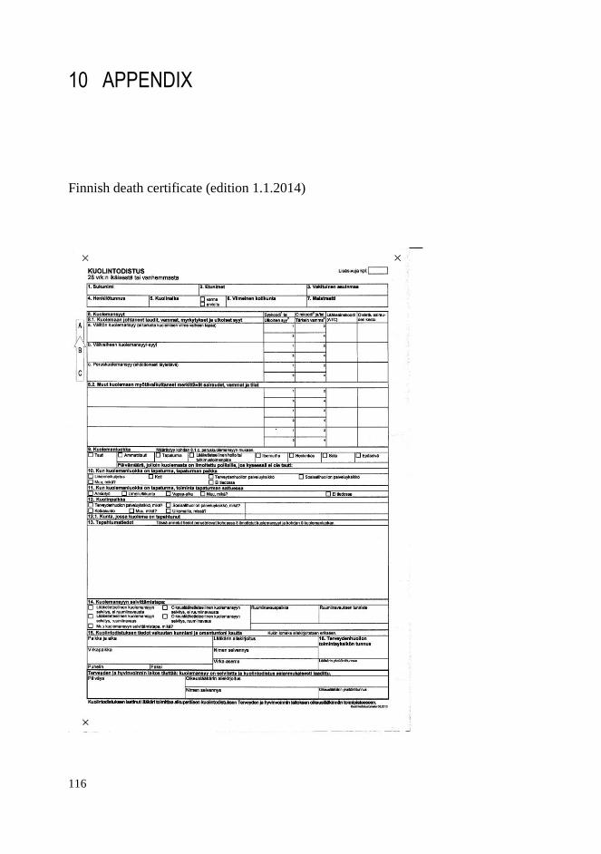
116
10 APPENDIX
Finnish death certificate (edition 1.1.2014)

117

Clinical Study
Fatal cervical spine injuries: a Finnish nationwide register-basedepidemiologic study on data from 1987 to 2010
Tuomo Thesleff, MD*, Tero Niskakangas, MD, Teemu M. Luoto, MD, PhD,Juha Öhman, MD, PhD, Antti Ronkainen, MD, PhD
Department of Neurosurgery, Tampere University Hospital, P.O. Box 2000, Tampere FI-33521, Finland
Received 29 May 2015; revised 26 August 2015; accepted 19 November 2015
Abstract BACKGROUND CONTEXT: The number of cervical spine injuries (CSIs) is increasing. Cervi-cal spine injuries are associated with high morbidity and mortality. Identifying those who are at riskfor CSI-related death can help develop national and international interventions and policies to reducemortality.PURPOSE: This study aimed to determine the trends in the incidence and the characteristics offatal CSIs in Finland over a 24-year study period from 1987 to 2010.STUDY DESIGN/SETTING: A large nationwide, retrospective, register-based study was carriedout.PATIENT SAMPLE: The population-based sample was collected from death certificates issued inFinland between 1987 and 2010. The death certificates were obtained from the official Cause-of-Death Register, coordinated by Statistics Finland, which covers all deaths occurring in Finland.OUTCOME MEASURES: Sociodemographics and injury- and death-related data were used foroutcome measures.METHODS: All death certificates issued in Finland (1987–2010) containing a CSI as the cause ofdeath were carefully reviewed.RESULTS: A total of 2,041 fatal CSIs were identified. These constituted 0.17% of all deaths inFinland within the study period. The average annual incidence of fatal CSIs was 16.5 per million(range: 12.5–21.2). The majority of the victims were male (72.9%) and had concurrent spinal cordinjury (83.0%). Traffic accidents (40.1%) and falls (45.0%) were the most common injury mecha-nisms. Almost one-third (29.8%) of the deaths were alcohol-related. Among the young victims (<60years) with upper CSI (C0–C2), the majority (91.8%) died within 24 hours post-injury. One-thirdof elderly victims’ (≥60 years) CSI-related deaths occurred after 1 week post-injury and were mostly(74.2%) caused by respiratory and circulatory system diseases. Within the 24-year period, the inci-dence of fatal CSIs (+2/million), as well as the average age of sustaining a fatal CSI (+13.5 years),increased markedly. Fall-induced accidents among elderly males were the most prominently increas-ing subpopulation of fatal CSI victims.CONCLUSIONS: In recent decades, fatal CSI incidence (death certificate-based) has increased,being 18.6 per million in Finland in 2010. Victims of fatal CSIs tend to be older than in the past,and for a substantial number of males, low-energy falls lead to cervical trauma and death. © 2015Elsevier Inc. All rights reserved.
Keywords: Autopsy; Epidemiology; Mortality; Registries; Spinal injuries; Spinal cord injuries
FDA device/drug status: Not applicable.Author disclosures:TT:Nothing to disclose.TN:Nothing to disclose.TML:
Nothing to disclose. JÖ: Nothing to disclose. AR: Nothing to disclose.No competing financial interests exist.
* Corresponding author. Department of Neurosurgery, TampereUniversity Hospital, P.O. Box 2000, Tampere FI-33521, Finland. Tel.: +358400763034.
E-mail address: [email protected] (T. Thesleff)
http://dx.doi.org/10.1016/j.spinee.2015.11.0541529-9430/© 2015 Elsevier Inc. All rights reserved.
The Spine Journal 16 (2016) 918–926
Introduction
Globally, traumatic injuries are the leading cause of deathamong people aged between 5 and 44 years [1]. Cervical spineinjuries (CSIs) constitute a considerable number of these fatalinjuries. Cervical spine injuries are most often caused by motorvehicle accidents and falls, and they are associated with sig-nificant morbidity and mortality [2,3]. The overall prevalenceof CSIs among all trauma patients is estimated to be about3.7% [4], and the annual incidence of CSIs in the general pop-ulation is reported to be 12 per 100,000 [5,6]. The overallincidence of fatal CSIs in developed countries is not wellknown. The majority of epidemiologic CSI studies focus onhospital-admitted patients [5–7]. However, it has been notedthat a substantial number of all injury-related (including CSI-related) deaths occur outside hospitals [8–10]. Therefore, manyCSIs go unrecorded unless postmortem examinations areconducted.
Medicolegal autopsies are crucial for the accuracy of deathcertification. The number of medicolegal autopsies per-formed in Finland is considerably higher than in many otherdeveloped countries [11]. In Finland, the indications for medi-colegal autopsies are strictly controlled by legislation [11].Medicolegal autopsies are performed in up to 87.2% of allunintentional injury-related deaths, 98.3% of homicides, and99.5% of suicides [11]. This results in highly controlled andcomprehensive national mortality statistics.
Acknowledging the fatal nature of CSIs and the relative-ly high incidence of concurrent mortality, there is a need forpreventive measures for CSIs. Identifying those who are atrisk for CSI-related death can help develop national and in-ternational interventions and policies to reduce mortality. Weaimed to determine the trends in CSI incidence and the char-acteristics of patients with fatal CSIs over two decades byexploiting the inclusive population-based mortality statis-tics of Finland. Our data were drawn from the entire populationof Finland; therefore, the absolute numbers and incidencesof CSIs in this study represent complete population-basedresults, not cohort-based estimates. We hypothesized that thenumber of fall-related fatal CSIs among elderly people(60 years and older) had increased over the last two decades.
Materials and methods
Study frame and ethics
This population-based sample was collected from deathcertificates issued in Finland between 1987 and 2010. Thedeath certificates were obtained from the official Cause-of-Death Register, coordinated by Statistics Finland, which coversall deaths occurring in Finland. The Finnish official cause-of-death statistics are, in practice, 100% complete [12].According to Finnish legislation, a medicolegal autopsy shouldbe performed in the following circumstances: when death iscaused or suspected to be caused by (i) a crime, (ii) a suicide,(iii) an accident, (iv) poisoning, (v) an occupational disease,or (vi) medical treatment, or when death has (vii) not been
caused by a disease, or when (viii) during the last illness, thedeceased had not been treated by a doctor within 3 months,or when (ix) the death was otherwise unexpected. The studywas approved by the Ethics Committee of Pirkanmaa Hos-pital District, Tampere, Finland.
Data collection
All death certificates containing CSI as an immediate, in-termediate, main, or related cause of death were carefullyreviewed by the first author (T.T.). During this period, boththe International Classification of Diseases (ICD)-9 and theICD-10 codes were in use. In our study, CSI was defined asan injury to the cervical spine, including (i) fractures, (ii) dis-locations, (iii) fractures with spinal cord injury, (iv) isolatedspinal cord injury, or (v) a combination of the aforemen-tioned. The corresponding ICD-10 diagnosis codes were S12.0,S12.1, S12.7, S12.9, S13.0, S13.1, S13.2, S13.3, S14.0, andS14.1, and the ICD-9 diagnosis codes were 805.0–805.18,806.0–806.19, 839.0–839.18, and 952.0–952.09. Combinedskull and cervical fractures (T01.1–T06.0), whiplash, nerveroot injuries, and non-traumatic spinal cord lesions were notused for the screening of the death certificates. The collectedvariables included diagnosis (ICD codes), gender, age, timeof injury, place of injury, time between injury and death, cause
ContextThe authors present results of an epidemiological inves-tigation regarding the incidence and clinical characteristicsof fatal cervical spine injuries in Finland between 1987and 2010.
ContributionA total of 2,041 fatal cervical injuries were identified. Fatalcervical injuries have increased over the course of the time-period studied, particularly among older males. Nearly one-third of deaths were alcohol related.
ImplicationsThe results presented here dovetail to some extent withmaterial published from the United States and else-where, particularly with respect to an increasing incidenceof fatal cervical injury among elderly males. Nonethe-less, there is little evidence that the information in thisanalysis can be generalized beyond the Finnish popula-tion and the findings are likely not translatable to othersocio-ethnic and demographic contexts, particularly withregard to factors associated with cervical spine injury. Whilepresenting interesting information from the perspective ofclinical epidemiology, the evidence associated with thiswork must be considered no higher than Level IV.
—The Editors
919T. Thesleff et al. / The Spine Journal 16 (2016) 918–926
of death, type of injury, alcohol and drug consumption at thetime of injury, type of death certification, presence of spinalcord injury, and the level of cervical injury. Fractures of theupper cervical spine (C0–C2) were regarded as high injuriesand the rest of the cervical fractures (C3–C7) as low injuries.Substance abuse was considered positive if it was mentionedin the death certificate regardless of the method of affirmation.
Statistical analyses
IBM SPSS Statistics 20.0 (IBM Corp. Armonk, NY, USA)was used to perform the analyses. The frequencies and per-centage of the analyzed variables were formed using descriptivestatistics.
Results
The incidence of fatal CSI in the whole population
The average population of Finland during the 24-year(1987–2010) study period was 5,157,442, and altogether1,179,305 deaths were reported during that time [12]. In ourstudy, a total of 2,041 death certificates were found to reportCSI, and they contributed to 0.17% of all deaths. In the studypopulation, medicolegal autopsy was performed in 1,907(93.4%) cases and clinical autopsy in 5 (0.2%) cases. In 121(5.9%) cases, the death certification was written by the treat-ing physician based on clinical findings without an autopsy.In eight (0.4%) cases, the type of death certification wasunknown. Cervical spine injury was the main cause of deathin 1,847 (90.5%) cases, a related cause of death in 193 (9.5%)cases, and an intermediate cause of death in 1 case. Theaverage annual incidence of fatal CSI was 16.5 per million(range: 12.5–21.2) during the study period. Of all the fatalCSIs, 83.0% (n=1,694) had a cervical spinal cord injury. Theaverage annual incidence of fatal cervical spinal cord injurywas 13.7 per million (range: 10.8–16.6).
Characteristics of the study sample
The main characteristics of the study sample are summa-rized in Table 1. Of all 2,041 CSI victims, 72.9% were maleand the overall male-to-female ratio was 2.6:1. The mean ageat death was 59.4 years, and 57.3% of the victims were over60 years old. Ninety-one (4.5%) victims were children (<18years). Alcohol detection was reported on 608 (29.8%) of thedeath certificates and its incidence was highest among youngmales (46.2%). The proportion of spinal cord-injured victimsdecreased with increasing age (Tables 1 and 2). Among theyoung victims (<60 years) who had an upper CSI (C0–C2),the majority (91.8%, n=402) died within 24 hours post-injury, and 96.1% (n=424) of these had a spinal cord injury.The mechanism of injury and cause of death varied signifi-cantly by age. Young and middle-aged victims (<60 years)were often injured in fatal traffic accidents or in suicides, andolder (≥60 years) victims were injured most often in low-energy falls. Sports-related injuries presented only 0.6% ofall accidents. There were significant differences in the place
of death for age groups. Of the older victims, 74.3% died attheir home or residence or in a medical facility, whereas foryounger victims, the place of death was coded as “other” (asopposed to “medical facility,” “home or residence,” or“abroad”) in 71.9% of cases. The mean time between injuryand death was 46.7 days (median: 0 days, min: 0 days, andmax: 37.9 years). This time was much shorter among youngervictims since 86% of them died within 24 hours post-injury, whereas only 48% of the older (≥60 years) victimsdied during the day of injury. A substantial number of oldervictims (n=388, 33.2%) died after 1 week post-injury. Amongthose, the immediate causes of death were provided in 93.0%(361/388) of cases and were most often diseases of the re-spiratory system (n=234, 60.3%) and diseases of the circulatorysystem (n=54, 13.9%). Respiratory system diseases mostlyincluded pneumonia (n=216, 55.7%), whereas any kind of heartdisease (n=27, 7.0%), stroke (n=13, 3.4%), and pulmonaryembolus (n=12, 3.1%) accounted for the majority of the cir-culatory system diseases. Most of the injuries happened inJuly (10.1%), and the most frequent day of the week for injurywas Saturday (18.0%).
Changes in the characteristics of the fatal CSI victimsduring the study period
The death certificate-based number and incidence of CSIvictims began to increase at the end of 1990s (Fig. 1), andthey have been on average 99.8 and 19.0 per million, respec-tively, during the last 10 years (2001–2010) of the study period.Fig. 2 shows the clear increase in the mean age at the timeof death—from 54.4 years to 67.9 years—during the studyperiod. The changes in the frequency of the most commoninjury types are shown in Fig. 3. The number of fall-relatedCSIs exceeded traffic accident-related CSIs in 1998. The in-crease in the total number of CSI victims is explained by theincrease in fall-related accidents of elderly males, which isshown in Fig. 4, Top. The number of pediatric patients de-creased toward the end of the study period (Table 2).
Discussion
In this nationwide cause-of-death register-based epide-miologic study, we used the death certificates of the entireFinnish population to describe the trends in the incidence andcharacteristics of fatal CSI victims. Between 1987 and 2010,we found 2,041 deaths that were related to CSI. The averageincidence of fatal CSI in Finland was 16.5 per million. Wehave shown that the number and incidence of CSI-relateddeaths increased, especially after the year 1999, and duringthe last 10 years, there have been about 100—or 19 permillion—annually. Falls, traffic accidents, and suicides werethe leading causes of injury.
The causes of injury and the age of those deceased changednotably during the observation period. The mean age at thetime of death increased from 54.4 years to 67 years, and thenumber of pediatric patients decreased significantly. Falls ex-ceeded traffic accidents as the most common cause of injury
920 T. Thesleff et al. / The Spine Journal 16 (2016) 918–926
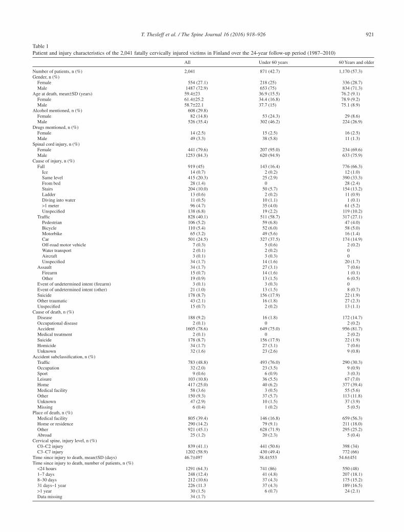
Table 1Patient and injury characteristics of the 2,041 fatally cervically injured victims in Finland over the 24-year follow-up period (1987–2010)
All Under 60 years 60 Years and older
Number of patients, n (%) 2,041 871 (42.7) 1,170 (57.3)Gender, n (%)
Female 554 (27.1) 218 (25) 336 (28.7)Male 1487 (72.9) 653 (75) 834 (71.3)
Age at death, mean±SD (years) 59.4±23 36.9 (15.5) 76.2 (9.1)Female 61.4±25.2 34.4 (16.8) 78.9 (9.2)Male 58.7±22.1 37.7 (15) 75.1 (8.9)
Alcohol mentioned, n (%) 608 (29.8)Female 82 (14.8) 53 (24.3) 29 (8.6)Male 526 (35.4) 302 (46.2) 224 (26.9)
Drugs mentioned, n (%)Female 14 (2.5) 15 (2.5) 16 (2.5)Male 49 (3.3) 38 (5.8) 11 (1.3)
Spinal cord injury, n (%)Female 441 (79.6) 207 (95.0) 234 (69.6)Male 1253 (84.3) 620 (94.9) 633 (75.9)
Cause of injury, n (%)Fall 919 (45) 143 (16.4) 776 (66.3)
Ice 14 (0.7) 2 (0.2) 12 (1.0)Same level 415 (20.3) 25 (2.9) 390 (33.3)From bed 28 (1.4) 0 28 (2.4)Stairs 204 (10.0) 50 (5.7) 154 (13.2)Ladder 13 (0.6) 2 (0.2) 11 (0.9)Diving into water 11 (0.5) 10 (1.1) 1 (0.1)>1 meter 96 (4.7) 35 (4.0) 61 (5.2)Unspecified 138 (6.8) 19 (2.2) 119 (10.2)
Traffic 828 (40.1) 511 (58.7) 317 (27.1)Pedestrian 106 (5.2) 59 (6.8) 47 (4.0)Bicycle 110 (5.4) 52 (6.0) 58 (5.0)Motorbike 65 (3.2) 49 (5.6) 16 (1.4)Car 501 (24.5) 327 (37.5) 174 (14.9)Off-road motor vehicle 7 (0.3) 5 (0.6) 2 (0.2)Water transport 2 (0.1) 2 (0.2) 0Aircraft 3 (0.1) 3 (0.3) 0Unspecified 34 (1.7) 14 (1.6) 20 (1.7)
Assault 34 (1.7) 27 (3.1) 7 (0.6)Firearm 15 (0.7) 14 (1.6) 1 (0.1)Other 19 (0.9) 13 (1.5) 6 (0.5)
Event of undetermined intent (firearm) 3 (0.1) 3 (0.3) 0Event of undetermined intent (other) 21 (1.0) 13 (1.5) 8 (0.7)Suicide 178 (8.7) 156 (17.9) 22 (1.9)Other traumatic 43 (2.1) 16 (1.8) 27 (2.3)Unspecified 15 (0.7) 2 (0.2) 13 (1.1)
Cause of death, n (%)Disease 188 (9.2) 16 (1.8) 172 (14.7)Occupational disease 2 (0.1) 0 2 (0.2)Accident 1605 (78.6) 649 (75.0) 956 (81.7)Medical treatment 2 (0.1) 0 2 (0.2)Suicide 178 (8.7) 156 (17.9) 22 (1.9)Homicide 34 (1.7) 27 (3.1) 7 (0.6)Unknown 32 (1.6) 23 (2.6) 9 (0.8)
Accident subclassification, n (%)Traffic 783 (48.8) 493 (76.0) 290 (30.3)Occupation 32 (2.0) 23 (3.5) 9 (0.9)Sport 9 (0.6) 6 (0.9) 3 (0.3)Leisure 103 (10.8) 36 (5.5) 67 (7.0)Home 417 (25.0) 40 (6.2) 377 (39.4)Medical facility 58 (3.6) 3 (0.5) 55 (5.6)Other 150 (9.3) 37 (5.7) 113 (11.8)Unknown 47 (2.9) 10 (1.5) 37 (3.9)Missing 6 (0.4) 1 (0.2) 5 (0.5)
Place of death, n (%)Medical facility 805 (39.4) 146 (16.8) 659 (56.3)Home or residence 290 (14.2) 79 (9.1) 211 (18.0)Other 921 (45.1) 628 (71.9) 295 (25.2)Abroad 25 (1.2) 20 (2.3) 5 (0.4)
Cervical spine, injury level, n (%)C0–C2 injury 839 (41.1) 441 (50.6) 398 (34)C3–C7 injury 1202 (58.9) 430 (49.4) 772 (66)
Time since injury to death, mean±SD (days) 46.7±497 38.4±553 54.6±451Time since injury to death, number of patients, n (%)<24 hours 1291 (64.3) 741 (86) 550 (48)1–7 days 248 (12.4) 41 (4.8) 207 (18.1)8–30 days 212 (10.6) 37 (4.3) 175 (15.2)31 days–1 year 226 (11.3 37 (4.3) 189 (16.5)>1 year 30 (1.5) 6 (0.7) 24 (2.1)Data missing 34 (1.7)
921T. Thesleff et al. / The Spine Journal 16 (2016) 918–926
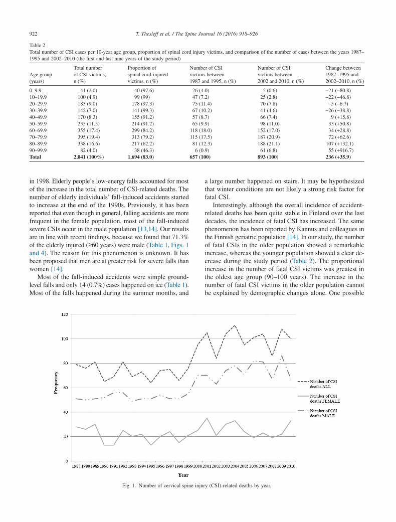
in 1998. Elderly people’s low-energy falls accounted for mostof the increase in the total number of CSI-related deaths. Thenumber of elderly individuals’ fall-induced accidents startedto increase at the end of the 1990s. Previously, it has beenreported that even though in general, falling accidents are morefrequent in the female population, most of the fall-inducedsevere CSIs occur in the male population [13,14]. Our resultsare in line with recent findings, because we found that 71.3%of the elderly injured (≥60 years) were male (Table 1, Figs. 1and 4). The reason for this phenomenon is unknown. It hasbeen proposed that men are at greater risk for severe falls thanwomen [14].
Most of the fall-induced accidents were simple ground-level falls and only 14 (0.7%) cases happened on ice (Table 1).Most of the falls happened during the summer months, and
a large number happened on stairs. It may be hypothesizedthat winter conditions are not likely a strong risk factor forfatal CSI.
Interestingly, although the overall incidence of accident-related deaths has been quite stable in Finland over the lastdecades, the incidence of fatal CSI has increased. The samephenomenon has been reported by Kannus and colleagues inthe Finnish geriatric population [14]. In our study, the numberof fatal CSIs in the older population showed a remarkableincrease, whereas the younger population showed a clear de-crease during the study period (Table 2). The proportionalincrease in the number of fatal CSI victims was greatest inthe oldest age group (90–100 years). The increase in thenumber of fatal CSI victims in the older population cannotbe explained by demographic changes alone. One possible
Table 2Total number of CSI cases per 10-year age group, proportion of spinal cord injury victims, and comparison of the number of cases between the years 1987–1995 and 2002–2010 (the first and last nine years of the study period)
Age group(years)
Total numberof CSI victims,n (%)
Proportion ofspinal cord-injuredvictims, n (%)
Number of CSIvictims between1987 and 1995, n (%)
Number of CSIvictims between2002 and 2010, n (%)
Change between1987–1995 and2002–2010, n (%)
0–9.9 41 (2.0) 40 (97.6) 26 (4.0) 5 (0.6) −21 (−80.8)10–19.9 100 (4.9) 99 (99) 47 (7.2) 25 (2.8) −22 (−46.8)20–29.9 183 (9.0) 178 (97.3) 75 (11.4) 70 (7.8) −5 (−6.7)30–39.9 142 (7.0) 141 (99.3) 67 (10.2) 41 (4.6) −26 (−38.8)40–49.9 170 (8.3) 155 (91.2) 57 (8.7) 66 (7.4) 9 (+15.8)50–59.9 235 (11.5) 214 (91.2) 65 (9.9) 98 (11.0) 33 (+50.8)60–69.9 355 (17.4) 299 (84.2) 118 (18.0) 152 (17.0) 34 (+28.8)70–79.9 395 (19.4) 313 (79.2) 115 (17.5) 187 (20.9) 72 (+62.6)80–89.9 338 (16.6) 217 (62.2) 81 (12.3) 188 (21.1) 107 (+132.1)90–99.9 82 (4.0) 38 (46.3) 6 (0.9) 61 (6.8) 55 (+916.7)Total 2,041 (100%) 1,694 (83.0) 657 (100) 893 (100) 236 (+35.9)
Fig. 1. Number of cervical spine injury (CSI)-related deaths by year.
922 T. Thesleff et al. / The Spine Journal 16 (2016) 918–926

reason might be the increase of spondylosis changes in thespine with age.
Moreover, Lieutaud and colleagues observed that eventhough the total number of road traffic accidents has fallenin recent years, the incidence and mortality of spinal cord in-juries sustained in those accidents has not shown significantreduction [15]. These patients also tended to be older thanin the past and more often had associated multiple trauma[15]. Our results support this finding because in Finland, road
traffic mortality decreased from 581 per year to 272 per yearbetween 1987 and 2010 [12]. During the same period, fatalCSIs induced by traffic accidents decreased only marginal-ly (Fig. 3). Sports have been reported to be a significant causeof cervical injury in previous epidemiologic reports [2,3,7].For example, in a multicenter cohort study from 1988 to 2009by Hasler and colleagues, 7.6% of cervical injuries were dueto sports-related accidents. Surprisingly, in our series, sports-related injuries were almost totally absent, with only 9 (0.4%)
Fig. 2. Mean age at death by year.
Fig. 3. Number of cervical spine injury (CSI)-related deaths by injury mechanism by year.
923T. Thesleff et al. / The Spine Journal 16 (2016) 918–926

cases present. The reason for this difference may lie in thedivergent coding habits between studies, as some of the sportsinjuries may have been coded as leisure activity injuries inour series.
Alcohol is a major risk factor for CSI [2,16], and in general,every third fatal injury in Finland occurs under the influ-ence of alcohol [17]. Our findings corresponded with previousreports, since alcohol was mentioned in 29.8% of all casesand in 46.2% of young (<60 years) male victims.
The incidence of cervical spinal cord injury in the settingof CSI is reported to be 10%–50% [3,6,18]. We found a spinalcord injury in 83% of all victims and in 91.2% of those who
died in less than 24 hours. Among the young victims (<60years) who had an upper CSI, the majority (91.8%, n=402)died within 24 hours post-injury, and 96.1% (n=424) of thesevictims had a spinal cord injury. On the contrary, in the oldergroup (≥60 years), only about half of the victims sustainingan upper CSI (49.6%, n=194) died in less than 24 hours,and 65.3% (n=260) of these had a spinal cord injury. Thedifference is most likely due to different accidentprofiles: high-energy traumas often causing occipitocervicaldissociation in young patients and low energy traumas oftencausing C2 dens fracture (often not immediately fatal) in olderpatients.
Fig. 4. (Top) Number of cervical spine injuries (CSIs) caused by fall-induced accidents by year in <60- and ≥60-year-old victims. (Bottom) Number of CSIinjuries caused by traffic accidents by year in <60- and ≥60-year-old victims.
924 T. Thesleff et al. / The Spine Journal 16 (2016) 918–926

The epidemiology of traumatic deaths in general has beenstudied extensively. In 1977, Baker and colleagues de-scribed the classic trimodal distribution of trauma deaths, wherethe first peak represented the immediate deaths, the secondpeak included early hospital deaths, and the third peak in-cluded late deaths [19]. The trimodal distribution has laterbeen challenged, for example, by Sauaia and Evans [8,9].Evans and colleagues found that in Australia, 66% of pa-tients die before hospitalization and only 2% die after 7 days.They also reported that none of the low-energy trauma pa-tients (who had all sustained a fall of less than 1 m) died beforehospitalization. Our results for CSI victims differ, becausewe found that 460 (22.9%) of all CSI victims and 388 (33.2%)of the older group (≥60 years) died after 7 days. Further-more, 147 of 457 (32.2%) low-energy trauma deaths occurredin less than 24 hours post-injury. The majority (97%) of thedeaths that occurred in less than 24 hours post-injury wereprehospital deaths.
Most of the CSI-related deaths among the young popu-lation take place outside of medical facilities within 24 hourspost-injury. Therefore, accident prevention and behavior mod-ification is crucial in reducing the number of victims in thissubgroup. In the older population, one-third (n=388, 33.2%)of the CSI-related deaths occur more than 7 days post-injury. In 74.2% of these cases, the immediate cause of deathwas either due to diseases of the respiratory or the circula-tory system (including eg, pneumonia, heart attack, andpulmonary embolism). This is important, as these are poten-tially preventable or treatable medical conditions if they arerecognized early. A fortiori, as the proportion of elderly pop-ulation among CSI victims is steadily increasing.
Identifying those who are at risk for a cervical injury-related death can help in developing interventions and policiesintended to reduce the mortality among this patient group.We studied all victims who had sustained a fatal CSI—eitherwith or without a spinal cord injury—coded in their deathcertificates. The strength of the present study is that it is basedon the whole population of one nation over an observationperiod of 24 years. Moreover, it has been shown that the qualityand rate of performed medicolegal autopsies in Finland isamong the highest in the world [11].
Limitations of the study
We based our study on death certificates that were se-lected by diagnosis codes. This method carries a risk of missingcases. It has been shown that because of the failure of someclinicians to take into account the contribution of trauma tosubsequent death, some of the deaths may have been erro-neously diagnosed and may therefore remain outside oursample [10,20]. This especially applies to fall-induced acci-dents among the elderly. In the present study, we tried to avoidthis by also assessing the related cause of death. We did notinclude ICD-10 codes T01.0–T06.0 (including multiple traumato head and neck), so some of the cases (mainly prehospitaldeaths due to traffic accidents) may have been missed. Due
to the aforementioned reasons, the number of CSIs reportedin our study may be underestimated. It must also be noticedthat in those 193 (9.5%) death certificates that reported CSIas a related cause of death, cervical injury by itself was notnecessarily fatal but a contributing factor to the following death.
Conclusions
During the 24-year observation period, fatal (deathcertificate-based) CSI incidence has increased. In 2010, thepopulation-based incidence of CSI in Finland was 18.6 permillion. This study highlights that victims of CSI tend to beolder than in the past, and a substantial number of cervicaltraumas and deaths result from low-energy falls by males.
Acknowledgments
The authors wish to thank biostatistician Mika Helminenfor his assistance. This study was made as part of the firstauthor’s (TT) PhD dissertation research program, and it waspartly presented at the National Neurosurgical Education Days,September 2014, in Tampere, Finland.
References
[1] World Health Organization. World health report 2009. 2009. Availableat: www.who.int. Accessed November 30 2015.
[2] Sekhon LH, Fehlings MG. Epidemiology, demographics, andpathophysiology of acute spinal cord injury. Spine 2001;26(24 Suppl.):S2–12.
[3] Leucht P, Fischer K, Muhr G, Mueller EJ. Epidemiology of traumaticspine fractures. Injury 2009;40:166–72.
[4] Milby AH, Halpern CH, Guo W, Stein SC. Prevalence of cervical spinalinjury in trauma. Neurosurg Focus 2008;25:E10.
[5] Hu R, Mustard CA, Burns C. Epidemiology of incident spinal fracturein a complete population. Spine 1996;21:492–9.
[6] Fredo HL, Rizvi SA, Lied B, Ronning P, Helseth E. The epidemiologyof traumatic cervical spine fractures: a prospective population studyfrom Norway. Scand J Trauma Resusc Emerg Med 2012;20:85.7241-20-85.
[7] Hasler RM, Exadaktylos AK, Bouamra O, et al. Epidemiology andpredictors of cervical spine injury in adult major trauma patients:a multicenter cohort study. J Trauma Acute Care Surg 2012;72:975–81.
[8] Sauaia A, Moore FA, Moore EE, et al. Epidemiology of trauma deaths:a reassessment. J Trauma 1995;38:185–93.
[9] Evans JA, van Wessem KJ, McDougall D, Lee KA, Lyons T, BaloghZJ. Epidemiology of traumatic deaths: comprehensive population-basedassessment. World J Surg 2010;34:158–63.
[10] Gedeborg R, Chen LH, Thiblin I, et al. Prehospital injurydeaths—strengthening the case for prevention: nationwide cohort study.J Trauma Acute Care Surg 2012;72:765–72.
[11] Lunetta P, Lounamaa A, Sihvonen S. Surveillance of injury-relateddeaths: medicolegal autopsy rates and trends in Finland. Inj Prev2007;13:282–4.
[12] Official Statistics of Finland (OSF). 2011. Population structure[e-publication]. ISSN:1797-5395. Helsinki: Statistics Finland. Availableat: http://www.tilastokeskus.fi/til/vaerak/tau_en.html. AccessedNovember 16, 2013.
[13] Kannus P, Parkkari J, Niemi S, Palvanen M. Fall-induced deaths amongelderly people. Am J Public Health 2005;95:422–4.
925T. Thesleff et al. / The Spine Journal 16 (2016) 918–926

[14] Kannus P, Palvanen M, Niemi S, Parkkari J. Alarming rise in thenumber and incidence of fall-induced cervical spine injuries among olderadults. J Gerontol A Biol Sci Med Sci 2007;62:180–3.
[15] Lieutaud T, Ndiaye A, Laumon B, Chiron M. Spinal cord injuriessustained in road crashes are not on the decrease in France: a studybased on epidemiological trends. J Neurotrauma 2012;29:479–87.
[16] Burke DA, Linden RD, Zhang YP, Maiste AC, Shields CB. Incidencerates and populations at risk for spinal cord injury: a regional study.Spinal Cord 2001;39:274–8.
[17] Tiirikainen K. Tapaturmat suomessa. terveyden ja hyvinvoinnin laitos.Helsinki: Edita Prima Oy; 2009.
[18] Bohlman HH. Acute fractures and dislocations of the cervical spine.An analysis of three hundred hospitalized patients and review of theliterature. J Bone Joint Surg Am 1979;61:1119–42.
[19] Baker CC, Oppenheimer L, Stephens B, Lewis FR, Trunkey DD.Epidemiology of trauma deaths. Am J Surg 1980;140:144–50.
[20] Hodgson NF, Stewart TC, Girotti MJ. Autopsies and death certificationin deaths due to blunt trauma: what are we missing? Can J Surg2000;43:130–6.
926 T. Thesleff et al. / The Spine Journal 16 (2016) 918–926

Accepted Manuscript
Title: Preventable diagnostic errors in fatal cervical spine injuries: a nationwide
register-based study from 1987 to 2010
Author: Tuomo Thesleff, Tero Niskakangas, Teemu Luoto, Grant L Iverson,
Juha Öhman, Antti Ronkainen
PII: S1529-9430(17)30905-1
DOI: http://dx.doi.org/doi: 10.1016/j.spinee.2017.08.231
Reference: SPINEE 57460
To appear in: The Spine Journal
Received date: 13-4-2017
Revised date: 17-7-2017
Accepted date: 9-8-2017
Please cite this article as: Tuomo Thesleff, Tero Niskakangas, Teemu Luoto, Grant L Iverson,
Juha Öhman, Antti Ronkainen, Preventable diagnostic errors in fatal cervical spine injuries: a
nationwide register-based study from 1987 to 2010, The Spine Journal (2017),
http://dx.doi.org/doi: 10.1016/j.spinee.2017.08.231.
This is a PDF file of an unedited manuscript that has been accepted for publication. As a service
to our customers we are providing this early version of the manuscript. The manuscript will
undergo copyediting, typesetting, and review of the resulting proof before it is published in its
final form. Please note that during the production process errors may be discovered which could
affect the content, and all legal disclaimers that apply to the journal pertain.
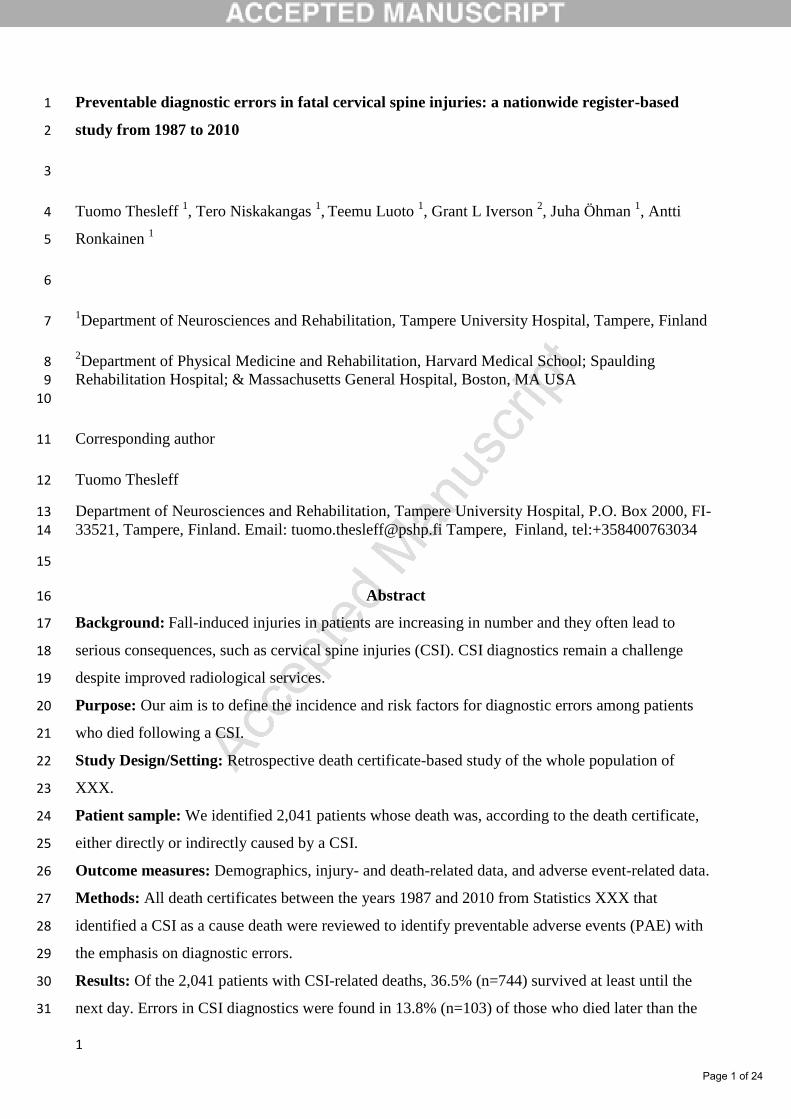
1
Preventable diagnostic errors in fatal cervical spine injuries: a nationwide register-based 1
study from 1987 to 2010 2
3
Tuomo Thesleff 1, Tero Niskakangas
1, Teemu Luoto
1, Grant L Iverson
2, Juha Öhman
1, Antti 4
Ronkainen 1 5
6
1Department of Neurosciences and Rehabilitation, Tampere University Hospital, Tampere, Finland 7
2Department of Physical Medicine and Rehabilitation, Harvard Medical School; Spaulding 8
Rehabilitation Hospital; & Massachusetts General Hospital, Boston, MA USA 9
10
Corresponding author 11
Tuomo Thesleff 12
Department of Neurosciences and Rehabilitation, Tampere University Hospital, P.O. Box 2000, FI-13
33521, Tampere, Finland. Email: [email protected] Tampere, Finland, tel:+358400763034 14
15
Abstract 16
Background: Fall-induced injuries in patients are increasing in number and they often lead to 17
serious consequences, such as cervical spine injuries (CSI). CSI diagnostics remain a challenge 18
despite improved radiological services. 19
Purpose: Our aim is to define the incidence and risk factors for diagnostic errors among patients 20
who died following a CSI. 21
Study Design/Setting: Retrospective death certificate-based study of the whole population of 22
XXX. 23
Patient sample: We identified 2,041 patients whose death was, according to the death certificate, 24
either directly or indirectly caused by a CSI. 25
Outcome measures: Demographics, injury- and death-related data, and adverse event-related data. 26
Methods: All death certificates between the years 1987 and 2010 from Statistics XXX that 27
identified a CSI as a cause death were reviewed to identify preventable adverse events (PAE) with 28
the emphasis on diagnostic errors. 29
Results: Of the 2,041 patients with CSI-related deaths, 36.5% (n=744) survived at least until the 30
next day. Errors in CSI diagnostics were found in 13.8% (n=103) of those who died later than the 31
Page 1 of 24

2
day of injury. Those with diagnostic errors were significantly older (median age 79.4 years, 95% CI 1
75.9-80.1 vs. 74.9, 95% CI 70.2-72.9, p<0.001) and the mechanism of injury was significantly more 2
often a fall (86.4%, n=89 vs. 69.7%, n=447, p=0.002) compared to those who did not have a 3
diagnostic error. The incidence of diagnostic errors increased slightly during the 24–year study 4
period. 5
Conclusions: Cervical spine injury diagnostics remain difficult despite improved radiological 6
services. The majority of the patients subjected to diagnostic errors are fragile elderly people with 7
reduced physical capacity. In our analysis, preventable adverse events and diagnostic errors were 8
most commonly associated with ground-level falls. 9
10
Key Words: Spinal Injuries; Diagnostic Errors; Adverse Effects; Causes of Death; Accidental Falls 11
12
Page 2 of 24

3
Introduction 1
Cervical spine injury (CSI) is a potentially devastating injury. Approximately 2-4% of blunt 2
trauma patients suffer CSI.(1-4) Falls are common in the elderly, and elderly patients may sustain a 3
CSI after a seemingly low energy trauma—including ground-level falls.(5, 6) It is estimated that 4
30% of people aged 65 or older fall every year,(7, 8) and about 5-10% of falls lead to serious 5
injuries.(9, 10) The incidence of fall-related CSIs among older adults has increased since the 6
1970´s.(6, 11) Patients aged 65 years or older have a relative risk for CSI twice that of younger 7
trauma patients.(12, 13) The associated mortality rate in this age group is approximately 24%.(14, 8
15) About 10%–50% of patients with CSIs suffer a concomitant spinal cord injury, which may lead 9
to complete or incomplete tetraplegia or death.(16-18) Nevertheless, isolated CSIs without a spinal 10
cord injury lead to high mortality among elderly.(19, 20) 11
The detection of CSI is especially challenging among the elderly, patients with alterations in 12
consciousness, or patients with distracting injuries.(13, 21) The gold standard of both pre-hospital 13
and in-hospital trauma care is cervical spine immobilization to prevent further neurological 14
deterioration.(15, 19, 22) Emergency cervical spine computed tomography (CT) is the cornerstone 15
of CSI diagnostics in order to triage the need for immediate care. Failure to suspect or detect a CSI 16
as early as possible leads to increased morbidity and mortality.(20, 23, 24) Diagnostic errors occur 17
in every medical specialty. The error rate in clinical medicine is estimated at 5-15%.(25, 26) In 18
blunt trauma patients, accurate confirmation of the absence of CSI remains a challenge despite 19
clinical decision rules and sophisticated radiological techniques.(21, 27-32) Historically, in the era 20
of plain roentgenograms, misdiagnosis of a CSI was estimated in up to one-third of cases.(17, 20, 21
24, 33) Currently, in the era of CT, the rate of misdiagnosis is reported to range from approximately 22
0 to 5% depending on the population studied,(33, 34) and a failure to identify a CSI is considered a 23
diagnostic error. These diagnostic errors might result in fatal adverse events (AE). An AE is usually 24
defined as an unintended injury or complication, caused by health care management rather than the 25
Page 3 of 24

4
patient’s underlying disease. These events result in prolonged hospital stay, disability at the time of 1
discharge, or death.(35-37) In this study, diagnostic errors were considered as AEs regardless of 2
their consequences. AEs may or may not be considered preventable (PAEs = preventable adverse 3
events). PAEs include preventable diagnostic errors, medical errors, and so-called iatrogenic errors. 4
In general, the incidence of in-hospital AEs is estimated around 10% of which a substantial 5
proportion are considered preventable.(38, 39) In surgical and orthopedic care the rate of AEs is 6
even greater (up to 30%), of which the majority are considered preventable.(40-43) 7
The primary purpose of this nationwide retrospective study was to define the incidence and 8
characteristics of CSI diagnostic errors (only diagnostic errors that were considered preventable 9
were included), in patients whose death was directly or indirectly related to a CSI according to their 10
death certificates. The secondary aim was to define the incidence and characteristics of other kinds 11
of PAEs (other than diagnostic errors). We hypothesized that the number of preventable errors in 12
CSI diagnostics decreased over the years (from 1987 to 2010) due to improved radiological services 13
and more objective diagnostics. 14
Materials and Methods 15
This population-based sample was collected from death certificates issued in XXX between 16
1987 and 2010. The death certificates were obtained from the official Cause-of-Death Register, 17
coordinated by Statistics XXX (XXXX, XXXX), which has filed each death certificate in XXX 18
since 1936.(44) The XXX official cause-of-death statistics are, in practice, 100% complete.(44) 19
Medicolegal autopsies are crucial for the accuracy of death certification. The number of 20
medicolegal autopsies performed in XXX is considerably higher than in many other developed 21
countries.(45) In XXX, the indications for medicolegal autopsies are strictly controlled by 22
legislation.(45) Medicolegal autopsies are performed in up to 87.2% of all unintentional injury-23
related deaths, 98.3% of homicides, and 99.5% of suicides.(45) According to XXX legislation, a 24
medicolegal autopsy should be performed in the following circumstances: when death is caused or 25
Page 4 of 24

5
suspected to be caused by (i) a crime, (ii) a suicide, (iii) an accident, (iv) poisoning, (v) an 1
occupational disease, or (vi) medical treatment, or when death has (vii) not been caused by a 2
disease, or when (viii) during the last illness, the deceased had not been treated by a doctor within 3 3
months, or when (ix) the death was otherwise unexpected. Each death certificate includes among 4
other information a short narrative that contains additional relevant information (including key 5
findings from autopsy) on the circumstances leading to death. All relevant information on the 6
events leading to a patient’s death are supposed to be summarized in the death certificates. At the 7
National Institute for Health and Welfare (XXX,XXX), forensic pathologists review each death 8
certificate and ensure that the recorded causes of death are accurate, complete, and consistent.(44) 9
This process results in highly controlled and comprehensive national mortality statistics. The 10
main/underlying cause of death is the disease or injury that initiated the series of illnesses leading 11
directly to death. The immediate cause of death refers to the disease, failure, or injury that causes 12
the person to die. The intermediate cause of death refers to the condition that arise from or after the 13
main/underlying cause to become the immediate cause of death. The related causes of death are 14
other significant circumstances that contributed to the death.(44) This study was approved by the 15
Ethics Committee of XXXX Hospital District, XXXX, XXXX. 16
Data Collection 17
In the first stage of the study, all death certificates containing CSI as an immediate, 18
intermediate, main, or related cause of death were reviewed carefully by the first author (TT). 19
During the study period, both the 9th
and 10th
Edition of the International Statistical Classification of 20
Diseases and Related Health Problems (ICD-9 and ICD-10) were in use. In our study, CSI was 21
defined as an injury to the cervical spine, including (i) fractures, (ii) dislocations, (iii) fractures with 22
spinal cord injury, (iv) isolated spinal cord injury, or (v) a combination of those injuries. The 23
corresponding ICD-10 diagnosis codes were: S12.0, S12.1, S12.2, S12.7, S12.9, S13.0, S13.1, 24
S13.2, S13.3, S14.0, and S14.1. Similarly, the ICD-9 diagnosis codes were: 805.0–805.18, 806.0–25
Page 5 of 24

6
806.19, 839.0–839.18, and 952.0–952.09. Combined skull and cervical fractures (T01.1–T06.0), 1
whiplash, nerve root injuries, and non-traumatic spinal cord lesions were not used for the screening 2
of the death certificates. The collected variables included diagnosis (ICD codes), gender, age, time 3
of injury, place of injury, time between injury and death, cause of death, type of injury, alcohol and 4
drug consumption at the time of injury, type of death certification (medicolegal autopsy, hospital 5
autopsy, autopsy not performed, or other), presence of spinal cord injury, and approximate level of 6
cervical injury. Injuries at cervical levels C0-C2 were regarded as high injuries and injuries at levels 7
C3-C7 as low injuries. Cases where the level of injury was undetermined or the injury consisted of 8
multiple levels were considered as low injuries. Substance abuse was considered positive if it was 9
mentioned in the death certificate regardless of the method of verification. 10
In the second stage of the study, all cases in which the date of death was different from the 11
date of injury were selected for further analysis. By doing the selection we excluded people who, in 12
majority of cases, succumbed at the accident scene and therefore had a minor chance for PAE. The 13
death reports of those who survived at least until the next day were examined (by XXXX and 14
XXXX) to detect reports with a suspicion of any kind of PAE (including preventable diagnostic 15
errors and other medical errors). Preventability was defined as care that fell below the level of 16
expected performance for practitioners or systems at the time of the PAE. If the death report did not 17
provide enough information for a solid conclusion, it was considered as a non-PAE or a PAE 18
undetermined. PAEs were categorized in two main groups (diagnostic errors and other events) and 19
six subcategories (surgical treatment related events, diagnostic errors, health care facility related 20
events, secondary complications, treatment-related events, and other). The location of the PAE 21
occurrence was documented. If more than one PAE was detected for an individual patient, only the 22
most severe one was coded. The focus of the current study was on preventable diagnostic errors. 23
Page 6 of 24
7
Patient Involvement 1
Patients were not involved in setting the research question or the outcome measures, nor were 2
they involved in the design, or implementation of the study. No patients were asked to advice on 3
interpretation or writing up of results. There are no plans to disseminate the results of this research 4
to study participants or the relevant patient community. 5
Statistical Analyses 6
IBM SPSS Statistics 20.0 (IBM Corp. Armonk, NY, USA) was used to perform the 7
analyses. The frequencies and percentage of the analyzed variables were calculated using 8
descriptive statistics. The normality of the variable distributions was tested using the Kolmogorov-9
Smirnov and Shapiro-Wilk tests. Continuous variables were analyzed with the Pearson (normal 10
distribution) and Spearman (skewed distribution) correlation coefficients. Group comparisons were 11
tested with the Student’s t-test (normal distribution) and the Mann-Whitney U-test (skewed 12
distribution). The statistical significance level was set at p < 0.05. 13
Results 14
A total of 2,041 death certificates reported a CSI in XXXX during the 24-year study period 15
(1987-2010). The average population of XXX during the same time was about five million. Of the 16
2,041 CSI-related deaths, 36.5% (n = 744) survived at least until the next day (date of death was 17
later than date of injury). The differences in patient characteristics of those who died on the day of 18
injury and those who died after the day of injury are presented in Table 1. Those who died on the 19
day of injury were significantly younger (median age = 55.0 years) and the most common injury 20
mechanism was a traffic accident (51.7%, n = 670) whereas those who survived at least until the 21
next day were elderly (median age = 75.7 years) and the injury mechanism was usually a fall 22
(72.0%, n = 536). The location of death for those who survived at least until the next day was a 23
medical facility in 93.8% (n = 698), whereas only 8.2% (n = 107) of those who died on the day of 24
injury did so in a medical facility. Medicolegal autopsies were performed in 99.5% (n = 1,291) of 25
Page 7 of 24

8
the patients who died on the day of injury and in 82.8% (n = 616) of those who survived at least 1
until the next day. Of those who survived at least until the next day, a PAE was identified in 27.7% 2
(n = 206), of which half (n = 103) were considered diagnostic errors (Fig 1). In 5% (n = 37), a PAE 3
could not be determined as present or not present. 4
Insert Figure 1 here 5
Insert Table 1 here 6
Preventable Diagnostic Adverse Events 7
Patient characteristics, injury-related information, and diagnostic error data are presented in 8
Table 2. CSI patients with diagnostic errors were significantly older (median age 79.4 years, 95% 9
CI 75.9-80.1 versus 74.9, 95% CI 70.2-72.9, p<0.001), and the injury mechanism was significantly 10
more often a fall (86.4%, n=89 versus 69.7%, n=447, p=0.002), compared to those who did not 11
have a diagnostic error. The median time from injury to death was significantly less in the 12
diagnostic error group (seven days compared to 17 days, p < 0.001). Examples of typical cases with 13
a diagnostic error are presented in the online supplementary material. The number of PAEs and 14
diagnostic errors increased during the study period, but so did the number of all patients with CSIs. 15
The PAEs / diagnostic error ratio in relation to the number of all patients with CSIs increased 16
slightly during the study years (Fig 2). The PAEs (n = 206) were classified into six different 17
categories as presented in Table 3. The most frequent recorded PAE types were related to CSI 18
diagnostics and events related to health care facilities. It is worth noting that 1,297 patients with 19
CSIs died on the day of injury, and 107 (8.2%) of them died at a medical facility. It is possible that 20
some of these 1,297 patients experienced PAEs that are not included in our study. 21
Insert Figure 2 about here 22
Insert Table 2 about here 23
Insert Supplement about here 24
Insert Table 3 about here 25
Page 8 of 24
9
Discussion 1
Principal Findings 2
In this nationwide, retrospective, death certificate-based study, we found preventable 3
diagnostic errors in 13.8% of those who survived at least until the next day after a CSI. The patients 4
who experienced diagnostic errors were significantly older, and the mechanism of injury was more 5
often a fall compared to patients who did not experience diagnostic errors. Diagnostic errors were 6
associated with significantly less time from injury to death. Contrary to our hypothesis, the 7
incidence of diagnostic errors did not decrease during the 24–year study period from 1987 to 2010 8
despite advancements in radiological services and increased availability of CT for CSI diagnostics. 9
At least four factors might have contributed to this. First, the number of elderly and presumably 10
more frail people (who are prone to AEs) in our society has increased. Patients included in this 11
study were older toward the end of the study period (see data presented in our previous article).(11) 12
Second, a considerable number of the diagnostic errors resulted from the lack of suspicion of CSI 13
(throughout the study period), thus not even conventional roentgenograms of the neck were 14
performed (online supplement, case examples 1,4,6,8, and 9). Therefore, greater availability of CT 15
over time could not influence the error rate of these and similar patients. Third, many death reports 16
from the late 80’s and early 90’s were briefer and less informative than the more recent ones, which 17
made it impossible to draw solid conclusions on possible AEs. Therefore, the number of cases in the 18
early years of the study may be artificially lower. Fourth, in XXX the overall rate of medicolegal 19
autopsies has increased during the study period,(46) making it more likely that errors would be 20
detected over time. We suspect that diagnostic accuracy has likely improved over time, but this is 21
not reflected in this death certificate based study because medicolegal autopsy was performed on 22
82.8% of all patients with CSIs who died after the day of injury. 23
We suggest that diagnostic errors are a substantial cause of preventable harm in primary and 24
in secondary care of patients with CSIs. We found a high frequency of diagnostic errors following 25
Page 9 of 24
10
ground-level falls among older individuals, whose physical frailty may increase the risk of these 1
relatively low-impact injuries. It has been previously shown that evidence-based clinical decision 2
rules for CSI, applied to elderly patients, have failed to predict injury in some cases.(47, 48) In 3
addition to the physical frailty associated with senescence, older patients might provide vague 4
complaints, while younger patients might be more explicit about describing neck pain and/or 5
neurological symptoms. However, the incidence of diagnostic errors in younger populations might 6
also be higher than presented in this death certificate based study, but because of their better general 7
health status, the errors (leading to tetraparesis, for example) are more rarely fatal and therefore not 8
shown in this study. Diagnostic errors are important AE types because they are in most cases 9
preventable and their consequences are often severe. 10
The number of other PAEs, separate from diagnostic errors, was also rather high. Events in 11
health care facilities (including falling in nursing homes) were the second largest category of PAEs 12
after diagnostic errors. Improvement of personnel resources in some health care facilities, such as 13
under-resourced nursing homes, might reduce the rate of these events. Fall prevention in general is 14
crucial to reduce the number of CSIs. 15
Strengths and Weaknesses of the Study 16
To the best of our knowledge, this is the first death certificate-based nationwide study to 17
examine the number of diagnostic errors in patients with CSIs. A medicolegal autopsy was 18
performed on 92.2% of patients who experienced a diagnostic error and on 82.8% of all patients 19
with CSIs who died after the day of injury. The XXX official cause-of-death statistics are, in 20
practice, 100% complete and the autopsy rates in XXX are considerably higher than in many other 21
western countries, which strengthen our results.(45) In addition, all residents in XXX are covered 22
by public health care which means that no patients are denied medical care (including for example 23
CT imaging) based on socio-economic status, age, or other reasons. Total health spending 24
accounted for 9.3% of the GDP in 2012, which is slightly lower than the average in Organization 25
Page 10 of 24
11
for Economic Cooperation and Development countries. The quality of healthcare in XXX is 1
generally considered good.(49) 2
However, this study has several limitations. First, the study’s data extraction was restricted to 3
death certificates only. Information was sometimes limited for drawing a solid conclusion about 4
possible adverse events. Because we did not have access to the full autopsy reports, it is possible 5
that we are underestimating PAEs. Second, the identification and classification of PAEs was 6
susceptible to subjective considerations from the authors (XXXX and XXXX), and as such could 7
have introduced bias. Both authors have considerable experience in diagnosing and treating patients 8
with CSIs, but inter-rater reliability was not measured. One could, for example, argue about the 9
possible preventability of an elderly patient falling in a nursing home. In this study, we focused on 10
diagnostic errors because the available data from death certificates was often limited for drawing 11
conclusions about other kinds of PAEs. In contrast, the identification and classification of 12
diagnostic errors was straightforward in most cases. It should be noted that the PAEs or diagnostic 13
errors did not necessarily influence the death of individual patients, even though the median time 14
between injury and death was significantly less for those with diagnostic errors. 15
Strengths and Weaknesses in Relation to Other Studies 16
During the 24-year observation period, the number of CSI-related diagnostic errors was 17
clearly greater than in previous more general studies conducted in hospital settings.(35, 36, 38, 50) 18
However, our findings are in line with prior autopsy studies showing that major diagnostic 19
discrepancies are commonly identified in more than 10% of cases.(51-54) 20
In the history of medicine, autopsy has been described as the most powerful tool and the 21
“gold standard” for detecting diagnostic errors.(55, 56) It is shown that a substantial proportion of 22
fatal spinal cord injuries are without radiographic abnormalities at CT.(57) Nevertheless, it is 23
essential to understand that autopsy studies only provide the error rate in patients who die. Because 24
the diagnostic error rate is almost certainly lower among patients who are still alive, error rates 25
Page 11 of 24

12
derived solely from autopsy data are inflated. For example, whereas autopsy studies suggest that 1
fatal pulmonary embolism is misdiagnosed approximately 55% of the time, the misdiagnosis rate 2
for all cases of pulmonary embolism is only 4%.(52, 58) Feedback from autopsies is a helpful 3
component of physician training, but the declining number of autopsies performed in many 4
countries limits this opportunity. 5
We analyzed diagnostic errors from only a subpopulation that died after the day of injury 6
(93.8% of those died in a medical facility). If the entire population of people who died following 7
CSIs (n = 2,041) had been analyzed, the percentage of diagnostic errors likely would have been 8
lower, given that over 90% of patients who died on the day of injury died outside of hospitals and 9
therefore were less likely to have diagnostic errors. 10
Unanswered Questions and Future Research 11
Re-examining the death certificates of the 1,297 patients with CSIs who died during the day 12
of injury would yield more data on the possible PAEs that happened in the field by first aid 13
personnel. That might provide us with the number of PAEs in the total population who died and 14
more data about the PAEs by first aid personnel. We believe that the high number of missed CSIs in 15
this study is not a country specific finding considering the generally good XXXX healthcare 16
system. This could be determined by performing similar death certificate based studies in other 17
countries. Further prospective studies are also needed to evaluate the signs and symptoms of elderly 18
patients with CSIs. In a retrospective setting, global trigger tools or other computer-assisted chart 19
review methods might be helpful for identifying risk factors for diagnostic errors in patients with 20
CSIs.(59) 21
Conclusions 22
Cervical spine injury diagnostics remain difficult despite improved radiological services. 23
Frail elderly people with diminished physical capacity represent the majority of patients who 24
experienced diagnostic errors in this population of patients with CSIs who died. In our analysis, 25
Page 12 of 24
13
preventable adverse events and diagnostic errors were most commonly associated with ground-level 1
falls. Although diagnostic errors cannot be eliminated, adherence to evidence-based guidelines and 2
acknowledging their limitations especially among elderly patients might reduce the frequency of 3
some of these events and errors.(47, 48, 60, 61) Continuing medical education and quality 4
improvement initiatives might also reduce adverse events and diagnostic errors. 5
“What this paper adds” box 6
Section 1: Cervical spine injury (CSI) diagnostics have improved with advancements in 7
radiological services. However, this death-certificate based nationwide study of the incidence and 8
risk factors for diagnostic errors among patients who die following a CSI indicates that the 9
diagnostic error rate is not declining. Hence, CSI diagnostics remain problematic and diagnostic 10
errors may lead to excess harm for patients. 11
Section 2: The injury mechanism of patients who were identified as experiencing preventable 12
diagnostic errors was most often a ground-level fall. We emphasize that special consideration in 13
cervical spine evaluation should be given to elderly patients who sustain a low energy trauma. 14
15
Footnotes 16
Acknowledgements 17
The authors wish to thank Statistics XXX for the provision of data. 18
Funding 19
No external funding. 20
Competing interests 21
All authors have completed the ICMJE uniform disclosure form at 22
www.icmje.org/coi_disclosure.pdf (available on request from the corresponding author) and 23
declare: no support from any organization for the submitted work; no financial relationships with 24
any organizations that might have an interest in the submitted work in the previous three years; no 25
other relationships or activities that could appear to have influenced the submitted work. 26
27
Page 13 of 24
14
Transparency declaration 1
The lead author affirms that the manuscript is an honest, accurate, and transparent account of 2
the study being reported; that no important aspects of the study have been omitted; and that any 3
discrepancies from the study as planned have been explained. 4
Data sharing 5
No additional data available. 6
Ethical approval 7
This study was approved by the Ethics Committee of XXXX Hospital District, XXXX, 8
XXXX (reference number: R12215) 9
Contributors 10
TN and TT conceived the idea for the study. JÖ, AR, and TL also made important contributions to 11
the concept and design of the study. TT and TN had full access to all of the data in the study and 12
take responsibility for the integrity of the data and the accuracy of the data analysis. TT is the 13
guarantor. All authors contributed to the analysis and interpretation of the data. TT drafted the 14
initial manuscript and all authors contributed to the critical revision of the manuscript. 15
16
References 17
1. Hasler RM, Exadaktylos AK, Bouamra O, Benneker LM, Clancy M, Sieber R, et al. 18
Epidemiology and predictors of cervical spine injury in adult major trauma patients: A multicenter 19
cohort study. J Trauma Acute Care Surg. 2012 Apr;72(4):975-81. 20
2. Milby AH, Halpern CH, Guo W, Stein SC. Prevalence of cervical spinal injury in trauma. 21
Neurosurg Focus. 2008;25(5):E10. 22
3. Bayless P, Ray VG. Incidence of cervical spine injuries in association with blunt head trauma. 23
Am J Emerg Med. 1989 Mar;7(2):139-42. 24
4. Grogan EL, Morris JA,Jr, Dittus RS, Moore DE, Poulose BK, Diaz JJ, et al. Cervical spine 25
evaluation in urban trauma centers: Lowering institutional costs and complications through helical 26
CT scan. J Am Coll Surg. 2005 Feb;200(2):160-5. 27
Page 14 of 24
15
5. Wang H, Coppola M, Robinson RD, Scribner JT, Vithalani V, de Moor CE, et al. Geriatric 1
trauma patients with cervical spine fractures due to ground level fall: Five years experience in a 2
level one trauma center. J Clin Med Res. 2013 Apr;5(2):75-83. 3
6. Kannus P, Palvanen M, Niemi S, Parkkari J. Alarming rise in the number and incidence of fall-4
induced cervical spine injuries among older adults. J Gerontol A Biol Sci Med Sci. 2007 5
Feb;62(2):180-3. 6
7. Nevitt MC, Cummings SR, Kidd S, Black D. Risk factors for recurrent nonsyncopal falls. A 7
prospective study. JAMA. 1989 May 12;261(18):2663-8. 8
8. Hoidrup S, Sorensen TI, Gronbaek M, Schroll M. Incidence and characteristics of falls leading to 9
hospital treatment: A one-year population surveillance study of the danish population aged 45 years 10
and over. Scand J Public Health. 2003;31(1):24-30. 11
9. Tinetti ME, Williams CS. Falls, injuries due to falls, and the risk of admission to a nursing home. 12
N Engl J Med. 1997 Oct 30;337(18):1279-84. 13
10. Kannus P, Sievanen H, Palvanen M, Jarvinen T, Parkkari J. Prevention of falls and consequent 14
injuries in elderly people. Lancet. 2005 Nov 26;366(9500):1885-93. 15
11. Thesleff T, Niskakangas T, Luoto TM, Ohman J, Ronkainen A. Fatal cervical spine injuries: A 16
finnish nationwide register-based epidemiological study on data from 1987 to 2010. Spine J. 2015 17
Dec 7. 18
12. Lowery DW, Wald MM, Browne BJ, Tigges S, Hoffman JR, Mower WR, et al. Epidemiology 19
of cervical spine injury victims. Ann Emerg Med. 2001 Jul;38(1):12-6. 20
13. Goode T, Young A, Wilson SP, Katzen J, Wolfe LG, Duane TM. Evaluation of cervical spine 21
fracture in the elderly: Can we trust our physical examination? Am Surg. 2014 Feb;80(2):182-4. 22
14. Spivak JM, Weiss MA, Cotler JM, Call M. Cervical spine injuries in patients 65 and older. 23
Spine (Phila Pa 1976). 1994 Oct 15;19(20):2302-6. 24
15. Damadi AA, Saxe AW, Fath JJ, Apelgren KN. Cervical spine fractures in patients 65 years or 25
older: A 3-year experience at a level I trauma center. J Trauma. 2008 Mar;64(3):745-8. 26
16. Leucht P, Fischer K, Muhr G, Mueller EJ. Epidemiology of traumatic spine fractures. Injury. 27
2009 Feb;40(2):166-72. 28
17. Bohlman HH. Acute fractures and dislocations of the cervical spine. an analysis of three 29
hundred hospitalized patients and review of the literature. J Bone Joint Surg Am. 1979 30
Dec;61(8):1119-42. 31
18. Fredo HL, Rizvi SA, Lied B, Ronning P, Helseth E. The epidemiology of traumatic cervical 32
spine fractures: A prospective population study from norway. Scand J Trauma Resusc Emerg Med. 33
2012 Dec 21;20:85,7241-20-85. 34
Page 15 of 24
16
19. Golob JF,Jr, Claridge JA, Yowler CJ, Como JJ, Peerless JR. Isolated cervical spine fractures in 1
the elderly: A deadly injury. J Trauma. 2008 Feb;64(2):311-5. 2
20. Reid DC, Henderson R, Saboe L, Miller JD. Etiology and clinical course of missed spine 3
fractures. J Trauma. 1987 Sep;27(9):980-6. 4
21. Hoffman JR, Wolfson AB, Todd K, Mower WR. Selective cervical spine radiography in blunt 5
trauma: Methodology of the national emergency X-radiography utilization study (NEXUS). Ann 6
Emerg Med. 1998 Oct;32(4):461-9. 7
22. Weller SJ, Malek AM, Rossitch E,Jr. Cervical spine fractures in the elderly. Surg Neurol. 1997 8
Mar;47(3):274,80; discussion 280-1. 9
23. Levi AD, Hurlbert RJ, Anderson P, Fehlings M, Rampersaud R, Massicotte EM, et al. 10
Neurologic deterioration secondary to unrecognized spinal instability following trauma--a 11
multicenter study. Spine (Phila Pa 1976). 2006 Feb 15;31(4):451-8. 12
24. Davis JW, Phreaner DL, Hoyt DB, Mackersie RC. The etiology of missed cervical spine 13
injuries. J Trauma. 1993 Mar;34(3):342-6. 14
25. Berner ES, Graber ML. Overconfidence as a cause of diagnostic error in medicine. Am J Med. 15
2008 May;121(5 Suppl):S2-23. 16
26. Schwartz A, Elstein AS. Clinical reasoning in medicine. Clinical reasoning in the health 17
professions.3rd ed.Edinburgh: Elsevier. 2008:223-34. 18
27. Anderson PA, Gugala Z, Lindsey RW, Schoenfeld AJ, Harris MB. Clearing the cervical spine in 19
the blunt trauma patient. J Am Acad Orthop Surg. 2010 Mar;18(3):149-59. 20
28. Anderson PA, Muchow RD, Munoz A, Tontz WL, Resnick DK. Clearance of the asymptomatic 21
cervical spine: A meta-analysis. J Orthop Trauma. 2010 Feb;24(2):100-6. 22
29. Stiell IG, Clement CM, McKnight RD, Brison R, Schull MJ, Rowe BH, et al. The canadian C-23
spine rule versus the NEXUS low-risk criteria in patients with trauma. N Engl J Med. 2003 Dec 24
25;349(26):2510-8. 25
30. Raza M, Elkhodair S, Zaheer A, Yousaf S. Safe cervical spine clearance in adult obtunded blunt 26
trauma patients on the basis of a normal multidetector CT scan--a meta-analysis and cohort study. 27
Injury. 2013 Nov;44(11):1589-95. 28
31. Sweeney JF, Rosemurgy AS, Gill S, Albrink MH. Is the cervical spine clear? undetected 29
cervical fractures diagnosed only at autopsy. Ann Emerg Med. 1992 Oct;21(10):1288-90. 30
32. Michaleff ZA, Maher CG, Verhagen AP, Rebbeck T, Lin CW. Accuracy of the canadian C-31
spine rule and NEXUS to screen for clinically important cervical spine injury in patients following 32
blunt trauma: A systematic review. CMAJ. 2012 Nov 6;184(16):E867-76. 33
33. Platzer P, Hauswirth N, Jaindl M, Chatwani S, Vecsei V, Gaebler C. Delayed or missed 34
diagnosis of cervical spine injuries. J Trauma. 2006 Jul;61(1):150-5. 35
Page 16 of 24
17
34. Patel MB, Humble SS, Cullinane DC, Day MA, Jawa RS, Devin CJ, et al. Cervical spine collar 1
clearance in the obtunded adult blunt trauma patient: A systematic review and practice management 2
guideline from the eastern association for the surgery of trauma. J Trauma Acute Care Surg. 2015 3
Feb;78(2):430-41. 4
35. Brennan TA, Leape LL, Laird NM, Hebert L, Localio AR, Lawthers AG, et al. Incidence of 5
adverse events and negligence in hospitalized patients. results of the harvard medical practice study 6
I. N Engl J Med. 1991 Feb 7;324(6):370-6. 7
36. Thomas EJ, Studdert DM, Burstin HR, Orav EJ, Zeena T, Williams EJ, et al. Incidence and 8
types of adverse events and negligent care in utah and colorado. Med Care. 2000 Mar;38(3):261-71. 9
37. Wilson RM, Runciman WB, Gibberd RW, Harrison BT, Newby L, Hamilton JD. The quality in 10
australian health care study. Med J Aust. 1995 Nov 6;163(9):458-71. 11
38. de Vries EN, Ramrattan MA, Smorenburg SM, Gouma DJ, Boermeester MA. The incidence and 12
nature of in-hospital adverse events: A systematic review. Qual Saf Health Care. 2008 13
Jun;17(3):216-23. 14
39. Soop M, Fryksmark U, Koster M, Haglund B. The incidence of adverse events in swedish 15
hospitals: A retrospective medical record review study. Int J Qual Health Care. 2009 16
Aug;21(4):285-91. 17
40. Merten H, Johannesma PC, Lubberding S, Zegers M, Langelaan M, Jukema GN, et al. High risk 18
of adverse events in hospitalised hip fracture patients of 65 years and older: Results of a 19
retrospective record review study. BMJ Open. 2015 Sep 7;5(9):e006663,2014-006663. 20
41. Rutberg H, Borgstedt-Risberg M, Gustafson P, Unbeck M. Adverse events in orthopedic care 21
identified via the global trigger tool in sweden - implications on preventable prolonged 22
hospitalizations. Patient Saf Surg. 2016 Oct 26;10:23. 23
42. Nilsson L, Risberg MB, Montgomery A, Sjodahl R, Schildmeijer K, Rutberg H. Preventable 24
adverse events in surgical care in sweden: A nationwide review of patient notes. Medicine 25
(Baltimore). 2016 Mar;95(11):e3047. 26
43. Anderson O, Davis R, Hanna GB, Vincent CA. Surgical adverse events: A systematic review. 27
Am J Surg. 2013 Aug;206(2):253-62. 28
44. Official statistics of finland (OSF): Deaths [e-publication].ISSN=1798-2545. 2015, quality 29
description, deaths 2015 . helsinki: Statistics finland [referred: 15.11.2016].access method: 30
Http://Www.stat.fi/til/kuol/2015/kuol_2015_2016-04-14_laa_001_en.html [Internet]. 31
45. Lunetta P, Lounamaa A, Sihvonen S. Surveillance of injury-related deaths: Medicolegal autopsy 32
rates and trends in finland. Inj Prev. 2007 Aug;13(4):282-4. 33
46. Official statistics of finland (OSF): Population projection [e-publication].ISSN=1798-5153. 34
2015. helsinki: Statistics finland [referred: 1.5.2017].access method: 35
Http://Www.stat.fi/til/vaenn/2015/vaenn_2015_2015-10-30_tie_001_en.html [Internet]. 36
Page 17 of 24

18
47. Healey CD, Spilman SK, King BD, Sherrill JE,2nd, Pelaez CA. Asymptomatic cervical spine 1
fractures: Current guidelines can fail older patients. J Trauma Acute Care Surg. 2017 Apr 20. 2
48. Denver D, Shetty A, Unwin D. Falls and implementation of NEXUS in the elderly (the FINE 3
study). J Emerg Med. 2015;49(3):294-300. 4
49. OECD, EU. Health at a glance: Europe 2016. Organisation for Economic Co-operation and 5
Development; 2016. 6
50. Zegers M, de Bruijne MC, de Keizer B, Merten H, Groenewegen PP, van der Wal G, et al. The 7
incidence, root-causes, and outcomes of adverse events in surgical units: Implication for potential 8
prevention strategies. Patient Saf Surg. 2011 May 20;5:13,9493-5-13. 9
51. Aalten CM, Samson MM, Jansen PA. Diagnostic errors; the need to have autopsies. Neth J 10
Med. 2006 Jun;64(6):186-90. 11
52. Shojania KG, Burton EC, McDonald KM, Goldman L. The autopsy as an outcome and 12
performance measure. Evid Rep Technol Assess (Summ). 2002 Oct;(58)(58):1-5. 13
53. Burton EC, Troxclair DA, Newman WP,3rd. Autopsy diagnoses of malignant neoplasms: How 14
often are clinical diagnoses incorrect? JAMA. 1998 Oct 14;280(14):1245-8. 15
54. Sonderegger-lseli K, Burger S, Muntwyler J, Salomon F. Diagnostic errors in three medical 16
eras: A necropsy study. The Lancet. 2000;355(9220):2027-31. 17
55. Cabot RC. A study of mistaken diagnosis: Based on the analysis of 1000 autopsies and a 18
comparison with the clinical findings. JAMA. 1910;55:1343-1350. 19
56. Cabot RC. Diagnostic pitfalls identified during a study of three thousand autopsies. JAMA. 20
1912;59:2295-2298. 21
57. Makino Y, Yokota H, Hayakawa M, Yajima D, Inokuchi G, Nakatani E, et al. Spinal cord 22
injuries with normal postmortem CT findings: A pitfall of virtual autopsy for detecting traumatic 23
death. Am J Roentgenol. 2014;203(2):240-4. 24
58. Pineda LA, Hathwar VS, Grant BJ. Clinical suspicion of fatal pulmonary embolism. Chest. 25
2001 Sep;120(3):791-5. 26
59. Adler L, Denham CR, McKeever M, Purinton R, Guilloteau F, Moorhead JD, et al. Global 27
trigger tool: Implementation basics. Journal of Patient Safety. 2008;4(4):245-9. 28
60. Hoffman JR, Mower WR, Wolfson AB, Todd KH, Zucker MI. Validity of a set of clinical 29
criteria to rule out injury to the cervical spine in patients with blunt trauma. N Engl J Med. 30
2000;343(2):94-9. 31
61. Stiell IG, Wells GA, Vandemheen KL, Clement CM, Lesiuk H, De Maio VJ, et al. The canadian 32
C-spine rule for radiography in alert and stable trauma patients. JAMA. 2001;286(15):1841-8. 33
34
Page 18 of 24

19
1
Figure 1. Flowchart of the patients with CSIs in the study 2
CSI = Cervical Spine Injury; PAE = Preventable Adverse Event 3
Figure 2. The annual percentages of patients with preventable adverse events (PAE) and diagnostic 4
errors for all patients with CSIs who died after the day of injury (n = 744) 5
6
7
8
9
Page 19 of 24

20
Table 1. Comparison of characteristics of patients who died on the day of injury and patients who 1 died after the day of injury. 2
Patients
who died
on the day of injury
n=1,297 (63.5%)
Patients
who died
after the day of injury
n=744 (36.5%)
n % n % p
Gender, 0.013
Female 376 29 178 23.9
Male 921 71 566 76.1
Age at death, years, median (95% CI) 55.0 (50.7-53.2) 75.7 (71.3-73.6) <0.001
Alcohol mentioned 482 37.2 126 16.9 <0.001
Drugs mentioned 59 4.5 4 0.5 <0.001
CSI as main cause of death 1,243 95.8 604 81.2 <0.001
C0-C2 injury 598 46.1 241 32.4 <0.001
Spinal cord injury 1,182 91.1 512 68.8 <0.001
Cause of injury <0.001
Fall 383 29.5 536 72.0
Traffic accident 670 51.7 158 21.2
Suicide 173 13.3 5 0.7
Other traumatic or unspecified 71 5.5 45 6.0
Place of death <0.001
Medical facility 107 8.2 698 93.8
Home or residence 261 20.1 29 3.9
Other 907 69.6 14 1.9
Abroad 22 1.7 3 0.4
Death certification <0.001
No autopsy 1 0,1 120 16.1
Hospital autopsy 0 0 5 0.7
Medicoloegal autopsy 1,291 99.5 616 82.8
Other 5 0.4 3 0.4
3
4
5
Page 20 of 24
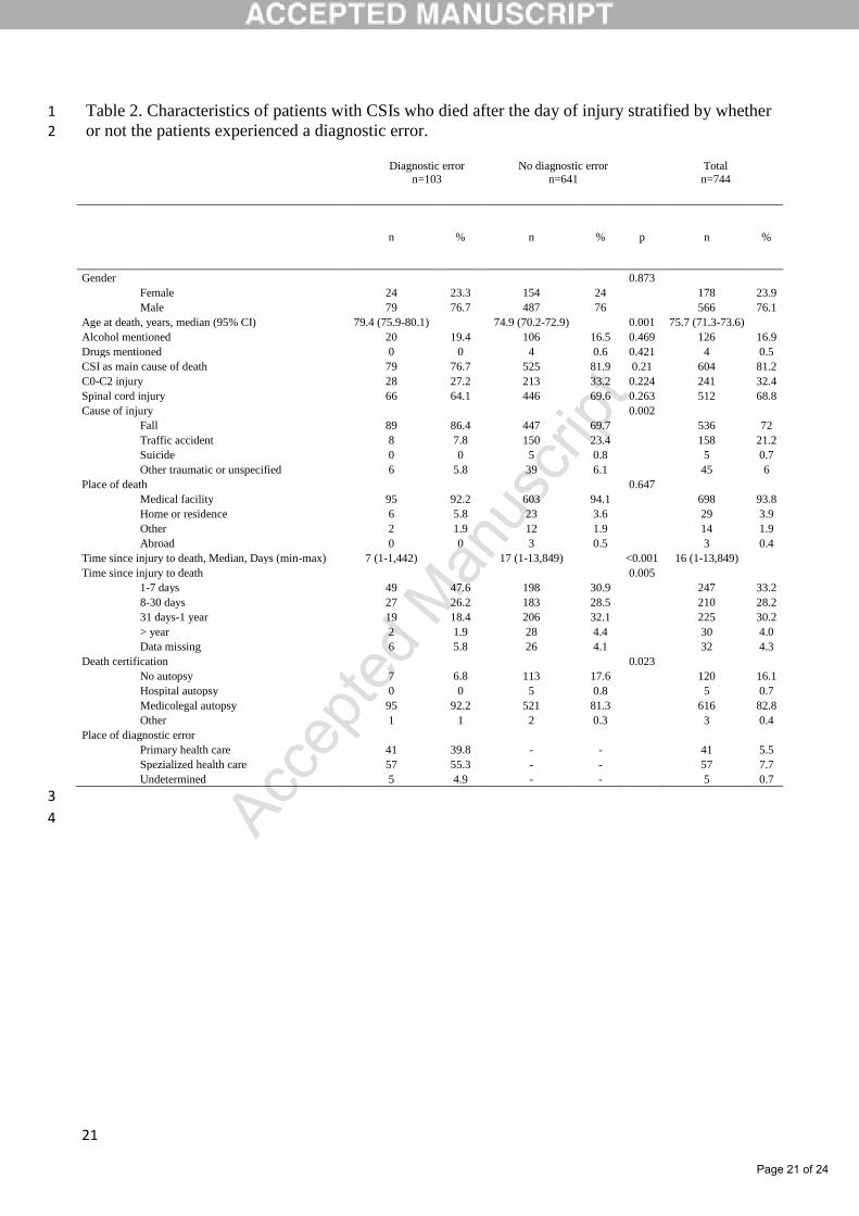
21
Table 2. Characteristics of patients with CSIs who died after the day of injury stratified by whether 1 or not the patients experienced a diagnostic error. 2
Diagnostic error
n=103
No diagnostic error
n=641
Total
n=744
n % n % p n %
Gender 0.873
Female 24 23.3 154 24 178 23.9
Male 79 76.7 487 76 566 76.1
Age at death, years, median (95% CI) 79.4 (75.9-80.1) 74.9 (70.2-72.9) 0.001 75.7 (71.3-73.6)
Alcohol mentioned 20 19.4 106 16.5 0.469 126 16.9
Drugs mentioned 0 0 4 0.6 0.421 4 0.5
CSI as main cause of death 79 76.7 525 81.9 0.21 604 81.2
C0-C2 injury 28 27.2 213 33.2 0.224 241 32.4
Spinal cord injury 66 64.1 446 69.6 0.263 512 68.8
Cause of injury 0.002
Fall 89 86.4 447 69.7 536 72
Traffic accident 8 7.8 150 23.4 158 21.2
Suicide 0 0 5 0.8 5 0.7
Other traumatic or unspecified 6 5.8 39 6.1 45 6
Place of death 0.647
Medical facility 95 92.2 603 94.1 698 93.8
Home or residence 6 5.8 23 3.6 29 3.9
Other 2 1.9 12 1.9 14 1.9
Abroad 0 0 3 0.5 3 0.4
Time since injury to death, Median, Days (min-max) 7 (1-1,442) 17 (1-13,849) <0.001 16 (1-13,849)
Time since injury to death 0.005
1-7 days 49 47.6 198 30.9 247 33.2
8-30 days 27 26.2 183 28.5 210 28.2
31 days-1 year 19 18.4 206 32.1 225 30.2
> year 2 1.9 28 4.4 30 4.0
Data missing 6 5.8 26 4.1 32 4.3
Death certification 0.023
No autopsy 7 6.8 113 17.6 120 16.1
Hospital autopsy 0 0 5 0.8 5 0.7
Medicolegal autopsy 95 92.2 521 81.3 616 82.8
Other 1 1 2 0.3 3 0.4
Place of diagnostic error
Primary health care 41 39.8 - - 41 5.5
Spezialized health care 57 55.3 - - 57 7.7
Undetermined 5 4.9 - - 5 0.7
3
4
Page 21 of 24

22
Table 3. Preventable adverse events (n=206) stratified into six categories. 1
Preventable adverse event category n %
Total 206 100.0
Diagnostic errors 103 50.0
CSI diagnosis missed during lifetime 59 28.6
CSI diagnosis delayed 41 20.0
Diagnostic other 3 1.5
Health care facility related events (fall at the ward, fall from bed, wrong place of
treatment, or fell from wheelchair)
36 17.5
Treatment related events (wrong treatment, CSI operation not performed, supine skull
traction for elderly, medication related, related to halo for stabilizing head and neck,
iatrogenic aspiration of blood, too early extubation, or post-operative bleeding related to
anticoagulant treatment)
31 15.0
Secondary complication (pressure ulcer, pulmonary embolism, or preventable aspiration) 20 9.7
Surgical treatment related events (peri-operative death, surgical hardware error,
suboptimal operation, or post-operative infection/hematoma)
12 5.8
Other (post-surgery brain infarction, removal of collar by patient, or
NORO-diarrhea)
3 1.5
2
Page 22 of 24
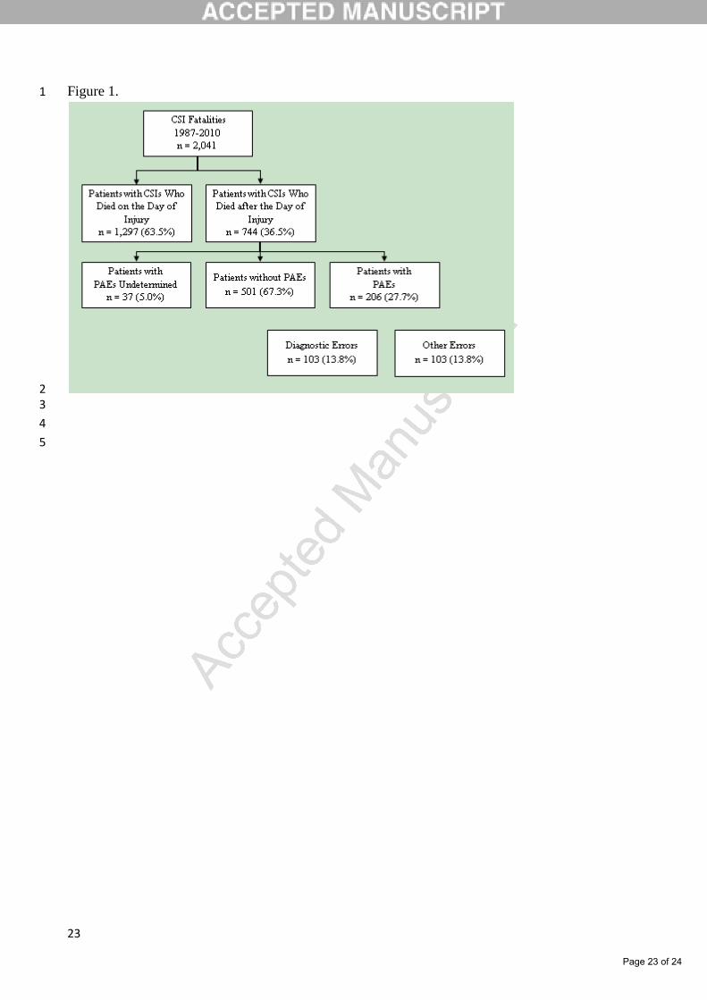
23
Figure 1. 1
2 3
4
5
Page 23 of 24
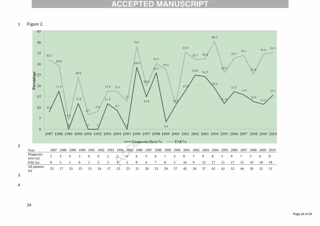
24
Figure 2. 1
2 Year 1987 1988 1989 1990 1991 1992 1993 1994 1995 1996 1997 1998 1999 2000 2001 2002 2003 2004 2005 2006 2007 2008 2009 2010
Diagnostic
error (n) 2 3 0 3 0 0 2 2 0 6 3 6 1 3 8 7 9 8 5 9 7 5 6 8
PAE (n) 8 5 1 6 1 2 3 4 3 8 4 7 8 3 16 9 12 17 11 17 15 10 18 18
All patients
(n) 25 17 23 25 15 24 17 23 23 21 20 23 29 27 45 28 37 42 42 52 44 39 52 51
3
4
Page 24 of 24
ORIGINAL ARTICLE - BRAIN INJURY
Head injuries and the risk of concurrent cervical spine fractures
Tuomo Thesleff1 & Anneli Kataja2 & Juha Öhman1& Teemu M. Luoto1
Received: 9 November 2016 /Accepted: 20 February 2017 /Published online: 3 March 2017# Springer-Verlag Wien 2017
AbstractBackground Cervical spine injuries of variable severity arecommon among patients with an acute traumatic brain injury(TBI). We hypothesised that TBI patients with positive headcomputed tomography (CT) scans would have a significantlyhigher risk of having an associated cervical spine fracturecompared to patients with negative head CT scans.Method This widely generalisable retrospective sample wasderived from 3,023 consecutive patients, who, due to an acutehead injury (HI), underwent head CT at the EmergencyDepartment of Tampere University Hospital (August 2010–July 2012). Medical records were reviewed to identify theindividuals whose cervical spine was CT-imaged within1 week after primary head CT due to a clinical suspicion ofa cervical spine injury (CSI) (n = 1,091).
Results Of the whole cranio-cervically CT-imaged sample(n = 1,091), 24.7% (n = 269) had an acute CT-positive TBI.Car accidents 22.4% (n = 244) and falls 47.8% (n = 521) werethe most frequent injury mechanisms. On cervical CT, anytype of fracture was found in 6.6% (n = 72) and dislocationand/or subluxation in 2.8% (n = 31) of the patients. The pa-tients with acute traumatic intracranial lesions had significant-ly (p = 0.04; OR = 1.689) more cervical spine fractures (9.3%,n = 25) compared to head CT-negative patients (5.7%, n = 47).On an individual cervical column level, head CT positivitywas especially related to C6 fractures (p = 0.031,OR = 2.769). Patients with cervical spine fractures (n = 72)had altogether 101 fractured vertebrae, which were most oftenC2 (22.8, n = 23), C7 (19.8%, n = 20) and C6 (16.8%, n = 17).Conclusions Head trauma patients with acute intracranial le-sions on CT have a higher risk for cervical spine fractures incomparison to patients with a CT-negative head injury.Although statistically significant, the difference in fracture ratewas small. However, based on these results, we suggest thatcervical spine fractures should be acknowledged when treatingCT-positive TBIs.
Keywords Brain injuries . Computed tomography . Headinjury . Spinal injuries . Cervical spine fractures
Introduction
The evaluation of the cervical spine among emergency traumapatients is challenging and is frequently complicated byextracervical injuries and other comorbid conditions [1].Undetected cervical spine injuries (CSIs) may have cata-strophic consequences, leading to serious neurological impair-ment or even death [22].
Parts of this work were presented as a poster at the 16th EuropeanCongress of Neurosurgery in Athens, Greece, in September 2016 andNational Neurotrauma Symposium in Santa Fe, New Mexico, USA, inJune 2015.
* Tuomo [email protected]
Anneli [email protected]
Juha Ö[email protected]
Teemu M. [email protected]
1 Department of Neuroscience and Rehabilitation, Tampere UniversityHospital, P.O. Box 2000, FI-33521 Tampere, Finland
2 Medical Imaging Centre, Department of Radiology, TampereUniversity Hospital, P.O. Box 2000, FI-33521 Tampere, Finland
Acta Neurochir (2017) 159:907–914DOI 10.1007/s00701-017-3133-0
According to the National Emergency X-RadiographyUtilization Study (NEXUS), a clinically significant CSI canbe excluded without imaging if none of the following fivecriteria are fulfilled: (1) focal neurological deficits, (2) midlinespinal tenderness, (3) altered level of consciousness, (4) intox-ication or (5) distracting injury [11]. However, in many clini-cal settings, these criteria are not operable. Patients with headinjury (HI) and/or traumatic brain injury (TBI) comprise thelargest group of patients seen in emergency departments,where clinical examination alone is not sufficient to rule outCSI. The rule of thumb is that all patients with HIs should betreated as if a concomitant CSI is present until proven other-wise. Inmultitrauma patients, a whole-body computed tomog-raphy (CT) from head to pelvis should be obtained on arrivalat the emergency department (ED) [3, 9, 16, 21]. However,patients with less serious or isolated HIs comprise the largestgroup of patients when it comes to the dilemma of cervicalspine clearance. It remains rather unclear which HI patientsshould undergo routine cervical spine CT imaging. Excessiveand liberal use of CT raises direct medical costs and increasesthe risk of radiation-induced malignancies [17].
The incidence of CSI in HI patients has been reported torange from 4 to 8% [3, 4, 10, 18, 20, 26]. To what extent headtrauma severity is associated with concomitant CSIs is contro-versial and depends largely upon the study methods and pop-ulation studied. Vahldiek et al. [25] did not find an associationbetween positive head CTs and CSIs in their study that com-prised only low-energy injuries. Several studies have includedonly CT-positive or unconscious HI patients [2, 8, 12, 23, 24].Moreover, many studies have been large multicentre registryor databank studies in which the details of CSIs and HIs havebeen limited [5, 8, 20]. In general, several factors in traumapatients have been reported to associate with CSI; for exam-ple, age, lowered Glasgow Coma Scale score (GCS), injurymechanism, facial fracture and hypotension [8, 12, 20, 24].Table 1 summarises the central original publications related tothe topic and their key findings.
We hypothesised that HI patients with positive head CTscans would have a significantly higher risk of having a cer-vical spine fracture than HI patients with negative head CTscans. Due to the suboptimal accuracy of CT in detectingvertebral ligament injuries, our study is focused on fractures.
Materials and methods
Study design and setting
This study is a part of the Tampere Traumatic Head and BrainInjury Study. The patient sample in the registry includes allconsecutive patients (n = 3,023) with HI who underwent headCT at the Tampere University Hospital’s ED between August2010 and July 2012. The patients were prospectively enrolled
from the ED and the data were retrospectively recorded. In thestudy, the minimum diagnostic criteria for TBI were based onthe WHO Neurotrauma Task Force recommendation [13].Referral criteria for acute head CT were based on the formerScandinavian guidelines for initial management of minimal,mild and moderate head injuries [14]. Ethics approval (code,R10027) for the study was obtained from the ethical commit-tee of the Pirkanmaa Hospital District in Tampere, Finland.
Clinical data
Data collected from the registry included subject- andinjury-related data, clinical information from the ED,and data on neurosurgical interventions. Also, the mech-anisms of injury and time intervals (injury—ED admis-s ion—head CT—ED discharge) were recorded .Destination after the ED was categorised into four groups:home, hospital ward, local health centre or death. In ret-rospect, the medical records of all these patients(n = 3,023) were carefully reviewed to select those indi-viduals whose cervical spine was CT-imaged due to aclinical suspicion of a CSI within 1 week after primaryhead CT. Cervical CT was performed primarily accordingto the NEXUS recommendations [11]. On arrival,multitrauma patients underwent whole-body CT (compris-ing cervical spine) according to international recommen-dations [3, 9, 16, 21]. A total of 1,091 (36.1%) cervicalspine CT-imaged patients were identified and includedinto the current study. The majority of the patients(96.5%, n = 1,053) were cervically CT-imaged within24 h after primary head CT and the rest in 1–4 days’ time.Of the CSI patients, the presence of possible spinal cordinjury and radiculopathy, as well as the American SpinalInjury Association (ASIA) scores, were recorded [15].Medical records from a period of 1 year post-injury werereviewed to collect information on possible cervical spinesurgery due to the index injury.
Imaging data
In the ED, an emergency non-contrast head CT scan was per-formed as per Scandinavian guidelines for all patients, using a64-row CT scanner (Lightspeed VCT; GE, Wisconsin, USA)[14]. In a non-on-call setting, all head CTscans were analysedand systematically coded by two neuroradiologists using astructured data collection form. Acute traumatic intracraniallesions included subdural haematoma and effusion (SDH),epidural haematoma and effusion (EDH), diffuse axonal inju-ry (DAI) lesions, oedema, compression of the cerebrospinalfluid spaces, midline shift, contusions, pneumocephalus, skullfracture and traumatic subarachnoid haemorrhage.
Cervical CT imaging was performed with the same scanneras for the head CTs. CSI was defined as a fracture or
908 Acta Neurochir (2017) 159:907–914

subluxation of any of the cervical vertebrae.Whiplash injurieswithout radiological findings were not included in the analy-sis. The injured cervical spine level, including occipital
condyle (C0) fracture, together with a detailed anatomic de-scription of each vertebra and CT-detectible ligament injury,was recorded systematically by the first author (T.T.). On
Table 1 List of relevant publications on association of head injury and cervical spine injury
Publication Study type Study population Key findings
Bayless et al. [2] Single centre, retrospective 228 significant blunt headtrauma patients
Only 1.7% of the patients with a significant blunthead trauma had a CSI.
Fujii et al. [5] National trauma databank 550,313 trauma cases Incidence of CSI in TBI patients was 8.6%. CSIincidence was significantly higher among TBIpatients than among other trauma patients.
Gbaanador et al. [6] Trauma centre, retrospective 406 patients with HI CSI occurred in only 1.2% of HI cases. Acutecervical radiography was not efficacious andshould not be routinely used in the emergencymanagement of head trauma.
Hasler et al. [8] Multicentre trauma registry 250,584 major trauma patients Incidence of CSI in all trauma patients was 3.5%.Patients with lowered GCS or systolic bloodpressure, severe facial fractures, dangerousinjury mechanism, male gender and/or\ age ≥35 years have an increased risk for CSI.HI was not an independent predictor of CSI.
Hills et al. [10]a Single centre 8,285 blunt trauma patients CSI occurred in 4.5% of HI patients. Patientswith clinically significant head injury were atgreater risk for CSI. Patients with a GCS ≤ 8were at even greater risk (7.8%).
Holly et al. [12] 2 centres, retrospective 447 consecutive moderate-severehead trauma patients
Incidence of CSI in head trauma patients was5.4%. GCS ≤ 8 or motor vehicle accident wererisk factors for CSI.
Michael et al. [18] Single centre, retrospective 359 patients with HI and 92 patients with CSI CSI occurred in 6% of head injured patients.Coincidence of head injury and CSI incomatose patients was estimated 2.4%. Allseriously head injured patients should betreated as having concomitant CSI untilproven otherwise.
Milby et al. [19] Review article 281,864 trauma patients CSI occurred in 3.7% of all trauma patients andin 7.7% of unevaluable patients (distractingpainful injury, intoxication or concomitantHI).
Mulligan et al. [20] Databank 1.3 million trauma patients CSI occurred in 7.0% of head injuries. Aneffective identification protocol for CSI in caseof HI is proposed.
Soicher et al. [23] Single centre, prospective 260 patients from falls or traffic accidentswith a significant HI
CSI occurred in 3.5% of significant HI patients.No association between severity of HI and theincidence of CSI.
Tian et al. [24] Single centre, prospective 1,026 comatose TBI patients Incidence of CSI in comatose TBI patients was6.9%. Patients with a low GCS, motorcycleaccident as the mechanism of injury andwith askull base fracture had an increased risk forCSI.
Vahldiek et al. [25] 3 centres, retrospective 1,342 minor blunt trauma patients No association between HI and CSI. Only onepatient had combined craniocervical injury.
Williams et al. [26] Single centre, retrospective 5,021 trauma patients CSI occurred in 4.8% of HI patients. Nosignificant difference in CSI incidencebetween HI and non-HI patients. GCS < 14associated with CSI in HI and non-HI patients.
TBI traumatic brain injury, CSI cervical spine injury, HI head injury, GCS Glasgow Coma Scalea No full text available
Acta Neurochir (2017) 159:907–914 909
clinical basis, magnetic resonance imaging was performed onthe patients with spinal cord injury, but their results were notanalysed in this study.
Statistical analyses
The normality of the variable distributions was tested usingthe Kolmogorov-Smirnov and Shapiro-Wilk tests.Continuous variables were analysed with the Pearson (normaldistribution) and Spearman (skewed distribution) correlationcoefficients. Group comparisons were tested with theStudent’s t-test (normal distribution) and the Mann–WhitneyU test (skewed distribution).
Results
Characteristics of the study sample
The main clinical characteristics of the study sample (n = 1,091)are presented in Table 2 and CSI-related characteristics inTable 3. For comparison, the characteristics of the patients withpositive head CT scans (n= 269) are presented parallel with thepatients with negative head CT scans (n= 822) and the wholestudy sample (n = 1,091). Of all the patients (n = 1,091), 607(55.6%) fulfilled the clinical criteria for mild TBI, 201 (18.4%)had amoderate or severe TBI and the rest (n= 283, 25.9%) had ahead trauma without clear signs of TBI. Cervical spinal cordinjury was found in 13 (16.9%) of the CSI patients. In one pa-tient, the existence of a possible spinal cord injury remainedunknown due to early death caused by a severe TBI.
Cervical CT findings
On cervical CT, CSI was found in 7.1% (n = 77) and cervicalspine fracture in 6.6% (n = 72) of the patients (n = 1,091). Intotal, these patients sustained 101 fractured vertebrae. Fivepatients (0.5%) had a CSI with only a CT-detectible ligamentinjury. Of the patients with CSI, 31 (40.3%) had dislocationsand/or subluxations. The mean dislocation was 3 mm (range,1–10 mm). The distribution of fractures and dislocations/subluxations is presented in Fig. 1.
Cervical spine fractures in patients with CT-positiveversus CT-negative head injuries
Patients with CT-positive acute traumatic intracranial lesions(9.3%, n = 25) had significantly more cervical spine fractures(Pearson chi-squared, p = 0.04; OR = 1.689, 95% CI = 1.019–2.802) compared with head CT-negative HI patients (5.7%,n = 47). Interestingly, patients with positive head CT scans hadfewer spinal cord injuries (0.4%, n = 1 compared with 1.5%,n = 12; statistical comparison not done due to small group sizes).
Moreover, positive head CT findings had significant associationwith C6 vertebra fractures (3.0%, n = 8 vs 1.1%, n = 9; p = 0.031,OR= 2.769, 95% CI = 1.057–7.250), but not with other cervicalvertebra fractures (C0-5 and C7) or dislocations/subluxationsalone. The distribution of fractures between the upper (C0-C2)and lower (C3-C7) cervical spine did not differ between the twogroups. Patients with positive head CT scans (2.2%, n= 6) didnot have more neurosurgical operations within 1 year post-HIdue to cervical spine fractures compared with patients with neg-ative head CT scans (2.8%, n = 23; p = 0.614).
Risk of cervical spine fractures in relation to neurologicalpicture and mechanism of injury
Patients with cervical spine fractures had more frequently mod-erate to severe TBIs (22.2%, n = 16 vs 18.2%, n= 185) but thedifference did not reach statistical significance (p = 0.376). Also,GCS scores (p = 0.464) and number of patients with loss ofconsciousness (15.3%, n = 11 vs 23.7%, n = 241, p = 0.264)were similar between the groups. However, data on GCS andloss of consciousness were frequently missing (30.6%, n= 334;and 42.4%, n= 463, respectively).
The gender (males 73.6%, n = 53 vs 64.9%, n = 661,p = 0.132) and mechanism of injury (p = 0.352) distributionswere not related to cervical spine fractures. Patients with cer-vical fractures were hospitalised longer (median 96.4 h vs21.3 h, p < 0.001) and underwent more neurosurgical proce-dures (19.4%, n = 37 vs 7.9%, n = 81, p < 0.001) than the pa-tients without fractures.
Discussion
For clinicians working in the ED setting, detecting patientswith a possible CSI is of the utmost importance. Acute HI isone of the most common causes of ED admissions, and thefailure to detect CSI, especially in patients with a decreasedlevel of consciousness, can have catastrophic consequences.We found that HI patients with acute intracranial lesions onCT had almost a twofold risk of cervical spine fractures incomparison to CT-negative head trauma patients. In particular,C6 fractures were associated with head CT positivity. Becauseof the suboptimal accuracy of CT in detecting ligament inju-ries, we concentrated mainly on fractures in the risk analysis.Compared with prior studies, our results might have superiorgeneralisability as our sample is non-selected and consists ofHIs ranging in severity from minimal to severe.
In contrast to our results, there are several reports in theliterature stating that HI is not an independent risk factor forCSI [6, 8, 23, 25, 26]. Some of the incoherence between thereported coincidence of TBI and CSI may derive from thedifferences in the study populations and methods used in dif-ferent studies. For example, Hasler et al. [8] studied an
910 Acta Neurochir (2017) 159:907–914
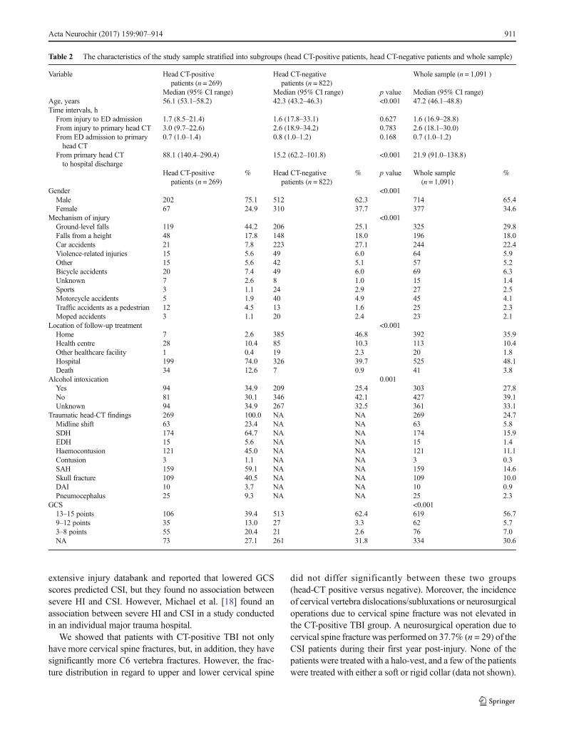
extensive injury databank and reported that lowered GCSscores predicted CSI, but they found no association betweensevere HI and CSI. However, Michael et al. [18] found anassociation between severe HI and CSI in a study conductedin an individual major trauma hospital.
We showed that patients with CT-positive TBI not onlyhave more cervical spine fractures, but, in addition, they havesignificantly more C6 vertebra fractures. However, the frac-ture distribution in regard to upper and lower cervical spine
did not differ significantly between these two groups(head-CT positive versus negative). Moreover, the incidenceof cervical vertebra dislocations/subluxations or neurosurgicaloperations due to cervical spine fracture was not elevated inthe CT-positive TBI group. A neurosurgical operation due tocervical spine fracture was performed on 37.7% (n = 29) of theCSI patients during their first year post-injury. None of thepatients were treated with a halo-vest, and a few of the patientswere treated with either a soft or rigid collar (data not shown).
Table 2 The characteristics of the study sample stratified into subgroups (head CT-positive patients, head CT-negative patients and whole sample)
Variable Head CT-positivepatients (n = 269)
Head CT-negativepatients (n = 822)
Whole sample (n = 1,091 )
Median (95% CI range) Median (95% CI range) p value Median (95% CI range)Age, years 56.1 (53.1–58.2) 42.3 (43.2–46.3) <0.001 47.2 (46.1–48.8)Time intervals, hFrom injury to ED admission 1.7 (8.5–21.4) 1.6 (17.8–33.1) 0.627 1.6 (16.9–28.8)From injury to primary head CT 3.0 (9.7–22.6) 2.6 (18.9–34.2) 0.783 2.6 (18.1–30.0)From ED admission to primaryhead CT
0.7 (1.0–1.4) 0.8 (1.0–1.2) 0.168 0.7 (1.0–1.2)
From primary head CTto hospital discharge
88.1 (140.4–290.4) 15.2 (62.2–101.8) <0.001 21.9 (91.0–138.8)
Head CT-positivepatients (n = 269)
% Head CT-negativepatients (n = 822)
% p value Whole sample(n = 1,091)
%
Gender <0.001Male 202 75.1 512 62.3 714 65.4Female 67 24.9 310 37.7 377 34.6
Mechanism of injury <0.001Ground-level falls 119 44.2 206 25.1 325 29.8Falls from a height 48 17.8 148 18.0 196 18.0Car accidents 21 7.8 223 27.1 244 22.4Violence-related injuries 15 5.6 49 6.0 64 5.9Other 15 5.6 42 5.1 57 5.2Bicycle accidents 20 7.4 49 6.0 69 6.3Unknown 7 2.6 8 1.0 15 1.4Sports 3 1.1 24 2.9 27 2.5Motorcycle accidents 5 1.9 40 4.9 45 4.1Traffic accidents as a pedestrian 12 4.5 13 1.6 25 2.3Moped accidents 3 1.1 20 2.4 23 2.1
Location of follow-up treatment <0.001Home 7 2.6 385 46.8 392 35.9Health centre 28 10.4 85 10.3 113 10.4Other healthcare facility 1 0.4 19 2.3 20 1.8Hospital 199 74.0 326 39.7 525 48.1Death 34 12.6 7 0.9 41 3.8
Alcohol intoxication 0.001Yes 94 34.9 209 25.4 303 27.8No 81 30.1 346 42.1 427 39.1Unknown 94 34.9 267 32.5 361 33.1
Traumatic head-CT findings 269 100.0 NA NA 269 24.7Midline shift 63 23.4 NA NA 63 5.8SDH 174 64.7 NA NA 174 15.9EDH 15 5.6 NA NA 15 1.4Haemocontusion 121 45.0 NA NA 121 11.1Contusion 3 1.1 NA NA 3 0.3SAH 159 59.1 NA NA 159 14.6Skull fracture 109 40.5 NA NA 109 10.0DAI 10 3.7 NA NA 10 0.9Pneumocephalus 25 9.3 NA NA 25 2.3
GCS <0.00113–15 points 106 39.4 513 62.4 619 56.79–12 points 35 13.0 27 3.3 62 5.73–8 points 55 20.4 21 2.6 76 7.0NA 73 27.1 261 31.8 334 30.6
Acta Neurochir (2017) 159:907–914 911
In the light of our findings, it can be speculated that althoughhead CT positivity is associated with an increased risk ofcervical spine fractures, these fractures are equally severe clin-ically as measured by the need for surgery. Interestingly, therewere also fewer cervical spinal cord injuries in CT-positiveTBI patients. This may be due to differences in injury mech-anisms. Injury mechanism among patients with a spinal cordinjury was remarkably often a simple ground-level fall (n = 6,46.2%), whereas among patients without a spinal cord injuryground-level falls accounted for only 29.6% (n = 319) of thecases. Moreover, patients with a spinal cord injury were older(median age, 65.9 vs 47.0 years) than patients without a spinalcord injury. It may be speculated that falling accidents of el-derly people are more likely to cause sole cervical spine inju-ries than CT-positive craniocervical injuries. In a previousstudy of more than 5,000 trauma patients, Williams et al.[26] found a negative association between TBI and spinal cordinjury, which is in line with our findings.
In order to identify risk factors for cervical spine fractures,we compared patients with a fracture to patients who did nothave a fracture. In contrast to the majority of the prior litera-ture, we could not find an association between neurologicalstatus and fracture risk [8, 12, 24, 26]. However, the amount ofmissing data on the GCS and loss of consciousness was
considerably high and prevents drawing a reliable conclusionin this regard. Injury mechanisms were also somewhat similarbetween the groups. Ground level falls accounted for aboutone-third of the cases in both groups and car accidents weremore frequent in the fracture group (30.6%, n = 22 vs 21.8%,n = 222). However, the difference in injury mechanisms wasnot statistically significant. Nevertheless, it is widely acceptedthat high-energy injury mechanism increases the risk of cervi-cal spine fractures.
Our results on the distribution of cervical spine fractureswere mostly in concordance with the previous reports on trau-ma patients, although the number of studies focusing on HIpatients is scarce [7]. In our study, 30.6% (n = 22) of the cer-vical fracture patients had a multilevel injury. C2 (includingodontoid) was the most commonly fractured vertebraconsisting 22.8% (n = 23) of all (n = 101) fractured individualvertebrae. Subaxial fractures (n = 64) consisted 63.4% of allfractured vertebrae. Vertebrae C6 (n = 17, 16.8%) and C7(n = 20, 19.8%) were the most commonly fractured subaxialvertebrae, whereas C3 was the vertebra least likely to be in-jured (n = 6, 5.9%). Dislocations and subluxations occurredmost frequently in C1/C2 (n = 9, 29%) and C4/C5 (n = 7,22.6%) levels. In the study by Goldberg et al. [7] on the dis-tribution and patterns of blunt CSIs, the findings were slightly
Table 3 The CSI characteristics of the study sample stratified into subgroups (head CT-positive patients, head CT-negative patients and whole sample)
Variable Head CT-positivepatients (n = 269)
% Head CT-negativepatients (n = 822)
% p value Whole sample(n = 1,091)
%
Cervical spine surgery within 1 year post injury 6 2.2 23 2.8 0.614 29 2.7
Cervical spine fracture 25 9.3 47 5.7 0.04 72 6.6
Cervical dislocation/subluxation 8 3.0 23 2.8 0.880 31 2.8
Number of fractured cervical vertebrae 35 13.0 66 8.0 0.014 101 9.3
C0 2 0.7 2 0.2 0.431 4 0.4
C1 5 1.9 5 0.6 0.062 10 0.9
C2 (odontoid) 5 1.9 8 1.0 0.245 13 1.2
C2 (non-odontoid) 5 1.9 11 1.3 0.538 16 1.5
C3 2 0.7 4 0.5 0.621 6 0.5
C4 1 0.4 8 1.0 0.344 9 0.8
C5 4 1.5 8 1.0 0.483 12 1.1
C6 8 3.0 9 1.1 0.031 17 1.6
C7 5 1.9 15 1.8 0.971 20 1.8
Multilevel cervical spine fracture (>1 levels) 9 3.3 13 1.6 0.074 22 2.0
Patients with lower level cervical fracture (C3-C7) 16 5.9 31 3.8 0.127 47 4.3
Patients with upper level cervical fracture (C0-C2) 11 4.1 19 2.3 0.122 30 2.7
Spinal cord injury 1 0.4 12 1.5 13 1.2
ASIA scale 0.490
A 1 0.4 2 0.2 3 0.3
B 0 0 4 0.5 4 0.4
C 0 0 5 0.6 5 0.5
D 0 0 1 0.1 1 0.1
E or unknown 268 99.6 810 98.5 1078 98.8
912 Acta Neurochir (2017) 159:907–914

different compared to our results. In line with our results,Goldberg et al. reported C2 to be the most commonly frac-tured cervical vertebra. On the contrary, they found C6 to bethe most common site of subaxial fracture, whereas in ourseries the most common site was C7, followed by C6 frac-tures. Different study populations (blunt trauma in the studyby Goldberg et al. vs HI patients in our study) and differentinjury coding habits may explain the small differences in thefracture distribution.
Our study represents a large population-based HI sample thatis commonly seen in EDs internationally. The sample includes awide severity spectrum of HIs that were treated at the ED atvarying time delays post-injury. Although not unique, our find-ings support the prior studies on the association between TBI andCSI [3, 10, 12, 18]. However, the main finding of our study(increased risk of cervical spine fractures among headCT-positive TBI patients) must be interpreted with caution giventhe subtle difference in fracture rate that is only slightly statisti-cally significant (p = 0.04). The main limitation of our study isthe retrospective nature, and therefore the number ofmissing data(e.g. GCS and alcohol consumption) is relatively large, whichmay influence the main conclusions. Secondly, head-CT criteriain minimal, mild and moderate HIs in the ED were based on theScandinavian guidelines. Apparently, some patients in the sam-ple did not fulfil the Scandinavian CT criteria and wereCT-imaged without solid indications. In contrast, possibly some
HI patients did not undergo CT imaging although they met thecriteria. Thirdly, cervical ligament, and soft tissue injuries mayhave beenmissed since patients were not systematically assessedwith MRI. However, the main emphasis of this study was onfractures. Most probably, clinically significant CSIs were notmissed, since our study included patients whose cervical spinewas CT-imaged up to 1 week post-injury and the care of CSIs inthe study catchment area is centred to our hospital.
Conclusions
Head trauma patients with acute intracranial lesions on CThave a higher risk for cervical spine fractures in comparisonto patients with a CT-negative head injury. However, thesefractures did not cause more spinal cord injuries or requiremore neurosurgical spinal intervention compared to headCT-negative cervical spine fractures. Although statisticallysignificant, the difference in fracture rate is small (9.3% vs5.7%). Nevertheless, cervical spine fracture should be ac-knowledged when treating CT-positive TBI patients. CT im-aging of the cervical spine in the case of CT-positive TBIshould be considered based on these findings.
Acknowledgements The authors wish to thank nurse Anne Simi forher assistance in data collection.
Compliance with ethical standards
Conflict of interest All authors certify that they have no affiliationswith or involvement in any organisation or entity with any financialinterest (such as honoraria; educational grants; participation in speakers’bureaus; membership, employment, consultancies, stock ownership, orother equity interest; and expert testimony or patent-licensing arrange-ments), or non-financial interest (such as personal or professional rela-tionships, affiliations, knowledge or beliefs) in the subject matter or ma-terials discussed in this manuscript.
Funding No funding was received for this work.
Ethical approval For this type of study formal consent is not required.
References
1. Anderson PA, Muchow RD, Munoz A, Tontz WL, Resnick DK(2010) Clearance of the asymptomatic cervical spine: a meta-anal-ysis. J Orthop Trauma 24:100–106
2. Bayless P, Ray VG (1989) Incidence of cervical spine injuries inassociation with blunt head trauma. Am J Emerg Med 7:139–142
3. Blackmore CC, Emerson SS, Mann FA, Koepsell TD (1999)Cervical spine imaging in patients with trauma: determination offracture risk to optimize use. Radiology 211:759–765
4. Drainer EK, Graham CA, Munro PT (2003) Blunt cervical spineinjuries in Scotland 1995–2000. Injury 34:330–333
4
10
23
6
9
12
17
20
0
5
10
15
20
25
Num
ber
of f
ract
ured
ver
tebr
ae(n
=101
)
Level of fractured vertebra
1
9
3
1
7
5
3
2
0
1
2
3
4
5
6
7
8
9
10
Num
ber
of d
islo
cati
ons/
subl
uxat
ions
(n=3
1)
Level of dislocations/subluxations
a
b
Fig. 1 a The distribution of cervical spine fractures. b The distribution ofcervical spine dislocations/subluxations
Acta Neurochir (2017) 159:907–914 913
5. Fujii T, Faul M, Sasser S (2013) Risk factors for cervical spineinjury among patients with traumatic brain injury. J EmergTrauma Shock 6:252–258
6. GbaanadorGB, FruinAH, TaylonC (1986) Role of routine emergencycervical radiography in head trauma. Am J Surg 152:643–648
7. Goldberg W, Mueller C, Panacek E, Tigges S, Hoffman JR, MowerWR, NEXUS Group (2001) Distribution and patterns of blunt trau-matic cervical spine injury. Ann Emerg Med 38:17–21
8. Hasler RM, Exadaktylos AK, Bouamra O, Benneker LM, ClancyM, Sieber R, Zimmermann H, Lecky F (2012) Epidemiology andpredictors of cervical spine injury in adult major trauma patients: amulticenter cohort study. J Trauma Acute Care Surg 72:975–981
9. Hessmann MH, Hofmann A, Kreitner KF, Lott C, Rommens PM(2006) The benefit of multislice CT in the emergency room man-agement of polytraumatized patients. Acta Chir Belg 106:500–507
10. Hills MW, Deane SA (1993) Head injury and facial injury: is therean increased risk of cervical spine injury? J Trauma 34:549–553,discussion 553–4
11. Hoffman JR, Wolfson AB, Todd K, Mower WR (1998) Selectivecervical spine radiography in blunt trauma: methodology of thenational emergency X-radiography utilization study (NEXUS).Ann Emerg Med 32:461–469
12. Holly LT, Kelly DF, Counelis GJ, Blinman T, McArthur DL, CryerHG (2002) Cervical spine trauma associated with moderate andsevere head injury: incidence, risk factors, and injury characteris-tics. J Neurosurg 96:285–291
13. Holm L, Cassidy JD, Carroll LJ, Borg J, Neurotrauma Task Force onMild Traumatic Brain Injury of the WHOCollaborating Centre (2005)Summary of the WHO collaborating centre for neurotrauma task forceon mild traumatic brain injury. J Rehabil Med 37:137–141
14. Ingebrigtsen T, Romner B, Kock-Jensen C (2000) Scandinavian guide-lines for initial management of minimal, mild, and moderate head inju-ries. The scandinavian neurotrauma committee. J Trauma 48:760–766
15. Kirshblum SC, Burns SP, Biering-Sorensen F, Donovan W, GravesDE, Jha A, Johansen M, Jones L, Krassioukov A, Mulcahey MJ,Schmidt-ReadM,WaringW (2011) International standards for neu-rological classification of spinal cord injury (revised 2011). J SpinalCord Med 34:535–546
16. Linsenmaier U, Krotz M, Hauser H, Rock C, Rieger J, Bohndorf K,Pfeifer KJ, Reiser M (2002) Whole-body computed tomography inpolytrauma: techniques and management. Eur Radiol 12:1728–1740
17. Mathews JD, Forsythe AV, Brady Z, Butler MW, Goergen SK,Byrnes GB, Giles GG, Wallace AB, Anderson PR, Guiver TA,McGale P, Cain TM, Dowty JG, Bickerstaffe AC, Darby SC(2013) Cancer risk in 680,000 people exposed to computed tomog-raphy scans in childhood or adolescence: data linkage study of 11million Australians. BMJ 346:f2360
18. Michael DB, Guyot DR, DarmodyWR (1989) Coincidence of headand cervical spine injury. J Neurotrauma 6:177–189
19. Milby AH, Halpern CH, Guo W, Stein SC (2008) Prevalence ofcervical spinal injury in trauma. Neurosurg Focus 25:E10
20. Mulligan RP, Friedman JA, Mahabir RC (2010) A nationwide re-view of the associations among cervical spine injuries, head inju-ries, and facial fractures. J Trauma 68:587–592
21. Schmidt OI, Gahr RH, Gosse A, Heyde CE (2009) ATLS(R) anddamage control in spine trauma. World J Emerg Surg 4:9
22. Schoenfeld AJ, Bono CM, McGuire KJ, Warholic N, Harris MB(2010) Computed tomography alone versus computed tomographyand magnetic resonance imaging in the identification of occult in-juries to the cervical spine: a meta-analysis. J Trauma 68:109–113,discussion 113–4
23. Soicher E, Demetriades D (1991) Cervical spine injuries in patientswith head injuries. Br J Surg 78:1013–1014
24. Tian HL, Guo Y, Hu J, Rong BY,Wang G, GaoWW, Chen SW, ChenH (2009) Clinical characterization of comatose patients with cervicalspine injury and traumatic brain injury. J Trauma 67:1305–1310
25. Vahldiek JL, Thieme S, Hamm B, Niehues SM (2016) Incidence ofcombined cranial and cervical spine injuries in patients with blunt mi-nor trauma: are combined CT examinations of the head and cervicalspine justified?. Acta Radiol. doi:10.1177/0284185116673120
26. Williams J, Jehle D, Cottington E, Shufflebarger C (1992) Head,facial, and clavicular trauma as a predictor of cervical-spine injury.Ann Emerg Med 21:719–722
Comments
Analysing a large 2-year emergency room cohort retrospectively, theauthors demonstrate that head injured patients with intracranial lesionson CT harbour an 1.7-fold increased risk of cervical fractures in compar-ison to CT-negative patients. The conclusion that cervical fractures shouldbe acknowledged when treating CT-positive TBIs can obviously be sup-ported and is kept in mind intuitively in general practice already. Thestudy provides further proof for this concept. However, the risk differenceof 9.3% versus 5.7% is too small to make a real difference in the ER. Thec-spine clearance rules still have to be applied. Surprisingly, the rate ofspinal cord injury was higher in the CT-negative cohort (1.5% vs 0.4%,i.e. 12 patients versus one single patient). It could be speculated that thismay be due to central cord syndromes after minor falls in the elderly andis an interesting finding.
Claudius Thome
Tirol, Austria
914 Acta Neurochir (2017) 159:907–914


![[Treating frostbite injuries]](https://static.fdokumen.com/doc/165x107/633ff39332b09e4bae09a1b5/treating-frostbite-injuries.jpg)


