Hildegard Bingeniläisen visiot aikalaiskontekstissa ja ... - Trepo
Upload
khangminh22Category
view
0download
0
������������ ����������������
�������������� ������� ������ ����������� ����������� ��������������������������� ����
A c t a U n i v e r s i t a t i s T a m p e r e n s i s 925
U n i v e r s i t y o f T a m p e r eT a m p e r e 2 0 0 3
��������� ������������������������ ������������������
������������������������������������������������
�������������������������������������������������������
�����������������������������������������
������������������������������ �����!"���"##�����!"��$�����%
�������������

� ��� ��� ��
��������� ��� ���������������� � &�'%(%� ��)� *!+��#!,� ��������� ��� ������������
-�������������&���� ����
'����� ��������� �������������������������."/0��1�./!2,,2/*,,2#0��1� !,//2!*!*
��������� �������������(�� &������ '����������"##�
���%� � 3�/4� �5"!/� *#//��)� � 3�/4� �� "!/5+*4/�6�7��%����899�����%��%��
:�������� ��������� ���:�������������������������������",/0��1�./!2,,2/*,/2.0��1�!,/*2./,;��899���%��%��
��������� � ����������� ���� ������������������ �� ���!�� ������� �� ���� ��� "��� �� # � ���
������ ���� ��$��������� ����� �� ������ ���� ��� ��� ������
�� �%��� ��$��������� $�&&�� ��� ' ���"� � �& � �� ���� ��� ��� ��!�� �(�$��������� ���� � ��� )'�� �� ���� ��� ��� "� � �&

TABLE OF CONTENTS
3
TABLE OF CONTENTS
ABSTRACT...........................................................................................................................5
ABBREVIATIONS ...............................................................................................................7
LIST OF ORIGINAL COMMUNICATIONS.......................................................................9
1. INTRODUCTION ...........................................................................................................11
2. REVIEW OF THE LITERATURE..................................................................................13
2.1 Nitric Oxide ...............................................................................................................13
2.1.1 Synthesis of NO..................................................................................................13
2.1.2 Signal transduction by NO .................................................................................15
2.1.3 NO in inflammation............................................................................................16
2.2 NO in Inflammatory Lung Diseases ..........................................................................17
2.2.1 Asthmatic inflammation .....................................................................................17
2.2.2 Inflammation in cryptogenic fibrosing alveolitis ...............................................19
2.2.3 Inflammation in extrinsic allergic alveolitis.......................................................19
2.2.4 NO in pulmonary physiology and pathophysiology...........................................20
2.2.5 NO in asthmatic inflammation ...........................................................................21
2.2.6 NO and inflammation in CFA and EAA ............................................................23
2.3 Exhaled NO Measurement.........................................................................................23
2.3.1 Basic methodology of exhaled NO measurement ..............................................25
2.3.2 International guidelines on exhaled NO measurement.......................................28
2.4 Exhaled NO Concentration in Inflammatory Lung Diseases.....................................29
2.4.1 Exhaled NO in asthma........................................................................................29
2.4.2 Exhaled NO in CFA and EAA ...........................................................................32
2.4.3 Exhaled NO in other pulmonary diseases...........................................................33
2.5 Mathematical Modelling of Pulmonary NO Dynamics .............................................33
3. AIMS OF THE STUDY...................................................................................................39
4. SUBJECTS AND METHODS.........................................................................................41
4.1 Subjects and Study Protocols.....................................................................................41
4.1.1 Two-compartment model in alveolar and bronchial inflammation (I, II) ..........41
4.1.2 Effect of treatment on alveolar and bronchial NO output in asthma and alveolitis
(II, III)..........................................................................................................................42
4.1.3 Relation of alveolar and bronchial NO output to nocturnal symptoms of asthma
(IV) ..............................................................................................................................42
4.1.4 Alveolar and bronchial NO output in patients with asthmatic symptoms but
normal lung function (V).............................................................................................43
4.2 Methods .....................................................................................................................43
4.2.1 Exhaled NO measurement..................................................................................43
4.2.2 Lung function .....................................................................................................45
4.2.3 Allergy testing ....................................................................................................45
4.2.4 Inflammatory markers in blood and urine ..........................................................45
4.2.5 Asthmatic symptoms ..........................................................................................46
4.2.6 Statistics..............................................................................................................46
5. SUMMARY OF RESULTS.............................................................................................47
5.1 Flow Dependency of Exhaled NO Concentration .....................................................47
5.2 Alveolar and Bronchial NO Output in Asthma and Alveolitis ..................................48
5.3 The Effect of Treatment on Alveolar and Bronchial NO Output in Asthma and
Alveolitis..........................................................................................................................49

TABLE OF CONTENTS
4
5.4 Relation of Alveolar and Bronchial NO Output to Nocturnal Symptoms in Patients
with Asthma .....................................................................................................................51
5.5 Alveolar and Bronchial NO Output in Patients with Asthmatic Symptoms but
Normal Lung Function.....................................................................................................52
6. DISCUSSION ..................................................................................................................55
6.1 Flow Rate Dependency of Exhaled NO Concentration .............................................55
6.2 Critique of the Two-Compartment Model .................................................................56
6.3 Exhaled NO Measurement at Multiple Flow Rates in Assessing Alveolar and
Bronchial Inflammation ...................................................................................................58
6.3.1 Alveolar and bronchial NO in asthma ................................................................58
6.3.2 Alveolar and bronchial NO in alveolitis .............................................................60
6.3.3 Responsiveness of alveolar and bronchial NO output to alleviation of
inflammation................................................................................................................62
6.3.4 Alveolar and bronchial inflammation in patients with asthmatic symptoms but
normal lung function ...................................................................................................63
6.3.5 Critique of the Present Method...........................................................................63
6.4 Single or Multiple Flow Rates in Exhaled NO Measurement?..................................64
7. SUMMARY AND CONCLUSIONS...............................................................................69
ACKNOWLEDGEMENTS .................................................................................................71
REFERENCES.....................................................................................................................73
APPENDIX 1 .......................................................................................................................91
ORIGINAL COMMUNICATIONS.....................................................................................93

ABSTRACT
5
ABSTRACT
Chronic inflammation is a key feature in
many lung diseases, for example asthma.
Currently, the diagnosis and follow-up of
inflammatory lung diseases are based largely
on radiography and measurements of lung
function. However, these methods do not
measure the inflammatory process but
secondary changes caused by it. Direct
measures of inflammation would therefore
provide more precise information on the
underlying pathogenetic process and the
activity of the disease.
Nitric oxide (NO) is an endogenous
signalling molecule in the lungs, produced in
small amounts in the physiological state. In
inflammatory lung diseases, pulmonary NO
production increases and inflammation can
be detected based on an increased
concentration of NO in exhaled breath.
Exhaled NO concentration is usually
measured at repeated exhalations at a single
low flow rate. This method can be used to
detect pulmonary inflammation, but it
cannot discriminate between alveolar and
bronchial contributions to exhaled NO, and
therefore cannot differentiate between
alveolar and bronchial components in the
inflammatory process. Mathematical models
of pulmonary NO dynamics suggest that the
alveolar and bronchial contributions to
exhaled NO concentration can be calculated
on the basis of exhaled NO measurements at
multiple different exhalation flow rates. The
aim of the present study was to establish
whether assessment of alveolar and
bronchial NO output could be used to
differentiate between alveolar and bronchial
inflammation.
Alveolar NO concentration and
bronchial NO flux were calculated in
steroid-naïve patients with asthma, patients
with alveolitis, and healthy subjects.
Asthmatics had increased bronchial NO flux
but on average normal alveolar NO
concentration, whereas patients with
alveolitis showed increased alveolar NO
concentration but normal bronchial NO flux.
In asthma and alveolitis, bronchial NO flux
and alveolar NO concentration, respectively,
correlated with markers of disease severity.
Anti-inflammatory treatment reduced the
bronchial NO flux in asthma and the
alveolar NO concentration in alveolitis
simultaneously with improvement of the
disease state. Alveolar NO concentration
was also increased in asthmatics with
nocturnal symptoms, which is consistent
with the peripheral inflammation detected in
nocturnal asthma. Alveolar and bronchial
NO output were also increased in subjects
presenting with asthmatic symptoms but not
fulfilling the lung function criteria for
asthma.
It is concluded that assessment of
alveolar and bronchial NO output by
measuring exhaled NO concentration at
multiple exhalation flow rates can be used to
differentiate between the alveolar and
bronchial components of pulmonary
inflammation. Exhaled NO measurement at
multiple flow rates provides more detailed
information on the inflammatory process
than the currently recommended single flow
rate method.

6

ABBREVIATIONS
7
ABBREVIATIONS
ANOVA Analysis of variance BAL Bronchoalveolar lavage CAlv Steady state nitric oxide concentration in alveolar air [ppb] CFA Cryptogenic fibrosing alveolitis cGMP Cyclic guanosine monophosphate cNOS Constitutive nitric oxide synthase COPD Chronic obstructive pulmonary disease CW Nitric oxide concentration in bronchial wall tissue [ppb] DNO,Alv Alveolar diffusing capacity of nitric oxide [nl/s/ppb] DNO,Br Bronchial wall diffusing capacity of nitric oxide [nl/s/ppb] DLCO Pulmonary diffusing capacity of carbon monoxide [ml/s/mmHg] DRS Dose-response slope EAA Extrinsic allergic alveolitis ECP Eosinophil cationic protein eNOS Endothelial nitric oxide synthase EPX Eosinophil protein X FASSc Fibrosing alveolitis associated with systemic sclerosis FEF50% Forced expiratory flow when 50 % of VC has been exhaled [l/s] FEF75% Forced expiratory flow when 75 % of VC has been exhaled [l/s] FEV1 Forced expiratory volume in 1 second [l] FVC Forced vital capacity [l] HRCT High-resolution computed tomography hɜ Energy of electromagnetic radiation, where h is Planck’s coefficient
and ɜ is the frequency of the radiation IgE Immunoglobulin E IL-1 Interleukin 1 IL-6 Interleukin 6 iNOS Inducible nitric oxide synthase JNO,Br Maximal diffusion rate of NO from bronchial wall to luminal air,
bronchial NO flux [nl/s] JNO,Alv Diffusion rate of NO from alveolar tissue to alveolar air during steady
state [nl/s] LTE4 Leukotriene E4
MPO Myeloperoxidase NF-kB Nuclear factor kBnNOS Neuronal nitric oxide synthase NO Nitric oxide NO2 Nitrogen dioxide O2 Molecular oxygen O3 Ozone p Probability value PD20FEV1 Provocative dose causing a 20 % decrease in FEV1
PEF Peak expiratory flow [l/min] r Pearson’s correlation coefficient SEM Standard error of mean TNF-a Tumour necrosis factor aUIP Usual interstitial pneumonia VC Vital capacity [l]
NOV# Total nitric oxide output from lower respiratory tract [nl/s] V# Exhalation flow rate [l/s]

8

LIST OF ORIGINAL COMMUNICATIONS
9
LIST OF ORIGINAL COMMUNICATIONS
This thesis is based on the following original communications, referred to in the text by their
Roman numerals (I – V). In addition, some unpublished data are presented.
I. Lehtimäki L, Turjanmaa V, Kankaanranta H, Saarelainen S, Hahtola P and Moilanen E
(2000). Increased bronchial nitric oxide production in patients with asthma measured
with a novel method of different exhalation flow rates. Annals of Medicine, 32: 417-423.
II. Lehtimäki L, Kankaanranta H, Saarelainen S, Hahtola P, Järvenpää R, Koivula T,
Turjanmaa V and Moilanen E (2001). Extended exhaled NO measurement differentiates
between alveolar and bronchial inflammation. American Journal of Respiratory and
Critical Care Medicine, 163: 1557-1561.
III. Lehtimäki L, Kankaanranta H, Saarelainen S, Turjanmaa V and Moilanen E (2001).
Inhaled fluticasone decreases bronchial but not alveolar nitric oxide output in asthma.
European Respiratory Journal, 18: 635-639.
IV. Lehtimäki L, Kankaanranta H, Saarelainen S, Turjanmaa V and Moilanen E (2002).
Increased alveolar nitric oxide concentration in asthmatic patients with nocturnal
symptoms. European Respiratory Journal, 20: 841-845.
V. Lehtimäki L, Kankaanranta H, Saarelainen S, Turjanmaa V and Moilanen E. Peripheral
inflammation in patients with asthmatic symptoms but normal lung function. Submitted
for publication.

10

INTRODUCTION
11
1. INTRODUCTION
Inflammation is an important host response
elicited by tissue injury or invading
pathogens. Although inflammation is a vital
defence response, e.g. during infection,
chronic inflammation also has many
detrimental effects. Inflammation is a key
feature in the pathogenesis of many lung
diseases such as asthma, chronic obstructive
pulmonary disease (COPD), extrinsic
allergic alveolitis (EAA) and cryptogenic
fibrosing alveolitis (CFA). The prevalence
of asthma and COPD is increasing, and
these diseases constitute a major burden to
patients and health care organisations in
many countries. Inflammatory lung diseases
are nowadays mainly diagnosed using
radiography and indicating impaired
pulmonary function, a secondary change
caused by chronic inflammation. There is,
however, a clear need for widely accessible
direct measures of pulmonary inflammation
to allow more precise diagnosis and follow-
up of inflammatory lung diseases. Such
measures would also facilitate the
assessment of the efficacy of anti-
inflammatory treatment. During recent
years, considerable interest has focused on
developing novel non-invasive methods to
measure pulmonary inflammation through
analysis of inflammatory markers in exhaled
breath.
Nitric oxide (NO) is an endogenous
signalling molecule produced in the human
body. In the physiological state, low
amounts of NO are produced for regulatory
purposes. In inflammation, on the other
hand, NO production increases and induces
cytotoxic effects against both invading
organisms and host cells. A small fraction of
the NO synthesised in the lungs diffuses into
the pulmonary air and endogenous NO can
be detected in exhaled air. In inflammatory
lung diseases, pulmonary NO production
increases considerably and the inflammatory
process can be detected on the basis of the
increased NO concentration in exhaled air.
The interest in exhaled NO measurement as
a non-invasive measure of inflammation has
been intense, and exhaled NO measurement
is now considered as a promising novel tool
in the diagnosis and follow-up of various
inflammatory lung diseases. The aim of the
present study was to further develop exhaled
NO measurement by using a mathematical
model of pulmonary NO dynamics, which
provides a theoretical means to assess
alveolar and bronchial components in
pulmonary inflammation separately.

12

REVIEW OF THE LITERATURE
13
2. REVIEW OF THE LITERATURE
2.1 Nitric Oxide
Nitric oxide (NO) is a small diatomic
molecule formed by nitrogen and oxygen
atoms covalently bound to each other. It is a
colourless gas at room temperature and
pressure. NO has one unpaired electron and
is thus a highly reactive molecule.
NO was first discovered as an
endogenous signalling molecule in 1987,
when the endothelium-derived relaxing
factor, an important vasodilatory signalling
molecule, was revealed to be NO (Ignarro et
al. 1987, Palmer et al. 1987). Since then, this
small gaseous molecule has been found to
play a crucial role in human physiology and
pathophysiology. NO is an important
regulator of blood pressure and platelet
aggregation, it acts as a neurotransmitter in
both central and peripheral nervous systems
and regulates immune responses (Moncada
et al. 1991, Christopherson and Bredt 1997,
Marin and Rodriguez-Martinez 1997,
Moilanen et al. 1999, Bogdan 2001). In
1998, the Nobel Prize for Physiology or
Medicine was awarded to three
pharmacologists, Robert R. Furchgott, Louis
J. Ignarro and Ferid Murad, for their
fundamental work in exploring the role of
NO in cardiovascular physiology.
2.1.1 Synthesis of NO
NO is synthesised from amino acid L-
arginine by a family of nitric oxide synthase
(NOS) enzymes. The reaction catalysed by
NO synthases requires oxygen and NADPH
to convert L-arginine into NO and the amino
acid citrulline. NO synthases are dimeric
haeme containing enzymes composed of
oxygenase and reductase domains which
possess binding sites for flavin adenine
dinucleotide (FAD), flavin mononucleotide
(FMN), calmodulin (CaM) and
tetrahydrobiopterin (BH4) (Janssens et al.
1992, Lowenstein et al. 1992, Knowles and
Moncada 1994, Alderton et al. 2001) (Figure
1).
Three NOS isoforms encoded by distinct
genes are known to date, namely neuronal
NOS (nNOS), inducible NOS (iNOS) and
endothelial NOS (eNOS). Although
originally found in neuronal and endothelial
cells, nNOS and eNOS are expressed in a
variety of cell types throughout the human
body (Föstermann et al. 1998, Alderton et al.
2001).
nNOS and eNOS are so-called
constitutive NO synthases (cNOS), i.e. they
are expressed in several cell types in
response to physiological stimuli, and they
are thought to produce the fairly low
amounts of NO needed in different
physiological processes (Kleinert et al.
2000). After expression, the enzymatic
activity of the constitutive NO synthases is
strictly regulated by the intra-cellular Ca2+
concentration (Bredt and Snyder 1990,
Busse and Mulsch 1990, Alderton et al.
2001). eNOS- and nNOS-dependent NO
production in endothelial and neural cells is
activated by signalling molecules such as
acetylcholine, bradykinin and glutamate,
which increase the intracellular Ca2+
concentration through receptor-associated

REVIEW OF THE LITERATURE
14
mechanisms (Johns et al. 1987, Garthwaite
et al. 1988, Christopherson and Bredt 1997,
Marin and Rodriguez-Martinez 1997).
By contrast, iNOS is not typically
present in the physiological state. Its
expression is induced in many cell types by
bacterial products (e.g. endotoxin) and
inflammatory cytokines such as IL-1, IFN-gor TNF-a (Kleinert et al. 2000, Alderton et
al. 2001). Once expressed, iNOS produces
NO for prolonged periods. iNOS produces
NO in much higher quantities as compared
with cNOS (Kleinert et al. 2000).
iNOS differs from the constitutive
enzyme forms also in that its expression is
suppressed by anti-inflammatory treatment
with glucocorticoids, whereas cNOSs are
not steroid-sensitive (Radomski et al. 1990).
Glucocorticoids have been shown to reduce
iNOS expression by inhibiting NF-kB, an
important transcription factor for iNOS
(Kleinert et al. 1996), and by undermining
the stability of iNOS mRNA (Korhonen et
al. 2002).
A variety of different NOS inhibitors
have been developed and used as research
tools to extend our knowledge of the
physiological role of NO. Most of these
inhibitors bind at the arginine-binding site of
the enzyme and act competitively (Alderton
et al. 2001). Some of the inhibitors, for
example L-NMMA (NG-monomethyl-L-
arginine), inhibit all NOS isoforms with
similar potency, whereas others are isoform-
specific and provide possibilities to study
the role of different isoforms. iNOS-specific
inhibitors, e.g. L-NIL (L-N6-(1-
iminoethyl)lysine), are widely used to study
the role of iNOS-dependent high NO
production in inflammatory and other
conditions.
Figure 1. Nitric oxide synthase, its cofactors and the overall reaction catalysed. Electrons are donated
by NADPH to the reductase domain of the enzyme and proceed via FAD and FMN to the oxygenase
domain. There they interact with the haem iron and BH4 at the active site to catalyse the reaction of
oxygen with L-arginine, generating citrulline and NO as products. Electron flow through the reductase
domain requires the presence of bound Ca2+
/ calmodulin (Alderton et al. 2001).
Calmodulin
Oxygenase
Reductase
BH4
FAD
FMN
Fe
NADPH
Arginine
O2
Citrulline
NO
e-
e-
e-
Ca2+
NADP
H
+
+
Nitric oxide synthase

REVIEW OF THE LITERATURE
15
2.1.2 Signal transduction by NO
Unlike the classical intercellular signalling
molecules, NO has no specific receptor
mediating its action. Being a small gaseous
molecule it diffuses easily through cell
membranes and alters cell function by
affecting cellular proteins. Signal
transduction by NO can be divided into
direct and indirect mechanisms. In direct
effects, NO itself reacts with the key
molecules to mediate the biological
function. In indirect effects, on the other
hand, NO first reacts with other molecules
like oxygen or superoxide to form
compounds such as peroxynitrite, which in
turn react with the target molecules
(Grisham et al. 1999). At low physiological
concentrations, the direct mechanisms of
NO signalling predominate. In states of
inflammation, where iNOS is expressed and
high amounts of NO are produced, indirect
effects are thought to take over (Miranda et
al. 2000) (Figure 2).
NO reacts directly with many enzymes,
and the most important pathway in its
physiology is the activation of guanylate
cyclase (Arnold et al. 1977, Grisham et al.
1999). Activation of guanylate cyclase leads
to increased cGMP production in target
cells, mediating e.g. relaxation of smooth
muscle in blood vessels and airways and
inhibition of platelet aggregation (Furlong et
al. 1987, Ignarro et al. 1987, Palmer et al.
1987, Pohl and Busse 1989). NO also reacts
avidly with oxyhaemoglobin, forming nitrate
and methaemoglobin (Doyle and Hoekstra
1981, Miranda et al. 2000). This is an
important inactivation route of NO in the
body.
Figure 2. Direct and indirect effects of NO. Modified from Grisham et al. (1999) and Miranda et al.
(2000). O2-, superoxide; N2O3, dinitrogen trioxide; ONOO
-, peroxynitrite.
NOHigh [NO]
(iNOS)Low [NO](cNOS)
Direct effects
Reactions with metal complexes
Radical reactions
(primarily associated with physiological
regulation)
e.g. activation of guanylate cyclase and
cyclooxygenase
e.g. inhibition of lipid peroxidation
Indirect effects(associated with
pathophysiological effects, immune response and cytotoxicity)
Nitrosation, oxidation and
nitratione.g. formation of nitrotyrosine and
nitrosamines, DNA breaks and lipid
peroxidation
O or O2 2
-
N O and ONOO2 3
-
REVIEW OF THE LITERATURE
16
NO reacts with molecular oxygen (O2)
and superoxide anion (O2-) to form reactive
nitrogen species such as dinitrogen trioxide
(N2O3) and peroxynitrite (ONOO-), which
mediate the indirect effects of NO.
Formation of peroxynitrite is abundant in
inflammatory states associated with an
increased production of superoxide anion.
Dinitrogen trioxide causes nitrosation of
amines and thiols, whereas peroxynitrite
causes nitration of e.g. tyrosine residues.
Peroxynitrite is also a powerful oxidant
which initiates lipid peroxidation and
cleaves DNA (Pryor and Squadrito 1995,
Beckman and Koppenol 1996, Wink et al.
1997). NO in small concentrations regulates
mitochondrial respiration by reversibly
inhibiting cytochrome oxidase, the terminal
complex of the mitochondrial electron
transport chain (Brown and Cooper 1994,
Cleeter et al. 1994). At higher NO
concentrations peroxynitrite is formed, and
irreversibly inhibits several complexes of the
respiratory chain and therefore hampers
mitochondrial respiration (Lizasoain et al.
1996). This may play a significant role in
causing tissue damage in disease states such
as ischemia-reperfusion injury and
endotoxin-induced or haemorrhagic shock
(Liaudet et al. 2000). These indirect effects
are thought to mediate most of the
detrimental and cytotoxic effects of NO in
inflammation (Grisham et al. 1999, Miranda
et al. 2000).
2.1.3 NO in inflammation
Nitric oxide plays a dual role in the
inflammatory process. Constitutive low NO
production exerts many protective effects
against inflammatory changes, whereas the
increased NO and peroxynitrite production
following upon iNOS expression is
associated with cytotoxicity and potentiation
of many detrimental events.
Constitutive NO production by eNOS
has a protective role against
microcirculatory damage and oedema
formation during the early phase of
inflammation. After a few hours iNOS is
expressed in vascular cells. High amounts of
NO produced by iNOS exacerbate oedema
and iNOS inhibition has been shown to
reduce microvascular leakage in
inflammation (Boughton-Smith et al. 1993,
Laszlo et al. 1994, Moilanen et al. 1999).
NO also exerts anti-inflammatory effects
by reducing leukocyte extravasation.
Endogenous NO production or NO-releasing
drugs reduce the expression of adhesion
molecules in endothelial cells and thereby
suppress leukocyte adhesion to the
endothelium and their extravasation (Kubes
et al. 1991, Kosonen et al. 2000).
NO suppresses lymphocyte proliferation
in a GMP-independent manner (Albina et al.
1991, Kosonen et al. 1997, Kosonen et al.
1998a). It might also participate in
regulation of the Th1/Th2 lymphocyte
balance, as it inhibits IL-2 and IFN-gsecretion by Th1 cells but has no effect on
the cytokine production of Th2 cells (Taylor-
Robinson et al. 1994). NO would therefore
favour the Th2 response and might
participate in the development of atopic
diseases.
Inflammatory cytokines and microbial
products induce iNOS expression in
macrophages (Lyons et al. 1992, Xie et al.
1992, Bogdan 2001, Thomassen and Kavuru
2001). The high amount of NO produced is
REVIEW OF THE LITERATURE
17
thought to play an important role in
cytostatic-cytotoxic effects of macrophages
against tumour cells and microbes
(MacMicking et al. 1997, Hibbs et al. 1988).
Induction of iNOS expression is better
known in rodent than in human
macrophages, but the latter also have been
reported to express iNOS after a variety of
stimuli (Moilanen et al. 1997, Thomassen
and Kavuru 2001). Human alveolar
macrophages express iNOS in patients with
cryptogenic fibrosing alveolitis (Saleh et al.
1997) or after induction by e.g.
mycobacteria (Nicholson et al. 1996, Nozaki
et al. 1997).
The high NO level prevailing in
inflammation are thought to exert cytotoxic
effects not only against invading
microorganisms but also against host cells.
Many of these aspects are mediated by
peroxynitrite and N2O3 formed by reactions
between NO and superoxide or O2. These
compounds cause oxidative stress and
nitrosylation of many important host
proteins, leading e.g. to inhibition of cellular
respiration and cell necrosis (Szabo 2000).
NO can also modulate the inflammatory
response by altering the activity of other
mediator pathways. NO reduces leukotriene
synthesis by inhibiting 5-lipoxygenase
(Moilanen et al. 1993, Kanner et al. 1992,
Maccarrone et al. 1996, Brunn et al. 1997).
NO has been reported either to inhibit or
activate cyclo-oxygenase (COX), which
converts arachidonic acid into prostanoids
(Salvemini et al. 1993, Kosonen et al.
1998b, Stadler et al. 1993). The effect of NO
on COX activity seems to be concentration-
dependent such that low NO amounts
activate COX while higher levels inhibit
COX activity (Liaudet et al. 2000). NO
might thus have important regulatory effects
on inflammation in affecting the activity of
these lipid mediator pathways.
2.2 NO in Inflammatory Lung
Diseases
Inflammation is an essential host response to
a variety of insults. It constitutes a host
defence against microorganisms and is also
necessary to tissue repair after mechanical or
chemical injury. Inflammation involves a
complex interplay between structural and
inflammatory cells and includes a wide
variety of mediators needed for inter-cell
signalling (Cotran et al. 1999).
Chronic inflammation also has
detrimental effects and constitutes a key
feature in many pulmonary diseases.
Sustained inflammation may cause
functional and structural changes such as
airway hyperresponsiveness, airway wall
thickening, alveolar wall destruction and
parenchymal fibrosis (Keane et al. 2000).
Early suppression of the inflammatory
process is believed to prevent these injuries.
2.2.1 Asthmatic inflammation
Asthma is defined as a chronic inflammatory
disorder of the airways in which many cells
and cellular elements play a role, in
particular, mast cells, eosinophils, T
lymphocytes, macrophages, neutrophils, and
epithelial cells (National Heart Lung and
Blood Institute 1997). The complex
inflammatory process underlying asthma can
be divided into fairly distinct components,
which are responsible for the different

REVIEW OF THE LITERATURE
18
clinical characteristics of the disease. These
are the acute IgE-mediated allergic reaction,
chronic inflammation, and airway
remodelling (Bousquet et al. 2000).
In sensitised subjects, inhaled allergens
bind to specific IgE molecules on the cell
surface receptors of different inflammatory
cells, particularly mast cells, which become
activated and release proinflammatory
substances such as histamine and
eicosanoids. These substances lead to
bronchoconstriction, mucus secretion and
vasodilatation, and may provoke an acute
asthmatic attack. In the so-called late-phase
reaction 6 to 9 hours after the allergen
challenge, many inflammatory cell types are
recruited into the airways. These include
eosinophils, CD4+ T cells, basophils,
macrophages and neutrophils (Boushey et al.
2000, Bousquet et al. 2000).
In addition to allergic reactions, chronic
airway inflammation is a key feature in
asthma. Eosinophils and Th2 cells play a
major role in this condition (Boushey et al.
2000, Bousquet et al. 2000), but also
neutrophils are present in great numbers
especially in the severe, steroid-resistant
form (Wenzel et al. 1997, Jatakanon et al.
1999b). Chronic inflammation causes non-
specific airway hyperresponsiveness and
structural changes such as an increase in
smooth muscle mass and number of mucous
glands, epithelial shedding and thickening of
the basement membrane (Jeffery 1998,
Bousquet et al. 2000). It is thought that early
intervention with anti-inflammatory
treatment turns off the chronic inflammatory
Figure 3. Asthmatic inflammation is a complex interplay of various inflammatory cells and signalling
molecules (Boushey et al. 2000). GM-CSF, granulocyte-macrophage colony-stimulating factor; LTB4,
leukotriene B4; MBP, major basic protein; PGD2, prostaglandin D2; RANTES, regulated on activation,
normal T cell expressed and secreted.
Allergen
Chemotaxis
Eosinophil
Th2
B cell
Mast cell
Epithelium
Antigen-presenting cell
IgE
GM-CSFIL-3IL-5
MBPECP
RANTESGM-CSF
IL-8
LTB
IL-84
IL-4IL-13
HistaminePGD
Cysteinyl-LTs2
Bronchoconstriction

REVIEW OF THE LITERATURE
19
process and thereby prevents permanent
structural changes in asthmatic airways.
Asthmatic inflammation appears to
affect the whole lower respiratory tract.
Although asthma has classically been
thought to affect mainly the large airways,
there are data suggesting the presence of
inflammation also in the small peripheral
airways and alveolar tissue in stable chronic
asthma (Kraft et al. 1996). Furthermore,
night-time enhancement of alveolar
inflammation has been associated with
nocturnal asthma (Martin et al. 1991, Kraft
et al. 1996, Kraft et al. 1999).
2.2.2 Inflammation in cryptogenic
fibrosing alveolitis
Cryptogenic fibrosing alveolitis (CFA), as
the name implies, is an inflammatory and
fibrosing condition of unknown origin
affecting the pulmonary parenchyma. Its
pathophysiology involves a combination of
tissue damage, inflammation and excess
fibrosis. Although the initial damaging agent
is not known, several predisposing factors
have been suggested. These include a
number of organic and inorganic dusts, viral
factors and smoking (American Thoracic
Society and European Respiratory Society
2000).
The inflammatory cells involved in the
pathogenesis of CFA include macrophages,
neutrophils, eosinophils and lymphocytes.
Fibroblasts and myofibroblasts are
responsible for producing the excess
collagen characteristic of the disease.
Especially macrophages, but also other
inflammatory cells, are capable of producing
cytokines which can increase collagen
synthesis and induce proliferation of
fibroblasts. Inflammatory cells also secrete
collagenases and thereby regulate collagen
turnover. The imbalance between synthesis
and degradation of collagen resulting from
altered secretion of collagenases and
profibrotic substances is considered a central
factor underlying the pathogenesis of CFA
(Chan et al. 1998, King 1998).
The histopathological features of the
disease vary between cellular and fibrotic
patterns, where inflammatory and fibrotic
changes, respectively, are the major
characteristics at microscopic level. The
cellular pattern is usually associated with
better prognosis and response to drug
treatment (American Thoracic Society and
European Respiratory Society 2000).
2.2.3 Inflammation in extrinsic
allergic alveolitis
Extrinsic allergic alveolitis (EAA) is an
inflammatory reaction caused by an
immunological response to inhaled organic
dusts. The inflammation in this condition
affects alveolar, bronchiolar and
peribronchiolar tissue. There are many
names for EAA depending on the offending
antigen and the setting in which the
exposure takes place. The most common of
these is farmers’ lung, caused by exposure to
mouldy hay, straw or grain in agriculture.
The inhaled allergens are spores of
thermophilic actinomycetes, particularly
Micropolyspora faeni and
Thermoactinomyces vulgaris. Other
examples of EAA are bird fanciers’ lung,
caused by inhalation of pigeon serum
proteins in their bloom, and malt workers’

REVIEW OF THE LITERATURE
20
lung caused by inhalation of spores from
mouldy malts. However, the
histopathological features of EAA are the
same regardless of the causative antigen
(Selman 1998).
The characteristic pathological findings
in EAA are peribronchiolar granulomatous
inflammation and bronchiolocentric
alveolitis, characterised by an accumulation
of plasma cells, T lymphocytes and
macrophages in the inflammatory exudates
(Selman 1998). The immunopathogenesis is
thought to be driven by immunocomplexes
formed by the antigen and specific IgG
molecules. Activated T lymphocytes are
thought to play a key role in the formation of
granulomas. However, neither of these is
specific to EAA, as both increased T cell
counts in BAL and circulating specific IgG
can be found in exposed subjects without
active EAA, and it is not known what are the
triggering factors needed to cause EAA in
exposed subjects (Selman 1998, Rose 2000).
EAA is usually a benign disease as
compared with CFA. If the offending
antigen is avoided, the granulomas and
inflammation resolve. However, with
prolonged exposure, fibrosis and irreversible
changes may evolve in the peripheral lung
(Selman 1998).
2.2.4 NO in pulmonary physiology
and pathophysiology
All three isoforms of NOS have been
detected in the human lung (Hamid et al.
1993, Kobzik et al. 1993, Asano et al. 1994).
Although iNOS expression is usually
associated with inflammatory states, some
“constitutive” iNOS expression seems to be
present in the airway epithelium of healthy
subjects (Saleh et al. 1998). This might
reflect the continuous inhalation of irritant
factors which induce iNOS expression, as in
ex vivo cultures airway epithelial cells from
healthy subjects soon lose iNOS expression
(Guo et al. 1995).
NO and NO-releasing compounds relax
bronchial smooth muscle in a cGMP-
dependent manner and cause
bronchodilatation (Di Maria et al. 2000). NO
acts as an endogenous neurotransmitter of
the so-called non-adrenergic non-cholinergic
nerves (NANC-nerves) which relax smooth
muscle in the bronchial tree (Belvisi et al.
1992). NO is an important endogenous
molecule counteracting bronchoconstriction
in the human airways. Inhalation of NOS
inhibitor considerably increases bronchial
responsiveness to inhaled bradykinin or
methacholine in asthma (Sterk et al. 1999),
whereas inhalation of a high NO
concentration after such a provocation
attenuates bronchoconstriction (Högman et
al. 1993, Kacmarek et al. 1996).
Constitutive NO synthesis is an
important factor maintaining microvascular
integrity in the bronchial circulation.
However, increased NO production in
inflammation dilates the arterioles and
increases blood flow to leaky post-capillary
venules and thus exacerbates airway oedema
(Bernareggi et al. 1997). NO is also an
important vasodilating mediator in the
pulmonary circulation, and inhaled NO in
high concentrations can be used to improve
pulmonary blood oxygenation and to treat
pulmonary hypertension (Hurford et al.
2000).
NO has a dual role in bronchial mucus
secretion. Low constitutive NO production

REVIEW OF THE LITERATURE
21
inhibits this process (Ramnarine et al. 1996),
whereas increased NO production in
inflammation enhances it (Adler et al. 1995,
Barnes 1998). NO is thought also to regulate
mucociliary clearance, as NOS inhibitors
have been reported to reduce ciliary beat
frequency after stimulation by bradykinin or
substance P (Jain et al. 1993).
High levels of NO in inflammation are
thought to have cytotoxic effects on the
bronchial epithelium, and this might cause
epithelial shedding found e.g. in asthma
(Barnes 1998). These effects are likely to be
mediated by peroxynitrite, which is formed
by NO reacting with the superoxide
produced in high amounts during
inflammation.
2.2.5 NO in asthmatic inflammation
As compared with healthy subjects,
increased iNOS expression has been
reported in bronchial biopsies from
asthmatic subjects (Hamid et al. 1993, Saleh
et al. 1998, Redington et al. 2001).
Furthermore, iNOS expression can be
reduced by treatment with glucocorticoids
(Saleh et al. 1998, Redington et al. 2001).
iNOS expression in asthmatic airways has
been shown in epithelial cells, macrophages,
eosinophils, neutrophils, vascular
endothelium and smooth muscle cells
(Hamid et al. 1993, Saleh et al. 1998, ten
Hacken et al. 2000, Redington et al. 2001).
The increased amount of NO in the exhaled
air of asthmatic subjects is largely produced
by the iNOS pathway, as inhalation or an
oral dose of selective iNOS inhibitors causes
a significant reduction in exhaled NO level
(Yates et al. 1996, Erin et al. 2002).
High NO production by iNOS is thought
to have many detrimental effects in
asthmatic airways, for example oedema
formation by reduced vascular integrity,
epithelial cell toxicity and increased
hyperresponsiveness caused by peroxynitrite
(Heiss et al. 1994, Curran 1996, Sadeghi-
Hashjin et al. 1996).
NO has been found to inhibit
proliferation of murine Th1 lymphocytes and
their secretion of IL-2 and IFN-g. By
contrast, no effect on Th2 lymphocytes was
evidenced (Taylor-Robinson et al. 1994). It
has thus been suggested that NO plays a role
in regulating the balance between Th1 and
Th2 lymphocytes also in humans. By
inhibiting the Th1 cell response, NO would
favour the Th2 response and secretion of IL-
4 and IL-5, further leading to IgE production
and eosinophil recruitment into the airways
(Barnes and Liew 1995). However, studies
assessing Th1 / Th2 response in murine cells
may not be conclusive for human
lymphocytes, as there are considerable inter-
species differences in the regulation of the
immune response.
The role of NO in asthmatic
inflammation and airway
hyperresponsiveness has been studied in
animal models of asthma using either NOS
inhibitors or NOS knockout mice (Folkerts
et al. 2001). The non-selective NOS
inhibitor L-NMMA and the partially cNOS-
selective L-NAME reduce experimentally
induced airway hyperresponsiveness and
eosinophilic airway inflammation in
ovalbumin-induced asthma models in guinea
pigs (Iijima et al. 1998), rats (Ferreira et al.
1998) and mice (Feder et al. 1997, Trifilieff
et al. 2000), although contradictory results
have also been presented (Mehta et al. 1997,

REVIEW OF THE LITERATURE
22
Tulic et al. 2000). Selective iNOS inhibitors
have been used to determine the specific role
of iNOS-derived NO in these ovalbumin-
induced asthma models. Aminoguanidine
reduced airway hyperresponsiveness in
guinea pigs (Iijima et al. 1998) and
eosinophilic airway inflammation in both
rats (Tulic et al. 2000) and mice (Feder et al.
1997). The more selective iNOS inhibitors
L-NIL and 1400W had no effect on
eosinophilic inflammation in mice or rats
but reduced airway hyperresponsiveness
(Feder et al. 1997, Muijsers et al. 2001,
Eynott et al. 2002).
Knockout mice of each NOS isoform
have been studied in an ovalbumin-induced
asthma model. Surprisingly, nNOS knockout
mice showed lower airway responsiveness
than wild-type controls (De Sanctis et al.
1999). iNOS knock-outs were reported to
have either reduced or similar eosinophilic
airway inflammation as in wild-type
controls, but no change was found in airway
responsiveness (De Sanctis et al. 1999,
Xiong et al. 1999).
The conflicting results from these animal
studies might reflect problems with the
specificity of NOS inhibitors, or differences
between the asthma models used. Although
these animal studies give in vivo information
on the role of different NOS isoforms, their
interpretation in terms of human asthma is
complicated.
In humans, inhalation of the non-
selective NOS inhibitor L-NAME has been
found to have no effect on allergen
challenge-related airway obstruction either
in the early or late asthmatic reaction
(Taylor et al. 1998). A significant decrease
in exhaled NO level was evidenced, as
expected, but whether NOS inhibition
caused changes in the recruitment of
inflammatory cells was not measured.
Recent findings also link NO to the
pathogenesis of asthma by showing a
relation between certain nNOS and eNOS
gene polymorphisms and asthma (Gao et al.
2000, Grasemann et al. 2000, Lee et al.
2000). Gao and colleagues studied variants
of nNOS exon 2 and found that a 183 bp
allele was significantly more frequently
present in asthmatics than in controls.
Grasemann and colleagues studied CA
repeat polymorphism in the exon 29 of the
nNOS gene, and found that in asthmatics the
allele with 18 repeats was significantly less
common whereas that with 17 repeats was
significantly more common than in controls.
However, the functional significance of
these polymorphisms in either exon 2 or 29
is not known and their role in the
pathogenesis of asthma cannot thus be
addressed. A defect in nNOS-dependent
bronchodilatory innervation could, in theory,
be one mechanism predisposing to
symptomatic asthma.
Lee and colleagues studied variation in
exon 4 of eNOS and found two alleles of
different lengths; allele a (573 bp) and allele
b (604 bp). Genotype b/b was significantly
more frequent in asthmatics than in controls
(Lee et al. 2000). The b/b genotype has been
shown to be associated with higher
circulating NO metabolite levels than the b/a
or a/a genotypes (Tsukada et al. 1998), but
how this might affect the pathogenesis of
asthma is not known.

REVIEW OF THE LITERATURE
23
2.2.6 NO and inflammation in CFA
and EAA
In patients with CFA, iNOS expression has
been shown by immunohistochemistry in
airway and alveolar epithelial cells,
macrophages, neutrophils, and in some cases
also in lymphocytes, vascular endothelium
and smooth muscle of airways and vessels.
Also nitrotyrosine was shown in the same
cells. Further, immunostaining for both
iNOS and nitrotyrosine was higher in CFA
patients with early to intermediate disease
stage as compared with patients having end-
stage fibrosis (Saleh et al. 1997). In patients
with CFA or fibrosing alveolitis associated
with systemic sclerosis (FASSc), activated
fibroblasts in the early disease stage showed
immunostaining for iNOS, whereas in late-
stage fibrosis with less active fibroblasts,
only little iNOS was found (Romanska et al.
2000b). Stimulation of human pulmonary
fibroblasts with cytokines induces iNOS
expression and increases the cell population,
whereas the NOS inhibitor L-NMMA
reverses the cytokine effect on cell count
(Romanska et al. 2000a). iNOS expression
and detrimental effects mediated by a high
NO production rate seem to be associated
with active inflammation and fibrogenesis of
the disease process.
Intra-tracheal bleomycin administration
is used to induce a model of pulmonary
fibrosis in rodents. Inhibition of iNOS by
aminoguanidine prevents fibrotic changes
and reduces the pulmonary content of
hydroxyproline in this model (de Rezende et
al. 2000, Giri et al. 2000), suggesting that
iNOS-dependent high NO production is
detrimental in the pathogenesis of
pulmonary fibrosis.
A recent interesting finding suggests a
protective role for constitutive NO
production in pulmonary fibrosis. In
transgenic mice overexpressing eNOS,
bleomycin administration causes
significantly less fibrosis and
hydroxyproline production than in wild-type
mice. NOS inhibition by L-NAME abolishes
the protecting effect of eNOS
overexpression (Yoshimura et al. 2002). NO
might thus have a dual role also in the
pathogenesis of pulmonary fibrosis as in
inflammation, constitutive NO production
protects from fibrotic events whereas high
iNOS-dependent NO production is
detrimental.
There are no published papers assessing
the role of NO in EAA, but one abstract
reports increased levels of nitrate in the BAL
fluid of patients with EAA (Lange et al.
2000). However, as iNOS has been detected
by immunohistochemistry in granulomas in
patients with sarcoidosis or pulmonary
tuberculosis (Moodley et al. 1999), there is
probably iNOS activation also in the
granulomas in EAA, although the
granulomas differ between these diseases.
2.3 Exhaled NO Measurement
Part of the NO produced in the lungs
diffuses into the pulmonary air. In 1991,
Gustafsson and colleagues reported that
exhaled air of rabbits, guinea pigs and
humans contains endogenously produced
NO (Gustafsson et al. 1991). They found
NO in exhaled air from all three species
although the inhaled air was free of NO.
NOS inhibitors administered to rabbits and
guinea pigs reduced exhaled NO

REVIEW OF THE LITERATURE
24
concentrations, while administration of L-
arginine increased the exhaled NO
concentration back towards normal. Based
on these results it was assumed that
pulmonary diseases associated with altered
NO production rate could be assessed by
measuring the NO concentration in exhaled
air.
This assumption was confirmed by two
study groups, who reported that patients
with asthmatic airway inflammation have
increased exhaled NO concentrations as
compared with healthy subjects (Alving et
al. 1993, Persson et al. 1994). In 1994,
Kharitonov and colleagues further reported
that steroid-naive asthmatics exhibited an
increased exhaled NO concentration, while
asthmatics treated with inhaled steroids had
normal concentrations (Kharitonov et al.
1994b).
In these early studies exhaled air was
found to contain endogenously produced
NO, whose concentration was increased in
patients with untreated asthmatic airway
inflammation, while anti-inflammatory drug
treatment of the inflammation reduced the
concentration back towards normal. These
findings made exhaled NO measurement a
promising new non-invasive method to
assess airway inflammation in pulmonary
diseases. Since then, great interest has been
shown towards exhaled NO measurement in
inflammatory lung diseases, and several
hundred scientific papers have hitherto been
published on the topic. Basic methodology,
most important results and recent progress in
exhaled NO measurement will be reviewed
in this chapter.
Figure 4. Schematic illustration of an ozone-chemiluminescence NO analyser. Red and infrared light is
emitted as NO reacts with ozone. The NO concentration in the sample gas can be calculated by
quantifying the amount of light emitted.
NO + O NO * + O3 2 2NO NO + h2 2* n
Ozonegenerator
Sampleinlet
Photomultiplier
Signal amplificationand processing
O2
Computer

REVIEW OF THE LITERATURE
25
2.3.1 Basic methodology of exhaled
NO measurement
NO analysis. The nitric oxide concentration
in exhaled air is usually measured by the
ozone-chemiluminescence method. This
approach is based on the reaction between
NO and ozone (O3) forming nitrogen dioxide
(NO2), some of which is in excited state
(NO2*). Red and infrared light (~ 640 – 3000
nm) is emitted as the excited form of
nitrogen dioxide regains its stable ground
state (NO2* NO2 + hɜ) (Hampl et al.
1996). The amount of light can be quantified
by a photomultiplier. In stable conditions,
the amount of light emitted is proportional to
the amount of NO in the specimen gas,
making ozone-chemiluminescence a precise
and sensitive method for measuring the NO
content of exhaled air (Figure 4).
Exhalation manoeuvre. In the early
studies, exhaled NO was measured during
tidal breathing or uncontrolled slow vital
capacity manoeuvres (Alving et al. 1993,
Kharitonov et al. 1994a, Kharitonov et al.
1994b). During tidal breathing, the measured
NO concentration varies considerably in the
course of the breathing cycle. Changes in
breathing pattern also cause variation in
exhaled NO levels, and more repeatable
manoeuvres were sought.
Real-time measurement of NO
concentration during a single slow
exhalation from total lung capacity is now
preferred (Kharitonov et al. 1997a,
American Thoracic Society 1999). The
initial part of the NO curve representing the
dead-space air is somewhat variable, but the
end-expiratory plateau in the NO curve
during a single exhalation is highly
repeatable. A high initial peak in the curve
may be caused by accumulation of NO in
the airways during a breath-hold before
exhalation or by a high NO level in inhaled
air (high ambient NO level or inhalation
through the nose where the NO production
Figure 5. Exhaled NO concentration curves during single exhalations after oral inhalation and nasal
inhalation. Inhalation of nasal air with high NO concentration causes an initial peak in the exhaled NO
profile, but the end-expiratory plateau is not different from that after oral inhalation. Modified from
American Thoracic Society (1999).
28
24
20
16
12
8
4
0
Exhale
d N
O c
oncentr
ation (
ppb)
After oralinhalation
After nasalinhalation
0 10 20 30 40
Time (s)
REVIEW OF THE LITERATURE
26
rate is high). However, even after such a
peak the normal end-expiratory plateau
representing the endogenous NO production
in the lower airways is reached (Silkoff et al.
1997) (Figure 5).
With the single exhalation manoeuvre,
certain important factors such as exhalation
flow rate and discarding of nasal NO need to
be taken into account (as discussed in
greater detail below). In small children who
are not able to control their breathing and
cannot produce a long steady exhalation
with a fixed flow rate, NO measurement has
to be done during tidal breathing. Manually
adjustable flow restrictors have been used in
small children to fix the exhalation flow rate
externally to the desired level during tidal
breathing (Buchvald and Bisgaard 2001).
This improves the repeatability of tidal
breathing measurements by eliminating the
variability in NO values caused by variation
in exhalation flow rate. Off-line
measurements with collection of exhaled air
into a reservoir to be transported for later
analysis allow NO measurement in subjects
away from the actual NO analyser. Off-line
measurements have a good correlation with
on-line measurements, provided that the
exhalation flow rate and sufficient
exhalation pressure are strictly controlled in
both methods (Kissoon et al. 2000, Kissoon
et al. 2002).
Exhalation flow rate. The first exhaled
NO measurements were carried out in
patients breathing normal tidal breathing or
during slow uncontrolled vital capacity
manoeuvres. In later studies, the exhalation
flow rate was found to have a major effect
on the NO concentration in exhaled air
(Byrnes et al. 1997, Högman et al. 1997,
Silkoff et al. 1997). The exhaled NO
concentration decreases with increasing flow
rates, while the total NO output increases
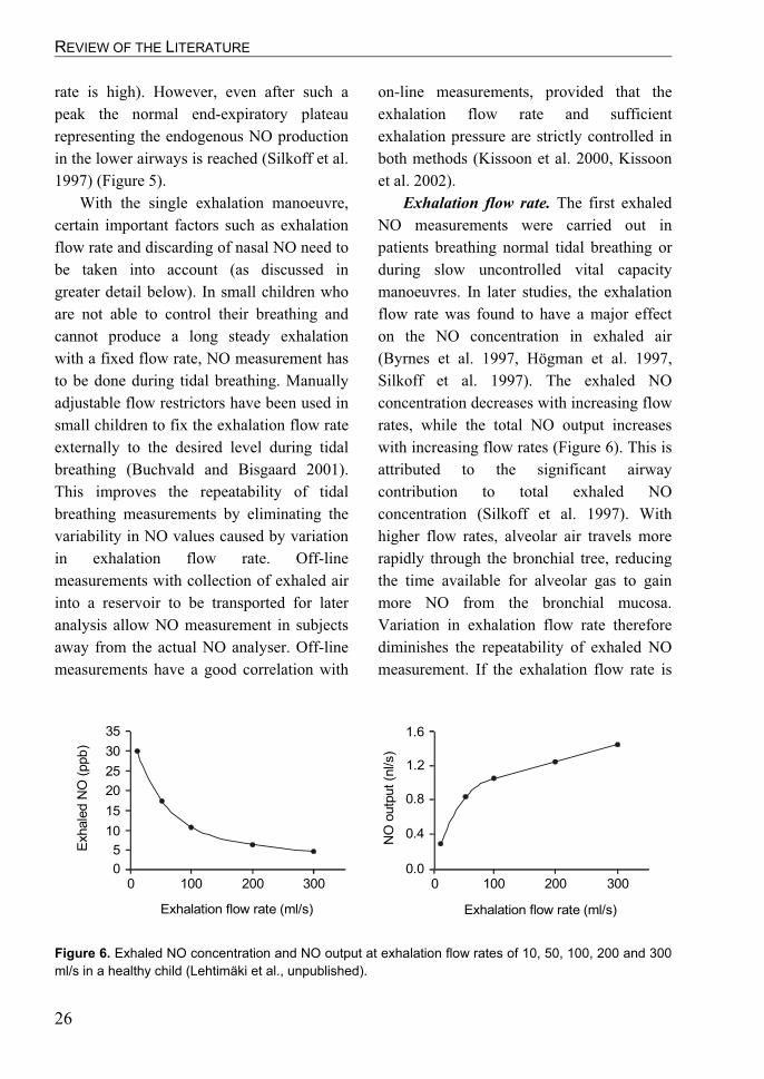
with increasing flow rates (Figure 6). This is
attributed to the significant airway
contribution to total exhaled NO
concentration (Silkoff et al. 1997). With
higher flow rates, alveolar air travels more
rapidly through the bronchial tree, reducing
the time available for alveolar gas to gain
more NO from the bronchial mucosa.
Variation in exhalation flow rate therefore
diminishes the repeatability of exhaled NO
measurement. If the exhalation flow rate is
Figure 6. Exhaled NO concentration and NO output at exhalation flow rates of 10, 50, 100, 200 and 300
ml/s in a healthy child (Lehtimäki et al., unpublished).
35
30
25
20
15
10
00 100 200 300
Exhalation flow rate (ml/s)
Exhale
d N
O (
ppb
)
0.00 100 200 300
Exhalation flow rate (ml/s)
0.4
0.8
1.2
1.6
NO
outp
ut (n
l/s)
5

REVIEW OF THE LITERATURE
27
adequately controlled, exhaled NO
measurement is highly repeatable (Silkoff et
al. 1997, Gabbay et al. 1998, Ekroos et al.
2000, Ekroos et al. 2002). ERS guidelines
from 1997 recommend a constant flow rate
from 10 to 15 l/min (167-250 ml/s)
(Kharitonov et al. 1997a), while more recent
guidelines by ERS and ATS recommend a
flow rate of 50 ml/s (American Thoracic
Society 1999, Baraldi and de Jongste 2002).
Mouth pressure. The mucosa in the
nasal cavity and paranasal sinuses produces
considerable amounts of NO (Lundberg et
al. 1995) which accumulates in high
concentrations in nasal air compared with air
from the lower respiratory tract (Kimberly et
al. 1996). When measuring exhaled NO
concentration from the lower respiratory
tract, contamination of the sample with nasal
air should be avoided. A low positive mouth
pressure (º4 cmH2O) during exhalation is
sufficient to close up the soft palate and
hence separate the nasal cavity from the oral
cavity (Kharitonov and Barnes 1997b,
Silkoff et al. 1997) (Figure 7). This ensures
that there is no leak of nasal air into the
sample air from the lower respiratory tract,
and nasal NO contamination can be avoided.
An internal resistance in the exhalation
circuit of the measurement device is
therefore recommended to obtain mouth
pressures from 5 to 20 cmH2O during
exhaled NO measurement (American
Thoracic Society 1999).
NO in ambient air. Inhalation of NO-
free air (< 5 ppb) is recommended in
exhaled NO measurement (American
Thoracic Society 1999), although it has been
shown that even high levels of ambient NO
(up to 150 ppb) (Piacentini et al. 1998) or
inhalation of a gas mixture with a high NO
concentration (up to 1 000 ppb) have no
effect on the end-expiratory NO plateau
(Silkoff et al. 1997). However, inhalation of
air with a high NO concentration causes a
high initial peak in the exhaled NO
concentration curve (Silkoff et al. 1997).
Diet. NO may be produced in the oral
cavity not only by NOS enzymes but also in
a non-enzymatic reduction from nitrite. The
Figure 7. Exhalation against a resistance during NO measurement produces a low positive mouth
pressure which closes up the soft palate and hence separates nasal air with high NO concentration from
the air coming from the lower respiratory tract. Modified from Barnes (1998).
Nasal NO
Soft palateclosed
Back pressure
Exhaled NO
Exhalation againsta resistance
NO analyser

REVIEW OF THE LITERATURE
28
salivary glands excrete nitrate from the
circulation into saliva, and bacteria in the
oral cavity convert nitrate into nitrite. In an
acidic oral environment nitrite is further
reduced to form NO. Ingestion of potassium
nitrate or a meal rich in nitrate prior to NO
measurement has been shown to increase the
exhaled NO level, whereas rinsing the
mouth with anti-bacterial or basic solution
reduces the level (Zetterquist et al. 1999,
Olin et al. 2001). It is thus recommended
that patients should refrain from eating and
drinking 1 h prior to exhaled NO
measurement (American Thoracic Society
1999), and rinsing of the mouth with basic
solution may be used to reduce the non-
enzymatic oral NO output.
Pulmonary function testing. Repeated
forced vital capacity manoeuvres with
spirometry have been shown to lower
exhaled NO levels, whereas inhaled b2-
agonists after spirometry increase the level
(Silkoff et al. 1999). Exhaled NO
measurement should thus be performed prior
to any possible measures of lung function.
Smoking. Cigarette smoking has been
found to reduce exhaled NO levels, while
discontinuation of smoking increases
exhaled NO levels gradually towards normal
levels (Persson et al. 1994, Kharitonov et al.
1995b, Robbins et al. 1996, Robbins et al.
1997). As cigarette smoke contains high
levels of NO (Borland and Higenbottam
1987), it has been suggested that
endogenous pulmonary NO production is
reduced in smokers due to the
downregulation of NOS activity by
exogenous NO (Assreuy et al. 1993,
Kharitonov et al. 1995b). Smoking has also
been shown to reduce the bioavailability of
BH4, an important cofactor in NO synthesis
(Higman et al. 1996, Heitzer et al. 2000,
Ueda et al. 2000). NO synthesis might
therefore be reduced in smokers regardless
of normal or even enhanced NOS
expression. However, exhaled breath
condensate from active smokers contains
normal levels of NO metabolites such as
nitrite and nitrate despite the lower level of
exhaled NO (Balint et al. 2001). This
suggests that the lower NO level in exhaled
air of smokers might not be explained by
decreased NO production rate but rather by
increased metabolic consumption of NO
(e.g. reacting with superoxide to form
peroxynitrite).
2.3.2 International guidelines on
exhaled NO measurement
As discussed above, a number of important
technical factors need to be taken into
account while measuring the NO
concentration in exhaled air. Most important
of these are the inverse relation between
exhalation flow rate and NO concentration
in exhaled air, and the need for a positive
mouth pressure closing up the soft palate to
avoid nasal contamination. Before these
factors were strictly controlled in earlier
studies, there was significant variation
between study groups in measurement
technique, causing conflicting results. To
overcome this problem, international
guidelines have been published to
standardise the measurement technique.
Nowadays exhaled NO concentration is
recommended to be measured during a
single breath at a constant exhalation flow
rate of 50 ml/s against a low mouth pressure
of 5 – 20 cmH2O (American Thoracic

REVIEW OF THE LITERATURE
29
Society 1999, Baraldi and de Jongste 2002).
An abnormally high exhaled NO
concentration is interpreted as a sign of
ongoing inflammation in the lower
respiratory tract.
2.4 Exhaled NO Concentration in
Inflammatory Lung Diseases
The concentration of NO in exhaled air has
been assessed in many different
inflammatory lung conditions. In most
studies, the exhaled NO concentration has
been measured at a single exhalation flow
rate. Early studies used uncontrolled slow
exhalations or tidal breathing, but the
recommended single-breath method with
counterpressure and fixed exhalation flow
rate is nowadays the approach mostly used.
The latest progress in exhaled NO
measurement technique is the use of
multiple different exhalation flow rates to
differentiate between alveolar and bronchial
NO output. The theory underlying this
method will be reviewed in the next section
(2.5 Mathematical Modelling of Pulmonary
NO Dynamics). The most important results
on exhaled NO measurement at a single
exhalation flow rate in asthma and alveolitis
are reviewed here, other lung diseases being
only briefly mentioned.
2.4.1 Exhaled NO in asthma
Asthma was the first lung disease in which
exhaled NO levels were reported. An
increased exhaled NO concentration was
found during tidal breathing in patients with
asthma in 1993 (Alving et al. 1993), and
soon after, asthmatic patients treated with
inhaled glucocorticoids were reported to
have lower peak exhaled NO values than
steroid-naïve patients (Kharitonov et al.
1994b). Increased levels of exhaled NO have
since been found in many studies utilising
the single-breath technique in both adults
and children with asthma (Horvath et al.
1998, Mattes et al. 1999, Silkoff 2000,
Kharitonov and Barnes 2001). The increased
exhaled NO concentration in asthma has
been attributed to enhanced iNOS
expression in the airway epithelium and
inflammatory cells in asthmatic airways
(Hamid et al. 1993, Saleh et al. 1998). A
considerable number of papers have been
published concerning exhaled NO
measurement in asthma, and it is the most
widely studied pulmonary disease in terms
of exhaled NO measurement.
Atopy, asthma and exhaled NO.
Exhaled NO levels are higher in atopic than
in non-atopic asthma (Frank et al. 1998, Ho
et al. 2000a). Exhaled NO levels among
asthmatics are correlated with the number of
positive skin prick tests (Ho et al. 2000a),
blood eosinophil count (Silvestri et al. 1999)
and serum levels of IgE (Simpson et al.
1999, Ho et al. 2000a). The reason for the
higher NO levels in atopic versus non-atopic
asthmatics is not known, but the finding
might be related to different inflammatory
mechanisms in these patient groups.
Increased exhaled NO levels have been
found even in atopic adults (Horvath and
Barnes 1999) and children (Franklin et al.
1999) without symptoms of asthma. This
might be related to subclinical airway
inflammation in the lower respiratory tract
without detectable changes in airway
function.

REVIEW OF THE LITERATURE
30
A challenge test with specific allergens
in atopic asthmatics does not affect the
exhaled NO concentration during early
asthmatic reaction but increases exhaled NO
levels during late asthmatic reaction in
association with decreased FEV1
(Kharitonov et al. 1995a, Piipari et al. 2002).
In atopic asthmatics, the exhaled NO
concentration increases in association with
enhanced airway inflammation during a low-
dose allergen exposure even in the absence
of changes in lung function (de Kluijver et
al. 2002). Likewise naturally occurring
allergen exposure increases exhaled NO
levels. Atopic asthmatics with significant
indoor exposure to their specific allergens
have higher exhaled NO levels than
unexposed atopic asthmatics (Simpson et al.
1999). In asthmatic children sensitised to the
house-dust mite, moving to a residential
house with low levels of mite allergen
lowers exhaled NO levels (Piacentini et al.
1999), while moving back to high allergen
exposure increases the concentration back to
high levels (Piacentini et al. 1999, Piacentini
et al. 2000). The increase in exhaled NO
upon returning to high allergen exposure can
be avoided by inhaled glucocorticoid
treatment (Piacentini et al. 2000). Atopic
asthmatics have shown higher exhaled NO
levels associated with increased symptoms
during the pollen season than outside the
season, even though no change was
evidenced in FEV1 during pollen exposure
(Baraldi et al. 1999). These findings support
the role of exhaled NO measurement in
assessing the severity of allergic airway
inflammation in atopic asthmatics.
Exhaled NO and other markers of
asthma. Either a weak (Sippel et al. 2000) or
no correlation (Horvath et al. 1998,
Jatakanon et al. 1998, Ho et al. 2000a) has
been found between exhaled NO
concentration and spirometric parameters of
lung function (FEV1) in asthma. Although
the exhaled NO concentration does not
appear to reflect fixed airflow obstruction, it
is positively correlated with diurnal PEF
variation (Al-Ali et al. 1998, Lim et al.
1999) and with bronchial
hyperresponsiveness to histamine (Dupont et
al. 1998) or methacholine (Lim et al. 1999).
However, while exhaled NO levels decrease
after treatment with inhaled glucocorticoids,
the correlation between exhaled NO and
bronchial responsiveness is also disturbed
(Dupont et al. 1998, Lim et al. 1999). This
might be explained by a more rapid response
of exhaled NO to anti-inflammatory
treatment, or that bronchial
hyperresponsiveness cannot always be
totally abolished by anti-inflammatory
treatment if airway remodelling has
occurred. Also the significant improvement
in FEV1 after inhaled b2-agonist is related to
a higher exhaled NO concentration (Sippel
et al. 2000). The baseline exhaled NO level
also correlates with the degree of exercise-
induced bronchoconstriction (Scollo et al.
2000, Terada et al. 2001). It would thus
appear that exhaled NO concentration is not
a good marker of fixed airway obstruction,
but it is related to bronchial
hyperresponsiveness and reversibility or
variability in airflow obstruction associated
with inflammatory activity.
Exhaled NO levels have also been found
to correlate with more direct measures of
asthmatic airway inflammation such as the
number of eosinophils in sputum (Jatakanon
et al. 1998, Chan-Yeung et al. 1999, Mattes
et al. 1999) and BAL fluid (Lim et al. 1999,

REVIEW OF THE LITERATURE
31
Warke et al. 2002). A correlation has also
been found between exhaled NO levels and
indices of eosinophilic inflammation in
bronchial mucosal biopsies (Payne et al.
2001, van den Toorn et al. 2001), but not in
all studies (Lim et al. 2000).
Drug treatment and exhaled NO in
asthma. Inhaled glucocorticoids in patients
with asthma reduce the exhaled NO
concentration while alleviating asthmatic
airway inflammation (Kharitonov et al.
1996a, Lim et al. 1999, van Rensen et al.
1999). Also oral and parenteral
glucocorticoids lower exhaled NO levels in
asthma (Massaro et al. 1995, Baraldi et al.
1997, Nelson et al. 1997). If inhaled
glucocorticoids are withdrawn after a short
treatment period, exhaled NO rises back to
pre-treatment level, and the reduction in
exhaled NO level can be repeated in a
similar manner after re-introducing inhaled
steroids (Silkoff et al. 2001). The decrease in
exhaled NO concentration after inhaled
glucocorticoids is dose-dependent, such that
a higher dose of inhaled glucocorticoids
reduces the exhaled NO concentration more
and faster than a lower dose (Jatakanon et al.
1999a, Silkoff et al. 2001, Jones et al. 2002,
Kharitonov et al. 2002). Exhaled NO
concentration is sensitive to the anti-
inflammatory effect of inhaled
glucocorticoids even in low doses
(Jatakanon et al. 1999a, Silkoff et al. 2001).
Exhaled NO level also responds to inhaled
glucocorticoids very rapidly, showing a
significant decrease already 6 hours after
introducing a high dose of inhaled
budesonide (Tsai et al. 2001). The reducing
effect of inhaled glucocorticoids on exhaled
NO can be explained by reduced iNOS
expression in the asthmatic airways
following the treatment (Saleh et al. 1998).
Glucocorticoids may reduce iNOS
expression directly by interfering with the
transcription and translation of iNOS
(inhibition of NF-kB or destabilising of
mRNA), or indirectly by reducing the
production of the pro-inflammatory
cytokines (e.g. IL-1, TNF-a) responsible for
induction of iNOS expression.
Treatment with leukotriene receptor
antagonists has been reported to reduce
exhaled NO concentrations (Bisgaard et al.
1999, Bratton et al. 1999, Wilson et al.
2001), although this has not been the case in
all studies (Yamauchi et al. 2001, Dempsey
et al. 2002). The potency of antileukotrienes
to reduce exhaled NO levels seems to be
weaker than that of inhaled glucocorticoids
(Bisgaard et al. 1999). Neither low-dose
theophylline (Lim et al. 2001) nor
nedocromil has any effect on exhaled NO in
patients with asthma (Carra et al. 2001). The
anti-inflammatory effect of these agents is
also weaker than that of steroids.
Inhaled b2-agonists have no long term
effect on the exhaled NO concentration
(Yates et al. 1997, Fuglsang et al. 1998,
Aziz et al. 2000). This is comprehensible,
since these drugs have no major anti-
inflammatory effect. However, while
bronchoconstriction after several different
types of stimuli slightly reduces exhaled NO
levels (de Gouw et al. 1998, Scollo et al.
2000, Terada et al. 2001), inhaled b2-
agonists after challenge increase exhaled NO
levels with increasing FEV1 (Ho et al.
2000b). This is very likely due to enhanced
bronchial diffusion of NO after dilating the
airways and thus increasing the mucosal
surface area available for this diffusion.

REVIEW OF THE LITERATURE
32
Exhaled NO and asthma control.
Reduction of dose or discontinuation of
treatment with inhaled glucocorticoids
increases both exhaled NO concentration
and asthmatic symptoms, while re-
introduction of the previous dose reduces the
exhaled NO level and symptoms
(Kharitonov et al. 1996b, Silkoff et al.
2001). Higher exhaled NO levels are
associated with recent asthmatic symptoms
and with the need for rescue b2-agonists
(Stirling et al. 1998, Artlich et al. 1999,
Sippel et al. 2000). Exhaled NO
measurement might thus serve to indicate
whether a given dose of inhaled
glucocorticoids is sufficient.
Increased exhaled NO levels in
asthmatics on regular inhaled
glucocorticoids can be used to predict the
beneficial effect of adding oral prednisolone
to their conventional therapy. Patients with
an increased exhaled NO level while on
inhaled glucocorticoids have shown
improved lung function after adding oral
prednisolone 30 mg/day for two weeks to
their therapy. Patients with low exhaled NO
concentration during their normal inhaled
glucocorticoid treatment did not benefit
from adding oral steroids (Little et al. 2000).
This suggests that exhaled NO measurement
could be used to find the optimal steroid
dose.
Recent results suggest that the exhaled
NO concentration can also be used to predict
loss of asthma control after discontinuation
of drug treatment. Both a high exhaled NO
level before withdrawal of medication and a
significant increase in exhaled NO after the
withdrawal can be used to predict loss of
asthma control (Jones et al. 2001). Jones and
colleagues found that loss of asthma control
could be predicted by exhaled NO
measurement as reliably as with measures of
sputum eosinophils or bronchial
responsiveness. However, exhaled NO level
before discontinuing inhaled glucocorticoids
has not proved useful in predicting loss of
asthma control in all studies (Leuppi et al.
2001).
2.4.2 Exhaled NO in CFA and EAA
Only a few studies have assessed exhaled
NO concentrations in patients with fibrosing
or allergic alveolitis. A slightly increased
concentration has been found in patients
with CFA (Montuschi et al. 1998, Paredi et
al. 1999). Exhaled NO in CFA correlates
positively with the BAL fluid level of 8-
isoprostane, a marker of oxidative stress
(Montuschi et al. 1998). In patients with
fibrosing alveolitis associated with systemic
sclerosis, exhaled NO levels are increased
(Rolla et al. 2000) especially in subjects
with signs of active inflammation in BAL
fluid (Paredi et al. 1999). These findings are
in line with those of enhanced iNOS
expression in the alveolar epithelium and
inflammatory cells in patients with an active
inflammatory process of CFA, as compared
with low iNOS expression in patients with
end-stage disease associated with
honeycomb fibrosis (Saleh et al. 1997).
No peer-reviewed data on exhaled NO
levels in patients with extrinsic allergic
alveolitis have been published, but some
abstracts report either increased (Lange et al.
2000) or normal (Boyd et al. 2000) exhaled
NO levels in these subjects.

REVIEW OF THE LITERATURE
33
2.4.3 Exhaled NO in other pulmonary
diseases
Several studies have assessed exhaled NO
levels in COPD, with conflicting results.
Normal or slightly increased levels of
exhaled NO have been reported (Kanazawa
et al. 1998, Maziak et al. 1998, Rutgers et al.
1998, Ichinose et al. 2000). The
discrepancies very likely reflect differences
in the methodology used and different
phenotypes of patients with this
heterogeneous disease. Although increased
pulmonary NO production has been reported
in COPD (increased iNOS expression,
nitrotyrosine and nitrite/nitrate in sputum),
exhaled NO levels are normal or only
slightly increased (Kanazawa et al. 1998,
Ichinose et al. 2000). This might be
explained by an increased production of
superoxide which reacts with NO and thus
scavenges NO from diffusion into the gas
phase. Increased exhaled NO levels are
associated with unstable COPD (Maziak et
al. 1998) and with reversible airflow
obstruction and eosinophilic inflammation,
which predict a favourable response to
steroid treatment (Chanez et al. 1997, Papi et
al. 2000). Exhaled NO measurement might
thus aid in distinguishing patients who are
likely to benefit from inhaled
glucocorticoids.
Increased exhaled NO levels also have
been found in other inflammatory airway
diseases like sarcoidosis, bronchiectasis and
pneumonia (Kharitonov et al. 1995c,
Moodley et al. 1999, Adrie et al. 2001).
Decreased exhaled NO levels are found in
cystic fibrosis and primary ciliary dyskinesia
(Grasemann et al. 1997, Loukides et al.
1998, Thomas et al. 2000, Kharitonov and
Barnes 2001). In cystic fibrosis, the low
exhaled NO values might be due to low
iNOS expression or a high reaction rate of
NO within the airways (Kharitonov and
Barnes 2001, Morrissey et al. 2002).
2.5 Mathematical Modelling of
Pulmonary NO Dynamics
Mathematical models of pulmonary NO
dynamics have been developed to allow
more sophisticated interpretation of exhaled
NO measurements. In 1997, Hyde and
colleagues presented a model describing NO
dynamics in the alveolar region (Hyde et al.
1997). They assumed that NO produced in
alveolar tissue diffuses into alveolar air at a
certain rate (JNO,Alv), causing NO to
accumulate in alveolar air up to a certain
concentration (CAlv). NO in alveolar air can
either be exhaled or it diffuses into the
pulmonary blood circulation and is
scavenged by haemoglobin. The diffusion
rate of alveolar NO to the pulmonary
capillaries is a product of the alveolar
diffusing capacity of NO (DNO,Alv) and
alveolar NO concentration. After inhalation
of air with a low NO concentration, NO
from the alveolar tissue diffuses into the
alveolar air and a steady-state dynamic
equilibrium between tissue and gas phase is
soon achieved. During the steady state, the
diffusion of NO into the capillaries equals
the diffusion of NO from the tissue to
luminal air (Hyde et al. 1997) (Figure 8),
giving:
CAlv ³ DNO,Alv = JNO,Alv (1)

REVIEW OF THE LITERATURE
34
CAlv can be assessed by measuring the
end-exhalation NO plateau at a high
exhalation flow rate (minimal airway
contribution to exhaled NO concentration),
and DNO,Alv can be measured using NO as the
tracer gas in determining pulmonary
diffusing capacity. With equation (1), JNO,Alv
can be calculated based on these
measurements, allowing approximation of
alveolar NO production.
Experimental studies showing an inverse
relation between exhaled NO concentration
and exhalation flow rate led to the
conclusion that a significant portion of NO
in exhaled air originates from the conducting
airways (Högman et al. 1997, Silkoff et al.
1997). The model by Hyde and colleagues
described above considered only the alveolar
region as a possible source of NO and
neglected conducting airways.
In 1998, Tsoukias and George published
a two-compartment model of pulmonary NO
exchange dynamics which takes into account
both the alveolar region and conducting
airways (Tsoukias and George 1998). In the
model, the lung is divided into two separate
compartments, namely the alveolar and the
bronchial compartments. The flexible or
expansile alveolar compartment represents
respiratory bronchioles and the alveolar
region (generations 18 and beyond
according to Weibel (1963)) where gas
diffusion between alveolar air and
pulmonary circulation takes place. The
bronchial compartment is a single rigid
cylindrical tube representing conducting
airways larger than respiratory bronchioles
(from trachea to generation 17 according to
Weibel (1963)). Both compartments are
surrounded by tissue layers capable of
producing NO (alveolar membrane and
bronchial mucosa, respectively). Both tissue
layers, in turn, are surrounded by blood
representing the bronchial circulation in the
bronchial compartment and the pulmonary
circulation in the alveolar compartment
(Figure 9). NO produced in the tissue layers
can follow one of the following paths: 1)
consumption through reactions within the
tissue, 2) diffusion into the surrounding
blood circulation where NO is rapidly
scavenged by haemoglobin, or 3) diffusion
into the luminal air (alveolar or bronchial).
In both compartments there is a net
diffusion of NO between tissue and gas
phases, and the direction of the flux depends
on the relative concentrations of NO in the
tissue and gas phases. Alveolar air contains
a certain concentration of NO (CAlv)
depending on alveolar NO dynamics as in
the above-described model by Hyde and
colleagues. The final NO concentration in
exhaled air is dependent on two
C DAlv NO,Alv³
CAlv
JNO,Alv
Capillary
Alveolartissue
Figure 8. Model of pulmonary NO dynamics by
Hyde et al. (1997). NO produced in alveolar
tissue diffuses to alveolar air (JNO,Alv), causing a
certain concentration of NO in the alveolar air
(CAlv). The diffusion of NO from alveolar air to the
pulmonary circulation equals the product of CAlv
and the alveolar diffusing capacity of NO (DNO,Alv).

REVIEW OF THE LITERATURE
35
mechanisms: 1) NO concentration in
alveolar air, and 2) conditioning of alveolar
air while it travels through bronchial
compartment during exhalation.
Accumulation of NO from the bronchial
wall to exhaled air while it travels through
bronchial tree can be modelled by dividing
the bronchial compartment into infinitely
short differential units. Upon entry to the
bronchial compartment, the luminal air NO
concentration equals the alveolar NO
concentration. In the first unit NO diffuses
from the bronchial wall to the luminal air,
and at the entry to the second unit the
luminal NO concentration equals the
alveolar NO concentration + NO diffused in
the first unit. Upon entry to the third unit the
luminal NO concentration equals the
alveolar NO concentration + NO diffused to
luminal air in the first two units, and so
forth. In every differential unit the diffusion
of NO from bronchial wall to luminal air is
driven by the NO concentration gradient
between these two. The diffusion rate is
determined by the bronchial diffusing
capacity of NO (DNO,Br). Conditioning of
alveolar air in the bronchial compartment
thus depends on the transit time of the
alveolar air through conducting airways
during exhalation (inverse to exhalation flow
rate), the airway wall NO concentration
(CW), and the bronchial diffusing capacity of
NO (DNO,Br).
By formulating a differential equation
based on the two-compartment model, the
final NO concentration in exhaled air (FENO)
can be expressed as a function of alveolar
NO concentration (CAlv), bronchial wall NO
concentration (CW), bronchial diffusing
capacity of NO (DNO,Br) and exhalation flow
rate ( V# ), as follows (Tsoukias and George
1998, Högman et al. 2000, Silkoff et al.
2000):
V
D
AlvV
D
WNO
Br,NOBr,NO
eCe1CFE##
-³
-+ö
ö÷
õææç
å-= (2)
Figure 9. Schematic illustration of the two-compartment model of pulmonary NO dynamics (Tsoukias
and George 1998, Silkoff et al. 2000). See text for details.
FENO
Bronchial compartment
CAlv
Alveolarmembrane
Blood inpulmonary capillaries
Bronchial mucosa
Blood inbronchial circulation
C DAlv NO,Alv³
JNO,Alv
JNO,Br
DNO,BrCW
Alveolarcompartment
(Exhalation)
DNO,Alv

REVIEW OF THE LITERATURE
36
As the total NO output from the lower
respiratory tract is a product of exhaled NO
concentration and exhalation flow rate
( VFEV NONO ## ³= ), the total NO output can
be written based on equation (2) as follows:
VeCe1CV V
D
AlvV
D
WNO
Br,NOBr,NO
## ## ³öö÷
õææç
å+ö
ö÷
õææç
å-=
-³
-(3)
These fundamental equations of the two-
compartment model are able to simulate the
experimentally demonstrated inverse
relationship between FENO and V# , and the
positive relationship between total NO
output and V# (Figure 10). After measuring
the exhaled NO concentration (FENO) at
several different known flow rates ( V# ), the
unknown parameters (CAlv, CW and DNO,Br)
can be solved using the basic equations. This
allows separate assessment of alveolar and
bronchial NO dynamics.
At sufficiently high exhalation flow
rates, when Br,NODV >># , the exponential
function can be substituted with its
approximate V
D1e
Br,NOV
D Br,NO
## -=
- (Högman
et al. 2000). Equation (3) is then rearranged
to give:
Br,NOAlvWAlvNO D)CC(VCV -+³= ## (4)
Equation (4) presents NO output as a
sum of two factors, namely NO convected
from the alveolar compartment ( VCAlv#³ )
and NO diffused from the bronchial wall to
the luminal air ((CW-CAlv)DNO,Br). The latter
term describes the theoretical maximum
bronchial NO flux (JNO,Br) reached at an
infinitely high exhalation flow rate when the
bronchial luminal NO concentration does
not rise above the alveolar NO
concentration. By introducing JNO,Br into
equation (4), it can be rearranged to give:
Br,NOAlvNO JVCV +³= ## (5)
Equation (5) presents NOV# as a linear
function of V# , where CAlv is the slope of the
line and JNO,Br is the intercept between the
line and the y-axis. CAlv and JNO,Br can thus
Figure 10. Simulation of exhaled NO concentration and NO output as functions of exhalation flow rate
by the two-compartment model. Exhaled NO concentration and NO output are calculated according to
equations 2 and 3 based on arbitrary values CAlv = 2 ppb, CW = 100 ppb and DNO,Br = 10 pl/s/ppb.
120
100
80
60
40
20
00 50 100 150 200 250
Exhalation flow rate (ml/s)
Exhale
d N
O (
ppb
)
0.00 50 100 150 200 250
Exhalation flow rate (ml/s)
0.4
0.8
1.2
1.6
NO
outp
ut (n
l/s)

REVIEW OF THE LITERATURE
37
be solved by means of a regression line.
After measuring the exhaled NO
concentration at several different exhalation
flow rates, NOV# is calculated for each V#
used. NOV# is then plotted against V# and a
regression line is set. The slope of the
regression line is an approximate of CAlv and
the intercept is an approximate of JNO,Br
(Tsoukias and George 1998, Högman et al.
2000) (Figure 11). Tsoukias and colleagues
have further suggested that the parameters
for alveolar and bronchial NO dynamics
could be determined by measuring exhaled
NO at a single exhalation with a
dynamically changing exhalation flow rate
instead of using several exhalations at
different constant flow rates (Tsoukias et al.
2001b). This method, however, requires
more complicated mathematics.
As shown in equation (1), the alveolar
NO concentration can be increased by two
factors; either enhanced NO production in
alveolar tissue increases the diffusion rate of
NO from alveolar tissue to alveolar air
(increased JNO,Alv), or an inflammatory /
fibrosing process thickens the alveolar
membrane and reduces the diffusing
capacity of NO from alveolar air to
pulmonary capillaries (decreased DNO,Alv).
Assessment of CAlv and measurement of
DNO,Alv would allow calculation of JNO,Alv to
approximate the alveolar tissue NO
production rate. As the pulmonary diffusing
capacity of NO (DNO,Alv) is approximately
CODL4³ (Borland and Higenbottam 1989),
equation (1) can be rearranged to give
JNO,Alv = CAlv ³ 4 ³ DLCO (6)
Equation (6) allows calculation of JNO,Alv
based on CAlv and measurement of DLCO,
which is a routine test in pulmonary function
laboratories. The alveolar NO production
rate can thereby be assessed without
measuring DNO,Alv, which is not normally
available.
The mathematical models above might
help us better to understand the complicated
dynamics of NO in the lungs, and thus hold
a promise of more accurate interpretation of
the results of exhaled NO measurement. If
the exhaled NO concentration is measured
only at a single exhalation flow rate, as
recommended by current guidelines
(Kharitonov et al. 1997a, American Thoracic
Society 1999, Baraldi and de Jongste 2002),
the presence of pulmonary inflammation can
be detected based on an increased NO
concentration in exhaled air. However, this
method cannot be used to differentiate
between alveolar and bronchial sources of
NO, nor to differentiate between alveolar
Figure 11. Plot of three NO output
measurements (&) against flow rate. At higher
flow rates (>50 ml/s), where NO output (solid
curve) is almost linear, the bronchial NO flux
approaches its theoretical maximum and can be
considered constant. The slope and intercept of a
regression line (dotted line) between NO output
and flow rate are approximates of bronchial NO
flux and alveolar NO concentration, respectively
(Tsoukias and George 1998).
0.00 50 100 150 200 250
Exhalation flow rate (ml/s)
0.4
0.8
1.2
1.6
NO
outp
ut (n
l/s)
Slope
Intercept

REVIEW OF THE LITERATURE
38
and bronchial inflammation. The
mathematical models suggest that we could
separate between alveolar and bronchial
inflammation by measuring the exhaled NO
concentration at multiple exhalation flow
rates and then calculating the alveolar and
bronchial contributions to the exhaled NO
concentration. The aim of the present work
was to test this possibility in practice. When
this project was started, there were only
theoretical publications concerning the
possibility of differentiating between
alveolar and bronchial inflammation by
exhaled NO measurement. Other study
groups have since published their results on
applying the two-compartment model in
patients with lung diseases. These results
will be discussed in connection with the
results of the present study in the
Discussion.
AIMS OF THE STUDY
39
3. AIMS OF THE STUDY
It was hypothesised that calculation of
alveolar NO concentration and bronchial NO
flux by the mathematical model of
pulmonary NO dynamics might serve to
differentiate between the alveolar and
bronchial components of pulmonary
inflammation. The aim of the present study
was to test this hypothesis and to study the
clinical applicability of the two-
compartment model in assessing alveolar
and bronchial NO output and inflammation
in asthma and alveolitis. The detailed aims
were:
1. to study whether calculation of alveolar
and bronchial NO parameters based on
exhaled NO measurement at multiple
flow rates can be used to differentiate
between alveolar and bronchial
inflammation. (I and II)
2. to study whether alveolar and bronchial
NO output are related to other markers of
inflammation and disease severity in
asthma and alveolitis. (II)
3. to study the effect of anti-inflammatory
treatment on alveolar and bronchial NO
output in alveolitis and asthma. (II and
III)
4. to study whether alveolar and bronchial
NO output are related to nocturnal
symptoms among asthmatic patients. (IV)
5. to study whether alveolar or bronchial
NO output is altered in patients showing
symptoms of asthma but normal lung
function (asthma-like syndrome). (V)

40

SUBJECTS AND METHODS
41
4. SUBJECTS AND METHODS
4.1 Subjects and Study Protocols
4.1.1 Two-compartment model in
alveolar and bronchial inflammation
(I, II)
To test whether the two-compartment model
could be used to differentiate between
alveolar and bronchial components in
pulmonary inflammation, exhaled NO was
measured at multiple exhalation flow rates
in 40 patients with asthma, 17 patients with
alveolitis and 57 healthy age-matched (° 5
years) and sex-matched controls. Alveolar
and bronchial NO output was calculated for
each subject. The correlation of alveolar and
bronchial NO output to lung function,
bronchial responsiveness and several
inflammatory markers in serum and urine
was studied in asthma. In alveolitis, the
correlation of NO parameters to lung
volumes and pulmonary diffusing capacity
was studied.
To find patients with newly diagnosed
asthma, 65 non-smoking steroid-naïve
patients with asthmatic symptoms, who were
referred for diagnostic evaluation from
primary health care units to the Department
of Respiratory Medicine at Tampere
University Hospital, were recruited. The
diagnostic investigations included thorough
medical history, chest X-ray, physical
examination, spirometry, methacholine
challenge test and two-week PEF
monitoring. Atopy was tested for by skin-
prick test. An exercise test was performed if
considered necessary based on patients’
medical history. The diagnosis of asthma
was based on symptoms and reversible or
variable airway obstruction (b2-agonist-
induced increase in FEV1 >12% and >200
ml, or b2-agonist-induced increase in PEF or
FVC >15%, or diurnal variation in PEF >
20% on three days during two weeks, or an
exercise-induced decrease in FEV1 > 15%)
(American Thoracic Society 1991, National
Heart Lung and Blood Institute 1997). Forty
of the 65 patients met the diagnostic criteria
for asthma and two had other disease
causing their symptoms (one with gastro-
oesophageal reflux and one with COPD); 23
patients had symptoms of asthma but did not
fulfil the diagnostic criteria for asthma, and
no other diseases causing their symptoms
were found in clinical investigations.
Patients with alveolitis were also
recruited from the Department of
Respiratory Medicine at Tampere University
Hospital. Their lung function was measured
(spirometry and diffusing capacity) and
plain radiograph or high resolution
computed tomography used for radiological
assessment of their disease. Seven patients
were diagnosed with EAA (Terho 1986) and
ten with CFA (American Thoracic Society
and European Respiratory Society 2000).
All EAA patients were farmers. Six of them
were predisposed to mouldy hay and one to
mouldy wood chips. Three of the patients
with CFA had a biopsy-proven diagnosis of
usual interstitial pneumonia (UIP), and the
others had a clinical presentation typical of
UIP. Five of the patients with alveolitis were
already on anti-inflammatory medication
when recruited into the study.

SUBJECTS AND METHODS
42
4.1.2 Effect of treatment on alveolar
and bronchial NO output in asthma
and alveolitis (II, III)
To study the effect of anti-inflammatory
medication on alveolar and bronchial NO
output in asthma, 16 asthmatic patients were
treated with inhaled fluticasone (Flixotide
Diskus, GlaxoWellcome, Ware, UK) for
eight weeks (500 mg bid for weeks 1-4, 250
mg bid for weeks 5-8). Spirometry and
exhaled NO were measured and asthma
symptoms assessed before treatment and at
weekly control visits during the eight-week
treatment period. Inflammatory markers in
blood and urine were measured before and
after treatment.
To study the effect of anti-inflammatory
treatment on alveolar and bronchial NO
output in alveolitis, pulmonary diffusing
capacity and exhaled NO were measured
before and after two months of drug
treatment and/or allergen avoidance in 7
patients with alveolitis. Five of the patients
had EAA and 2 had CFA. None was on anti-
inflammatory medication prior to the study.
The intervention included either allergen
avoidance (three patients with EAA) or
allergen avoidance combined with oral
prednisolone (2 patients with EAA), or oral
prednisolone with or without azathioprine (2
patients with CFA).
4.1.3 Relation of alveolar and
bronchial NO output to nocturnal
symptoms of asthma (IV)
As a part of a written symptom
questionnaire, nocturnal asthma symptoms
Group N Age(yrs)
Males(N)
FEV
(% pred)1 VC
(% pred)DL
(% pred)COPD FEV
<2.6 mg (N)20 1
TABLE 1. Characteristics of patient groups in studies II - V.
Study
Asthma 40 32 2°8 91 2° 29 - -II
Asthmaticsymptoms
23 36 3° 5 92 2° 5 - -V
Alveolitis 17 58 3°10 76 4° 52 3° - -II
IVAsthmawith NS
19 38 3°3 93 2° 16 - -
Asthmawithout NS
21 28 2°5 89 2° 13 - -IV
IIIAsthma
before steroids16 36 3°5 85 3° - --
*
*
NS, nocturnal symptoms; *, metacholine challenge was not carried out in three subjects due to severe airway obstruction.

SUBJECTS AND METHODS
43
were assessed among the 40 asthmatic
patients by asking whether wheezing, cough
or chest tightness had disturbed the patient’s
sleep during the last week. Alveolar and
bronchial NO output and serum levels of
inflammatory markers were compared
between patients with nocturnal symptoms
and those without.
4.1.4 Alveolar and bronchial NO
output in patients with asthmatic
symptoms but normal lung function
(V)
While recruiting the asthmatic patients,
diagnostic evaluation of asthma was also
conducted in 23 patients who had typical
symptoms of asthma but who did not fulfil
the lung function criteria for asthma. No
other diseases causing their symptoms were
found in routine clinical investigations.
Alveolar and bronchial NO output, lung
function, bronchial responsiveness, atopy
and inflammatory markers in blood and
urine were assessed in these subjects and
compared to those of the 40 asthmatics and
40 healthy controls. Also correlations of
alveolar and bronchial NO output to
spirometric parameters and inflammatory
markers were studied.
All the studies were approved by the
Ethics Committee of Tampere University
Hospital, and the subjects gave written
informed consent. All subjects were non-
smokers. Subject characteristics of patients
in studies II – V are summarised in Table 1.
4.2 Methods
4.2.1 Exhaled NO measurement
Real-time measurements of exhaled NO
concentration were made with a Sievers
NOA 280 chemiluminescence NO analyser,
with a sensitivity of < 1ppb, measurement
range from < 1ppb to 500 000 ppb, response
time of 200 ms and repeatability of ° 1 ppb.
The lag time of the measuring and sampling
system was 1.5 s with a sample gas flow of
200 ml/min. The test subjects were asked to
breathe through a T-shaped two-way non-
rebreathing valve with separate inhalation
port, exhalation port and mouth port tube
(Figure 12). The mouth port tube had fittings
for the sample line to the NO analyser and
for a line to a pressure transducer within the
analyser. The exhalation port was equipped
with a Luer adapter for installation of
different mechanical flow restrictors. The
inhalation port allowed inhalation of
atmospheric air. The NO analyser was
connected to a computer, which functioned
as a feedback display unit to provide real-
time visual guidance for the test subject to
maintain exhalation pressure steady and at
the desired level during the measurement.
The computer also displayed mouth pressure
and NO concentration curves against a time
scale for final assessment of exhaled NO
level. Different mechanical flow restrictors
were used in the exhalation port of the
breathing valve to obtain the desired
exhalation flow rates at certain exhalation
pressures. The flow characteristics of the
restrictors were measured with an electric
manometer and a mass flow meter at the
Tampere Laboratory of Technical Research
Centre of Finland.

SUBJECTS AND METHODS
44
The analyser was calibrated daily using
NO at known concentration (96.9, 101.0 or
103.0 ppm, AGA, Lidingö, Sweden) and the
zero calibration was run before every subject
using filtered NO-free air (Sievers
Instruments filter). All subjects were seated
during the measurement and nose clamps
were not worn. Subjects were asked to
inhale through the mouth to total lung
capacity and then to exhale into the
mouthpiece as long as they could maintain
the exhalation pressure at the desired level.
The NO concentration at the end-expiratory
plateau during the last quarter of the total
exhalation time was used in results. The
mean of three successful NO measurements
at each exhalation flow rate was used in
results.
In studies applying the two-compartment
model, exhaled NO was measured at flow
rates of 40, 100, 175 and 370 ml/s with a
mouth pressure of 9 cmH2O. For each
subject, NO output at every flow rate was
calculated by multiplying the exhaled NO
concentration by the flow rate used. A
regression line was set between NO output
and exhalation flow rate, and the slope and
the intercept were calculated. The slope and
the intercept are approximates of alveolar
NO concentration and bronchial NO flux,
respectively (Tsoukias and George 1998)
(see 2.5 Mathematical Modelling of
Pulmonary NO Dynamics for a more
detailed introduction to the method). All
four flow rates were used in the first study.
However, as NO output is not a linear
Figure 12. Schematic illustration representing the connections between breathing valve, NO analyser
and PC as used in the study.
NO analyser
Sample line toNO analyser
Line to pressuretransducer
Exchangeableflow restrictor
Inhalationport
Exhalationport
Mouthpiece
PC with real-time display of exhalationpressure and NO concentration curves
Pressure
NO
Pressure
One-wayvalve
5
10
15
20

SUBJECTS AND METHODS
45
function of exhalation flow rate at lower
flow rates, only the three highest flow rates
were used for the linear fitting in studies II –
V.
4.2.2 Lung function
Spirometry (Vmax 20C, SensorMedics,
Yorba Linda, CA, USA) was applied before
and after 400 mg of inhaled salbutamol
(Ventoline, GlaxoWellcome, Evreux,
France), and the results were compared to
normal values in the Finnish population
(Viljanen et al. 1982).
Bronchial responsiveness was studied by
allowing the patients to inhale increasing
doses of methacholine and measuring
spirometry after each dose (Nieminen 1992).
A provocative dose of methacholine causing
a 20 % decrease in FEV1 (PD20FEV1) of less
than 2.6 mg was considered indicative of
bronchial hyperresponsiveness. As a 20 %
decrease in FEV1 was not reached in every
patient, the PD20FEV1 of methacholine could
not be calculated in all patients. Thus, the
logarithm of dose-response slope (DRS =
percent decline in FEV1 divided by the total
dose of methacholine inhaled) was
calculated for each subject and used in
statistical analysis (O'Connor et al. 1987).
The pulmonary diffusing capacity of
carbon monoxide (DLCO) was measured by
the single-breath technique (Vmax 22 or
model 2200, SensorMedics) (American
Thoracic Society 1995) and the results
compared to normal values in the Finnish
population (Viljanen et al. 1982).
To analyse diurnal PEF variation in the
diagnostic evaluation of asthma, the patients
were requested to record their morning and
evening PEF values during 2 weeks. During
the second week, they were asked to
measure PEF values before and after
inhaling 200 mg of salbutamol.
4.2.3 Allergy testing
In patients sent for diagnostic evaluation of
asthma, atopy was tested for by skin-prick
test. Eleven common animal and aero-
allergens (cat, dog, house dust mite
(Dematophagoides pteronyssinus and D.
farinae), alder, birch, timothy, smooth
meadow-grass, mugwort, aspergillus,
cladosporium) were tested (ALK-Abelló,
Hørsholm, Denmark). A weal diameter at
least 3 mm was considered a positive result.
4.2.4 Inflammatory markers in blood
and urine
Venous blood was collected into Vacutainer
K3 EDTA tubes (Becton Dickinson, Franklin
Lakes, NJ, USA) for analysis of blood
eosinophil count (analysed in the laboratory
at Tampere University Hospital), and into 4
ml Vacutainer Hemogaard SST tubes
(Becton Dickinson) to obtain serum. The
sample in the serum tubes was allowed to
clot for 60 minutes at 22º C and then
centrifuged at 1300g for 10 min to separate
serum. Serum level of IgE was measured by
immunoluminometry (analysed in the
laboratory at Tampere University Hospital).
Serum levels of ECP, EPX and MPO were
measured by radio-immuno-assay (ECP-
RIA, EPX-RIA and MPO-RIA, Pharmacia
AB, Uppsala, Sweden). The serum level of
IL-6 was measured by enzyme-linked-

SUBJECTS AND METHODS
46
immonosorbent-assay (PeliPair ELISA,
CLB, Amsterdam, Netherlands).
Urinary concentrations of EPX and
LTE4 were measured (EPX-RIA, Pharmacia
AB, Uppsala, Sweden; LTE4-EIA, Cayman
Chemical, Ann Arbor, MI, USA) and
normalised to the urinary concentration of
creatinine to assess the urinary excretion rate
of these markers.
Detection limits and inter-assay
coefficients of variation in immunological
analysis were 2 mg/l and 4.2% for ECP-RIA,
3 mg/l and 5.4% for EPX-RIA, 8 mg/l and
6.2% for MPO-RIA, 7.8 ng/l and 8.1% for
LTE4-EIA, 0.6 ng/l and 4.1% for IL-6
ELISA.
4.2.5 Asthmatic symptoms
Asthmatic symptoms were assessed by
means of a written questionnaire. Cough,
chest tightness and wheezing during the last
week were each scored as none (0), on one
day (1), on more than one day (2) or every
day (3). Nocturnal symptoms were assessed
by asking whether the above-mentioned
symptoms had disturbed the patient’s sleep
during the last week. Nocturnal symptoms
were scored as no nocturnal symptoms (0),
symptoms on one night (1), symptoms on
more than one night (2), or symptoms every
night (3). The maximum symptom score was
thus 12. The questionnaire and its English
translation are given in Appendix 1.
4.2.6 Statistics
In studies concerning the two-compartment
model, after log-transforming DRS of
methacholine and serum levels of IgE, all
parameters were normally distributed
(Kolmogorov-Smirnov test) and parametric
methods were used in statistical analysis.
ANOVA with post-test (Bonferroni, Games-
Howell, or Least significant difference) or t-
test, where appropriate, was used to study
differences in parameters of interest between
subject groups. Correlation between
different parameters was studied by
Pearson’s r. Repeated measures ANOVA
with Dunnet’s post-test or paired t-test, as
appropriate, was used to study the effect of
drug treatment on different parameters. A p-
value of less than 0.05 was considered
statistically significant. The results are given
as mean ° SEM (standard error of the
mean). SPSS 9.01 (SPSS Inc., Chicago, IL,
USA) and InStat 3.05 (GraphPad Software
Inc., San Diego, CA, USA) softwares were
used in analysis.

SUMMARY OF RESULTS
47
5. SUMMARY OF RESULTS
5.1 Flow Dependency of Exhaled
NO Concentration
The exhalation flow rate was found to have
a major effect on exhaled NO concentration.
Exhaled NO concentration decreased
exponentially with increasing exhalation
flow rate in patients with asthma or
alveolitis and in healthy subjects (I) (Figure
13).
Patients with asthma had higher exhaled
NO concentrations than healthy subjects at
all four exhalation flow rates (40, 100, 175
and 370 ml/s). Patients with alveolitis had
significantly increased exhaled NO
concentration only at the two highest flow
rates. Exhaled NO concentration was higher
in asthmatics than in patients with alveolitis,
but the difference was not significant at the
highest flow rate (Figure 14).
Figure 13. The exhaled NO concentration was
highly dependent on the exhalation flow rate in
healthy controls and patients with asthma or
alveolitis. (Reprinted with permission from:
Lehtimäki et al. 2000, Ann Med, 32: 417-423. ÒTaylor & Francis)
0 100 200 300 400
Exhalation flow rate (ml/s)
0
20
40
60
80
Exhale
d N
O c
onc. (p
pb)
Asthma
Alveolitis
Controls
Figure 14. Exhaled NO concentration in 57 healthy controls, 17 patients with alveolitis and 40 patients
with asthma at exhalation flow rates of 40, 100, 175 and 370 ml/s.
40 ml/s 100 ml/s 175 ml/s 370 ml/s
p<0.001
p=0.075p<0.001
p<0.001
p<0.001p=0.035
p<0.001
p<0.001p=0.086
p<0.001
p<0.001p=0.545
Exhale
d N
O c
oncentr
ation
(ppb
)
70
60
50
40
30
20
10
Healthy
AlveolitisAsthma
0

SUMMARY OF RESULTS
48
5.2 Alveolar and Bronchial NO
Output in Asthma and Alveolitis
The exhaled NO concentration was
measured at different exhalation flow rates,
and the alveolar NO concentration and
bronchial NO flux were calculated for each
subject according to the mathematical model
of pulmonary NO dynamics. Patients with
asthma had increased bronchial NO flux but
normal alveolar NO concentration, whereas
those with alveolitis had increased alveolar
NO concentration but normal bronchial NO
flux, as compared with healthy controls
(Figure 15) (I, II).
In patients with asthma, the increased
bronchial NO flux correlated positively with
bronchial hyperresponsiveness (Log DRS of
methacholine), blood eosinophil count,
serum levels of ECP and EPX, Log serum
level of IgE and urinary excretion of EPX
(Figure 16). The alveolar NO concentration
correlated with none of these markers.
Spirometric measures of lung function did
not correlate to either bronchial or alveolar
NO output (II, V).
In patients with alveolitis, the increased
alveolar NO concentration correlated
inversely with pulmonary diffusing capacity
(DLCO % of predicted), vital capacity and
alveolar volume (VA) (Figure 17). Bronchial
NO flux did not correlate with these factors
(II).
The increased alveolar NO concentration
in patients with alveolitis may be attributed
to one of two factors; either enhanced NO
production in the alveolar tissue increases
the diffusion rate of NO from alveolar tissue
to alveolar air (increased JNO,Alv), or the
inflammatory / fibrosing process thickens
the alveolar membrane and thereby lowers
the diffusing capacity of NO from alveolar
air to pulmonary capillaries (decreased
DNO,Alv). Judging from the reduced DLCO
Figure 15. Alveolar NO concentration and bronchial NO flux in 17 patients with alveolitis, 40 patients
with asthma and 57 healthy controls. (Reprinted with permission from: Lehtimäki et al. 2001, Am J
Respir Crit Care Med 163: 1557-1561. Ò American Thoracic Society)
Alv
eola
r N
O c
onc. (p
pb
)
Bro
nchia
l N
O flu
x (
nl/s)
Alveo
litis
Hea
lthy
Ast
hma
p<0.001
p<0.001
p=0.992 p=0.854
p<0.001
p<0.0013.0
2.5
2.0
1.5
1.0
0.5
0.0
5.0
4.0
3.0
2.0
1.0
0.0
Alveo
litis
Hea
lthy
Ast
hma

SUMMARY OF RESULTS
49
values measured in these patients, DNO,Alv
may also be assumed to be reduced.
Whether enhanced JNO,Alv also participates in
increasing the alveolar NO concentration in
alveolitis was estimated using equation (6)
(page 37) to calculate JNO,Alv based on CAlv
and DLCO measurements. In healthy controls
and patients with asthma DLCO was not
measured but was assumed to equal the age-
and sex-dependent normal value, and JNO,Alv
was also calculated in these subject groups.
Patients with alveolitis had significantly
higher JNO,Alv than those with asthma or
healthy controls (II).
5.3 The Effect of Treatment on
Alveolar and Bronchial NO Output
in Asthma and Alveolitis
The increased bronchial NO flux in patients
with asthma was reduced back to normal
level by treatment with inhaled fluticasone.
However, the treatment had no effect on
alveolar NO concentration which was
normal already before treatment. Bronchial
Figure 16. Correlation between bronchial NO flux and Log DRS of methacholine (A, n=37) and serum
level of EPX (B, n=40) in patients with asthma. (Reprinted with permission from: Lehtimäki et al. 2001,
Am J Respir Crit Care Med 163: 1557-1561. Ò American Thoracic Society)
Bro
nch
ial N
O f
lux (
nl/s)
Log DRS of methacholine
A
S-EPX ( g/ml)m
B
Bro
nchia
l N
O flu
x (
nl/s)
r = 0.60p<0.001
r = 0.55p<0.001
8.0
6.0
4.0
2.0
0.0
1 2 3 4
8.0
6.0
4.0
2.0
0.0
0 10050
Figure 17. Correlation between alveolar NO concentration and DLCO (A) and alveolar volume (B) in 17
patients with alveolitis. (Reprinted with permission from: Lehtimäki et al. 2001, Am J Respir Crit Care
Med 163: 1557-1561. Ò American Thoracic Society)
Alv
eo
lar
NO
co
nc. (p
pb)
DL % of pred.CO
A
Alveolar volume (l)
B
Alv
eola
r N
O c
onc. (p
pb)
r = -0.54p= 0.027
r = -0.55p= 0.022
7.0
6.0
5.0
4.0
1.0
20% 40% 80% 100%
3.0
2.0
60%
7.0
6.0
5.0
4.0
1.0
0.0 2.0 6.0 8.0
3.0
2.0
4.0

SUMMARY OF RESULTS
50
NO flux was significantly decreased already
after one week of treatment, and was no
longer statistically different from that in
healthy controls. Inhaled fluticasone also
improved FEV1 and lowered the asthma
symptom score and serum levels of ECP and
EPX in these subjects. Bronchial NO flux
decreased most dramatically during the first
2 to 3 weeks of treatment, simultaneously
with a decrease in symptom score, but the
Figure 18. Bronchial NO flux (A), alveolar NO concentration (B), asthma symptom score (C) and FEV1
% of predicted (D) before and during treatment with inhaled fluticasone in 16 patients with asthma. **
p<0.01 as compared with baseline. (Reprinted with permission from: Lehtimäki et al. 2001, Eur Respir J
18: 635-639. Ò ERS Journals Ltd)
Before 1 2 3 4 6 7 85
Weeks of fluticasone treatment
Asth
ma
sym
pto
m s
co
re C
****
** ** ** ** **
**
10
8
6
4
2
0
Alv
eola
r N
O c
on
c.
B4.0
3.0
2.0
1.0
0.0Bro
nchia
l N
O flu
x (
nl/s) A
**** ** ** ** ** ** **
5.0
4.0
3.0
2.0
1.0
0.0
Before 1 2 3 4 6 7 85
Weeks of fluticasone treatmentF
EV
% p
redic
ted
1
D
** ** ** ** **** **
110
100
90
80
70
Figure 19. Alveolar NO concentration (A) and DLCO % of predicted (B) in 7 patients with alveolitis before
and after 2 months of allergen avoidance or drug treatment. (Reprinted with permission from: Lehtimäki
et al. 2001, Am J Respir Crit Care Med 163: 1557-1561. Ò American Thoracic Society)
Alv
eo
lar
NO
con
c.
(ppb)
Before After
A
DL
% o
f pre
dic
ted
CO
B
Before After
p = 0.011 p = 0.006
7.0
6.0
5.0
4.0
3.0
2.0
1.0
0.0
100%
80%
60%
40%
20%
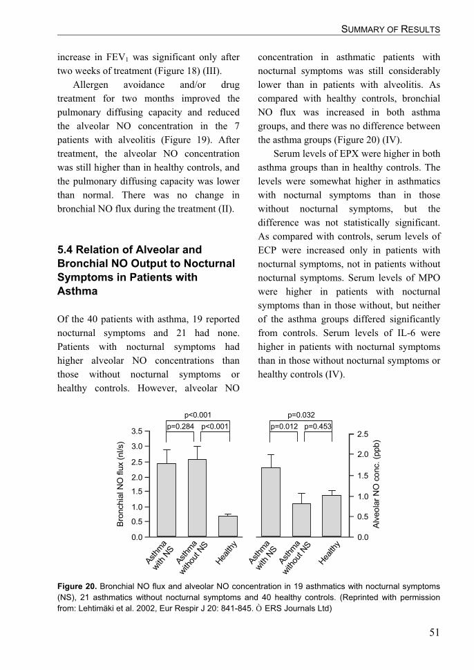
SUMMARY OF RESULTS
51
increase in FEV1 was significant only after
two weeks of treatment (Figure 18) (III).
Allergen avoidance and/or drug
treatment for two months improved the
pulmonary diffusing capacity and reduced
the alveolar NO concentration in the 7
patients with alveolitis (Figure 19). After
treatment, the alveolar NO concentration
was still higher than in healthy controls, and
the pulmonary diffusing capacity was lower
than normal. There was no change in
bronchial NO flux during the treatment (II).
5.4 Relation of Alveolar and
Bronchial NO Output to Nocturnal
Symptoms in Patients with
Asthma
Of the 40 patients with asthma, 19 reported
nocturnal symptoms and 21 had none.
Patients with nocturnal symptoms had
higher alveolar NO concentrations than
those without nocturnal symptoms or
healthy controls. However, alveolar NO
concentration in asthmatic patients with
nocturnal symptoms was still considerably
lower than in patients with alveolitis. As
compared with healthy controls, bronchial
NO flux was increased in both asthma
groups, and there was no difference between
the asthma groups (Figure 20) (IV).
Serum levels of EPX were higher in both
asthma groups than in healthy controls. The
levels were somewhat higher in asthmatics
with nocturnal symptoms than in those
without nocturnal symptoms, but the
difference was not statistically significant.
As compared with controls, serum levels of
ECP were increased only in patients with
nocturnal symptoms, not in patients without
nocturnal symptoms. Serum levels of MPO
were higher in patients with nocturnal
symptoms than in those without, but neither
of the asthma groups differed significantly
from controls. Serum levels of IL-6 were
higher in patients with nocturnal symptoms
than in those without nocturnal symptoms or
healthy controls (IV).
Figure 20. Bronchial NO flux and alveolar NO concentration in 19 asthmatics with nocturnal symptoms
(NS), 21 asthmatics without nocturnal symptoms and 40 healthy controls. (Reprinted with permission
from: Lehtimäki et al. 2002, Eur Respir J 20: 841-845. Ò ERS Journals Ltd)
p<0.001p=0.284 p=0.453
p=0.032
p=0.012
Alv
eo
lar
NO
co
nc. (p
pb)
Ast
hma
with
NS
Ast
hma
with
out N
S
Hea
lthy
Ast
hma
with
NS
Ast
hma
with
out N
S
Hea
lthy
Bro
nchia
l N
O flu
x (
nl/s)
3.5
3.0
2.5
2.0
1.5
0.0
1.0
0.5
2.5
2.0
1.5
0.0
1.0
0.5
p<0.001
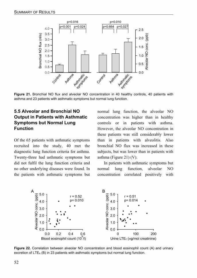
SUMMARY OF RESULTS
52
5.5 Alveolar and Bronchial NO
Output in Patients with Asthmatic
Symptoms but Normal Lung
Function
Of the 65 patients with asthmatic symptoms
recruited into the study, 40 met the
diagnostic lung function criteria for asthma.
Twenty-three had asthmatic symptoms but
did not fulfil the lung function criteria and
no other underlying diseases were found. In
the patients with asthmatic symptoms but
normal lung function, the alveolar NO
concentration was higher than in healthy
controls or in patients with asthma.
However, the alveolar NO concentration in
these patients was still considerably lower
than in patients with alveolitis. Also
bronchial NO flux was increased in these
subjects, but was lower than in patients with
asthma (Figure 21) (V).
In patients with asthmatic symptoms but
normal lung function, alveolar NO
concentration correlated positively with
Figure 21. Bronchial NO flux and alveolar NO concentration in 40 healthy controls, 40 patients with
asthma and 23 patients with asthmatic symptoms but normal lung function.
p=0.024p<0.001 p=0.027
p=0.010
p=0.664
Alv
eola
r N
O c
onc. (p
pb)
Con
trol
Ast
hma
Ast
hmat
ic
sym
ptom
s
Bro
nchia
l N
O flu
x (
nl/s)
2.5
2.0
1.5
0.0
1.0
0.5
p=0.016
Con
trol
Ast
hma
Ast
hmat
ic
sym
ptom
s
Figure 22. Correlation between alveolar NO concentration and blood eosinophil count (A) and urinary
excretion of LTE4 (B) in 23 patients with asthmatic symptoms but normal lung function.
Alv
eola
r N
O c
onc. (p
pb)
Blood eosinophil count (10 /l)9
A
Urine LTE ( g/mol creatinine)4 m
B
r = 0.51p= 0.014
r = 0.52p= 0.010
5.0
3.0
4.0
2.0
0.0
0.0 0.2 0.4 0.6 0 200
1.0
Alv
eola
r N
O c
onc. (p
pb
) 5.0
3.0
4.0
2.0
0.0
1.0
100

SUMMARY OF RESULTS
53
blood eosinophil count and urinary LTE4
(Figure 22). Alveolar NO concentration
correlated negatively with measures of
small-airway function (FEF50% and FEF75%)
(Figure 23). Bronchial NO flux correlated
positively with bronchial
hyperresponsiveness (Log DRS of
methacholine) (V).
Figure 23. Correlation between alveolar NO concentration and FEF50% (A) and FEF75% (B) in 23 patients
with asthmatic symptoms but normal lung function.
Alv
eola
r N
O c
onc. (p
pb)
FEF (l/s)50%
A
FEF (l/s)75%
B
r = -0.55p= 0.009
r = -0.50p= 0.018
5.0
3.0
4.0
2.0
0.0
1.0 3.0 5.0 7.0 0.5 3.01.0
1.0
Alv
eola
r N
O c
onc. (p
pb
) 5.0
3.0
4.0
2.0
0.0
1.0
2.52.01.5

54

DISCUSSION
55
6. DISCUSSION
Theoretical considerations regarding the
two-compartment model of pulmonary NO
dynamics suggest that the alveolar and
bronchial contributions to exhaled NO can
be calculated by measuring the exhaled NO
concentration at multiple exhalation flow
rates. Studies I and II of this series were the
first publications applying the model to
healthy controls and patients with alveolar
inflammation (alveolitis) or bronchial
inflammation (asthma) in clinical setting.
The main finding was that assessment of
alveolar and bronchial NO output by the
multiple flow rate method differentiates
between bronchial and alveolar / peripheral
components in pulmonary inflammation.
6.1 Flow Rate Dependency of
Exhaled NO Concentration
In the present study, exhaled NO
concentration was found to be inversely
related to exhalation flow rate in both
healthy and diseased subjects. The results
are in line with previous reports (Högman et
al. 1997, Silkoff et al. 1997) and confirm the
role of conducting airways as a major source
of exhaled NO.
Exhaled NO concentration is a sum of
both alveolar and bronchial NO output. At
higher exhalation flow rates, the exhaled NO
concentration reflects predominantly the
alveolar NO concentration, as exhaled air
travels through the conducting airways very
rapidly and there is little time for more NO
to be taken up from the bronchial mucosa.
At lower exhalation flow rates, the passage
of exhaled air through the conducting
airways takes longer and there is more time
for NO to diffuse from the bronchial mucosa
to the luminal air. At low exhalation flow
rates, the exhaled NO concentration thus
reflects predominantly bronchial NO
dynamics.
In view of the above said, the magnitude
of the chosen exhalation flow rate is clearly
a critical factor determining where in the
respiratory tract inflammatory changes can
be detected. If a high flow rate is chosen, the
exhaled NO concentration reflects NO
dynamics mainly in the alveolar region and
is not sensitive to detect increased bronchial
NO production. Conversely, if a low flow
rate is chosen, the exhaled NO concentration
will reflect mainly the bronchial
compartment and will be insensitive to
changes in alveolar NO dynamics. The
exhaled NO concentration at the currently
recommended flow rate of 50 ml/s reflects
mostly bronchial NO output and cannot be
used to study alveolar NO dynamics. This
conception is supported by the results of this
series. Exhaled NO concentration at flow
rates of 40 and 100 ml/s could differentiate
asthmatics from healthy controls and from
patients with alveolitis, but patients with
alveolitis could not be distinguished from
healthy subjects. At higher flow rates of 175
and 370 ml/s also patients with alveolitis
could be differentiated from healthy
subjects, but the difference between asthma
and alveolitis was no longer significant at
the highest flow rate. It is therefore likely
that alveolar inflammation is missed by
measuring exhaled NO concentration only at

DISCUSSION
56
a flow rate of 50 ml/s, but at higher flow
rates alveolar inflammation cannot be
differentiated from bronchial inflammation.
It is here that the multiple flow rate method
shows its superiority, as it can be used to
assess the alveolar and bronchial
components in pulmonary inflammation
separately.
6.2 Critique of the Two-
Compartment Model
The two-compartment model of pulmonary
NO dynamics was developed to allow more
precise interpretation of exhaled NO
measurements. Earlier models regarded only
the alveolar region as a source of NO and
neglected the bronchial tree (Hyde et al.
1997). However, experimental observations
showed an inverse relation between
exhalation flow rate and exhaled NO
concentration, suggesting that the
conducting airways are a major contributor
to the exhaled NO concentration (Högman et
al. 1997, Silkoff et al. 1997). The two-
compartment model considers both alveolar
and bronchial regions as possible sources of
NO, and also provides a theoretical means to
separate between alveolar and bronchial NO
production (Tsoukias and George 1998).
To retain mathematical simplicity, the
model is based on a robust simplification of
the human lung and cannot therefore be
considered to describe pulmonary NO
dynamics in all its details. The millions of
tiny alveoli and alveolar sacs together with
respiratory bronchioles are reduced to a
single huge alveolus, and the branching
bronchial tree is represented by a single rigid
cylindrical tube (Tsoukias and George
1998). Alveolar and bronchial NO
parameters are therefore only average values
describing the whole alveolar or bronchial
compartment, respectively, and local
differences in NO dynamics within a
compartment cannot be assessed.
As all the alveoli are represented by a
single well-mixed alveolus, the model
neglects possible differences in the NO
dynamics of parallel alveoli and also the
sequential filling of the alveoli. During
normal respiration, the alveoli which are
filled first during inhalation are emptied last
during exhalation (usually the apical region
of the lung), while the alveoli which are
filled last are emptied first (usually the basal
region) (Fukuchi et al. 1980, Tsoukias et al.
2000b). Thus, air from basal alveoli
predominates in the early expiration,
whereas air from apical alveoli predominates
in the late phase. If there is a significant
difference in alveolar NO concentration
between basal and apical lung, this might
cause a downward or upward slope in the
plateau NO concentration during a single
exhalation. A model taking into account the
sequential filling of the lung might be able
to differentiate between apical and basal
inflammatory processes.
The two-compartment model considers
convection as the only mode of axial gas
transport and neglects axial diffusion of NO
in the gas phase (e.g. diffusion of NO from
bronchial air with higher NO concentration
backwards to alveolar air with lower NO
concentration). However, diffusion has been
shown to be a major contributor to gas
transport particularly in the very small
airways and the alveolar region. It has been
suggested that neglect of axial diffusion in
the two-compartment model leads to a four-

DISCUSSION
57
fold underestimation of maximal bronchial
NO flux (Shin and George 2002a).
However, this underestimation depends
markedly on the significant contribution of
small airways to bronchial NO flux. If
bronchial NO flux is mainly derived from
the large airways, as in healthy subjects
(Silkoff et al. 1998), neglecting axial
diffusion will cause no major
underestimation of bronchial NO flux (Shin
and George 2002a).
The two-compartment model assumes
alveolar and bronchial NO parameters to be
constant while the lung volume decreases
during exhalation. There are, however, data
showing that the alveolar diffusing capacity
of NO (DNO,Alv) decreases with decreasing
alveolar volume during exhalation (Borland
and Higenbottam 1989, Tsoukias et al.
2000a). According to equation (1) (page 33),
the alveolar NO concentration is inversely
related to DNO,Alv. The alveolar NO
concentration should therefore increase
during a single exhalation as the decreasing
alveolar volume also reduces DNO,Alv. This
would tend to increase the total exhaled NO
concentration towards end-exhalation.
However, during the plateau phase of the
exhaled NO curve, the NO value usually
decreases slightly with decreasing
pulmonary volume towards end-exhalation
(Silkoff et al. 1997, Tsoukias et al. 1998). It
is therefore suggested that the flux of NO
from alveolar tissue to alveolar air (JNO,Alv)
would also decrease with decreasing
alveolar volume such that the ratio between
JNO,Alv and DNO,Alv would be constant during
exhalation (Tsoukias and George 2001a).
This would lead to a constant alveolar NO
concentration, as CAlv is equal to JNO,Alv /
DNO,Alv (see equation (1) on page 33).
Although the bronchial compartment in
the two-compartment model is a rigid tube
whose volume does not change during the
respiratory cycle, the small bronchioles very
likely decrease in diameter as pulmonary
volume is reduced during exhalation. The
decreased diameter of the small bronchioles
would also reduce the mucosal surface area
of the bronchial compartment. Bronchial NO
flux is determined by the bronchial wall NO
concentration and the bronchial diffusing
capacity of NO, which in turn is dependent
on the mucosal surface area available for
diffusion (Tsoukias and George 1998). The
decreasing bronchial surface area during
exhalation would therefore tend to reduce
the bronchial NO flux and exhaled NO
concentration towards end-exhalation
(Tsoukias and George 2001a). This might be
the explanation for the observed decrease in
exhaled NO concentration during the plateau
phase of the NO curve of a single
exhalation. The exhaled NO concentration
has been found to decrease after induction of
bronchoconstriction (de Gouw et al. 1998,
Scollo et al. 2000, Terada et al. 2001), while
attenuation of the bronchoconstriction with
inhaled b2-agonists increases exhaled NO
levels back to normal (Ho et al. 2000b).
These changes in exhaled NO concentration
can also be explained by corresponding
changes in the bronchial mucosal surface
area after bronchoconstriction or
bronchodilatation.
While it is evident that this simplified
model cannot describe pulmonary NO
dynamics in all detail, the important
question is whether it still yields clinically
useful information. For example, the widely
used measures of pulmonary diffusing
capacity are also based on a robustly

DISCUSSION
58
simplified model of the lung, but the clinical
value of these measurements is indisputable.
The results of the present study suggest that
the relatively simple equations of the two-
compartment model do indeed provide
clinically useful information and allow
differentiation between alveolar and
bronchial inflammation. Further studies are
needed to establish whether taking into
account the parameters neglected by the
two-compartment model would provide
additional clinically useful information on
pulmonary NO dynamics.
6.3 Exhaled NO Measurement at
Multiple Flow Rates in Assessing
Alveolar and Bronchial
Inflammation
The exhaled NO concentration was
measured at multiple exhalation flow rates
in patients with asthma and alveolitis. Using
the linear equation derived from the two-
compartment model (Eq (5) on page 36),
alveolar NO concentration and bronchial NO
flux were calculated for each subject. The
increased alveolar NO concentration in
alveolitis and the increased bronchial NO
flux in asthma suggest that exhaled NO
measurement at multiple exhalation flow
rates can be used to differentiate between
alveolar and bronchial inflammation.
Further, alveolar NO concentration and
bronchial NO flux correlated with disease
severity and responded to anti-inflammatory
treatment in alveolitis and asthma. However,
biopsy studies are needed to further assess
the relation of alveolar and bronchial NO
parameters to NOS expression and indices
of inflammatory activity in the alveolar and
bronchial compartments.
6.3.1 Alveolar and bronchial NO in
asthma
Asthmatic patients had approximately 3.5
times higher bronchial NO flux than healthy
subjects, but there was some overlap in
individual values between the groups. The
mean alveolar NO concentration in patients
with asthma was not different from that in
healthy subjects, but variation in asthmatics
was somewhat higher.
An increased bronchial NO flux in
asthma has lately also been found by other
study groups utilising the multiple flow rate
method (Silkoff et al. 2000, Högman et al.
2001, Delclaux et al. 2002). If only
relatively high exhalation flow rates (> 50
ml/s) are used, exhaled NO can be
partitioned into alveolar NO concentration
and bronchial NO flux by the linear equation
(Eq (5) on page 36) (Tsoukias and George
1998). However, including also very low
flow rates (down to 5 - 10 ml/s) allows
application of the non-linear equation (Eq
(2), page 35) to divide bronchial NO flux
into its two components: bronchial wall NO
concentration and bronchial diffusing
capacity of NO (Högman et al. 2000, Silkoff
et al. 2000). As only relatively high
exhalation flow rates were used in the
present study, it was not possible to address
the question whether the increased bronchial
NO flux in asthma was due to increased
airway wall NO concentration or increased
bronchial diffusing capacity of NO. By also
including very low flow rates, Silkoff and
colleagues found an increased bronchial wall

DISCUSSION
59
diffusing capacity of NO and a slightly but
not significantly increased bronchial wall
NO concentration in asthma (Silkoff et al.
2000). Högman and colleagues reported that
the increased bronchial NO output in asthma
is due to an increase in both bronchial wall
NO concentration and the bronchial wall
diffusing capacity of NO (Högman et al.
2002b).
Increased bronchial wall NO
concentration in asthma can be argued to
represent a higher NO production rate
caused by enhanced iNOS expression in the
asthmatic airways (Hamid et al. 1993, Saleh
et al. 1998). The increased bronchial wall
diffusing capacity of NO is more difficult to
interpret in terms of pathological changes in
asthmatic airways. In theory, the bronchial
wall diffusing capacity of NO can be
increased due to either physical changes in
the mucosa improving the actual diffusivity
of NO, or an increase in the mucosal surface
area producing NO (Tsoukias and George
1998, Silkoff et al. 2000). Silkoff and
colleagues proposed that the main source of
bronchial NO in healthy subjects would be
the proximal largest airways; lobar bronchi,
main bronchi and trachea (Silkoff et al.
1998, Silkoff et al. 2000). They attribute the
increased bronchial wall diffusing capacity
of NO in asthma to an increased total airway
area producing NO, i.e. spreading of the
NO-producing area also to smaller airways.
This increased NO production area would be
a reflection of NO-related bronchodilatory
defence mechanism against
bronchoconstriction. So far there are no
means of separating between production
area and physical diffusivity as components
in the bronchial wall diffusing capacity of
NO. More studies with advanced
measurement techniques are needed to
establish how these two factors are affected
in asthmatic airway inflammation.
In this study bronchial NO flux
correlated positively with several markers of
asthmatic inflammation, for example blood
eosinophil count, serum levels of ECP and
EPX, and urinary excretion of EPX. This is
in line with results obtained by Högman and
colleagues showing a positive correlation
between bronchial NO flux and percentage
of sputum eosinophils in asthma (Högman et
al. 2001). In addition, several studies using
the single flow rate method have found
exhaled NO to correlate with direct indices
of eosinophilic inflammation in sputum,
BAL and bronchial biopsies (Jatakanon et al.
1998, Chan-Yeung et al. 1999, Lim et al.
1999, Mattes et al. 1999, Payne et al. 2001,
van den Toorn et al. 2001, Warke et al.
2002). These results support the role of
bronchial NO flux as a measure of bronchial
inflammation in asthma. There was also a
positive correlation between bronchial NO
flux and serum level of IgE in this study. No
previous studies have assessed the relation
between these measures, but studies using
the single flow rate method have shown a
correlation between exhaled NO
concentration and the serum level of IgE
(Simpson et al. 1999). The positive
correlation between bronchial NO flux and
serum IgE levels is in line with studies
showing higher exhaled NO levels in atopic
than non-atopic asthmatics.
Bronchial NO flux correlated positively
with bronchial hyperresponsiveness (Log
DRS of methacholine). Thus, the bronchial
NO flux was higher in patients with more
responsive airways. The exhaled NO
concentration at a single flow rate has also

DISCUSSION
60
been found to correlate with bronchial
responsiveness (Dupont et al. 1998, Lim et
al. 1999). Asthmatic inflammation induces
bronchial iNOS expression and also
enhances bronchial responsiveness. The
correlation between bronchial NO flux and
hyperresponsiveness is thus what would be
expected. However, Silkoff and colleagues
showed a positive correlation between PC20
of methacholine and DNO,Br³CW (ºbronchial
NO flux) after treatment with inhaled
glucocorticoids, i.e. bronchial NO flux after
steroid treatment was higher in patients with
less reactive airways. As inhaled
glucocorticoids lower iNOS but not cNOS
expression in the airways (Saleh et al. 1998),
the authors concluded that the higher
bronchial NO flux after steroids would
reflect more intense bronchoprotective
nNOS expression. This would explain the
less reactive airways in subjects with higher
bronchial NO flux after steroid treatment.
The increased bronchial NO flux but
normal average alveolar NO concentration
in asthma found in this study is in line with
the classic view of asthma as an
inflammatory disease of the large airways.
Other study groups have likewise reported
that alveolar NO concentration in asthmatics
does not differ from that in healthy controls
(Högman et al. 2001, Delclaux et al. 2002).
However, alveolar inflammation has been
found in transbronchial biopsies from
patients with stable chronic asthma (Kraft et
al. 1996) and nocturnal asthma is associated
with further enhancement of alveolar
inflammation (Martin et al. 1991). This is in
line with the increased alveolar NO
concentration seen in asthmatics with
nocturnal symptoms in the present study. As
bronchial NO flux in asthmatics with
nocturnal symptoms was not higher than in
patients without these symptoms, exhaled
NO measurement at a single flow rate could
not differentiate between these two asthma
groups. The results suggest that
measurement of alveolar NO concentration
could be used to assess the peripheral
component in asthmatic inflammation
associated with nocturnal asthma.
6.3.2 Alveolar and bronchial NO in
alveolitis
Patients with CFA or EAA had increased
alveolar NO concentrations but normal
bronchial NO flux. Girgis and colleagues
have reported increased alveolar NO
concentration but slightly decreased
bronchial NO flux in patients with FASSc
(Girgis et al. 2002). The increased alveolar
NO concentration in patients with alveolitis
in the present study was not entirely
explained by the decreased pulmonary
diffusing capacity of NO, but the results
suggest that the actual alveolar production
rate of NO was also increased. The
increased production rate of NO is
consistent with the iNOS expression found
in the alveolar epithelium of patients with
CFA (Saleh et al. 1997).
The alveolar NO concentration in
alveolitis correlated negatively with DLCO
and alveolar volume. Also Girgis and
colleagues found a negative correlation
between alveolar NO concentration and
DLCO in patients with FASSc (Girgis et al.
2002). The correlation between alveolar NO
concentration and DLCO might be explained
by a thickening of the alveolar membrane
reducing diffusion of both CO and NO. A

DISCUSSION
61
reduced diffusing capacity of NO would
increase the alveolar NO concentration (see
Eq (1) on page 33), and therefore decreased
DLCO and increased alveolar NO
concentration would both reflect the same
pathophysiological feature, namely impaired
diffusion of gases in the alveolar region.
However, the results of the present study
suggest that the alveolar production rate of
NO was also increased. This is supported by
the increased iNOS expression in the
alveolar epithelium of patients with CFA
(Saleh et al. 1997). Thus, increased alveolar
NO concentration might not only reflect a
decreased alveolar diffusing capacity of NO
but would also serve as a marker of
inflammatory activity in the alveolar
compartment. There are also animal data
suggesting that an increased alveolar
production rate of NO might be one of the
factors causing inflammation and excess
fibrosis in alveolar tissue (de Rezende et al.
2000, Giri et al. 2000), and might thus
actually be one of the factors causing
decreased DLCO in fibrosing alveolitis.
In the present work, the alveolar NO
concentration was not compared to direct
measures of pulmonary inflammation in
patients with alveolitis. Previously, the
exhaled NO concentration at a single flow
rate has been compared to inflammatory
indices in BAL fluid in patients with FASSc.
The exhaled NO concentration was higher in
patients with signs of active inflammation in
BAL than in patients with normal BAL, and
there was a correlation between exhaled NO
concentration and BAL lymphocyte cell
count (Paredi et al. 1999). In CFA or
FASSc, the iNOS expression in alveolar
tissue is more pronounced in patients with
an active inflammatory pattern in biopsies as
compared with patients having end-stage
fibrosis (Saleh et al. 1997, Romanska et al.
2000b). An increased alveolar NO
concentration might thus be used as a
marker of on-going active inflammation in
patients with fibrosing alveolitis. As
inflammatory-stage rather than end-stage
fibrosis is thought to be more responsive to
drug treatment (American Thoracic Society
and European Respiratory Society 2000),
measurement of the alveolar NO
concentration might serve as a useful tool in
identifying patients most likely to benefit
from drug treatment.
Inflammation in EAA is not restricted to
alveolar tissue only; also bronchiolitis and
peribronchiolar granulomas are typically
found in the disease. However, bronchial
NO flux was not increased in these subjects.
There are two possible reasons for this.
Firstly, the most peripheral bronchioles are
included in the alveolar compartment in the
two-compartment model of pulmonary NO
dynamics. Secondly, the exhaled NO
concentration reflects mostly NO production
in the airway epithelium rather than in
deeper tissue layers (Gabbay et al. 1999).
NO produced in the peribronchiolar
granulomas might thus not, due to the
relatively long distance, diffuse into the
airway lumen in high enough quantities to
affect bronchial NO flux. Studies with larger
numbers of subjects and assessment of
bronchiolitis are needed to establish whether
profound bronchiolitis increases bronchial
NO flux in EAA.

DISCUSSION
62
6.3.3 Responsiveness of alveolar and
bronchial NO output to alleviation of
inflammation
There is a clinical need for a marker of
airway inflammation which would allow
assessment of the efficacy of anti-
inflammatory treatment in lung diseases. In
asthma, the exhaled NO concentration
measured at a single exhalation flow rate
decreases dose-dependently during treatment
with inhaled glucocorticoids, while
discontinuation of the treatment increases
the exhaled NO level together with increase
in symptoms (Kharitonov et al. 1996b,
Silkoff et al. 2001, Jones et al. 2002,
Kharitonov et al. 2002). Patients with CFA
using anti-inflammatory medication show
lower levels of exhaled NO than patients
without such medication (Paredi et al. 1999).
These observations suggest that NO
production might serve as a surrogate
marker for inflammatory activity during
drug treatment.
It was found in the present study that
anti-inflammatory treatment reduces
bronchial NO flux in asthma and the
alveolar NO concentration in alveolitis. The
responsiveness of these parameters to anti-
inflammatory treatment in the compartment
where the disease process is considered
mainly to take place supports the role of
alveolar and bronchial NO parameters as
inflammatory markers of the respective
compartments.
In asthma, inhaled fluticasone reduced
bronchial NO flux but had no effect on
alveolar NO concentration. This is
understandable, as the alveolar NO
concentration was already normal before
treatment. Silkoff and colleagues have
reported that the decreased bronchial NO
flux in asthma following inhaled
glucocorticoids is due to a decreased
bronchial wall NO concentration (Silkoff et
al. 2000). This is in line with biopsy studies
showing decreased iNOS expression in
asthmatic airways after inhaled
glucocorticoids (Saleh et al. 1998).
Glucocorticoids may reduce iNOS
expression in at least two ways; directly by
interfering with the transcription and
translation of iNOS (inhibition of NF-kB or
destabilisation of iNOS mRNA) (Kleinert et
al. 1996, Korhonen et al. 2002), or indirectly
by lowering the production rate of
inflammatory cytokines needed to induce
iNOS expression in airway cells.
The effect of inhaled fluticasone was
rapid. A significant decrease in bronchial
NO flux was already evident at the first
control visit after one week of treatment, and
bronchial NO flux was no longer statistically
different from that in healthy controls. Other
studies have shown that a high dose of
inhaled budesonide reduces the exhaled NO
concentration already after 6 hours (Tsai et
al. 2001). In the present study, bronchial NO
flux decreased simultaneously with
asthmatic symptoms and preceded the
increase in FEV1.
In patients with alveolitis, anti-
inflammatory treatment reduced the alveolar
NO concentration but had no effect on
bronchial NO flux. There was also an
increase in pulmonary diffusing capacity
during the treatment. However, even after
the treatment, alveolar NO concentration
was higher than in healthy controls, but the
diffusing capacity in patients was still lower
than normal. Anti-inflammatory treatment
may reduce the alveolar NO concentration

DISCUSSION
63
by two different mechanisms. It might lower
the actual alveolar production rate of NO, or
reduce alveolar oedema and therefore
increase the diffusion rate of NO from
alveolar air to pulmonary blood to be
scavenged by haemoglobin.
These results suggest that bronchial NO
flux and alveolar NO concentration could be
used in assessing the efficacy of anti-
inflammatory treatment in patients with
bronchial or alveolar inflammation.
6.3.4 Alveolar and bronchial
inflammation in patients with
asthmatic symptoms but normal lung
function
In patients with asthmatic symptoms but
normal lung function, bronchial NO flux
was higher than in healthy subjects but
lower than in patients with asthma. This
suggests that the asthmatic symptoms group
had bronchial inflammation but that this was
milder than in asthmatic subjects. This is in
line with other studies in such patients,
showing sputum eosinophils to be increased
but lower than in asthma (Gibson et al.
1989, Rytilä et al. 2000). Bronchial
responsiveness was also increased in the
asthmatic symptoms group but was lower
than in patients with asthma. The exhaled
NO concentration at a single exhalation flow
has been shown to be increased in a similar
group of patients with asthmatic symptoms
but normal lung function (Ekroos et al.
2002). These results suggest that functional
criteria of asthma are not sufficient to detect
all the patients with airway inflammation,
but inflammatory markers are needed.
In addition, the alveolar NO
concentration was slightly increased in the
asthmatic symptoms group, and it correlated
negatively with the spirometric indices of
small airway function. This would imply
that the asthmatic symptoms group had a
more peripheral inflammation than patients
fulfilling the current diagnostic criteria of
asthma. The results also suggest that among
patients with asthma-like symptoms, those
with more central airway inflammation show
impaired large airway function (changes in
PEF and FEV1) and fulfil the lung function
criteria for asthma, while subjects with more
peripheral inflammation and milder
inflammation in the large airways do not
have impaired large airway function and
thus do not fulfil the current criteria for
asthma. However, further studies with
biopsies would help to validate alveolar and
bronchial NO output measures against
inflammatory activity in these patient
groups.
6.3.5 Critique of the Present Method
In the present study, alveolar NO
concentration and bronchial NO flux were
calculated using the linear equation (Eq (5)
on page 36) of the two-compartment model
based on exhaled NO concentrations
measured at exhalation flow rates of 100,
175 and 370 ml/s (also 40 ml/s in the first
paper). The exhalation flow rates were
achieved by means of different mechanical
flow restrictors for each flow rate at the
exhalation port of the breathing valve, and
the patients were requested to keep
exhalation pressure steady at a target level.
The achieved exhalation flow rate is
DISCUSSION
64
dependent on the patient’s ability to
maintain exhalation pressure steady at the
desired level. There was thus probably some
deviation in the flow rate values achieved,
which might have affected the results.
The alveolar NO concentrations in
healthy subjects and asthmatics in the
present study are somewhat lower than those
published by other study groups using the
linear fitting method (Tsoukias et al. 1998,
Högman et al. 2001, Delclaux et al. 2002,
Girgis et al. 2002). Differences in
measurement technique (e.g. the exhalation
flow rates used) might be the reason. The
linear function between NO output and
exhalation flow rate is based on the
assumption that the NO concentration in the
bronchial luminal air is not significantly
higher than the alveolar NO concentration,
so that the bronchial NO flux would equal
its maximum value ((CW-CAlv) ³ DNO,Br). At
very high exhalation flow rates this is
roughly the case, but at lower flow rates the
NO concentration in the bronchial luminal
air increases and diffusion of NO from
bronchial mucosa to luminal air decreases.
Thus, the linear fitting between NO output
and exhalation flow rate should be used only
at such high flow rates that the assumption
of maximum bronchial NO flux is justified
and the relation between NO output and
exhalation flow rate is linear. How high such
flow rates are, depends on the magnitude of
bronchial NO flux. The higher the bronchial
NO flux, the higher should be the flow rates.
Usually only flow rates higher than 50 ml/s
are recommended for the linear fitting.
Based on the present data, 100 ml/s might be
a useful lower limit especially in asthmatics.
Approximates of alveolar NO
concentration and bronchial NO flux
calculated by the linear fitting method are
affected by the magnitude of the chosen
exhalation flow rates. Inclusion of too low
flow rates causes overestimation of the
alveolar NO concentration and
underestimation of the bronchial NO flux.
Exclusion of the lowest exhalation flow rate
(40 ml/s) in the first paper of the present
series would increase the bronchial NO flux
from 2.6 to 2.8 nl/s and lower the alveolar
NO concentration from 1.5 to 0.7 ppb in
patients with asthma. This might also be
explained in part by inaccuracies in
exhalation flow rates.
At higher flow rates the problem of
overestimating the alveolar NO
concentration and underestimating the
bronchial NO flux diminishes. However, at
higher flow rates the duration of a single
exhalation is shorter by reason of the limited
pulmonary capacity, and also exhaled NO
concentrations are lower. The shorter
duration of exhalation and low NO levels
cause inaccuracy in NO measurement due to
the limited performance of current NO
analysers. Differences in chosen flow rates
might cause differences in results between
study groups, so that standardisation of the
method is clearly needed. There is also a
need for improved measurement equipment
allowing easy use of different exhalation
flow rates without the need for the patient to
maintain a steady exhalation effort.
6.4 Single or Multiple Flow Rates
in Exhaled NO Measurement?
Currently, the exhaled NO concentration is
recommended to be measured at a single
exhalation flow rate of 50 ml/s (American

DISCUSSION
65
Thoracic Society 1999, Baraldi and de
Jongste 2002). This method can be used to
detect pulmonary inflammation, but it
cannot differentiate between the bronchial
and alveolar components in the
inflammatory process. The single flow rate
method is also insensitive to alveolar /
peripheral inflammation (see chapter 6.1).
However, the results of the present study
and those obtained by other groups show
that application of the two-compartment
model by measuring exhaled NO
concentration at multiple flow rates provides
the advantage of differentiating between
alveolar and bronchial components in
pulmonary inflammation (Jörres 2000,
Högman et al. 2000, Högman et al. 2001,
Delclaux et al. 2002). If only higher flow
rates (> 50 ml/s) are used, the exhaled NO
concentration can be partitioned into
alveolar NO concentration and bronchial NO
flux (Tsoukias and George 1998). However,
if also very low flow rates are included,
bronchial NO flux can further be divided
into its two components: bronchial wall NO
concentration and bronchial diffusing
capacity of NO (Högman et al. 2000, Silkoff
et al. 2000). Different methods of exhaled
NO measurement according to flow rates
used are compared in Table 2.
In addition to the results presented in
this thesis, also other study groups have
applied exhaled NO measurement at
multiple flow rates to different pulmonary
conditions. The results are summarised in
Table 3. Increased bronchial NO flux but
normal alveolar NO concentration has
consistently been found in asthma, which is
in line with increased exhaled NO
concentration at a single flow rate. In
addition, in the present study increased
alveolar NO concentration was found in
nocturnal asthmatics and subjects with
asthmatic symptoms but normal lung
function. Although asthmatics can be
differentiated from healthy subjects by
measuring the exhaled NO concentration at
a single flow rate, this peripheral component
of asthmatic inflammation cannot be found.
Patients with FASSc have been found to
have increased alveolar NO concentration,
which is in line with the results on alveolitis
in this work. Increased NO production has
also been found in FASSc by measuring the
exhaled NO concentration at a single flow
rate (Paredi et al. 1999), but, again, the
inflammatory process cannot be
anatomically located by the single flow rate
Exhalation flow rates used
Single flow rate
Multiple relativelyhigh flow rates (> 50 ml/s)
Multiple flow rates,both low and high
Parameters gained
-Exhaled NO concentration
-Alveolar NO concentration,-Bronchial NO flux
-Alveolar NO concentration,-Bronchial wall NO concentration,-Bronchial diffusing capacity of NO
Information gained
-Presence / absence of pulmonary inflammation(not sensitive for alveolar/peripheral inflammation)
-Presence / absence of pulmonary inflammation,-Differentiation between alveolar and bronchial inflammation
-Presence / absence of pulmonary inflammation,-Differentiation between alveolar and bronchial inflammation,
-Whether changes in bronchial NO flux are due tochanges in tissue concentration or diffusion of NO
Table 2. Gained parameters and information by exhaled NO measurement at different techniques according to the exhalation flow rates used.

DISCUSSION
66
method.
Shin and colleagues estimated
parameters of alveolar and bronchial NO
dynamics in children with cystic fibrosis and
in healthy children (Shin et al. 2002b) by
measuring exhaled NO with a new
application of the two-compartment model
utilising a single exhalation with
dynamically decreasing flow rate (Tsoukias
et al. 2001b). They found an increased
bronchial wall diffusing capacity of NO, but
a decreased alveolar NO concentration and
bronchial wall NO concentration in cystic
fibrosis. The authors attribute these changes
to increased chemical consumption of NO
by e.g. superoxide, whose production is
increased in cystic fibrosis. The increased
chemical reaction rate of NO in the tissue
phase would reduce the tissue concentration
of NO, this explaining the decreased CW and
CAlv. An increased reaction rate of NO
would also alter the tissue concentration
Disease state
TABLE 3. Alveolar and bronchial NO parameters in different pulmonary diseases as compared with healthy subjects.
CAlvJNO,Br CW DNO,Br
Asthma
 »
ª ¬
ª ¬
ª ¬ ¬
ª ¬
¬¬Asthma-likesymptoms*
C , alveolar NO concentration; JAlv NO,Br W NO,Br, bronchial NO flux; C , bronchial wall NO concentration; D , bronchial wall
diffusing capacity of NO; CFA, cryptogenic fibrosing alveolitis; EAA, extrinsic allergic alveolitis; FASSc, fibrosing
alveolitis associated with systemic sclerosis; *, patients with asthmatic symptoms but normal lung function; , not
different from healthy subjects; , higher than in healthy subjects; , lower than in healthy subjects.
ª¬ ®
COPD
Smoking
ª ®
Cystic fibrosis ª ¬® ®
¬ ®
ª ® ª
¬ ªª
FASSc ®¬
¬ ªCFA & EAA
¬¬
Reference
Silkoff et al. 2000
Delclaux et al. 2002
Högman et al. 2001
Högman et al. 2002b
Lehtimäki et al. 2000 (I) & 2001 (II)
Lehtimäki et al. 2003 (V)
Delclaux et al. 2002
Shin et al. 2002
Högman et al. 2002a
Högman et al. 2002b
Högman et al. 2002b
Girgis et al. 2002
Lehtimäki et al. 2000 (I) & 2001 (II)
Lehtimäki et al. 2002 (IV)Nocturnal asthma
DISCUSSION
67
profile of NO in the bronchial wall, leading
to an increased bronchial wall diffusing
capacity of NO. This together with a
decreased tissue concentration of NO would
tend to balance out each other’s effects on
bronchial NO flux. The slightly decreased
alveolar NO concentration and fairly normal
bronchial NO flux would lead to normal or
slightly decreased exhaled NO concentration
at a single flow rate. Thus, patients with
cystic fibrosis cannot necessarily be
distinguished from healthy subjects by
exhaled NO measurement at a single flow
rate, although considerable changes in
pulmonary NO dynamics can be shown by
separately assessing the alveolar and
bronchial NO parameters (Shin et al.
2002b). This example again nicely indicates
the superiority of the extended NO
measurements over the single flow rate
method.
Högman and colleagues have reported
an increased bronchial wall diffusing
capacity of NO also in patients with allergic
rhinitis without asthma (Högman et al.
2002b). On the other hand, the bronchial
wall NO concentration in these patients was
normal. The results suggest that the
increased exhaled NO concentration found
in allergic rhinitis (Henriksen et al. 1999,
Gratziou et al. 2001) is caused by an
increased bronchial mucosal area producing
NO rather than by increased intensity in NO
production.
Delclaux and colleagues have found
increased alveolar NO concentrations but
normal bronchial NO output in patients with
liver cirrhosis (Delclaux et al. 2002). The
hepatopulmonary syndrome caused by liver
cirrhosis is associated with increased
vascular endothelial NO production
(Albillos et al. 1995) and increased iNOS
expression in macrophages in pulmonary
capillaries (Nunes et al. 2001), which
presumably explains the increased alveolar
NO concentration in cirrhotic patients.
Increased exhaled NO concentrations in
liver cirrhosis have also been shown at a
single flow rate (Cremona et al. 1995,
Matsumoto et al. 1995, Sogni et al. 1995)
but, again, these measurements cannot
indicate where in the lung NO dynamics is
altered.
Smoking has been shown to reduce
bronchial NO parameters (Delclaux et al.
2002, Högman et al. 2002b), and one study
also shows increased alveolar NO
concentration as an early sign of smoking-
induced pulmonary damage (Högman et al.
2002a). The decreased bronchial NO flux in
smokers is consistent with the decreased
exhaled NO concentration at single flow
rate, but the single flow rate method might
not be useful in detecting the increased
alveolar NO concentration in these subjects.
Högman and colleagues also report
increased alveolar NO concentrations but
normal bronchial NO parameters in patients
with COPD (Högman et al. 2002b). This
increased alveolar NO concentration in
COPD might reflect a decreased alveolar
diffusing capacity of NO due to emphysema
or increased alveolar NO production
associated with inflammation. The exhaled
NO concentration at a low exhalation flow
rate reflects bronchial NO dynamics, while
the contribution from the alveolar level
increases with increasing flow rate. This
might explain why COPD has been
associated with increased exhaled NO
concentrations in studies using a single
higher flow rate (º 100 ml/s) (Maziak et al.

DISCUSSION
68
1998, Paredi et al. 2000) but with normal
exhaled NO levels in studies using a single
lower flow rate (º 50 or 42 ml/s) (Kanazawa
et al. 1998, Ichinose et al. 2000).
The difference between measuring
exhaled NO at a single or at multiple flow
rates can be compared to the difference
between measuring peak expiratory flow
(PEF) or spirometric flow-volume curves.
PEF measurements are obviously of clinical
value in detecting impaired lung function,
but the parameters obtained from flow-
volume spirometry are more sensitive and
give more detailed information on airway
function. Similarly, exhaled NO
measurement at a single flow rate can be
used to detect pulmonary inflammation,
although it is not sensitive to peripheral
inflammation at the currently recommended
flow rate (50 ml/s), but NO measurement at
multiple flow rates gives more detailed
information of pulmonary NO dynamics and
on the underlying inflammatory process. It is
therefore likely that the multiple flow rate
method will displace the single flow rate
method. International guidelines on exhaled
NO measurement should be revised to
include the multiple flow rate method, which
significantly improves the clinical value of
exhaled NO measurement.
There have been several different
applications of the multiple flow rate
method. Some have used only higher flow
rates, others also include very low flow
rates, and a dynamically changing flow rate
during a single exhalation has also been
used. Further studies are needed to assess
how the results are affected by the number
and magnitude of chosen flow rates, and
whether the flow rates are to be achieved
during a single or multiple exhalations.
SUMMARY AND CONCLUSIONS
69
7. SUMMARY AND CONCLUSIONS
Currently the diagnosis and follow-up of
inflammatory lung diseases are largely based
on radiography and measures of lung
function to detect secondary changes caused
by the inflammatory process. There is a need
to develop methods allowing direct
measurement of pulmonary inflammation.
The present study focused on calculating
alveolar NO concentration and bronchial NO
flux based on measuring the exhaled NO
concentration at multiple exhalation flow
rates. The aim was to test whether
assessment of alveolar NO concentration
and bronchial NO flux could be used to
differentiate between alveolar and bronchial
components in inflammation, and to
establish whether alveolar and bronchial NO
output correlate with disease severity and
respond to anti-inflammatory treatment in
asthma and alveolitis. Alveolar and
bronchial NO output were also studied in
nocturnal asthma and in patients with
asthmatic symptoms but normal lung
function (asthma-like syndrome). The
summary and conclusions of the main
results of the present study are as follows:
1. Patients with asthma had increased
bronchial NO flux but on average normal
alveolar NO concentration, whereas
patients with alveolitis had increased
alveolar NO concentration but normal
bronchial NO flux. In asthma, increased
bronchial NO flux correlated with
markers of eosinophilic inflammation
and bronchial hyperresponsiveness. In
alveolitis, increased alveolar NO
concentration correlated negatively with
pulmonary diffusing capacity and
alveolar volume. It is concluded that
measurement of the alveolar NO
concentration and bronchial NO flux by
the multiple flow rate method can be
used to differentiate between alveolar and
bronchial inflammation and to assess
disease severity in asthma and alveolitis.
2. Treatment of asthma with inhaled
glucocorticoids reduced the bronchial
NO flux back to normal while alleviating
symptoms and improving lung function.
Anti-inflammatory treatment in alveolitis
lowered the alveolar NO concentration
towards normal and improved the
pulmonary diffusing capacity. It is
concluded that measurement of alveolar
NO concentration and bronchial NO flux
can be used to study the efficacy of anti-
inflammatory treatment in alveolitis and
asthma, respectively.
3. Consistently with the intensified
alveolar inflammation reported in
nocturnal asthma, asthmatic patients with
nocturnal symptoms had higher alveolar
NO concentrations but bronchial NO flux
similar to that in asthmatics with only
daytime symptoms. It is concluded that
measurement of the alveolar NO
concentration can be used to detect the
peripheral component in asthmatic
inflammation associated with nocturnal
asthma.
4. Subjects with asthmatic symptoms but
normal lung function (asthma-like

SUMMARY AND CONCLUSIONS
70
syndrome) had slightly increased
bronchial NO flux and also enhanced
alveolar NO concentration. It is
concluded that measurements of lung
function are not sufficient to detect all
patients with airway inflammation, but a
direct measure of inflammation should be
used to identify those with an
inflammatory process underlying their
asthma-like symptoms. The results also
suggest that this subject group had more
peripheral airway inflammation than
patients fulfilling the current diagnostic
criteria for asthma.
The results of the present study series
show that the multiple flow rate method in
exhaled NO measurement can be used to
assess the presence and severity of alveolar
and bronchial components in pulmonary
inflammation separately in a non-invasive
manner. Alveolar / peripheral inflammation
is not detectable by exhaled NO at a single
low flow rate, while at a single high flow
rate it cannot be differentiated from
bronchial inflammation. The multiple flow
rate method thereby gives clinically more
useful information on pulmonary
inflammation than the single flow rate
method of exhaled NO measurement. Future
studies should seek to provide information
on the applicability of the multiple flow rate
method to other pulmonary diseases. There
is also a need to standardise the method by
studying which and how many flow rates
should be used, and to ascertain what is the
additional clinical value of dividing
bronchial NO flux further into its
components (bronchial wall NO
concentration and NO diffusing capacity).
Measuring equipment allowing easy use of
different exhalation flow rates should be
developed to facilitate research and clinical
practice in the field.
ACKNOWLEDGEMENTS
71
ACKNOWLEDGEMENTS
This study was carried out at the Department
of Pharmacology, Medical School,
University of Tampere, and the departments
of Respiratory Medicine, Clinical
Physiology and Clinical Chemistry,
Tampere University Hospital during 1998 –
2003.
I am deeply grateful to my supervisor
Professor Eeva Moilanen, MD for all the
guidance and support during the years, and
for providing me with the facilities needed.
I express my gratitude also to Docent
Hannu Kankaanranta, MD for his expert
comments and guidance, and for his
contribution to patient recruitment.
I wish to thank Seppo Saarelainen, MD
for sharing his expertise in respiratory
medicine and for his contribution to patient
recruitment.
I warmly thank Professor Väinö
Turjanmaa, MD, Docent Timo Koivula,
MD, Paula Hahtola, MD, and Ritva
Järvenpää, MD for their collaboration and
co-authorship.
I am deeply grateful to Marja-Leena
Lampén and Paula Pirttimäki for their
invaluable help with patient work and
laboratory analyses. I also thank Heli Määttä
for all the help and guidance in practical
matters.
I am grateful to the reviewers of the
thesis, Professor Anssi Sovijärvi, MD and
Professor Pekka Meriläinen, PhD for their
constructive comments. I am also grateful to
Professor Aarne Lahdensuo, MD for help
and comments when I was writing the thesis.
I wish to thank all the members of the
Immunopharmacological Research Group
for their friendship and for the stimulating
atmosphere.
Finally, I would like to thank my family
and especially my wife Terhi for their
support.
The study was supported by grants from
the National Technology Agency,
GlaxoSmithKline Oy, Fuchs Medical Oy,
the Academy of Finland, the Medical
Research Fund of Tampere University
Hospital, the Tampere Tuberculosis
Foundation, the Väinö and Laina Kivi
Foundation, the Instrumentarium Science
Foundation, the Finnish Medical Society
Duodecim and the Ida Montin Foundation.
Tampere, March 2003
Lauri Lehtimäki

72

REFERENCES
73
REFERENCES
Adler KB, Fischer BM, Li H, Choe NH and
Wright DT (1995): Hypersecretion of mucin
in response to inflammatory mediators by
guinea pig tracheal epithelial cells in vitro is
blocked by inhibition of nitric oxide
synthase. Am J Respir Cell Mol Biol
13:526-530.
Adrie C, Monchi M, Dinh-Xuan AT, Dall'Ava-
Santucci J, Dhainaut JF and Pinsky MR
(2001): Exhaled and nasal nitric oxide as a
marker of pneumonia in ventilated patients.
Am J Respir Crit Care Med 163:1143-1149.
Al-Ali MK, Eames C and Howarth PH (1998):
Exhaled nitric oxide; relationship to
clinicophysiological markers of asthma
severity. Respir Med 92:908-913.
Albillos A, Rossi I, Cacho G, Martinez MV,
Millan I, Abreu L, Barrios C and Escartin P
(1995): Enhanced endothelium-dependent
vasodilation in patients with cirrhosis. Am J
Physiol 268:G459-G464
Albina JE, Abate JA and Henry WL Jr. (1991):
Nitric oxide production is required for
murine resident peritoneal macrophages to
suppress mitogen-stimulated T cell
proliferation. Role of IFN-gamma in the
induction of the nitric oxide-synthesizing
pathway. J Immunol 147:144-148.
Alderton WK, Cooper CE and Knowles RG
(2001): Nitric oxide synthases: structure,
function and inhibition. Biochem J 357:593-
615.
Alving K, Weitzberg E and Lundberg JM (1993):
Increased amount of nitric oxide in exhaled
air of asthmatics. Eur Respir J 6:1368-1370.
American Thoracic Society (1991): Lung
function testing: selection of reference
values and interpretative strategies. Am Rev
Respir Dis 144:1202-1218.
American Thoracic Society (1995): Single-breath
carbon monoxide diffusing capacity
(transfer factor). Recommendations for a
standard technique--1995 update. Am J
Respir Crit Care Med 152:2185-2198.
American Thoracic Society (1999):
Recommendations for standardized
procedures for the online and offline
measurement of exhaled lower respiratory
nitric oxide and nasal nitric oxide in adults
and children. Am J Respir Crit Care Med
160:2104-2117.
American Thoracic Society and European
Respiratory Society (2000): Idiopathic
pulmonary fibrosis: diagnosis and treatment.
Am J Respir Crit Care Med 161:646-664.
Arnold WP, Mittal CK, Katsuki S and Murad F
(1977): Nitric oxide activates guanylate
cyclase and increases guanosine 3':5'-cyclic
monophosphate levels in various tissue
preparations. Proc Natl Acad Sci USA
74:3203-3207.
Artlich A, Busch T, Lewandowski K, Jonas S,
Gortner L and Falke KJ (1999): Childhood
asthma: exhaled nitric oxide in relation to
clinical symptoms. Eur Respir J 13:1396-
1401.
Asano K, Chee CB, Gaston B, Lilly CM, Gerard
C, Drazen JM and Stamler JS (1994):
Constitutive and inducible nitric oxide
synthase gene expression, regulation, and
activity in human lung epithelial cells. Proc
Natl Acad Sci USA 91:10089-10093.
Assreuy J, Cunha FQ, Liew FY and Moncada S
(1993): Feedback inhibition of nitric oxide

REFERENCES
74
synthase activity by nitric oxide. Br J
Pharmacol 108:833-837.
Aziz I, Wilson AM and Lipworth BJ (2000):
Effects of once-daily formoterol and
budesonide given alone or in combination
on surrogate markers in asthmatic adults.
Chest 118:1049-1058.
Balint B, Donnelly LE, Hanazawa T, Kharitonov
SA and Barnes PJ (2001): Increased nitric
oxide metabolites in exhaled breath
condensate after exposure to tobacco
smoke. Thorax 56:456-461.
Baraldi E, Azzolin NM, Zanconato S, Dario C
and Zacchello F (1997): Corticosteroids
decrease exhaled nitric oxide in children
with acute asthma. J Pediatr 131:381-385.
Baraldi E, Carra S, Dario C, Azzolin N, Ongaro
R, Marcer G and Zacchello (1999): Effect
of natural grass pollen exposure on exhaled
nitric oxide in asthmatic children. Am J
Respir Crit Care Med 159:262-266.
Baraldi E and de Jongste JC (2002):
Measurement of exhaled nitric oxide in
children, 2001. Eur Respir J 20:223-237.
Barnes PJ (1998): Nitric Oxide. In: Asthma:
Basic Mechanisms and Clinical
Management, pp. 369-388. Eds. Barnes,
Rodger and Thomson, Academic Press,
Cambridge.
Barnes PJ and Liew FY (1995): Nitric oxide and
asthmatic inflammation. Immunol Today
16:128-130.
Beckman JS and Koppenol WH (1996): Nitric
oxide, superoxide, and peroxynitrite: the
good, the bad, and ugly. Am J Physiol
271:C1424-C1437
Belvisi MG, Stretton CD, Miura M, Verleden
GM, Tadjkarimi S, Yacoub MH and Barnes
PJ (1992): Inhibitory NANC nerves in
human tracheal smooth muscle: a quest for
the neurotransmitter. J Appl Physiol
73:2505-2510.
Bernareggi M, Mitchell JA, Barnes PJ and
Belvisi MG (1997): Dual action of nitric
oxide on airway plasma leakage. Am J
Respir Crit Care Med 155:869-874.
Bisgaard H, Loland L and Oj JA (1999): NO in
exhaled air of asthmatic children is reduced
by the leukotriene receptor antagonist
montelukast. Am J Respir Crit Care Med
160:1227-1231.
Bogdan C (2001): Nitric oxide and the immune
response. Nat Immunol 2:907-916.
Borland C and Higenbottam T (1987): Nitric
oxide yields of contemporary UK, US and
French cigarettes. Int J Epidemiol 16:31-34.
Borland CD and Higenbottam TW (1989): A
simultaneous single breath measurement of
pulmonary diffusing capacity with nitric
oxide and carbon monoxide. Eur Respir J
2:56-63.
Boughton-Smith NK, Evans SM, Laszlo F,
Whittle BJ and Moncada S (1993): The
induction of nitric oxide synthase and
intestinal vascular permeability by
endotoxin in the rat. Br J Pharmacol
110:1189-1195.
Boushey HA, Corry DB and Fahy JV (2000):
Asthma. In: Textbook of respiratory
medicine, pp. 1247-1289. Eds. JF Murray
and JA Nadel, W.B. Saunders Company,
Philadelphia.
Bousquet J, Jeffery PK, Busse WW, Johnson M
and Vignola AM (2000): Asthma. From
bronchoconstriction to airways
inflammation and remodeling. Am J Respir
Crit Care Med 161:1720-1745.

REFERENCES
75
Boyd, G, Barrchlough, R.M., McLeod, K.,
Esfandiari, E., Anderson, K., Lynch, C.,
McSharry, C.P. and Bourke, S.J. (2000):
Nitric oxide in hypersensitivity
pneumonitis. Eur Respir J, 31:S41
(Abstract).
Bratton DL, Lanz MJ, Miyazawa N, White CW
and Silkoff PE (1999): Exhaled nitric oxide
before and after montelukast sodium
therapy in school-age children with chronic
asthma: a preliminary study. Padiatr Pulm
28:402-407.
Bredt DS and Snyder SH (1990): Isolation of
nitric oxide synthetase, a calmodulin-
requiring enzyme. Proc Natl Acad Sci USA
87:682-685.
Brown GC and Cooper CE (1994): Nanomolar
concentrations of nitric oxide reversibly
inhibit synaptosomal respiration by
competing with oxygen at cytochrome
oxidase. FEBS Lett 356:295-298.
Brunn G, Hey C, Wessler I and Racke K (1997):
Endogenous nitric oxide inhibits leukotriene
B4 release from rat alveolar macrophages.
Eur J Pharmacol 326:53-60.
Buchvald F and Bisgaard H (2001): FeNO
measured at fixed exhalation flow rate
during controlled tidal breathing in children
from the age of 2 yr. Am J Respir Crit Care
Med 163:699-704.
Busse R and Mulsch A (1990): Calcium-
dependent nitric oxide synthesis in
endothelial cytosol is mediated by
calmodulin. FEBS Lett 265:133-136.
Byrnes CA, Dinarevic S, Busst CA, Shinebourne
EA and Bush A (1997): Effect of
measurement conditions on measured levels
of peak exhaled nitric oxide. Thorax
52:697-701.
Carra S, Gagliardi L, Zanconato S, Scollo M,
Azzolin N, Zacchello F and Baraldi E
(2001): Budesonide but not nedocromil
sodium reduces exhaled nitric oxide levels
in asthmatic children. Respir Med 95:734-
739.
Chan-Yeung M, Obata H, Dittrick M, Chan H
and Abboud R (1999): Airway
inflammation, exhaled nitric oxide, and
severity of asthma in patients with western
red cedar asthma. Am J Respir Crit Care
Med 159:1434-1438.
Chan ED, Worthen GS, Augustin A, Lapadat R
and Riches DWH (1998): Inflammation in
the pathogenesis of interstitial lung disease.
In: Interstitial lung disease. pp. 135-164.
Eds. MI Schwarz and TEJ King, BC Decker
Inc, London.
Chanez P, Vignola AM, O'Shaugnessy T,
Enander I, Li D, Jeffery PK and Bousquet J
(1997): Corticosteroid reversibility in
COPD is related to features of asthma. Am J
Respir Crit Care Med 155:1529-1534.
Christopherson KS and Bredt DS (1997): Nitric
oxide in excitable tissues: physiological
roles and disease. J Clin Invest 100:2424-
2429.
Cleeter MW, Cooper JM, Darley-Usmar VM,
Moncada S and Schapira AH (1994):
Reversible inhibition of cytochrome c
oxidase, the terminal enzyme of the
mitochondrial respiratory chain, by nitric
oxide. Implications for neurodegenerative
diseases. FEBS Lett 345:50-54.
Cotran RS, Kumar V and Collins T (1999):
Robbins pathologic basis of disease. W.B.
Saunders Company, Philadelphia.
Cremona G, Higenbottam TW, Mayoral V,
Alexander G, Demoncheaux E, Borland C,
Roe P and Jones GJ (1995): Elevated

REFERENCES
76
exhaled nitric oxide in patients with
hepatopulmonary syndrome. Eur Respir J
8:1883-1885.
Curran AD (1996): The role of nitric oxide in the
development of asthma. Int Arch Allergy
Immunol 111:1-4.
De Gouw HM, Hendriks J, Woltman AM, Twiss
IM and Sterk PJ (1998): Exhaled nitric
oxide (NO) is reduced shortly after
bronchoconstriction to direct and indirect
stimuli in asthma. Am J Respir Crit Care
Med 158:315-319.
De Kluijver J, Evertse CE, Schrumpf JA, van der
Veen H, Zwinderman AH, Hiemstra PS,
Rabe KF and Sterk PJ (2002):
Asymptomatic worsening of airway
inflammation during low-dose allergen
exposure in asthma: protection by inhaled
steroids. Am J Respir Crit Care Med
166:294-300.
De Rezende MC, Martinez JA, Capelozzi VL,
Simoes MJ and Beppu OS (2000):
Protective effect of aminoguanidine in a
murine model of pulmonary fibrosis
induced by bleomycin. Fundam Clin
Pharmacol 14:561-567.
De Sanctis GT, MacLean JA, Hamada K, Mehta
S, Scott JA, Jiao A, Yandava CN, Kobzik L,
Wolyniec WW, Fabian AJ, Venugopal CS,
Grasemann H, Huang PL and Drazen JM
(1999): Contribution of nitric oxide
synthases 1, 2, and 3 to airway
hyperresponsiveness and inflammation in a
murine model of asthma. J Exp Med
189:1621-1630.
Delclaux C, Mahut B, Zerah-Lancner F,
Delacourt C, Laoud S, Cherqui D, Duvoux
C, Mallat A and Harf A (2002): Increased
Nitric Oxide Output from Alveolar Origin
during Liver Cirrhosis versus Bronchial
Source during Asthma. Am J Respir Crit
Care Med 165:332-337.
Dempsey OJ, Kennedy G and Lipworth BJ
(2002): Comparative efficacy and anti-
inflammatory profile of once-daily therapy
with leukotriene antagonist or low-dose
inhaled corticosteroid in patients with mild
persistent asthma. J Allergy Clin Immunol
109:68-74.
Di Maria GU, Spicuzza L, Mistretta A and
Mazzarella G (2000): Role of endogenous
nitric oxide in asthma. Allergy 55 Suppl
61:31-35.
Doyle MP and Hoekstra JW (1981): Oxidation of
nitrogen oxides by bound dioxygen in
hemoproteins. J Inorg Biochem 14:351-358.
Dupont LJ, Rochette F, Demedts MG and
Verleden GM (1998): Exhaled nitric oxide
correlates with airway hyperresponsiveness
in steroid-naive patients with mild asthma.
Am J Respir Crit Care Med 157:894-898.
Ekroos H, Tuominen J and Sovijärvi AR (2000):
Exhaled nitric oxide and its long-term
variation in healthy non-smoking subjects.
Clin Physiol 20:434-439.
Ekroos H, Karjalainen J, Sarna S, Laitinen LA
and Sovijärvi AR (2002): Short-term
variability of exhaled nitric oxide in young
male patients with mild asthma and in
healthy subjects. Respir Med 96:895-900.
Erin, E M, Hansel, T.T., Kharitonov, S.A.,
Donnelly, L.E., Currie, M.G., Manning,
P.T., Recker, D.P. and Barnes, P.J. (2002):
A selective inhibitor of inducible nitric
oxide synthase inhibits exhaled breath nitric
oxide in asthma. Am J Respir Crit Care
Med, 165:A187 (Abstract).
Eynott PR, Groneberg DA, Caramori G, Adcock
IM, Donnelly LE, Kharitonov S, Barnes PL

REFERENCES
77
and Chung KF (2002): Role of nitric oxide
in allergic inflammation and bronchial
hyperresponsiveness. Eur J Pharmacol
452:123-133.
Feder LS, Stelts D, Chapman RW, Manfra D,
Crawley Y, Jones H, Minnicozzi M,
Fernandez X, Paster T, Egan RW, Kreutner
W and Kung TT (1997): Role of nitric oxide
on eosinophilic lung inflammation in
allergic mice. Am J Respir Cell Mol Biol
17:436-442.
Ferreira HH, Bevilacqua E, Gagioti SM, De Luca
IM, Zanardo RC, Teixeira CE, Sannomiya
P, Antunes E and De Nucci G (1998): Nitric
oxide modulates eosinophil infiltration in
antigen-induced airway inflammation in
rats. Eur J Pharmacol 358:253-259.
Folkerts G, Kloek J, Muijsers RB and Nijkamp
FP (2001): Reactive nitrogen and oxygen
species in airway inflammation. Eur J
Pharmacol 429:251-262.
Frank TL, Adisesh A, Pickering AC, Morrison
JJ, Wright T, Francis H, Fletcher A, Frank
PI and Hannaford P (1998): Relationship
between exhaled nitric oxide and childhood
asthma. Am J Respir Crit Care Med
158:1032-1036.
Franklin PJ, Taplin R and Stick SM (1999): A
community study of exhaled nitric oxide in
healthy children. Am J Respir Crit Care
Med 159:69-73.
Fuglsang G, Vikre-Jorgensen J, Agertoft L and
Pedersen S (1998): Effect of salmeterol
treatment on nitric oxide level in exhaled air
and dose-response to terbutaline in children
with mild asthma. Padiatr Pulm 25:314-321.
Fukuchi Y, Cosio M, Murphy B and Engel LA
(1980): Intraregional basis for sequential
filling and emptying of the lung. Respir
Physiol 41:253-266.
Furlong B, Henderson AH, Lewis MJ and Smith
JA (1987): Endothelium-derived relaxing
factor inhibits in vitro platelet aggregation.
Br J Pharmacol 90:687-692.
Föstermann U, Boissel JP and Kleinert H (1998):
Expressional control of the 'constitutive'
isoforms of nitric oxide synthase (NOS I
and NOS III). FASEB J 12:773-790.
Gabbay E, Fisher AJ, Small T, Leonard AJ and
Corris PA (1998): Exhaled single-breath
nitric oxide measurements are reproducible,
repeatable and reflect levels of nitric oxide
found in the lower airways. Eur Respir J
11:467-472.
Gabbay E, Haydn WE, Orsida B, Whitford H,
Ward C, Kotsimbos TC, Snell GI and
Williams TJ (1999): In stable lung
transplant recipients, exhaled nitric oxide
levels positively correlate with airway
neutrophilia and bronchial epithelial iNOS.
Am J Respir Crit Care Med 160:2093-2099.
Gao PS, Kawada H, Kasamatsu T, Mao XQ,
Roberts MH, Miyamoto Y, Yoshimura M,
Saitoh Y, Yasue H, Nakao K, Adra CN,
Kun JF, Moro-oka S, Inoko H, Ho LP,
Shirakawa T and Hopkin JM (2000):
Variants of NOS1, NOS2, and NOS3 genes
in asthmatics. Biochem Biophys Res
Commun 267:761-763.
Garthwaite J, Charles SL and Chess-Williams R
(1988): Endothelium-derived relaxing factor
release on activation of NMDA receptors
suggests role as intercellular messenger in
the brain. Nature 336:385-388.
Gibson PG, Dolovich J, Denburg J, Ramsdale
EH and Hargreave FE (1989): Chronic
cough: eosinophilic bronchitis without
asthma. Lancet i:1346-1348.
Girgis RE, Gugnani MK, Abrams J and Mayes
MD (2002): Partitioning of alveolar and

REFERENCES
78
conducting airway nitric oxide in
scleroderma lung disease. Am J Respir Crit
Care Med 165:1587-1591.
Giri, S N, Biring, I., Nguyen, T., Xie, Y., Wang,
Q. and Hyde, D.M. (2000): Effects of nitric
oxide synthase inhibitor, aminoguanide on
bleomycin-induced lung fibrosis in mice.
Am J Respir Crit Care Med, 161:A752
(Abstract).
Grasemann H, Michler E, Wallot M and Ratjen F
(1997): Decreased concentration of exhaled
nitric oxide (NO) in patients with cystic
fibrosis. Padiatr Pulm 24:173-177.
Grasemann H, Yandava CN, Storm V, Deykin A,
Pillari A, Ma J, Sonna LA, Lilly C,
Stampfer MJ, Israel E, Silverman EK and
Drazen JM (2000): A neuronal NO synthase
(NOS1) gene polymorphism is associated
with asthma. Biochem Biophys Res
Commun 272:391-394.
Gratziou C, Rovina N, Lignos M, Vogiatzis I and
Roussos C (2001): Exhaled nitric oxide in
seasonal allergic rhinitis: influence of pollen
season and therapy. Clin Exp Allergy
31:409-416.
Grisham MB, Jourd'Heuil D and Wink DA
(1999): Nitric oxide. I. Physiological
chemistry of nitric oxide and its
metabolites:implications in inflammation.
Am J Physiol 276:G315-G321
Guo FH, De Raeve HR, Rice TW, Stuehr DJ,
Thunnissen FB and Erzurum SC (1995):
Continuous nitric oxide synthesis by
inducible nitric oxide synthase in normal
human airway epithelium in vivo. Proc Natl
Acad Sci USA 92:7809-7813.
Gustafsson LE, Leone AM, Persson MG,
Wiklund NP and Moncada S (1991):
Endogenous nitric oxide is present in the
exhaled air of rabbits, guinea pigs and
humans. Biochem Biophys Res Commun
181:852-857.
Hamid Q, Springall DR, Riveros-Moreno V,
Chanez P, Howarth P, Redington A,
Bousquet J, Godard P, Holgate S and Polak
JM (1993): Induction of nitric oxide
synthase in asthma. Lancet 342:1510-1513.
Hampl V, Walters CL and Archer SL (1996):
Determination of nitric oxide by the
chemiluminescence reaction with ozone. In:
Methods in nitric oxide reasearch, pp. 309-
318. Eds. M Feelisch and JS Stamler, John
Wiley & Sons Ltd.,
Heiss LN, Lancaster JRJ, Corbett JA and
Goldman WE (1994): Epithelial
autotoxicity of nitric oxide: role in the
respiratory cytopathology of pertussis. Proc
Natl Acad Sci USA 91:267-270.
Heitzer T, Brockhoff C, Mayer B, Warnholtz A,
Mollnau H, Henne S, Meinertz T and
Munzel T (2000): Tetrahydrobiopterin
improves endothelium-dependent
vasodilation in chronic smokers: evidence
for a dysfunctional nitric oxide synthase.
Circ Res 86:E36-E41
Henriksen AH, Sue-Chu M, Holmen TL,
Langhammer A and Bjermer L (1999):
Exhaled and nasal NO levels in allergic
rhinitis: relation to sensitization, pollen
season and bronchial hyperresponsiveness.
Eur Respir J 13:301-306.
Hibbs JBJ, Taintor RR, Vavrin Z and Rachlin
EM (1988): Nitric oxide: a cytotoxic
activated macrophage effector molecule.
Biochem Biophys Res Commun 157:87-94.
Higman DJ, Strachan AM, Buttery L, Hicks RC,
Springall DR, Greenhalgh RM and Powell
JT (1996): Smoking impairs the activity of
endothelial nitric oxide synthase in

REFERENCES
79
saphenous vein. Arterioscler Thormb Vasc
Biol 16:546-552.
Ho LP, Wood FT, Robson A, Innes JA and
Greening AP (2000a): Atopy influences
exhaled nitric oxide levels in adult
asthmatics. Chest 118:1327-1331.
Ho LP, Wood FT, Robson A, Innes JA and
Greening AP (2000b): The current single
exhalation method of measuring exhales
nitric oxide is affected by airway calibre.
Eur Respir J 15:1009-1013.
Horvath I and Barnes PJ (1999): Exhaled
monoxides in asymptomatic atopic subjects.
Clin Exp Allergy 29:1276-1280.
Horvath I, Donnelly LE, Kiss A, Kharitonov SA,
Lim S, Fan CK and Barnes PJ (1998):
Combined use of exhaled hydrogen
peroxide and nitric oxide in monitoring
asthma. Am J Respir Crit Care Med
158:1042-1046.
Hurford WE, Steudel W and Zapol WM (2000):
Clinical therapy with inhaled nitric oxide in
respiratory diseases. In: Nitric oxide:
Biology and pathobiology, pp. 931-946.
Eds. LJ Ignarro, Academic Press, San
Diego.
Hyde RW, Geigel EJ, Olszowka AJ, Krasney JA,
Forster RE 2nd, Utell MJ and Frampton
MW (1997): Determination of production of
nitric oxide by lower airways of humans--
theory. J Appl Physiol 82:1290-1296.
Högman M, Frostell CG, Hedenström H and
Hedenstierna G (1993): Inhalation of nitric
oxide modulates adult human bronchial
tone. Am Rev Respir Dis 148:1474-1478.
Högman M, Strömberg S, Schedin U, Frostell C,
Hedenstierna G and Gustafsson LE (1997):
Nitric oxide from the human respiratory
tract efficiently quantified by standardized
single breath measurements. Acta Physiol
Scand 159:345-346.
Högman M, Drca N, Ehrstedt C and Meriläinen
P (2000): Exhaled nitric oxide partitioned
into alveolar, lower airways and nasal
contributions. Respir Med 94:985-991.
Högman M, Lúdvíksdóttir D, Anderson SD,
George S, Håkansson L, Chan HK,
Meriläinen P and Hedenström H (2001):
Inhaled mannitol shifts exhaled nitric oxide
in opposite directions in asthmatics and
healthy subjects. Respir Physiol 124:141-
150.
Högman M, Holmkvist T, Walinder R,
Meriläinen P, Lúdvíksdóttir D, Håkansson L
and Hedenström H (2002a): Increased nitric
oxide elimination from the airways after
smoking cessation. Clin Sci 103:15-19.
Högman M, Holmkvist T, Wegener T, Emtner
M, Andersson M, Hedenström H and
Meriläinen P (2002b): Extended NO
analysis applied to patients with COPD,
allergic asthma and allergic rhinitis. Respir
Med 96:24-30.
Ichinose M, Sugiura H, Yamagata S, Koarai A
and Shirato K (2000): Increase in reactive
nitrogen species production in chronic
obstructive pulmonary disease airways. Am
J Respir Crit Care Med 162:701-706.
Ignarro LJ, Buga GM, Wood KS, Byrns RE and
Chaudhuri G (1987): Endothelium-derived
relaxing factor produced and released from
artery and vein is nitric oxide. Proc Natl
Acad Sci USA 84:9265-9269.
Iijima H, Uchida Y, Endo T, Xiang A, Shirato
M, Nomura A and Hasegawa S (1998): Role
of endogenous nitric oxide in allergen-
induced airway responses in guinea-pigs. Br
J Pharmacol 124:1019-1028.

REFERENCES
80
Jain B, Rubinstein I, Robbins RA, Leise KL and
Sisson JH (1993): Modulation of airway
epithelial cell ciliary beat frequency by
nitric oxide. Biochem Biophys Res
Commun 191:83-88.
Janssens SP, Shimouchi A, Quertermous T,
Bloch DB and Bloch KD (1992): Cloning
and expression of a cDNA encoding human
endothelium-derived relaxing factor/nitric
oxide synthase. J Biol Chem 267:14519-
14522.
Jatakanon A, Lim S, Kharitonov SA, Chung KF
and Barnes PJ (1998): Correlation between
exhaled nitric oxide, sputum eosinophils,
and methacholine responsiveness in patients
with mild asthma. Thorax 53:91-95.
Jatakanon A, Kharitonov S, Lim S and Barnes PJ
(1999a): Effect of differing doses of inhaled
budesonide on markers of airway
inflammation in patients with mild asthma.
Thorax 54:108-114.
Jatakanon A, Uasuf C, Maziak W, Lim S, Chung
KF and Barnes PJ (1999b): Neutrophilic
inflammation in severe persistent asthma.
Am J Respir Crit Care Med 160:1532-1539.
Jeffery PK (1998): Airway pathology in asthma.
In: Asthma. Basic mechanisms and clinical
manegement., pp. 47-64. Eds. PJ Barnes,
IW Rodger and NC Thomson, Academic
Press, London.
Johns A, Lategan TW, Lodge NJ, Ryan US, Van
Breemen C and Adams DJ (1987): Calcium
entry through receptor-operated channels in
bovine pulmonary artery endothelial cells.
Tissue Cell 19:733-745.
Jones SL, Kittelson J, Cowan JO, Flannery EM,
Hancox RJ, McLachlan CR and Taylor DR
(2001): The predictive value of exhaled
nitric oxide measurements in assessing
changes in asthma control. Am J Respir Crit
Care Med 164:738-743.
Jones SL, Herbison P, Cowan JO, Flannery EM,
Hancox RJ, McLachlan CR and Taylor DR
(2002): Exhaled NO and assessment of anti-
inflammatory effects of inhaled steroid:
dose-response relationship. Eur Respir J
20:601-608.
Jörres RA (2000): Modelling the production of
nitric oxide within the human airways. Eur
Respir J 16:555-560.
Kacmarek RM, Ripple R, Cockrill BA, Bloch
KJ, Zapol WM and Johnson DC (1996):
Inhaled nitric oxide. A bronchodilator in
mild asthmatics with methacholine-induced
bronchospasm. Am J Respir Crit Care Med
153:128-135.
Kanazawa H, Shoji S, Yoshikawa T, Hirata K
and Yoshikawa J (1998): Increased
production of endogenous nitric oxide in
patients with bronchial asthma and chronic
obstructive pulmonary disease. Clin Exp
Allergy 28:1244-1250.
Kanner J, Harel S and Granit R (1992): Nitric
oxide, an inhibitor of lipid oxidation by
lipoxygenase, cyclooxygenase and
hemoglobin. Lipids 27:46-49.
Keane MP, Henson PM and Strieter RM (2000):
Inflammation, injury and repair. In:
Textbook of respiratory medicine, pp. 495-
538. Eds. JF Murray and JA Nadel, W.B.
Saunders Company, Philadelphia.
Kharitonov SA, Logan-Sinclair RB, Busset CM,
Shinebourne and EA (1994a): Peak
expiratory nitric oxide differences in men
and women: relation to the menstrual cycle.
Br Heart J 72:243-245.
Kharitonov SA, Yates D, Robbins RA, Logan-
Sinclair R, Shinebourne EA and Barnes PJ
REFERENCES
81
(1994b): Increased nitric oxide in exhaled
air of asthmatic patients. Lancet 343:133-
135.
Kharitonov SA, O'Connor BJ, Evans DJ and
Barnes PJ (1995a): Allergen-induced late
asthmatic reactions are associated with
elevation of exhaled nitric oxide. Am J
Respir Crit Care Med 151:1894-1899.
Kharitonov SA, Robbins RA, Yates D, Keatings
V and Barnes PJ (1995b): Acute and
chronic effects of cigarette smoking on
exhaled nitric oxide. Am J Respir Crit Care
Med 152:609-612.
Kharitonov SA, Wells AU, O'Connor BJ, Cole
PJ, Hansell, DM, Logan-Sinclair RB and
Barnes PJ (1995c): Elevated levels of
exhaled nitric oxide in bronchiectasis. Am J
Respir Crit Care Med 151:1889-1893.
Kharitonov SA, Yates DH and Barnes PJ
(1996a): Inhaled glucocorticoids decrease
nitric oxide in exhaled air of asthmatic
patients. Am J Respir Crit Care Med
153:454-457.
Kharitonov SA, Yates DH, Chung KF and
Barnes PJ (1996b): Changes in the dose of
inhaled steroid affect exhaled nitric oxide
levels in asthmatic patients. Eur Respir J
9:196-201.
Kharitonov SA, Alving K and Barnes PJ
(1997a): Exhaled and nasal nitric oxide
measurements: recommendations. The
European Respiratory Society Task Force.
Eur Respir J 10:1683-1693.
Kharitonov SA and Barnes PJ (1997b): Nasal
contribution to exhaled nitric oxide during
exhalation against resistance or during
breath holding. Thorax 52:540-544.
Kharitonov SA and Barnes PJ (2001): Exhaled
markers of pulmonary disease. Am J Respir
Crit Care Med 163:1693-1722.
Kharitonov SA, Donnelly LE, Montuschi P,
Corradi M, Collins JV and Barnes PJ
(2002): Dose-dependent onset and cessation
of action of inhaled budesonide on exhaled
nitric oxide and symptoms in mild asthma.
Thorax 57:889-896.
Kimberly B, Nejadnik B, Giraud GD and Holden
WE (1996): Nasal contribution to exhaled
nitric oxide at rest and during breathholding
in humans. Am J Respir Crit Care Med
153:829-836.
King TEJ (1998): Idiopathic pulmonary fibrosis.
In: Interstitial Lung Disease, pp. 597-644.
Eds. MI Schwarz and TEJ King, BC Decker
Inc, London.
Kissoon N, Duckworth LJ, Blake KV, Murphy
SP, Taylor CL and Silkoff PE (2000): FENO:
relationship to exhalation rates and online
versus bag collection in healthy adolescents.
Am J Respir Crit Care Med 162:539-545.
Kissoon N, Duckworth LJ, Blake KV, Murphy
SP, Taylor CL, DeNicola LR and Silkoff PE
(2002): Exhaled nitric oxide concentrations:
online versus offline values in healthy
children. Padiatr Pulm 33:283-292.
Kleinert H, Euchenhofer C, Ihrig-Biedert I and
Forstermann (1996): Glucocorticoids inhibit
the induction of nitric oxide synthase II by
down-regulating cytokine-induced activity
of transcription factor nuclear factor-kappa
B. Mol Pharmacol 49:15-21.
Kleinert H, Boissel J-P, Schwartz PM and
Föstermann U (2000): Regulation of the
expression of nitric oxide synthase
isoforms. In: Nitric oxide: Biology and
pathobiology, pp. 105-128. Eds. LJ Ignarro,
Academic Press, San Diego.

REFERENCES
82
Knowles RG and Moncada S (1994): Nitric
oxide synthases in mammals. Biochem J
298:249-258.
Kobzik L, Bredt DS, Lowenstein CJ, Drazen J,
Gaston B, Sugarbaker D and Stamler JS
(1993): Nitric oxide synthase in human and
rat lung: immunocytochemical and
histochemical localization. Am J Respir
Cell Mol Biol 9:371-377.
Korhonen R, Lahti A, Hämäläinen M,
Kankaanranta H and Moilanen E (2002):
Dexamethasone inhibits inducible nitric-
oxide synthase expression and nitric oxide
production by destabilizing mRNA in
lipopolysaccharide-treated macrophages.
Mol Pharmacol 62:698-704.
Kosonen O, Kankaanranta H, Vuorinen P and
Moilanen E (1997): Inhibition of human
lymphocyte proliferation by nitric oxide-
releasing oxatriazole derivatives. Eur J
Pharmacol 337:55-61.
Kosonen O, Kankaanranta H, Lähde M,
Vuorinen P, Ylitalo P and Moilanen E
(1998a): Nitric oxide-releasing oxatriazole
derivatives inhibit human lymphocyte
proliferation by a cyclic GMP-independent
mechanism. J Pharmacol Exp Ther 286:215-
220.
Kosonen O, Kankaanranta H, Malo-Ranta U,
Ristimäki A and Moilanen E (1998b):
Inhibition by nitric oxide-releasing
compounds of prostacyclin production in
human endothelial cells. Br J Pharmacol
125:247-254.
Kosonen O, Kankaanranta H, Uotila J and
Moilanen E (2000): Inhibition by nitric
oxide-releasing compounds of E-selectin
expression in and neutrophil adhesion to
human endothelial cells. Eur J Pharmacol
394:149-156.
Kraft M, Djukanovic R, Wilson S, Holgate ST
and Martin RJ (1996): Alveolar tissue
inflammation in asthma. Am J Respir Crit
Care Med 154:1505-1510.
Kraft M, Martin RJ, Wilson S, Djukanovic R and
Holgate ST (1999): Lymphocyte and
eosinophil influx into alveolar tissue in
nocturnal asthma. Am J Respir Crit Care
Med 159:228-234.
Kubes P, Suzuki M and Granger DN (1991):
Nitric oxide: an endogenous modulator of
leukocyte adhesion. Proc Natl Acad Sci
USA 88:4651-4655.
Lange, C, Satake, N., Liu, Y., Ratjen, F.,
Guzman, J. and Costabel, U. (2000):
Exhaled nitric oxide and nitrate levels in
BAL and induced sputum in interstitial lung
disease. Eur Respir J, 16:S214 (Abstract).
Laszlo F, Whittle BJ and Moncada S (1994):
Time-dependent enhancement or inhibition
of endotoxin-induced vascular injury in rat
intestine by nitric oxide synthase inhibitors.
Br J Pharmacol 111:1309-1315.
Lee YC, Cheon KT, Lee HB, Kim W, Rhee YK
and Kim DS (2000): Gene polymorphisms
of endothelial nitric oxide synthase and
angiotensin-converting enzyme in patients
with asthma. Allergy 55:959-963.
Leuppi JD, Salome CM, Jenkins CR, Anderson
SD, Xuan W, Marks GB, Koskela H,
Brannan JD, Freed R, Andersson M, Chan
HK and Woolcock AJ (2001): Predictive
markers of asthma exacerbation during
stepwise dose reduction of inhaled
corticosteroids. Am J Respir Crit Care Med
163:406-412.
Liaudet L, Soriano FG and Szabo C (2000):
Biology of nitric oxide signaling. Crit Care
Med 28:N37-N52

REFERENCES
83
Lim S, Jatakanon A, John M, Gilbey T,
O'Connor BJ, Chung KF and Barnes PJ
(1999): Effect of inhaled budesonide on
lung function and airway inflammation -
Assessment by various inflammatory
markers in mild asthma. Am J Respir Crit
Care Med 159:22-30.
Lim S, Jatakanon A, Meah S, Oates T, Chung
KF and Barnes PJ (2000): Relationship
between exhaled nitric oxide and mucosal
eosinophilic inflammation in mild to
moderately severe asthma. Thorax 55:184-
188.
Lim S, Tomita K, Carramori G, Jatakanon A,
Oliver B, Keller A, Adcock I, Chung KF
and Barnes PJ (2001): Low-dose
theophylline reduces eosinophilic
inflammation but not exhaled nitric oxide in
mild asthma. Am J Respir Crit Care Med
164:273-276.
Little SA, Chalmers GW, MacLeod KJ,
McSharry C and Thomson MC (2000):
Non-invasive markers of airway
inflammation as predictors of oral steroid
responsiviness in asthma. Thorax 55:232-
234.
Lizasoain I, Moro MA, Knowles RG, Darley-
Usmar V and Moncada S (1996): Nitric
oxide and peroxynitrite exert distinct effects
on mitochondrial respiration which are
differentially blocked by glutathione or
glucose. Biochem J 314:877-880.
Loukides S, Kharitonov S, Wodehouse T, Cole
PJ, Barnes and PJ (1998): Effect of arginine
on mucociliary function in primary ciliary
dyskinesia. Lancet 352:371-372.
Lowenstein CJ, Glatt CS, Bredt DS and Snyder
SH (1992): Cloned and expressed
macrophage nitric oxide synthase contrasts
with the brain enzyme. Proc Natl Acad Sci
USA 89:6711-6715.
Lundberg JO, Farkas-Szallasi T, Weitzberg E,
Rinder J, Lidholm J, Anggaard A, Hokfelt
T, Lundberg JM and Alving K (1995): High
nitric oxide production in human paranasal
sinuses. Nat Med 1:370-373.
Lyons CR, Orloff GJ and Cunningham JM
(1992): Molecular cloning and functional
expression of an inducible nitric oxide
synthase from a murine macrophage cell
line. J Biol Chem 267:6370-6374.
Maccarrone M, Corasaniti MT, Guerrieri P,
Nistico G and Finazzi AA (1996): Nitric
oxide-donor compounds inhibit
lipoxygenase activity. Biochem Biophys
Res Commun 219:128-133.
MacMicking J, Xie QW and Nathan C (1997):
Nitric oxide and macrophage function.
Annu Rev Immunol 15:323-350.
Marin J and Rodriguez-Martinez MA (1997):
Role of vascular nitric oxide in
physiological and pathological conditions.
Pahrmacol Therapeut 75:111-134.
Martin RJ, Cicutto LC, Smith HR, Ballard RD
and Szefler SJ (1991): Airways
inflammation in nocturnal asthma. Am Rev
Respir Dis 143:351-357.
Massaro AF, Gaston B, Kita D, Fanta C, Stamler
JS and Drazen JM (1995): Expired nitric
oxide levels during treatment of acute
asthma. Am J Respir Crit Care Med
152:800-803.
Matsumoto A, Ogura K, Hirata Y, Kakoki M,
Watanabe F, Takenaka K, Shiratori Y,
Momomura S and Omata M (1995):
Increased nitric oxide in the exhaled air of
patients with decompensated liver cirrhosis.
Ann Intern Med 123:110-113.
Mattes J, Storm v', Reining U, Alving K, Ihorst
G, Henschen M and Kuehr J (1999): NO in

REFERENCES
84
exhaled air is correlated with markers of
eosinophilic airway inflammation in
corticosteroid-dependent childhood asthma.
Eur Respir J 13:1391-1395.
Maziak W, Loukides S, Culpitt S, Sullivan P,
Kharitonov, SA and Barnes PJ (1998):
Exhaled nitric oxide in chronic obstructive
pulmonary disease. Am J Respir Crit Care
Med 157:998-1002.
Mehta S, Lilly CM, Rollenhagen JE, Haley KJ,
Asano K and Drazen JM (1997): Acute and
chronic effects of allergic airway
inflammation on pulmonary nitric oxide
production. Am J Physiol 272:L124-L131
Miranda KM, Espey MG, Jourd'heuil D,
Grisham MB, Fukuto JM, Feelisch M and
Wink DA (2000): The chemical biology of
nitric oxide. In: Nitric oxide: Biology and
pathobiology, pp. 41-56. Eds. LJ Ignarro,
Academic Press, San Diego.
Moilanen E, Vuorinen P, Kankaanranta H,
Metsä-Ketelä T and Vapaatalo H (1993):
Inhibition by nitric oxide-donors of human
polymorphonuclear leucocyte functions. Br
J Pharmacol 109:852-858.
Moilanen E, Moilanen T, Knowles R, Charles I,
Kadoya Y, al-Saffar N, Revell PA and
Moncada S (1997): Nitric oxide synthase is
expressed in human macrophages during
foreign body inflammation. Am J Pathol
150:881-887.
Moilanen E, Whittle BRJ and Moncada S (1999):
Nitric oxide as a factor in inflammation. In:
Inflammation: Basic Principles and Clinical
Correlates, pp. 787-801. Eds. JI Gallin, S
Snyderman, DT Fearon, BF Hayes and C
Nathan, Williams & Wilkins, Philadelphia.
Moncada S, Palmer RM and Higgs EA (1991):
Nitric oxide: physiology, pathophysiology,
and pharmacology. Pharmacol Rev 43:109-
142.
Montuschi P, Ciabattoni G, Paredi P, Pantelidis
P, du BR, Kharitonov SA and Barnes PJ
(1998): 8-Isoprostane as a biomarker of
oxidative stress in interstitial lung diseases.
Am J Respir Crit Care Med 158:1524-1527.
Moodley YP, Chetty R and Lalloo UG (1999):
Nitric oxide levels in exhaled air and
inducible nitric oxide synthase
immunolocalization in pulmonary
sarcoidosis. Eur Respir J 14:822-827.
Morrissey B, Schilling K, Weil J, Silkoff P and
Rodman D (2002): Nitric oxide and protein
nitration in the cystic fibrosis airway. Arch
Biochem Biophys 406:33
Muijsers RB, van Ark I, Folkerts G, Koster AS,
van Oosterhout AJ, Postma DS and
Nijkamp FP (2001): Apocynin and 1400 W
prevents airway hyperresponsiveness during
allergic reactions in mice. Br J Pharmacol
134:434-440.
National Heart Lung and Blood Institute (1997):
Guidelines for the diagnosis and
management of asthma. NIH publication
No. 97-4051, July 1997. National Institutes
of Health, Bethesda, MD.
Nelson BV, Sears S, Woods J, Ling CY, Hunt J,
Clapper LM and Gaston B (1997): Expired
nitric oxide as a marker for childhood
asthma. J Pediatr 130:423-427.
Nicholson S, Bonecini-Almeida MG, Lapa,
Nathan C, Xie QW, Mumford R, Weidner
JR, Calaycay J, Geng J and Boechat N
(1996): Inducible nitric oxide synthase in
pulmonary alveolar macrophages from
patients with tuberculosis. J Exp Med
183:2293-2302.

REFERENCES
85
Nieminen MM (1992): Unimodal distribution of
bronchial hyperresponsiveness to
methacholine in asthmatic patients. Chest
102:1537-1543.
Nozaki Y, Hasegawa Y, Ichiyama S, Nakashima
I and Shimokata K (1997): Mechanism of
nitric oxide-dependent killing of
Mycobacterium bovis BCG in human
alveolar macrophages. Infectiona and
Immunity 65:3644-3647.
Nunes H, Lebrec D, Mazmanian M, Capron F,
Heller J, Tazi KA, Zerbib E, Dulmet E,
Moreau R, Dinh-Xuan AT, Simonneau G
and Herve P (2001): Role of nitric oxide in
hepatopulmonary syndrome in cirrhotic rats.
Am J Respir Crit Care Med 164:879-885.
O'Connor G, Sparrow D, Taylor D, Segal M and
Weiss S (1987): Analysis of dose-response
curves to methacholine. An approach
suitable for population studies. Am Rev
Respir Dis 136:1412-1417.
Olin AC, Aldenbratt A, Ekman A, Ljungkvist G,
Jungersten L, Alving K and Toren K
(2001): Increased nitric oxide in exhaled air
after intake of a nitrate-rich meal. Respir
Med 95:153-158.
Palmer RM, Ferrige AG and Moncada S (1987):
Nitric oxide release accounts for the
biological activity of endothelium-derived
relaxing factor. Nature 327:524-526.
Papi A, Romagnoli M, Baraldo S, Braccioni F,
Guzzinati I, Saetta M, Ciaccia A and Fabbri
LM (2000): Partial reversibility of airflow
limitation and increased exhaled NO and
sputum eosinophilia in chronic obstructive
pulmonary disease. Am J Respir Crit Care
Med 162:1773-1777.
Paredi P, Kharitonov SA, Loukides S, Pantelidis
P, du Bois RM and Barnes PJ (1999):
Exhaled nitric oxide is increased in active
fibrosing alveolitis. Chest 115:1352-1356.
Paredi P, Kharitonov SA, Leak D, Ward S,
Cramer D and Barnes PJ (2000): Exhaled
ethane, a marker of lipid peroxidation, is
elevated in chronic obstructive pulmonary
disease. Am J Respir Crit Care Med
162:369-373.
Payne DN, Adcock IM, Wilson NM, Oates T,
Scallan M and Bush A (2001): Relationship
between exhaled nitric oxide and mucosal
eosinophilic inflammation in children with
difficult asthma, after treatment with oral
prednisolone. Am J Respir Crit Care Med
164:1376-1381.
Persson MG, Zetterstrom O, Agrenius V, Ihre E
and Gustafsson LE (1994): Single-breath
nitric oxide measurements in asthmatic
patients and smokers. Lancet 343:146-147.
Piacentini GL, Bodini A, Vino L, Zanolla L,
Costella S, Vicentini L and Boner AL
(1998): Influence of environmental
concentrations of NO on the exhaled NO
test. Am J Respir Crit Care Med 158:1299-
1301.
Piacentini GL, Bodini A, Costella S, Vicentini L,
Peroni D, Zanolla L and Boner AL (1999):
Allergen avoidance is associated with a fall
in exhaled nitric oxide in asthmatic children.
J Allergy Clin Immunol 104:1323-1324.
Piacentini GL, Bodini A, Costella S, Vicentini L,
Mazzi P, Suzuki Y, Peroni D and Boner AL
(2000): Exhaled nitric oxide in asthmatic
children exposed to relevant allergens:
effect of flunisolide. Eur Respir J 15:730-
734.
Piipari R, Piirilä P, Keskinen H, Tuppurainen M,
Sovijärvi A and Nordman H (2002):
Exhaled nitric oxide in specific challenge
REFERENCES
86
tests to assess occupational asthma. Eur
Respir J 20:1532-1537.
Pohl U and Busse R (1989): EDRF increases
cyclic GMP in platelets during passage
through the coronary vascular bed. Circ Res
65:1798-1803.
Pryor WA and Squadrito GL (1995): The
chemistry of peroxynitrite: a product from
the reaction of nitric oxide with superoxide.
Am J Physiol 268:L699-L722
Radomski MW, Palmer RM and Moncada S
(1990): Glucocorticoids inhibit the
expression of an inducible, but not the
constitutive, nitric oxide synthase in
vascular endothelial cells. Proc Natl Acad
Sci USA 87:10043-10047.
Ramnarine SI, Khawaja AM, Barnes PJ and
Rogers DF (1996): Nitric oxide inhibition of
basal and neurogenic mucus secretion in
ferret trachea in vitro. Br J Pharmacol
118:998-1002.
Redington AE, Meng QH, Springall DR, Evans
TJ, Creminon C, Maclouf J, Holgate ST,
Howarth PH and Polak JM (2001):
Increased expression of inducible nitric
oxide synthase and cyclo-oxygenase-2 in
the airway epithelium of asthmatic subjects
and regulation by corticosteroid treatment.
Thorax 56:351-357.
Robbins RA, Floreani AA, Von Essen SG,
Sisson JH, Hill GE, Rubinstein I and
Townley RG (1996): Measurement of
exhaled nitric oxide by three different
techniques. Am J Respir Crit Care Med
153:1631-1635.
Robbins RA, Millatmal T, Lassi K, Rennard S
and Daughton D (1997): Smoking cessation
is associated with an increase in exhaled
nitric oxide. Chest 112:313-318.
Rolla G, Colagrande P, Scappaticci E, Chiavassa
G, Dutto L, Cannizzo S, Bucca C, Morello
M, Bergerone S, Bardini D, Zaccagna A,
Puiatti P, Fava C and Cortese G (2000):
Exhaled nitric oxide in systemic sclerosis:
relationships with lung involvement and
pulmonary hypertension. J Rheumatol
27:1693-1698.
Romanska, H M, Coleman, R.A., James, R.S.,
Harmer, D.W., Allen, J.C., Polak, J.M. and
Bishop, A.E. (2000a): Cytokine-induced
expression of the inducible nitric oxide
synthase (iNOS) gene is associated with the
onset of proliferation of human lung
fibroplasts. Am J Respir Crit Care Med,
161:A375 (Abstract).
Romanska, H M, Nicholson, A.G., du Bois,
R.M., Polak, J.M. and Bishop, A.E.
(2000b): Up-regulation of inducible nitric
oxide synthase (iNOS) expression is
associated with active fibrosing alveolitis.
Am J Respir Crit Care Med, 161:A827
(Abstract).
Rose CS (2000): Hypersensitivity pneumonitis.
In: Textbook of respiratory medicine., pp.
1867-1884. Eds. JF Murray and JA Nadel,
W.B. Saunders Company, Philadelphia.
Rutgers SR, Meijer RJ, Kerstjens HA, van der
Mark TW, Koeter GH and Postma DS
(1998): Nitric oxide measured with single-
breath and tidal-breathing methods in
asthma and COPD. Eur Respir J 12:816-
819.
Rytilä P, Metso T, Heikkinen K, Saarelainen P,
Helenius, IJ and Haahtela T (2000): Airway
inflammation in patients with symptoms
suggesting asthma but with normal lung
function. Eur Respir J 16:824-830.
Sadeghi-Hashjin G, Folkerts G, Henricks PA,
Verheyen AK, van der Linde HJ, van Ark I,
Coene A and Nijkamp FP (1996):

REFERENCES
87
Peroxynitrite induces airway
hyperresponsiveness in guinea pigs in vitro
and in vivo. Am J Respir Crit Care Med
153:1697-1701.
Saleh D, Barnes PJ and Giaid A (1997):
Increased production of the potent oxidant
peroxynitrite in the lungs of patients with
idiopathic pulmonary fibrosis. Am J Respir
Crit Care Med 155:1763-1769.
Saleh D, Ernst P, Lim S, Barnes PJ and Giaid A
(1998): Increased formation of the potent
oxidant peroxynitrite in the airways of
asthmatic patients is associated with
induction of nitric oxide synthase: effect of
inhaled glucocorticoid. FASEB J 12:929-
937.
Salvemini D, Misko TP, Masferrer JL, Seibert K,
Currie MG and Needleman P (1993): Nitric
oxide activates cyclooxygenase enzymes.
Proc Natl Acad Sci USA 90:7240-7244.
Scollo M, Zanconato S, Ongaro R, Zaramella C,
Zacchello F and Baraldi E (2000): Exhaled
nitric oxide and exercise-induced
bronchoconstriction in asthmatic children.
Am J Respir Crit Care Med 161:1047-1050.
Selman M (1998): Hypersensitivity pneumonitis.
In: Interstitial Lung Disease, pp. 393-422.
Eds. MI Schwarz and TEJ King, BC Decker
Inc, London.
Shin HW and George SC (2002a): Impact of
axial diffusion on nitric oxide exchange in
the lungs. J Appl Physiol 93:2070-2080.
Shin HW, Rose-Gottron CM, Sufi RS, Perez F,
Cooper DM, Wilson AF and George SC
(2002b): Flow-independent Nitric Oxide
Exchange Parameters in Cystic Fibrosis.
Am J Respir Crit Care Med 165:349-357.
Silkoff PE (2000): Noninvasive measurement of
airway inflammation using exhaled nitric
oxide and induced sputum. Clin Chest Med
21:345-360.
Silkoff PE, McClean PA, Slutsky AS, Furlott
HG, Hoffstein E, Wakita S, Chapman KR,
Szalai JP and Zamel N (1997): Marked
flow-dependence of exhaled nitric oxide
using a new technique to exclude nasal
nitric oxide. Am J Respir Crit Care Med
155:260-267.
Silkoff PE, McClean PA, Caramori M, Slutsky
AS and Zamel N (1998): A significant
proportion of exhaled nitric oxide arises in
large airways in normal subjects. Respir
Physiol 113:33-38.
Silkoff PE, Wakita S, Chatkin J, Ansarin K,
Gutierrez C, Caramori M, McClean P,
Slutsky AS, Zamel N and Chapman KR
(1999): Exhaled nitric oxide after beta2-
agonist inhalation and spirometry in asthma.
Am J Respir Crit Care Med 159:940-944.
Silkoff PE, Sylvester JT, Zamel N and Permutt S
(2000): Airway nitric oxide diffusion in
asthma: Role in pulmonary function and
bronchial responsiveness. Am J Respir Crit
Care Med 161:1218-1228.
Silkoff PE, McClean P, Spino M, Erlich L,
Slutsky AS and Zamel N (2001): Dose-
response relationship and reproducibility of
the fall in exhaled nitric oxide after inhaled
beclomethasone dipropionate therapy in
asthma patients. Chest 119:1322-1328.
Silvestri M, Spallarossa D, Yourukova VF,
Battistini E, Fregonese B and Rossi GA
(1999): Orally exhaled nitric oxide levels
are related to the degree of blood
eosinophilia in atopic children with mild-
intermittent asthma. Eur Respir J 13:321-
326.
Simpson A, Custovic A, Pipis S, Adisesh A,
Faragher B and Woodcock A (1999):

REFERENCES
88
Exhaled nitric oxide, sensitization, and
exposure to allergens in patients with
asthma who are not taking inhaled steroids.
Am J Respir Crit Care Med 160:45-49.
Sippel JM, Holden WE, illes SA, O´Hollaren M,
Cook J, Thukkani N, Priest J, Nelson B and
Osborne ML (2000): Exhaled nitric oxide
levels correlate with measures of disease
control in asthma. J Allergy Clin Immunol
106:645-650.
Sogni P, Garnier P, Gadano A, Moreau R,
Dall'Ava-Santucci J, Dinh-Xuan AT and
Lebrec D (1995): Endogenous pulmonary
nitric oxide production measured from
exhaled air is increased in patients with
severe cirrhosis. J Hepatol 23:471-473.
Stadler J, Harbrecht BG, Di Silvio M, Curran
RD, Jordan ML, Simmons RL and Billiar
TR (1993): Endogenous nitric oxide inhibits
the synthesis of cyclooxygenase products
and interleukin-6 by rat Kupffer cells. J
Leukocyte Biol 53:165-172.
Sterk PJ, De Gouw HW, Ricciardolo FL and
Rabe KF (1999): Exhaled nitric oxide in
COPD: glancing through a smoke screen.
Thorax 54:565-567.
Stirling RG, Kharitonov SA, Campbell D,
Robinson DS, Durham SR, Chung KF and
Barnes PJ (1998): Increase in exhaled nitric
oxide levels in patients with difficult asthma
and correlation with symptoms and disease
severity despite treatment with oral and
inhaled corticosteroids. Thorax 53:1030-
1034.
Szabo C (2000): Pathophysiological roles of
nitric oxide in inflammation. In: Nitric
oxide: Biology and pathobiology, pp. 841-
872. Eds. LJ Ignarro, Academic Press, San
Diego.
Taylor-Robinson AW, Liew FY, Severn A, Xu
D, McSorley SJ, Garside P, Padron J and
Phillips RS (1994): Regulation of the
immune response by nitric oxide
differentially produced by T helper type 1
and T helper type 2 cells. Eur J Immunol
24:980-984.
Taylor DA, McGrath JL, O'Connor BJ and
Barnes PJ (1998): Allergen-induced early
and late asthmatic responses are not affected
by inhibition of endogenous nitric oxide.
Am J Respir Crit Care Med 158:99-106.
ten Hacken NH, Postma DS, Drok G, Smith M,
Kraan J and Timens W (2000): Increased
vascular expression of iNOS at day but not
at night in asthmatic subjects with increased
nocturnal airway obstruction. Eur Respir J
16:445-451.
Terada A, Fujisawa T, Togashi K, Miyazaki T,
Katsumata H, Atsuta J, Iguchi K, Kamiya H
and Togari H (2001): Exhaled nitric oxide
decreases during exercise-induced
bronchoconstriction in children with
asthma. Am J Respir Crit Care Med
164:1879-1884.
Terho EO (1986): Diagnostic criteria for farmer's
lung disease. Am J Ind Med 10:329
Thomas SR, Kharitonov SA, Scott SF, Hodson
ME and Barnes PJ (2000): Nasal and
exhaled nitric oxide is reduced in adult
patients with cystic fibrosis and does not
correlate with cystic fibrosis genotype.
Chest 117:1085-1089.
Thomassen MJ and Kavuru MS (2001): Human
alveolar macrophages and monocytes as a
source and target for nitric oxide. Int
Immunopharmacol 1:1479-1490.
Trifilieff A, Fujitani Y, Mentz F, Dugas B,
Fuentes M and Bertrand C (2000): Inducible
nitric oxide synthase inhibitors suppress

REFERENCES
89
airway inflammation in mice through down-
regulation of chemokine expression. J
Immunol 165:1526-1533.
Tsai YG, Lee MY, Yang KD, Chu DM, Yuh YS
and Hung CH (2001): A single dose of
nebulized budesonide decreases exhaled
nitric oxide in children with acute asthma. J
Pediatr 139:433-437.
Tsoukias NM and George SC (1998): A two-
compartment model of pulmonary nitric
oxide exchange dynamics. J Appl Physiol
85:653-666.
Tsoukias NM, Tannous Z, Wilson AF and
George SC (1998): Single-exhalation
profiles of NO and CO2 in humans: effect
of dynamically changing flow rate. J Appl
Physiol 85:642-652.
Tsoukias NM, Dabdub D, Wilson AF and
George SC (2000a): Effect of alveolar
volume and sequential filling on the
diffusing capacity of the lungs: II.
Experiment. Respir Physiol 120:251-271.
Tsoukias NM, Wilson AF and George SC
(2000b): Effect of alveolar volume and
sequential filling on the diffusing capacity
of the lungs: I. theory. Respir Physiol
120:231-249.
Tsoukias NM and George SC (2001a): Impact of
volume-dependent alveolar diffusing
capacity on exhaled nitric oxide
concentration. Ann Biomed Eng 29:731-
739.
Tsoukias NM, Shin HW, Wilson AF and George
SC (2001b): A single-breath technique with
variable flow rate to characterize nitric
oxide exchange dynamics in the lungs. J
Appl Physiol 91:477-487.
Tsukada T, Yokoyama K, Arai T, Takemoto F,
Hara S, Yamada A, Kawaguchi Y, Hosoya
T and Igari J (1998): Evidence of
association of the ecNOS gene
polymorphism with plasma NO metabolite
levels in humans. Biochem Biophys Res
Commun 245:190-193.
Tulic MK, Wale JL, Holt PG and Sly PD (2000):
Differential effects of nitric oxide synthase
inhibitors in an in vivo allergic rat model.
Eur Respir J 15:870-877.
Ueda S, Matsuoka H, Miyazaki H, Usui M,
Okuda S and Imaizumi T (2000):
Tetrahydrobiopterin restores endothelial
function in long-term smokers. J Am Coll
Cardiol 35:71-75.
van den Toorn LM, Overbeek SE, de Jongste JC,
Leman K, Hoogsteden HC and Prins JB
(2001): Airway inflammation is present
during clinical remission of atopic asthma.
Am J Respir Crit Care Med 164:2107-2113.
van Rensen EJ, Straathof KM, Veselic-Charvat
MA, Zwinderman AH, Bel EH and Sterk PJ
(1999): Effect of inhaled steroids on airway
hyperresponsiveness, sputum eosinophils,
and exhaled nitric oxide levels in patients
with asthma. Thorax 54:403-408.
Viljanen AA, Halttunen PK, Kreus KE and
Viljanen BC (1982): Spirometric studies in
non-smoking, healthy adults. Scand J Clin
Lab Invest 42: suppl. 159, 5-20.
Warke TJ, Fitch PS, Brown V, Taylor R, Lyons
JD, Ennis M and Shields MD (2002):
Exhaled nitric oxide correlates with airway
eosinophils in childhood asthma. Thorax
57:383-387.
Weibel E (1963): Morphometry of the human
lung. Springer-Verlag, New York.
Wenzel SE, Szefler SJ, Leung DY, Sloan SI, Rex
MD and Martin RJ (1997): Bronchoscopic
evaluation of severe asthma. Persistent

REFERENCES
90
inflammation associated with high dose
glucocorticoids. Am J Respir Crit Care Med
156:737-743.
Wilson AM, Dempsey OJ, Sims EJ and Lipworth
BJ (2001): A comparison of topical
budesonide and oral montelukast in seasonal
allergic rhinitis and asthma. Clin Exp
Allergy 31:616-624.
Wink DA, Cook JA, Kim SY, Vodovotz Y,
Pacelli R, Krishna MC, Russo A, Mitchell
JB, Jourd'Heuil D, Miles AM and Grisham
MB (1997): Superoxide modulates the
oxidation and nitrosation of thiols by nitric
oxide-derived reactive intermediates.
Chemical aspects involved in the balance
between oxidative and nitrosative stress. J
Biol Chem 272:11147-11151.
Xie QW, Cho HJ, Calaycay J, Mumford RA,
Swiderek KM, Lee TD, Ding A, Troso T
and Nathan C (1992): Cloning and
characterization of inducible nitric oxide
synthase from mouse macrophages. Science
256:225-228.
Xiong Y, Karupiah G, Hogan SP, Foster PS and
Ramsay AJ (1999): Inhibition of allergic
airway inflammation in mice lacking nitric
oxide synthase 2. J Immunol 162:445-452.
Yamauchi K, Tanifuji Y, Pan LH, Yoshida T,
Sakurai S, Goto S, Kuroda S, Kobayashi H
and Inoue H (2001): Effects of pranlukast, a
leukotriene receptor antagonist, on airway
inflammation in mild asthmatics. J Asthma
38:51-57.
Yates DH, Kharitonov SA, Thomas PS and
Barnes PJ (1996): Endogenous nitric oxide
is decreased in asthmatic patients by an
inhibitor of inducible nitric oxide synthase.
Am J Respir Crit Care Med 154:247-250.
Yates DH, Kharitonov SA and Barnes PJ (1997):
Effect of short- and long-acting inhaled
beta2-agonists on exhaled nitric oxide in
asthmatic patients. Eur Respir J 10:1483-
1488.
Yoshimura, S, Nishimura, Y., Nishiuma, T.,
Takenaka, K., Kobayashi, K., Urata, Y.,
Shimada, T., Funada, Y. and Yokoyama, M.
(2002): Overexpression of nitric oxide
synthase by the endothelium attenuates
bleomycin induced lung fibrosis. Am J
Respir Crit Care Med, 165:A381 (Abstract).
Zetterquist W, Pedroletti C, Lundberg JN and
Alving K (1999): Salivary contribution to
exhaled nitric oxide. Eur Respir J 13:327-
333.

APPENDIX 1
91
APPENDIX 1
Kyselykaavake astmaoireista
Astman tyypillisiä oireita ovat mm. yskä, hengityksen ajoittainen vinkuminen ja
hengenahdistus. Alla esitetään neljä astmaoireita koskevaa kysymystä, joihin
jokaiseen on neljä vastausvaihtoehtoa. Ympyröikää niistä Teidän tilannettanne
viimeisen viikon aikana parhaiten kuvaava vaihtoehto.
1. Onko Teillä viimeisen viikon aikana ollut yskää?
0 Ei ollenkaan.
1 Yhtenä päivänä.
2 Kahtena tai useampana päivänä.
3 Joka päivä.
2. Onko Teillä viimeisen viikon aikana ollut hengityksenvinkumiskohtauksia?
0 Ei ollenkaan.
1 Yhtenä päivänä.
2 Kahtena tai useampana päivänä.
3 Joka päivä.
3. Onko Teillä viimeisen viikon aikana ollut hengenahdistusta?
0 Ei ollenkaan.
1 Yhtenä päivänä.
2 Kahtena tai useampana päivänä.
3 Joka päivä.
4. Ovatko astmaoireet (yskä, hengenahdistus tai hengityksen vinkuminen)
haitanneet nukkumistanne viimeisen viikon aikana?
0 Ei ollenkaan.
1 Yhtenä yönä.
2 Kahtena tai useampana yönä.
3 Joka yö.

APPENDIX 1
92
English translation of the asthma symptom questionnaire
Cough, wheezing of breath and chest tightness are typical symptoms of asthma. Below
are four questions concerning asthma symptoms, each having four optional answers.
Please choose the answer option on each point that best describes your symptoms
during the last week.
1. Have you had cough during the last week?
0 Not at all.
1 On one day.
2 On 2 or more days.
3 Every day.
2. Have you had wheezing of breath during the last week?
0 Not at all.
1 On one day.
2 On 2 or more days.
3 Every day.
3. Have you had chest tightness during the last week?
0 Not at all.
1 On one day.
2 On 2 or more days.
3 Every day.
4. Have symptoms of asthma (cough, wheezing of breath, or chest tightness)
disturbed your sleep during the last week?
0 Not at all.
1 On one night.
2 On 2 or more nights.
3 Every night.

ORIGINAL COMMUNICATIONS
93
ORIGINAL COMMUNICATIONS
Copyright © 2022 FDOKUMEN