Large-Scale Field Application of RNAi Technology Reducing Israeli Acute Paralysis Virus Disease in...
10
Large-Scale Field Application of RNAi Technology Reducing Israeli Acute Paralysis Virus Disease in Honey Bees (Apis mellifera, Hymenoptera: Apidae) Wayne Hunter 1 *, James Ellis 2 , Dennis vanEngelsdorp 3 *, Jerry Hayes 4 , Dave Westervelt 4 , Eitan Glick 5 , Michael Williams 3 , Ilan Sela 6 , Eyal Maori 6 , Jeffery Pettis 7 , Diana Cox-Foster 3 , Nitzan Paldi 5 * 1 United States Department of Agriculture (USDA), Agricultural Research Service (ARS), U.S. Horticultural Research Lab, Fort Pierce, Florida, United States of America, 2 University of Florida, Department of Entomology and Nematology, Gainesville, Florida, United States of America, 3 Department of Entomology, The Pennsylvania State University, University Park, Pennsylvania, United States of America, 4 Florida Department of Agriculture, Bureau of Plant and Apiary Inspection, Apiary Inspection Section, Division of Plant Industry, Gainesville, Florida, United States of America, 5 Beeologics Inc., Miami, Florida, United States of America, 6 Robert H. Smith Institute for Plant Sciences and Genetics in Agriculture, Virus Laboratory, Faculty of Agricultural, Food and Environmental Quality Sciences, The Hebrew University of Jerusalem, Rehovot, Israel, 7 USDA, ARS, Bee Research Laboratory, Beltsville, Maryland, United States of America Abstract The importance of honey bees to the world economy far surpasses their contribution in terms of honey production; they are responsible for up to 30% of the world’s food production through pollination of crops. Since fall 2006, honey bees in the U.S. have faced a serious population decline, due in part to a phenomenon called Colony Collapse Disorder (CCD), which is a disease syndrome that is likely caused by several factors. Data from an initial study in which investigators compared pathogens in honey bees affected by CCD suggested a putative role for Israeli Acute Paralysis Virus, IAPV. This is a single stranded RNA virus with no DNA stage placed taxonomically within the family Dicistroviridae. Although subsequent studies have failed to find IAPV in all CCD diagnosed colonies, IAPV has been shown to cause honey bee mortality. RNA interference technology (RNAi) has been used successfully to silence endogenous insect (including honey bee) genes both by injection and feeding. Moreover, RNAi was shown to prevent bees from succumbing to infection from IAPV under laboratory conditions. In the current study IAPV specific homologous dsRNA was used in the field, under natural beekeeping conditions in order to prevent mortality and improve the overall health of bees infected with IAPV. This controlled study included a total of 160 honey bee hives in two discrete climates, seasons and geographical locations (Florida and Pennsylvania). To our knowledge, this is the first successful large-scale real world use of RNAi for disease control. Citation: Hunter W, Ellis J, vanEngelsdorp D, Hayes J, Westervelt D, et al. (2010) Large-Scale Field Application of RNAi Technology Reducing Israeli Acute Paralysis Virus Disease in Honey Bees (Apis mellifera, Hymenoptera: Apidae). PLoS Pathog 6(12): e1001160. doi:10.1371/journal.ppat.1001160 Editor: David S. Schneider, Stanford University, United States of America Received May 12, 2010; Accepted September 23, 2010; Published December 23, 2010 This is an open-access article distributed under the terms of the Creative Commons Public Domain declaration which stipulates that, once placed in the public domain, this work may be freely reproduced, distributed, transmitted, modified, built upon, or otherwise used by anyone for any lawful purpose. Funding: In-house funds were used from USDA, ARS; Univeristy of Florida; and Beeologics. Dr. Jeffery Pettis, USDA, ARS has established a CRADA agreement with Beeologics, post project completion, focusing on Nosema. Only the research leaders from each institution established the study design, data collection and analyses. No other funds were used, and did not influence the decision to publish, or in the preparation of the manuscript. Competing Interests: Dr. Cox-Foster and J. Hayes are on the technical advisory board for Beeologics, Dr. Ilan Sela is the CSO and co-founder, and Eitan Glick and Nitzan Paldi are employees of Beeologics. Dr. Jeffery Pettis is the Research Leader responsible for bee research at the Bee Lab, USDA, ARS, Maryland, and has established a cooperative agreement with Beeologics, post completion of this research. * E-mail: [email protected] (NP); [email protected] (DV); [email protected] (WH) Introduction The importance of honey bees as pollinators of crops to the global economy far surpasses their contributions in terms of honey production [1]. In all, 52 of the world’s 115 leading agricultural crops rely on honey bee pollination to some extent. These crops represent approximately 35% of the human diet [2]. Insect pollination, which is provided predominately by honey bees, is estimated to have a value of US$ 212 billion [3]. Honey bee populations have been decreasing globally in recent years [4]. Since fall 2006, honey bees overwintering in the U.S.A. have faced unusually high rates of mortality, in part because of a phenomenon now known as Colony Collapse Disorder (CCD) [5]. Several hypotheses have been offered to explain CCD and existing and emerging pathogens have been implicated either directly or indirectly [6]. Colonies affected by CCD are infected with larger numbers of pathogenic organisms than control colonies, yet no single pathogen was found associated with all affected colonies [7]. In another effort, researchers did find that single-stranded RNA viruses, specifically picorna-like viruses, occurred at elevated levels in CCD colonies. These elevated levels of viruses may interfere with gene transcription, thus reducing immune response compe- tence and pesticide detoxification capabilities, subsequently leading to premature death of infected bees [8]. Honey bees are susceptible to a host of picorna-like viruses, including the closely related Acute Bee Paralysis Virus (ABPV), Kashmir Bee Virus (KBV), and Israeli Acute Paralysis Virus (IAPV) [9,10]. The latter of these three viruses was identified as a good marker for CCD in initial studies, especially when found in association with the microsporidia Nosema sp. [6]. While IAPV is probably not the sole cause of CCD [7], its ability to cause increased mortality in honey bees has been established [11]. The process of post-transcriptional gene silencing is thought to be an evolutionarily-conserved cellular defense mechanism used PLoS Pathogens | www.plospathogens.org 1 December 2010 | Volume 6 | Issue 12 | e1001160
Transcript of Large-Scale Field Application of RNAi Technology Reducing Israeli Acute Paralysis Virus Disease in...
Large-Scale Field Application of RNAi TechnologyReducing Israeli Acute Paralysis Virus Disease in HoneyBees (Apis mellifera, Hymenoptera: Apidae)Wayne Hunter1*, James Ellis2, Dennis vanEngelsdorp3*, Jerry Hayes4, Dave Westervelt4, Eitan Glick5,
Michael Williams3, Ilan Sela6, Eyal Maori6, Jeffery Pettis7, Diana Cox-Foster3, Nitzan Paldi5*
1 United States Department of Agriculture (USDA), Agricultural Research Service (ARS), U.S. Horticultural Research Lab, Fort Pierce, Florida, United States of America,
2 University of Florida, Department of Entomology and Nematology, Gainesville, Florida, United States of America, 3 Department of Entomology, The Pennsylvania State
University, University Park, Pennsylvania, United States of America, 4 Florida Department of Agriculture, Bureau of Plant and Apiary Inspection, Apiary Inspection Section,
Division of Plant Industry, Gainesville, Florida, United States of America, 5 Beeologics Inc., Miami, Florida, United States of America, 6 Robert H. Smith Institute for Plant
Sciences and Genetics in Agriculture, Virus Laboratory, Faculty of Agricultural, Food and Environmental Quality Sciences, The Hebrew University of Jerusalem, Rehovot,
Israel, 7 USDA, ARS, Bee Research Laboratory, Beltsville, Maryland, United States of America
Abstract
The importance of honey bees to the world economy far surpasses their contribution in terms of honey production; they areresponsible for up to 30% of the world’s food production through pollination of crops. Since fall 2006, honey bees in theU.S. have faced a serious population decline, due in part to a phenomenon called Colony Collapse Disorder (CCD), which is adisease syndrome that is likely caused by several factors. Data from an initial study in which investigators comparedpathogens in honey bees affected by CCD suggested a putative role for Israeli Acute Paralysis Virus, IAPV. This is a singlestranded RNA virus with no DNA stage placed taxonomically within the family Dicistroviridae. Although subsequent studieshave failed to find IAPV in all CCD diagnosed colonies, IAPV has been shown to cause honey bee mortality. RNA interferencetechnology (RNAi) has been used successfully to silence endogenous insect (including honey bee) genes both by injectionand feeding. Moreover, RNAi was shown to prevent bees from succumbing to infection from IAPV under laboratoryconditions. In the current study IAPV specific homologous dsRNA was used in the field, under natural beekeepingconditions in order to prevent mortality and improve the overall health of bees infected with IAPV. This controlled studyincluded a total of 160 honey bee hives in two discrete climates, seasons and geographical locations (Florida andPennsylvania). To our knowledge, this is the first successful large-scale real world use of RNAi for disease control.
Citation: Hunter W, Ellis J, vanEngelsdorp D, Hayes J, Westervelt D, et al. (2010) Large-Scale Field Application of RNAi Technology Reducing Israeli Acute ParalysisVirus Disease in Honey Bees (Apis mellifera, Hymenoptera: Apidae). PLoS Pathog 6(12): e1001160. doi:10.1371/journal.ppat.1001160
Editor: David S. Schneider, Stanford University, United States of America
Received May 12, 2010; Accepted September 23, 2010; Published December 23, 2010
This is an open-access article distributed under the terms of the Creative Commons Public Domain declaration which stipulates that, once placed in the publicdomain, this work may be freely reproduced, distributed, transmitted, modified, built upon, or otherwise used by anyone for any lawful purpose.
Funding: In-house funds were used from USDA, ARS; Univeristy of Florida; and Beeologics. Dr. Jeffery Pettis, USDA, ARS has established a CRADA agreement withBeeologics, post project completion, focusing on Nosema. Only the research leaders from each institution established the study design, data collection andanalyses. No other funds were used, and did not influence the decision to publish, or in the preparation of the manuscript.
Competing Interests: Dr. Cox-Foster and J. Hayes are on the technical advisory board for Beeologics, Dr. Ilan Sela is the CSO and co-founder, and Eitan Glick andNitzan Paldi are employees of Beeologics. Dr. Jeffery Pettis is the Research Leader responsible for bee research at the Bee Lab, USDA, ARS, Maryland, and hasestablished a cooperative agreement with Beeologics, post completion of this research.
* E-mail: [email protected] (NP); [email protected] (DV); [email protected] (WH)
Introduction
The importance of honey bees as pollinators of crops to the
global economy far surpasses their contributions in terms of honey
production [1]. In all, 52 of the world’s 115 leading agricultural
crops rely on honey bee pollination to some extent. These crops
represent approximately 35% of the human diet [2]. Insect
pollination, which is provided predominately by honey bees, is
estimated to have a value of US$ 212 billion [3]. Honey bee
populations have been decreasing globally in recent years [4].
Since fall 2006, honey bees overwintering in the U.S.A. have faced
unusually high rates of mortality, in part because of a phenomenon
now known as Colony Collapse Disorder (CCD) [5]. Several
hypotheses have been offered to explain CCD and existing and
emerging pathogens have been implicated either directly or
indirectly [6]. Colonies affected by CCD are infected with larger
numbers of pathogenic organisms than control colonies, yet no
single pathogen was found associated with all affected colonies [7].
In another effort, researchers did find that single-stranded RNA
viruses, specifically picorna-like viruses, occurred at elevated levels
in CCD colonies. These elevated levels of viruses may interfere
with gene transcription, thus reducing immune response compe-
tence and pesticide detoxification capabilities, subsequently
leading to premature death of infected bees [8].
Honey bees are susceptible to a host of picorna-like viruses,
including the closely related Acute Bee Paralysis Virus (ABPV),
Kashmir Bee Virus (KBV), and Israeli Acute Paralysis Virus
(IAPV) [9,10]. The latter of these three viruses was identified as a
good marker for CCD in initial studies, especially when found in
association with the microsporidia Nosema sp. [6]. While IAPV is
probably not the sole cause of CCD [7], its ability to cause
increased mortality in honey bees has been established [11].
The process of post-transcriptional gene silencing is thought to
be an evolutionarily-conserved cellular defense mechanism used
PLoS Pathogens | www.plospathogens.org 1 December 2010 | Volume 6 | Issue 12 | e1001160
to prevent the expression of foreign genes and is commonly
shared by diverse flora and phyla [12]. The presence of long
double-stranded RNAs in cells stimulates the activity of a
ribonuclease III, Dicer, which is involved in the processing of
the double stranded RNA (dsRNA) into short interfering RNAs
(siRNAs). The RNAi response also features an endonuclease
complex, commonly referred to as an RNA-induced silencing
complex (RISC), which mediates cleavage of target ssRNA
having sequence complementary to the antisense strand of the
siRNA duplex.[13,14,15,16].
In a variety of organisms, exogenously applied dsRNA or their
siRNA derivatives, can be used to arrest, retard or even prevent a
variety of pathogens. In some of these organisms, such as plants
and the nematode C. elegans, an amplification stage follows the
initiation stage of gene silencing, involving an RNA dependent
RNA Polymerase (RdRp), which may lead subsequently to
degradation of RNAs outside the initial dsRNA region of
homology [17]. RNAi can spread from the initial site of dsRNA
delivery, producing interference phenotypes throughout the
treated animal. To serve as a preventive or curative strategy,
amplification and systemic spread of the silencing signal are both
paramount. In some invertebrates, including honey bees, a
systemic interference defective (SID) gene encodes a transmem-
brane protein that is an important participator in the systemic
RNAi pathway. Apparently, these SID1-like proteins channel
dsRNAs between cells, enabling systemic spread of the silencing
signal [18,19]. Although a canonical invertebrate RNA dependent
RNA Polymerase (RdRP) homologue has not yet been described,
there is evidence that such RdRp activity may occur via other
enzymes, leading to amplification of the silencing signal in insects
[20].
IAPV specific dsRNA (Remebee-IAPV or herein Remebee-I)
was used successfully to prevent bees from succumbing to infection
from IAPV in small scale lab experiments whereas bees fed Green
Fluorescent Protein (GFP) dsRNA and virus died in a manner
similar to the IAPV fed control bees [12]. Although these results
were exciting per-se, transferring RNAi from a well characterized
and efficient tool in the lab and making it successful in preventing
the adverse effects of virus infection in the field, remains
notoriously difficult.
We present the first large-scale real world successful use of
RNAi for disease control. We attempted to determine if IAPV
specific homologous dsRNA can be used to reduce impacts from
IAPV infection in 160 honey bee hives in two discrete climates,
seasons and geographical locations (Florida and Pennsylvania). To
our knowledge, this is the first successful demonstration of the use
of RNAi as a preventative treatment for an insect disease on such a
large scale.
Results
Brief overall set up, feeding, and monitoring of bees, hivehealth, honey production
The field demonstration in FL was designed in a manner that
permitted us to follow IAPV-infested bee colonies (some given
Remebee-I and others not) for six weeks. One hundred standard
colonies of honey bees were split into 5 groups with 20 colonies per
group. Four groups were located within 100 m of one another
(non-isolated) while a 5th group was isolated from the remaining
four by at least 3.2 km to measure any environmental effects due
to location.
Treatment allocations (20 colonies per treatment) were as
follows:
Treatment 1 – no treatment – non isolated
Treatment 2 – Remebee-I only – non isolated
Treatment 3 – Remebee-I+IAPV – non isolated
Treatment 4 – IAPV only (fed in sugar water solution) – non
isolated
Treatment 5 – no treatment– isolated
Treatment effects on colony strength parameters- Beehealth
Brood area. This parameter, cm2 brood area, expressed as a
cumulative percentage of coverage of each frame in a hive, was
measured to assess the potential effects of Remebee-I on queen
fecundity and can be used as a proxy to subsequent potential hive
population (see Supporting Information S1). In Florida (FL), no
significant difference was observed in the area of capped brood in
Remebee-I treated versus non-treated controls (F4,95 = 1.2;
P = 0.30). In Pennsylvania (PA), a BACI design analysis (see
Statistical Analysis) was employed, and no significant difference in
the change in brood area was identified between treatment groups
(F2,57 = 0.05; P = 0.9495).
Remebee-I has no negative effect on honey bees. Remebee-I
only without IAPV challenge was only applied in FL and not in
PA. There was no significant difference in all parameters checked
(total population, forager activity and total honey produced)
between Remebee-I only treatment compared with non-treated,
non IAPV challenged control hives. For continuity of figures thus
this data is represented by the non IAPV challenged control hives.
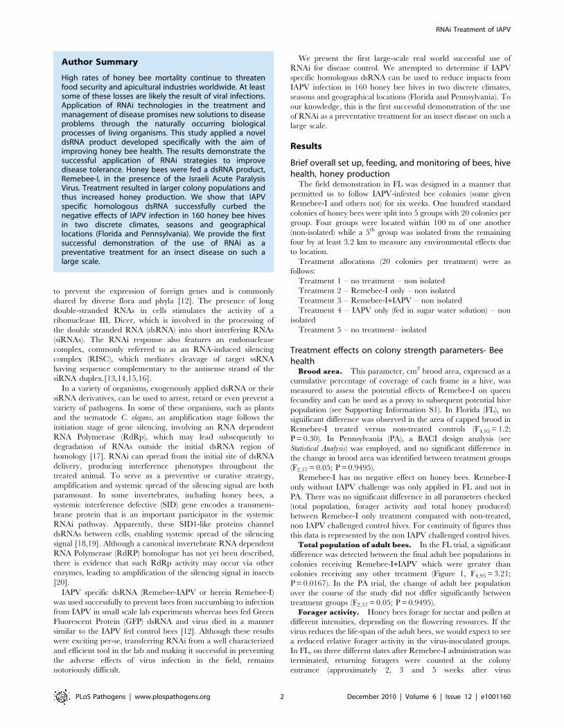
Total population of adult bees. In the FL trial, a significant
difference was detected between the final adult bee populations in
colonies receiving Remebee-I+IAPV which were greater than
colonies receiving any other treatment (Figure 1, F4,95 = 3.21;
P = 0.0167). In the PA trial, the change of adult bee population
over the course of the study did not differ significantly between
treatment groups (F2,57 = 0.05; P = 0.9495).
Forager activity. Honey bees forage for nectar and pollen at
different intensities, depending on the flowering resources. If the
virus reduces the life-span of the adult bees, we would expect to see
a reduced relative forager activity in the virus-inoculated groups.
In FL, on three different dates after Remebee-I administration was
terminated, returning foragers were counted at the colony
entrance (approximately 2, 3 and 5 weeks after virus
Author Summary
High rates of honey bee mortality continue to threatenfood security and apicultural industries worldwide. At leastsome of these losses are likely the result of viral infections.Application of RNAi technologies in the treatment andmanagement of disease promises new solutions to diseaseproblems through the naturally occurring biologicalprocesses of living organisms. This study applied a noveldsRNA product developed specifically with the aim ofimproving honey bee health. The results demonstrate thesuccessful application of RNAi strategies to improvedisease tolerance. Honey bees were fed a dsRNA product,Remebee-I, in the presence of the Israeli Acute ParalysisVirus. Treatment resulted in larger colony populations andthus increased honey production. We show that IAPVspecific homologous dsRNA successfully curbed thenegative effects of IAPV infection in 160 honey bee hivesin two discrete climates, seasons and geographicallocations (Florida and Pennsylvania). We provide the firstsuccessful demonstration of the use of RNAi as apreventative treatment for an insect disease on such alarge scale.
RNAi Treatment of IAPV
PLoS Pathogens | www.plospathogens.org 2 December 2010 | Volume 6 | Issue 12 | e1001160
administration, Supporting Information S1). This was done
simultaneously by several observers who moved between the
colonies and counted returning foragers for one minute for each
hive, and then rotated between the treatments to preclude any
possibility of bias. Although immediately after virus infection there
were no significant differences in forager counts, increasingly
larger significant differences in forager activity were noted as the
experiment progressed. In the first counting done 2 weeks after the
virus administration, mean number of returning foragers per
minute in the Remebee-I+IAPV was 37 (S.E.+/22.4) and the
comparable mean for the IAPV only group was 34 (S.E.+/21.88)
(t-test p.0.15 N.S.). However, 3 weeks post virus inoculation the
mean number of returning foragers per minute in the Remebee-
I+IAPV was 56 (S.E.+/22.8), whereas it totaled only 43 for IAPV
only (S.E.+/22.8) (t-test, p,0.01). At five weeks post virus
infection, the mean number of returning foragers per minute in
the Remebee-I+IAPV was 37 (S.E.+/21.8) yet only 22 in the
IAPV only group (S.E.+/21.6), (t-test, p,0.0001). This greater
relative activity in the Remebee-I+IAPV treatment in relation to
the IAPV only was somewhat correlated (rsqu = 0.35, p,0.0001)
with the greater adult bee population in the Remebee-I+IAPV
treatment in relation to the other treatments at the end of the
experiment (Figure 1). In the PA site, extreme variability in
returning forager counts, both at different times and between
observers precluded the use of this data.
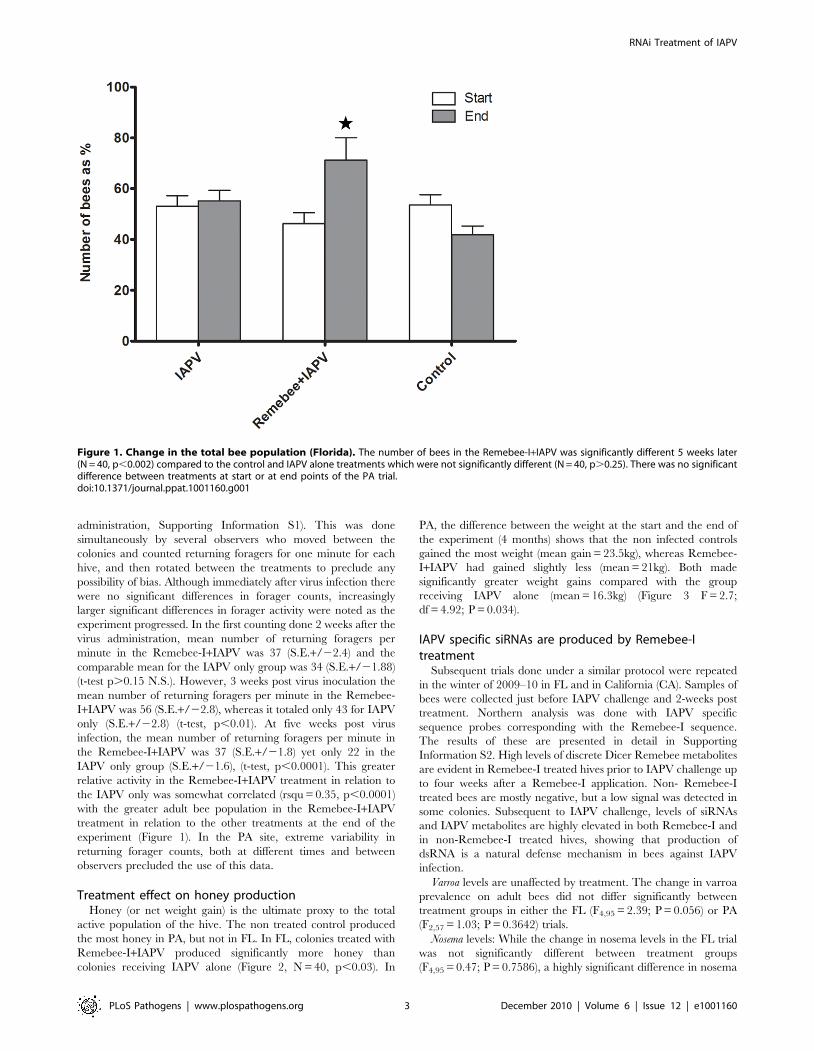
Treatment effect on honey productionHoney (or net weight gain) is the ultimate proxy to the total
active population of the hive. The non treated control produced
the most honey in PA, but not in FL. In FL, colonies treated with
Remebee-I+IAPV produced significantly more honey than
colonies receiving IAPV alone (Figure 2, N = 40, p,0.03). In
PA, the difference between the weight at the start and the end of
the experiment (4 months) shows that the non infected controls
gained the most weight (mean gain = 23.5kg), whereas Remebee-
I+IAPV had gained slightly less (mean = 21kg). Both made
significantly greater weight gains compared with the group
receiving IAPV alone (mean = 16.3kg) (Figure 3 F = 2.7;
df = 4.92; P = 0.034).
IAPV specific siRNAs are produced by Remebee-Itreatment
Subsequent trials done under a similar protocol were repeated
in the winter of 2009–10 in FL and in California (CA). Samples of
bees were collected just before IAPV challenge and 2-weeks post
treatment. Northern analysis was done with IAPV specific
sequence probes corresponding with the Remebee-I sequence.
The results of these are presented in detail in Supporting
Information S2. High levels of discrete Dicer Remebee metabolites
are evident in Remebee-I treated hives prior to IAPV challenge up
to four weeks after a Remebee-I application. Non- Remebee-I
treated bees are mostly negative, but a low signal was detected in
some colonies. Subsequent to IAPV challenge, levels of siRNAs
and IAPV metabolites are highly elevated in both Remebee-I and
in non-Remebee-I treated hives, showing that production of
dsRNA is a natural defense mechanism in bees against IAPV
infection.
Varroa levels are unaffected by treatment. The change in varroa
prevalence on adult bees did not differ significantly between
treatment groups in either the FL (F4,95 = 2.39; P = 0.056) or PA
(F2,57 = 1.03; P = 0.3642) trials.
Nosema levels: While the change in nosema levels in the FL trial
was not significantly different between treatment groups
(F4,95 = 0.47; P = 0.7586), a highly significant difference in nosema
Figure 1. Change in the total bee population (Florida). The number of bees in the Remebee-I+IAPV was significantly different 5 weeks later(N = 40, p,0.002) compared to the control and IAPV alone treatments which were not significantly different (N = 40, p.0.25). There was no significantdifference between treatments at start or at end points of the PA trial.doi:10.1371/journal.ppat.1001160.g001
RNAi Treatment of IAPV
PLoS Pathogens | www.plospathogens.org 3 December 2010 | Volume 6 | Issue 12 | e1001160
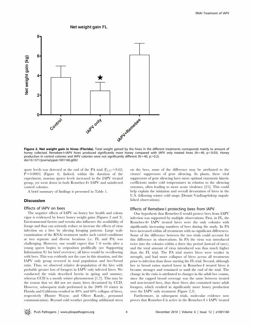
spore levels was detected at the end of the PA trial (F2,57 = 8.62;
P = 0.0005) (Figure 4). Indeed, within the duration of the
experiment, nosema spores levels increased in the IAPV treated
group, yet went down in both Remebee-I+ IAPV and uninfected
control colonies.
A brief summary of findings is presented in Table 1.
Discussion
Effects of IAPV on beesThe negative effects of IAPV on honey bee health and colony
vigor is evidenced by lower honey weight gains (Figures 2 and 3).
Environmental factors and terrain also influence the availability of
forage and thus can seriously reduce or increase the effects of virus
infection on a hive by altering foraging patterns. Large scale
examination of the RNAi treatment under such varied conditions
at two separate and diverse locations (i.e. FL and PA) was
challenging. However, one would expect that 7–8 weeks after a
young queen begins to oviposition prolifically (see Supporting
Information S1 for brief overview), all hives would be overflowing
with bees. This was evidently not the case in this situation, and the
IAPV only group reversed in total population and bee/brood
ratio. Thus, we observed relative de-population of the hive with
probable greater loss of foragers in IAPV only infected hives. We
conducted the trials described herein in spring and summer,
whereas CCD is a mostly winter phenomenon [1,7]. This may be
the reason that we did not see many hives devastated by CCD.
However, subsequent trials performed in the 2009–10 winter in
Florida and California resulted in 40% and 60% collapse of hives,
respectively (Hunter Wayne. and Oliver Randy., personnel
communication). Beyond cold weather providing additional stress
on the bees, some of the difference may be attributed to the
viruses’ suppressors of gene silencing. In plants, these viral
suppressors of gene silencing have more optimal enzymatic kinetic
coefficients under cold temperatures in relation to the silencing
enzymes, often leading to more acute virulence [21]. This could
help explain the initiation and overall devastation of hives in the
U.S. following winter cold snaps (Dennis VanEngelsdorp unpub-
lished observations).
Effects of Remebee-I protecting bees from IAPVOur hypothesis that Remebee-I would protect bees from IAPV
infection was supported by multiple observations: First, in FL, the
Remebee-I+ IAPV treated hives were the only colonies with
significantly increasing numbers of bees during the study. In PA
bees increased within all treatments with no significant differences.
Some of the difference between the two trials could account for
this difference in observations. In PA the virus was introduced
twice into the colonies within a three day period (instead of once),
and the total amount of virus introduced was thus much higher
than the FL trial. The PA trial starter hives were weaker in
strength, and had more collapses of hives across all treatments
prior to infection than those starting the FL trial. Second, although
bee to brood ratios started lower in Remebee-I treated hives it
became stronger and remained so until the end of the trial. The
change in the ratio is attributed to changes in the adult bee counts,
since the capped brood coverage was the same between treated
and non-treated bees, thus these hives also contained more adult
foragers, which resulted in significantly more honey production
over the IAPV only treatment (Figure 2,3).
Furthermore, in subsequent trials, molecular evidence now
proves that Remebee-I is active in the Remebee-I + IAPV treated
Figure 2. Net weight gain in hives (Florida). Total weight gained by the hives in the different treatments corresponds mainly to amount ofhoney collected. Remebee-I+IAPV hives produced significantly more honey compared with IAPV only treated hives (N = 40, p,0.03). Honeyproduction in control colonies and IAPV colonies were not significantly different (N = 40, p.0.2).doi:10.1371/journal.ppat.1001160.g002
RNAi Treatment of IAPV
PLoS Pathogens | www.plospathogens.org 4 December 2010 | Volume 6 | Issue 12 | e1001160

groups, as determined by the presence of siRNA (see Supporting
Information S2). The strong presence of siRNAs probably restricts
the severity of the disease in the bees leading to a longer life-span
and subsequently to an overall greater number of bees, with more
foragers and consequently a greater yield of honey. It is interesting
to note the natural occurrence of these siRNAs in bees receiving
IAPV challenge. Presence of these siRNA in non- Remebee-I
treated hives prior to infection may be a result of natural virus
infection prior to the challenge, or by transcription of integrated
viral sequences in the bee genome [22].
Honey production. The most obvious measure of bee hive
health is honey production. While overall honey production was
not on the levels of commercial production, due to the remote
locations and time of year, the Remebee-I + IAPV treated hives
still produced 30–300% more honey than the IAPV-only treated
hives in PA and FL respectively (Figures 2,3).
Natural modes of infection. Some bees in hives which were
neither treated with Remebee-I nor fed IAPV eventually became
infected with virus. There are several ways this may have
occurred. First, healthy bees and IAPV-infected bees may forage
on the same flowers, thus facilitating pathogen transmission
[7,23,24]. Secondly, bees may drift randomly between colonies.
Finally, viruses may be transferred through the residual varroa
mites [25]. Foragers from the Remebee-I + IAPV treatment may
have been protected from acute viral disease because they were
chronically infected with virus at very low levels, thus failing to
develop full symptoms and remaining apparently healthy. This
may be due to the virus being introduced together with Remebee-
I, which serves as a template for dsRNA/siRNA amplification,
thereby providing continued silencing of the virus. The recent
results of the dramatic amplification effect of the virus on the
siRNAs demonstrate this clearly (Supporting Information S2).
Remebee-I safety to bees. Remebee-I was shown to be
active in the Remebee-I+IAPV treated colonies by the presence of
siRNA in treated bees (Supporting Information S2). The high
titers of siRNAs probably restrict the severity of IAPV in bees. The
protection provided by Remebee-I appears to last throughout the
lifespan of an individual bee. Thus, after several weeks without the
application of Remebee-I to colonies, newly emerged bees which
were not fed the treatment may not be protected.
Notwithstanding, some evidence points to the possibility of
providing protection from virus to the adult bees through larval
feeding by the nurse bees [26].
Varroa mites. Varroa mites are arguably one of the largest
challenges to beekeepers. However, prevention of Varroa is
intensively practiced by beekeepers using a variety of miticides
and by application of best management practices. While Varroa
levels were found at equal levels in CCD and non-CCD colonies at
the time of sample collection, coumaphos, a miticide commonly
used by beekeepers, was found at higher levels in non-CCD versus
CCD colonies. This may suggest that non-CCD colonies had mite
levels more aggressively controlled some time prior to sample
collection, and the increased viral loads in CCD colonies is a
legacy effect of different mite levels some time before sample
collection [27]. Indeed, most ubiquitous bee viruses, with IAPV
amongst them, have been found in the Varroa mite and
transmission has been demonstrated [24,28].
CCD multi-pathogen syndrome. The evidence which ties
virus with nosema to CCD continues to increase. Nosema is one of
the most prevalent adult honey bee diseases and is caused by two
Figure 3. Net weight gain in hives (Pennsylvania). Total weight gained by the hives in the different treatments corresponds mainly to amountof honey collected. The difference between the weight at the start and the end of the experiment (4 months) shows that the non infected controls(mean = 23.5kg) and the Remebee-I+IAPV (mean = 21kg) made significantly more honey than hives receiving IAPV alone (mean = 16.3kg) (F = 2.7;df = 4.92; P = 0.034).doi:10.1371/journal.ppat.1001160.g003
RNAi Treatment of IAPV
PLoS Pathogens | www.plospathogens.org 5 December 2010 | Volume 6 | Issue 12 | e1001160
described species of microsporidia (Nosema apis and Nosema ceranae,
N. apis and N. ceranae, respectively). Researchers from Spain
showed that natural N. ceranae infection can induce the sudden
collapse of bee colonies, establishing a direct correlation between
N. ceranae infection and the death of honey bee colonies [29].
Nosema ceranae has existed in honey bees within the United States
since at least 1996 [30] without any reported dramatic colony
declines until recently. However, an association between viruses
(IAPV and others) with Nosema in CCD colonies has been
established [7,11]. Our data further supports this association. In
the PA trial, colonies treatment with Remebee-I, which reduced
virus levels, may have led to a concurrent reduction in nosema
levels comparable to that of the control non-virus inoculated
colonies (Figure 4), further supporting the association of acute
virus disease and elevated Nosema levels in hive collapse. Recently it
was shown that an RNAi strategy against Nosema is also efficacious
[31], so using RNAi to target both viruses and Nosema in concert is
now feasible.
We postulate that foragers from Remebee-I + IAPV treated
hives were protected from this acute viral disease, because the
siRNAs they produced protected them and enabled chronic
infection with virus at very low levels. These bees thus failed to
develop symptoms and remained sufficiently healthy to support
longer forager activity. This may be due to the concurrent
Figure 4. Nosema spore counts per bee. Nosema spore counts at the 4 weeks and at the end point of the Pennsylvania trial. At 4 weeks after virusintroduction, IAPV only and Remebee-I+IAPV Nosema spore counts were elevated in contrast to the uninfected control, but insignificantly so becauseof the large variance. However, 8 weeks later, levels of Nosema spores were significantly greater in the IAPV only group (1.02 million per bee) relativeto the uninfected control (0.36 million per bee) (N = 31, p,0.006). Remarkably, spore levels in the treatment group receiving Remebee-I+IAPV (0.33million per bee) were not significantly different from the uninfected control (N = 30, p.0.89), but were significantly different from the IAPV onlycluster (N = 27, p,0.007). BACI design; Proc mixed = repeated measured analysis with unstructured covariance matrix. No effect of treatment overtime. Df = 4, 95; F = 0.47; P = 0.7586.doi:10.1371/journal.ppat.1001160.g004
RNAi Treatment of IAPV
PLoS Pathogens | www.plospathogens.org 6 December 2010 | Volume 6 | Issue 12 | e1001160
presence of virus together with Remebee-I whereby the replicating
viral genome serves as a template for dsRNA/siRNA amplification
via virus or host encoded RdRp that may be amplifying the
silencing signal.
ConclusionsIAPV specific dsRNA (Remebee-I) was used successfully to
prevent bees from succumbing to infection from IAPV. The results
further demonstrate the possibility to produce targeted treatments
for bee pathogenic diseases. These field results demonstrate the
successful application of dsRNA as a viable treatment to solve a
real world problem, which may further lead to concerted efforts to
utilize this ubiquitous natural mechanism, RNAi, for the benefit of
the bees, beekeepers, and hopefully to other applications in
agriculture and veterinary health.
Materials and Methods
To determine if IAPV can be silenced using RNAi technology,
we had to (1) purify IAPV from honey bees, (2) infect honey bee
colonies with IAPV and/or Remebee-I and (3) determine IAPV
presence in experimental colonies.
dsRNA synthesisEssentially as described in [11].
IAPV purificationApproximately 40 adult forager honey bees were collected from
10 colonies in a Florida bee yard (apiary) where CCD had been
reported. Each bee was processed individually and tested using
rtPCR for the presence of Israeli Acute Paralysis Virus (IAPV),
genome – NC_009025; Acute Bee Paralysis Virus (ABPV) genome
– NC_002548; Kashmir Bee Virus (KBV) genome – NC_004807;
Black Queen Cell Virus (BQCV), genome-NC_003784; Deformed
Wing Virus (DWV) genome NC_004830. All bees had more than
one virus detected so inoculum was prepared from bees which
tested positive only for IAPV+KBV by homogenizing the bees
with glass beads in small amounts of 10 mM buffer phosphate,
pH 7.2 containing 0.02% DETCA (Sigma-Aldrich Cat #22,868-
0). Inoculum was prepared by passing the virus solution through a
syringe filter, 0.45 mm, to remove bacteria, after which ,10 ml
were administered by microinjection along the lateral side of the
abdomen of ,700 pupae using a Hamilton syringe with a 30GxK
gauge sterile needle. Inoculated pupae were kept in petri dishes
covered with slightly damp filter paper and maintained at 21–
23uC for three days to permit virus replication.
Virus purificationOn the third day, batches of about 50 pupae were homogenized
with glass beads. Small amounts of 10 mM buffer phosphate
(pH 7.2 contained 0.02% DETCA, Sigma-Aldrich Cat #22,868-
0) were added to the homogenates. The homogenates were
collected in a beaker volume adjusted to ,350 ml with buffer (see
above) and mixed. Each sample was split into two 250 ml
centrifuge tubes and centrifuged at 3006g (,1,400 rpm) on a
GSA rotor for 20 min. The supernatant (S1) was collected and
kept at 4uC for 3 d. The pellet (P1) was recovered and saved at
4uC. Since some precipitation was noticed after 3 d, the
supernatant was centrifuged again as before for 10 min to remove
debris. Supernatant (S1) next was transferred to 12 ultracentrifuge
tubes (about 26 ml/tube) (Beckman Cat #355618) and centri-
fuged for 4 h, 4uC, at 37,000 rpm (,124,5006g) (Beckman Type
50.2 Ti rotor, Beckman Optima L-70K Ultracentrifuge). After 4 h
the supernatant (S2) was removed and saved. The pellet (P2) was
resuspended in 10 mM phosphate buffer containing 4% Brij 58
(Aldrich Cat #388831) and 0.4% Sodium deoxycholate (Sigma-
Aldrich D6750): about 1 ml of buffer was used per tube. It was
necessary to insert a spatula to help pellet into solution and this
was followed by vortexing the suspension. The content from each
tube was transferred to clean 50 ml centrifuge tubes. The process
was repeated twice but only buffer phosphate was added the final
time. Because the final solution was very thick, buffer was added to
increase the final volume to ,30 ml and this was mixed by
inversion. The tube was centrifuged for 15 min at ,10uC, 8006g
(Beckman Coulter Allegra 25R), to remove debris. The pellet (P3)
was saved at 4uC. (the pellet saved as a backup). The supernatant
(S2) was transferred into two clean 50 ml tubes and 13.2 g CsCl
(Amresco Cat #0415) were added to each tube. To ensure the
right CsCl concentration, 13.2 g CsCl were added to ,10 g
sample; however the final volume was adjusted to 24 ml with
buffer and gently mixed (up/down). The second tube was set by
adding CsCl to the remaining sample. This final preparation was
transferred to two ultracentrifuge tubes ,25 ml (Beckman Cat
#355618) and centrifuged at 37,000 rpm (,124,5006g), 18uC for
24 h. After 24 h centrifugation, the tubes were removed carefully
from the rotor and the whitish virus band collected by insertion of
a needle attached to a syringe. Two more fractions were recovered
for analyses: (1) the ‘‘liquid’’ part left after removing the virus band
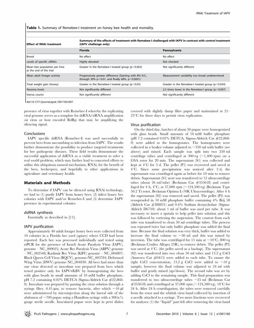
Table 1. Summary of Remebee-I treatment on honey bee health and mortality.
Effect of RNAi treatmentSummary of the effects of treatment with Remebee-I challenged with IAPV in contrast with control treatment(IAPV challenge only)
Florida Pennsylvania
Brood No effect No effect
Levels of specific siRNAs Highly elevated Not checked
Mean bee population per hiveat the end of the trial
Greater in the Remebee-I treated group (p,0.002) Not significantly different
Mean adult forager activity Progressively greater difference (Starting with 8% N.S.,through 30% p,0.01, and finally 68%, p,0.0001)
Measurement variability too broad undetermined
Total weight gain (honey) Greater in the Remebee-I treated group (p,0.03) Greater in the Remebee-I treated group (p,0.034)
Nosema levels Not significantly different 2.5 times lower in the Remebee-I group (p,0.007)
Varroa counts Not significantly different Not significantly different
doi:10.1371/journal.ppat.1001160.t001
RNAi Treatment of IAPV
PLoS Pathogens | www.plospathogens.org 7 December 2010 | Volume 6 | Issue 12 | e1001160
and (2) the ‘‘pellet’’ (P4) attached to the bottom of tube: Each
fraction was transferred to dialysis tubes (Thomas Scientific Cat
#3787-F42) and dialyzed overnight against nanopure filtered
water followed by 3–4 additional changes in water the following
day. After dialysis, content from the tubes was collected in 15 ml
clean tubes and the volumes were measured. A subsample of 20 ml
from each fraction was tested for virus presence.
RNA extractionAdult bees were transferred to 1.5 ml centrifuge tubes. Tri
Reagent (Sigma Cat #T9424), was added and individual bees
were homogenized in 0.5 ml Tri reagent using disposable pestles
and glass beads. Homogenates were frozen at 220uC if needed.
Samples then were centrifuged 10 min at 12,0006g, at 4uC. The
clear supernatant was transferred to a new tube and left at least
5 min at room temperature (RT). Next, 0.2 ml chloroform was
added and samples were shaken vigorously. This was followed by
a10–15 min incubation at RT. Tubes were centrifuged 15 min at
12,0006g at 4uC. The colorless upper aqueous phase was
transferred to a new tube and 0.5 ml isopropanol was added.
After mixing, samples were allowed to stand for 10 min then spun
10 min at 12,0006g at 4uC. The supernatant was removed and
the pellet containing the RNA was washed with 1 ml 75% ethanol.
After 5 min centrifugation at 7,5006g at 4uC, the RNA was
allowed to dry (5–10 min) and reconstitute in ,30 ml Nuclease
free water (Qiagen). RNA concentrations were measured in a
Nanodrop, ND-1000 Spectrophotometer. Samples were diluted in
Nuclease free water.
Overall set up, feeding, monitoring of bees, hive health,and honey production
The field demonstration in FL was designed in a manner that
permitted us to follow IAPV-infested bee colonies (some given
Remebee-I and others not) for six weeks. One hundred standard
colonies of honey bees were split into 5 groups with 20 colonies per
group. Four groups were located within 100 m of one another (non-
isolated) while a 5th group was isolated from the remaining four by at
least 3.2 km to measure any environmental effects due to location.
Treatment allocations (20 colonies per treatment) were as
follows:
Treatment 1 – no treatment – non isolated
Treatment 2 – Remebee-I only – non isolated
Treatment 3 – Remebee-I+IAPV – non isolated
Treatment 4 – IAPV only (fed in sugar water solution) – non
isolated
Treatment 5 – no treatment– isolated
Colonies were equalized according to standard protocols prior
to the beginning of the study (frames of bees/brood moved
between colonies until populations leveled) and were managed
optimally for honey production. Data collected at the beginning,
middle, and end of the study included: frames of adult bees, cm2
brood, the presence of other bee maladies (nosema, varroa, and
tracheal mites), bee activity, honey production and IAPV
presence/absence and titer. The study lasted 6 weeks from the
date of colony inoculation with IAPV and was replicated in PA
with the following modifications: Only Treatment groups 1, 3 and
4 were established and the trial lasted 12 weeks after inoculation to
enable the bees to take advantage of a honeyflow (Tables 2 and 3).
In PA the virus was introduced twice into the colonies within a
three day period (instead of once as in FL), and the total amount of
virus introduced was thus much higher than the FL trial. We
calculated all test/sampling dates below from the date of the last
treatment with Remebee-I. Controls 1, 2, and 5, accounted for
‘within treatments’, a Remebee-I alone treatment to evaluate any
potential detrimental effects to bees, and a distant control to
measure environmental effects in the absence of IAPV.
Frames of adult bees. This parameter was measured only at
weeks 2.5 and 5. Two observers estimated (in tenths) the amount
of frame area covered by bees for every frame in each colony. The
scores from both observers were averaged and converted to
number of bees per colony [32]. Estimates as % of total frame.
Each frame is raised and the observer notes percentage coverage
on both sides of the frame. If all the frame side is completely
covered with bees, the observer scores 1, and if there are no bees
on the frame side, he scores 0. Any other coverage is estimated by
the observer from 0 to 1 in whole numbers. Two trial observers
went through the hives independently, both at interim and final
dates. [33]
Total brood area. This parameter was measured only at
weeks 2.5 and 5. Two observers estimated total capped brood as %
frame coverage. Each frame is raised and the observer notes %
coverage on both sides of the frame. If all the frame side is covered
with capped brood, the observer scores 1, and if there is no capped
brood at all, he scores 0. Any other coverage is estimated (in
tenths) the amount of brood (of any stage in development) covering
both sides of a single frame for every frame in each colony. Since a
queen excluder was placed between the deep and the honey super,
there was no brood in the honey super at the final date. The scores
from both observers were averaged and converted to cm2 brood
based on the observation that one deep Langstroth comb (both
sides) = 1754 cm2 [33].
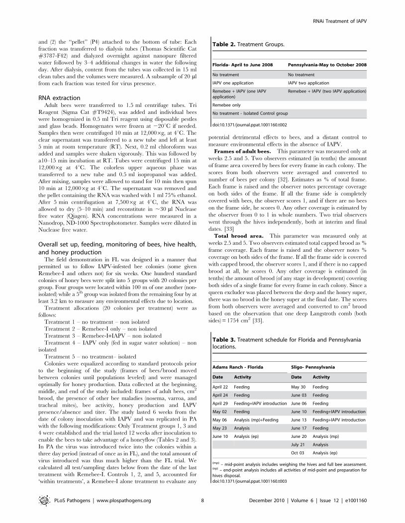
Table 2. Treatment Groups.
Florida- April to June 2008 Pennsylvania-May to October 2008
No treatment No treatment
IAPV one application IAPV two application
Remebee + IAPV (one IAPVapplication)
Remebee + IAPV (two IAPV application)
Remebee only
No treatment - Isolated Control group
doi:10.1371/journal.ppat.1001160.t002
Table 3. Treatment schedule for Florida and Pennsylvanialocations.
Adams Ranch - Florida Sligo- Pennsylvania
Date Activity Date Activity
April 22 Feeding May 30 Feeding
April 24 Feeding June 03 Feeding
April 29 Feeding+IAPV introduction June 06 Feeding
May 02 Feeding June 10 Feeding+IAPV introduction
May 06 Analysis (mp)+Feeding June 13 Feeding+IAPV introduction
May 23 Analysis June 17 Feeding
June 10 Analysis (ep) June 20 Analysis (mp)
July 21 Analysis
Oct 03 Analysis (ep)
(mp) – mid-point analysis includes weighing the hives and full bee assessment.(ep) – end-point analysis includes all activities of mid-point and preparation forhives disposal.doi:10.1371/journal.ppat.1001160.t003
RNAi Treatment of IAPV
PLoS Pathogens | www.plospathogens.org 8 December 2010 | Volume 6 | Issue 12 | e1001160
Honey production. Deep and Super boxes were weighed in
kilograms. Prior to introducing the super on top of each deep, the
super with the empty frames was weighed in the field and marked.
Deeps were numbered 1–100 according to hive numbers, whereas
supers were numbered 101–200, respectively. At the final end point
both deeps and supers were weighed again, and the net gain was
recorded. In PA, to determine the amount of honey produced by
each colony, empty supers were weighed empty and added to
colonies prior to any nectar flow. They were added to colonies as
needed throughout the study. Due to delayed flower bloom the
weights were taken as scheduled, but then again 4 weeks later to
adjust for environmental differences between PA and FL seasons.Bee activity counts. Hive activity at all colonies was
determined at weeks 0, 2.5 and 5 by having multiple observers
simultaneously counting the number of bees landing at the hive
entrance for 60 seconds, for all hives in all treatments (Supporting
Information S1).Presence of other bee maladies. Samples of 300 bees were
placed in 70% ethanol on 2 April (before any feeding with
Remebee-I and/or virus) and at the end of week 5. The samples
were used to determine initial and ending nosema, varroa mite,
and tracheal mite loads. Samples were analyzed at the University
of Florida Honey Bee Research and Extension Lab.Feeding regimes. After feeding colonies once with 0.5 L of
sugar water (66% sugar by volume) in glass feeding jars to
ascertain that all hives were consuming the food, another five (FL)
or six (PA) consecutive feedings of 0.5 L were given to colonies
over a 2–3 week period. Thereafter, an empty honey super was
added to all colonies and no further feedings occurred.
1) No treatment = Five or six feedings of 0.5 L sugar water per
hive (FL and PA).
2) IAPV only treatment = In FL: five feedings of 0.5 L sugar
water per hive, with feedings three, four, and five containing
500 micrograms IAPV per hive, and two of only sugar
water. In PA: five feedings of 0.5 L sugar water per hive,
with both feedings four and five supplemented with 500
micrograms of virus per hive.
3) Remebee-I + IAPV = Five or six feedings with 0.5 L sugar
water supplemented with 10 mg Remebee-I per hive per
feeding, with feeding three (FL) or both feedings four and
five (PA) having an addition of 500 microgram purified
IAPV per hive, per application.
4) Remebee-I only treatment = Five or six feedings with 0.5 L
sugar water supplemented with 10 mg Remebee-I per hive
per feeding (FL only).
5) Remote Control = The isolated untreated control of 20 hives
was placed ,1.8 miles due East from the test site, and provided
five feedings of 0.5 L of sugar water per hive (FL only).
Statistical analysisColonies were equalized at the beginning of the studies, starting
and ending colony strength parameters were compared using
ANOVA recognizing treatment as the main effect (PROC GLM).
Honey gains in treated colonies in FL and PA were compared
identically. However, to compare colony size measures in the PA
trial and for levels of nosema and varroa mites, a Before-After
Control-Impact (BACI) design [33,34] was used. A BACI design
us a way of comparing data that are measured before treatment
with data obtained after treatment. In general, it can be described
as a repeated measures analysis of variance (ANOVA) which is
performed using colonies as replicates and the covariance structure
that best suits the data (PROC MIXED, SAS Institute). Each
variable is measured at the start of the experiment to show existing
conditions before treatment and then after a treatment. The
analysis then looks at whether the change in variable measures was
different between treatment groups. A repeated measures analysis
of variance [34] was performed using colonies as replicates and an
unstructured covariance structure was performed using SAS
statistical software (PROC MIXED) [35].
Accession numbers mentioned in textIsraeli Acute Paralysis Virus (IAPV) genome – NC_009025
(RefSec); Acute Bee Paralysis Virus (ABPV) genome – NC_002548
(RefSec); Kashmir Bee Virus (KBV) genome – NC_004807
(RefSec); Black Queen Cell Virus (BQCV), genome-NC_003784
(RefSec); Deformed Wing Virus (DWV) genome NC_004830
(RefSec); RNA dependent RNA Polymerase protein (C. elegans)–
NP_492131 (RefSec); SID-1 protein (A. mellifera) – XP_395167
(RefSec); GFP nucleotide sequence – U87625 (GenBank).
Supporting Information
Supporting Information S1 Bee hives, feeding unit and field
set up.
Found at: doi:10.1371/journal.ppat.1001160.s001 (0.43 MB PDF)
Supporting Information S2 Detection of siRNAs in bee
samples.
Found at: doi:10.1371/journal.ppat.1001160.s002 (0.99 MB PDF)
Acknowledgments
We thank Maria Gonzalez, Christine Lynch, Biological science techni-
cians, USDA, ARS, Ft. Pierce, FL; Hanna O’Malley, Renee Cole,
technicians University of Florida, Gainesville, FL; and Dr. Yuval Peretz,
and Dr. Rita Koch Mozes, Alex Inberg. We also acknowledge
contributions from the Adams Ranch, FL and RFI Energy, PA for
providing property and resources to conduct experimental trial. Special
thanks to Dave Hackenberg and David Mendes for providing 100+ hives
each used in PA and FL respectively. Dr. Michael Braverman, Head of
biopesticide division at the Federal IR4 project for regulatory information.
Author Contributions
Conceived and designed the experiments: JE DV JH JP NP. Performed the
experiments: WH JE DV JH DW EG MW IS EM JP DCF NP. Analyzed
the data: DV NP. Contributed reagents/materials/analysis tools: WH JE
DV JH DW EG MW IS JP DCF NP. Wrote the paper: WH JE DV EM
NP. Organized regulatory collaborations and inspections: JE DV JH DW
MW NP.
References
1. vanEngelsdorp D, Meixner MD (2010) A historical review of managed honey
bee populations in Europe and the United States and the factors that may affect
them. J Invertebr Pathol 103 Suppl 1: S80–95.
2. Klein AM, Vaissiere BE, Cane JH, Steffan-Dewenter I, Cunningham SA, et al.
(2007) Importance of pollinators in changing landscapes for world crops. Proc
Royal Soc London Series B, Biol Sci 274: 303–313.
3. Gallai N, Salles J-M, Settele J, Vaissiere BE (2009) Economic valuation of the
vulnerability of world agriculture confronted with pollinator decline. Ecol Econ
68: 810–821.
4. Neumann P, Carreck NL (2010) Honey bee colony losses. J Apic Res 49: 1–6.
5. vanEngelsdorp D, Underwood R, Caron D, Hayes J, Jr. (2007) An estimate of
managed colony losses in the winter of 2006–2007: A report commissioned by
the Apiary Inspectors of America. Amer Bee Jour 147: 599–603.
6. Cox-Foster DL, Conlan S, Holmes EC, Palacios G, Evans JD, et al. (2007) A
metagenomic survey of microbes in honey bee colony collapse disorder. Science318: 283–287.
7. vanEngelsdorp D, Evans JD, Saegerman C, Mullin C, Haubruge E, et al. (2009)
Colony Collapse Disorder: A Descriptive Study. PloS ONE 4: e6481.
RNAi Treatment of IAPV
PLoS Pathogens | www.plospathogens.org 9 December 2010 | Volume 6 | Issue 12 | e1001160
8. Johnson RM, Evans JD, Robinson GE, Berenbaum MR (2009) Changes in
transcript abundance relating to colony collapse disorder in honey bees (Apis
mellifera). Proc Natl Acad Sci USA 106: 14790–14795.
9. Ellis JD, Munn PA (2005) The worldwide health status of honey bees. Bee World
86: 88–101.
10. Highfield AC, El Nagar A, Mackinder LC, Noel LM, Hall MJ, et al. (2009)
Deformed wing virus implicated in overwintering honeybee colony losses. Appl
Environ Microbiol 75: 7212–7220.
11. Maori E, Paldi N, Shafir S, Kalev H, Tsur E, et al. (2009) IAPV, a bee-affecting
virus associated with Colony Collapse Disorder can be silenced by dsRNA
ingestion. Ins Mol Biol 18: 55–60.
12. Price DR, Gatehouse JA (2008) RNAi-mediated crop protection against insects.
Trends Biotech 26: 393–400.
13. Mathieu O, Bender J (2004) RNA-directed DNA methylation. J Cell Sci 117:
4881–4888.
14. Matzke M, Aufsatz W, Kanno T, Daxinger L, Papp I, et al. (2004) Genetic
analysis of RNA-mediated transcriptional gene silencing. Biochim Biophys Acta
1677: 129–141.
15. Brodersen P, Voinnet O (2006) The diversity of RNA silencing pathways in
plants. Trends Genet 22: 268–280.
16. Zaratiegui M, Irvine DV, Martienssen RA (2007) Noncoding RNAs and gene
silencing. Cell 128: 763–776.
17. Siomi H, Siomi M (2009) On the road to reading the RNA-interference code.
Nature 457: 396–404.
18. The Honey Bee Consortium (2006) Insights into social insects from the genome
of the honeybee Apis mellifera. Nature 443: 931–949.
19. Aronstein K, Pankiw T, Saldivar E (2006) SID-I is implicated in systemic gene
silencing in the honey bee. J Apiculture Res Bee World 45: 20–24.
20. Lipardi C, Paterson BM (2009) Identification of an RNA-dependent RNA
polymerase in Drosophila involved in RNAi and transposon suppression. PNAS
106: 15645–15650.
21. Feng Q, Morris TJ (2005) Suppressors of RNA silencing encoded by plant
viruses and their role in viral infections. FEBS Letters 579: 5958–5964.
22. Maori E, Lavi S, Mozes-Koch R, Gantman Y, Peretz Y, et al. (2007) Isolation
and characterization of Israeli acute paralysis virus, a dicistrovirus affecting
honeybees in Israel: evidence for diversity due to intra- and inter-species
recombination. J Gen Virol 88: 3428–3438.23. Cox-Foster D, vanEngelsdorp D (2009) Solving the mystery of the disappearing
bees. Sci Amer 300: 40–47.
24. vanEngelsdorp D, Hayes J, Jr., Underwood RM, Pettis J (2008) A survey ofhoney bee colony losses in the US, fall 2007 to spring 2008. PLoS One 3: e4071.
25. Chen YP, Pettis JS, Collins A, Feldlaufer MF (2006) Prevalence and transmissionof honeybee viruses. Appl Environ Microbiol 72: 606–611.
26. Liu X, Zhang Y, Yan X, Han R (2010) Prevention of Chinese sacbrood virus
infection in Apis ceranae using RNA interference. Curr Microbiol (online) DOI10.1007/s00284-010-9633-2.
27. Rosenkranz P, Aumeier P, Ziegelmann B (2010) Biology and control of Varroa
destructor. J Invertebr Pathol 103 Suppl 1: S96–119.
28. Bowen-Walker PL, Martin SJ, Gunn A (1999) The transmission of deformedwing virus between honeybees (Apis mellifera L.) by the ectoparasitic mite Varroa
jacobsoni Oud. J Invertebr Pathol 73: 101–106.
29. Higes M, Martin-Hernandez R, Botias C, Bailon EG, Gonzalez-Porto AV, et al.(2008) How natural infection by Nosema ceranae causes honeybee colony collapse.
Environ Microbiol 10: 2659–2669.30. Chen YP, Evans JD, Smith IB, Pettis JS (2008) Nosema ceranae is a long-present
and wide-spread microsporidian infection of the European honey bee (Apis
mellifera) in the United States. J Invertebr Pathol 97: 186–188.31. Paldi N, Glick E, Oliva M, Zilberberg Y, Aubin L, Pettis JS, Chen YP, Evans JD
(2010) Effective gene silencing of a microsporidian parasite associated withhoney bee (Apis mellifera) colony declines. Appl Environ Microbiol 76:
5960–5964.32. Burgett M, Burikam I (1985) Number of adult honey bees (Hymenoptera:
Apidae) occupying a comb: a standard for estimating colony populations. J Econ
Entomol 78: 1154–1156.33. Stewart-Oaten A, Murdoch WW, Parker KR (1986) Environmental impact
assessment: ‘‘Pseudoreplication’’ in Time? Ecology 67: 929–940.34. Smith EP (2002) BACI design. In: El-Shaarawi AH, Piegorsch WW, eds.
Encyclopedia of Environmetrics. New York, New York, USA: John Wiley and
Sons. pp 141–148.35. Institute SAS (2004) SAS computer program, version 9.1.3 By Institute, S,
Carry, NC.
RNAi Treatment of IAPV
PLoS Pathogens | www.plospathogens.org 10 December 2010 | Volume 6 | Issue 12 | e1001160