Oocytes, Larvae, and Cleptoparasitic Behavior of Biastes emarginatus (Hymenoptera: Apidae:...
16
PUBLISHED BY THE AMERICAN MUSEUM OF NATURAL HISTORY CENTRAL PARK WEST AT 79TH STREET, NEW YORK, NY 10024 Number 3667, 15 pp., 30 figures, 1 table November 30, 2009 Oocytes, Larvae, and Cleptoparasitic Behavior of Biastes emarginatus (Hymenoptera: Apidae: Nomadinae: Biastini) JEROME G. ROZEN, JR., 1 JAKUB STRAKA, 2 AND KATERINA REZKOVA 2 ABSTRACT We present information on the nest-searching and parasitic behavior of the European cleptoparasitic bee Biastes emarginatus (Schenck), found attacking nests of Rophites quinquespi- nosus Spinola in the Czech Republic. Its mature oocyte, first instar, and last larval instar are described and illustrated by SEM micrographs, microphotographs, and diagrams. These stages are compared with those of other members of the Biastini. Because the first instar of the related Neopasites cressoni had not been described before, its description is appended, so that comparisons can be made with B. emarginatus. In most respects, the biology and immature stages of B. emarginatus closely resemble what is known concerning other tribal members, but we note that the mature larva, though agreeing morphologically with those of close relatives, has an anatomy that invites investigation into adaptive function. INTRODUCTION This study reports on the biology and imma- ture stages of the cleptoparasitic bee Biastes emarginatus (Schenck) and compares the infor- mation with existing knowledge concerning other members of the Biastini, one of the numerous tribes of the Nomadinae, all of which are cleptoparasites. This small tribe of three genera totalling12 named species is restricted to the Holarctic region, with Neopasites and Rhopalolemma in North America and Biastes in the Palearctic (Michener, 2007). Known hosts of all biastines belong to the Rophitinae (Halictidae), as follows (host names in parenthe- ses): Neopasites (Dufourea), Rhopalolemma (Protodufourea), Biastes (Systropha, Rophites, and Dufourea) (Michener, 2007; Westrich, 1989). Copyright E American Museum of Natural History 2009 ISSN 0003-0082 1 Division of Invertebrate Zoology, American Museum of Natural History ([email protected]). 2 Faculty of Science, Charles University in Prague, Department of Zoology, Vinic ˇna ´ 7, 128 44 Praha 2, Czech Republic ([email protected]).
Transcript of Oocytes, Larvae, and Cleptoparasitic Behavior of Biastes emarginatus (Hymenoptera: Apidae:...

PUBLISHED BY THE AMERICAN MUSEUM OF NATURAL HISTORY
CENTRAL PARK WEST A T 79TH STREET, NEW YOR K, NY 10024
Number 3667, 15 pp., 30 figures, 1 table November 30, 2009
Oocytes, Larvae, and Cleptoparasitic Behavior ofBiastes emarginatus (Hymenoptera: Apidae:
Nomadinae: Biastini)
JEROME G. ROZEN, JR.,1 JAKUB STRAKA,2 AND KATERINA REZKOVA2
ABSTRACT
We present information on the nest-searching and parasitic behavior of the Europeancleptoparasitic bee Biastes emarginatus (Schenck), found attacking nests of Rophites quinquespi-nosus Spinola in the Czech Republic. Its mature oocyte, first instar, and last larval instar aredescribed and illustrated by SEM micrographs, microphotographs, and diagrams. These stages arecompared with those of other members of the Biastini. Because the first instar of the relatedNeopasites cressoni had not been described before, its description is appended, so that comparisonscan be made with B. emarginatus. In most respects, the biology and immature stages of B.emarginatus closely resemble what is known concerning other tribal members, but we note that themature larva, though agreeing morphologically with those of close relatives, has an anatomy thatinvites investigation into adaptive function.
INTRODUCTION
This study reports on the biology and imma-ture stages of the cleptoparasitic bee Biastesemarginatus (Schenck) and compares the infor-mation with existing knowledge concerningother members of the Biastini, one of thenumerous tribes of the Nomadinae, all of whichare cleptoparasites. This small tribe of three
genera totalling12 named species is restricted tothe Holarctic region, with Neopasites andRhopalolemma in North America and Biastesin the Palearctic (Michener, 2007). Knownhosts of all biastines belong to the Rophitinae(Halictidae), as follows (host names in parenthe-ses): Neopasites (Dufourea), Rhopalolemma(Protodufourea), Biastes (Systropha, Rophites,and Dufourea) (Michener, 2007; Westrich, 1989).
Copyright E American Museum of Natural History 2009 ISSN 0003-0082
1 Division of Invertebrate Zoology, American Museum of Natural History ([email protected]).2 Faculty of Science, Charles University in Prague, Department of Zoology, Vinicna 7, 128 44 Praha 2, Czech Republic

Beyond host associations, biological infor-mation concerning these cleptoparasitic gen-era is limited. Torchio et al. (1967) describedhost-searching behavior of adults, the eggs,egg-laying habits, and larval behavior ofNeopasites cressoni Crawford. Rozen et al.(1997) reported on various aspects of thebiology of Rhopalolemma rotundiceps Roig-Alsina. However, details concerning the biol-ogy of Biastes have been lacking.
The immature stages of these three generahave been examined on a number of occasions.Egg/mature oocytes of exemplars of all generahave been described. Most of these descriptionswere accompanied by information concerningthe number of ovarioles per ovary found indissections because among the Nomadinae thereis extensive variation in ovariole counts fromone species to the next. Egg/oocyte descriptionsare as follows: Neopasites cressoni (Torchio etal., 1967; Rozen et al., 1997); Rhopalolemmarotundiceps (Rozen et. al., 1997); Biastes brevi-cornis (Panzer) (Rozen and Ozbek, 2003).Information concerning ovary statistics is sum-marized in Rozen (2003). Descriptions of firstinstars of the Biastini have been lacking untilnow. Because we describe here the first instar ofBiastes emarginatus, we append a comparativeaccount of the first instar of N. cressoni. Maturebiastine larvae have been described, as follows:N. cressoni (Rozen, 1966, 1996a; McGinley,1981) and R. rotundiceps (Rozen et al., 1997).An intermediate stage larva of Biastes emargi-natus was treated by Rozen (1993), as was thepupa of N. cressoni (Rozen, 1997).
BIOLOGICAL OBSERVATIONS
J.S. and K.R. made the following observa-tions on Biastes emarginatus at a dense nest-ing aggregation of Rophites quinquespinosusSpinola in a large, unattended garden inPraha-Miskovice, a district of Prague, CzechRepublic. This nesting site was discovered in2006 and checked yearly since then. TheRophites nest aggregation occupied about a 33 3 m section of a 40–45u sloping surface on thesunny edge of a deciduous forest. More than 40nests were observed there at a single time duringthe course of our observations. The site waspartly shielded by the forest canopy from rain,particularly by one old linden tree (Tilia cordata
Miller), but much of the area received consid-erable sunlight during a large part of sunnydays. Ground cover, mostly tall grasses (ca30 cm high), obscured much of the surface, sothat many nest entrances were partly orcompletely hidden. Nest entrances, surroundedby tumuli when fresh, were left opened duringforaging. The main burrows of host nestsdescended vertically with sharp turns to avoidrocks. Cells in a nest were arranged singly (i.e.,not in linear series). Scattered nests were founda few meters away from the main site, butactivity of B. emarginatus was low there. Atleast 10 Biastes females flew over the main partof the nest aggregation during the peak ofactivity on sunny days.
The aggregation was studied during 3–18August 2008. Rophites females were activefrom early morning to late afternoon, flyingeven when the sky was overcast. In contrast,flight activity of Biastes depended on weatherconditions. They flew only during warm,sunny, windless days (temperatures 25–30uC),and on such days they preferred host nestssituated in sunny places. In addition, search-ing Biastes females moved with the sunnyareas as these areas shifted during the day.Only during the warmest days, when thetemperature rose over 30uC, did Biastesfemales prefer shaded parts of the nesting site.When the temperature fell below 25uC, thebees flew strictly in shafts of sunlight or wereinactive, behaving similarly to the thermoph-ilous behavior of another cleptoparasitic beeEpeoloides coecutiens (Fabricius) (Straka andBogusch, 2007a). Because the site was com-pletely shaded in the morning and lateafternoon, Biastes females were active onlyfrom 10:30 A.M. to 4:00–5:00 P.M. (CEST).
While searching Biastes females usually flewslowly close to the vegetation, scanning theground surface for host nests. Because nestswere often well hidden in the grass, Biasteswould usually land when they spotted suspi-cious places and finish their inspections bycrawling through the vegetation. The efficien-cy of finding nests seemed rather low, in thatcleptoparasites usually spent a long timesearching before finding active host nests.Females of Biastes always immediately entera newly discovered nest and probably check itscontents. Of 21 visits, most took only a very
2 AMERICAN MUSEUM NOVITATES NO. 3667

short time (5–47 sec, on average 29 sec),probably indicating that the cell conditionswere inadequate for successful egg laying.Seven attempts were probably successful asthey took on average 1 min, 48 sec (thelongest took 2 min, 30 sec). Interestingly, inabout half these cases, the cleptoparasitewaited 3–8 sec at the nest entrance beforeflying away, probably checking the conditionsoutside but risking confrontation with thehost. If the entrance became shaded during theinspection, the parasite waited at the entranceseveral more seconds.
Parasitic behavior at the aggregation oftenresulted in conflict. Owners of active nests visittheir nests many times a day, in that a femaleof Rophites quinquespinosus spends approxi-mately 15 min inside her nest and 45 minforaging for every hour of daily activity. Thismeans that the nest is protected at least onequarter of the time. Females of Biastesemarginatus fly away immediately when theyencounter host females at the nests. A conflictsituation happens when a host female returnswhile a Biastes female is inside the nest. Thehost grabs the parasite with her mandibles andthrows her from the nest entrance.
No egg was found even when cell walls werecarefully checked. The eggs could be wellhidden or possibly laid on/in the sphericalprovision mass.
We encountered six first instars of Biastesemarginatus in four cells; two first instars werefound on the surface of the food sphere ineach of two cells and a single first instar wasfound in each of the other two cells. In onecase of dual occupancy one larva appeared tobe attacking the other (fig. 2). This discoverydemonstrates that in two cases two eggs hadbeen deposited in the chamber, but whether bya single female or separate females remainsunknown. It almost certainly indicates that onhatching, this species, like most cleptopara-sites, immediately rids the host cell of all butone individual, as is necessary if there is storedfood adequate for only one individual.
OVARIAN STATISTICS ANDMATURE OOCYTE
We dissected and later examined the ovariesof two adult females of Biastes emarginatus to
obtain mature oocytes and data concerningthe number of ovarioles and mature oocytes(summarized in table 1). The number ofovarioles is expressed by a notation thatprovides the number of ovarioles in each ofa female’s two ovaries. For example, theovarian formula 4:4 refers to four ovariolesin each of the female’s two ovaries; thisnumber is the plesiomorphic number for theApidae (Michener, 2007). One female of B.emarginatus possessed an ovarian formula of7:6 (or 7) and the other female was 8:8.Although these counts are uncertain becauseof difficulty interpreting the maze of ovarioleson such small specimens, they confirm that
Figs. 1, 2. Microphotographs of Biastes emar-ginatus. 1. Mature oocyte, lateral view, showingtransparent, glassy chorion. 2. First instar appar-ently attacking another first instar.
2009 ROZEN ET AL.: BEHAVIOR OF BIASTES EMARGINATUS 3

like most Nomadinae B. emarginatus has farmore ovarioles per ovary than the plesio-morphic number (i.e., 4:4) found in manyother Apidae (Rozen, 2003). The numbers forB. emarginatus, however, are lower than thosefound for B. brevicornis (see table 1 forcomparisons with other known Biastini).
As also shown in table 1 the average numberof mature oocytes in the two females of Biastesemarginatus appears to be 14.5, although it isdifficult to determine with certainty whichoocytes were completely encased by chorion.With this species, as with B. brevicornis (Rozenand Ozbek, 2003) (and probably any speciesthat has a thick chorion), there is a gradation inthe development of the thickness of the chorionand especially of the anterior transverse lamel-late tubercle. Although an oocyte is consideredmature when it is surrounded by chorion, fulldeposition of a thick chorion comes laterbecause it requires time after maturity andbefore oviposition.
The egg index of Biastes emarginatus,calculated by dividing the length of the largestoocyte (0.64 mm) by the distance between theouter rims of the female’s tegulae (2.05 mm,2.19 mm) (Iwata and Sakagami, 1966), is 0.29or 0.31 (average 0.30) depends on whichfemale is used. The index places the specieswell within the dwarf category (ibid.: table 2)with most of the other Nomadinae (Rozen,2003: table 1).
The egg index of Biastes emarginatus issubstantially higher than 0.18, the index of B.brevicornis (table 1). The meaning of thisdifference is not clear but might, in part, be
an artifact of using egg length, a singledimension, as the criterion for volume, whenin fact the mature oocyte of B. brevicornis ismuch wider than that of B. emarginatus. Afurther explanation may be forthcomingwhen eggs of the two species are found in situin host cells. The eggs of Neopasites cressoniCrawford (Torchio et al., 1967) and Rhopa-lolemma rotundiceps (Rozen et al., 1997) aredeposited so their dorsal surfaces are parallelto and flush with the host cell wall. Rozen andOzbek (2003) suggested the same arrangementfor B. brevicornis because of the great thick-ness of the dorsal chorion of that species.However, the thick anterior chorion of themature oocyte of B. emarginatus describedbelow does not seems to fit that scenario. Ifthe thick chorion functions to protect the eggof the cleptoparasite from the mandibles of areturning host female, chorion thickness onlyat the anterior end may suggest that the egg is‘‘nailed,’’ perhaps obliquely, into the cell wallor pollen mass with its anterior end partlyexposed. A cleptoparasitic egg with only oneend exposed in a host cell would more likelyescape detection by a returning host female,than a large cleptoparasitic egg with its entiredorsal surface exposed: hence the need for B.brevicornis to have extremely small eggs, so aslittle chorion as possible is exposed to possiblehost detection. It will be interesting to learnmore about this matter.
The following is formatted in accordancewith the descriptions of the egg/mature oo-cytes of other cleptoparasitic bees in Rozenand Ozbek (2003).
TABLE 1Egg Indices, Number of Mature Oocytes, and Number Ovarioles of Biastes emarginatus
Compared with Other BiastiniFigures in first three columns are averages; data for other Biastini from Rozen (2003: table 1).
Taxon Egg index
Total mature
oocytes Per
specimen
Mature oocytes
per ovariole Ovariole formula No. of specimens
Biastes emarginatus 0.30 14.5 1.00 7:6–7 2
8:8
Biastes brevicornis 0.18 32 1.60 ,10:,10 1
Neopasites cressoni 0.31 14.5 1.16 6:6 2
7:6 3
5:6 1
Rhopalolemma rotundiceps 0.38 20.7 0.58a 17:14+a 3
aBased on one specimen.
4 AMERICAN MUSEUM NOVITATES NO. 3667

MATURE OOCYTE OF BIASTES EMARGINATUS
Figures 1, 3–7
DIAGNOSIS: The known eggs/mature oo-cytes of the Biastini show considerable chori-onic variation when one compares Rhopa-lolemma rotundiceps (Rozen et al., 1997: figs15, 19, 20), Neopasites cressoni (ibid.: figs. 18,26), Biastes emarginatus (figs. 1, 3, 4), and B.brevicornis (Rozen and Ozbek, 2003: figs. 17,18). Despite these differences, as a group, theyall uniquely possess a transverse median tuber-cle (described as lamellate in Biastes), whichdistinguishes them from all other cleptoparasi-tic bee eggs/mature oocytes studied to date(Rozen and Ozbek, 2003; Rozen, 2003).
The much more elongate shape of themature oocyte of Biastes emarginatus imme-diately distinguishes it from the stouter oocyteof B. brevicornis, as do the differences inthickened chorion in the two species (figs. 1, 3,4; Rozen and Ozbek, 2003: fig. 17). Thebroader, anteriorly projecting median lamel-
late tubercle arising from the semitruncateanterior end of the oocyte of B. emarginatus(figs. 5, 6) contrasts with the somewhatsmaller, more dorsally projecting medianlamellate tubercle on the rounded front endof the mature oocytes of B. brevicornis (Rozenand Ozbek, 2003: figs. 17, 18).
DESCRIPTION: Length measured from pos-terior end to apex of lamellate tubercle/micropylar process 0.64 mm; maximum widthlateral view, measured at right angle to longaxis 0.18 mm; egg index 0.30. Shape (based onfully developed mature oocyte) elongate (ap-proximately 3.5 3 as long as maximumdiameter), gently curved in lateral view(fig. 1), with presumed dorsal surface in-curved; widest part in anterior half, rear partgradually, evenly tapering posteriorly untilrounded posterior end; anterior end obliquelysubtruncate, flattened, bearing micropylarprocess on lower edge and with large, thinbut broad, lamellate tubercle extending fromapproximate center of truncation (as if toshelter micropylar process) (figs. 5, 6); anteri-
Figs. 3–7. SEM micrographs of mature oocytes of Biastes emarginatus. 3. Entire oocyte, dorsal view,anterior end to the right. 4. Same, approximate ventral view. 5. Close-up of anterior end, near lateral view. 6.Close-up of micropyle and lamellate tubercle from fig. 5. 7. Anterior end of another oocyte, showingvariation in shape of lamellate tubercle, anterodorsal view.
2009 ROZEN ET AL.: BEHAVIOR OF BIASTES EMARGINATUS 5

or edge of lamellate tubercles gently curved(fig. 7) to somewhat notched (fig. 3) in dorsal/ventral views; periphery of flattened anteriorend without flange, such as found in manyAmmobatini (Rozen and Ozbek, 2003); mi-cropylar process knoblike, elongate, forwardprojecting, with numerous holes (fig. 6).Under stereoscope, chorion clear, shiny,glassy, and faintly tinted where thick (fig. 1);chorion thickest only at front includingflattened anterior surface and chorion justposterior to this surface; elsewhere chorionevident but evenly thin, without increasedthickness of entire dorsal surface, thus unlikein B. brevicornis (Rozen and Ozbek, 2003: fig.17); under SEM examination smooth exceptwith faint polygonal pattern.
MATERIAL STUDIED: Oocytes from twofemales, Czech Republic: Praha-Miskovice,3-VIII-2008 (K. Rezkova and J. Straka lgt.).
REMARKS: Although we had hoped tocompare the mature oocytes of Biastes emar-ginatus and its host, Rophites quinquespinosus,the one preserved specimen of the hostcontained a single large oocyte, which appar-ently was still not mature.
FIRST INSTAR OF BIASTESEMARGINATUS
Figures 8–19
The format used here follows that in thedescriptions of first instars of cleptoparasiticApidae (Rozen, 1991). The only other avail-able first instar is Neopasites cressoni, whosedescription is appended. These are the firstSEM studies of any first-stage larva of theNomadinae.
DIAGNOSIS: Although slightly larger andwith parietals somewhat more swollen com-pared with the first instar of Neopasitescressoni (description appended below), thesetwo larvae are similar when examined by lightmicroscope. The head shapes in lateral viewappear to differ in that the front of the head offirst-instar Biastes emarginatus in outline isstraight as it rises to the curvature at thecrown (fig. 18) whereas it is gently curved inN. cressoni (fig. 20). The paired labral tuber-cles of B. emarginatus are more slender thanthose of N. cressoni. Although the labiomax-illary region of N. cressoni may project
ventrally more than that of B. emarginatus,this matter will need to be checked again whenmore material becomes available. Underlimited SEM examination no feature couldbe found whereby the two taxa could beseparated, thus leaving the impression of closeagreement.
With the exception of its greatly reducedmaxillary palpus, the first instar of Biastesemarginatus fits the description of theNomadinae based on first instars in Rozen(1991).
DESCRIPTION: Length of newly eclosedlarva 0.53 mm; other first stage specimenslarger.
HEAD: Maximum transverse width of headcapsule 0.19 mm; maximum transverse fora-men width 0.11 mm. Shape slightly progna-thous because of ventral surface extension ofposteroventral constriction of parietals andpresence of partial or complete postoccipitalbridge (figs. 12, 14); head capsule not dorso-ventrally flattened; parietals swollen laterallyand dorsolaterally immediately in front ofposterior constriction at rear of head capsule;vertex appearing bilobed because of mediandepression; in lateral view (figs. 8, 18) headshape similar to that of Townsendiella pulchraCrawford (Rozen, 1991: fig. 9). Integument ofhead capsule strongly sclerotized, moderatelypigmented, as characteristic of hospicidalnomadine first instars; sclerotization endingat posterior margin of head capsule; capsulewithout ring of sharp-pointed spinulae acrossvertex as in Melectini (Rozen, 1991) but withlinear row of simple setiform sensilla arisingfrom shallow pits, extending across top ofcranium from mandibular bases (fig. 9).Tentorium weak, not certainly complete;anterior tentorial pit not evident on SEMmicrographs, but present as small spot abovebase of mandible somewhat mesad of imagi-nary line through antenna paralleling medianline; posterior tentorial pits small put present(fig. 12); hypostomal groove/ridge present(figs. 8, 12, 14), darkly pigmented; sclerotiza-tion extending mesad (below in lateral view) ofeach hypostomal groove, presumably repre-senting fused cardo and stipes, describedbelow; pleurostomal and epistomal ridgesnot evident because of general thickness ofintegumental sclerotization; ecdysial line not
6 AMERICAN MUSEUM NOVITATES NO. 3667

Figs. 8–15. SEM micrographs of head of first instar Biastes emarginatus. 8. Entire head lateral view. 9.Close-up of top of head showing position of antenna and linear row of sensilla across top. 10. Close-up ofantenna with 4 sensilla. 11. Close-up of labrum, approximate lateral view. 12. Ventral view of head frombehind showing mandibles and labiomaxillary region. 13. Close-up of labrum, approximate ventral view. 14.Close-up of labiomaxillary region, identified by rectangle in fig. 8, dashed line demarking boundary ofsclerotized (above) and membranous (below) areas. 15. Close-up of membranous area of figure 14identifying palpi and salivary opening. PTP 5 posterior tentorial pit.
2009 ROZEN ET AL.: BEHAVIOR OF BIASTES EMARGINATUS 7

evident; posterior margin of head capsule atapproximate right angle to hypostomal groovein lateral view (figs. 8, 18). Antenna (figs. 8–10) small, scarcely protuberant, inconspicu-ous, with 3 or 4 sensilla. Labrum membranousexcept for pair of elongate, sclerotized tuber-cles apically beset with fine, bristlelike sensilla(figs. 11–13); epipharyngeal surface (figs. 11,13) with small median protuberance beneathbase of tubercles, laterad of which a pair ofminute but elongate double sensilla on eachside. Mandible (figs. 8, 12) elongate, manytimes longer than basal diameter, apicallyslender, tapering, curved, bearing a fewsharply pointed sensilla at midlength but moreat base. Labiomaxillary region scarcely pro-tuberant in lateral view (figs. 8, 12, 18) withcardo and lateral part of stipes fused as singlesclerite attached to parietal (figs. 8, 12, 14);posterior part of this sclerite extending mesadto form ventral postoccipital bridge or partialbridge to head (figs. 12, 14); rest of labiomax-illary region U-shaped, membranous, bearingmaxillary and labial palpi as well as mediansalivary opening (figs. 14, 15); maxillary pal-pus (fig. 15) apparently a small tuberclewith single long seta; two smaller setae(fig. 15) posterior to palpus presumably onmaxilla, rather than on palpus; labial palpus asingle seta on small tubercle posterior to andlaterad of salivary opening. Hypopharynxapparently sclerotized with simple, curvedanterior edge forming ventral edge of mouth(figs. 13, 19).
BODY: Form elongate, tapering posteriorly(newly eclosed) to straight sided (after somefeeding), without paired lateral tubercles onsegments; abdominal segments 8–10 becomingprogressively smaller, giving pointed appear-ance to posterior part of body; segment 10small, bearing apically paired lobes, possiblyeversible, presumably functional as pygopod.Integument with a few minute setae anteriorlybut extensively covered, especially ventrallyand laterally, with minute transverse, pos-teriorly sloping projections (thus cleatlike),each consisting of rows of 1–5 or 6 sharppoints visible only with SEM (figs. 16, 17).Information concerning spiracles not avail-able. Position of anus not identified.
MATERIAL STUDIED: Three first instars,Czech Republic: Praha-Miskovice, 3-VIII-2008 (K. Rezkova and J. Straka lgt.).
REMARKS: The differences in body sizeamong three specimens demonstrate the flex-ibility of the body wall to expand as well as asubstantial intake of food during the firststadium.
The small, transverse, dentate, integumentalprojections (figs. 16, 17) presumably are mod-ified spicules, which probably assist the larvain crawling as it searches for the host’soffspring or larval competitors. Since similarprojections occur on first instars of Neopasitescressoni, we wonder how extensively they willbe found among other Nomadinae.
There is a striking resemblance amongknown first instars of Neolarra probably
Figs. 16, 17. SEM micrographs of first instar of Biastes emarginatus. 16. Integument of first abdominalsegment showing transverse, multidentate spicules. 17. Close-up of area identified by rectangle in fig. 16.
8 AMERICAN MUSEUM NOVITATES NO. 3667

vigilans (Cockerell)3, Townsendiella pulchra(Rozen, 1991), Neopasites cressoni (descrip-tion appended), and Biastes emarginatus in theshape of the head in lateral view. It is createdby the extreme elevation of the rear part of thehead capsule resulting in a more-or-lessstraight lateral profile from the base of theclypeus to the highest point of the headcapsule followed by an abrupt decline to thepostoccipital ridge. Although it is tempting tospeculate that these shared similarities areevidence of close relationships expressed in
various cladograms in Rozen (1996a) and fig.45 in Rozen et al. (1997), an examination of abroad spectrum of nomadine taxa suggeststhat a more thorough and detailed examina-tion of their anatomy needs to be pursuedbefore conclusions should be drawn.
POSTDEFECATING LARVA
Figures 21, 27–30
DIAGNOSIS: The mature larvae of theBiastini share a number of features withNeolarrini and Townsendiellini. Charactersheld in common by the three tribes include
Figs. 18, 19. Microphotographs of cleared head of Biastes emarginatus, lateral (18) and ventral (19)views, respectively. Fig. 20. Microphotographs of cleared head of Neopasites cressoni, lateral view, showinggreater curvature to frontal profile. Fig. 21. Microphotograph of side of cleared head of mature larvae ofBiastes emarginatus, showing texture of integument associate with rear part of head. Fig. 22.Microphotograph of spiracle of mature larva of Biastes emarginatus, side view. PTP 5 posterior tentorial pit.
3 Specimen in the collection of the American Museum ofNatural History.
2009 ROZEN ET AL.: BEHAVIOR OF BIASTES EMARGINATUS 9

the strongly produced frontal area on thehead, recessed labroclypeal area in lateralview, apex of mandible thin and attenuate,maxillary palpus not projecting and nearlyabsent (visible as sensilla-bearing pale area oncleared head capsule), and protruding venterof abdominal segment 9 (abdominal segment10 attached dorsally). In Neolarra pruinosa theanus is apical on a prolonged abdominalsegment 10 (Rozen, 1966: fig. 79). The veryshort abdominal segment 10 (fig. 27) isanother distinctive feature of the Biastinishared with Townsendiella (Rozen andMcGinley, 1991: fig. 7), and, except forNeopasites cressoni (Rozen, 1966: fig. 72),the anus is positioned on the sloping dorsalsurface of segment 10 (fig. 27). In Town-sendiella pulchra the head capsule is extremelybroad in frontal view with the top of the
parietals higher than the vertex at midline(Rozen and McGinley, 1991: fig. 8), unlike thesimple, evenly curved vertex of the Biastini(fig. 29). The mature larva of Biastes emargi-natus will key successfully to Rhopalolemma inRozen (2001). Whereas the paired dorsal bodytubercles of Neopasites cressoni are scarcelyevident (Rozen et al., 1997: fig. 32)4, those ofB. emarginatus and R. rotundiceps are low butobvious on most segments (fig. 27). Theclypeus is strongly recessed in R. rotundiceps,so that labrum is produced slightly beyond theclypeus in lateral view (ibid., fig. 35) unlike inB. emarginatus.
Figs. 23–26. SEM micrographs of first instar of Neopasites cressoni. 23. Head, near frontal view. 24.Close-up of part of left side of head in fig. 23. 25. Head, ventrolateral view. 26. Head, of another specimen,approximate ventrolateral view. ATP 5 anterior tentorial pit.
4 The tubercles seem more pronounced in the illustrationof Rozen (1966: fig. 72), which questions the reliability ofthis character.
10 AMERICAN MUSEUM NOVITATES NO. 3667

Figs. 27–30. Diagrams of postdefecating larva of Biastes emarginatus. 27. Entire larva, lateral view. 28.Right mandible, outer view. 29, 30. Head, frontal and lateral views respectively. ATP 5 anterior tentorialpit; PTP 5 posterior tentorial pit.
2009 ROZEN ET AL.: BEHAVIOR OF BIASTES EMARGINATUS 11
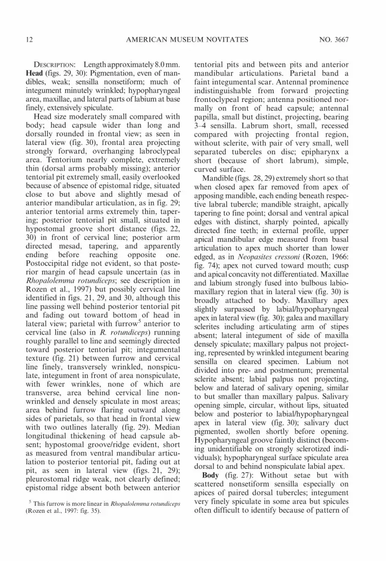
DESCRIPTION: Length approximately 8.0 mm.Head (figs. 29, 30): Pigmentation, even of man-dibles, weak; sensilla nonsetiform; much ofintegument minutely wrinkled; hypopharyngealarea, maxillae, and lateral parts of labium at basefinely, extensively spiculate.
Head size moderately small compared withbody; head capsule wider than long anddorsally rounded in frontal view; as seen inlateral view (fig. 30), frontal area projectingstrongly forward, overhanging labroclypealarea. Tentorium nearly complete, extremelythin (dorsal arms probably missing); anteriortentorial pit extremely small, easily overlookedbecause of absence of epistomal ridge, situatedclose to but above and slightly mesad ofanterior mandibular articulation, as in fig. 29;anterior tentorial arms extremely thin, taper-ing; posterior tentorial pit small, situated inhypostomal groove short distance (figs. 22,30) in front of cervical line; posterior armdirected mesad, tapering, and apparentlyending before reaching opposite one.Postoccipital ridge not evident, so that poste-rior margin of head capsule uncertain (as inRhopalolemma rotundiceps; see description inRozen et al., 1997) but possibly cervical lineidentified in figs. 21, 29, and 30, although thisline passing well behind posterior tentorial pitand fading out toward bottom of head inlateral view; parietal with furrow5 anterior tocervical line (also in R. rotundiceps) runningroughly parallel to line and seemingly directedtoward posterior tentorial pit; integumentaltexture (fig. 21) between furrow and cervicalline finely, transversely wrinkled, nonspicu-late, integument in front of area nonspiculate,with fewer wrinkles, none of which aretransverse, area behind cervical line non-wrinkled and densely spiculate in most areas;area behind furrow flaring outward alongsides of parietals, so that head in frontal viewwith two outlines laterally (fig. 29). Medianlongitudinal thickening of head capsule ab-sent; hypostomal groove/ridge evident, shortas measured from ventral mandibular articu-lation to posterior tentorial pit, fading out atpit, as seen in lateral view (figs. 21, 29);pleurostomal ridge weak, not clearly defined;epistomal ridge absent both between anterior
tentorial pits and between pits and anteriormandibular articulations. Parietal band afaint integumental scar. Antennal prominenceindistinguishable from forward projectingfrontoclypeal region; antenna positioned nor-mally on front of head capsule; antennalpapilla, small but distinct, projecting, bearing3–4 sensilla. Labrum short, small, recessedcompared with projecting frontal region,without sclerite, with pair of very small, wellseparated tubercles on disc; epipharynx ashort (because of short labrum), simple,curved surface.
Mandible (figs. 28, 29) extremely short so thatwhen closed apex far removed from apex ofapposing mandible, each ending beneath respec-tive labral tubercle; mandible straight, apicallytapering to fine point; dorsal and ventral apicaledges with distinct, sharply pointed, apicallydirected fine teeth; in external profile, upperapical mandibular edge measured from basalarticulation to apex much shorter than loweredged, as in Neopasites cressoni (Rozen, 1966:fig. 74); apex not curved toward mouth; cuspand apical concavity not differentiated. Maxillaeand labium strongly fused into bulbous labio-maxillary region that in lateral view (fig. 30) isbroadly attached to body. Maxillary apexslightly surpassed by labial/hypopharyngealapex in lateral view (fig. 30); galea and maxillarysclerites including articulating arm of stipesabsent; lateral integument of side of maxilladensely spiculate; maxillary palpus not project-ing, represented by wrinkled integument bearingsensilla on cleared specimen. Labium notdivided into pre- and postmentum; prementalsclerite absent; labial palpus not projecting,below and laterad of salivary opening, similarto but smaller than maxillary palpus. Salivaryopening simple, circular, without lips, situatedbelow and posterior to labial/hypopharyngealapex in lateral view (fig. 30); salivary ductpigmented, swollen shortly before opening.Hypopharyngeal groove faintly distinct (becom-ing unidentifiable on strongly sclerotized indi-viduals); hypopharyngeal surface spiculate areadorsal to and behind nonspiculate labial apex.
Body (fig. 27): Without setae but withscattered nonsetiform sensilla especially onapices of paired dorsal tubercles; integumentvery finely spiculate in some area but spiculesoften difficult to identify because of pattern of
5 This furrow is more linear in Rhopalolemma rotundiceps(Rozen et al., 1997: fig. 35).
12 AMERICAN MUSEUM NOVITATES NO. 3667

fine, transverse wrinkles that tend to beparallel except on apices of dorsolateraltubercles, where wrinkles are multidirectional;integument without spines or sclerotizedtubercles. Body form moderate in robustness,not quite as slender as that of Neopasitescressoni or Rhopalolemma rotundiceps (Rozenet al., 1997: figs. 32, 33); intersegmental linesweakly incised between most body segments;dorsolateral intrasegmental lines not evident;each thoracic segment with weakly evident,paired dorsolateral tubercles; abdominal seg-ments 1–7 with paired dorsolateral tuberclessomewhat more evident but absent on seg-ments 8–10; all dorsolateral tubercles moder-ately low, conical (as opposed to transverse);venter of abdominal segment 9 in lateral view(fig. 27) appearing somewhat produced be-cause of dorsal attachment of segment 10;segment 10, short with length much less thanone-half height in lateral view (fig. 27), at-tached dorsally to 9 (fig. 27); anus a transverseslit on sloping dorsal surface of 10 (fig. 27);perianal area with small dorsal lip immediate-ly above anus that is to width of anus.Spiracles (fig. 22) subequal and moderate insize, projecting beyond body wall, with rims;peritreme present, moderately wide; atriumglobular, pigmented; atrial wall without den-ticles or rings; primary spiracular openingwith collar; subatrium moderately short, inside view tapering from body surface inward,with about 8 chambers. Male with medianintegumental scar toward posterior margin ofabdominal segment 9; female sex charactersunknown.
MATERIAL STUDIED: Six postdefecating lar-vae, Czech Republic: Praha-Miskovice, 1-IX-2007, 18-VIII-2008 (K. Rezkova and J. Strakalgt.); host: Rophites quinquespinosus.
REMARKS: The description of an immaturelarva of this species from Switzerland (Rozen,1993), though far less complete, is in agree-ment with the above description.
DISCUSSION AND CONCLUSIONS
Similarity in morphology both of firstinstars and of mature larvae among the tribesBiastini, Neolarrini, and Townsendiellini mayindicate a close relationship of these bees. Thissimilarity was recognized several times in
cladistic analyses (Alexander, 1990; Rozen,1996a; Rozen et al., 1997) with high bootstrapsupport for the Neolarrini-Biastini relation-ship (Townsendiellini not included) (Strakaand Bogusch, 1997b). Five characters listed inthe diagnosis of mature larvae can be expand-ed by a specific combination of adult charac-ters shared by bees of these three tribes:propodeal profile with a nearly horizontalbasal zone, a gonocoxite with an internalseptum, and the presence of a ventral para-pennial lobe with strong setae of the gonocox-ite. The only bee with a similar adult charactercombination is Hexepeolus rhodogyne (Linsleyand Michener); however, its mature larvae arelargely different (see Rozen, 1996b).
Biastini, Townsendiella, and Neolarra prob-ably form a well recognizable clade like themorphologically diverse clades within thetribes Ammobatini, Ammobatoidini, Bra-chynomadini, and Epeolini. However, a com-prehensive phylogenetic analysis based on beemorphology and possibly DNA sequences isneeded.
The nest-searching behavior we describehere for Biastes emarginatus is similar to thatof many small nomadine cleptoparasites thatattack nests of ground-nesting solitary bees.Like many cleptoparasitic bees that deposittheir eggs in cells that are still open and thuslikely revisited by host females, their eggs/mature oocytes are small and possess a thick,presumably protective chorion against attackby host females. The configuration of thechorion suggests that the eggs may beembedded in the pollen sphere and/or the cellwall, to make them less obvious to hostfemales. The first instars are small as probablydictated by the small egg size and, like those ofother Nomadinae, possess adaptations forseeking out and battling eggs/larvae of thehost or of competing cleptoparasites. Theseadaptations consist of a heavily sclerotizedhead capsule, large, curved, sharply pointedmandibles, elongate labral tubercles, and theability to crawl, presumably with the aid of aterminal pygopod and transverse, cleatlikespicules (reported here for the first time) onthe body surface.
As with many cleptoparasitic bees withspecialized hospicidal early instars, the larvalinstars of Biastes emarginatus exhibit a sub-
2009 ROZEN ET AL.: BEHAVIOR OF BIASTES EMARGINATUS 13

stantial anatomical modification between thehospicidal stage and subsequent instars, whichpresumably are devoted to consuming provi-sions. One would assume that the anatomy ofthe last larval instar would reveal obvious,perhaps plesiomorphic features for ingestingprovisions, such as scoop-shaped mandiblespowered by mandibular muscles attached tothe parietals, and parietal architecture thatsupports associated muscular contraction forbiting into the provisions. As an example weclearly see this is the situation with Coelioxys(Megachilidae) (e.g., Baker, 1971). However,this does not seem to be the case for Biastesemarginatus, Neopasites cressoni, and likelyfor some (if not many) other Nomadinae.With B. emarginatus, the mandibles of the lastinstar are so short that in repose their apicesare far separated, they are not scoop shaped,and the head capsule is thinly sclerotized andso week that not only is it not constrictedbehind (suggesting weak musculature), but theposterior margin of the capsule cannot becertainly identified (is it the cervical line or theanterior, faint furrow that is directed towardthe posterior tentorial pits? if the former, howthen do we account for the anterior furrow?).The postoccipital ridge is absent. The intrigu-ing questions are: how does this animal feedand how do we explain its peculiar morphol-ogy? The answers will probably come fromfieldwork: observing them as they feed, notonly the first and last instars, but theintermediate ones as well. Because mandiblesof the last instar seem ineffectual for chewinginto food and forcing the food into theesophagus, might an internal study of thisstructure reveal some sort of cibarial oresophageal pump, thereby explaining therotund projection of the front of the headcapsule?
ACKNOWLEDGMENTS
Illustrations were arranged by Steve Thurston,Senior Scientific Assistant, American Museumof Natural History (A.M.N.H.), who alsophotographed figure 2. Christine LeBeau,Scientific Assistant, A.M.N.H., took all SEMmicrophotographs in the A.M.N.H. Microscopyand Imaging Facility (Rebecca Rudolph,Laboratory Manager).
The project was partly supported by theResearch Program of the Ministry of Educationof the Czech Republic No. 0021620828 (forJakub Straka).
REFERENCES
Alexander, B. 1990. A cladistic analysis of thenomadine bees (Hymenoptera: Apoidea). Sys-tematic Entomology 15: 121–152.
Baker, J.R. 1971. Development and sexual dimor-phism of larvae of the bee genus Coelioxys.Journal of the Kansas Entomological Society44: 225–235.
Iwata, K., and S.E. Sakagami. 1966. Gigantism anddwarfism in bee eggs in relation to the mode oflife, with notes on the number of ovarioles.Japanese Journal of Ecology 16: 4–16.
McGinley, R.J. 1981. Systematics of the Colletidaebased on mature larvae with phenetic analysisof apoid larvae. University of CaliforniaPublications in Entomology 91: i–xvi + 1–307.
Michener, C.D. 2007. The bees of the world. 2nded. Baltimore, MD: Johns Hopkins UniversityPress, 953 pp.
Rozen, J.G., Jr. 1966. The larvae of the Antho-phoridae (Hymenoptera, Apoidea). Part 2. TheNomadinae. American Museum Novitates 2244:1–38.
Rozen, J.G., Jr. 1991. Evolution of cleptoparasitismin anthophorid bees as revealed by their modeof parasitism and first instars (Hymenoptera:Apoidea). American Museum Novitates 3029:1–36.
Rozen, J.G., Jr. 1993. Nesting biologies and im-mature stages of the rophitine bees (Halic-tidae) with notes on the cleptoparasite Biastes(Anthophoridae) (Hymenoptera: Apoidea).American Museum Novitates 3066: 1–28.
Rozen, J.G., Jr. 1996a. Phylogenetic analysis ofthe cleptoparasitic bees belonging to theNomadinae based on mature larvae (Apoidea:Apidae). American Museum Novitates 3180:1–39.
Rozen, J.G., Jr. 1996b. First and last larval instars ofthe cleptoparasitic bee Hexepeolus (Hymenop-tera: Apidae: Nomadinae). Memoirs of theEntomological Society of Washington 17:188–193.
Rozen, J.G., Jr. 1997. Pupal description of Neopasitescressoni (Apidae: Nomadinae: Biastini). Journalof the Kansas Entomological Society 70: 76–78.
Rozen, J.G., Jr. 2001. Taxonomic key to maturelarvae of cleptoparasitic bees (Apoidea).American Museum Novitates 3309: 1–27.
Rozen, J.G., Jr. 2003. Eggs, ovariole numbers, andmodes of parasitism of cleptoparasitic bees, with
14 AMERICAN MUSEUM NOVITATES NO. 3667

emphasis on Neotropical species (Hymenop-tera:). American Museum Novitates 3413:1–36.
Rozen, J.G., Jr., and R.J. McGinley. 1991. Biologyand larvae of the cleptoparasitic bee Townsendiellapulchra and nesting biology of its host Hesper-apis larreae (Hymenoptera: Apoidea). AmericanMuseum Novitates 3005: 1–11.
Rozen, J.G., Jr., and H. Ozbek. 2003. Oocytes,eggs, and ovarioles of some long-tongued bees(Hymenoptera: Apoidea). Appendix: Param-mobatodes rozeni, a new bee species from Israel,by M. Schwarz. American Museum Novitates3393: 1–35.
TownsendiellaRozen, J.G., Jr., A. Roig-Alsina, andB.A. Alexander. 1997. The cleptoparasitic beegenus Rhopalolemma, with reference to otherNomadinae (Apidae) and biology of its hostProtodufourea (Halictidae: Rophitinae). Ameri-can Museum Novitates 3194: 1–28.
TownsendiellaStraka, J., and P. Bogusch. 2007a.Description of immature stages of cleptopar-asitic bees Epeoloides coecutiens and Leiopodustrochantericus (Hymenoptera: Apidae: Osirini,Protepeolini) with remarks to their unusualbiology. Entomologica Fennica 18: 242–254.
TownsendiellaStraka, J., and P. Bogusch. 2007b.Phylogeny of the bees of the family Apidaebased on larval characters with focus onthe origin of cleptoparasitism (Hymenoptera:Apiformes). Systematic Entomology 32: 700–711.
TownsendiellaTorchio, P.F., J.G. RozenJr., G.E.Bohart, and M.S. Favreau. 1967. Biology ofDufourea and of its cleptoparasite, Neopasites(Hymenoptera: Apoidea). Journal of the NewYork Entomological Society 75: 132–146.
Westrich, P. 1989. Die Wildbienen Baden-Wurttembergs. Allgemeiner Teil: 1–431, Speziel-ler Teil: 432–972. Stuttgart: Eugene Ulmer.
APPENDIX
FIRST INSTAR OF NEOPASITES CRESSONI
Figures 20, 23–26
The first instar of Neopasites cressoni is describedhere to allow a comparison with the first instar ofBiastes emarginatus. The description is basedmostly on a cleared specimen, but the SEMmicrographs are of two of seven specimens founddead in a single cell, presumably having been killedearlier by other cleptoparasites. While heavilysclerotized areas were relatively unaffected, mem-branous areas, including the labrum and labiomax-illary regions shrank and became distorted throughpostmortem processes. Many setae were alsobroken, for reasons not understood.
DIAGNOSIS: See Diagnosis of first instar ofBiastes emarginatus above.
DESCRIPTION: Length of newly eclosed specimen0.44 mm.
HEAD: Maximum transverse width of headcapsule 0.18 mm; maximum transverse foramenwidth 0.13 mm. Shape about as described forBiastes emarginatus except foramen magnum notnearly as constricted and parietals not swollen asgreatly dorsolaterally, so the median depression ofvertex less exaggerated, only faintly lower thanlateral parts of vertex in frontal view; integument ofhead capsule about as described for Biastesemarginatus; capsule clearly with simple setae, notspinulae but arrangement uncertain. Tentoriumweak, not certainly complete; anterior (fig. 24)and posterior tentorial pits present; hypostomal
groove present, darkly pigmented; sclerotizationmesad (below) of each hypostomal groove presentas in B. emarginatus; other features of parietalabout as described for B. emarginatus. Antenna(fig. 24) as described for B. emarginatus. Labrum asdescribed for B. emarginatus except paired tuberclesstouter at base and information about epipharyn-geal surface unknown. Mandible as described for B.emarginatus. Labiomaxillary region probably muchlike that of B. emarginatus because of fusion ofcardo with lateral parts of stipes while rest of regionremaining membranous except perhaps membra-nous part more ventrally protuberant althoughSEM micrographs (figs. 23, 25, 26) show this areabadly desiccated due to postmortem deterioration;posterior part of fused cardo/stipes sclerite formingventral postoccipital bridge (fig. 23), except bridgeperhaps not fused medially; identification ofmaxillary and labial palpi as well as median salivaryopening uncertain (figs. 23, 25, 26). Hypopharynxapparently sclerotized as in B. emarginatus.
BODY: Form elongate, linear, as described forBiastes emarginatus. Integument with a few minutesetae anteriorly and with minute, transverse, poste-riorly sloping projections consisting of rows of 1–5+sharp points visible only with SEM, as in Biastesemarginatus (fig. 17). Spiracular peritreme flush withbody wall (fig. 25). Position of anus unknown.
MATERIAL STUDIED: Seven first instars, USA:Arizona: Cochise Co., 3 mi SW of Rodeo, HidalgoCo., New Mexico, V-1-1965 (J.G. Rozen), presum-ably all collected dead; one first instar, same dataexcept IV-27-1966 (J.G. Rozen, M. Favreau),collected alive.
2009 ROZEN ET AL.: BEHAVIOR OF BIASTES EMARGINATUS 15

This paper meets the requirements of ANSI/NISO Z39.48-1992 (Permanence of Paper).
Complete lists of all issues of the Novitates and the Bulletin are available at World Wide
Web site http://library.amnh.org/pubs. Inquire about ordering printed copies via e-mail from
[email protected] or via standard mail from: American Museum of Natural History,
LibraryÐScientific Publications, Central Park West at 79th St., New York, NY 10024. TEL:
(212) 769-5545. FAX: (212) 769-5009.