Zfp521 antagonizes Runx2, delays osteoblast differentiation in vitro, and promotes bone formation in...
21
Zfp521 antagonizes Runx2, delays osteoblast differentiation in vitro, and promotes bone formation in vivo Meilin Wu 1,3 , Eric Hesse 1,2 , Frederic Morvan 1,4 , Jian-Ping Zhang 1 , Diego Correa 1,2 , Glenn C. Rowe 1,2 , Riku Kiviranta 1,2 , Lynn Neff 1 , William M. Philbrick 1 , William C. Horne 1,2 , and Roland Baron 1,2 1 Yale University School of Medicine, New Haven, Connecticut 06510, USA 2 Harvard School of Dental Medicine and Harvard Medical School, Boston, Massachusetts 02115, USA Abstract Zfp521, a 30 C2H2 Kruppel-like zinc finger protein, is expressed at high levels at the periphery of early mesenchymal condensations prefiguring skeletal elements and in all developing bones in the perichondrium and periosteum, in osteoblast percursors and osteocytes, and in chondroblast percursors and growth plate prehypertrophic chondrocytes. Zfp521 expression in cultured mesenchymal cells is decreased by BMP-2 and increased by PTHrP, which promote and antagonize osteoblast differentiation, respectively. In vitro, Zfp521 overexpression reduces the expression of several downstream osteoblast marker genes and antagonizes osteoblast differentiation. Zfp521 binds Runx2 and represses its transcriptional activity, and Runx2 dose-dependently rescues Zfp521’s inhibition of osteoblast differentiation. In contrast, osteocalcin promoter-targeted overexpression of Zfp521 in osteoblasts in vivo results in increased bone formation and bone mass. We propose that Zfp521 regulates the rate of osteoblast differentiation and bone formation during development and in the mature skeleton, in part by antagonizing Runx2. Keywords Zfp521; Runx2; osteoblast; bone formation; differentiation Introduction OBs arise from mesenchymal stem cells that can differentiate into a number of specialized cell types [1,2]. The progression from multipotent cells to the fully differentiated and bone matrix- producing osteoblast is regulated by numerous transcription factors, among them Runx2, a member of the Runt domain family of transcription factors considered to be a master regulator that is essential for osteoblast commitment and early stages of osteoblast differentiation [3– Corresponding author: Roland Baron, Department of Oral Medicine, Infection and Immunity, Harvard School of Dental Medicine, 188 Longwood Ave., REB309, Boston, MA 02115, Phone: (617) 432-7320, Fax: (617) 432-1897, [email protected]. 3 Present addresses: Cardiovascular Division, Molecular Cardiology Research Center, 957 Biomedical Research Building II/III, 421 Curie Blvd., Philadelphia, PA 19104-6160, USA; 4 Novartis Institutes for Biomedical Research, Musculoskeletal Disease Area, WKL-125.9.01, Klybeckstrasse 141, CH-4057 Basel, Switzerland. Publisher's Disclaimer: This is a PDF file of an unedited manuscript that has been accepted for publication. As a service to our customers we are providing this early version of the manuscript. The manuscript will undergo copyediting, typesetting, and review of the resulting proof before it is published in its final citable form. Please note that during the production process errors may be discovered which could affect the content, and all legal disclaimers that apply to the journal pertain. NIH Public Access Author Manuscript Bone. Author manuscript; available in PMC 2010 April 1. Published in final edited form as: Bone. 2009 April ; 44(4): 528–536. doi:10.1016/j.bone.2008.11.011. NIH-PA Author Manuscript NIH-PA Author Manuscript NIH-PA Author Manuscript
-
Upload
independent -
Category
Documents
-
view
5 -
download
0
Transcript of Zfp521 antagonizes Runx2, delays osteoblast differentiation in vitro, and promotes bone formation in...
Zfp521 antagonizes Runx2, delays osteoblast differentiation invitro, and promotes bone formation in vivo
Meilin Wu1,3, Eric Hesse1,2, Frederic Morvan1,4, Jian-Ping Zhang1, Diego Correa1,2, GlennC. Rowe1,2, Riku Kiviranta1,2, Lynn Neff1, William M. Philbrick1, William C. Horne1,2, andRoland Baron1,21 Yale University School of Medicine, New Haven, Connecticut 06510, USA2 Harvard School of Dental Medicine and Harvard Medical School, Boston, Massachusetts 02115,USA
AbstractZfp521, a 30 C2H2 Kruppel-like zinc finger protein, is expressed at high levels at the periphery ofearly mesenchymal condensations prefiguring skeletal elements and in all developing bones in theperichondrium and periosteum, in osteoblast percursors and osteocytes, and in chondroblastpercursors and growth plate prehypertrophic chondrocytes. Zfp521 expression in culturedmesenchymal cells is decreased by BMP-2 and increased by PTHrP, which promote and antagonizeosteoblast differentiation, respectively. In vitro, Zfp521 overexpression reduces the expression ofseveral downstream osteoblast marker genes and antagonizes osteoblast differentiation. Zfp521 bindsRunx2 and represses its transcriptional activity, and Runx2 dose-dependently rescues Zfp521’sinhibition of osteoblast differentiation. In contrast, osteocalcin promoter-targeted overexpression ofZfp521 in osteoblasts in vivo results in increased bone formation and bone mass. We propose thatZfp521 regulates the rate of osteoblast differentiation and bone formation during development andin the mature skeleton, in part by antagonizing Runx2.
KeywordsZfp521; Runx2; osteoblast; bone formation; differentiation
IntroductionOBs arise from mesenchymal stem cells that can differentiate into a number of specialized celltypes [1,2]. The progression from multipotent cells to the fully differentiated and bone matrix-producing osteoblast is regulated by numerous transcription factors, among them Runx2, amember of the Runt domain family of transcription factors considered to be a master regulatorthat is essential for osteoblast commitment and early stages of osteoblast differentiation [3–
Corresponding author: Roland Baron, Department of Oral Medicine, Infection and Immunity, Harvard School of Dental Medicine, 188Longwood Ave., REB309, Boston, MA 02115, Phone: (617) 432-7320, Fax: (617) 432-1897, [email protected] addresses: Cardiovascular Division, Molecular Cardiology Research Center, 957 Biomedical Research Building II/III, 421 CurieBlvd., Philadelphia, PA 19104-6160, USA;4Novartis Institutes for Biomedical Research, Musculoskeletal Disease Area, WKL-125.9.01, Klybeckstrasse 141, CH-4057 Basel,Switzerland.Publisher's Disclaimer: This is a PDF file of an unedited manuscript that has been accepted for publication. As a service to our customerswe are providing this early version of the manuscript. The manuscript will undergo copyediting, typesetting, and review of the resultingproof before it is published in its final citable form. Please note that during the production process errors may be discovered which couldaffect the content, and all legal disclaimers that apply to the journal pertain.
NIH Public AccessAuthor ManuscriptBone. Author manuscript; available in PMC 2010 April 1.
Published in final edited form as:Bone. 2009 April ; 44(4): 528–536. doi:10.1016/j.bone.2008.11.011.
NIH
-PA Author Manuscript
NIH
-PA Author Manuscript
NIH
-PA Author Manuscript
6] and thus essential for bone formation. Runx2 induces the expression of Osterix, atranscription factor specifically expressed in all developing bones [7], which together with theautocrine Wnt-induced activation of β-catenin [8] promotes the progression to fully matureosteoblasts. The activities of these key transcription factors are modulated by a variety offactors and mechanisms [9–13].
Zfp521 (also termed Evi3 in mice and EHZF in humans) is a zinc finger protein consistingalmost entirely of 30 C2H2 Kruppel-like zinc fingers [14,15]. Both Zfp521 and the homologousZfp423 (also termed OLF/EBF associated-zinc finger protein [OAZ]) inhibit celldifferentiation in some cell lineages [15–17]. We have now found that Zfp521 is expressed inosteoblast and chondrocyte precursors in the periosteum, endosteum and perichondrium, inprehypertrophic chondrocytes in the growth plate, and in osteoblasts and osteocytes in bone.Zfp521 expression increased during in vitro osteoblast differentiation, and BMPs and PTHrPregulated its expression. Zfp521 bound Runx2 and repressed the expression of severalosteoblast marker genes and in vitro osteoblast differentiation, while overexpressed Runx2dose-dependently reversed this effect. In contrast to the effect of Zfp521 on in vitro osteoblastdifferentiation, however, targeted overexpression of Zfp521 in mature osteoblasts in transgenicmice resulted in marked increases in bone formation. We propose that the balance betweenZfp521 and Runx2 contributes to the regulation of the rate of osteoblast differentiation andbone formation during development and in the mature skeleton.
Materials and MethodsPlasmids
Zfp521 was cloned from a mouse brain lambda phage library (Clontech, Mountain View, CA).The open reading frame was amplified by PCR (HA-Zfp521 F and HA-Zfp521 R primers;Table 1) and ligated into the pCMV-HA vector (Clontech) between the Sal1/Not1 sites. P.Ducy (Columbia University, New York) provided pCMV-FLAG-Runx2 encoding the p57 typeII Runx2 isoform (N-terminal MASNSL..) and OG2-luciferase, G. Rawadi (Galapagos,Romainville, France) provided Runx2-luciferase, and S. Warming (National Cancer Institute,Frederick, MD) provided mEBFAZ cDNA, which was subcloned into the pCMV-HA vector.Zfp521 shRNA constructs were generated by subcloning annealed oligonucleotides (PS3;Table 1) into the pSilencer vector, resulting in shRNA with a 19-nucleotide sequence that NCBIBlast analysis showed to be specific for Zfp521.
Cell culturePrimary calvarial osteoblasts were obtained from 1–3 day-old mice as previously described[18]. All cells were cultured in α-MEM with 10% fetal bovine serum (FBS), 100 units/mlpenicillin and 100 μg/ml streptomycin (all Invitrogen, Carlsbad, CA). Differentiation wasinduced by adding BMP-2 (100 ng/ml; Galapagos, Romainville, France) to confluent cells (day0). Calvarial osteoblasts and MC3T3-E1 cells stably transfected with Zfp521 weredifferentiated in 50 μM ascorbic acid and 5 mM β-glycerolphosphate (Sigma-Aldrich).
Cell transfectionCells at 80% confluence were transfected using a ratio of 2 μg DNA to 3 μl FuGENE6transfection reagent (Roche) as described by the manufacturer.
Generation of stable cell linesC3H10T1/2 and MC3T3-E1 cells were infected with concentrated Zfp521 or lacZ lentivirusand cultured overnight in 6-well dishes (1 ml/well containing 6 mg/ml polybrene). Fresh
Wu et al. Page 2
Bone. Author manuscript; available in PMC 2010 April 1.
NIH
-PA Author Manuscript
NIH
-PA Author Manuscript
NIH
-PA Author Manuscript
medium was added one day post-infection. Cells were cultured for 3 weeks in 2 mg/ml(C3H10T1/2) or 5 mg/ml (MC3T3-E1) blasticidin to eliminate uninfected cells.
Retrovirus and adenovirus production and gene transductionshRNA and control retroviruses were generated by transfecting PhoenixE cells usingLipofectamine 2000 according to the manufacturer’s instructions (Invitrogen). 72 hours post-transfection, retrovirus was harvested, combined with medium and polybrene (4 mg/ml) in atotal volume of 4 ml, and immediately overlaid on CD1 calvarial osteoblasts plated the previousnight at 275,000 cells/6 cm culture dish. After 24 hours, medium was changed to α-MEM with10% FBS, 100 units/ml penicillin and 100 μg/ml streptomycin. One day later, cells were splitinto two 10 cm dishes and placed in selection medium containing 2 μg/ml puromycin. Cellswere cultured until uninfected cells died, then harvested, counted and replated for experiments.
To produce Runx2 adenovirus, the Runx2 open reading frame of the pCMV-FLAG-Runx2plasmid (type II isoform) was amplified and cloned into the pShuttle-CMV vector (Qbiogene).Linearized plasmid (Pme1 - New England Biolabs, Ipswich, MA) was purified andelectroporated into E. coli strain BJ5183-AD-1 (Stratagene) for recombination with thepAdEasy-1 plasmid. Positive colonies were identified by restriction digestion with Pac1 (NewEngland Biolabs). Adenovirus was generated as previously described [19]. Calvarialosteoblasts were seeded at a density of 50,000 cells/well in a 6-well tissue culture dish andinfected overnight once confluent.
AntibodiesMouse monoclonal anti-HA, mouse monoclonal anti-Myc, mouse polyclonal anti-PEBP2aA,goat anti-OAZ and mouse monoclonal anti-C23 were obtained from Santa Cruz Biotechnology(Santa Cruz, CA), mouse monoclonal anti-actin from Chemicon International (Temecula, CA),and mouse anti-V5 from Invitrogen. Affinity-purified rabbit polyclonal antibodies againstZfp521, produced by Bethyl Laboratories (Montgomery, TX) using peptide antigensCT369MVEAAPPIPKSRGR [anti-Zfp521(369)] and CQ1177VSPMPRISPSQSDEKK [anti-Zfp521(1177)], were used at 1:500 for Western blotting, 5 μg/ml for immunoprecipitation and20 μg/ml for immunofluorescence. Antibody specificity, including lack of cross-reactivity withthe closely related Zfp423, was confirmed by Western blot (data not shown).
In-situ hybridizationTo generate Zfp521 probes, the 907–1219 and 3306–3611 fragments of the cDNA wereamplified by PCR with primers containing HindIII and BamH1 restriction sites and insertedinto the HindIII/BamH1 sites of pBSKII. 35S-labeled sense and antisense riboprobes weresynthesized from linearized plasmids using the Promega transcription kit and 35S-UTP(Amersham). In situ hybridization of Zfp521 mRNA with cRNA probes was performed onsections from E12.5, E15.5 and E18.5 mouse embryo and neonatal mouse skeleton aspreviously described [20]. Sections were digitally photographed and analyzed using AdobePhotoshop 6.0.
Immunofluorescence microscopyHumeri of 6 month old mice were fixed and decalcified in 4.1% EDTA, 5% PVP (pH 7.4) for3–4 days. Tissue samples and bones were washed in PBS after fixation and kept in 40% sucroseuntil sectioned. Cells were plated on glass coverslips and cultured overnight, then fixed in 3.7%formaldehyde/PBS for 10 min and washed twice with PBS. Nuclei were stained with ToPro-3iodide (1:1000, Molecular Probes, Eugene, OR). Zfp521 was labeled with the anti-Zfp521(369) antibody. Cells and cryostat sections were mounted in FluorSave (Calbiochem, SanDiego, CA) and imaged using an LSM 510 Meta confocal microscope (Carl Zeiss Inc.,
Wu et al. Page 3
Bone. Author manuscript; available in PMC 2010 April 1.
NIH
-PA Author Manuscript
NIH
-PA Author Manuscript
NIH
-PA Author Manuscript
Thornwood, NY). Images were recorded, composite images were compiled and enhancementsperformed using Adobe Photoshop 8.0.
Analysis of alkaline phosphatase activity and mineralized nodulesCells were washed twice with PBS and fixed for 10 minutes in 3.7% formaldehyde, then stainedusing Fast BlueAlkaline phosphatase (Sigma-Aldrich) according to the manufacturer’sinstructions. Mineralized nodules were stained by the Von Kossa method [18], or with alizarinred by incubating cells with 2% (w/v) of alizarin red S (Sigma-Aldrich) in water at pH 4.2 for10 minutes.
Northern blot analysisRNA was isolated and Northern blots were performed using a gel electrophoresis-purifiedZfp521 probe excised from pASV4 by EcoR1 digestion as previously described [18].
Real-time PCRRNA was isolated using the RNeasy Mini Kit (Qiagen, Valencia, CA). cDNA was synthesizedusing 1 μg of total RNA and SuperScript 2 (Invitrogen) according to the manufacturer’sinstructions. 50 ng of cDNA was mixed with 12.5 μl iQ SYBR green supermix (BioRad,Hercules, CA), 10.5 μl of water, and 0.5 μl of sense and antisense oligonucleotide (10 μM;Table 1), denatured at 95 °C for 3 minutes and amplified by 40 cycles of 95 °C/57 °C/72 °Cfor 30 seconds each using an iCycler (BioRad). All samples were normalized to GAPDH andrelative expression of Zfp521 was determined using the 2−ΔΔCT method. Data are presentedas fold change relative to control samples.
Transcriptional activation assayMC3T3-E1 cells were plated in 6-well tissue culture plates (1×10⁵ cells/well) and transfectedwith pCMV-HA-Zfp521 (2.0 μg) and pCMV-FLAG-Runx2 (1.5 μg) per well for 24h usingFuGENE 6 at a 6:1 FuGENE 6 to DNA ratio. The Runx2-luc and OG2-luc constructs wereused at 1.0 μg/well. Renilla luciferase cDNA (0.017 μg/well, Promega) was included as controlfor transfection efficiency. Total DNA was maintained at 4.5 μg/well with empty vector(pcDNA3.1). Dual-Luciferase assay (Promega) was performed according to themanufacturer’s instructions. Results corrected for transfection efficiency were normalized toempty vector.
Transgenic miceOG2-tTA mice were described elsewhere [21]. TetOp-Zfp521 mice were generated bysubcloning the Sal1-Not1 fragment of Zfp521 from pCMV-HA-Zfp521 into the TetOP-tTAvector (E. Nestler, Southwestern Medical Center, Dallas) from which the tTa gene was excised.Linearized DNA was microinjected into pronuclei of oocytes from SJL × C57BL6 mice F2mouse ova as previously described [21]. When crossed with the OG2-tTA mice, two TetOp-Zfp521 lines (03 and 05) generated double-transgenic OG2-tTA × TetOp-Zfp521 (OG2 ×Zfp521) mice that expressed Zfp521. Animal protocols were approved by the Yale AnimalCare and Use Committee.
Bone histomorphometryHistomorphometric analysis of 4 and 12 week-old OG2 × Zfp521 mice and control littermateswas performed as previously described [18].
Wu et al. Page 4
Bone. Author manuscript; available in PMC 2010 April 1.
NIH
-PA Author Manuscript
NIH
-PA Author Manuscript
NIH
-PA Author Manuscript
In vivo bone resorption analysisCollagen type 1 C-telopeptide fragments in serum samples collected at 4 and 12 weeks of agewere analyzed using the RatLaps ELISA kit (Nordic Biosciences Diagnostics) according tothe manufacturer’s instructions.
Statistical analysisStatistical significance was calculated using the Student’s t test. Standard error was calculatedusing one-tailed analysis.
ResultsZfp521 is expressed in osteoblasts and mesenchymal cells
Zfp521 is present in many cell types [14], and Northern blots revealed that it is expressed athigh levels in calvarial osteoblasts as well as in C3H10T1/2 and MC3T3-E1 mesenchymal celllines (Fig. 1A). In contrast, Zfp521 was not detected in mature osteoclasts generated in vitro,osteoclast precursors or the monocytic RAW 264.7 cell line (not shown), indicating that theprotein is either not expressed or expressed at much lower levels in osteoclasts than inosteoblasts.
In situ hybridization confirmed these findings in developing skeletal elements (Fig 1B–F), withhigh expression of Zfp521 at the periphery of mesenchymal condensations (Fig. 1B), in thedeveloping skull (Fig. 1C) and in perichondral and periosteal cells (Fig. 1D–F), suggesting apossible role in regulating early skeletal development. Immunocytochemistry using affinity-purified antibodies directed against Zfp521 (Fig. 1G) confirmed the pattern of expression indeveloping long bones seen by in situ hybridization. In the growth plate, Zfp521 expressionwas low in resting and proliferating chondrocytes, markedly higher in the prehypertrophicchondrocytes, then low again in hypertrophic cells (upper panels). In postnatal bone, Zfp521was strongly expressed in the periosteum and the endosteum where it was restricted toosteoblasts and one or two layers of pre-osteoblastic cells (middle panels), and in osteocytes(lower panels). No staining was detected in osteoclasts.
Zfp521 is a nuclear protein in the chondrocyte and osteoblast lineagesConsistent with Zfp521 being a nuclear transcription factor, Zfp521 was most highly enrichedin the nuclei of C3H10T1/2 cells, especially the nucleoli (Fig. 1H). Some Zfp521 was alsodetected in the cytoplasm. Together, the immunocytochemical and in situ hybridization datasuggested a regulated pattern of expression with high levels in partially differentiatedchondrocytes (but not in hypertrophic cells), as well as in pre-osteoblasts, mature osteoblastsand osteocytes. The nuclear localization of Zfp521 suggests that it plays a role in the regulationof gene transcription during osteoblast differentiation.
Zfp521 antagonizes osteoblast differentiationThe expression of Zfp521 at the periphery of mesenchymal condensations, in areas of transitionfrom precursors to more mature chondrocytes or osteoblasts (growth plate, perichondrium andperiosteum), and in mesenchymal cell lines suggested that it might regulate the differentiationof early mesenchymal cells during bone development and formation. To determine whetherZfp521 affected the process of osteoblastogenesis positively or negatively, we used smallhairpin RNA (shRNA) to deplete Zfp521 mRNA by approximately 40% in calvarial OBs (Fig.2A). The shRNA increased nodule formation and mineralization (Fig. 2B), indicating thatZfp521 has an inhibitory effect on osteoblast differentiation and maturation in cultures of earlyosteoblast precursors.
Wu et al. Page 5
Bone. Author manuscript; available in PMC 2010 April 1.
NIH
-PA Author Manuscript
NIH
-PA Author Manuscript
NIH
-PA Author Manuscript
The effect of Zfp521 on alkaline phosphatase (ALP) production, a marker of early osteoblastdifferentiation, was tested by transiently transfecting wild-type calvarial cells with Zfp521 andstaining for ALP activity 6 days after transfection. The Zfp521-transfected cells had less ALPactivity than mock-transfected cells (Fig. 2C). Moreover, stable overexpression of Zfp521 (Fig.2D) reduced ALP expression in MC3T3-E1 cells and in BMP-2-treated C3H10T1/2 cells(which do not express ALP unless treated with BMP-2) (Fig. 2E). Thus, Zfp521 delaysdifferentiation in calvarial osteoblasts and mesenchymal cell lines.
Zfp521 expression is regulated during osteoblast differentiationIf Zfp521 is a physiologically important brake on osteoblast differentiation, its expressionmight be regulated during differentiation of osteoblast precursors. We therefore determined ifagents that modulate osteoblast differentiation affected endogenous Zfp521 expression (Fig.3). Endogenous Zfp521 mRNA levels increased steadily with time in culture in mesenchymalcell lines and primary calvarial osteoblasts. Treatment with BMP-2, which induces osteoblastdifferentiation and maturation, repressed Zfp521 expression (Fig. 3A, B). In contrast,continuous treatment of calvarial osteoblasts with PTHrP 1–34, which favors proliferation[22] but antagonizes osteoblast differentiation [23], increased Zfp521 expression (Fig. 3C).
Zfp521 interacts with Runx2 and inhibits Runx2-induced activation of the Runx2 andosteocalcin promoters
Runx2 is a key regulatory factor downstream of BMPs and PTHrP [24]. We therefore examinedthe effect of Zfp521 on Runx2-induced activation of Runx2-luciferase (Runx2-luc) andosteocalcin-luciferase (OG2-luc) reporter genes in MC3T3-E1 cells (Fig. 4A). As previouslyreported [3,6,25], Runx2 activated both reporter genes. Zfp521 alone had little effect on eitherreporter, but it strongly repressed the activation of the two reporters by Runx2, indicating thatZfp521 indeed antagonizes Runx2 transcriptional activity. The antagonism of Runx2 activityby Zfp521 suggested that the two proteins might interact, either directly or indirectly, and weconfirmed this by co-immunoprecipitating HA-tagged Zfp521 and FLAG-tagged Runx2 fromtransiently transfected 293VNR cell lysates (Fig. 4B). Thus, Zfp521 both associates withRunx2 and represses Runx2 transcriptional activity, indicating that Zfp521’s ability toantagonize osteoblast differentiation could be a consequence, at least in part, of its interactionwith Runx2.
Osteoblast-targeted overexpression of Zfp521 inhibits ex vivo osteoblast differention butpromotes in vivo bone formation
Zfp521’s antagonism of in vitro osteoblast differentiation and Runx2 transcriptional activitysuggested that overexpressing the protein would inhibit bone formation in vivo. To test thispossibility and since we had observed Zfp52 expressed in mature osteoblasts, we generatedTetOP-Zfp521 mice and crossed them with the osteoblast-restricted OG2-tTA mice, in orderto express the protein specifically in osteoblasts [26]. Two lines of OG2-tTA × TetOp-Zfp521(OG2 × Zfp521) mice expressed two-fold more Zfp521 in bone but not in fat or heart (Fig.5A), consistent with the previous characterization of the OG2-tTA mice [21].
Before examining the effect of the overexpressed Zfp521 on the in vivo bone formation, weexamined the ex vivo differentiation of calvarial cells from the mice to confirm that theoverexpressed Zfp521 antagonized the differentiation of calvarial cells in a manner consistentwith our earlier observations. Prior to induction of osteogenic differentiation, the cells derivedfrom transgenic mice already expressed 1.5- to 2-fold more Zfp521 than cells from controllittermates (data not shown), notwithstanding the characterization of osteocalcin as a markerof relatively mature osteoblasts [27]. The OG2 × Zfp521 cultures had less nodule formationand mineralization than the control cultures (Fig. 5B), consistent with our in vitro results (Fig.2). Moreover, expression of several early osteoblast marker genes and Runx2 target genes
Wu et al. Page 6
Bone. Author manuscript; available in PMC 2010 April 1.
NIH
-PA Author Manuscript
NIH
-PA Author Manuscript
NIH
-PA Author Manuscript
(Runx2, Osterix, alkaline phosphatase, osteopontin, osteocalcin, bone sialoprotein) wasdecreased in the Zfp521 calvarial cells (Fig. 5C), further confirming that Zfp521 antagonizesRunx2 and in vitro osteoblast differentiation.
We also examined the effect of expressing increasing amounts of Runx2, using a Runx2adenovirus, on the differentiation of the OG2 × Zfp521 calvarial cells, in order to determineif Runx2 could reverse the inhibitory effect of Zfp521. Runx2 dose-dependently reversed theZfp521-induced inhibition of ALP expression by Runx2 (Fig. 5D), providing further evidencethat Zfp521 acts at least in part by antagonizing Runx2 activity.
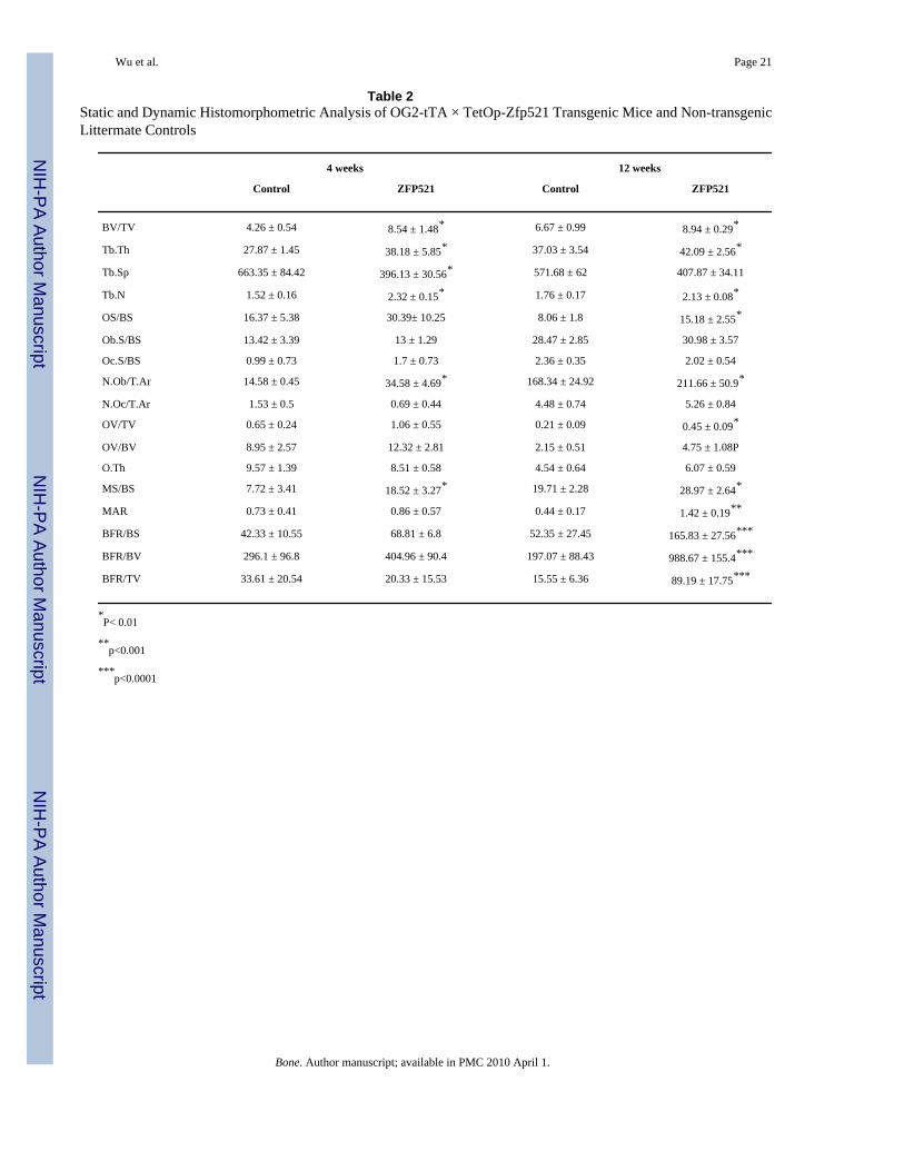
Unexpectedly, however, Von Kossa-stained sections of proximal tibiae from the OG2 × Zfp521animals showed denser trabecular bone and thicker cortices (Fig. 6A). Histomorphometricanalysis of tibiae from 4 and 12 week-old OG2 × Zfp521 mice and non-Zfp521 littermates(Fig. 6B, Table 2) revealed increased bone volume, osteoblast number and bone formation ratein the OG2 × Zfp521 mice. Osteoclast number and serum collagen cross-links, a marker ofbone resorption, were essentially unchanged (Table 2, Fig. 6C), indicating that Zfp521overexpression did not alter bone resorption and that osteocalcin promoter-drivenoverexpression of Zfp521 leads to an increase in bone mass due to a marked increase inosteoblast numbers and bone-forming activity. Thus, osteocalcin promoter-driven stableexpression of Zfp521 in osteoblast precursors hindered the progression of both calvarial cellsand mesenchymal cell lines towards a mature osteoblast phenotype in vitro, but promoted boneformation in the whole animal.
DiscussionOur results show that Zfp521 inhibits in vitro osteoblast differentiation and that overexpressingRunx2, whose transcriptional activity is decreased by Zfp521, reverses this inhibition. Inaddition, Zfp521 regulates in vivo bone formation when its expression is targeted to osteoblastsvia the osteocalcin promoter, although in this case, Zfp521 increases bone formation, theopposite of what would be predicted from its antagonism of in vitro osteoblast differentiation.Thus, this report establishes Zfp521 as a novel and important player in the regulation ofosteoblast differentiation and bone formation, which on the one hand antagonizes Runx2 buton the other hand favors bone formation in vivo, at least when expression is targeted toosteoblasts by the osteocalcin promoter.
Both Zfp521 and the homologous Zfp423 exert inhibitory effects on the differentiation andmaturation of several cell types [14,16,17,28], suggesting that this family of proteins mightfunction generally as “switch proteins”, controlling the transition from immature to maturecells. Several of our observations suggest that Zfp521 restricts the developmental progressionof mesenchymal cells in the developing skeleton. We found that it is expressed in earlymesenchymal cell precursors during skeletal development, around early mesenchymalcondensations, in the perichondrium, periosteum, and specific regions of the growth plate ofdeveloping bones, in osteocytes, and also in primary osteoblasts and mesenchymal cell linesin vitro. Its expression was strongly repressed by treatment of the cells with BMP-2, whichinduces osteoblast differentiation, and increased by PTHrP, which prevents differentiation infavor of proliferation. Overexpressing Zfp521 in primary osteoblasts and a multipotentmesenchymal cell line delayed or reduced the expression of early osteoblast differentiationmarkers (Runx2, alkaline phosphatase, collagen 1) and the formation of mineralized nodules.Conversely, depleting Zfp521 with shRNA favored osteoblast differentiation andmineralization. Together, these results strongly suggest that Zfp521 acts to maintain osteoblastprecursors in a less differentiated state.
Wu et al. Page 7
Bone. Author manuscript; available in PMC 2010 April 1.
NIH
-PA Author Manuscript
NIH
-PA Author Manuscript
NIH
-PA Author Manuscript
Appropriately balancing the advance of some cells towards a more mature state while retainingothers in a less differentiated state requires tight regulation of the relative activities of factorsthat favor the maturation of the cells and those that arrest differentiation. Multiple transcriptionfactors (i.e., Msx, Sox9, Runx2, β-catenin, Osterix, members of the AP-1 family) act at highlyspecific steps during the differentiation of early mesenchymal cells to mature chondrocytes orosteoblasts to either retain or advance the cell through specific checkpoints [2]. Appropriatetiming of the activity of these key transcription factors is critical to the proper coordination ofgenes that maintain the balance of proliferation and commitment of osteoblast precursors andthe progression to mature bone-forming cells. Runx2 regulates both chondrogenesis, where itis required for final maturation of hypertrophic chondrocytes, and osteogenesis, where it playsa dual role, first as a “master regulator” that is required for osteoblast formation [3–6] and lateras an antagonist of late stages of osteoblast maturation [29–31]. Similar early and late oppositeeffects have recently been suggested for Wnt signaling [32,33]. Since Runx2 activates manyof the osteoblast marker genes that we found to be down-regulated by Zfp521, we hypothesizedthat Zfp521’s inhibitory effect on osteoblast differentiation could result from antagonism ofRunx2 activity, and indeed, we observed that Zfp521 bound Runx2 and inhibited the Runx2-induced activation of OG2-luc and Runx2-luc reporter genes. In addition, overexpressed Runx2dose-dependently reversed the repression of ALP expression in calvarial cells from Zfp521transgenic mice. Thus, Zfp521 inhibition of in vitro osteoblast differentiation appears to be atleast in part a consequence of antagonizing Runx2 activity.
Unexpectedly, however, overexpressing Zfp521 in osteoblasts in transgenic mice under thecontrol of the osteocalcin promoter strongly increased bone formation. This difference in thein vitro and in vivo effects of overexpressed Zfp521 was not due to the use of differentpromoters, since differentiation of calvarial osteoblast precursors from OG2 × Zfp521 micerecapitulated the results with the cell lines that expressed Zfp521 under the control of unrelatedpromoters, notwithstanding the characterization of the osteocalcin gene as a relatively latemarker of osteoblast differentiation [26]. Moreover, driving Zfp521 expression with the morewidely expressed ENO2 promoter [18] resulted in an even stronger increase in bone formationin vivo (data not shown). We cannot rule out, however, the possibility that expressing theZfp521 transgene in vivo under the control of a promoter that is specifically expressed earlierin osteoblast differentiation (e.g., Osterix, Collagen type 1) would result in reduced bone, andwe are currently preparing to evaluate this possibility.
It is not clear how Zfp521’s antagonism of Runx2 could contribute to the high bone phenotype.Other proteins that decrease Runx2 levels (i.e., Schnurri3) or antagonize its activity (i.e., Twist)appear to down-regulate bone formation in vivo, consistent with their actions in vitro, sincedeleting either gene increases bone formation [10,11]. It is however important to note here thatthese genetic experiments simply removed the gene of interest in an untimed manner, therebyenhancing Runx2 levels from early time points, and only at the early time points whenSchnurri3 or Twist are normally expressed. It is therefore not surprising that these loss-of-function experiments lead to increased bone formation. It is however well established thatRunx2 antagonizes the final steps of osteoblast maturation and/or the bone-forming activity offully differentiated osteoblasts [29–31,34] in addition to promoting the progression of earlyosteoblast precursors [3–5]. Thus, a factor that antagonizes Runx2 might promote boneformation if expressed in more mature cells, as we did here for Zfp521 driven by theosteocalcin promoter. This proposed mechanism does not, however, exclude the involvementof other proteins in the increased bone formation, since Zfp521 also interacts with Smads andEBFs [15], both of which affect bone formation [35–37]. Future studies will determine therespective contribution of these pathways to the observed in vivo phenotype.
In conclusion, our data show that Zfp521 is expressed at high levels in mesenchymal precursorcells, and that BMP and PTHrP, which modulate osteoblast proliferation and differentiation,
Wu et al. Page 8
Bone. Author manuscript; available in PMC 2010 April 1.
NIH
-PA Author Manuscript
NIH
-PA Author Manuscript
NIH
-PA Author Manuscript
regulate its expression. We also found that Zfp521 slows the progression of mesenchymalprecursors towards the osteoblast phenotype in vitro, but promotes bone formation in vivo.Finally, Zfp521 interacts with and inhibits the transcriptional activity of Runx2. Thus, thisreport establishes Zfp521 as an important player in bone formation that modulates the actionsof several key regulators of osteoblast differentiation and function, including Runx2.
AcknowledgmentsWe thank K. Ford for help with animal care. Support was provided by grants from the National Institutes of Healthto R.B. (AR48218), W.M.P. (DE12616) and the Yale Core Center for Musculoskeletal Diseases (AR46032).Additional support was provided the Deutsche Forschungsgemeinschaft (E.H.; HE 5208/1-1), the International Boneand Mineral Society Gideon & Sevgi Rodan Fellowship (E.H. and R.K.), a National Institutes of Health training grant(D.C.), the Gustavus and Louise Pfeiffer Research Foundation’s George Robert Pfeiffer Fellowship (D.C.), a UNCF-Merck Dissertation Fellowship (G.C.R.), and the Academy of Finland (R.K.).
References1. Karsenty G, Wagner EF. Reaching a genetic and molecular understanding of skeletal development.
Dev Cell 2002;2:389–406. [PubMed: 11970890]2. Harada S, Rodan GA. Control of osteoblast function and regulation of bone mass. Nature
2003;423:349–55. [PubMed: 12748654]3. Ducy P, Zhang R, Geoffroy V, Ridall AL, Karsenty G. Osf2/Cbfa1: a transcriptional activator of
osteoblast differentiation. Cell 1997;89:747–54. [PubMed: 9182762]4. Komori T, Yagi H, Nomura S, Yamaguchi A, Sasaki K, Deguchi K, Shimizu Y, Bronson RT, Gao Y-
H, Inada M, Sato M, Okamoto R, Kitamura Y, Yoshiki S, Kishimoto T. Targeted disruption of Cbfa1results in a complete lack of bone formation owing to maturational arrest of osteoblasts. Cell1997;89:755–64. [PubMed: 9182763]
5. Otto F, Thornell AP, Crompton T, Denzel A, Gilmour KC, Rosewell IR, Stamp GWH, BeddingtonRSP, Mundlos S, Olsen BR, Selby PB, Owen MJ. Cbfa1, a candidate gene for cleidocranial dysplasiasyndrome, is essential for osteoblast differentiation and bone development. Cell 1997;89:765–71.[PubMed: 9182764]
6. Ducy P, Starbuck M, Priemel M, Shen J, Pinero G, Geoffroy V, Amling M, Karsenty G. A Cbfa1-dependent genetic pathway controls bone formation beyond embryonic development. Genes Dev1999;13:1025–36. [PubMed: 10215629]
7. Nakashima K, Zhou X, Kunkel G, Zhang Z, Deng JM, Behringer RR, de Crombrugghe B. The novelzinc finger-containing transcription factor Osterix is required for osteoblast differentiation and boneformation. Cell 2002;108:17–29. [PubMed: 11792318]
8. Rawadi G, Vayssiere B, Dunn F, Baron R, Roman-Roman S. BMP-2 controls alkaline phosphataseexpression and osteoblast mineralization by a Wnt autocrine loop. J Bone Miner Res 2003;18:1842–53. [PubMed: 14584895]
9. Aberle H, Bauer A, Stappert J, Kispert A, Kemler R. b-catenin is a target for the ubiquitin-proteasomepathway. EMBO J 1997;16:3797–804. [PubMed: 9233789]
10. Bialek P, Kern B, Yang X, Schrock M, Sosic D, Hong N, Wu H, Yu K, Ornitz DM, Olson EN, JusticeMJ, Karsenty G. A twist code determines the onset of osteoblast differentiation. Dev Cell2004;6:423–35. [PubMed: 15030764]
11. Jones DC, Wein MN, Oukka M, Hofstaetter JG, Glimcher MJ, Glimcher LH. Regulation of adultbone mass by the zinc finger adapter protein Schnurri-3. Science 2006;312:1223–7. [PubMed:16728642]
12. Thompson BA, Tremblay V, Lin G, Bochar DA. CHD8 is an ATP-dependent chromatin remodelingfactor that regulates b-catenin target genes. Mol Cell Biol 2008;28:3894–904. [PubMed: 18378692]
13. Wu X, Tu X, Joeng KS, Hilton MJ, Williams DA, Long F. Rac1 activation controls nuclearlocalization of b-catenin during canonical Wnt signaling. Cell 2008;133:340–53. [PubMed:18423204]
Wu et al. Page 9
Bone. Author manuscript; available in PMC 2010 April 1.
NIH
-PA Author Manuscript
NIH
-PA Author Manuscript
NIH
-PA Author Manuscript
14. Warming S, Liu P, Suzuki T, Akagi K, Lindtner S, Pavlakis GN, Jenkins NA, Copeland NG. Evi3,a common retroviral integration site in murine B-cell lymphoma, encodes an EBFAZ-relatedKruppel-like zinc finger protein. Blood 2003;101:1934–40. [PubMed: 12393497]
15. Bond HM, Mesuraca M, Carbone E, Bonelli P, Agosti V, Amodio N, De Rosa G, Di Nicola M, GianniAM, Moore MAS, Hata A, Grieco M, Morrone G, Venuta S. Early hematopoietic zinc finger protein(EHZF), the human homolog to mouse Evi3, is highly expressed in primitive human hematopoieticcells. Blood 2004;103:2062–70. [PubMed: 14630787]
16. Tsai RYL, Reed RR. Cloning and functional characterization of Roaz, a zinc finger protein thatinteracts with O/E-1 to regulate gene expression: implications for olfactory neuronal development.J Neurosci 1997;17:4159–69. [PubMed: 9151733]
17. Hata A, Seoane J, Lagna G, Montalvo E, Hemmati-Brivanlou A, Massague J. OAZ uses distinctDNA- and protein-binding zinc fingers in separate BMP-Smad and Olf signaling pathways. Cell2000;100:229–40. [PubMed: 10660046]
18. Sabatakos G, Sims NA, Chen J, Aoki K, Kelz MB, Amling M, Bouali Y, Mukhopadhyay K, Ford K,Nestler EJ, Baron R. Overexpression of DFosB transcription factor(s) increases bone formation andinhibits adipogenesis. Nat Med 2000;6:985–90. [PubMed: 10973317]
19. Miyazaki T, Sanjay A, Neff L, Tanaka S, Horne WC, Baron R. Src kinase activity is essential forosteoclast function. J Biol Chem 2004;279:17660–6. [PubMed: 14739300]
20. Zhang SX, Zhang J-P, Fletcher DL, Zoeller RT, Sun GY. In situ hybridization of mRNA expressionfor IP3 receptor and IP3-3-kinase in rat brain after transient focal cerebral ischemia. Mol Brain Res1995;32:252–60. [PubMed: 7500836]
21. Kveiborg M, Sabatakos G, Chiusaroli R, Wu M, Philbrick WM, Horne WC, Baron R. ΔFosB inducesosteosclerosis and decreases adipogenesis by two independent cell-autonomous mechanisms. MolCell Biol 2004;24:2820–30. [PubMed: 15024071]
22. Datta NS, Pettway GJ, Chen C, Koh AJ, McCauley LK. Cyclin D1 as a target for the proliferativeeffects of PTH and PTHrP in early osteoblastic cells. J Bone Miner Res 2007;22:951–64. [PubMed:17501623]
23. van der Horst G, Farih-Sips H, Lowik CWG, Karperien M. Multiple mechanisms are involved ininhibition of osteoblast differentiation by PTHrP and PTH in KS483 Cells. J Bone Miner Res2005;20:2233–44. [PubMed: 16294276]
24. Franceschi RT, Xiao G. Regulation of the osteoblast-specific transcription factor, Runx2:responsiveness to multiple signal transduction pathways. J Cell Biochem 2003;88:446–54. [PubMed:12532321]
25. Xiao ZS, Hinson TK, Quarles LD. Cbfa1 isoform overexpression upregulates osteocalcin geneexpression in non-osteoblastic and pre-osteoblastic cells. J Cell Biochem 1999;74:596–605.[PubMed: 10440929]
26. Lian JB, Stein GS, Stein JL, van Wijnen AJ. Osteocalcin gene promoter: unlocking the secrets forregulation of osteoblast growth and differentiation. J Cell Biochem 1998;30–31(Suppl):62–72.
27. Stein GS, Lian JB, van Wijnen AJ, Stein JL. The osteocalcin gene: a model for multiple parametersof skeletal-specific transcriptional control. Mol Biol Rep 1997;24:185–96. [PubMed: 9291092]
28. Liberg D, Sigvardsson M, Akerblad P. The EBF/Olf/Collier family of transcription factors: regulatorsof differentiation in cells originating from all three embryonal germ layers. Mol Cell Biol2002;22:8389–97. [PubMed: 12446759]
29. Liu W, Toyosawa S, Furuichi T, Kanatani N, Yoshida C, Liu Y, Himeno M, Narai S, Yamaguchi A,Komori T. Overexpression of Cbfa1 in osteoblasts inhibits osteoblast maturation and causesosteopenia with multiple fractures. J Cell Biol 2001;155:157–66. [PubMed: 11581292]
30. Geoffroy V, Kneissel M, Fournier B, Boyde A, Matthias P. High bone resorption in adult agingtransgenic mice overexpressing Cbfa1/Runx2 in cells of the osteoblastic lineage. Mol Cell Biol2002;22:6222–33. [PubMed: 12167715]
31. Maruyama Z, Yoshida CA, Furuichi T, Amizuka N, Ito M, Fukuyama R, Miyazaki T, Kitaura H,Nakamura K, Fujita T, Kanatani N, Moriishi T, Yamana K, Liu W, Kawaguchi H, Nakamura K,Komori T. Runx2 determines bone maturity and turnover rate in postnatal bone development and isinvolved in bone loss in estrogen deficiency. Dev Dyn 2007;236:1876–90. [PubMed: 17497678]
Wu et al. Page 10
Bone. Author manuscript; available in PMC 2010 April 1.
NIH
-PA Author Manuscript
NIH
-PA Author Manuscript
NIH
-PA Author Manuscript
32. Li X, Liu P, Liu W, Maye P, Zhang J, Zhang Y, Hurley M, Guo C, Boskey A, Sun L, Harris SE,Rowe DW, Ke HZ, Wu D. Dkk2 has a role in terminal osteoblast differentiation and mineralizedmatrix formation. Nat Genet 2005;37:945–52. [PubMed: 16056226]
33. van der Horst G, van der Werf SM, Farih-Sips H, van Bezooijen RL, Lowik CW, Karperien M.Downregulation of Wnt signaling by increased expression of Dickkopf-1 and -2 is a prerequisite forlate-stage osteoblast differentiation of KS483 cells. J Bone Miner Res 2005;20:1867–77. [PubMed:16160745]
34. Merciris D, Marty C, Collet C, de Vernejoul M-C, Geoffroy V. Overexpression of the transcriptionalfactor Runx2 in osteoblasts abolishes the anabolic effect of parathyroid hormone in vivo. Am J Pathol2007;170:1676–85. [PubMed: 17456773]
35. Canalis E, Economides AN, Gazzerro E. Bone morphogenetic proteins, their antagonists, and theskeleton. Endocr Rev 2003;24:218–35. [PubMed: 12700180]
36. Hesslein DGT, Xi Y, Zhou S, Gundberg CM, Grosschedl R, Lorenzo JA, Schatz DG, Horowitz MC.The transcription factor EBF-1 regulates osteoblast and adipocyte development. J Bone Miner Res2005;20 (Suppl 1):S47.
37. Kieslinger M, Folberth S, Dobreva G, Dorn T, Croci L, Erben R, Consalez GG, Grosschedl R. EBF2regulates osteoblast-dependent differentiation of osteoclasts. Dev Cell 2005;9:757–67. [PubMed:16326388]
Wu et al. Page 11
Bone. Author manuscript; available in PMC 2010 April 1.
NIH
-PA Author Manuscript
NIH
-PA Author Manuscript
NIH
-PA Author Manuscript
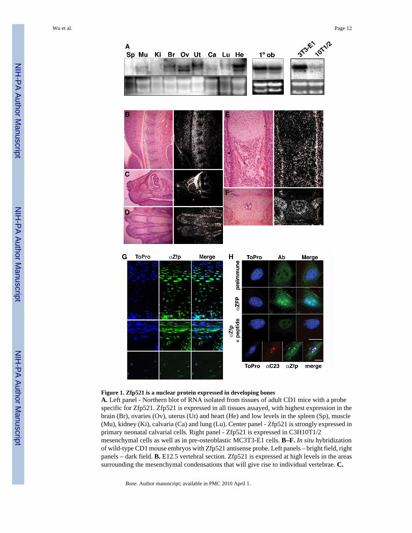
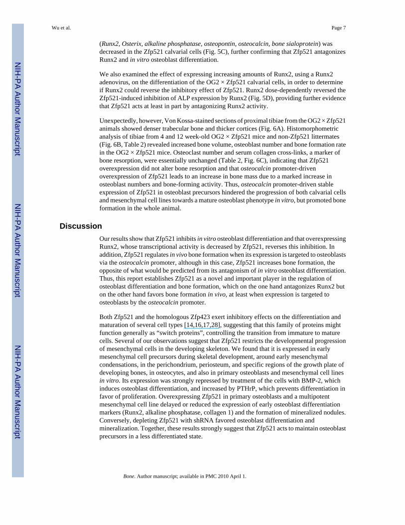
Figure 1. Zfp521 is a nuclear protein expressed in developing bonesA. Left panel - Northern blot of RNA isolated from tissues of adult CD1 mice with a probespecific for Zfp521. Zfp521 is expressed in all tissues assayed, with highest expression in thebrain (Br), ovaries (Ov), uterus (Ut) and heart (He) and low levels in the spleen (Sp), muscle(Mu), kidney (Ki), calvaria (Ca) and lung (Lu). Center panel - Zfp521 is strongly expressed inprimary neonatal calvarial cells. Right panel - Zfp521 is expressed in C3H10T1/2mesenchymal cells as well as in pre-osteoblastic MC3T3-E1 cells. B–F. In situ hybridizationof wild-type CD1 mouse embryos with Zfp521 antisense probe. Left panels – bright field, rightpanels – dark field. B. E12.5 vertebral section. Zfp521 is expressed at high levels in the areassurrounding the mesenchymal condensations that will give rise to individual vertebrae. C.
Wu et al. Page 12
Bone. Author manuscript; available in PMC 2010 April 1.
NIH
-PA Author Manuscript
NIH
-PA Author Manuscript
NIH
-PA Author Manuscript
Oblique section through E15.5 skull. Zfp521 is highly expressed in all the craniofacialelements, most strongly in the developing skull. D. E15.5 metatarsals. Zfp521 is highlyexpressed in the perichondrium of each developing bone in the developing limb. E. E15.5humerus. Zfp521 is expressed in both the perichondrium and periosteum of the bone collar.F. D.0 vertebrae. Zfp521 is expressed in the perichondrium as well as the endosteal surface ofthe developing marrow space. G. Immunohistochemistry with anti-Zfp521(369) antibody.Zfp521 is highly expressed in prehypertrophic chondrocytes of the growth plate (neonatalmetatarsals, top panels), in osteoblasts and one or two layers of pre-osteoblastic cells in theperiosteum and the endosteum (center panels), and in osteocytes (humeri of 6 month-old mice,lower panels). H. Confocal immunofluorescence of Zfp521 in C3H10T1/2 cells. The anti-Zfp521(369) antibody stained the nuclei, particularly in nucleoli, with weaker cytoplasmicstaining. The peptide antigen blocked the staining. Pre-immune serum gave a low diffusesignal, demonstrating specificity. Similar staining was seen with the anti-Zfp521(1177)antibody (data not shown). Bottom panels -C3H10T1/2 cells co-labeled with anti-nucleolin(αC23) and anti-Zfp521(369) antibodies, demonstrating the nucleolar localization of Zfp521(scale bar, 5 μm).
Wu et al. Page 13
Bone. Author manuscript; available in PMC 2010 April 1.
NIH
-PA Author Manuscript
NIH
-PA Author Manuscript
NIH
-PA Author Manuscript
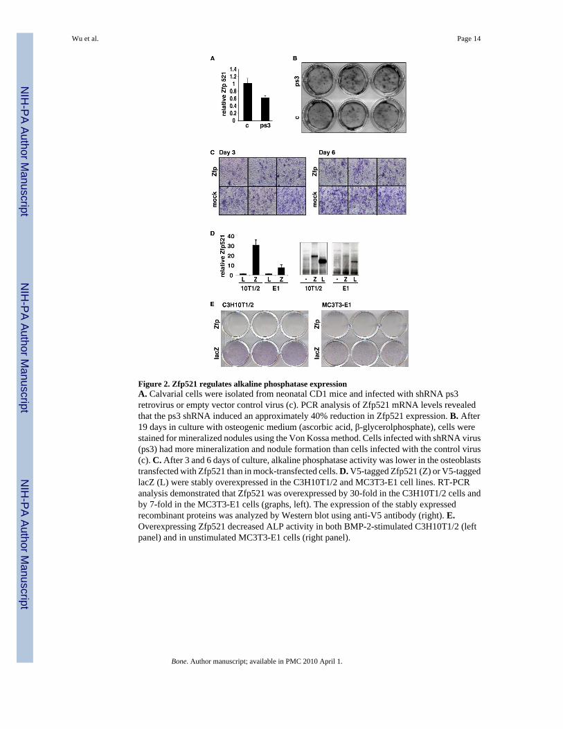
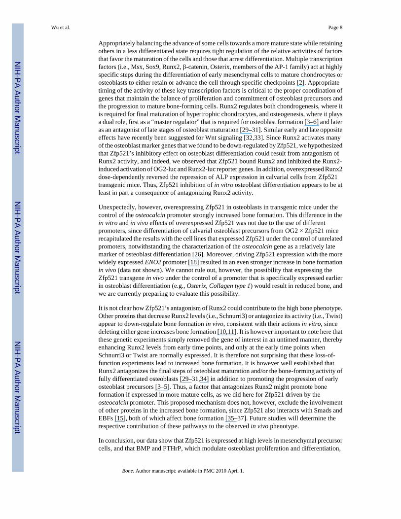
Figure 2. Zfp521 regulates alkaline phosphatase expressionA. Calvarial cells were isolated from neonatal CD1 mice and infected with shRNA ps3retrovirus or empty vector control virus (c). PCR analysis of Zfp521 mRNA levels revealedthat the ps3 shRNA induced an approximately 40% reduction in Zfp521 expression. B. After19 days in culture with osteogenic medium (ascorbic acid, β-glycerolphosphate), cells werestained for mineralized nodules using the Von Kossa method. Cells infected with shRNA virus(ps3) had more mineralization and nodule formation than cells infected with the control virus(c). C. After 3 and 6 days of culture, alkaline phosphatase activity was lower in the osteoblaststransfected with Zfp521 than in mock-transfected cells. D. V5-tagged Zfp521 (Z) or V5-taggedlacZ (L) were stably overexpressed in the C3H10T1/2 and MC3T3-E1 cell lines. RT-PCRanalysis demonstrated that Zfp521 was overexpressed by 30-fold in the C3H10T1/2 cells andby 7-fold in the MC3T3-E1 cells (graphs, left). The expression of the stably expressedrecombinant proteins was analyzed by Western blot using anti-V5 antibody (right). E.Overexpressing Zfp521 decreased ALP activity in both BMP-2-stimulated C3H10T1/2 (leftpanel) and in unstimulated MC3T3-E1 cells (right panel).
Wu et al. Page 14
Bone. Author manuscript; available in PMC 2010 April 1.
NIH
-PA Author Manuscript
NIH
-PA Author Manuscript
NIH
-PA Author Manuscript
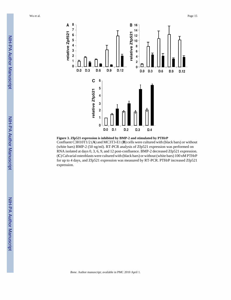
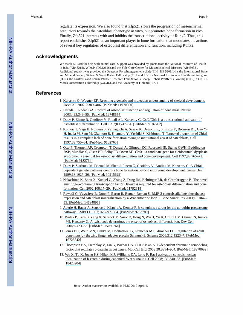
Figure 3. Zfp521 expression is inhibited by BMP-2 and stimulated by PTHrPConfluent C3H10T1/2 (A) and MC3T3-E1 (B) cells were cultured with (black bars) or without(white bars) BMP-2 (50 ng/ml). RT-PCR analysis of Zfp521 expression was performed onRNA isolated at days 0, 3, 6, 9, and 12 post-confluence. BMP-2 decreased Zfp521 expression.(C) Calvarial osteoblasts were cultured with (black bars) or without (white bars) 100 nM PTHrPfor up to 4 days, and Zfp521 expression was measured by RT-PCR. PTHrP increased Zfp521expression.
Wu et al. Page 15
Bone. Author manuscript; available in PMC 2010 April 1.
NIH
-PA Author Manuscript
NIH
-PA Author Manuscript
NIH
-PA Author Manuscript
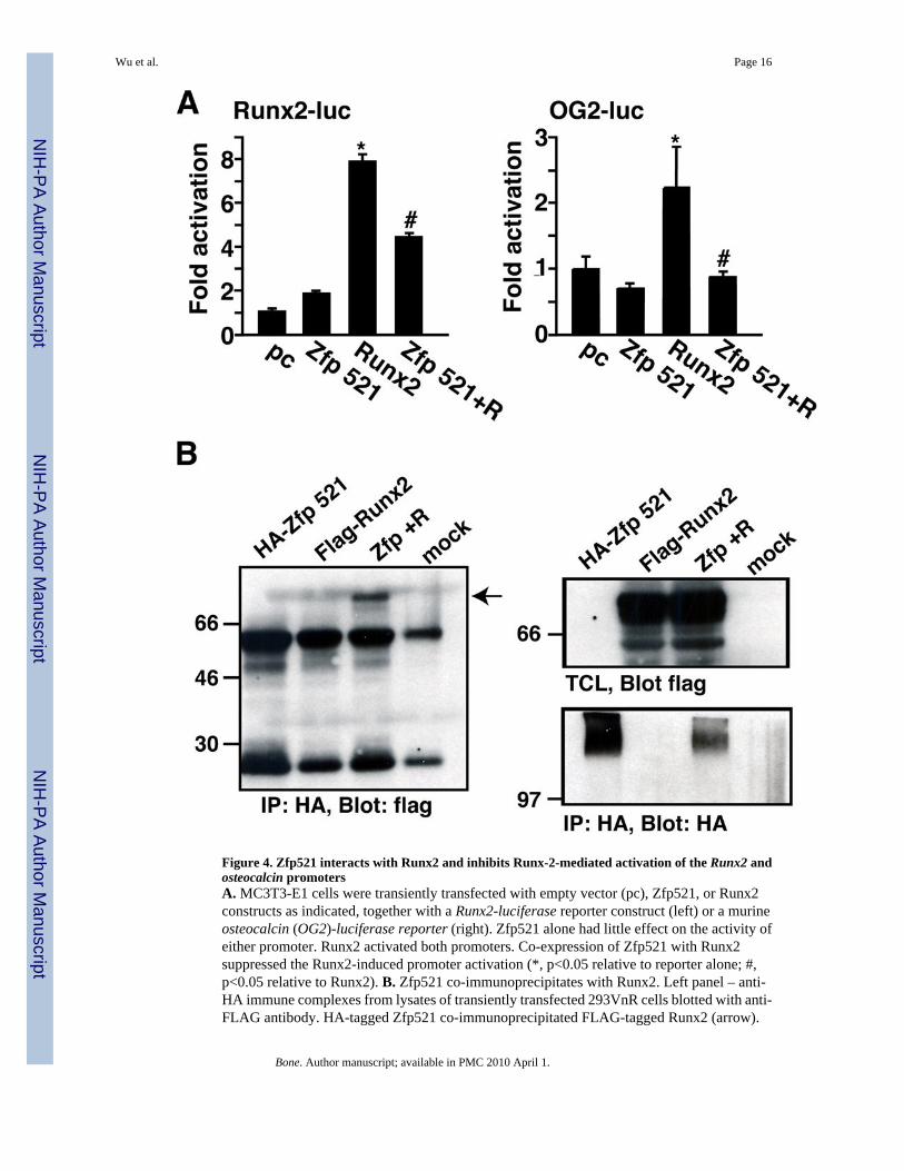
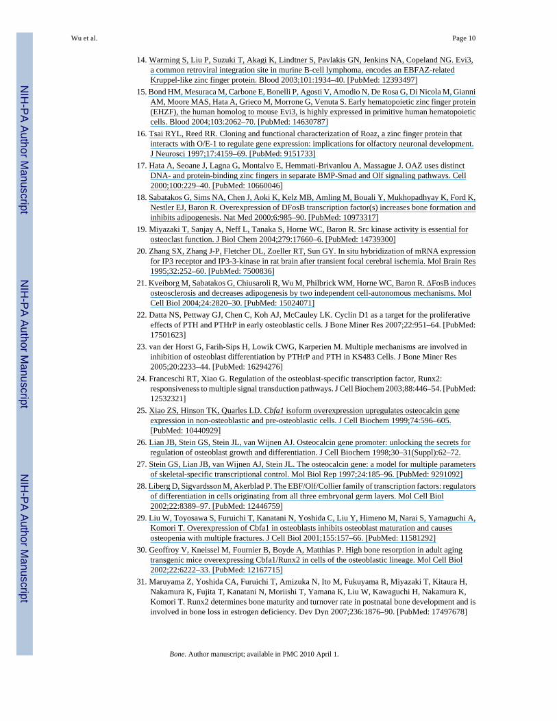
Figure 4. Zfp521 interacts with Runx2 and inhibits Runx-2-mediated activation of the Runx2 andosteocalcin promotersA. MC3T3-E1 cells were transiently transfected with empty vector (pc), Zfp521, or Runx2constructs as indicated, together with a Runx2-luciferase reporter construct (left) or a murineosteocalcin (OG2)-luciferase reporter (right). Zfp521 alone had little effect on the activity ofeither promoter. Runx2 activated both promoters. Co-expression of Zfp521 with Runx2suppressed the Runx2-induced promoter activation (*, p<0.05 relative to reporter alone; #,p<0.05 relative to Runx2). B. Zfp521 co-immunoprecipitates with Runx2. Left panel – anti-HA immune complexes from lysates of transiently transfected 293VnR cells blotted with anti-FLAG antibody. HA-tagged Zfp521 co-immunoprecipitated FLAG-tagged Runx2 (arrow).
Wu et al. Page 16
Bone. Author manuscript; available in PMC 2010 April 1.
NIH
-PA Author Manuscript
NIH
-PA Author Manuscript
NIH
-PA Author Manuscript

Right top panel – total cell lysates (TCL) blotted with anti-FLAG antibody to demonstrateFLAG-Runx2 expression. Right bottom panel – Anti-HA immune complexes blotted with anti-HA antibody to demonstrate HA-Zfp521.
Wu et al. Page 17
Bone. Author manuscript; available in PMC 2010 April 1.
NIH
-PA Author Manuscript
NIH
-PA Author Manuscript
NIH
-PA Author Manuscript
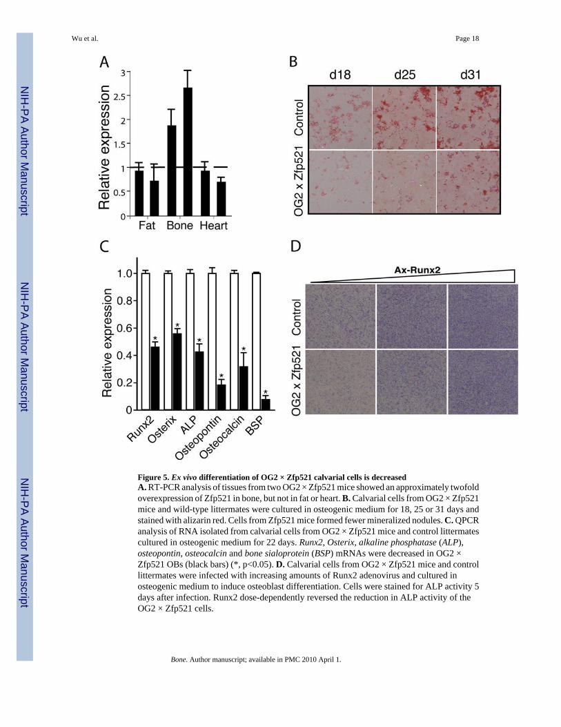
Figure 5. Ex vivo differentiation of OG2 × Zfp521 calvarial cells is decreasedA. RT-PCR analysis of tissues from two OG2 × Zfp521 mice showed an approximately twofoldoverexpression of Zfp521 in bone, but not in fat or heart. B. Calvarial cells from OG2 × Zfp521mice and wild-type littermates were cultured in osteogenic medium for 18, 25 or 31 days andstained with alizarin red. Cells from Zfp521 mice formed fewer mineralized nodules. C. QPCRanalysis of RNA isolated from calvarial cells from OG2 × Zfp521 mice and control littermatescultured in osteogenic medium for 22 days. Runx2, Osterix, alkaline phosphatase (ALP),osteopontin, osteocalcin and bone sialoprotein (BSP) mRNAs were decreased in OG2 ×Zfp521 OBs (black bars) (*, p<0.05). D. Calvarial cells from OG2 × Zfp521 mice and controllittermates were infected with increasing amounts of Runx2 adenovirus and cultured inosteogenic medium to induce osteoblast differentiation. Cells were stained for ALP activity 5days after infection. Runx2 dose-dependently reversed the reduction in ALP activity of theOG2 × Zfp521 cells.
Wu et al. Page 18
Bone. Author manuscript; available in PMC 2010 April 1.
NIH
-PA Author Manuscript
NIH
-PA Author Manuscript
NIH
-PA Author Manuscript
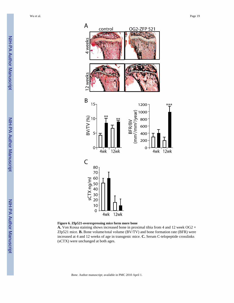
Figure 6. Zfp521-overexpressing mice form more boneA. Von Kossa staining shows increased bone in proximal tibia from 4 and 12 week OG2 ×Zfp521 mice. B. Bone volume/total volume (BV/TV) and bone formation rate (BFR) wereincreased at 4 and 12 weeks of age in transgenic mice. C. Serum C-telopeptide crosslinks(sCTX) were unchanged at both ages.
Wu et al. Page 19
Bone. Author manuscript; available in PMC 2010 April 1.
NIH
-PA Author Manuscript
NIH
-PA Author Manuscript
NIH
-PA Author Manuscript
NIH
-PA Author Manuscript
NIH
-PA Author Manuscript
NIH
-PA Author Manuscript
Wu et al. Page 20
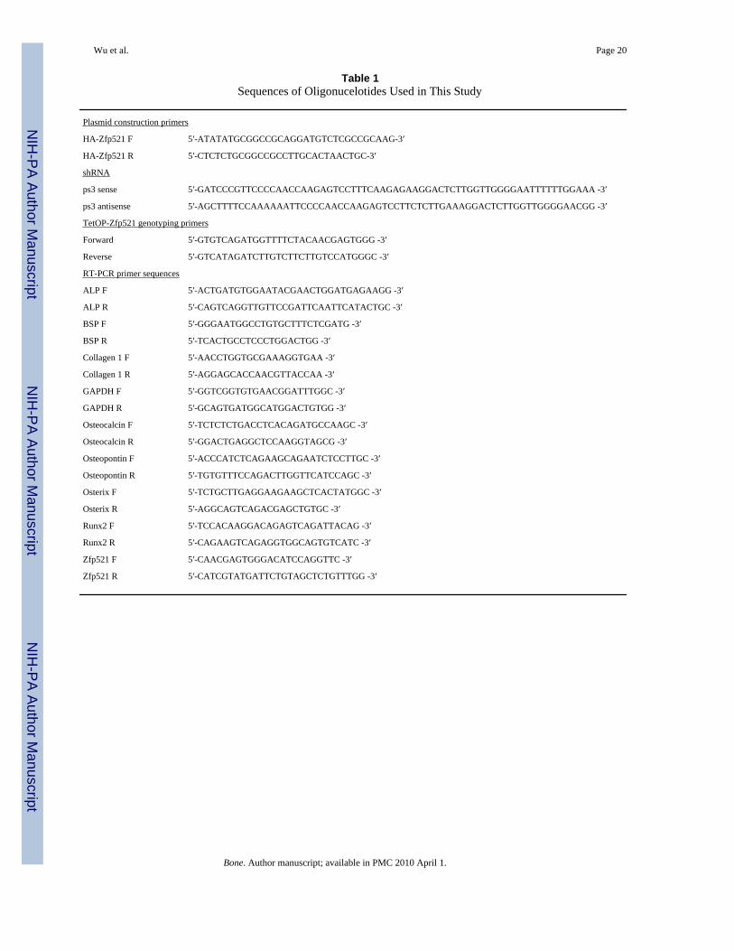
Table 1Sequences of Oligonucelotides Used in This Study
Plasmid construction primers
HA-Zfp521 F 5′-ATATATGCGGCCGCAGGATGTCTCGCCGCAAG-3′
HA-Zfp521 R 5′-CTCTCTGCGGCCGCCTTGCACTAACTGC-3′
shRNA
ps3 sense 5′-GATCCCGTTCCCCAACCAAGAGTCCTTTCAAGAGAAGGACTCTTGGTTGGGGAATTTTTTGGAAA -3′
ps3 antisense 5′-AGCTTTTCCAAAAAATTCCCCAACCAAGAGTCCTTCTCTTGAAAGGACTCTTGGTTGGGGAACGG -3′
TetOP-Zfp521 genotyping primers
Forward 5′-GTGTCAGATGGTTTTCTACAACGAGTGGG -3′
Reverse 5′-GTCATAGATCTTGTCTTCTTGTCCATGGGC -3′
RT-PCR primer sequences
ALP F 5′-ACTGATGTGGAATACGAACTGGATGAGAAGG -3′
ALP R 5′-CAGTCAGGTTGTTCCGATTCAATTCATACTGC -3′
BSP F 5′-GGGAATGGCCTGTGCTTTCTCGATG -3′
BSP R 5′-TCACTGCCTCCCTGGACTGG -3′
Collagen 1 F 5′-AACCTGGTGCGAAAGGTGAA -3′
Collagen 1 R 5′-AGGAGCACCAACGTTACCAA -3′
GAPDH F 5′-GGTCGGTGTGAACGGATTTGGC -3′
GAPDH R 5′-GCAGTGATGGCATGGACTGTGG -3′
Osteocalcin F 5′-TCTCTCTGACCTCACAGATGCCAAGC -3′
Osteocalcin R 5′-GGACTGAGGCTCCAAGGTAGCG -3′
Osteopontin F 5′-ACCCATCTCAGAAGCAGAATCTCCTTGC -3′
Osteopontin R 5′-TGTGTTTCCAGACTTGGTTCATCCAGC -3′
Osterix F 5′-TCTGCTTGAGGAAGAAGCTCACTATGGC -3′
Osterix R 5′-AGGCAGTCAGACGAGCTGTGC -3′
Runx2 F 5′-TCCACAAGGACAGAGTCAGATTACAG -3′
Runx2 R 5′-CAGAAGTCAGAGGTGGCAGTGTCATC -3′
Zfp521 F 5′-CAACGAGTGGGACATCCAGGTTC -3′
Zfp521 R 5′-CATCGTATGATTCTGTAGCTCTGTTTGG -3′
Bone. Author manuscript; available in PMC 2010 April 1.
NIH
-PA Author Manuscript
NIH
-PA Author Manuscript
NIH
-PA Author Manuscript
Wu et al. Page 21
Table 2Static and Dynamic Histomorphometric Analysis of OG2-tTA × TetOp-Zfp521 Transgenic Mice and Non-transgenicLittermate Controls
4 weeks 12 weeks
Control ZFP521 Control ZFP521
BV/TV 4.26 ± 0.54 8.54 ± 1.48* 6.67 ± 0.99 8.94 ± 0.29*
Tb.Th 27.87 ± 1.45 38.18 ± 5.85* 37.03 ± 3.54 42.09 ± 2.56*
Tb.Sp 663.35 ± 84.42 396.13 ± 30.56* 571.68 ± 62 407.87 ± 34.11
Tb.N 1.52 ± 0.16 2.32 ± 0.15* 1.76 ± 0.17 2.13 ± 0.08*
OS/BS 16.37 ± 5.38 30.39± 10.25 8.06 ± 1.8 15.18 ± 2.55*
Ob.S/BS 13.42 ± 3.39 13 ± 1.29 28.47 ± 2.85 30.98 ± 3.57
Oc.S/BS 0.99 ± 0.73 1.7 ± 0.73 2.36 ± 0.35 2.02 ± 0.54
N.Ob/T.Ar 14.58 ± 0.45 34.58 ± 4.69* 168.34 ± 24.92 211.66 ± 50.9*
N.Oc/T.Ar 1.53 ± 0.5 0.69 ± 0.44 4.48 ± 0.74 5.26 ± 0.84
OV/TV 0.65 ± 0.24 1.06 ± 0.55 0.21 ± 0.09 0.45 ± 0.09*
OV/BV 8.95 ± 2.57 12.32 ± 2.81 2.15 ± 0.51 4.75 ± 1.08P
O.Th 9.57 ± 1.39 8.51 ± 0.58 4.54 ± 0.64 6.07 ± 0.59
MS/BS 7.72 ± 3.41 18.52 ± 3.27* 19.71 ± 2.28 28.97 ± 2.64*
MAR 0.73 ± 0.41 0.86 ± 0.57 0.44 ± 0.17 1.42 ± 0.19**
BFR/BS 42.33 ± 10.55 68.81 ± 6.8 52.35 ± 27.45 165.83 ± 27.56***
BFR/BV 296.1 ± 96.8 404.96 ± 90.4 197.07 ± 88.43 988.67 ± 155.4***
BFR/TV 33.61 ± 20.54 20.33 ± 15.53 15.55 ± 6.36 89.19 ± 17.75***
*P< 0.01
**p<0.001
***p<0.0001
Bone. Author manuscript; available in PMC 2010 April 1.