Herschel map of Saturn's stratospheric water, delivered by the ...
Upload
independentCategory
view
0download
0
Cbfa1/Runx2-deficiency delays bone wound healing and locallydelivered Cbfa1/Runx2 promotes bone repair in animal models
Qisheng Tu, MD, PhD1,*, Jin Zhang, DDS1,2,*, Laji James, DDS3, Julia Dickson, DDS3, JeanTang, BS1, Pishan Yang, DDS2, and Jake Chen, DDS, PhD11 Division of Oral Biology, Tufts University School of Dental Medicine, Boston, Massachusetts2 School of Stomatology, Shandong University, Jinan, Shandong Province, China3 Department of Pediatric Dentistry, University of Texas Health Science Center at San Antonio,Texas
AbstractCore binding factor 1 (Cbfa1)/runt-related transcription factor 2 (Runx2) has been identified as a‘‘master gene’’ in osteoblastic differentiation. In this two-part study, part I of the study wasundertaken to test the hypothesis that bone regeneration is compromised in Cbfa1+/− mice. Comparedwith wild-type mice, wound healing was dramatically delayed in Cbfa1+/− mice characterized bythe presence of a small amount of bone near the base of the wounds. The bone defects were largelyfilled with fibrous connective tissues 3 weeks after surgery. Part II was performed to determine theeffects of Cbfa1 in enhancing bone wound healing using a gene-activated matrix (GAM) method.Cbfa1 cDNA was mixed with a biodegradable bovine type I collagen sponge and was inserted intothe periodontal window wounds of mice. Control sponges were collagen matrix without Cbfa1cDNA. Histological analysis and immunohistochemical staining demonstrated that compared withcontrols, there was increased new bone formation that almost filled the wound defects 14 days aftersurgery in the Cbfa1-GAM group. The collagen sponge matrix did not seem to elicit significantforeign body reaction in either group. In conclusion, the reduced expression of Cbfa1 interferes withthe process of bone wound healing, and local application of Cbfa1 cDNA incorporated into a collagenmatrix promotes bone tissue regeneration.
Millions of Americans are afflicted with periodontal diseases that cause destruction ofsupporting structures of teeth: periodontal ligament (PDL), alveolar bone, and cementum. Theprocess can lead to the loss of attachment, with destruction of the connective tissue matrix andcells. Loosening and eventual loss of teeth may follow. However, despite considerable researchefforts in this area, regeneration of the periodontium has still proved to be an elusive goal ofperiodontal therapy and remains a subject of intense interest to dentists and dental scientists,with the molecular and cellular bases of PDL formation, repair, and regeneration still poorlyunderstood.
In the 1980s, Gould observed that the progenitor cells for cementum, bone, and PDL fibroblastswere all contained in PDL tissue.1 He described generation of a new, functionally orientedPDL when teeth bearing cultured PDL cells were transplanted into bony sites. By contrast, thetransplantation of teeth bearing cultured gingival cells did not lead to regeneration of the PDL.These early studies indicated that regeneration of damaged periodontium can be achieved by
Reprint requests: Jake Chen, DDS, PhD, Department of General Dentistry, Division of Oral Biology, Tufts University School of DentalMedicine. One Kneeland Street. Boston, MA 02111. Tel: +1 617 636 2729; Fax: +1 617 636 0878; [email protected].*These two authors contributed to this work equally.
NIH Public AccessAuthor ManuscriptWound Repair Regen. Author manuscript; available in PMC 2010 March 12.
Published in final edited form as:Wound Repair Regen. 2007 ; 15(3): 404–412. doi:10.1111/j.1524-475X.2007.00243.x.
NIH
-PA Author Manuscript
NIH
-PA Author Manuscript
NIH
-PA Author Manuscript

the activity of the local stromal stem or progenitor cells found in paravascular locations of thePDL. These progenitor cells, which are stimulated to proliferate at wound sites, are derivedfrom adjacent, unwounded PDL. Selective repopulation of the root surface by PDL cells andsubsequent differentiation of these cells into fibroblasts, osteoblasts, and cementoblasts isbelieved to provide the basis for the regeneration of periodontal tissues and for the restorationof tissue domains.2
Similar to adipocyte3 and myoblast4 differentiation, it is apparent that components of theextracellular matrix are also critical for initiating cell differentiation during osteogenesis.5 Inthe 1990s, independent studies of a transcription factor that regulated the tissue-specificexpression of osteocalcin (OCN)6,7 and analysis of transgenic knockouts8,9 resulted in theidentification of core-binding factor 1 (Cbfa1), a runt domain transcription factor and itscognate enhancer. This transcription factor, also referred to as polyoma enhancer-bindingprotein (PEBP2aA), osteo-blast-specific factor (Osf2), and more recently as runt-relatedtranscription factor 2 (Runx2), is identified as a ‘‘master gene’’ required for osteoblasticdifferentiation.10 Cbfa1 is expressed during the early development of mesenchymal andepithelial tissues destined to form the mineralized tissues of the tooth and periodontal apparatus.11,12 In Cbfa1−/− mice,8,9 there is an almost complete lack of mineralized bone. The absenceof the osteodifferentiation markers, alkaline phosphatase and OCN, is consistent with an earlyblock in osteogenic development in these mice. Furthermore, numbers of in vitro studies haveshown that Cbfa1 is a positive regulator that can up-regulate the expression of bone matrixgenes, including type I collagen, osteopontin (OPN), OCN, fibronectin, and bone sialoprotein(BSP).13–16 And it is reported that various mutations in the human Cbfa1 gene locus, onchromosome 6p21, have been identified to correlate with the disease of cleidocranial dysplasia(CCD). This disease is an autosomal dominant disorder that is characterized by a short stature,varying degrees of loss of the clavicles, open cranial fontanels, and dental manifestationsincluding supernumerary teeth, delayed tooth eruption, tooth hypoplasia, and absence ofcellular cementum formation.9,17,18 Although evidences have shown that Cbfa1 has anessential role in osteogenesis, an unambiguous effect of Cbfa1 in promoting differentiation ofregenerative cells in periodontium has not yet been established. Therefore, to determine thefunction of Cbfa1 in bone repair and regeneration, we proposed a loss-of-function study to testthe hypothesis that bone regeneration is compromised in Cbfa1+/− mice.
In the second part of this research, a gain-of-function study was performed. It is well knownthat bone regeneration and tissue engineering are of enormous importance, particularly forreconstruction of oral and maxillofacial tissues that have been lost or damaged by diseases,trauma, or congenital disorders. The ultimate therapeutic goal of corrective surgery is toregenerate tissues to their normal or predisease state. Considering the key role of Cbfa1 inosteoblast differentiation, it can be deduced that Cbfa1 may also play an important role in boneregeneration and may induce an embryonic (regenerative) environment in the injured adulttissues. Here, in the second part of this study, the effects of overexpressed Cbfa1 in enhancingbone wound healing using a gene-activated matrix (GAM) method were determined.
MATERIALS AND METHODSAnimals
Six- to 8-week-old mice were maintained and used in accordance with recommendations inthe Guide for the Care and Use of Laboratory Animals, prepared by the Institute on LaboratoryAnimal Resources, National Research Council (DHHS Pub. NIH 86-23, 1985). The animalprotocol was approved by the Institutional Animal Use and Care Committee at the Tufts-NewEngland Medical Center in Boston (Massachusetts).
Tu et al. Page 2
Wound Repair Regen. Author manuscript; available in PMC 2010 March 12.
NIH
-PA Author Manuscript
NIH
-PA Author Manuscript
NIH
-PA Author Manuscript

Part IWound model—Periodontal window wounds and femoral defects were created in four Cbfa1+/− mice (a generous gift from Dr. Michael Owen, Imperial Cancer Research Fund, London,UK) and four wild mice under general anesthesia (a mixture of ketamine and xylazine, 110and 10 mg/kg). Periodontal window wounds (1 mm in diameter) were an established model asdescribed before.19,20 In brief, a skin flap was raised along the lower edge of the mandible toexpose the underlying bone. The site of the window wound was 1 mm away from the anterioredge of the mandible and between the superior margin of the alveolar bone overlying the mesialroot of the lower first molar and the foramen mentale. Under a dissecting microscope at ×10magnification, a dental burr (#329, Midwest, 0.6 mm in diameter) driven at a low speed by adental handpiece and cooled with phosphate-buffered saline (PBS) was used to penetrate theoverlying alveolar bone. The PDL was removed with a sharpened dental probe. Wounds wereuntreated and tissues were closed with 4–0 gut suture. Animals were closely observed for 1hour after surgery. Femoral defects (1 mm in diameter) were created using a 0.6 mm cuttingburr through a lateral incision after general anesthesia described above.
Tissue preparation—The animals were sacrificed 3 weeks after wounding. Aftereuthanasia, the part of the mandible between the central incisor and the third molar region wasexcised and for femoral defects, the whole femur was dissected. The tissues were fixedimmediately in periodate-lysine-paraformaldehyde21 for 24 hours. The tissue wasdemineralized in 0.2 N HCL for 2 days with constant stirring. Demineralized tissues werewashed in 0.1 M phosphate buffer (pH 7.2) overnight. The tissues were then dehydrated in anascending series of ethanol, cleared in xylene, and embedded in paraffin. As described inprevious publications,22 tissue sections, 6 μm thick, were mounted on glass slides.
Histomorphometric assessment—For each animal, three hematoxylin & eosin (H&E)-stained sections of each wound were analyzed morphometrically (Carl Zeiss Inc., Hamburg,Germany). With the aid of the computer software package, Image-pro plus (MediaCybernetics), image analysis was performed to assess: (a) the area of the drilled bone bydigitizing the reversal line in the bone at the cut margin that provided an estimate of the originalwound outline for each animal and the width of closure of the bony defect at the outer surfaceof the drilled site, (b) the area of the newly formed bone, (c) these two areas were expressedas a percentage (area of newly formed bone/area of defect × 100). Mean ± standard errors ofthe percentages were computed, and (d) the width of regenerating PDL was compared with theunwounded side (as a control) in the same sample.
Part IICbfa1 plasmid DNA preparation—Large-scale DNA preparation of the plasmid pCMV-Osf2/Cbfa1 (containing the complete cDNA of mouse Cbfa1, provided by Dr. Gerard Karsenty,Bayer College of Medicine, Houston, TX) was performed using an EndofreePlasmid Mega kit(Qiagen, Valencia, CA). The plasmid DNA was stored at −20°C in endotoxin-free TE bufferbefore use.
Assembly of Cbfa1 GAM prototypes—Cell Prime, an acid-soluble form of bovine typeI collagen, was purchased from Cohesion Technologies (Palo Alto, CA). To generate GAM,equal volumes of sterile tissue, culture medium (Dulbecco’s modified Eagle’s medium[DMEM]) and bovine collagen were mixed to achieve a pH of approximately 7.0. Sterile, pureCMV-Cbfa1 plasmid DNA in TE buffer was mixed with the neutralized collagen with aconcentration of 1.2 mg DNA in 1 mg collagen. The DNA and the collagen were mixed atroom temperature until a uniform distribution of plasmid DNA was achieved. The DNA–collagen mixture was then frozen at −86°C and lyophilized.
Tu et al. Page 3
Wound Repair Regen. Author manuscript; available in PMC 2010 March 12.
NIH
-PA Author Manuscript
NIH
-PA Author Manuscript
NIH
-PA Author Manuscript
Transplantation of Cbfa1-GAM into wounds—Periodontal window wounds werecreated in 22 seven-week-old mice as described in part I. The mice were divided into twogroups randomly. In group I, bone defects were filled with Cbfa1 GAM with a dose of 1.2 mgplasmid DNA per wound. Sterile GAM prototypes were placed and held in the defect sitesuntil they were surrounded by clotted blood. Control sponges in group II were comprised ofbiodegradable, biocompatible collagen without plasmid DNA. Animals were euthanized at 7and 14 days after the transplantation, respectively. These time points were chosen becauseprevious studies had shown that 1 week was required for initial healing and cell repopulationof wounded connective tissue (PDL in the periodontal wounds), and 2 weeks were requiredfor the initiation of mineralization of the regenerating new bone. After euthanasia, tissuepreparation and histomorphometric analysis were performed as described in Part I.
Immunohistochemical staining—The tissue slides were first deparaffinized andrehydrated, and then submerged in hydrogen peroxide for peroxidase quenching. Before usingthe primary antibodies, the slides were incubated with normal serum to block the nonspecificbindings. Monoclonal antibodies against BSP (LF120 from Dr. Larry Fisher, NIH/NIDCR)were then used at a dilution of 1 : 200. After overnight incubation, the biotinylated secondaryantibodies were applied to the slides. Finally, the substrate-chromogen 3-Amino-9-ethylcarbazole (AEC) was applied and the slides were counterstained with hematoxylin andmounted. Control sections were incubated with an irrelevant antibody (anti-human CD4lymphocyte antigen) to estimate background staining. All slides were randomly coded toprevent examiner’s bias.
The localization of BSP was studied on transverse sections of the first mandibular molar usingpreviously described semi-quantitative methods.19 In brief, the localization of BSP expressionwas studied in the newly formed alveolar bone (site 1) and in the unwounded alveolar bone(site 2). The unwounded side serves as an internal control for the assessment of the stainingintensity at the wounded side. The relative intensities of BSP staining were classified as intense(3), moderate (2), weak (1), or negative (0). For each slide the intensity of BSP staining wasdetermined by comparing the staining at each wounded site with unwounded alveolar bone inthe same section. Unwounded alveolar bone was classified as 2 (or moderate) staining intensityand experimental sites were compared with this internal standard. Stronger staining thaninternal controls was classified as 3 (intense), lower than internal controls as 1 (low), and nostaining as 0 or negative. This method is semi-quantitative and provides meaningfulcomparative data when differences of staining intensity are large.
Morphometric assessment of immunostained sections—At least three sections ofBSP-immunolabeled bone defects in each group were analyzed morphometrically. Slides werecoded so that time of killing, type of wound, and type of staining were unknown at the time ofmeasurement. Image analysis (Carl Zeiss Inc., Image-pro plus, Media Cybernetics) was usedto assess the area of the drilled bone by digitizing the reversal line in the bone at the cut marginthat provided an estimate of the original wound outline for each wound. Second, the area ofthe BSP-stained tissue in the bone compartment of the wound was digitized. Finally, these twoareas were expressed as a percentage (area of BSP staining/area of the defect×100),representing a newly formed bone ratio. Mean ± standard errors of the percentages werecomputed.
Total RNA extraction and reverse transcriptase polymerase-chain reaction (RT-PCR) analysis—For RT-PCR analysis, the implant samples (3 each time point) were excisedunder a dissecting microscope to avoid harvesting the host bone. Freshly dissected implanttissues were collected into ‘‘RNAlater’’ solution (Qiagen) at 4°C and homogenized in TRIzolsolution (Invitrogen, Carlsbad, CA), followed by the RNA isolation procedure recommendedby the manufacturer. Freshly isolated RNA was reverse transcribed with a SuperScript™ first-
Tu et al. Page 4
Wound Repair Regen. Author manuscript; available in PMC 2010 March 12.
NIH
-PA Author Manuscript
NIH
-PA Author Manuscript
NIH
-PA Author Manuscript

strand synthesis system (Invitrogen) following the manufacturer’s recommendations. Theresulting cDNA was then amplified by PCR with the Platinum PCR supermix (Invitrogen).The sequences of the primers for amplification of mouse Runx2/Cbfa1 were: 5′-GAGGCCGCCGCACG ACAACCG-3′ and 5′-CTCCGGCCCACAAATCTCAGA-3′(product size: 294 bp); for mouse glyceraldehyde-3-phosphate dehydrogenase (GAPDH) were:5′-ACCACAGTCCATGCCATCAC-3′ and 5′-TCCACCACCCT GTTGCTGTA-3′ (productsize: 450 bp). Linearity of the PCR conditions was determined for each primer pair inpreliminary experiments (data not shown). Images of the amplified products in 1.5% agarosegels were captured with a UVP bioimaging system and processed by Adobe Photoshop 6.0(Adobe Systems Incorporated, San Jose, CA) and Scion Image Beta 4.02 (Scion Image,Frederick, MD). The data are presented as the fold changes in gene expression afternormalization to the expression of G3PDH.
Statistical analysis—Raw data were kept separate and the means for each animal, type oftreatment, postwounding time, and site examined were computed. Mean data from each animalwere considered as independent samples. Data were assessed by analysis of variance(ANOVA) to evaluate the differences between the morphometric assessments andimmunostaining findings with respect to different sites, type of treatment, i.e., with vs. withoutCbfa1 cDNA transplantation, and postwounding time.
RESULTSIn the present two-part study, we used a mouse model to study tissue regeneration and woundrepair. Tissue sections were subsequently processed for histomorphometric analyses andimmunohistochemical staining of BSP to assess the effect of Cbfa1 on the regeneration ofperiodontal and femoral defects.
Part IIn wild-type (WT) mice, the relative volume of new bone matrix (Figure 1) was higher thanin the Cbfa1+/− mice (p < 0.05). The newly formed bone in WT mice showed a trabecularstructure, and the bone was well mineralized and merged with the normal bone at the defectedges. In contrast, wound healing was dramatically delayed in Cbfa1+/− mice characterizedby the presence of a small amount of bone near the base of the wounds. The alveolar bonedefects were largely filled with fibrous connective tissues 3 weeks after surgery (Figure 2).The bone wounds in femurs in WT mice showed callus formation at the inner side. In somecases, the defects almost healed completely. In Cbfa1+/− mice, the repair occurred at a muchslower pace and granulation tissues were seen at the wound sites (Figure 3). In some cases, thefemur wound was still widely open without any significant bone formed.
Part IIMorphometric assessment—At day 7, small islands of bone matrix could be seenscattered in the bone defects in all the Cbfa1-applied tissues and some of the controls (Figure4). However, the relative volume of new bone matrix in the defects of the Cbfa1 group washigher than in controls (> 1.5-fold) (p < 0.05). At day 14, bone formation in the Cbfa1 groupexhibited an even higher increase ( > twofold) compared with controls (p < 0.05) (Figure 5).And at this time, the newly formed bones almost filled the wound defects in the Cbfa1 group,while in controls, the newly formed bones were poorly organized and weakly stained. In somesamples of control tissues at day 14, there were large amounts of granulation tissues in thewound sites.
The PDL width was measured to assess the homeostasis of soft connective tissue domains. Atday 7, PDL width in the wounded sides significantly increased compared with unwounded
Tu et al. Page 5
Wound Repair Regen. Author manuscript; available in PMC 2010 March 12.
NIH
-PA Author Manuscript
NIH
-PA Author Manuscript
NIH
-PA Author Manuscript

sides in both groups (p < 0.05). At day 14, PDL width in the wounded sides decreased andexhibited values similar to unwound sides in the Cbfa1 group (p > 0.4). However, PL width inthe wounded sides remained significantly high compared with the unwounded side in thecontrol group at this time (p < 0.05). These measures were expressed as a percentage of thedigitized normal values for each time point (mean ± standard errors of the percentages) (Figure6).
The collagen sponge matrix did not elicit significant foreign body reaction in either theexperimental or control groups. Examination of wound sites showed that implantation of GAMcontaining Cbfa1 DNA promoted bone formation.
Cbfa1-GAM stimulates expression of BSP through up-regulation of Cbfa1 inperiodontal window wounds—BSP was used as a phenotypic marker of mineralizingconnective tissues to study cell differentiation in the periodontium. At day 7, staining for BSPwas weak in the newly formed alveolar bone in the control group. In some samples, there wasno new bone formed, and the staining for BSP was negative. In contrast, BSP staining in theCbfa1 group was more intense in the newly formed alveolar bone than in the unwoundedalveolar bone area (p < 0.05). At day 14, the staining intensity for BSP became much strongerin both groups in the newly formed alveolar bone, but particularly in the Cbfa1 group (p <0.05) (Figure 7). Consistently, the data demonstrated that the GAM significantly up-regulatedthe expression of cbfa1 in submandibular defects as early as day 7, and remained at a high leveluntil 14 days after surgery (Figure 8).
DISCUSSIONRunx2/Cbfa1 is a transcription factor that belongs to the runt domain gene family23 andfunctions by forming a heterodimer with Cbfb (core-binding factor beta).24 Cbfa1−/− micecompletely lack bone formation because of the maturational arrest of osteoblasts and die soonafter birth, indicating that Cbfa1 is an essential factor for osteoblast differentiation.8,9 Cbfa1heterozygous knockout mice (Cbfa1+/−) stay alive but show morphological defects in theskeletal system as observed in CCD in humans. In addition, chondrocyte maturation is alsodisturbed in Cbfa1−/− mice,25 and Cbfa1 has been shown to be an important factor forchondrocyte maturation.26 Although chondrocytes had matured, and the matrix wasmineralized in restricted parts of the skeleton of Cbfa1−/− mice, osteoclasts were completelyabsent, and no vascular invasion into the calcified cartilage occurs.8 Therefore, Cbfa1 playsimportant roles in multiple processes of endochondral ossification, including chondrocytematuration, vascular invasion into the cartilage, osteoclast differentiation, and osteoblastdifferentiation.27
In the first part of this study, data demonstrated that in contrast to the WT mice, wound healingwas dramatically delayed in Cbfa1+/− mice characterized by the presence of only a smallamount of bone near the base of the wounds. The alveolar bone defects were largely filled withfibrous connective tissues 3 weeks after surgery. Bone repair occurred at a much slower pacein these mice and granulation tissues were seen at the wound sites. In some cases, the femurwound was still widely open without any significant bone formed. This loss-of-function studyproved that bone regeneration is compromised in Cbfa1+/− mice, thus indicating that Cbfa1also plays an important role in the differentiation of osteogenic progenitor cells during boneregeneration.
Considering the essential role of Cbfa1 in osteogenesis and bone formation, several studieshave applied this transcription factor for tissue engineering. In a recent study, Byers et al.28
reported that primary rat bone marrow stromal cells transduced with Runx2 retroviral vectorwere seeded onto three-dimensional fused deposition-modeled polycaprolactone scaffolds.
Tu et al. Page 6
Wound Repair Regen. Author manuscript; available in PMC 2010 March 12.
NIH
-PA Author Manuscript
NIH
-PA Author Manuscript
NIH
-PA Author Manuscript

These Runx2-modified cells produced biologically equivalent mineralized matrices at nearlytwofold higher rates than control cells. Zhao et al.29 and Kojima et al.30 reported that primaryosteoprogenitor cells transduced with adenoviral vectors encoding Runx2 formed substantiallymore bone than cells transduced with control virus. However, there are safety concerns aboutthe clinical use of the viral method. Proviral DNA derived from a retrovirus can be randomlyintegrated into the target cell genome.31 Thus, the target cell’s genotype is permanently altered.Moreover, during integration of the proviral DNA, insertional mutagenesis can occur if theinserted DNA disrupts housekeeping genes or activates other genes, e.g., an oncogene.31
Furthermore, local toxicity can result from chronic overexpression of the expression cassetteproduct. In addition, most retrovirus can only infect, integrate, and express in dividing targetcells. Other limitations include low titers and sensitivity to inactivation.31 As for adenovirusvectors, although problems associated with insertional mutagenesis are avoided, they are stilllimited in use by the nonspecific immunologic reactions they elicit, as well as the potential forautoimmune reactions to the transgene-encoded proteins. Patient safety is a paramount issue,32 which considerably restricts the clinical application of the virus-based method.
The GAM method offers an alternative and safer approach to virus-based tissue-engineeringapplications.33,34 This method was designed specifically to provide an ideal environment fortissue regeneration and engineering. In some animal studies, plasmid genes can be delivered,in a polymer GAM, directly to actually injured bone, muscle, and ligament.35,36 The GAMcould serve as a platform technology for local gene delivery in various tissues and organs. AGAM consists of two ingredients: plasmid DNA and a biodegradable structural matrix carrier,e.g., a collagen sponge or gel. The carrier serves as a scaffold that holds DNA in situ untilendogenous wound-healing cells arrive. Up to 50% of available healing repair cells would betransfected.37 Wound repair cells possess relatively high levels of pinocytosis and potocytosis.38 This inherent ability may facilitate plasmid gene transfer and help explain the high level ofgene transfer efficiency associated with the GAM. The cells in the matrix carrier act as localin vivo bioreactors, producing plasmid-coding proteins that augment tissue repair andregeneration. GAM implantation at sites of bone injury was associated with retention andexpression of plasmid DNA for at least 6 weeks.34 In this study by Bonadio et al., GAMimplants with a pMat-1 plasmid DNA that encodes a secreted peptide fragment of humanparathyroid hormone (hPTH 1–34) induced predictable formation of centimeters of normalnew bone in a time-, plasmid dose-, and bone gap size-dependent manner.34
In the second part of the present study, Cbfa1 cDNA was locally delivered to the periodontalwindow wound using the GAM method to assess the effects of this transcription factor on thePDL, a tissue that is thought to contain the progenitor cells for cementum, bone, and PDLfibroblasts. At day 7, we observed that the GAM porous architecture provided scaffolding topromote cell ingrowth. The local granulation tissue fibroblasts, along with capillaries, migratedinto the GAM. The osteogenic progenitor cells within the tissue uptook the local plasmid DNAand transiently expressed the gene. The transfected reactive cells then secreted the plasmid-encoded proteins to stimulate and augment the bone regenerative cascade, which has beenobserved as the expression of both Cbfa1 and BSP increased in our study. As the GAM systemdelivers genes instead of recombinant proteins, researchers have reported longer-timeexpression of genes of interest (weeks vs. hours).34,39 In the present study, the relative volumeof newly formed bone was higher in the Cbfa1-GAM group than in the control group. At day14, the newly formed bones almost filled the wound defects in the Cbfa1 group, while incontrols, the newly formed bones were poorly organized and weakly stained. Furthermore,Cbfa1 and BSP expression was stronger in the Cbfa1 group than in the control group, whichindicated that Cbfa1 cDNA taken up by the PDL cells can be expressed in situ in an autocrine/intracrine manner40 that promoted phenotypic differentiation. As a result, expression of boneand cementum-specific proteins, e.g., BSP could be stimulated. This would lead to an increasein periodontal tissue regeneration.
Tu et al. Page 7
Wound Repair Regen. Author manuscript; available in PMC 2010 March 12.
NIH
-PA Author Manuscript
NIH
-PA Author Manuscript
NIH
-PA Author Manuscript
The PDL width was measured to assess the homeostasis of soft connective tissue domains. Atday 7, PDL width in the wounded sides significantly but slightly increased compared with theunwounded sides in both groups. However, at day 14, PDL width in the wounded sidesdecreased and exhibited values similar to the unwounded sides in the Cbfa1 group. Thesemeasures demonstrate that locally delivered Cbfa1 did not cause ankylosis in spite of inducingnew bone formation.
In conclusion, bone regeneration was compromised in Cbfa1+/− mice, and locally deliveredCbfa1 using the GAM approach promoted bone regeneration in the periodontal window wound.This study indicates that Cbfa1 also plays an important role in the differentiation of osteogenicprogenitor cells during bone regeneration.
AcknowledgmentsThis work was supported by NIH grants DE11088, DE14537 to JC.
References1. Gould TR, Melcher AH, Brunette DM. Migration and division of progenitor cell populations in
periodontal ligament after wounding. J Periodontal Res 1980;15:20–42. [PubMed: 6445968]2. Gould TR, Melcher AH, Brunette DM. Location of progenitor cells in periodontal ligament of mouse
molar stimulated by wounding. Anat Rec 1977;188:133–41. [PubMed: 869235]3. Loftus TM, Lane MD. Modulating the transcriptional control of adipogenesis. Curr Opin Genet Dev
1997;7:603–8. [PubMed: 9388775]4. Arnold HH, Winter B. Muscle differentiation: more complexity to the network of myogenic regulators.
Curr Opin Genet Dev 1998;8:539–44. [PubMed: 9794824]5. Owen TA, Aronow M, Shalhoub V, Barone LM, Wilming L, Tassinari MS, Kennedy MB, Pockwinse
S, Lian JB, Stein GS. Progressive development of the rat osteoblast phenotype in vitro: reciprocalrelationships in expression of genes associated with osteoblast proliferation and differentiation duringformation of the bone extracellular matrix. J Cell Physiol 1990;143:420–30. [PubMed: 1694181]
6. Ducy P, Karsenty G. Two distinct osteoblast-specific cis-acting elements control expression of a mouseosteocalcin gene. Mol Cell Biol 1995;15:1858–69. [PubMed: 7891679]
7. Merriman HL, van Wijnen AJ, Hiebert S, Bidwell JP, Fey E, Lian J, Stein J, Stein GS. The tissue-specific nuclear matrix protein, NMP-2, is a member of the AML/CBF/PEBP2/Runt domaintranscription factor family. Biochemistry 1995;34:13123–32.
8. Komori T, Yagi H, Nomura S, Yamaguchi A, Sasaki K, Deguchi K, Shimizu Y, Bronson RT, GaoYH, Inada M, Sato M, Okamoto R, Kitamura Y, Yoshiki S, Kishimoto T. Targeted disruption of Cbfa1results in a complete lack of bone formation owing to maturational arrest of osteoblasts. Cell1997;89:755–64. [PubMed: 9182763]
9. Otto F, Thornell AP, Crompton T, Denzel A, Gilmour KC, Rosewell IR, Stamp GW, Beddington RS,Mundlos S, Olsen BR, Selby PB, Owen MJ. Cbfa1, a candidate gene for cleidocranial dysplasiasyndrome, is essential for osteoblast differentiation and bone development. Cell 1997;89:765–71.[PubMed: 9182764]
10. Rodan GA, Harada S. The missing bone. Cell 1997;89:677–80. [PubMed: 9182754]11. Chen, J.; Jiang, H.; Sodek, J.; Karsenty, G.; Thomas, H.; Ranly, D. Comparison of expression patterns
of core binding factor (Osf2/Cbfa1) and bone sialoprotein (BSP). In: Goldberg, M.; Boskey, A.;Robinson, C., editors. Chemistry and biology of mineralized tissue. Vol. Chapter 24. Rosemont:American Academy of Orthopaedic Surgery; 2000. p. 149-54.
12. Jiang H, Sodek J, Karsenty G, Thomas H, Ranly D, Chen J. Expression of core binding factor Osf2/Cbfa1 and bone si-aloprotein in tooth development. Mech Dev 1999;81:169–73. [PubMed:10330494]
13. Sato M, Morii E, Komori T, Kawahata H, Sugimoto M, Terai K, Shimizu H, Yasui T, Ogihara H,Yasui N, Ochi T, Kitamura Y, Ito Y, Nomura S. Transcriptional regulation of osteopontin gene in
Tu et al. Page 8
Wound Repair Regen. Author manuscript; available in PMC 2010 March 12.
NIH
-PA Author Manuscript
NIH
-PA Author Manuscript
NIH
-PA Author Manuscript
vivo by PEBP2alphaA/CBFA1 and ETS1 in the skeletal tissues. Oncogene 1998;17:1517–25.[PubMed: 9794229]
14. Xiao ZS, Hinson TK, Quarles LD. Cbfa1 isoform overex-pression upregulates osteocalcin geneexpression in non-osteoblastic and pre-osteoblastic cells. J Cell Biochem 1999;74:596–605.[PubMed: 10440929]
15. Kern B, Shen J, Starbuck M, Karsenty G. Cbfa1 contributes to the osteoblast-specific expression oftype I collagen genes. J Biol Chem 2001;276:7101–7. Epub December 5, 2000. [PubMed: 11106645]
16. Prince M, Banerjee C, Javed A, Green J, Lian JB, Stein GS, Bodine PV, Komm BS. Expression andregulation of Runx2/Cbfa1 and osteoblast phenotypic markers during the growth and differentiationof human osteoblasts. J Cell Biochem 2001;80:424–40. [PubMed: 11135373]
17. Lee B, Thirunavukkarasu K, Zhou L, Pastore L, Baldini A, Hecht J, Geoffroy V, Ducy P, KarsentyG. Missense mutations abolishing DNA binding of the osteoblast-specific transcription factor OSF2/CBFA1 in cleidocranial dysplasia. Nat Genet 1997;16:307–10. [PubMed: 9207800]
18. Cunningham ML, Seto ML, Hing AV, Bull MJ, Hopkin RJ, Leppig KA. Cleidocranial dysplasia withsevere parietal bone dysplasia: C-terminal RUNX2 mutations. Birth Defects Res A Clin Mol Teratol2006;76:78–85. [PubMed: 16463420]
19. Lekic P, Sodek J, McCulloch CA. Osteopontin and bone si-aloprotein expression in regenerating ratperiodontal ligament and alveolar bone. Anat Rec 1996;244:50–8. [PubMed: 8838423]
20. Lekic P, Rubbino I, Krasnoshtein F, Cheifetz S, McCulloch CA, Tenenbaum H. Bisphosphonatemodulates proliferation and differentiation of rat periodontal ligament cells during wound healing.Anat Rec 1997;247:329–40. [PubMed: 9066910]
21. McLean IW, Nakane PK. Periodate-lysine-paraformaldehyde fixative. A new fixation forimmunoelectron microscopy. J Histochem Cytochem 1974;22:1077–83. [PubMed: 4374474]
22. Sasaguri K, Jiang H, Chen J. The effect of altered functional forces on the expression of bone-matrixproteins in developing mouse mandibular condyle. Arch Oral Biol 1998;43:83–92. [PubMed:9569994]
23. Komori T, Kishimoto T. Cbfa1 in bone development. Curr Opin Genet Dev 1998;8:494–9. [PubMed:9729728]
24. Yoshida CA, Furuichi T, Fujita T, Fukuyama R, Kanatani N, Kobayashi S, Satake M, Takada K,Komori T. Core-binding factor beta interacts with Runx2 and is required for skeletal development.Nat Genet 2002;32:633–8. Epub November 18, 2002. [PubMed: 12434152]
25. Inada M, Yasui T, Nomura S, Miyake S, Deguchi K, Himeno M, Sato M, Yamagiwa H, Kimura T,Yasui N, Ochi T, Endo N, Kitamura Y, Kishimoto T, Komori T. Maturational disturbance ofchondrocytes in Cbfa1-deficient mice. Dev Dyn 1999;214:279–90. [PubMed: 10213384]
26. Enomoto H, Enomoto-Iwamoto M, Iwamoto M, Nomura S, Himeno M, Kitamura Y, Kishimoto T,Komori T. Cbfa1 is a positive regulatory factor in chondrocyte maturation. J Biol Chem2000;275:8695–702. [PubMed: 10722711]
27. Westendorf JJ. Transcriptional co-repressors of Runx2. J Cell Biochem 2006;98:54–64. [PubMed:16440320]
28. Byers BA, Guldberg RE, Hutmacher DW, Garcia AJ. Effects of Runx2 genetic engineering and invitro maturation of tissue-engineered constructs on the repair of critical size bone defects. J BiomedMater Res A 2006;76:646–55. [PubMed: 16287095]
29. Zhao Z, Zhao M, Xiao G, Franceschi RT. Gene transfer of the Runx2 transcription factor enhancesosteogenic activity of bone marrow stromal cells in vitro and in vivo. Mol Ther 2005;12:247–53.[PubMed: 16043096]
30. Kojima H, Uemura T. Strong and rapid induction of osteo-blast differentiation by Cbfa1/Til-1overexpression for bone regeneration. J Biol Chem 2005;280:2944–53. Epub November 10, 2004.[PubMed: 15537653]
31. Crystal RG. 1995 Transfer of genes to humans: early lessons and obstacles to success. Science1995;270:404–10. [PubMed: 7569994]
32. Winn SR, Hu Y, Sfeir C, Hollinger JO. Gene therapy approaches for modulating bone regeneration.Adv Drug Deliv Rev 2000;42:121–38. [PubMed: 10942818]
Tu et al. Page 9
Wound Repair Regen. Author manuscript; available in PMC 2010 March 12.
NIH
-PA Author Manuscript
NIH
-PA Author Manuscript
NIH
-PA Author Manuscript

33. Fang J, Zhu YY, Smiley E, Bonadio J, Rouleau JP, Goldstein SA, McCauley LK, Davidson BL,Roessler BJ. Stimulation of new bone formation by direct transfer of osteogenic plasmid genes. ProcNatl Acad Sci USA 1996;93:5753–8. [PubMed: 8650165]
34. Bonadio J, Smiley E, Patil P, Goldstein S. Localized, direct plasmid gene delivery in vivo: prolongedtherapy results in reproducible tissue regeneration. Nat Med 1999;5:753–9. [PubMed: 10395319]
35. Labhasetwar V, Bonadio J, Goldstein S, Chen W, Levy RJ. A DNA controlled-release coating forgene transfer: transfection in skeletal and cardiac muscle. J Pharm Sci 1998;87:1347–50. [PubMed:9811488]
36. Ochiya T, Takahama Y, Nagahara S, Sumita Y, Hisada A, Itoh H, Nagai Y, Terada M. New deliverysystem for plasmid DNA in vivo using atelocollagen as a carrier material: the Minipellet. Nat Med1999;5:707–10. [PubMed: 10371512]
37. Bonadio J. Tissue engineering via local gene delivery. J Mol Med 2000;78:303–11. [PubMed:11001527]
38. Martin P. Wound healing–aiming for perfect skin regeneration. Science 1997;276:75–81. [PubMed:9082989]
39. Giannobile WV, Hernandez RA, Finkelman RD, Ryan S, Kiritsy CP, D’Andrea M, Lynch SE.Comparative effects of platelet-derived growth factor-BB and insulin-like growth factor-I,individually and in combination, on periodontal regeneration in Macaca fascicularis. J PeriodontalRes 1996;31:301–12. [PubMed: 8858534]
40. McCauley LK, Somerman MJ. Biologic modifiers in periodontal regeneration. Dent Clin North Am1998;42:361–87. [PubMed: 9597341]
Tu et al. Page 10
Wound Repair Regen. Author manuscript; available in PMC 2010 March 12.
NIH
-PA Author Manuscript
NIH
-PA Author Manuscript
NIH
-PA Author Manuscript

Figure 1.Graph depicting the newly formed bone area in core binding factor 1 (Cbfa1)+/+ (wild-type[WT]) mice and in Cbfa1+/− mice. Newly formed bone area is expressed as a percentage (areaof newly formed bone/area of wound defect × 100). In WT mice, the relative volume of newbone matrix was higher than in the Cbfa1+/− mice (p < 0.05). *p < 0.05 Cbfa1+/+ vs. Cbfa1+/−.
Tu et al. Page 11
Wound Repair Regen. Author manuscript; available in PMC 2010 March 12.
NIH
-PA Author Manuscript
NIH
-PA Author Manuscript
NIH
-PA Author Manuscript

Figure 2.Hematoxylin & eosin staining of periodontal window wounds in wild-type (WT) mice (A) andin core binding factor 1(Cbfa1)+/− mice (B). (A) At 3 weeks after the wound, alveolar bonedefects almost healed in WT mice. The newly formed bone showed a trabecular structure andthe bone was well mineralized. At defect edges, the newly formed bone was merged with thenormal bone in the wound sites. Bone remodeling seemed to take place on the surface of thenewly formed bone. (B) In contrast, wound healing was dramatically delayed in Cbfa1+/−mice. There was only a small amount of bone spicules formed near the base of the wounds.Instead, the alveolar bone defects were filled with fibrous connective tissues. The bony cutmargin remained sharp without apparent bone growth or deposition 3 weeks after surgery. t,tooth; hb, host bone; ct, connective tissue; nb, newly formed bone. Original amplification, ×40.
Tu et al. Page 12
Wound Repair Regen. Author manuscript; available in PMC 2010 March 12.
NIH
-PA Author Manuscript
NIH
-PA Author Manuscript
NIH
-PA Author Manuscript
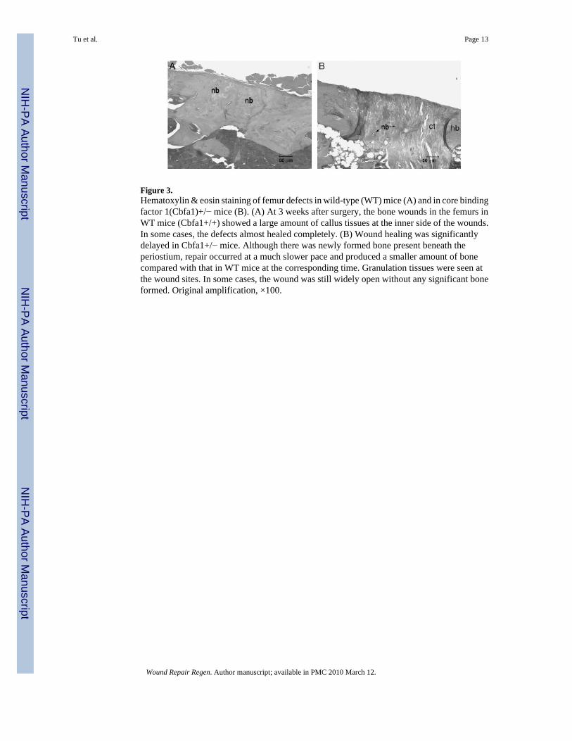
Figure 3.Hematoxylin & eosin staining of femur defects in wild-type (WT) mice (A) and in core bindingfactor 1(Cbfa1)+/− mice (B). (A) At 3 weeks after surgery, the bone wounds in the femurs inWT mice (Cbfa1+/+) showed a large amount of callus tissues at the inner side of the wounds.In some cases, the defects almost healed completely. (B) Wound healing was significantlydelayed in Cbfa1+/− mice. Although there was newly formed bone present beneath theperiostium, repair occurred at a much slower pace and produced a smaller amount of bonecompared with that in WT mice at the corresponding time. Granulation tissues were seen atthe wound sites. In some cases, the wound was still widely open without any significant boneformed. Original amplification, ×100.
Tu et al. Page 13
Wound Repair Regen. Author manuscript; available in PMC 2010 March 12.
NIH
-PA Author Manuscript
NIH
-PA Author Manuscript
NIH
-PA Author Manuscript

Figure 4.Hemotoxylin & eosin staining results of the periodontal window wounds in the core bindingfactor 1(Cbfa1)-gene activated matrix (GAM) group (A, E) and in the control group (C, G).Bone sialoprotein (BSP) staining results in the Cbfa1-GAM group (B, F) and in the controlgroup (D, H) are also shown. (A) At day 7, small islands of bone matrix could be seen scatteredin the bone defects in all the Cbfa1-applied tissues. (B) BSP staining in the Cbfa1 group wasmore intense in the newly formed alveolar bone. (C) In contrast, the newly formed bones wereless and (D) the BSP staining was weak in the control group at day 7. (E and F) At day 14, thenewly formed bones almost filled the wound defects in the Cbfa1 group. (G and H) In controls,the newly formed bones were poorly organized and weakly stained at day 14. In some samplesof control tissues at day 14, there were large amounts of granulation tissues in the wound sites.nb, newly formed bone; hb, host bone; t, tooth; ct, connective tissue; Col, collagen. Originalamplification, (A and B), ×100; (C–H), ×40.
Tu et al. Page 14
Wound Repair Regen. Author manuscript; available in PMC 2010 March 12.
NIH
-PA Author Manuscript
NIH
-PA Author Manuscript
NIH
-PA Author Manuscript

Figure 5.Newly formed bone area in alveolar bone defects. At day 7, the relative volume of new bonematrix in the defects of the core binding factor 1 (Cbfa1) group was higher than in controls(>1.5-fold) (p<0.05). At day 14, bone formation in the Cbfa1 group exhibited an even higherincrease (>twofold) compared with controls (p<0.05). *p < 0.05 Cbfa1 vs. control group.
Tu et al. Page 15
Wound Repair Regen. Author manuscript; available in PMC 2010 March 12.
NIH
-PA Author Manuscript
NIH
-PA Author Manuscript
NIH
-PA Author Manuscript

Figure 6.The periodontal ligament width was measured to assess the homeostasis of soft connectivetissue domains. At day 7, periodontal ligament (PDL) width in the wounded sides significantlyincreased compared with the unwounded sides in both groups (p < 0.05). At day 14, PL widthin wounded sides decreased and exhibited values similar to the unwounded sides in the Cbfa1group (p > 0.4). However, PDL width was still significantly increased compared with theunwounded sides in the control group at this time (p < 0.05). These measures were expressedas a percentage of the digitized normal values for each time point. *p < 0.05, wounded sidesvs. unwounded sides.
Tu et al. Page 16
Wound Repair Regen. Author manuscript; available in PMC 2010 March 12.
NIH
-PA Author Manuscript
NIH
-PA Author Manuscript
NIH
-PA Author Manuscript

Figure 7.Immunohistochemical staining for bone sialoprotein (BSP). At day 7, BSP staining in the newlyformed alveolar bone in the core binding factor 1 (Cbfa1) group was more intense than in theunwounded alveolar bone area (p < 0.05). But in the control group, staining for BSP was weakin the newly formed alveolar bone. At day 14, the staining intensity for BSP was much strongerin both groups compared with the internal control areas, but particularly in the Cbfa1 group(p < 0.05).
Tu et al. Page 17
Wound Repair Regen. Author manuscript; available in PMC 2010 March 12.
NIH
-PA Author Manuscript
NIH
-PA Author Manuscript
NIH
-PA Author Manuscript

Figure 8.The Core binding factor 1 (Cbfa1)-gene activated matrix (GAM) system induced high levelsof expression of cbfa1 in subman-dibular implant areas. (A) Representative image ofsemiquantitative reverse transcriptase polymerase-chain reaction (RT-PCR) analyses of cbfa1expression. (B) Levels of Cbfa1 were normalized with those of the loading control G3PDH inthree independent experiments. ap < 0.05 vs. non-GAM implant control.
Tu et al. Page 18
Wound Repair Regen. Author manuscript; available in PMC 2010 March 12.
NIH
-PA Author Manuscript
NIH
-PA Author Manuscript
NIH
-PA Author Manuscript
Copyright © 2022 FDOKUMEN