
HIV neutralising antibody delivered by gene therapy with a ...
255
HIV neutralising antibody delivered by gene therapy with a hybrid Vaccinia/retrovirus or BacMam/retrovirus expression systems A thesis submitted to The University of Manchester for the degree of Doctor of Philosophy in the Faculty of Biology, Medicine and Health 2018 LAYLA FAQIH Infection, Immunity and Respiratory Medicine
-
Upload
khangminh22 -
Category
Documents
-
view
1 -
download
0
Transcript of HIV neutralising antibody delivered by gene therapy with a ...
HIV neutralising antibody delivered by gene therapy
with a hybrid Vaccinia/retrovirus or BacMam/retrovirus
expression systems
A thesis submitted to The University of Manchester for the degree of Doctor of
Philosophy in the Faculty of Biology, Medicine and Health
2018
LAYLA FAQIH
Infection, Immunity and Respiratory Medicine
2
List of content
List of content ................................................................................................................................ 2
List of tables .................................................................................................................................. 7
List of figures ................................................................................................................................. 8
List of abbreviations .................................................................................................................... 13
General abstract .......................................................................................................................... 19
Declaration .................................................................................................................................. 21
Copyright Statement ................................................................................................................... 22
Acknowledgement ....................................................................................................................... 23
Dedication ................................................................................................................................... 24
Conference paper and publications ............................................................................................ 25
Chapter 1: General Introduction ...................................................................................... 26 1.
Human Immunodeficiency Virus (HIV) .................................................................... 26 1.1.
1.1.1. Overview ......................................................................................................... 26
1.1.2. Treatment ........................................................................................................ 28
1.1.3. Virus types ....................................................................................................... 29
1.1.4. Structure and genomic materials .................................................................... 29
Simian Immunodeficiency Virus (SIV) ..................................................................... 32 1.2.
1.2.1. Overview ......................................................................................................... 32
HIV and vaccine production .................................................................................... 34 1.3.
1.3.1. Overview ......................................................................................................... 34
1.3.2. HIV vaccine status and vaccine production challenges .................................. 35
1.3.3. Eliciting immune response using the current immunisation protocols ............ 37
Live attenuated vaccines............................................................................. 37 1.3.3.1.
Inactivated vaccines .................................................................................... 38 1.3.3.2.
Virus-like particles (VLP) ............................................................................. 40 1.3.3.3.
Subunit vaccines ......................................................................................... 41 1.3.3.4.
Envelope-based subunit vaccines .............................................................. 41 1.3.3.5.
Non-structural protein subunit vaccines ...................................................... 42 1.3.3.6.
Naked DNA vaccines .................................................................................. 44 1.3.3.7.
Live recombinant vaccines .......................................................................... 46 1.3.3.8.
1.3.3.8.1. Pox viruses ........................................................................................... 47
1.3.3.8.2. Adenoviruses ....................................................................................... 48
Prime-boost immunisation regimens ........................................................... 50 1.3.3.9.
Other vaccine approaches ........................................................................ 51 1.3.3.10.
1.3.4. Current status of HIV vaccines ........................................................................ 52
Humoral immune responses to HIV infection .......................................................... 54 1.4.
1.4.1. Overview ......................................................................................................... 54
1.4.2. Neutralising antibodies in HIV infection .......................................................... 56
1.4.3. HIV envelope protein ....................................................................................... 59
V3 loop ........................................................................................................ 59 1.4.3.1.
CD4 binding site .......................................................................................... 60 1.4.3.2.
Other V regions ........................................................................................... 61 1.4.3.3.
3
Gp41............................................................................................................ 61 1.4.3.4.
CD4 Induced neutralising antibodies .......................................................... 61 1.4.3.5.
1.4.4. HIV neutralising monoclonal antibodies .......................................................... 62
First-Generation broadly neutralising Abs against HIV ............................... 62 1.4.4.1.
Second-Generation broadly neutralising Abs against HIV.......................... 64 1.4.4.2.
1.4.5. Passive immunisation using neutralising antibodies ....................................... 66
Antibody gene cloning ............................................................................................. 68 1.5.
1.5.1. Antibody structure ........................................................................................... 68
1.5.2. Antibodies and molecular immunology ........................................................... 69
1.5.3. Antibody gene transfer for HIV immunoprophylaxis ........................................ 70
1.5.4. Expression vectors .......................................................................................... 73
1.5.5. Expression cells .............................................................................................. 74
1.5.6. Methods of co-expression of antibody heavy and light chains ....................... 74
1.5.7. PCR-based antibody gene cloning.................................................................. 77
1.5.8. HIV and human monoclonal antibody expression ........................................... 77
1.5.9. Regulatory and Enhancement Elements ........................................................ 79
Lentiviral vectors and gene therapy ........................................................................ 80 1.6.
1.6.1. Overview ......................................................................................................... 80
1.6.2. Lentiviruses as gene therapy vector ............................................................... 81
1.6.3. Pseudotyping of lentiviruses ........................................................................... 83
Chapter 2 ......................................................................................................................... 85 2.
HIV neutralising antibody delivered by gene therapy with a stable retroviral vector encoded in vaccinia expression systems....................................................................................................... 85
Abstract ................................................................................................................... 85 2.1.
Objectives and aims ................................................................................................ 87 2.2.
Introduction .............................................................................................................. 88 2.3.
2.3.1. Recombinant vaccinia viruses and vaccination .............................................. 88
Poxviruses in vaccination ............................................................................ 88 2.3.1.1.
Smallpox vaccination .................................................................................. 89 2.3.1.2.
Modified Vaccinia virus Ankara (MVA) and vaccination ............................. 91 2.3.1.3.
Vaccinia Retroviral Hybrid Vector ............................................................... 93 2.3.1.4.
Generation of recombinant MVA ................................................................. 94 2.3.1.5.
2.3.1.5.1. Insertion sites ....................................................................................... 97
2.3.1.5.2. Marker selection ................................................................................... 97
2.3.1.5.3. Promoters ............................................................................................. 98
2.3.1.5.4. T7 RNA polymerase hybrid system ..................................................... 99
2.3.1.5.5. Genes of interest ................................................................................ 100
Materials and Methods .......................................................................................... 102 2.4.
2.4.1. Molecular Cloning.......................................................................................... 102
Plasmid designing ..................................................................................... 102 2.4.1.1.
2.4.1.1.1. pLF-IgG1b12-mTK ............................................................................. 102
2.4.1.1.2. pLF-VSV-G ......................................................................................... 104
DNA extraction and purification from bacterial cells ................................. 105 2.4.1.2.
Preparation of vector and insert: Restrictions digest ................................ 105 2.4.1.3.
4
DNA End Modification: Dephosphorylation ............................................... 105 2.4.1.4.
Agarose gel electrophoresis and gel extraction ........................................ 105 2.4.1.5.
2.4.1.5.1. Agarose gel electrophoresis .............................................................. 105
2.4.1.5.2. Purification of vector and insert by DNA extraction from agarose gels 107
Ligation of vector and insert ...................................................................... 107 2.4.1.6.
Transformation .......................................................................................... 107 2.4.1.7.
Sequencing ............................................................................................... 108 2.4.1.8.
2.4.2. Generation and amplification of rMVA .......................................................... 108
Processing of primary Chicken Embryo Fibroblasts ................................. 108 2.4.2.1.
Virus stock ................................................................................................. 109 2.4.2.2.
2.4.2.2.1. Virus titration ...................................................................................... 109
2.4.2.2.2. Generation of recombinant MVA ........................................................ 110
2.4.2.2.3. Transfection of primary chicken embryo fibroblasts .......................... 110
2.4.2.2.4. Infection of pCEFs with wildtype MVA or recombinant MVA ............. 111
Plaque assay and rMVA isolation ............................................................. 111 2.4.2.3.
2.4.2.3.1. Antibiotic resistance system gpt selection ......................................... 112
2.4.2.3.2. Screening for transient β-galactosidase expression and plaque purification 112
2.4.2.3.3. Isolation of virus DNA from cells ........................................................ 113
2.4.2.3.4. Characterisation of recombinant MVA genomes by PCR .................. 113
2.4.3. Transfection using Lipfectamine 2000 .......................................................... 115
Results .................................................................................................................. 116 2.5.
2.5.1. Design and construction of MVA transfer plasmid vectors ........................... 116
pLF-IgG1b12-mTK .................................................................................... 116 2.5.1.1.
pLF-IgG1b12-LacZ-mTK ........................................................................... 120 2.5.1.2.
pLF-VSV-G ................................................................................................ 122 2.5.1.3.
pLF-VSV-G-Rev-D4 .................................................................................. 123 2.5.1.4.
pLF-IgG1b12-LacZ-D4 .............................................................................. 124 2.5.1.5.
2.5.2. Generation of recombinant MVA by homologous recombination ................. 125
2.5.3. Characterization of recombinant MVA genome by PCR ............................... 131
2.5.4. Transfection ................................................................................................... 133
Discussion and Conclusion ................................................................................... 135 2.6.
Chapter 3 ....................................................................................................................... 143 3.
HIV neutralising antibody delivered by gene therapy with a hybrid baculovirus/SIV vector ..... 143
Abstract ................................................................................................................. 143 3.1.
Objectives .............................................................................................................. 145 3.2.
Introduction ............................................................................................................ 147 3.3.
3.3.1. Baculovirus .................................................................................................... 147
3.3.2. Baculovirus Replication ................................................................................. 148
3.3.3. AcMNPV cell entry ........................................................................................ 150
3.3.4. Baculoviruses as Expression Vectors ........................................................... 152
3.3.5. Construction of recombinant baculoviruses .................................................. 155
Generating a Recombinant Virus by Homologous Recombination .......... 155 3.3.5.1.
Generating a Recombinant Virus by Site-Specific Transposition ............. 157 3.3.5.2.
5
3.3.6. Baculovirus and mammalian cell transduction .............................................. 158
3.3.7. Generation of VLPs using baculovirus expression ....................................... 161
3.3.8. Immune response and potential as gene therapy vectors and vaccine vectors 165
Material and methods: ........................................................................................... 169 3.4.
3.4.1. Virus like particle expression utilising the T7 RNA Polymerase/Promoter system 169
3.4.2. Virus like particle expression using a CMV Promoter ................................... 169
3.4.3. Molecular cloning .......................................................................................... 170
DNA extraction and purification from bacterial cells ................................. 171 3.4.3.1.
Preparation of vector and insert: Restriction digest .................................. 172 3.4.3.2.
DNA End Modification ............................................................................... 172 3.4.3.3.
3.4.3.3.1. Dephosphorylation ............................................................................. 172
3.4.3.3.2. Blunting/End-repair ............................................................................ 172
PCR Using Q5® High-Fidelity DNA polymerase ...................................... 172 3.4.3.4.
Agarose gel electrophoresis and gel extraction ........................................ 175 3.4.3.5.
3.4.3.5.1. Agarose gel electrophoresis .............................................................. 175
3.4.3.5.2. Purification of vector and insert by DNA extraction from agarose gels 175
Ligation of vector and insert ...................................................................... 176 3.4.3.6.
Transformation .......................................................................................... 176 3.4.3.7.
Sequencing ............................................................................................... 177 3.4.3.8.
Recombinant BacMam plasmids construction .......................................... 178 3.4.3.9.
3.4.3.9.1. Study : Expression under the T7 RNA Polymerase/Promoter system 178
3.4.3.9.2. Study : BacMam expression utilising a CMV promoter ................... 179
Generation of recombinant baculoviruses .............................................. 179 3.4.3.10.
Quantitative PCR (qPCR) titration .......................................................... 180 3.4.3.11.
Transduction of mammalian cells ........................................................... 180 3.4.3.12.
Immunohistochemistry ............................................................................ 181 3.4.3.13.
Western Blot Analysis ............................................................................. 182 3.4.3.14.
3.4.3.14.1. Protein lysis ...................................................................................... 182
3.4.3.14.2. Protein-electrophoresis .................................................................... 182
3.4.3.14.3. Protein Blotting ................................................................................. 183
Ultracentrifugation and Transmission Electron Microscopy (TEM) ......... 185 3.4.3.15.
Results .................................................................................................................. 186 3.5.
3.5.1. Design and construction of BacMam transfer plasmids ................................ 186
Study : T7 RNA Polymearse/Promoter system ....................................... 186 3.5.1.1.
3.5.1.1.1. pLF_T7_RNA-Polymerase ................................................................. 186
3.5.1.1.2. pLF_T7_IgGb12-GFP ........................................................................ 188
3.5.1.1.3. pLF_T7_Rev-VSV-G .......................................................................... 190
3.5.1.1.4. pLF_T7_Gag_Tat ............................................................................... 192
3.5.1.1.5. pLF_T7_Pol-Vpr-Vif ........................................................................... 194
Study , CMV promoter ............................................................................ 196 3.5.1.2.
6
3.5.1.2.1. pLF_CMV-Gag-IRES-Tat, pLF_CMV-RtInt-IRES-Vif and pLF_CMV-VSV-IRES-Rev ...................................................................................................... 196
3.5.1.2.2. pLF_CMV-IgG1b12 ............................................................................ 197
3.5.2. Generation of recombinant BacMam viruses ................................................ 203
3.5.3. Quantitative (qPCR) titration ......................................................................... 204
3.5.4. Immunohistochemistry .................................................................................. 204
Study ....................................................................................................... 204 3.5.4.1.
Study ..................................................................................................... 206 3.5.4.2.
3.5.5. Green-fluorescent protein (GFP) expression ................................................ 208
3.5.6. Western Blot .................................................................................................. 209
Study ....................................................................................................... 209 3.5.6.1.
Study ..................................................................................................... 209 3.5.6.2.
3.5.7. Electron microscopy ...................................................................................... 212
Discussion ............................................................................................................. 213 3.6.
Future work ........................................................................................................... 223 3.7.
Chapter 4 ....................................................................................................................... 225 4.
General discussion and future work .......................................................................................... 225
Refrences .................................................................................................................................. 237
Final word count: 52,485
7
List of tables
Table 1: Comparison of VAX004 and VAX003 studies ............................................................... 46
Table 2: Phase 2b or Phase 3 efficacy studies ........................................................................... 52
Table 3: Estimated frequency of postvaccinial encephalitis (pvE) and death after primary
vaccination with different vaccinia virus strains .......................................................................... 90
Table 4: Estimated frequency of specific postvaccinial complications after primary and re-
vaccination with NYCBH strain ................................................................................................... 90
Table 5: Recommended agarose gels for electrophoretic separation of DNA fragments ........ 106
Table 6: Primers used for sequencing ...................................................................................... 108
Table 7: PCR reaction setup ..................................................................................................... 113
Table 8: Thermocycling conditions for a routine PCR .............................................................. 114
Table 9: Primers used for PCR screening ................................................................................ 114
Table 10: Cell types that are permissive to baculovirus transduction ....................................... 159
Table 11: Promoter used in Baculovirus expression system .................................................... 160
Table 12: Generation of VLPs using baculovirus expression system ....................................... 163
Table 13: Comparison of Baculovirus and Other Viral Vectors ................................................ 166
Table 14: Reaction Setup.......................................................................................................... 173
Table 15: Thermocycling Conditions for a Routine PCR .......................................................... 173
Table 16: Recommended amounts of DNA template for a 50 µL reaction ............................... 173
Table 17: PCR primers used for inserts amplification ............................................................... 174
Table 18: Primers used for sequencing .................................................................................... 177
Table 19: List of primary and secondary antibodies ................................................................. 184
Table 20: Protein sizes .............................................................................................................. 184
Table 21: Titration results .......................................................................................................... 204
8
List of figures
Figure 1: Lentivirus virion.. .......................................................................................................... 30
Figure 2: Organization of the genome of the HIV provirus together with a summary description
of its genes and encoded proteins. ............................................................................................. 31
Figure 3: Organization of lentiviral genomes. ............................................................................. 32
Figure 4: Timeline of HIV vaccine efficacy trials. ........................................................................ 52
Figure 5: Function of antiviral antibodies. ................................................................................... 55
Figure 6: Sites of HIV‑1 vulnerability to neutralising antibodies. ................................................ 58
Figure 7: Antibody target sites on the HIV-1 envelope spike and schematic representation of the
HIV-1 envelopespike. .................................................................................................................. 59
Figure 8: Antibody structure. ....................................................................................................... 69
Figure 9: Examples of HIV prophylaxis approaches ................................................................... 70
Figure 10: Schematic representation of immunoadhesin constructs. ......................................... 71
Figure 11: Mono-cistronic: Light and heavy chain encoded on separate plasmids. .................. 75
Figure 12: Single plasmid cassettes for heavy and light chains expresion. ............................... 75
Figure 13: Bi-cistronic containing an internal ribosomal entry site (IRES). ................................. 76
Figure 14: Single Open Reading Frame (ORF). ......................................................................... 76
Figure 15: PCR cloning of the vector expressing the 2G12 light chain-2A-2G12 heavy chain. . 78
Figure 16: Dual-promoter rAAV antibody vector. ........................................................................ 79
Figure 17: Examples of special vector setups for stable cell line generation. ............................ 80
Figure 18: Schematic representation of the four generations of lentiviral packaging constructs 82
Figure 19: Electron micrographs of negatively stained, naturally released virions. .................... 88
Figure 20: Arrangement of HindIll genome DNA fragments of vaccinia virus CVA wild-type, CVA
382 and MVA.. ............................................................................................................................ 91
Figure 21: Generation of recombinant MVA virus for expression of foreign gene. ..................... 95
Figure 22: Conventional recombinant MVA production through spontaneous homologous
recombination. ............................................................................................................................. 96
Figure 23: Crystal structure of the intact human IgG1b12. ....................................................... 103
Figure 24: Heavy and light chain sequences after codon modification. ................................... 104
Figure 25: DNA Ladders. .......................................................................................................... 106
Figure 26: Tri-cistronic containing an internal ribosomal entry site (IRES) elements. .............. 117
9
Figure 27: Encephalomyocarditis virus (EMCV) IRES. ............................................................. 118
Figure 28: Schematic representation of the transfer plasmid pLF-IgG1b12-mTK. ................... 119
Figure 29: 1% agarose gels of DNA restriction digests product of pLF-IgG1b12-mTK.. .......... 120
Figure 30: Schematic representation of the construction of transfer plasmid pLF-IgG1b12-LacZ-
mTK. .......................................................................................................................................... 121
Figure 31: 1% agarose gel of DNA restriction digests product of pLF-IgG1b12-LacZ-mTK.. .. 121
Figure 32: Schematic representation of the transfer plasmid pLF-VGV-g. ............................... 122
Figure 33: 1% agarose gels of DNA restriction digests product of pLF-VGV-g. ....................... 122
Figure 34: Schematic representation of the construction of transfer plasmid pLF-VSV-G-Rev-D4.
.................................................................................................................................................. 123
Figure 35: 1% agarose gel of DNA restriction digests product of pLF-VSV-G-Rev-D4. ........... 123
Figure 36: Schematic representation of the construction of transfer plasmid pLF-IgG1b12-LacZ-
D4.. ............................................................................................................................................ 124
Figure 37: 1% agarose gels of DNA restriction digests product of pLF-IgG1b12-LacZ-D4.. .... 125
Figure 38: Schematic representation of the recombinant packaging MVA. .............................. 126
Figure 39: Schematic representation of the recombinant T7 MVA. .......................................... 126
Figure 40: Microscopic observation of established CPE where individual virus plaques
differentiated. ............................................................................................................................ 127
Figure 41: Blue plaque formation on 6-well dish. ...................................................................... 128
Figure 42: Schematic representation of the first rMVA (rMVA/gag/pol/VSV-G). ...................... 129
Figure 43: Schematic representation of the second rMVA (rMVA/IgG1b12/T7). ...................... 130
Figure 44: Schematic representation of the second rMVA (rMVA/IgG1b12/T7). ...................... 130
Figure 45: Agarose gel electrophoresis after PCR screening for the insertion of packaging
componants into the packaging virus. ....................................................................................... 132
Figure 46: Agarose gel electrophoresis after PCR screening for the insertion of antibody
expression cassette into TK of MVA forming the genomic virus............................................... 133
Figure 47: Fluorescent vero cells infected with pLF-IgG1b12-mTK.......................................... 134
Figure 48: Schematic representation of the hybrid dual-vector system (poxviral/retroviral
system) and generating HIV-neutralising monoclonal antibody.. .............................................. 136
Figure 49: Baculovirus. ............................................................................................................. 148
Figure 50: Key stages in the infection cycle of AcMNPV. ......................................................... 150
Figure 51: Internalisation of baculovirus into mammalian cells. ............................................... 152
10
Figure 52: Budded virus infection cycle in cultured cells. ......................................................... 154
Figure 53: Construction of baculovirus recombinants using three different systems. .............. 156
Figure 54: Generation of recombinant baculovirus and gene expression using Bac to Bac
sysem. ....................................................................................................................................... 158
Figure 55: pOET6 BacMAM map. ............................................................................................. 170
Figure 56: Cloning method for the first study utilising T7 promoter .......................................... 178
Figure 57: Cloning method for the second study utilising CMV promoter ................................ 179
Figure 58: Transduction of mammalian cells with baculovirus. ................................................ 181
Figure 59: ColorPlus™ Prestained Protein Ladder, Broad Range (10-230 kDa) ..................... 183
Figure 60: Schematic representation of pLF_T7_RNA-Polymerase cloning steps. ................. 186
Figure 61: 1% agarose gel of DNA restriction digests product cloning of pLF_T7_RNA-
Polymerase first step. ................................................................................................................ 187
Figure 62: 1% agarose gel of DNA restriction digests product cloning of pLF_T7_RNA-
Polymerase second step. M is a 1kb DNA ladder. ................................................................... 187
Figure 63: 1% agarose gel of DNA restriction digests product cloning of pLF_T7_RNA-
Polymerase final step. M is a 1kb DNA ladder.......................................................................... 187
Figure 64: Schematic representation of pLF_T7_IgGb12-GFP cloning steps.. ........................ 188
Figure 65: 1% agarose gel of DNA restriction digests product cloning of pLF_T7_IgGb12-GFP
first step. .................................................................................................................................... 189
Figure 66: 1% agarose gel of DNA restriction digests product cloning of pLF_T7_IgGb12-GFP
second step. .............................................................................................................................. 189
Figure 67: Schematic representation of pLF_T7_Rev-VSV-G cloning steps. .......................... 190
Figure 68: 1% agarose gel of DNA restriction digests product cloning of pLF_T7_Rev-VSV-G
first step.. ................................................................................................................................... 191
Figure 69: 1% agarose gel of DNA restriction digests product cloning of pLF_T7_Rev-VSV-G
second step.. ............................................................................................................................. 191
Figure 70: Schematic representation of pLF_T7_Gag_Tat cloning steps. ............................... 192
Figure 71: 1% agarose gel of DNA restriction digests product cloning of pLF_T7_Gag_Tat first
step. ........................................................................................................................................... 193
Figure 72: 1% agarose gel of DNA restriction digests product cloning of pLF_T7_Gag_Tat
second step.. ............................................................................................................................. 193
11
Figure 73: 1% agarose gel of DNA restriction digests product cloning of pLF_T7_Gag_Tat final
step.. .......................................................................................................................................... 193
Figure 74: Schematic representation of pLF_T7_Pol-Vpr-Vif cloning steps. ............................ 194
Figure 75: 1% agarose gel of DNA restriction digests product cloning of pLF_T7_Pol-Vpr-Vif first
step. ........................................................................................................................................... 195
Figure 76: 1% agarose gel of DNA restriction digests product cloning of pLF_T7_Pol-Vpr-Vif
second step.. ............................................................................................................................. 195
Figure 77: 1% agarose gel of DNA restriction digests product cloning of pLF_T7_Pol-Vpr-Vif
final step. ................................................................................................................................... 195
Figure 78: Schematic representation of pLF_CMV-Gag-IRES-Tat, pLF_CMV-RtInt-IRES-Vif and
pLF_CMV-VSV-IRES-Rev cloning steps. ................................................................................. 196
Figure 79: 1% agarose gels of DNA restriction digests product cloning of pLF_CMV-Gag-IRES-
Tat, pLF_CMV-RtInt-IRES-Vif and pLF_CMV-VSV-IRES-Rev. ................................................ 197
Figure 80: Highlights of the inaccuracy in the pLF_T7_IgG1b12.............................................. 197
Figure 81: Schematic representation of pOET6+A. .................................................................. 199
Figure 82: 1% agarose gel of DNA restriction digest product cloning of pLF_CMV-IgG1b12 first
step. ........................................................................................................................................... 200
Figure 83: Schematic representation of (A) fragment B (B) pOET6+A+B. ............................... 200
Figure 84: 1% agarose gel of DNA restriction digest product cloning of pLF_CMV-IgG1b12
second step. .............................................................................................................................. 200
Figure 85: Schematic representation of (A) fragment located between AflII and BstAPI from
pIgG+P2A (B) pLF_T7_IgG1b12 (C) pIgG+P2A....................................................................... 201
Figure 86: 1% agarose gel of DNA restriction digest product of pIgG+P2A. ............................ 201
Figure 87: Schematic representation of the final step of pLF_CMV-IgG1b12 cloning. ............ 202
Figure 88: 1% agarose gel of DNA restriction digest product cloning of pLF_CMV-IgG1b12 final
step. ........................................................................................................................................... 202
Figure 89: Sf9 cells visualised under the microscope 6 days after infection with BacMam
pOET6_T7. ................................................................................................................................ 203
Figure 90: Immunohistochemistry results for the first study, MRC5 cells. ................................ 205
Figure 91: Immunohistochemistry results for the first study, HEK cells. ................................... 206
Figure 92: Immunohistochemistry results for the second study, MRC5 cells. .......................... 207
12
Figure 93: Transfection of mammalian cells with BacMam pOET6_IgGb12-GFP recombinant
plasmid. ..................................................................................................................................... 208
Figure 94: SDS-PAGE analysis of cell extracts from transduced HEK cells. ........................... 209
Figure 95: Demonstration of SIVmac239 gag protein expressed from HEK cells. ................... 210
Figure 96: Demonstration of protein expressed from HEK cells. .............................................. 211
Figure 97: Demonstration of IgG1b12 expressed from HEK cells. ........................................... 211
Figure 98: TEM microphotographs of the VLP sample generated by BacMam system. .......... 212
Figure 99: Schematic representation of the hybrid dual-vector system (baculovirus/retrovirus
system) generating HIV-neutralising monoclonal antibody, utilising a T7 system. ................... 213
Figure 100: Schematic representation of the hybrid dual-vector system (baculovirus/retrovirus
system) and generating HIV-neutralising monoclonal antibody. ............................................... 217
Figure 101: Encephalomyocarditis virus (EMCV) internal ribosomal entry site (IRES) sequence
.................................................................................................................................................. 219
Figure 102: Schematic representation of the final structure of the corrected IgG1b12 cassette.
.................................................................................................................................................. 220
Figure 103: Requirements for vaccine-induced protection and control.. .................................. 228
13
List of abbreviations
AAV Adeno-associated virus
Ab Antibody
AcMNPV Autographa californica multiple nucleopolyhedrovirus
Ad5 Adenovirus type 5
ADCC Antibody-dependent cellular cytotoxicity
AIDS Acquired immune deficiency syndrome
ALVAC Canarypox virus vector
AMP Antibody mediated prevention
BHK Baby hamster kidney
bNAbs Broadly neutralising Abs
bp Base pair
BrdU Bromodeoxyuridine
BV Baculovirus
CA Capsid
cART combination Antiretroviral therapy
CBD1 Caveolin-1 binding domain of HIV-1 glycoprotein gp41
CD4 Cluster of differentiation 4
CD4BS CD4 binding site
CD4i CD4-induced
cDNA complementary DNA
CDRs Complementarity determining regions
CEA Carcinoembryonic Antigen
CEF Chicken embryo fibroblasts
CH Constant heavy chain
chiA Non-essential chitinase gene
CHO Chinese hamster ovary cells
CL Constant light chain
CMV Cytomegalovirus
CNS Central nervous system
14
COS African green monkey kidney
CPE Cytopathic effect
CTL Cytotoxic T lymphocytes
CVA Chorioallantois vaccine Ankara
CYP Cytochrome P450
DAF Decay-Accelerating Factor
DC Dendritic cells
Del Major Genomic Deletion region of MVA
DMEM Dulbecco modified Eagle medium
dNTP degrades deoxynucleoside triphosphate
EF Elongation Factors
eGFP enhanced Green Fluorescent Protein
EMCV Encephalomyocarditis virus
Env Envelope protein
Fab Fragment antigen-binding
Fc Fragment crystallisable region
FCS Foetal Calf Serum
FIV Feline immunodeficiency virus
FPV Fowlpox virus
Gag Gag polyprotein
GFP Green fluorescent protein
GMO Genetically Modified Organisms
GMP Good Manufacturing Practices
Gp Glycoprotein
gpt Xanthineguanine phosphoribosyl transferase
Gus E. coli β-glucuronidase A gene
HA Hemagglutinin gene
HAART Highly active antiretroviral therapy
HaCat Human keratinocyte cell line
HaSNPV Helicoverpa armigera single nucleopolyhedrovirus
HBsAg HBV surface antigen
15
HBV Hepatitis B virus
HCV Hepatitis C virus
HDAC Histone deacetylase
HEK293 Human embryonic kidney
HeLa Human cervix adenocarcinoma cell line
HEV Hepatitis E virus
HIV Human immunodeficiency virus
HLA human leukocyte antigen
hPGK Human Phosphoglycerate kinase
HRP Horseradish peroxidase
HTLV human T-cell leukemia virus
IAVI International AIDS Vaccine Initiative
ICAM Intercellular Adhesion Molecule
IE Immediate Early
IgG Immunoglobulin G
IGR Intergenic regions
IHC Immunohistochemistry
IL Interleukin
IN or Int Integrase
IRES Internal ribosomal entry site
Kb Kilobases
LacZ Gene for β-galactosidase
LAM-PCR Linear - amplification mediated PCR
LFA-1 Lymphocyte function-associated antigen 1
LTR Long terminal repeat
LV Lentiviral vectors
MA Matrix
mAbs Monoclonal antibodies
MCS Multiple cloning site
MEM Minimum Essential Media
MHC Major histocompatibility complex

16
MOI Multiplicity of Infection
MPA Mycophenolic acid
MPER Membrane proximal external region
MRC5 Human foetal lung fibroblast
mRNA Messenger RNA
MVA Modified Vaccinia Virus Ankara
NC Nucleocapsid
Nef Negative regulatory factor
NNRTI Non-nucleotide reverse transcriptase inhibitors
NRTI Nucleotide reverse transcriptase inhibitors
OB Occlusion Bodies
ODV Occlusion-Derived Virion
ORF Open reading frame
P Promoter
pA Polyadenylation
PBS Phosphate buffer saline
PBS-T Phosphate buffer saline with Tween
PCE post-transcriptional control element
pCEF Primary Chicken Embryo Fibroblast
PCR Polymerase chain reaction
PEC Peritoneal Macrophages
PI/r Ritonavir boosted protease inhibitor
PND Principal neutralising domain
Pol Polymerase
polh polyhedrin gene
PPT Polypurine tract
PR Protease
pvE Post-vaccinial Encephalitis
qPCR Quantitative PCR
rAAV recombinant Adeno-Associated Virus
rBV recombinant Baculovirus
17
RCL Replication-competent lentivirus
Rev Regulator of expression of virion proteins
rHA recombinant Trivalent Hemagglutinin
rMVA recombinant MVA
RNA Ribonucleic acid
RRE Rev-responsive element
rSAP recombinant Shrimp Alkaline Phosphatase
RSV Rous sarcoma virus
Rt Reverse transcriptase
SCID Severe combined immunodeficiency
Sf Spodoptera frugiperda
SHIV SIV/HIV hybrid viruses
SIN Self-inactivating LV
SIV Simian Immunodeficiency Virus
SIV mac SIV macaque species
SOC medium Super Optimal broth with Catabolite repression
ssRNA Single stranded RNA
SU Receptor binding domain
SV40 Simian virus 40
Tat Trans-activating protein
TBS Tris-buffered saline
TEM Transmission Electron Microscopy
TK Thymidine kinase
TM Transmembrane domain
UTR Untranslated regions
V Variable region of the HIV viron envelope glycoprotein
Vero African green monkey kidney cell line
VH Variable heavy chain
Vif Virion infectivity factor
VIP Vectored immunoprophylaxis
VL Variable light chain
18
VLP Virus-like particle
Vpr Viral protein R
Vpu Viral protein U
VSV-G G glycoprotein of the vesicular stomatitis virus
VV Vaccinia viruses
WB Western blot
WHO World Health Organisation
wtBV wild type Baculovirus
wtMVA wild type MVA
X-gal 5-bromo-4-chloro-3-indolyl-β-D-galactopyranoside
Ψ Packaging signal
19
General abstract
Production of an effective vaccine and long-term treatment against human
immunodeficiency virus (HIV) is elusive. In this thesis two different techniques were
used in an attempt to insert HIV-neutralising monoclonal antibody (IgG1b12)
sequences into a simian retroviral gene therapy agent pseudo-typed with vesicular
stomatitis virus glycoprotein. Genes were encoded in either a poxvirus split-vector
system or a baculovirus expression system. Both systems aim to produce replication
incompetent pseudotyped virus like particles with simian origin. It is believed that the
resulting non-infectious artificial lentivirus particles enter neighbouring cells, penetrate
the nucleus and insert genetic material (the antibody gene) into the mammalian
genome.
The poxvirus split-vector system used in this project was a Vaccinia Retroviral Hybrid
Vector, where recombinant modified vaccinia Ankara (MVA) is used to deliver the
simian immunodeficiency virus (SIV) like particles into mammalian cells. However, the
MVA system failed to express proteins of interest due to the instability of genetic
insertion into the recombinant MVA genome. As an alternative strategy, two different
BacMam systems were used to allow the production of VLPs, where mammalian cells
are co-transduced with different recombinant baculoviruses (rBVs). VLPs were
expressed either under the control of T7 RNA polymerase system or under the
cytomegalovirus immediate early gene promoter. The results from the first BacMam
system indicated that the T7 RNA polymerase system was not suitable to express
detectable levels of proteins. The results indicated that translation of the produced
mRNA by T7 promoter is inefficient, most likely because of the absence of RNA 5’ cap
structure. To overcome this hybrid BV–T7 system limitation, a different system was
developed. Proteins of interest from the second BacMam system were successfully
expressed and detected using western blot analysis. VLPs were generated and
visualised under electronic microscope. IgG1b12 was secreted in the supernatant of
20
the transduced mammalian cells. Mammalian cells were successfully transduced with
multiple different recombinant BVs simultaneously.
The study establishes the feasibility of antibody gene transfer, and demonstrates the
use of SIV like particles production to transduce mammalian cells using BacMam
technology. The technique may have application for use as an immunotherapy of HIV
infection as well providing long-acting prevention of HIV infection for those not yet
infected with HIV.

21
Declaration
I declare that no portion of the work referred to in the thesis has been submitted in
support of an application for another degree or qualification of this or any other
university or other institute of learning.
22
Copyright Statement
1. The author of this thesis (including any appendices and/or schedules to this thesis)
owns certain copyright or related rights in it (the “Copyright”) and s/he has given
The University of Manchester certain rights to use such Copyright, including for
administrative purposes.
2. Copies of this thesis, either in full or in extracts and whether in hard or electronic
copy, may be made only in accordance with the Copyright, Designs and Patents
Act 1988 (as amended) and regulations issued under it or, where appropriate, in
accordance with licensing agreements which the University has from time to time.
This page must form part of any such copies made.
3. The ownership of certain Copyright, patents, designs, trademarks and other
intellectual property (the “Intellectual Property”) and any reproductions of copyright
works in the thesis, for example graphs and tables (“Reproductions”), which may be
described in this thesis, may not be owned by the author and may be owned by
third parties. Such Intellectual Property and Reproductions cannot and must not be
made available for use without the prior written permission of the owner(s) of the
relevant Intellectual Property and/or Reproductions.
4. Further information on the conditions under which disclosure, publication and
commercialisation of this thesis, the Copyright and any Intellectual Property and/or
Reproductions described in it may take place is available in the University IP Policy
(see http://documents.manchester.ac.uk/DocuInfo.aspx?DocID=2442 0), in any
relevant Thesis restriction declarations deposited in the University Library, The
University Library’s regulations (see
http://www.library.manchester.ac.uk/about/regulations/) and in The University’s
policy on Presentation of Theses.

23
Acknowledgement
This work would not have been possible without the financial support of the Saudi
Arabian Cultural Bureau in London. I would like to express my gratitude to my
supervisory team Professor Pamela Vallely, Professor Paul Klapper and Dr Thomas
Blanchard for allowing me to pursue this PhD project at the University of Manchester
and for their support and guidance throughout my years of research. I am especially
indebted to Prof. Klapper, who have been supportive of my career goals and who
worked actively to provide me with the protected academic time to pursue those goals.
Thank you for your infinite patience, constant encouragement, immense support and
positive thinking. I would like to thank my advisor Dr Susan Shawcross for her support,
advice and for always beening there whenever I needed help. I would like to express
my sincere gratitude to Dr. Eddie McKenzie and Dr.Ruth Lopez at the Protein
Expression Facility for providing me support, help and guidance when I most needed it.
I am grateful to all the staff in the Medical Virology/Microbiology Unit at UoM, those with
whom I have had the pleasure to work. David, Yevone, Sally, Colin and Stuart each
one of you has provided me with extensive personal and professional guidance. You
made tea room the most enjoyable!
I would especially like to thank my previous colleague and currently best friend
Dr.Katharina Lauer. She has taught me more than I could ever give her credit for here.
She has shown me, by her example, what a good scientist (and person) should be.
Many thanks go to all the PhD students in the postgraduate office Ben, Roba, Wong,
Wanchat, Mohammed, Auhood, Mohanned, Radi and Ahmed who were always there to
help and support.
I am grateful to my sibling Abdulhadi and Lama who have provided me through
emotional support in my life. I am also grateful to my friends who have supported me
along the way. Thanks you Lama, Nahla, Roba and Hammody for all your support,
cakes and chocolates; I am sure that I could not do this without our adventurous trips.

24
Dedication
I want to dedicate this PhD thesis to my father Ahmed and my mother Elham the most
amazing supportive parents, whose love and guidance are with me in whatever I
pursue. You are the reason for who I am! Nobody has been more important to me in
the pursuit of this PhD than you. Thank you my parents, you are the ultimate role
models.
25
Conference paper and publications
Use of baculovirus expression system, utilising a CMV promoter, for generation of virus
like particles in mammalian cells
Poster at the 20th Annual Meeting of the ESCV, Journal of Clinical Virology,
2017.
HIV neutralising antibody delivered by gene therapy, with a stable retroviral vector
encoded in baculovirus expression system
Poster at the Postgraduate Summer Research Showcase, the University of
Manchester, 2017.
Oral presentation at the 19th Annual Meeting of the ESCV, Journal of Clinical
Virology, 2016.
Baculovirus expression systems for virus like particles production in mammalian cells
Poster at the Institute of Inflammation and Repair PGR Showcase, the
University of Manchester, 2016.
Long-term vaccine strategies for Ebola. Lancet infection diseases, 2015. K.B. Lauer, L.
Faqih, T.J. Blanchard.
Generation of marker-free rMVA by homologous recombination using a 50bp repetitive
homologous sequence
Poster at the London Vaccine conference, 2014.
26
Chapter 1: General Introduction 1.
Human Immunodeficiency Virus (HIV) 1.1.
1.1.1. Overview
In 1981, the first case of acquired immune deficiency syndrome (AIDS) was reported in
the United Kingdom in a patient characterised by profound immune deficiency (Collier
et al. 2011). Since then, a massive international effort has aimed to control HIV. HIV is
the leading cause of death in sub-Saharan Africa where it is endemic causing almost 2
million new infections every year. It is the fourth leading cause of death worldwide
(Girard et al. 2006; Collier et al. 2011; Greenwood et al 2012; Esparza 2001). In Asia
there are more than half a million new infections occurring every year. Significant
progress has been made over the past thirty years, especially in antiretroviral
treatment. However, the ability of the virus to develop drug-resistance during treatment
is of major concern (Churchill et al. 2016; World Health Organization Guideline 2016;
Seitz 2016).
HIV, together with the simian immunodeficiency virus (SIV) and the feline
immunodeficiency virus (FIV) are enveloped ssRNA viruses belonging to the genus
Lentivirus in the family Retroviridae. Lentiviruses possess a reverse transcriptase
enzyme that uses the viral RNA genome as a template and produces a proviral DNA
copy that integrates into the host cell chromosome. The provirus DNA is eventually
transcribed into a set of mRNAs that encode the viral proteins and progeny genomic
RNA (Klatt et al. 2012; Carter and Saunders 2013; Greenwood et al. 2012; Girard et al.
2006).
Lentiviruses produce characteristically slow progressive infections (prolonged clinical
latency), which have the ability to cause immunosuppression. After initial contact, HIV
is transported to secondary lymphoid organs where it infects and attaches to CD4
lymphocytes (T helper cells and macrophages) by using a viral envelope glycoprotein
27
(gp120). Fusion with the cell membrane is assisted by another glycoprotein (gp41).
Infected CD4 cells then spread the infection, sometimes through cell-to-cell fusion,
resulting in the formation of syncytia with other lymphocytes (Cho 2000; Levy 2007).
Once HIV enters a host cell, it integrates into the cell’s DNA and may persist for a long
and variable period before showing any pathogenic effect (Cho 2000; Girard et al.
2006). It was originally assumed that this latent period was characterised by a low level
of viral replication. However it is now clear that high rates of viral replication actually
occur early after infection.
HIV replicates in metabolically active cells (activated T lymphocytes and macrophages)
but remains latent in inactive cells. Infected CD4+ T memory lymphocytes (CD45+
R0+) do not proliferate and are metabolically inactive, thus these cells do not transcribe
the viral genome nor express the viral protein. As a result the cells remain
unrecognised by CTLs. The immune system is unable to eradicate HIV, partly because
of this latency phenomenon and partly because the virus continuously mutates its
sequence and thus escapes neutralising antibodies and CTLs. These mutant variants
are continuously selected in vivo by the pressure of the immune response. The infected
patient may be asymptomatic or paucisymptomatic for several years, but have plasma
viraemia and be infective. The phase of primary infection can be asymptomatic;
however, 30–70% of infected individuals experience an acute syndrome, characterised
by fever, fatigue, lymphoadenomegaly, maculopapular cutaneous eruption, and, in a
few cases, neurological involvement. In most cases, this condition is misdiagnosed or
unrecognised. As the infection progresses, the proportion of infected CD4+ cells and
the quantity of circulating virus rise, until the patient becomes symptomatic (Levy 2007;
Cho 2000; Giacca 2010).
HIV eventually damages the overall host immune response unless treated. The
malfunction of the immune system leaves the host at a high risk of infection, particularly
for opportunistic bacterial, viral, fungal and protozoan infections. The end stage of the
disease caused by HIV infection is AIDS, in which CD4 cells are significantly depleted

28
(<200 cells/mL). At this stage, opportunistic infections are much more likely to occur
(Girard et al. 2006; Collier et al. 2011; Giacca 2010; Capistrán 2010; Flint et al 2009).
1.1.2. Treatment
The major milestone in HIV treatment was the discovery of a multiple drug regimen
(previously referred to as highly active antiretroviral therapy, or HAART, now known as
combination antiretroviral therapy or cART) that has achieved remarkable success in
slowing the progression of AIDS. Antiretroviral therapies aim to arrest and reverse the
damage that results from the infection of the immune system, and thus reduce
infectivity and prolong survival.
In 1996, the 11th International Conference on AIDS in Vancouver was followed quickly
by sequential publications in The New England Journal of Medicine by Hammer &
colleagues and Gulick & co-investigators suggesting that in order to maximise the
effect of the treatment and to delay or to prevent the emergence of viral resistance, a
three drug regimen should be prescribed from at least two different drug classes
(Hammer et al. 1997; Gulick et al. 1997). This system of therapy allows continuous
suppression of plasma viral RNA to a level below the limits of detection afforded by
modern molecular assays (Greenwood et al 2012; World Health Organization Guideline
2016; Collier et al. 2011). Generally a combination of two nucleoside/ nucleotide
reverse transcriptase inhibitors (NRTI, e.g. tenofovir/emtricitabine, abacavir/lamivudine,
zidovudine/lamivudine) plus either a non-nucleoside reverse transcriptase inhibitor
(NNRTI, e.g. efavvirenz, nevirapine) or a ritonavir boosted protease inhibitor (PI/r, e.g.
darunavir/ritonavir, lopinavir/ritonavir, atazanavir/ritonavir) is currently recommended,
where the two NRTI agents, often referred to as the backbone, are preferably given as
a co-formulated preparation. A patient on combination therapy will have to adhere to a
strict regimen. However, drug resistance may be present. It is now standard practice to
test the patient’s viral population for drug resistance before initiation of the therapy
(Collier et al. 2011; World Health Organization Guideline 2016; Carter & Saunders
2013).
29
1.1.3. Virus types
HIV comprises two types—HIV type 1 and HIV type 2—which appear to have
indistinguishable clinical features and can both lead to the development of AIDS. Both
have the same modes of transmission: they can be transmitted through sexual contact
with an infected person; contaminated blood or blood clotting factor transfusions;
sharing contaminated needles, syringes or other injection equipment with infected
blood; and through maternal-fetal transmission (Collier et al. 2011; Levy 2007; Cho
2000). However, investigations indicate that HIV type 1 is more easily transmitted and
much more virulent. On the other hand, the period between the initial infection and the
appearance of symptoms is much longer in HIV type 2 (Carter & Saunders 2013).
Worldwide, HIV type 1 is the predominant type; most of the current studies focus on it
because HIV type 2 is relatively uncommon and geographically limited to West Africa
and former Portuguese colonies where it is endemic and rarely found elsewhere. In
general, when speaking of HIV without specifying the type of virus, HIV type 1 is being
referenced (Collier et al. 2011; Girard et al. 2011; Seitz 2016).
1.1.4. Structure and genomic materials
As with all retroviruses, HIV contains two copies of a dsRNA genome. The capsid is
cone-shaped with a diameter of 40-60 nm at the wide end, and about 20 nm at the
narrow end. Each protein has multiple roles and together they control virus gene
expression, modify the host’s immune response, and are involved in transporting virus
components within the cell. Both HIV and SIV encode a unique set of accessory
proteins that enhance viral replication in the host. HIV genome contains the three
genes common to all retroviruses (Gag, Pol, and Env) and six additional accessory
genes (Tat, Rev, Nef, Vpr, Vpu, and Vif) (Figure 1 and Figure 2) (Collier et al. 2011;
Carter & Saunders 2013).
The HIV genome is about 9.3 kb in length. In addition to the standard retroviral genes
gag, pol, and env, there are auxiliary genes. The genes encoding the virus proteins are
30
organised in three major regions of the genome; Gag (group specific antigen) internal
structural proteins, Pol (polymerase) enzymes and Env (envelope) envelope proteins.
The Gag gene codes for the Gag polyprotein (matrix, capsid, p2, nucleocapsid, p1 and
p6), which is then processed by the viral protease to generate proteins associated with
the viral mRNA and some may form a part of the viral capsid. The Pol gene
(polymerase), translated into a Gag-Pol polypeptide thanks to a ribosomal frame-shift,
codes for the viral enzymes reverse transcriptase RT (p55/p51), integrase IN (p32),
RNase H, and protease PR (p11). The Env (envelope) gene codes for a precursor Env
polyprotein which is cleaved by cellular proteases to generate TM (gp41) and SU
(gp120) (Collier et al. 2011; Carter & Saunders 2013).
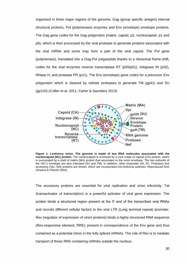
Figure 1: Lentivirus virion. The genome is made of two RNA molecules associated with the nucleocapsid (NC) protein. The nucleocapsid is enclosed by a core made of capsid (CA) protein, which
is surrounded by a shell of matrix (MA) protein that associates to the virion envelope. The two subunits of the HIV-1 envelope are also indicated (SU and TM). In addition, other enzymatic (IN, RT, Protease) and accessory (Vpr, Nef) proteins are shown, which are incorporated into lentivirus particles. Reproduced from (Greene & Peterlin 2002).
The accessory proteins are essential for viral replication and virion infectivity. Tat
(transactivator of transcription) is a powerful activator of viral gene expression. The
protein binds a structured region present at the 5’ end of the transcribed viral RNAs
and recruits different cellular factors to the viral LTR (Long terminal repeat) promoter.
Rev (regulator of expression of virion proteins) binds a highly structured RNA sequence
(Rev-responsive element, RRE), present in correspondence of the Env gene and thus
contained as a potential intron in the fully spliced mRNAs. The role of Rev is to mediate
transport of these RRE-containing mRNAs outside the nucleus.
31
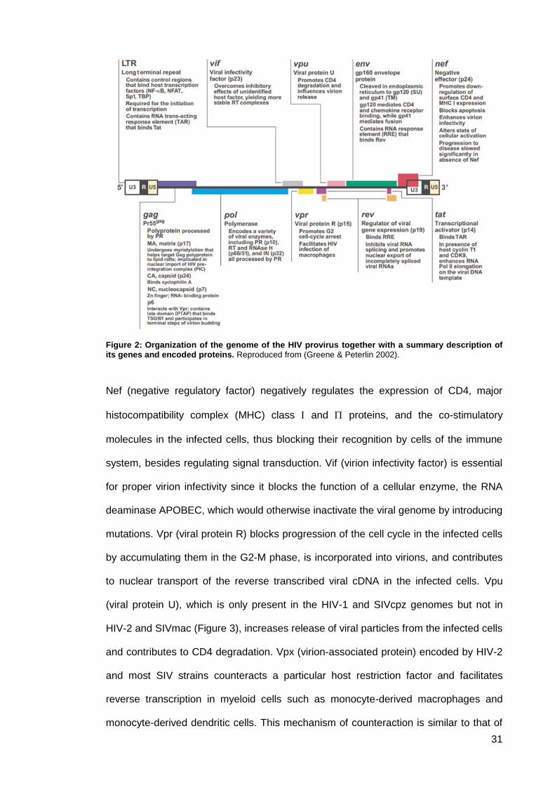
Figure 2: Organization of the genome of the HIV provirus together with a summary description of its genes and encoded proteins. Reproduced from (Greene & Peterlin 2002).
Nef (negative regulatory factor) negatively regulates the expression of CD4, major
histocompatibility complex (MHC) class and proteins, and the co-stimulatory
molecules in the infected cells, thus blocking their recognition by cells of the immune
system, besides regulating signal transduction. Vif (virion infectivity factor) is essential
for proper virion infectivity since it blocks the function of a cellular enzyme, the RNA
deaminase APOBEC, which would otherwise inactivate the viral genome by introducing
mutations. Vpr (viral protein R) blocks progression of the cell cycle in the infected cells
by accumulating them in the G2-M phase, is incorporated into virions, and contributes
to nuclear transport of the reverse transcribed viral cDNA in the infected cells. Vpu
(viral protein U), which is only present in the HIV-1 and SIVcpz genomes but not in
HIV-2 and SIVmac (Figure 3), increases release of viral particles from the infected cells
and contributes to CD4 degradation. Vpx (virion-associated protein) encoded by HIV-2
and most SIV strains counteracts a particular host restriction factor and facilitates
reverse transcription in myeloid cells such as monocyte-derived macrophages and
monocyte-derived dendritic cells. This mechanism of counteraction is similar to that of
32
the accessory proteins Vif and Vpu which antagonise other host factors. SAMHD1 a
protein that functions as a restriction factor counteracted by Vpx. SAMHD1 degrades
deoxynucleoside triphosphates (dNTPs), which are components of the viral genomic
cDNA, in order to deprive viruses of dNTPs. Vpx has also been shown to have an
apparent ability to enhance nuclear import of the viral genome in T lymphocytes (Collier
et al. 2011; Carter & Saunders 2013; Seitz 2016).
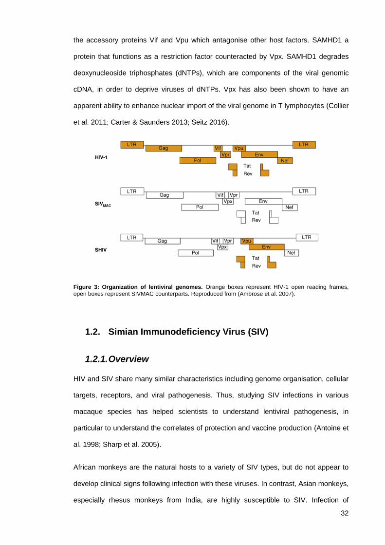
Figure 3: Organization of lentiviral genomes. Orange boxes represent HIV-1 open reading frames,
open boxes represent SIVMAC counterparts. Reproduced from (Ambrose et al. 2007).
Simian Immunodeficiency Virus (SIV) 1.2.
1.2.1. Overview
HIV and SIV share many similar characteristics including genome organisation, cellular
targets, receptors, and viral pathogenesis. Thus, studying SIV infections in various
macaque species has helped scientists to understand lentiviral pathogenesis, in
particular to understand the correlates of protection and vaccine production (Antoine et
al. 1998; Sharp et al. 2005).
African monkeys are the natural hosts to a variety of SIV types, but do not appear to
develop clinical signs following infection with these viruses. In contrast, Asian monkeys,
especially rhesus monkeys from India, are highly susceptible to SIV. Infection of
33
macaque species causes a progressive immunodeficiency syndrome, which closely
mimics human AIDS. In contrast, HIV does not usually infect Asian macaques and is
non-pathogenic when it does. Thus, evaluating experimental vaccines in these animals
requires a non-human analogue instead of the actual HIV vaccine candidate used in
clinical trials in humans (Greenwood et al 2012; Esparza 2001; Antoine et al. 1998;
Douglas et al. 1997).
SIV mac has been used extensively to model HIV in vaccine experiments. However,
studies have shown that it is not possible to evaluate the protective efficacy of an HIV
vaccine candidate using SIV mac. This is because there is a significant difference in
the proteins produced by SIV and HIV (Douglas et al. 1997; Johnston 2000; Sharp et
al. 2005). On the other hand, chimeric viruses can be used. SIV/HIV hybrid viruses
(SHIVs), where the Env gene as well as other genes including Tat, Rev, Vpr, and Vpu,
from an HIV-1 isolate were inserted in the backbone of an SIV genome (Ambrose et al.
2007; Johnston 2000) can allow the protective efficacy of vaccine candidates based on
envelope glycoprotein to be tested. Insertion of more HIV-1 genes into SIV genome
has been unsuccessful (Joag et al. 1997; Li et al. 1995; Shibata, Siemon, et al. 1997).
To varying degrees, SHIV can replicate in rhesus macaques causing no disease at the
start. However, serial in vivo passages eventually lead to the emergence of highly
pathogenic variants. These pathogenic SHIVs are capable of causing rapid depletion of
the circulating CD4+ T-cell population in the infected host, generating a severe
immunodeficiency syndrome and eventual death of the host within a year (Joag et al.
1996; Reimann et al. 1996; Igarashi et al. 1999; Johnston 2000). As such, SHIV could
mimic HIV to some extent. However, the relevance of using these pathogenic SHIVs, in
HIV vaccine protection experiments has been questioned. Studies argue that,
compared to SIV, SHIV containing HIV-1 Nef replicates insignificantly in vivo (Shibata,
Maldarelli, et al. 1997). Moreover, SHIV may demonstrate some host-range restriction
of growth (Shibata et al. 1991). As such, SHIV has been used less widely in HIV
vaccine experiments (Johnston 2000; Ambrose et al. 2007).
34
HIV and vaccine production 1.3.
1.3.1. Overview
Despite ongoing international efforts to control the HIV/AIDS pandemic through
individual action, behaviour modification and the screening of potentially contaminated
blood and blood products, more than 15,000 people become infected with HIV every
day. Most of them live in developing countries. The current studies of pandemics in
Africa and Asia indicate that HIV transmission is mainly heterosexual or vertical in
children (World Health Organization Guideline 2016; Greenwood et al 2012; Esparza
2001). The development of antiretroviral therapies to combat HIV infection has
dramatically decreased the morbidity and mortality among HIV-positive patients
(Churchill et al. 2016). However, HIV transmission has only been partially restricted by
the advent of antiretroviral treatment and changes in sexual behaviour. Effective
treatment of HIV-infected patients is limited by many factors, such as late diagnosis,
appearance of resistant mutations and poor adherence to treatment. HIV treatment is
expensive, and most HIV-infected patients cannot afford it as most live in low-income
countries (Greenwood et al 2012; Churchill et al. 2016; Esparza 2001).
The importance of developing a safe, effective, and affordable HIV vaccine cannot be
overemphasised as HIV is one of the world's deadliest scourges (Collier et al. 2011).
Since the discovery of HIV as the causative agent of AIDS in 1983–84, hopes for the
development of a HIV preventative vaccine have been raised, as the most potent
defence against viral infections are vaccines (Sarngadharan et al. 1984; Johnston &
Fauci 2007). A vaccine could be a valuable complement to other interventions that
significantly alter the chain of HIV transmission, the course of the disease, and its
infectiousness. Thus, this could provide positive health benefits for both infected
individuals as well as the community at large. Also, a well-conceived HIV immunisation
strategy could reach populations in which antiretroviral treatment and other
interventions are not sufficiently effective, such as in low-income countries (World
35
Health Organization Guideline 2016; Carter & Saunders 2013; Esparza 2001). Studies
suggest that a preventative HIV vaccine could possibly be used as a therapeutic
intervention in association with antiretroviral therapies. This could lead to lower cost of
the treatments and may increase the treatment’s long-term efficacy (Cho 2000; Di
Nunzio et al. 2012).
1.3.2. HIV vaccine status and vaccine production challenges
Vaccines have been our best application to protect us from most of the world’s
deadliest viral infectious diseases, including smallpox, polio, measles and yellow fever.
However, there are many confounding issues that prevent the production of a
sufficiently effective HIV vaccine (Morrow et al. 2012; Esparza 2001). Challenges
include HIV extreme antigenic variability, lack of understanding of immune correlates
for protection, limitations of available animal models, and the enormous constraints
associated with the probable need for multiple large-scale clinical trials in different parts
of the world.
The human body seems incapable of mounting an effective immune response against
HIV infection as the virus has a unique way of evading the immune system. The fact
that the virus infects the immune system itself presents a particular challenge. HIV
attacks T helper cells as they have the HIV-specific receptor CD4. HIV is taken up by
CD4+ T cells and macrophages following binding of viral glycoprotein (gp120) to CD4
and certain chemokine receptors (CXCR4 and CCR5). Thus, HIV specifically infects
the very cells necessary to activate both B-cell and cytotoxic T-cell immune responses.
T helper cells are a key part of the immune system; they help B-cells and effector T-
cells to combat infection. CD4+ T cells have been identified as a major effector cell
population in the response to many virus infections (Johnston & Fauci 2007; Levy
2007; Chhatbar et al. 2011; Cho 2000). However, HIV can also infect other cells of the
immune system, including macrophages, memory cells in the lymph node, dendritic
cells and brain microglial cells as well as its tendency to infect bone marrow-derived
cells and lymphocytes. In addition, HIV can minimise its recognition by CTL through its
36
ability to down regulate MHC class I molecules (Cho 2000; Levy 2007). To date, HIV
vaccine researchers have no human model with a full recovery from the infection and
subsequent protection from reinfection (Ambrose et al. 2007). Moreover, scientists lack
comprehensive information about the correlates of protective immunity to HIV (Esparza
2001).
HIV has a large genetic diversity as well as a high mutation rate. Inside the host, the
virus continually mutates and recombines, evolving new strains of virus that differ
slightly from the original infecting virus allows them to escape neutralising antibodies
and evade the immune response. The mutation usually occurs in those viral peptides
that bind to MHC class I molecules to which the initial T cell response arose which
results in a failure of T cell surveillance. As a result, quasi-species of viral infection are
usually present in HIV-infected patients. The extensive diversity of HIV poses a
challenge to designing a long-lasting HIV vaccine as it would need to protect against
the many different strains of the virus circulating throughout the world (Levy 2007; Cho
2000; Carter & Saunders 2013).
The lack of a suitable small-animal model to test vaccine candidates to predict the
effectiveness of an HIV vaccine in people hampers HIV vaccine development. The only
animals susceptible to experimental infection with HIV are chimpanzees and pigtail
macaques. Both can be infected with HIV, however, no clinical signs arise as they tend
to maintain low levels of persistent virus load and do not develop clinical manifestations
of AIDS. Currently, researchers rely on experiments using non-human primate models
infected with non-human lentivirus mainly SIV to somewhat mimic disease progression.
Thus, evaluating experimental vaccines in these animals requires a non-human analog
instead of the actual HIV vaccine candidate used in clinical trials in humans
(Greenwood et al 2012; Esparza 2001; Johnston & Fauci 2007; Ambrose et al. 2007;
Johnston 2000).
37
1.3.3. Eliciting immune response using the current
immunisation protocols
Live attenuated vaccines 1.3.3.1.
In general terms, live attenuated vaccines are composed of a live, but substantially
weakened pathogen through single or multiple mutations within its genome. The
attenuated pathogen will replicate in host cells causing limited and non-life threatening
infection. This vaccine strategy can elicit a strong and long-lasting immune response,
which can mimic the disease in very mild form and prevent reinfection with the same
pathogen (Niaid.nih.gov 2014; Cho 2000). However, in the case of HIV, serious
potential risks in humans are an important concern when the correlates of protection
are still largely unknown (Girard et al. 2011)
In favour of a live attenuated HIV vaccine approach, many studies have been
conducted using attenuated HIV-2 or SIV. Attenuation of these viruses was achieved
through induced deletions or mutations of the viral genome. Studies confirmed that the
least attenuated viruses elicited a better immune response (Lohman et al. 1994;
Johnson et al. 1999). However, as the virus integrates into the host genome following
infection, it is essential that the administered strain does not regress to its original
pathogenic form following vaccination.
Observations of several attenuated SIV or SHIV vaccine studies have produced
confounding results where macaques infected with attenuated SIV or SHIV exhibited
strong immune responses but various levels of protection (Girard et al. 2011). Although
sterilising immunity has not been achieved, immunisation studies with SIV nef-deleted
mutant indicate that SIV ∆nef confer a persistent, life-long, low-grade viral infection in
rhesus macaques that limits the development of AIDS. However, it does not protect the
vaccinated monkeys against infection with wild-type virus. In contrast, another study
indicated that infant monkeys developed AIDS when the SIV ∆nef vaccine was
administered orally. Prolonged observations of attenuated SIV with triple deletions,
38
where nef, vpr, and negative regulatory element was deleted from the virus genome,
indicated potential reversion of the virus to become pathogenic in neonates as well as
in adult macaques. Attenuated SIV with triple deletions is genetically unstable and can
evolve into a fast replicating variant (Baba et al. 1999; Bordería & Berkhout 2009; Joag
et al. 1998; Shibata, Maldarelli, et al. 1997; Shibata, Siemon, et al. 1997; Lohman et al.
1994).
Patients who were infected with naturally occurring attenuated HIV-1 isolates with a
deletion in the nef gene remain healthy substantially longer than those patients infected
with viruses with an intact nef gene. They have been shown to have high levels of CTL
activity against HIV-1 as well as long-term, non-progressive infection (Deacon et al.
1995; Dyer et al. 1999; Kirchhoff et al. 1995; Bogers et al. 1995; Greenough et al.
1999). However, recent reports show a low gradual decline in the number of CD4 + T
cells among those patients. As a result of these concerns, the live attenuated strategy
has not been used in HIV vaccination experiments, as some can evolve to virulent virus
over time (Girard et al. 2011).
Inactivated vaccines 1.3.3.2.
An inactivated vaccine is achieved either by heat or chemical treatment to ensure that
the virus has been killed and is no longer infectious. It is assumed that the native
structure of the epitopes concerned with eliciting protective immune responses are
preserved, despite the alteration of both the conformation and the antigenicity of the
viral envelope that occurs as a result of the various treatments (Cho 2000; Levy 2007).
A method has been developed to completely inactivate the infectivity of HIV or SIV
while keeping the envelope epitopes intact and functional (Rossio et al. 1998). This
was done by disrupting the two nucleocapsid protein zinc finger domains, which are
essential for virus infectivity, through mild oxidation or alkylation (Arthur et al. 1998;
Rossio et al. 1998).

39
Most of the vaccine studies using this approach used the macaque model with either
inactivated SIV or HIV-2 due to the potential danger resulting from the incomplete
inactivation of HIV-1. Since the virus has been killed and no longer replicates, this type
of vaccination will not elicit strong cellular immunity as there is no intracellular protein
expression, particularly affecting CTL responses. Immunisation studies using a whole
inactivated SIV vaccine have shown varied protection results: some have
demonstrated only partial protection, while others have reported complete protection
(Desrosiers et al. 1989; Sutjipto et al. 1990; Girard et al. 2011). Although most of the
vaccinated animals showed decreased viraemia and no significant depletion of CD4+ T
cells, they could still be infected with the wild-type virus. However, most of these
animals were protected from SIV grown in human cells, but not from the virus grown in
macaque peripheral blood mononuclear cells (PBMCs) (Cho 2000; Girard et al. 2006;
Girard et al. 2011).
In this regard, later studies indicated that it was likely that the protective immune
response in vaccinated animals was directed against the xenoantigen rather than the
viral antigens, as high levels of antibodies against human cellular proteins were
detected in all these animals and the degree of protection was shown to be correlated
with the titre level of these antibodies (Hirsch et al. 1996; Langlois et al. 1992).
Whether whole inactivated vaccines elicit a protective immune response or not, this
approach has been neglected due to the high risk of disease development, the possible
side effects of alloantigen immunisation, the resulting sporadic level of protection and
the associated use of various adjuvant treatments that need to be further evaluated
(Arthur et al. 1998; Girard et al. 2006; Rossio et al. 1998).
However, a recent study using a genetically modified and killed whole-HIV1 vaccine,
SAV001, describes a current ongoing Phase I human clinical trial, the first of its kind.
The study uses an inactivated and formulated killed whole-HIV (gmHIV-1NL4-3). The
virus was propagated in the A3.01 human T cell line followed by virus purification and
inactivation with aldrithiol-2 and γ-irradiation. The primary results suggested that the
40
vaccine had no potential toxicity and it triggered anti-HIV immune responses. However,
it is still to be determined whether this immune responses can prevent HIV infection
(Choi et al. 2016).
Virus-like particles (VLP) 1.3.3.3.
Virus-like particles (VLP) are multi-protein structures, which mimic the organisation and
the conformation of the original native viruses. VLPs lack the viral genome, and hence
yield potentially safer and cheaper vaccine candidates. It has been noticed that the
Gag and Env proteins of HIV or SIV, when expressed in cells, spontaneously assemble
to form VLPs that include only the virus envelope and the core proteins (Girard et al.
2006; Levy 2007).
In many studies, SIV VLPs have been tested as immunogens in non-human primate
models. These VLPs elicited low but to some extent, significant titres of neutralising
antibodies against both homologous and heterologous primary HIV-1 isolates.
However, the use of VLPs in vaccine production has failed due to the difficulty in
producing VLPs in sufficient quantities. This could be a result of inefficient particle
assembly and release, faulty Gag protein processing, or faulty incorporation of the virus
envelope. Moreover, studies show that it is almost impossible to produce VLPs without
contaminant cellular proteins that may produce complex immunogenicity (Cho 2000;
Levy 2007). Significant improvement in the HIV VLP vaccine approach using VLPs
containing different target antigens has been reported (Kushnir et al. 2012; Levy 2007).
Chimeric VLPs could be produced using homologous or heterologous retroviruses such
as the SIV Gag protein or the HIV Env protein. The assembled chimeric VLP could be
enveloped or non-enveloped. The preparation of chimeric VLPs is however,
substantially more difficult (Kushnir et al. 2012; Zhang et al. 2004). Compared to full
pathogen-based inactivated or live attenuated vaccines, immunogenicity of VLPs is
generally low thus requiring higher doses, booster administrations, co-administration of
adjuvants, or development of alternative approaches for enhancing target-specific
immunity.
41
Subunit vaccines 1.3.3.4.
Subunit vaccines are usually composed of a mixture of protein components from a
virus with no genetic material. The benefit of using such an approach is that these
subunits can elicit both humoral and cellular immune responses (Cho 2000). Large
amounts of viral peptides, which are needed for vaccine production, can be produced
either by using recombinant vehicles, such as bacteria, yeast, insect cells, or
mammalian cells, or they can be chemically synthesised (Levy 2007; Cho 2000). A set
of peptides used in the production of the subunit can be safely manipulated to contain
epitopes specific for B and/or T cells. As these epitopes lack the virus genome and are
not infectious, increased safety is one of the most important advantages of this
approach (Kushnir et al. 2012). Generally, subunit vaccines do not elicit an efficient
CTL response. However, studies show that if a strong immune response was elicited
when using a subunit vaccine, the purity of the vaccine preparation make it preferable
to more complex mixtures such as inactivated pathogens or recombinant vehicles that
would express pathogenic antigens (Emini et al. 1990; Murphey-Corb et al. 1991;
Berman et al. 1990).
Envelope-based subunit vaccines 1.3.3.5.
Recombinant envelope glycoproteins gp120 (exterior glycoproteins) and gp41
(transmembrane proteins) bear neutralisation epitopes that can elicit humoral immune
responses, which HIV-1-neutralising antibodies target (Mascola et al. 1996; Jeffs et al.
2004). According to Klein et al. 2001, at least 13 different envelope-based vaccines
have been evaluated in humans. Several trials have aimed to develop envelope-based
subunit vaccines by evaluating gp120 and gp140 (gp120 coupled with the ectodomain
of gp41) (Klein 2001; Girard et al. 2006). As reviewed in Girard et al. 2011, gp120
monomers from T-cell-line-adapted clade B isolates and gp140/160 oligomers have the
same immunogenicity (Girard et al. 2011).
42
The results of past studies indicate that all envelope proteins elicit neutralising
antibodies for homologous HIV strains, but not for heterologous ones. Despite the use
of adjuvant, CTL responses were not induced in these cases. In spite of the use of
booster immunisations, no trials have shown a statistically significant reduction of the
viral loads in vaccines. Other approaches that have aimed to elicit HIV-neutralising
antibody vaccine have been underdeveloped. For example, some studies have aimed
to overlap CD4-binding and expose neutralising sites by stabilising gb140 or
manipulating gp120. Higher titres of neutralising antibodies with limited ability to cross-
neutralise heterologous virus strains have been isolated (Girard et al. 2006; Cho 2000;
Levy 2007). In another example, developed by Merck and the University of Maryland,
the co-receptor binding site has been overlapped and neutralising epitopes have been
exposed by covalently linking gp120 or gp140 to soluble CD4 or to synthetic mimics of
the CD4 receptor. The results suggested that a broad neutralising antibody response
may be elicited (Devico et al. 1996); this finding has not been replicated (Girard et al.
2006). Targeting conserved epitopes on the CD4-binding site by engineering
hyperglycosylated derivatives of gp120 and immunisation with recombinant gp41 has
been studied to induce fusion-blocking antibodies. However, the extreme genetic
variability of HIV makes it difficult to use such approaches to develop a vaccine
capable of eliciting broadly neutralising antibodies that can challenge different primary
isolates and different clades of the virus (Girard et al. 2006; Girard et al. 2011; Di
Nunzio et al. 2012).
Non-structural protein subunit vaccines 1.3.3.6.
An example of the development of a non-structural protein subunit vaccine is
vaccination with a viral transactivator protein (Tat). Tat is a significant HIV virulence
factor helping in virus gene expression, replication, transmission and progression of
disease. Different approaches using Tat antigens have been developed (Levy 2007).
Studies indicate that both humoral and cellular immune responses are elicited when
cynomolgus monkeys are immunised using biologically active Tat antigens, while
43
immunised rhesus macaques are protected, to a certain level, from pathogenic SHIV
(Gallo 1999; Ferrantelli et al. 2004; Rezza et al. 2005; Maggiorella et al. 2004). On the
other hand, according to Liang et al., results concerning rhesus monkeys immunised
with adenovirus type 5 (Ad5)-HIV tat recombinant vaccine did not indicate any efficacy
against SHIV challenges, despite the elicitation of humoral and immune cellular
responses (Liang et al. 2005). However, different studies suggest that in intra-rectally
challenged rhesus macaques, inactivated Tat decreases the presence of the disease,
as well as reducing the viral load (Silvera et al. 2002).
Combined vaccines using Tat antigen have also been developed, where Tat-adenyl
cyclase fusion protein or a Tat–Nef fusion protein is combined with a recombinant
gp120 subunit vaccine in AS02A adjuvant. This combined vaccine has been able to
protect rhesus monkeys from the development of AIDS when they are challenged with
pathogenic SHIV. Although further evaluation is still needed, several clinical trials using
subunit Tat vaccine have indicated that the vaccine could be well-tolerated and would
produce a significant immune response (Girard et al. 2006; Girard et al. 2011). Another
approach is that of the AIDS Vaccine Integrated Project (AVIP), which is trying to
develop a novel vaccine approach based on the use of a trimeric gp140 molecule
deleted at the V2 loop (gp140 ∆V2 SF162) and mixing it with either Tat or Nef antigens.
It seems likely that a multicomponent vaccine will be more efficacious because more
epitopes will be presented. The anticipated diverse immune response should be less
susceptible to viral mutation and immune escape (Levy 2007; Girard et al. 2006).
Anti-Tat antibodies are rarely found in natural infection and when found correlate with
asymptomatic state and reduced disease progression, recent studies are focusing on
using Tat as an optimal candidate for therapeutic immunisation and cART
intensification more than a vaccination approach. Tat is produced very early upon
infection and continues to be expressed under cART. It is found that Tat expressed and
released extracellularly by infected cells, accumulates in tissues, induces immune
44
dysregulation, and promotes virus reactivation, entry and spreading (Ensoli et al. 2015;
Ensoli et al. 2010).
The introduction of anti-Tat antibodies results in pathogenesis regulation and inhibition
of disease progression thus helping in concentrating cART effectivity. Randomised
phase II clinical trial (ISS T-002, ClinicalTrials.gov NCT00751595) conducted in 168
cART-treated volunteers in Italy, found that Tat-based vaccination is safe,
immunogenic and capable of eliciting durable immune responses. Candidates used
were HIV-infected anti-Tat Abs negative, virologically suppressed cART-treated (mean
of 6 years) adult subjects. The vaccine induced anti-Tat Abs in 79% of patients. The
vaccine contributed in HIV containment by promoting a durable and significant
restoration of T, B, and natural killer (NK) cell numbers, increased CD4+ and CD8+
central memory subsets, and upregulating the expression of human leukocyte antigen-
D related (HLA-DR+) on CD8+ killer T cells. A 70% reduction of blood proviral DNA
was detected, which is significantly related to neutralization of Tat-mediated entry of
Env in dendritic cells (DCs). DCs resulted from the expression of anti-Tat in IgG and
IgM format (Ensoli et al. 2015; Ensoli et al. 2010; Longo et al. 2009).
Based on these data and to assess whether clade B Tat immunisation would be
effective also in patients with different genetic background and mainly infected with a C
clade virus, a phase II trial was conducted in South Africa (ISS T-003, ClinicalTrials.gov
NCT01513135). Results indicated that immunisation with B clade Tat induced
functionally effective cross-clade anti-Tat Abs and CD4+ T-cell responses. This finding
reinforces that clade B Tat is a suitable candidate for therapeutic immunisation against
different HIV clades in different geographical areas. As a result, phase III studies in
South Africa were established (Ensoli et al. 2016).
Naked DNA vaccines 1.3.3.7.
Evidence suggests that HIV-specific CD4+ and CD8+ T-cell immune responses are
associated with reduced viral load, caused by better controlled viral replication. Thus,
45
the cellular immune response is a key factor in controlling HIV. Although live attenuated
vaccines can elicit a strong cellular immune response, they can also evolve into virulent
viruses over time. An emphasis on the development of new strategies that could
produce a significant immune response against HIV without producing disease over
time is a primary goal of HIV immunisation (Levy 2007; Cho 2000; Girard et al. 2006).
Scientists have developed an immunisation strategy that is performed via the direct
injection of naked plasmid DNA that encodes viral genes of interest. This injection can
be made intramuscularly, intradermally, or via an epidermis gene gun.
It has been proven that using plasmid-encoding HIV envelope DNA vaccines in
nonhuman primates, along with a prime-boost immunisation regimen, can elicit both
humoral and cellular immune responses. This can be enhanced by cytokine
administration (Levy 2007; Robinson & Pertmer 2000; Girard et al. 2011). The
protective efficacy of DNA vaccines encoding envelope genes has been highly
variable. Researchers have argued that macaques and chimpanzees immunised with
plasmids encoding a viral genome can elicit cellular and relatively weak humoral
immune responses (Boyer et al. 1997; Fuller, Corb, et al. 1997; Fuller, Simpson, et al.
1997; Haigwood et al. 1999). As reviewed by Girard et al., DNA vaccines demonstrated
a higher rate of protection when immunised macaques were challenged by SHIV or
HIV-1. Immunised macaques with both DNA and recombinant gp160 showed
envelope-specific lyrnphoproliferative response and CTL activity (Girard et al. 2011;
Levy 2007). However, DNA vaccines produce significantly lower level of
immunogenicity in humans compared to animal models (Goyvaerts et al. 2013).
Wyeth Pharmaceuticals developed a naked DNA vaccine using an expressed HIV-1
gag gene with either IL-12 or IL-15 or both. The vaccine was tested on humans in
Phase I trials in the USA, Brazil and Thailand (Clinicaltrials.gov NCT00115960
NCT00111605). The Phase I clinical trial with HIV-1-infected human patients clearly
demonstrated the safety of DNA-based vaccines. IL-12 and IL-15 adjuvant were safe to

46
give, although offered limited ability to boost cellular immune responses (Kalams et al.
2012; Girard et al. 2006).
Table 1: Comparison of VAX004 and VAX003 studies
VAX004 Study VAX003 Study
Vaccine AIDSVAX® B/B′ AIDSVAX
® B/E
Vaccine design Bivalent rgp120 with alum
Vaccine antigens MNrgp120/HIV-1 plus GNE8
rgp120/HIV-1
(subtype B and B′)
MNrgp120/HIV-1 plus A244
rgp120/HIV-1
(subtype B and E)
rgp120 dose 300 µg (MN)/ 300 µg (GNE8) 300 µg (MN)/ 300 µg (A244)
Placebo alum
Randomization:
Vaccine to Placebo
2:1 1 :1
Vaccine administration Intramuscular
Between 1998 and 2003, the world’s first Phase III clinical trials of a gp 120 HIV/AIDS
vaccine (AIDSVAX) were conducted by VaxGen. Data from both VAX004 and VAX003
indicated no protective efficacy against acquisition of HIV infection (Table 1). AIDSVAX
B/E alone conferred no protection among Thai people who inject drugs (PWID)
(VAX003, Clinical-Trials.gov NCT00006327). Similarly, no overall significant protection
was seen with a similar regimen of bivalent gp120 (AIDSVAX B/B) alone (VAX004,
Clinical-Trials.gov NCT00002441) among mostly men who had sex with men (MSM) in
North America and The Netherlands. Despite intriguing findings in subgroup analyses
of VAX004, which suggested vaccine efficacy for acquisition of HIV infection in Blacks
or non-whites, women and high-risk volunteers, vaccine efficacy in the subgroups could
not be explained because of the small number of infections in each subgroup (Flynn et
al. 2005; Pitisuttithum et al. 2006).
Live recombinant vaccines 1.3.3.8.
Studies have suggested that the use of a vector can enhance vaccine protection level.
Recombinant viruses are the current vectors that are used to deliver selected genes
into the chosen host. Vectors are not usually derived directly from lentiviruses;
poxviruses, adenoviruses and retroviruses are commonly used (Levy 2007; Girard et
al. 2006). However, recently lentiviruses have been introduced as vaccine vectors.
47
Lentiviruses have successfully been used in gene therapy techniques, and their
subsequent use in vaccination has evolved from the demonstration of their capacity to
efficiently transduce dendritic cells.
In recombinant vaccination against HIV, envelope DNA is usually inserted into the
genome of the vector virus that has been selected to produce a recombinant vaccine.
The resulting virus will present envelope antigens to the host immune system, thus
eliciting cellular immune responses as well as humoral responses (Girard et al. 2006;
Morrow et al. 2012). DNA vaccines have been found useful when used within a prime-
boost regimen. Although DNA vaccination itself does not elicit a strong immune
response, it appears to prime the immune system quite effectively because a strong
anamnestic response is observed when boosting with recombinant protein or vaccinia
virus (Fuller, Simpson, et al. 1997; Fuller, Corb, et al. 1997; Haigwood et al. 1999). The
strength of the immune response is strongly influenced by the choice of the vector
system used, as well as the vaccination schedule (Fuller, Corb, et al. 1997; Fuller,
Simpson, et al. 1997; Girard et al. 2011). The list of virus vectors used to construct live
recombinant-vector-based HIV vaccines is long including lentiviruses, measles virus,
vesicular stomatitis virus (VSV), Sendai virus and Venezuelan equine encephalitis virus
(Sauter et al. 2005). Here I only highlight Pox viruses and Adenoviruses in detail, in
favour to the fact that they have been used in several vaccination trials.
1.3.3.8.1. Pox viruses
Pox viruses are one of the most important vectors used in recombinant vaccination
(Mercer et al. 2007). A variety of HIV antigens, such as Gag, Env, Pol and Nef, may be
expressed using such vectors. Recombinant canarypox virus was used to express
various HIV-2 antigens; high levels of antibody titres, together with specific HIV-2
CTLs, were detected. However, there was no correlation between immunologic
response and protection, despite the immunisation boost (Levy 2007). Vaccinia viruses
were the first to be used in vector based vaccines. In some early studies, a vaccinia
virus vector vaccine expressing HIV gp120 was shown to protect chimpanzees from
48
low-dose HIV challenge. In humans, a vaccinia-virus-based vaccine has been shown to
reduce the observed immune responses in subjects who had been previously
vaccinated for smallpox (Levy 2007). However, the use of a prime-boost regimen with
repeated boosting with gp160 has been shown to overcome this problem in the SIV
model (Pitisuttithum et al. 2006). Due to safety concerns, highly attenuated pox viruses
have been developed, such as modified vaccinia Ankara (MVA). Currently, several
vaccination studies have been developed involving macaques and using MVA as a
vector; MVA expresses multiple SIV antigens, most of which are provided within a
prime-boost regimen. Generally, the results indicate reduced viral load, as well as a
delay in the appearance of the disease among vaccinated macaques (Morrow et al.
2012; Girard et al. 2011). Promising studies in rhesus monkeys found that
immunisation with recombinant MVA vector expressing SIV mac 239 Gag-Pol and
HIV1 Env elicited strong Gag-specific CTL responses but did not create envelope-
neutralising antibodies.
The first evidence to confirm that HIV-1 vaccine could confer protective efficacy against
HIV-1 acquisition is provided by the Thai Phase III trial, RV144 (ClinicalTrials.gov
NCT00223080). This prime-boost vaccine regimen consisted of a non-replicating
recombinant canarypox vector (ALVAC; a Gag Pol Env prime and AIDSVAX gp120 B/E
boost). The trial demonstrated a 31.2% reduction in the frequency of acquisition of HIV
infection among vaccinated heterosexual men and women (refer to section 1.3.4).
1.3.3.8.2. Adenoviruses
An adenovirus-based vaccine system is of a major interest because it has effectively
protected chimpanzees from HIV-1 infection. Replication-deficient adenovirus type 5
(Ad5) represents one of the most promising live viral vectors in HIV vaccination. It was
first used by Merck Pharmaceuticals as a pilot monovalent Ad5 gag vaccine; it was
tested on mice, nonhuman primates, and human volunteers. Strong, long-lasting, HIV-
1-specific CD8+ T-cell responses were indicated in half of the volunteers (Casimiro et
al. 2003; Shiver et al. 2002; Shiver & Emini 2004).
49
It has been argued that vaccination with recombinant Ad5, either delivered alone or as
a boost after immunisation with a plasmid DNA vaccine, elicit significant immune
responses to HIV-1 Gag, in which high numbers of long-lasting T-cells were found in
human volunteer vaccine recipients (Levy 2007; Girard et al. 2006). However, frequent
pre-existing anti-vector immunity in humans limits the efficiency of the use of Ad5. To
overcome this problem, recombinant adenoviruses that are representative of less
human-prevalent strains are under investigation; for example, Ad6, Ad35, Ad11 and
Ad24. Non-replicative chimpanzee adenoviruses (AdC68, AdC6 and AdC7) could be
used or chimeric adenoviruses developed to evade the pre-existing anti-Ad5 immunity,
by replacing the fibre gene of an Ad5 vector with that of a rare Ad subtype. Adenovirus
chimeras such as Ad5/Ad11 or Ad5/Ad35 have been constructed and successfully
tested in animal models. Many other strategies using adenovirus vectors have been
developed to increase the protective efficiency of the use of adenoviruses as
recombinant vectors (Girard et al. 2006; Levy 2007; Girard et al. 2011).
Merck Pharmaceuticals, in collaboration with NIAID, engineered trivalent, recombinant
Ad5-expressing Gag, Pol and Nef antigens and used these in the STEP trial (HVTN
502/ Merck V520-023). This was the first completed efficacy evaluation of a CMI-based
HIV vaccine. It was conducted at 34 sites in North America, the Caribbean, South
America and Australia, where the predominant circulating HIV-1 subtype is B, and
enrolled 3,000 individuals. This Phase b trial started on Dec 2004 and terminated on
Sep 2007. The findings indicated that the MRKAd5 gag/pol/nef HIV-1 vaccine neither
prevented HIV-1 infection nor lowered the viral load (Buchbinder et al. 2008). The Step
study revealed an increased risk of HIV-1 acquisition in vaccinees who were
uncircumcised and/or Ad5 seropositive at baseline. However, later studies indicated
that pre-existing seropositivity to multiple Ad serotypes is not intrinsically associated
with increased risk of HIV-1 acquisition (Stephenson et al. 2012). The second Phase
b trail of the MRK rAd5 vaccine, Phambili (HVTN 503), was undertaken in South
Africa, where HIV-1 subtype c is predominant. Enrolment and vaccination was
50
terminated in Sep 2007 as a result of the STEP finding. Participants were followed-up
for up to 3.5 years. Long-term follow-up analysis indicated no significant efficacy.
Unlike the STEP study, statistical trends towards enhanced acquisition of HIV infection
in vaccinees who were Ad5-seropositive at baseline or men who were uncircumcised
were not observed (Gray et al. 2014) (refer to section 1.3.4).
Prime-boost immunisation regimens 1.3.3.9.
Most of the vaccine strategies used to elicit immune responses against HIV have been
shown to induce short–lived immune responses. In this regard, a prime-boost
vaccination regimen has been developed. There are many types of boosting regimens
(Girard et al. 2011). The initial use of a DNA vaccine candidate as a prime vaccine,
followed by a recombinant live virus vector as a booster vaccine, is one example.
Multiple studies concerned with HIV vaccination have used this approach, and it has
been proven that the cellular immune response is improved by using such a method
(Levy 2007). Synergy between DNA and live recombinant vectors has been observed,
regardless of the type of recombinant. For example, compared to the Ad5 recombinant
vaccine, the level of HIV-1 Env-specific T-cell responses is 7-fold higher in a DNA-Ad5
prime-boost regimen, which should dramatically increase the efficiency of the vaccine
(De Rosa et al. 2011).
A prime-boost vaccine regime can be achieved using two live recombinant vectors that
express the same antigens. The two live recombinants could be an Ad5 vector followed
by a recombinant poxvirus (Casimiro et al. 2003), two successive recombinant
poxviruses, such as MVA and FPV, or two successive recombinant adenoviruses, such
as Ad26 followed by Ad5 or Ad35. It has been argued that the use of a heterologous
prime-boost regimen, such as Ad5 followed by MVA or ALVAC, will elicit better cellular
immune responses than those elicited via the use of a homologous Ad5 only regimen
(Girard et al. 2011).

51
The final type of prime-boost immunisation regimen combined a DNA vaccine as a
prime and a protein subunit as a booster an immunisation schedule which should elicit
both cellular and humoral immune responses. It allows the use of a mixture of Env
DNA plasmids for priming and boosting with a mixture of gp120 proteins from various
HIV-1 clades in the hope of generating neutralising antibodies against primary
heterogeneous HIV-1 isolates. It has been found that when a DNA-gp120 prime
booster regimen was administered with the addition of an adjuvant, diverse antibody
response was elicited with high levels of antibody avidity and cross-clade neutralising
activity, as well as stronger cross-subtype T-cell responses (Girard et al. 2006; Levy
2007; Girard et al. 2011).
Other vaccine approaches 1.3.3.10.
Many other vaccine approaches are at an early stage of clinical development. Many
forms of vaccination use some combination of multi-epitopic peptides, fusion proteins
and long lipopeptides (lipid connected to a peptide). Also, the use of adjuvants can
enhance vaccine efficacy by stimulating the innate immune response thereby acting as
immune stimulatory components (Levy 2007). These are undergoing evaluation either
singly or in prime-boost recombinant virus regimens. It is argued that broad cellular
immune responses can be elicited when patients are immunised with a multi-epitope
DNA vaccine. Strong CD+8 T cell responses against HIV were detected in mice, higher
primates and humans when immunised with synthetic lipopeptides containing MHC
class Ι restricted T-cell epitopes without the addition of adjuvant (Girard et al. 2006).
A quite different method of vaccination is under development; it attempts to prevent the
depletion of CD+4 T-cells. This approach has been focused on in therapeutic HIV
vaccination. It is believed that using such a strategy would delay or prevent the onset of
immunodeficiency and disease in HIV-infected patients (Girard et al. 2006; Levy 2007;
Girard et al. 2011).

52
1.3.4. Current status of HIV vaccines
The six clinical efficacy trials (Figure 4) that have been completed to date and their
results are summarised in Table 2. Each study was performed as a result of promising
early pre-clinical and clinical data. The most promising of these efficacy studies was
the RV144 study, also known as the Thai Prime-Boost trial, which was the first AIDS
vaccine trial to show protection against HIV in humans. The prime-boost combination
reduced HIV risk by an estimated 31.2 % over three years of follow-up. To date, none
of these studies have supported the licencing of an HIV/AIDS vaccine regimen.
Figure 4: Timeline of HIV vaccine efficacy trials. Vaxgen: HIV gp120 protein, Merck/NIAID Step Trial:
Adenovirus type 5, Sanofi/MHRP/NIAID/Thai Ministry of Health: RV-144 Trial : Canarypox + gp120, HVTN 505: NIAID-VRC DNA + Adenovirus type 5. Adapted from (Corey et al. 2011).
Table 2: Phase 2b or Phase 3 efficacy studies
VAX004 VAX003 STEP Phambili
(HVTN 503)
RV144 HVTN 505
Phase 3
trial
Phase 3 trial Phase 2b
test-of-
concept
study
Phase 2b test-
of-concept
study
companion
study to the
STEP study
Phase 3 trial Phase 2b
Clinical
Trial
Sponsor
VaxGen VaxGen and
the Ministry of
Public Health
of Thailand
Merck and
NIAID
Merck and
NIAID
USMHRP, the
Ministry of
Public Health of
Thailand, Sanofi
and NIAID
NIAID and
VRC
Years of
enrolment
1998 -
2003
1999 - 2003 2004 - 2007 2007 - 2012 2003 - 2009 2009 - 2017
Place of
study
North
America
and the
Netherlan
ds
Thailand Australia,
the
Caribbean
and North
and South
America
South Africa Thailand U.S.
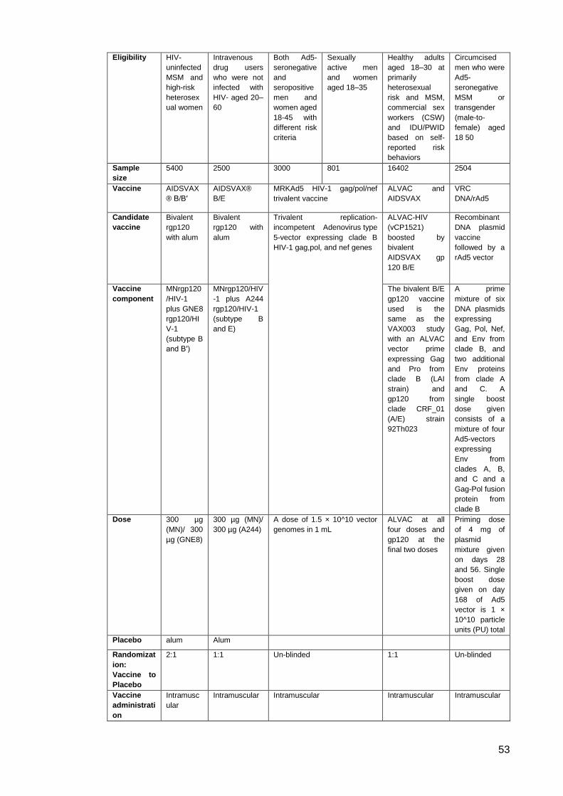
53
Eligibility HIV-
uninfected
MSM and
high-risk
heterosex
ual women
Intravenous
drug users
who were not
infected with
HIV- aged 20–
60
Both Ad5-
seronegative
and
seropositive
men and
women aged
18-45 with
different risk
criteria
Sexually
active men
and women
aged 18–35
Healthy adults
aged 18–30 at
primarily
heterosexual
risk and MSM,
commercial sex
workers (CSW)
and IDU/PWID
based on self-
reported risk
behaviors
Circumcised
men who were
Ad5-
seronegative
MSM or
transgender
(male-to-
female) aged
18 50
Sample
size
5400 2500 3000 801 16402 2504
Vaccine AIDSVAX
® B/B′
AIDSVAX®
B/E
MRKAd5 HIV-1 gag/pol/nef
trivalent vaccine
ALVAC and
AIDSVAX
VRC
DNA/rAd5
Candidate
vaccine
Bivalent
rgp120
with alum
Bivalent
rgp120 with
alum
Trivalent replication-
incompetent Adenovirus type
5-vector expressing clade B
HIV-1 gag,pol, and nef genes
ALVAC-HIV
(vCP1521)
boosted by
bivalent
AIDSVAX gp
120 B/E
Recombinant
DNA plasmid
vaccine
followed by a
rAd5 vector
Vaccine
component
MNrgp120
/HIV-1
plus GNE8
rgp120/HI
V-1
(subtype B
and B′)
MNrgp120/HIV
-1 plus A244
rgp120/HIV-1
(subtype B
and E)
The bivalent B/E
gp120 vaccine
used is the
same as the
VAX003 study
with an ALVAC
vector prime
expressing Gag
and Pro from
clade B (LAI
strain) and
gp120 from
clade CRF_01
(A/E) strain
92Th023
A prime
mixture of six
DNA plasmids
expressing
Gag, Pol, Nef,
and Env from
clade B, and
two additional
Env proteins
from clade A
and C. A
single boost
dose given
consists of a
mixture of four
Ad5-vectors
expressing
Env from
clades A, B,
and C and a
Gag-Pol fusion
protein from
clade B
Dose 300 µg
(MN)/ 300
µg (GNE8)
300 µg (MN)/
300 µg (A244)
A dose of 1.5 × 10^10 vector
genomes in 1 mL
ALVAC at all
four doses and
gp120 at the
final two doses
Priming dose
of 4 mg of
plasmid
mixture given
on days 28
and 56. Single
boost dose
given on day
168 of Ad5
vector is 1 ×
10^10 particle
units (PU) total
Placebo alum Alum
Randomizat
ion:
Vaccine to
Placebo
2:1 1:1 Un-blinded 1:1 Un-blinded
Vaccine
administrati
on
Intramusc
ular
Intramuscular Intramuscular Intramuscular Intramuscular
54
Description Vaccinatio
ns were
given at
months 0,
1, 6, 12,
18, 24 and
30, with
the final
study visit
at month
36
Vaccinations
were given at
0, 1, 6, 12, 18,
24, and 36
months
Vaccinations were given at 0,
1, 6 month schedule
Vaccinations
were given at 0,
4, 12, and 24
weeks
A single boost
dose given at
month six,
after a four-
month rest
from the
priming series
Efficacy No
significant
efficacy
was
observed
No significant
efficacy was
observed
No
significant
efficacy was
observed.
Statistical
trends
towards
enhanced
acquisition
of HIV
infection in
vaccinees
who were
Ad5-
seropositive
at baseline
or men who
were
uncircumcis
ed were not
observed
No significant
efficacy was
observed.
Unlike the
STEP study,
statistical
trends towards
enhanced
acquisition of
HIV infection
in vaccinees
who were
Ad5-
seropositive at
baseline or
men who were
uncircumcised
were not
observed
Estimated at
31.2% (95% CI
of 1.1–51.2%, p
= 0.04)
achieving
statistical
significance
Vaccine
efficacy was
estimated at
−25% (95%
CI, −121.2 to
29.3, p =
0.44). Overall,
there were 41
vaccinees and
31 placebo-
recipients who
had become
HIV infected,
thus the study
had to be
halted
Humoral immune responses to HIV infection 1.4.
1.4.1. Overview
Evidence from HIV-related animal models and other virus studies have suggested a
need for effective humoral-based protection, in addition to cellular responses, in order
to produce effective vaccination. The classical approach of mimicry, including
attenuated viruses, killed viruses and subunit viruses, failed because of the instability of
HIV glycoproteins and the low immunogenicity of the conserved regions of the
glycoproteins, especially compared to immune-dominant variable regions. The
discovery of neutralising antibodies in HIV infected patients during the 1980s raised
hopes that humoral immunity could be readily employed for passive immunisation.
Chronically HIV-1-infected serum were found to have moderate to broadly neutralising
antibodies as analysed by serum mapping, suggesting that the human immune system
is capable of generating broadly neutralising antibodies responses against HIV.
However, this proved far more challenging to achieve than imagined. The antibody
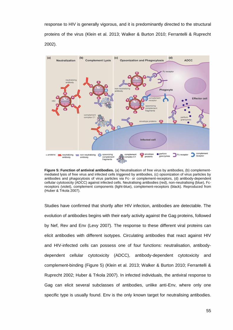
55
response to HIV is generally vigorous, and it is predominantly directed to the structural
proteins of the virus (Klein et al. 2013; Walker & Burton 2010; Ferrantelli & Ruprecht
2002).
Figure 5: Function of antiviral antibodies. (a) Neutralisation of free virus by antibodies, (b) complement-
mediated lysis of free virus and infected cells triggered by antibodies, (c) opsonization of virus particles by antibodies and phagocytosis of virus particles via Fc- or complement-receptors, (d) antibody-dependent cellular cytotoxicity (ADCC) against infected cells. Neutralising antibodies (red), non-neutralising (blue), Fc-receptors (violet), complement components (light-blue), complement-receptors (black). Reproduced from (Huber & Trkola 2007).
Studies have confirmed that shortly after HIV infection, antibodies are detectable. The
evolution of antibodies begins with their early activity against the Gag proteins, followed
by Nef, Rev and Env (Levy 2007). The response to these different viral proteins can
elicit antibodies with different isotypes. Circulating antibodies that react against HIV
and HIV-infected cells can possess one of four functions: neutralisation, antibody-
dependent cellular cytotoxicity (ADCC), antibody-dependent cytotoxicity and
complement-binding (Figure 5) (Klein et al. 2013; Walker & Burton 2010; Ferrantelli &
Ruprecht 2002; Huber & Trkola 2007). In infected individuals, the antiviral response to
Gag can elicit several subclasses of antibodies, unlike anti-Env, where only one
specific type is usually found. Env is the only known target for neutralising antibodies.
56
However, among the various isotypes, only IgG1 was found to play a dominant role at
all stages of infection (Walker & Burton 2010; Levy 2007; Kwong et al. 2013).
1.4.2. Neutralising antibodies in HIV infection
Studies have suggested that neutralisation is often considered to be the most efficient
mode of antibody-mediated defence against viral infections, and is therefore
considered the most significant humoral response elicited by vaccines. Although
antibodies can be detected during the first months of HIV infection, natural infection
predominantly induces non-neutralising or strain-specific antibodies. Recent studies
have argued that around 20% of HIV-1 chronically infected individuals elicit neutralising
antibodies (Klein et al. 2013; Yang & Wang 2014). Studies have shown that a small
fraction of these antibodies (2–4%) elicit a broadly neutralising response capable of
neutralising most tested HIV strains (Simek et al. 2009). It is argued that neutralising
antibodies could alter viral replication during the early asymptomatic stage of infection.
However, very low titres of these antibodies can be detected; this may favour the
selection of escape mutations. The mechanism of neutralisation lies either in blocking
the virus from its interaction with the specific receptors, preventing fusion or entry into
the target cell, or less commonly inhibiting transport to cytoplasm by preventing viral
budding or uncoating (Klein et al. 2013; Ferrantelli & Ruprecht 2002; Huber & Trkola
2007).
Recent studies have indicated that known HIV-1 neutralising antibodies bind to HIV
envelope glycoproteins—more specifically, functional viral envelope glycoprotein
spikes. Structural analyses of the HIV envelope proteins have indicated that many viral
neutralising epitopes are hidden from the immune system due to their spatial
arrangement. In addition, it has been found that the viral spikes can be protected from
neutralisation by evolving a shield of glycans, variable immunodominant loops and
conformational masking of important viral epitopes. However, studies have confirmed
that complete prevention can be achieved through passive delivery of neutralising
antibodies in animal models of HIV-1 transmission (Shibata et al. 1999; Mascola et al.

57
2000; Hessell et al. 2009; Mascola 2003). IgG has a higher general neutralising ability
than IgA (Kozlowski et al. 1994).
Passive transfer studies have suggested that relatively high concentrations of
neutralising antibodies are required for protection in the SHIV/macaque model
(Mascola et al. 2000; Burton & Parren 2000; Lu et al. 2003). However, there is some
indication that passive antibody protection requires antibody activity against infected
cells as well as free viruses (Hessell et al. 2007). In general, the level of antibodies
needed to provide benefit remains unclear as different factors can determine the
sensitivity of various HIV strains to antibody neutralisation. However, the ability to
neutralise HIV appears to require binding to most if not all of the surface envelope
glycoprotein molecules (spikes), which usually ranges from 7 to 14 per virion (Levy
2007; Klein et al. 2013).
Functional glycoprotein spikes on HIV particles are composed of trimeric heterodimer
of noncovalently attached surface subunit gp120, which is involved in CD4 receptor
recognition, and transmembrane subunit gp 41, which is responsible for the viral
fusion/entry and target cell membrane (Levy 2007; Kwong et al. 2013). Only antibodies
that interact with the intact trimer are considered to be neutralising. Following the
interaction of the virus spikes with the cell receptors and the co-receptors,
conformational changes occur to these spikes, exposing hidden neutralising epitopes
(Kwong et al. 2013).
Thus far, most studies have indicated that at least five epitopes of the viral envelope
could be involved in neutralisation (Figure 6 and Figure 7). Within gp120, there are four
regions involved in antibody neutralisation—the V3 loop, the V1/V2 loop, the CD4-
binding site (CD4BS) and the CD4-induced (CD4i) binding domain—which are involved
in the interaction with the co-receptors. On the other hand, only a few antibodies
directed to a specific region of the gp41—the membrane proximal external region
(MPER)- have been identified including the monoclonal antibodies 2F5, 4E10 and Z13.
Although MPER was found to have highly conserved structures, antibodies to this
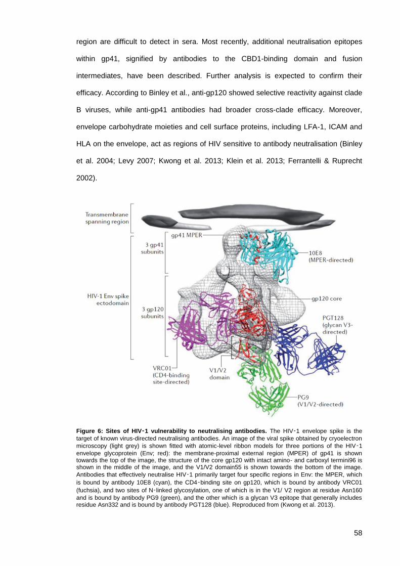
58
region are difficult to detect in sera. Most recently, additional neutralisation epitopes
within gp41, signified by antibodies to the CBD1-binding domain and fusion
intermediates, have been described. Further analysis is expected to confirm their
efficacy. According to Binley et al., anti-gp120 showed selective reactivity against clade
B viruses, while anti-gp41 antibodies had broader cross-clade efficacy. Moreover,
envelope carbohydrate moieties and cell surface proteins, including LFA-1, ICAM and
HLA on the envelope, act as regions of HIV sensitive to antibody neutralisation (Binley
et al. 2004; Levy 2007; Kwong et al. 2013; Klein et al. 2013; Ferrantelli & Ruprecht
2002).
Figure 6: Sites of HIV‑1 vulnerability to neutralising antibodies. The HIV‑1 envelope spike is the
target of known virus-directed neutralising antibodies. An image of the viral spike obtained by cryoelectron
microscopy (light grey) is shown fitted with atomic-level ribbon models for three portions of the HIV‑1
envelope glycoprotein (Env; red): the membrane-proximal external region (MPER) of gp41 is shown towards the top of the image, the structure of the core gp120 with intact amino- and carboxyl termini96 is shown in the middle of the image, and the V1/V2 domain55 is shown towards the bottom of the image.
Antibodies that effectively neutralise HIV‑1 primarily target four specific regions in Env: the MPER, which
is bound by antibody 10E8 (cyan), the CD4‑binding site on gp120, which is bound by antibody VRC01
(fuchsia), and two sites of N‑linked glycosylation, one of which is in the V1/ V2 region at residue Asn160
and is bound by antibody PG9 (green), and the other which is a glycan V3 epitope that generally includes residue Asn332 and is bound by antibody PGT128 (blue). Reproduced from (Kwong et al. 2013).
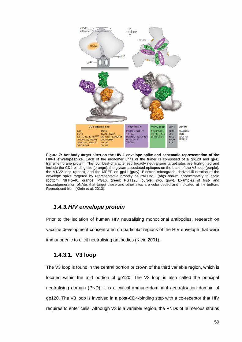
59
Figure 7: Antibody target sites on the HIV-1 envelope spike and schematic representation of the HIV-1 envelopespike. Each of the monomer units of the trimer is composed of a gp120 and gp41
transmembrane protein. The four best-characterised broadly neutralising target sites are highlighted and include the CD4-binding site (orange), the glycan-associated epitopes on the base of the V3 loop (purple), the V1/V2 loop (green), and the MPER on gp41 (gray). Electron micrograph–derived illustration of the envelope spike targeted by representative broadly neutralising F(ab)s shown approximately to scale (bottom: NIH45-46, orange; PG16, green; PGT128, purple; 2F5, gray). Examples of first- and secondgeneration bNAbs that target these and other sites are color-coded and indicated at the bottom. Reproduced from (Klein et al. 2013).
1.4.3. HIV envelope protein
Prior to the isolation of human HIV neutralising monoclonal antibodies, research on
vaccine development concentrated on particular regions of the HIV envelope that were
immunogenic to elicit neutralising antibodies (Klein 2001).
V3 loop 1.4.3.1.
The V3 loop is found in the central portion or crown of the third variable region, which is
located within the mid portion of gp120. The V3 loop is also called the principal
neutralising domain (PND); it is a critical immune-dominant neutralisation domain of
gp120. The V3 loop is involved in a post-CD4-binding step with a co-receptor that HIV
requires to enter cells. Although V3 is a variable region, the PNDs of numerous strains
60
differ only slightly in amino acid structure. It has been suggested that immunisation
within this region can elicit antibodies that neutralise a large number of HIV strains that
share this envelope domain (Levy 2007; Kwong et al. 2013).
Investigations found that homologous HIV-1 strains are not always neutralised by sera
with high titre antibodies to V3 peptides, suggesting that a V3 loop can have both
neutralising and non-neutralising epitopes. Neutralisation-escape studies have
indicated that the V3 loop rapidly mutates, leading to a reduction in its ability to be
neutralised. The V3 region can present both linear and conformational determinants
recognisable by antibodies. The neutralisation that resulted from using the V3 regions
might have resulted from inhibiting the V3 envelope cleavage, thus preventing viral
attachment. Changes in other envelope regions might also alter the neutralisation of
the V3 loop (Levy 2007; Kwong et al. 2013; Kwong et al. 2011),
CD4 binding site 1.4.3.2.
The CD4 binding domain, which is a large complex conformational region of the
envelope, is the second principal neutralising region in gp120. Polyclonal antibodies
that cross-neutralise a large number of primary- and laboratory-adapted strains,
including those with different V3 regions, are directed against this domain. Conversely,
sera from HIV infected individuals’ have neutralising monoclonal antibodies that
recognise discontinuous epitopes in the gp120 that is involved in CD4 receptor
interaction. The antibodies are thought to prevent the attachment of HIV strains to CD4.
However, broad antiviral activity by one type of monoclonal antibody might not be
achieved because numerous regions of the HIV envelope are involved and, thus,
numerous monoclonal antibodies might be needed to attain virus neutralisation.
Several studies indicate that conformational changes in gp120 result from point
mutations in gp41, as well as in the V3 loop, and affect the neutralisation ability in this
domain (Levy 2007; Kwong et al. 2013; Kwong et al. 2011; Zhou et al. 2007).
61
Other V regions 1.4.3.3.
The third and fourth principal neutralisation sites on gp120 are located in the V1 and V2
regions, involving both linear and conformational determinants. Antibodies that are
directed by both glycosylated and non-glycosylated epitopes have been found to
recognise such regions. Removal of the V2 loop from gp120 exposes epitopes on the
envelope that are conserved among various HIV-1 clades; this increases the virus’s
susceptibility to neutralisation. In contrast, neutralisation escape was observed in HIV
strains with a V1 deletion. It is still unclear whether V4 and V5 regions in HIV envelope
proteins contain domains that allow neutralisation (Levy 2007; Kwong et al. 2013;
Kwong et al. 2011).
Gp41 1.4.3.4.
Gp41 is a common fusion domain where only one specific region, which is named the
membrane proximal external region (MPER), is involved in neutralising antibody
binding. Immunisation studies of laboratory animals found that the amino terminal
portion of this region elicits antibodies against homologous and heterologous strains.
Certain broadly neutralising monoclonal antibodies target the MPER and interact with
the ectodomain of the HIV protein. Although the MPER displays highly conserved
structures, it poses the problem of auto-antigen mimicry. Antibodies to these structures
tend to be autoantibodies and react with normal cellular proteins (Levy 2007; Kwong et
al. 2013; Kwong et al. 2011).
CD4 Induced neutralising antibodies 1.4.3.5.
CD4-induced or CD4i antibodies are the antibodies that bind to HIV-1 gp120 after it
interacts with CD4. Studies have indicated that these epitopes compete with the co-
receptor binding sites and that CD4i antibodies may broadly neutralise different HIV-1
strains, as well as HIV-2. The need for further analysis of these epitopes remains,
although the cross-reacting neutralisation that was observed is encouraging and might
62
indicate a high degree of antigenic conservation that is linked to co-receptor binding
(Levy 2007; Kwong et al. 2013; Kwong et al. 2011).
1.4.4. HIV neutralising monoclonal antibodies
The definition and characterisation of monoclonal antibodies that mediate a broad
neutralisation range, as well as the structural basis for its interaction with the HIV
envelope, have provided opportunities for designing HIV vaccines that stimulate the
production of antibodies that are directed against specifically conserved regions of the
virus (Yang & Wang 2014; Kwong et al. 2011).
First-Generation broadly neutralising Abs against 1.4.4.1.
HIV
Initially only a few broadly neutralising monoclonal antibodies (mAbs) were isolated and
confirmed to neutralise primary isolates of HIV-1. These antibodies displayed
limitations in the overall potency or range of reactivity (Yang & Wang 2014; Kwong et
al. 2011).
The first broadly neutralising human Ab against HIV to be found was the antibody
IgG1b12 or IgGb12, which recognises a unique epitope on the gp120 surface of the
HIV Env and overlaps the CD4-binding site (Burton et al. 1991). IgG1b12 was isolated
from a clade B-infected patient by applying a novel method of phage screening of
libraries of Fab’s heavy and light chains. Because of the manner of their discovery, the
presence of IgG1b12 in nature has been questioned (Klein et al. 2013). Moreover, the
source material for the library was also unique as HIV-1- from an infected individual's
bone marrow RNA was used (Kessler et al. 1997). IgG1b12 can neutralise almost 50%
of clade B viral strains and approximately 30% of non-clade B strains (Yang & Wang
2014). In another study, the IgG1b12 antibody was found to neutralise approximately
35% of circulating isolates (Kwong et al. 2011).
63
Structural studies indicated that IgG1b12 differed from the naturally elicited Abs,
because it used the heavy chain only as a means of recognition. This dose did not
affect the site recognition ability of IgG1b12 (Zhou et al. 2007). However, antigenic
variation was found because of the extended recognition ability of IgG1b12, which
could recognise outside targets, especially around the CD4-binding loop, particularly in
the case of non-B clade isolates (Wu et al. 2009). Burton et al., indicated that the
human monoclonal antibody IgG1 b12 can prevent SHIV SF162 infection in rhesus
macaques. However, the dose of antibodies that is required to achieve complete
neutralisation was suggested to be extremely high, which is unlikely to be achieved
through current immunisation techniques (Burton et al. 1994; Davis et al. 2013; Kessler
et al. 1997). Few studies have been conducted so far that could effectively neutralise
HIV using IgG1b12. Hessell et al., found that the neutralising effect of IgG1b12 could
be decreased if the Fc region of the IgG was altered to elicit complement binding and
ADCC activity (Hessell et al. 2007).
Another broadly neutralising antibody that was isolated is IgG 2G12. IgG 2G12
recognises a unique cluster of high mannose carbohydrates of N-linked glycosylated
amino acid residues on the immunologically silent face of gp120 (Trkola et al. 1996;
Wyatt et al. 1998; Sanders et al. 2002). It was also found that 2G12 has broad
neutralising activity in vitro, where it binds to a glycan epitope, and that it can neutralise
numerous clade B-derived viral strains and, to a lesser extent, other HIV subtypes
(Burton et al. 1994; Trkola et al. 2005; Binley et al. 2004). IgG 2G12 has a unique
structure, where heavy and light chains lie vertically and adjacent to one another, and,
thus, are not associated in a traditional Y-like manner. It was suggested that this
unique configuration provides the antibody with flexibility and thus enables it to undergo
multivalent interactions with the gp120 oligomannose cluster (Calarese et al. 2003).
Phase I and I/II clinical trial studies have been conducted using this antibody
(Armbruster et al. 2002; Stiegler et al. 2002; Trkola et al. 2005).
64
The antibodies 2F5 (Muster et al. 1993; Purtscher et al. 1994; Trkola et al. 1996) and
4E10 (Stiegler et al. 2001; Zwick et al. 2001) were found to bind to both the gp41
MPER and its nearby lipids. Zhu et al. 2006, confirmed that the MPER constituted a
suitable target for HIV neutralising antibodies. The antibodies 2F5 and 4E10 are the
two most studied neutralising antibodies in this region (Zhu et al. 2006; Binley et al.
2004; Stiegler et al. 2001; Zwick et al. 2001; Muster et al. 1993). Although 4E10’s
potency is modest, it displayed a substantial range of neutralisation across several
clades. Both antibodies were successfully tested in Phase I studies (Armbruster et al.
2004; Armbruster et al. 2002).
An in vivo, passive transfer of IgG b12, IgG 2G12, IgG 2F5, or IgG 4E10 protected
against SHIV infection in macaques (Mascola et al. 2000; Hessell et al. 2009; Mascola
et al. 1999; Baba et al. 2000; Parren et al. 2002). A clinical study demonstrated that a
combination of IgG 2G12, IgG 2F5, and IgG 4E10 could delay a viral rebound in
numerous patients after cessation of a successful cART. An escape mutant analysis
suggested that the activity of IgG 2G12 was crucial for the in vivo effect of the
neutralising antibody cocktail in this trial (Trkola et al. 2005).
Second-Generation broadly neutralising Abs against 1.4.4.2.
HIV
Since 2009, following the application of new B cell technologies, a large panel of new
HIV-1 mAbs of substantial breadth were identified. It has been argued that numerous
additional neutralising mAbs might exist and will soon be detected. These new broadly
neutralising Abs (bNAbs) have greater neutralisation potency and breadth than the first
generation of bNAbs. They could provide an excellent resource for antibody gene
transfers to target HIV. However, to improve the knowledge of them, further structural
and escape mutant analytical studies are necessary (Klein et al. 2013; Yang & Wang
2014; Kwong et al. 2011).
65
Nussenzweig and colleagues combined the cell sorting of gp140-binding B cells and
single-cell antibody cloning to reveal that multiple antibodies targeting a range of Env
epitopes with broad neutralising activity could be detected in patients who were
infected with HIV (Mouquet et al. 2011; Walker et al. 2009). Burton and colleagues,
who screened over 30,000 activated memory B cells from clade A-infected African
donors by utilising clonal B cell culture, succeeded in identifying two broad neutralising
Abs, PG9, and PG16 that target the variable loops V2 and V3 of gp120 (Burton et al.
1991; Walker et al. 2009). The structure of this portion of the Env has yet to be
determined, and its functional importance remains unclear. Beside PG9 and PG16,
numerous antibodies have been identified that recognise this region, including human
monoclonal antibody (Klein et al. 2013) and a number of rhesus antibodies (Walker et
al. 2009). Studies have indicated that these antibodies vary dramatically in their
neutralisation breadth. Human monoclonal antibody 2909 was found to be extremely
strain-specific, whereas PG9/PG16 could neutralise 70%–80% of circulating isolates
(Honnen et al. 2007; Walker et al. 2009).
Antibody HJ16 is a natural human antibody that was isolated by direct assessment of
neutralisation coupled with single B cell antibody sequencing. Further structural
analysis is yet to be conducted to determine its characteristics and structure. However,
studies suggested that HJ16 targets the “loop D” region, which is outside the known
neutralisation sites. HJ16 neutralises 30%–40% of tested HIV strains (Klein et al. 2013;
Corti et al. 2010).
Antibodies VRC01, VRC02, and VRC03 targeted the site of vulnerability more
precisely. Studies suggested that they bind to a functionally conserved region of gp120
that interacts with the host cell receptor CD4 (Zhou et al. 2010; Wu et al. 2010; Zolla-
Pazner 2004; Pantophlet & Burton 2006; Mascola & Montefiori 2010). It is suggested
that these antibodies could neutralise over 90% of circulating isolates (Wu et al. 2010).
Structural studies reveal that the VRC01 heavy chain, in complex with gp120, mimics
the CD4 receptor (Zhou et al. 2010).
66
Recently, the 10E8 broad neutralising antibody has been identified. It targets the
MPER region and can neutralise more than 90% of tested HIV strains (Klein et al.
2013; Huang et al. 2012). The first monoclonal antibody against HIV applied in passive
immunisation in vivo was MAb F105, which binds to the CD4-binding site (Posner et al.
1991). The monoclonal antibodies 2G12, 2F5, 4E10, and F105 and the immunoglobulin
fusion protein PRO 542 have all been probed for efficacy in established HIV infection
(Trkola et al. 2005; Armbruster et al. 2004; Wolfe et al. 1996; Cavacini et al. 1998;
Jacobson et al. 2004; Jacobson et al. 2000). Recent studies isolated substantially more
broadly neutralising antibodies. The list of these new second-generation broadly
neutralising antibodies against HIV is lengthy; because most of them are yet to be
understood, further research is necessary (Klein et al. 2013).
1.4.5. Passive immunisation using neutralising antibodies
Passive immunisation, less commonly known as antibody mediated prevention (AMP),
is the transfer of pre-made antibodies to patients. Protection with transferred antibodies
can occur naturally, such as when maternal antibodies are transferred to the fetus
through the placenta, as well as artificially.
Several studies have shown that neutralising antibodies with broad specificity can
protect from the onset of HIV-1 infection if present at threshold concentration at the
time of the viral inoculum. Passive administration of broadly neutralising monoclonal
HIV-1 envelope-specific antibodies to macaques shortly before infection with SHIVs
carrying an HIV-1 envelope was found to prevent infection (Parren et al. 2001). Similar
results have been reported in macaque trials where vaginal administration of
neutralising antibodies before the viral challenge resulted in full protection from
systemic infection (Mascola et al. 2000; Pegu et al. 2017). Further evidence came from
mice with severe combined immunodeficiency (SCID) that were reconstituted with
human lymphoid tissue followed by infection with HIV-1. This experiment indicated that
67
the antibodies have to be inoculated and elicted before the virus encounter, as virus
escape mutation are quickly selected (Veazey et al. 2003). Levels of antibodies
declined over time, thus repeated doses are required to maintain a protection level.
Passive immunization clinical trials are under planning and additional research is
ongoing to find an approach to enhance the potency and half-life of existing bNAbs.
Two multinational clinical trials of an intravenously delivered investigational antibody for
preventing HIV infection have been launched, known as the AMP Studies, for antibody-
mediated prevention. The trials will evaluate the safety and efficacy of the human
monoclonal antibody (mAb) VRC-HIVMAB060-00-AB (VRC01) in preventing HIV-1
infection. The study will also examine safety of these antibody infusions and determine
the time the antibody is in the blood of study participants receiving different amounts or
doses of the antibody. The AMP Studies are being conducted jointly by the NIAID-
funded HIV Vaccine Trials Network (HVTN) and HIV Prevention Trials Network
(HPTN). Both are Phase b clinical trials. The first AMP Study, HVTN 704/HPTN 085
(ClinicalTrials.gov:NCT02716675), is taking place in Brazil, Peru and the United States.
Enrol 2,700 men and transgender people who have sex with men. The study started on
March 2016. The second of the two AMP Studies, HVTN 703/HPTN 081
(ClinicalTrials.gov:NCT02568215), started on April 2016, aiming to enrol 1,500 high
risk HIV-uninfected women. This study is being conducted in Botswana, Kenya,
Malawi, Mozambique, South Africa, Tanzania and Zimbabwe. The volunteers in both
studies are adults aged 18 to 50 years, at high risk for HIV infection, but HIV-negative
when they enter the study. In each trial, volunteers will be assigned at random to
receive an intravenous infusion of either VRC01 at a dose of 30 milligrams per kilogram
(mg/kg), VRC01 at a dose of 10 mg/kg, or a saline solution (a placebo). It is a double-
blind study where neither the volunteers nor the study investigators will know who
receives which type of infusion until the end of the study. Volunteers will receive a total
of 10 infusions, once every 8 weeks, and then will be followed for 20 more weeks. The
results of both trials are expected in 2022. If the current AMP Studies provides proof of
68
concept, it could lead to additional passive immunization trials, as well as research on
vaccines to help the body make these protective antibodies on its own
(https://ampstudy.org/ acceded on 07/05/18).
Antibody gene cloning 1.5.
1.5.1. Antibody structure
Immunoglobulins are Y-shaped molecules whose basic units are composed of 4
polypeptide chains arranged in 2 identical pairs of heavy (H) and light (L) chains
(Figure 8). Each antibody molecule has 2 Fab fragments that contain identical antigen
binding sites at their tips. IgG antibodies have one Fc (fragment crystallisable) fragment
joined to the Fabs by a flexible hinge (Fc has a propensity to self-associate and
crystallise into a lattice). The Fab region consists of the entire light chain combined with
part of the heavy chain. At the protein level, each of the polypeptide chains is folded
into discrete domains. The N-terminal variable regions (V-domains), VH and VL in the
heavy and light chains respectively, are variable in sequence. Within each V-domain
are 3 hypervariable regions (complementarity determining regions or CDRs) that
indicate the antibody specificity. The combination of the pairs of variable heavy and
light chains is referred to as the Fv of the antibody and is responsible for the binding of
antigens. In contrast, the C-terminal domains of both heavy and light chains are more
conservative and are called the constant regions (the C regions of the heavy chains
mediated effector functions).
There is only one constant region in the light chain's CL, while the IgG heavy chains
have 3 constant regions: CH1, CH2 and CH3. The constant regions of the
immunoglobulin are responsible for a variety of effector functions, including
complement fixation and clearing by cytotoxic mechanisms. Almost all antibodies share
the same structural characteristics but display remarkable variability in the regions that
bind the antigens (Davies & Chacko 1993; Travers et al. 1999).
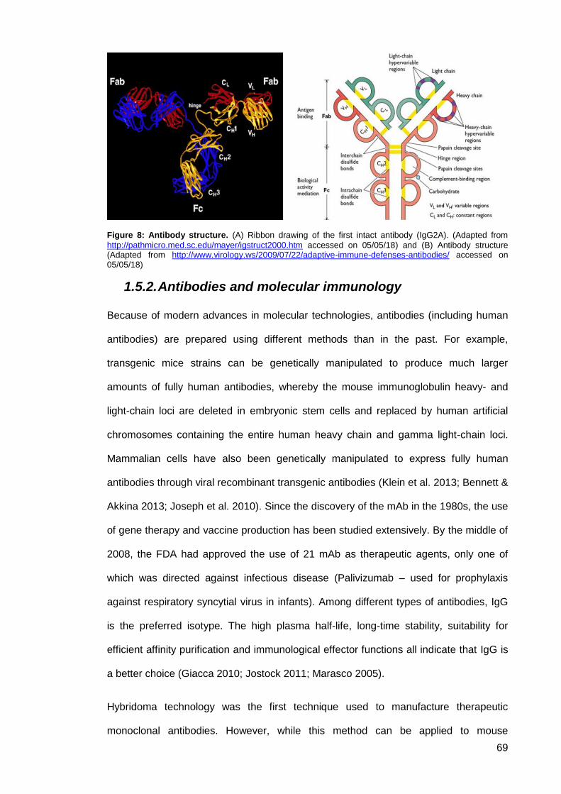
69
Figure 8: Antibody structure. (A) Ribbon drawing of the first intact antibody (IgG2A). (Adapted from
http://pathmicro.med.sc.edu/mayer/igstruct2000.htm accessed on 05/05/18) and (B) Antibody structure (Adapted from http://www.virology.ws/2009/07/22/adaptive-immune-defenses-antibodies/ accessed on 05/05/18)
1.5.2. Antibodies and molecular immunology
Because of modern advances in molecular technologies, antibodies (including human
antibodies) are prepared using different methods than in the past. For example,
transgenic mice strains can be genetically manipulated to produce much larger
amounts of fully human antibodies, whereby the mouse immunoglobulin heavy- and
light-chain loci are deleted in embryonic stem cells and replaced by human artificial
chromosomes containing the entire human heavy chain and gamma light-chain loci.
Mammalian cells have also been genetically manipulated to express fully human
antibodies through viral recombinant transgenic antibodies (Klein et al. 2013; Bennett &
Akkina 2013; Joseph et al. 2010). Since the discovery of the mAb in the 1980s, the use
of gene therapy and vaccine production has been studied extensively. By the middle of
2008, the FDA had approved the use of 21 mAb as therapeutic agents, only one of
which was directed against infectious disease (Palivizumab – used for prophylaxis
against respiratory syncytial virus in infants). Among different types of antibodies, IgG
is the preferred isotype. The high plasma half-life, long-time stability, suitability for
efficient affinity purification and immunological effector functions all indicate that IgG is
a better choice (Giacca 2010; Jostock 2011; Marasco 2005).
Hybridoma technology was the first technique used to manufacture therapeutic
monoclonal antibodies. However, while this method can be applied to mouse
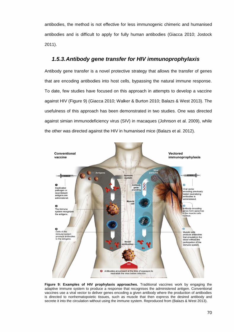
70
antibodies, the method is not effective for less immunogenic chimeric and humanised
antibodies and is difficult to apply for fully human antibodies (Giacca 2010; Jostock
2011).
1.5.3. Antibody gene transfer for HIV immunoprophylaxis
Antibody gene transfer is a novel protective strategy that allows the transfer of genes
that are encoding antibodies into host cells, bypassing the natural immune response.
To date, few studies have focused on this approach in attempts to develop a vaccine
against HIV (Figure 9) (Giacca 2010; Walker & Burton 2010; Balazs & West 2013). The
usefulness of this approach has been demonstrated in two studies. One was directed
against simian immunodeficiency virus (SIV) in macaques (Johnson et al. 2009), while
the other was directed against the HIV in humanised mice (Balazs et al. 2012).
Figure 9: Examples of HIV prophylaxis approaches. Traditional vaccines work by engaging the
adaptive immune system to produce a response that recognises the administered antigen. Conventional vaccines use a viral vector to deliver genes encoding a given antibody where the production of antibodies is directed to nonhematopoietic tissues, such as muscle that then express the desired antibody and secrete it into the circulation without using the immune system. Reproduced from (Balazs & West 2013).

71
Johnson et al. used adeno-associated virus (AAV) serotype 1 as a vector to deliver
genes encoding anti-SIV immunoadhesins, which are chimeric antibody-based
molecules that combine the functional domain of a binding protein with immunoglobulin
crystallizable fragment (Fc) domains (Figure 10), to protect macaques. This study
showed consistent results wherein macaques expressing the immunoadhesin 4L6 had
sustained circulating concentrations of approximately 20 mg/mL of 4L6 for 5 years with
no adverse health effects after 4 intramuscular injections given at the initiation of the
study. The macaques also demonstrated a stable erythropoietin expression for over 6
years (Johnson et al. 2009).
Figure 10: Schematic representation of immunoadhesin constructs. For molecules derived from
macaque Fabs (4L6, 8S, 5L7 and 3V), VH and VL domains were joined by a synthetic linker. Rhesus CD4 (domains 1 and 2 (D1 and D2)) was cloned as described in the Methods online. Antigen-binding domains were attached to the Fc fragment of a rhesus IgG2 molecule. Reproduced from (Johnson et al. 2009).
Burton presented early results of experiments where a remarkable protection in
macaques was demonstrated by giving different doses of PGT121. After intravaginal
challenge, all monkeys that received a dose of 5 mg/kg or 1 mg/kg remained
uninfected, whereas 3 of 5 monkeys that received a dose of 0.2 mg/kg were protected
from challenge. The antibody was not detected at the vaginal surface of any challenged
monkeys (Burton et al. 1994; Balazs & West 2013).

72
Baltimore also reported a study wherein humanised mice were given vector
immunoprophylaxis to express antibody b12 or VRC01, both challenged with the
REJO.c transmitted founder strain. In his experiment, substantial protection against
infection with REJO.c was noted in mice expressing VRC01 but not in those expressing
b12; this was consistent with results obtained in vitro for these antibody strain
combinations (Balazs & West 2013; Balazs et al. 2012).
Balazs et al. conducted a study based on vector-mediated gene transfer to engineer
secretion of broadly neutralising antibodies into the circulation and to describe the
practical implementation of vectored immunoprophylaxis (VIP) (Balazs et al. 2012). In
this study, humanised mice were found to induce long lasting expression of monoclonal
antibodies at high concentrations from a single intramuscular injection. Adeno-
associated viruses were used as optimised vectors to produce full-length antibody from
muscle tissue. Results from this study indicated that humanised mice receiving VIP
were fully protected from HIV infection. Intravenous challenge with very high doses of
replication-competent virus was successful. Based on this experiment, it was
suggested that effective prophylaxis against HIV could be achieved, as the feasibility of
direct translation of broadly neutralising antibodies (bNAbs) into functional
immunoprophylaxis using VIP in vivo was demonstrated.
The first phase 1 clinical trial involving the use of adeno-associated viruses (AAV) for
the expression of antibody from muscle tissues (Clinical Trials.gov: NCT01937455)
started on 2014 and estimated to be completed by 2017. It is a randomised (3:1)
blinded, dose-escalation study to evaluate the safety and tolerability of rAAV1-PG9DP
when administered intramuscularly at 4x10¹² vg, 4x10¹³ vg, 8x10¹³ vg and 1.2x10¹⁴ vg
in healthy male adults aged from 18 to 45 years. A total number of 24 volunteers were
screened up to 42 days before injection and followed for 12 months after the single
administration. The trial was held in the United Kingdom in the Surrey Clinical
Research Centre and was sponsored by IAVI, NIAID, and the Children's Hospital of
Philadelphia. Findings from the current PG9 study will provide important information
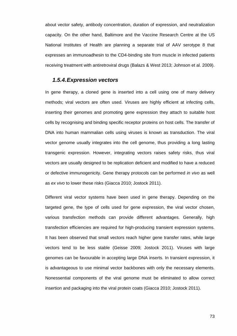
73
about vector safety, antibody concentration, duration of expression, and neutralization
capacity. On the other hand, Baltimore and the Vaccine Research Centre at the US
National Institutes of Health are planning a separate trial of AAV serotype 8 that
expresses an immunoadhesin to the CD4-binding site from muscle in infected patients
receiving treatment with antiretroviral drugs (Balazs & West 2013; Johnson et al. 2009).
1.5.4. Expression vectors
In gene therapy, a cloned gene is inserted into a cell using one of many delivery
methods; viral vectors are often used. Viruses are highly efficient at infecting cells,
inserting their genomes and promoting gene expression they attach to suitable host
cells by recognising and binding specific receptor proteins on host cells. The transfer of
DNA into human mammalian cells using viruses is known as transduction. The viral
vector genome usually integrates into the cell genome, thus providing a long lasting
transgenic expression. However, integrating vectors raises safety risks, thus viral
vectors are usually designed to be replication deficient and modified to have a reduced
or defective immunogenicity. Gene therapy protocols can be performed in vivo as well
as ex vivo to lower these risks (Giacca 2010; Jostock 2011).
Different viral vector systems have been used in gene therapy. Depending on the
targeted gene, the type of cells used for gene expression, the viral vector chosen,
various transfection methods can provide different advantages. Generally, high
transfection efficiencies are required for high-producing transient expression systems.
It has been observed that small vectors reach higher gene transfer rates, while large
vectors tend to be less stable (Geisse 2009; Jostock 2011). Viruses with large
genomes can be favourable in accepting large DNA inserts. In transient expression, it
is advantageous to use minimal vector backbones with only the necessary elements.
Nonessential components of the viral genome must be eliminated to allow correct
insertion and packaging into the viral protein coats (Giacca 2010; Jostock 2011).
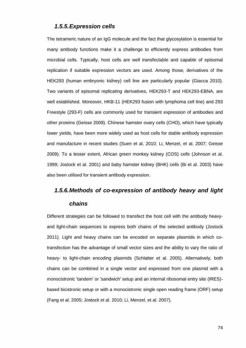
74
1.5.5. Expression cells
The tetrameric nature of an IgG molecule and the fact that glycosylation is essential for
many antibody functions make it a challenge to efficiently express antibodies from
microbial cells. Typically, host cells are well transfectable and capable of episomal
replication if suitable expression vectors are used. Among those, derivatives of the
HEK293 (human embryonic kidney) cell line are particularly popular (Giacca 2010).
Two variants of episomal replicating derivatives, HEK293-T and HEK293-EBNA, are
well established. Moreover, HKB-11 (HEK293 fusion with lymphoma cell line) and 293
Freestyle (293-F) cells are commonly used for transient expression of antibodies and
other proteins (Geisse 2009). Chinese hamster ovary cells (CHO), which have typically
lower yields, have been more widely used as host cells for stable antibody expression
and manufacture in recent studies (Suen et al. 2010; Li, Menzel, et al. 2007; Geisse
2009). To a lesser extent, African green monkey kidney (COS) cells (Johnson et al.
1999; Jostock et al. 2001) and baby hamster kidney (BHK) cells (Bi et al. 2003) have
also been utilised for transient antibody expression.
1.5.6. Methods of co-expression of antibody heavy and light
chains
Different strategies can be followed to transfect the host cell with the antibody heavy-
and light-chain sequences to express both chains of the selected antibody (Jostock
2011). Light and heavy chains can be encoded on separate plasmids in which co-
transfection has the advantage of small vector sizes and the ability to vary the ratio of
heavy- to light-chain encoding plasmids (Schlatter et al. 2005). Alternatively, both
chains can be combined in a single vector and expressed from one plasmid with a
monocistronic 'tandem' or 'sandwich' setup and an internal ribosomal entry site (IRES)-
based bicistronic setup or with a monocistronic single open reading frame (ORF) setup
(Fang et al. 2005; Jostock et al. 2010; Li, Menzel, et al. 2007).
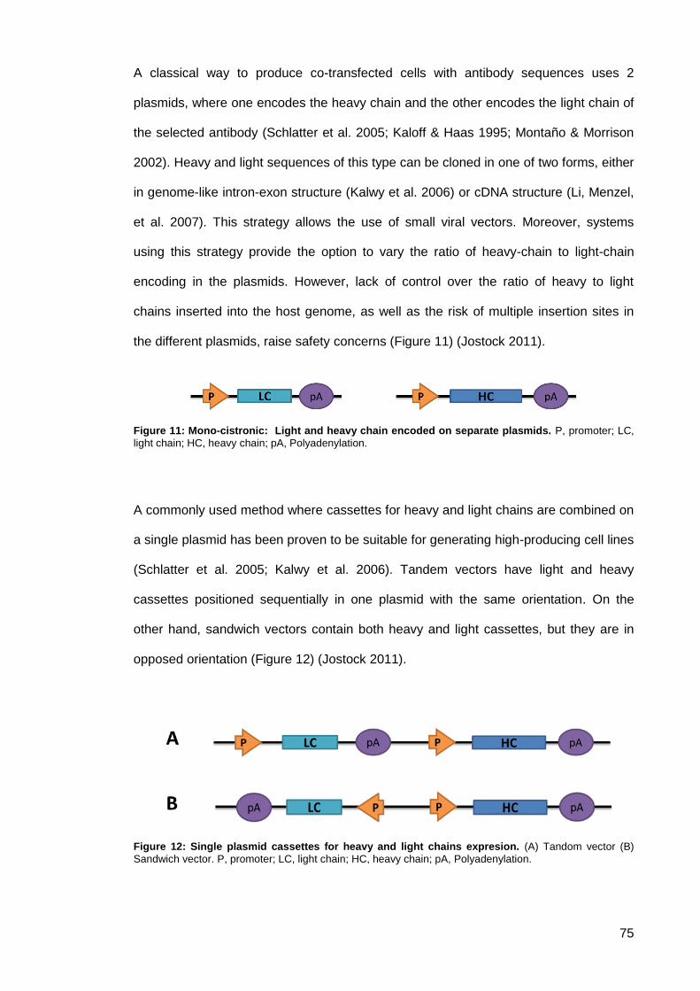
75
A classical way to produce co-transfected cells with antibody sequences uses 2
plasmids, where one encodes the heavy chain and the other encodes the light chain of
the selected antibody (Schlatter et al. 2005; Kaloff & Haas 1995; Montaño & Morrison
2002). Heavy and light sequences of this type can be cloned in one of two forms, either
in genome-like intron-exon structure (Kalwy et al. 2006) or cDNA structure (Li, Menzel,
et al. 2007). This strategy allows the use of small viral vectors. Moreover, systems
using this strategy provide the option to vary the ratio of heavy-chain to light-chain
encoding in the plasmids. However, lack of control over the ratio of heavy to light
chains inserted into the host genome, as well as the risk of multiple insertion sites in
the different plasmids, raise safety concerns (Figure 11) (Jostock 2011).
Figure 11: Mono-cistronic: Light and heavy chain encoded on separate plasmids. P, promoter; LC,
light chain; HC, heavy chain; pA, Polyadenylation.
A commonly used method where cassettes for heavy and light chains are combined on
a single plasmid has been proven to be suitable for generating high-producing cell lines
(Schlatter et al. 2005; Kalwy et al. 2006). Tandem vectors have light and heavy
cassettes positioned sequentially in one plasmid with the same orientation. On the
other hand, sandwich vectors contain both heavy and light cassettes, but they are in
opposed orientation (Figure 12) (Jostock 2011).
Figure 12: Single plasmid cassettes for heavy and light chains expresion. (A) Tandom vector (B)
Sandwich vector. P, promoter; LC, light chain; HC, heavy chain; pA, Polyadenylation.
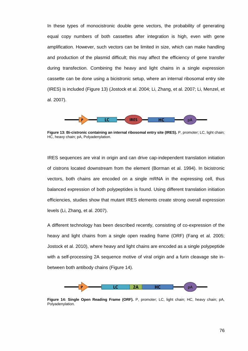
76
In these types of monocistronic double gene vectors, the probability of generating
equal copy numbers of both cassettes after integration is high, even with gene
amplification. However, such vectors can be limited in size, which can make handling
and production of the plasmid difficult; this may affect the efficiency of gene transfer
during transfection. Combining the heavy and light chains in a single expression
cassette can be done using a bicistronic setup, where an internal ribosomal entry site
(IRES) is included (Figure 13) (Jostock et al. 2004; Li, Zhang, et al. 2007; Li, Menzel, et
al. 2007).
Figure 13: Bi-cistronic containing an internal ribosomal entry site (IRES). P, promoter; LC, light chain;
HC, heavy chain; pA, Polyadenylation.
IRES sequences are viral in origin and can drive cap-independent translation initiation
of cistrons located downstream from the element (Borman et al. 1994). In bicistronic
vectors, both chains are encoded on a single mRNA in the expressing cell, thus
balanced expression of both polypeptides is found. Using different translation initiation
efficiencies, studies show that mutant IRES elements create strong overall expression
levels (Li, Zhang, et al. 2007).
A different technology has been described recently, consisting of co-expression of the
heavy and light chains from a single open reading frame (ORF) (Fang et al. 2005;
Jostock et al. 2010), where heavy and light chains are encoded as a single polypeptide
with a self-processing 2A sequence motive of viral origin and a furin cleavage site in-
between both antibody chains (Figure 14).
Figure 14: Single Open Reading Frame (ORF). P, promoter; LC, light chain; HC, heavy chain; pA,
Polyadenylation.
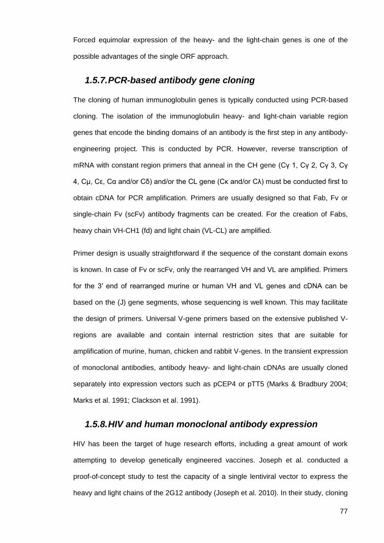
77
Forced equimolar expression of the heavy- and the light-chain genes is one of the
possible advantages of the single ORF approach.
1.5.7. PCR-based antibody gene cloning
The cloning of human immunoglobulin genes is typically conducted using PCR-based
cloning. The isolation of the immunoglobulin heavy- and light-chain variable region
genes that encode the binding domains of an antibody is the first step in any antibody-
engineering project. This is conducted by PCR. However, reverse transcription of
mRNA with constant region primers that anneal in the CH gene (Cγ 1, Cγ 2, Cγ 3, Cγ
4, Cμ, Cε, Cα and/or Cδ) and/or the CL gene (Cκ and/or Cλ) must be conducted first to
obtain cDNA for PCR amplification. Primers are usually designed so that Fab, Fv or
single-chain Fv (scFv) antibody fragments can be created. For the creation of Fabs,
heavy chain VH-CH1 (fd) and light chain (VL-CL) are amplified.
Primer design is usually straightforward if the sequence of the constant domain exons
is known. In case of Fv or scFv, only the rearranged VH and VL are amplified. Primers
for the 3′ end of rearranged murine or human VH and VL genes and cDNA can be
based on the (J) gene segments, whose sequencing is well known. This may facilitate
the design of primers. Universal V-gene primers based on the extensive published V-
regions are available and contain internal restriction sites that are suitable for
amplification of murine, human, chicken and rabbit V-genes. In the transient expression
of monoclonal antibodies, antibody heavy- and light-chain cDNAs are usually cloned
separately into expression vectors such as pCEP4 or pTT5 (Marks & Bradbury 2004;
Marks et al. 1991; Clackson et al. 1991).
1.5.8. HIV and human monoclonal antibody expression
HIV has been the target of huge research efforts, including a great amount of work
attempting to develop genetically engineered vaccines. Joseph et al. conducted a
proof-of-concept study to test the capacity of a single lentiviral vector to express the
heavy and light chains of the 2G12 antibody (Joseph et al. 2010). In their study, cloning
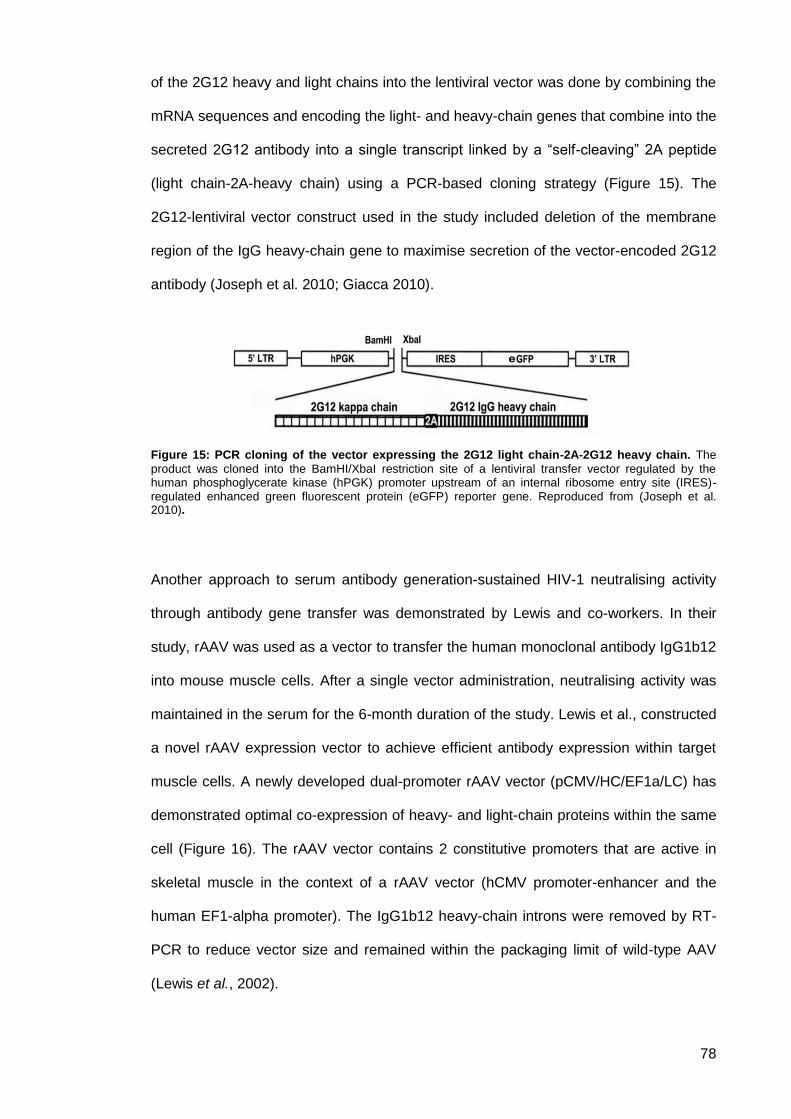
78
of the 2G12 heavy and light chains into the lentiviral vector was done by combining the
mRNA sequences and encoding the light- and heavy-chain genes that combine into the
secreted 2G12 antibody into a single transcript linked by a “self-cleaving” 2A peptide
(light chain-2A-heavy chain) using a PCR-based cloning strategy (Figure 15). The
2G12-lentiviral vector construct used in the study included deletion of the membrane
region of the IgG heavy-chain gene to maximise secretion of the vector-encoded 2G12
antibody (Joseph et al. 2010; Giacca 2010).
Figure 15: PCR cloning of the vector expressing the 2G12 light chain-2A-2G12 heavy chain. The
product was cloned into the BamHI/XbaI restriction site of a lentiviral transfer vector regulated by the human phosphoglycerate kinase (hPGK) promoter upstream of an internal ribosome entry site (IRES)-regulated enhanced green fluorescent protein (eGFP) reporter gene. Reproduced from (Joseph et al. 2010).
Another approach to serum antibody generation-sustained HIV-1 neutralising activity
through antibody gene transfer was demonstrated by Lewis and co-workers. In their
study, rAAV was used as a vector to transfer the human monoclonal antibody IgG1b12
into mouse muscle cells. After a single vector administration, neutralising activity was
maintained in the serum for the 6-month duration of the study. Lewis et al., constructed
a novel rAAV expression vector to achieve efficient antibody expression within target
muscle cells. A newly developed dual-promoter rAAV vector (pCMV/HC/EF1a/LC) has
demonstrated optimal co-expression of heavy- and light-chain proteins within the same
cell (Figure 16). The rAAV vector contains 2 constitutive promoters that are active in
skeletal muscle in the context of a rAAV vector (hCMV promoter-enhancer and the
human EF1-alpha promoter). The IgG1b12 heavy-chain introns were removed by RT-
PCR to reduce vector size and remained within the packaging limit of wild-type AAV
(Lewis et al., 2002).
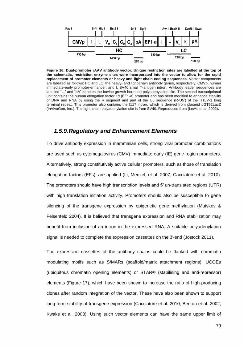
79
Figure 16: Dual-promoter rAAV antibody vector. Unique restriction sites are labelled at the top of the schematic, restriction enzyme sites were incorporated into the vector to allow for the rapid replacement of promoter elements or heavy and light chain coding sequences. Vector components
are labelled as follows: HC and LC, the heavy- and light-chain antibody genes, respectively; CMVp, human immediate-early promoter-enhancer; and I, SV40 small T-antigen intron. Antibody leader sequences are labelled “L,” and “pA” denotes the bovine growth hormone polyadenylation site. The second transcriptional unit contains the human elongation factor 1α (EF1-a) promoter and has been modified to enhance stability of DNA and RNA by using the R segment and part of the U5 sequence (R-U5’) of the HTLV-1 long terminal repeat. This promoter also contains the I117 intron, which is derived from plasmid pGT62LacZ (InVivoGen, Inc.). The light-chain polyadenylation site is from SV40. Reproduced from (Lewis et al. 2002).
1.5.9. Regulatory and Enhancement Elements
To drive antibody expression in mammalian cells, strong viral promoter combinations
are used such as cytomegalovirus (CMV) immediate early (IE) gene region promoters.
Alternatively, strong constitutively active cellular promoters, such as those of translation
elongation factors (EFs), are applied (Li, Menzel, et al. 2007; Cacciatore et al. 2010).
The promoters should have high transcription levels and 5′ un-translated regions (UTR)
with high translation initiation activity. Promoters should also be susceptible to gene
silencing of the transgene expression by epigenetic gene methylation (Mutskov &
Felsenfeld 2004). It is believed that transgene expression and RNA stabilization may
benefit from inclusion of an intron in the expressed RNA. A suitable polyadenylation
signal is needed to complete the expression cassettes on the 3′-end (Jostock 2011).
The expression cassettes of the antibody chains could be flanked with chromatin
modulating motifs such as S/MARs (scaffold/matrix attachment regions), UCOEs
(ubiquitous chromatin opening elements) or STAR® (stabilising and anti-repressor)
elements (Figure 17), which have been shown to increase the ratio of high-producing
clones after random integration of the vector. These have also been shown to support
long-term stability of transgene expression (Cacciatore et al. 2010; Benton et al. 2002;
Kwaks et al. 2003). Using such vector elements can have the same upper limit of
80
productivities to vectors without chromatin opening motifs. However, chromatin opening
motifs vectors need less clone screening to identify high producers. Such vectors can
therefore be useful in cases where clone-screening efforts are limited. Moreover, high
production stability could be obtained due to anti-repressor activities of such elements
(Kwaks et al. 2003).
Figure 17: Examples of special vector setups for stable cell line generation. (A) Chromatin
modulating elements: Flanking the antibody expression cassettes with chromatin modulating elements such as S/MAR, UCOE, STAR®. (B) STAR® select: Selection marker and antibody coding regions are positioned in single expression cassettes with the selection marker upfront. (C) Tri-cistronic vectors: IRES elements are used to combine antibody chains and selection marker a reporter gene like GFP in a single expression cassette and to drive translation initiation of the downstream cistrons. Reproduced from (Jostock 2011).
Lentiviral vectors and gene therapy 1.6.
1.6.1. Overview
Gene therapy was initially developed as a tool to deliver corrective genetic DNA into
cells in the hope of compensating for specific genetic mutations that cause cellular
dysfunction and the consequent development of systemic disease (Verma & Somia
1997). However, the principle of gene therapy can also be used to introduce protective
factors into the cells of healthy individuals and generate intracellular immunisation
(Baltimore 1988). This immunotherapy option aims to use genetically engineered
viruses (recombinant viruses) as vectors for delivery to the relevant genetic targets and
is concerned with eliciting cellular and humoral immune responses needed for
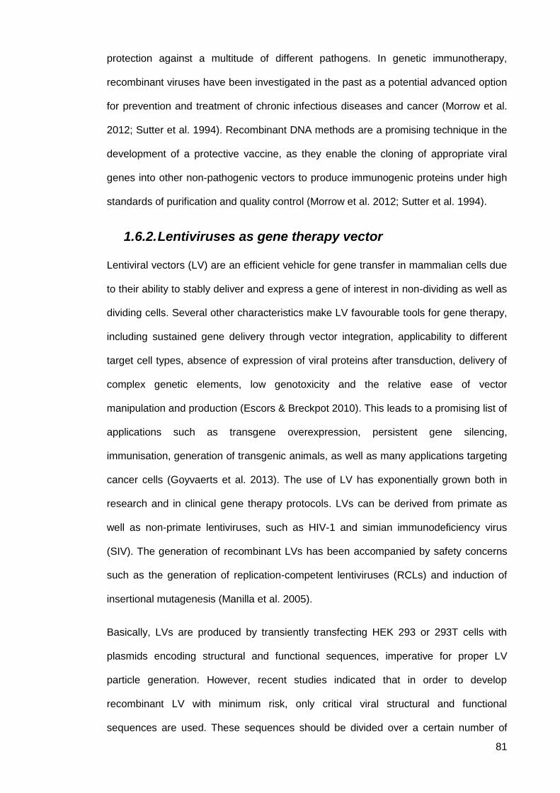
81
protection against a multitude of different pathogens. In genetic immunotherapy,
recombinant viruses have been investigated in the past as a potential advanced option
for prevention and treatment of chronic infectious diseases and cancer (Morrow et al.
2012; Sutter et al. 1994). Recombinant DNA methods are a promising technique in the
development of a protective vaccine, as they enable the cloning of appropriate viral
genes into other non-pathogenic vectors to produce immunogenic proteins under high
standards of purification and quality control (Morrow et al. 2012; Sutter et al. 1994).
1.6.2. Lentiviruses as gene therapy vector
Lentiviral vectors (LV) are an efficient vehicle for gene transfer in mammalian cells due
to their ability to stably deliver and express a gene of interest in non-dividing as well as
dividing cells. Several other characteristics make LV favourable tools for gene therapy,
including sustained gene delivery through vector integration, applicability to different
target cell types, absence of expression of viral proteins after transduction, delivery of
complex genetic elements, low genotoxicity and the relative ease of vector
manipulation and production (Escors & Breckpot 2010). This leads to a promising list of
applications such as transgene overexpression, persistent gene silencing,
immunisation, generation of transgenic animals, as well as many applications targeting
cancer cells (Goyvaerts et al. 2013). The use of LV has exponentially grown both in
research and in clinical gene therapy protocols. LVs can be derived from primate as
well as non-primate lentiviruses, such as HIV-1 and simian immunodeficiency virus
(SIV). The generation of recombinant LVs has been accompanied by safety concerns
such as the generation of replication-competent lentiviruses (RCLs) and induction of
insertional mutagenesis (Manilla et al. 2005).
Basically, LVs are produced by transiently transfecting HEK 293 or 293T cells with
plasmids encoding structural and functional sequences, imperative for proper LV
particle generation. However, recent studies indicated that in order to develop
recombinant LV with minimum risk, only critical viral structural and functional
sequences are used. These sequences should be divided over a certain number of
82
individual plasmids in either cis or trans acting manner, with as minimal overlap as
possible between viral sequences.
Figure 18: Schematic representation of the four generations of lentiviral packaging constructs. A)
First generation packaging vector. B) Second generation packaging vector. C) Third generation packaging vector. D) Fourth generation packaging vector. Reproduced from (Helio et al. 2013).
A system of three expression cassettes was developed in 1996 by Naldini et al where
LV production is accomplished using three different plasmids. First, to generate
functional particles a packaging plasmid encoding all viral structural and enzymatic
sequences in trans is developed. Then, a transfer plasmid providing the expression
cassette of gene of interest in cis is cloned into the non-coding regions of the original
lentiviral genome including a packaging signal and the two long terminal repeats
(LTRs) of which the promoter activity has been deleted from the 3’ LTR. Finally, an
envelope plasmid encoding an envelope glycoprotein (gp) consisting of a
transmembrane domain (TM) and a receptor binding domain (SU) is developed in
which the LVs’ tropism can be determined (Naldini et al. 1996). This system of three
expression cassettes is considered to be the first generation of LVs. Four generations
of LVs have now been described (Figure 18). In the first generation LV packaging
plasmids the entire gag and pol genes were encoded together with all accessory
83
regulatory and virulence genes. This system provided good antibody production, but its
level of safety was not very high as the chances of production of RCL were frequent.
To overcome safety concerns and to decrease the cytotoxicity of LVs, all accessory
genes that are not essential for viral replication were removed without any negative
effects on vector yield or infectivity, giving rise to the second generation vectors. In this
generation, multiplication was attenuated by removal of four virulence genes, but not
the regulatory ones (i.e. tat and rev) (Zufferey et al. 1997). However, this did not affect
the number of homologous events to generate RCL. A third generation was developed
in order to produce a more effective and safer version of the lentiviral gene delivery
vehicle. In this approach the rev gene is expressed from a separate plasmid and the tat
gene is removed by insertion of a strong constitutive promoter replacing the U3 region
in the 5’ LTR of the transfer plasmid (Dull et al. 1998). The U3 region of the 3’ LTR of
the transfer plasmid cancels the production of full-length vector RNA in transduced
cells. Vector of the third generation presents both high titres as well as a high level of
biosafety, thus they are now the most commonly used LVs. A further major
improvement was achieved with the development of self-inactivating LVs (SIN). That
minimised the production of RCLs and reduced the chance of viral LTR enhancer’s
interference with the expression cassette, which lowers detectable expression of
adjacent cellular coding regions. In contrast, in the fourth generation the homology
between constructs is severely reduced, but the titres of antibody produced have also
been affected (Romano et al. 2003).
1.6.3. Pseudotyping of lentiviruses
In addition to packaging and transfer plasmid optimisation, the envelope plasmid can
be modified by replacing the natural lentivirus envelope gp with an alternative gp gene,
most often the gp of vesicular stomatitis virus (VSV.G). This concept is called
pseudotyping. Pseudotyping of lentiviruses is mainly used to expand the host range of
retroviral vectors. The G glycoprotein of the VSV-G is almost always used to replace
the Env proteins of the lentiviruses. VSV-G pseudotyped lentiviral vectors have
84
expanded cell tropism and spurred applications for gene therapy that can infect most
types of cells. Studies have indicated that pseudotyped lentiviruses are particularly
stable (Burns et al. 1993).
Joseph et al. generated a pseudotyped HIV-based third-generation lentivirus carrying
the light-chain-2A-heavy-chain construct of the 2G12 antibody. In their study, co-
transfection of 293T cells with 4 plasmids was made under calcium phosphate-
mediation. A cytomegalovirus promoter was used to drive the packaging construct,
expressing the gag and pol genes. A Rous sarcoma virus (RSV) promoter was used to
drive construct expression of the rev gene. A cytomegalovirus promoter was also used
to drive expression of the VSV-G envelope gene. Finally, a self-inactivating transfer
construct was used, driven by the hPGK promoter containing HIV cis-acting sequences
and an expression cassette for the 2G12 light-chain-2A-heavy-chain coding sequence
inserted upstream of an IRES-regulated eGFP (2G12-lentivector) or an empty
expression plasmid (Joseph et al. 2010).
85
Chapter 2 2.
HIV neutralising antibody delivered by gene therapy
with a stable retroviral vector encoded in vaccinia
expression systems
Abstract 2.1.
Production of an effective vaccine against human immunodeficiency virus (HIV) is
elusive. This project used a novel replication-restricted, poxvirus split-vector system to
insert HIV-neutralising monoclonal antibody sequences into a simian retroviral gene
therapy agent pseudo-typed with vesicular stomatitis virus glycoprotein, all encoded
within a poxvirus. Recombination between split-vector recombinants does not produce
viable, fully replication-competent retroviruses. To circumvent this, a recombinant
modified vaccinia Ankara (MVA) was constructed for functional retrovirus production.
Production of these gene therapy retroviral particles delivered in vivo by a safe, stable
poxvirus promises to produce long-lasting, high titres of neutralising anti-HIV
monoclonal antibody. A hybrid dual-vector system using the large packaging capacity
of MVA was used. The expression cassette of the gene therapy agent was split into
two independent MVA vectors. The ultimate aim is to insert IgG1 b12 antibody genes
into the human genome. The first rMVA (rMVA/IgG1b12/T7) carried the HIV-
neutralising monoclonal antibody (mAb) sequence as well as a T7 polymerase for
delivery into the cytoplasm of mammalian cells. A second recombinant MVA
(rMVA/gag/pol/VSV-G) encoded a simian retroviral particle pseudo-typed with vesicular
stomatitis virus glycoprotein to facilitate the delivery of the mAb genes into the human
genome inside the nucleus. The second rMVA expressed heterologous polypeptides
capable of assembling into defective non -self-propagating viral particles mimicking the
packaging structure of SIV. The new non-infectious artificial lentivirus that results from

86
the hybrid dual-vector system infects neighbouring cells, penetrating the nucleus and
inserting its genetic material into the mammalian genome to function as a vehicle for
gene therapy. The insert comprised the expression cassette carrying a human CMV
promoter, allowing constant expression of the neutralising monoclonal antibody over a
prolonged period. This proof of principle will potentially allow subsequent development
of long-acting vaccines for prevention of HIV infection and long-lasting immunotherapy
of existing HIV infection.
87
Objectives and aims 2.2.
Generate a genomic rMVA/IgG1b12/T7 by homologous recombination between
wt MVA and a plasmid-based transfer vector encoding monoclonal antibody
IgG1b12, CMV promoter, T7 polymerase and a selective marker.
Generate a packaging rMVA by homologous recombination between
rMVA/gag/pol and a plasmid-based transfer vector encoding vesicular stomatitis
virus glycoprotein (VSV-G) and Rev.
Construct and validate recombinant MVA for functional gene therapy and
pseudotyped retrovirus production.
Produce gene therapy retroviral particles that will be delivered in vitro as a proof
of principle to demonstrate production of a long-acting immunotherapy vectors
for prevention of HIV infection and treatment of existing HIV infection.
88
Introduction 2.3.
2.3.1. Recombinant vaccinia viruses and vaccination
Poxviruses in vaccination 2.3.1.1.
Poxviruses, in particular Vaccinia viruses, are excellent candidates for the development
of gene vectors. Vaccinia viruses are members of the Orthopoxvirus genus of the
poxvirus family (Figure 19). They can be used in recombinant virus production to
express and deliver foreign genes for use in novel vaccines. Several characteristics
ensure the success of poxviruses in recombinant vaccination. The viruses have large
double-stranded DNA genomes with packaging capacity to accommodate a large
amount of foreign DNA (Sutter & Staib 2003; Amara et al. 2001).
Figure 19: Electron micrographs of negatively stained, naturally released virions. (A) Vaccinia virus.
(B) Parapoxvirus. (bar = 100 nm). Reproduced from (Baxby 1996).
Poxviruses replicate in the cytoplasm of mammalian cells and do not integrate into the
host genome while their gene expression is regulated by a strong poxvirus promoter.
Poxviruses are also known to have high immunogenicity, and are well suited to large
scale vaccine production (Blanchard et al. 1998; Mercer et al. 2007; Collier, L., Kellam,
Paul, Oxford, J. S. 2011; Collier et al. 2011). There are safety concerns around the use
of non-host range restricted vaccinia virus as human vaccines. These concerned are
exemplified by the observation of complications when chorioallantoic vaccine Ankara
(CVA) was administered as vaccine (Herrlich & Mayr 1957). It has been proven that
poxviruses, in particular vaccinia viruses (VV), can replicate in human cells and cause
89
significant harm; thus, they have to be handled under biosafety level 2 laboratory
conditions (Sutter & Staib 2003; Blanchard et al. 1998).
Smallpox vaccination 2.3.1.2.
Smallpox has been successfully eradicated by a global effort in the 1960s and 1970s
using a panel of live vaccinia virus vaccines. The last naturally occurring case was
reported in 1977 in Somalia and in May 1980 the World Health Organisation (WHO)
declared that smallpox had been eradicated (Breman & Arita 1980).
First generation smallpox vaccines where produced and successfully used during most
of the intensified eradication program. Various vaccinia virus strains were
demonstrated globally (e.g. Lister, NYCBH, Tiantan, EM63), manufactured on skin or
lymph of animals (e.g calf, sheep). However, towards the end of the eradication phase
a second generation vaccine was developed using culturing techniques and the same
smallpox vaccine strains employed for the manufacture of first generation vaccines or
clonal virus variants plaque purified from traditional vaccine stocks. Using cell culture
approach not only reduced the risk of contamination with adventitious agents but also
improved the manufacturing processes primarily in providing consistency between lots.
At the end of the eradication phase, replication deficient third generation smallpox
vaccines were developed which represent more attenuated vaccine strains, MVA
based vaccines, specifically developed as safer vaccines by further passaging in cell
culture or animals. Second and third generation vaccines are produced using modern
cell culture techniques and current standards of Good Manufacturing Practices (GMP).
Most adverse reactions to vaccinia virus/MVA administration involve the skin and
central nervous system (CNS). Expected vaccine-related common local and systemic
reactions with mild to moderate severity were detected. Local reactions included pain,
intense erythema and inflammation at the vaccination site. Systemic reactions included
fever, malaise, myalgia, headache, chills, nausea, fatique, and lymphadenopathy.
These reactions were usually self-limited and resolved within 2-3 weeks. Rare but
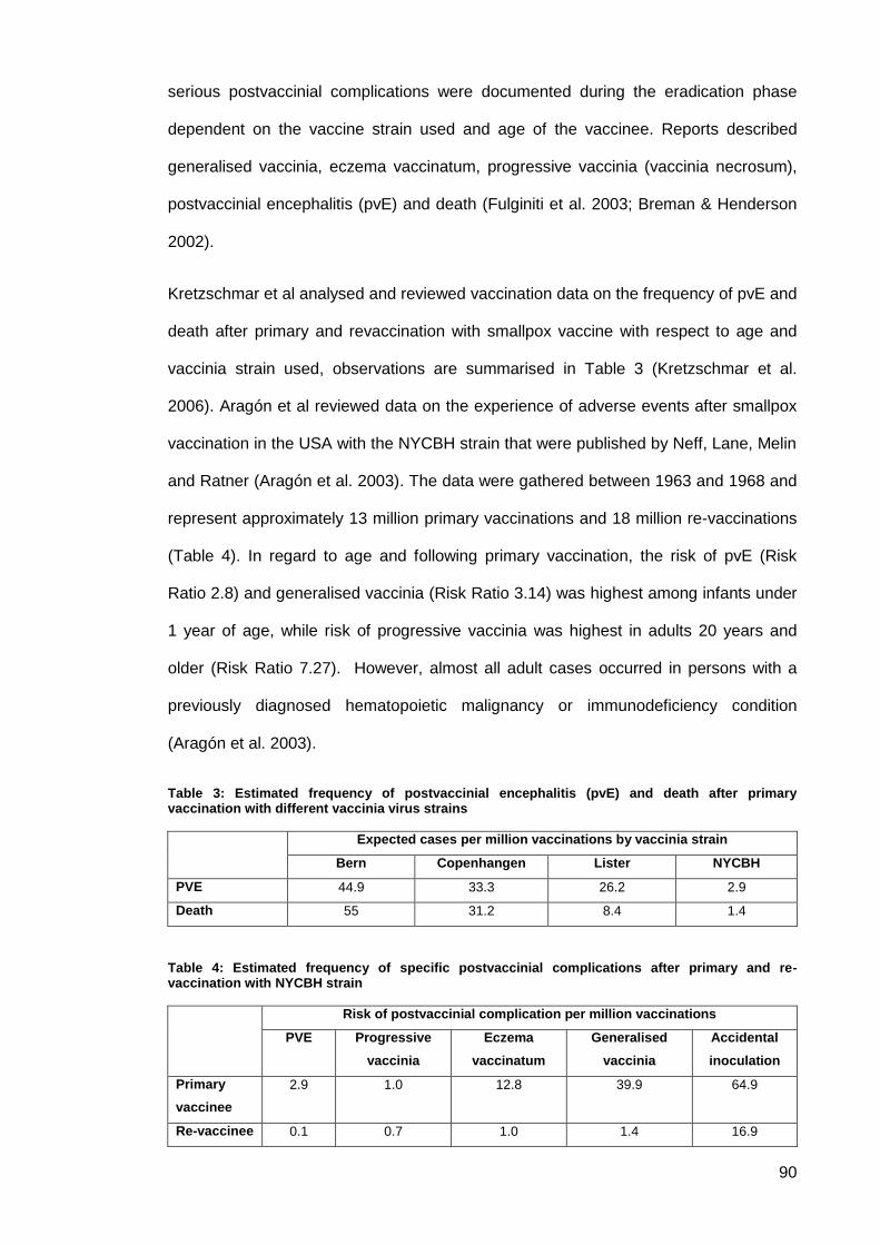
90
serious postvaccinial complications were documented during the eradication phase
dependent on the vaccine strain used and age of the vaccinee. Reports described
generalised vaccinia, eczema vaccinatum, progressive vaccinia (vaccinia necrosum),
postvaccinial encephalitis (pvE) and death (Fulginiti et al. 2003; Breman & Henderson
2002).
Kretzschmar et al analysed and reviewed vaccination data on the frequency of pvE and
death after primary and revaccination with smallpox vaccine with respect to age and
vaccinia strain used, observations are summarised in Table 3 (Kretzschmar et al.
2006). Aragón et al reviewed data on the experience of adverse events after smallpox
vaccination in the USA with the NYCBH strain that were published by Neff, Lane, Melin
and Ratner (Aragón et al. 2003). The data were gathered between 1963 and 1968 and
represent approximately 13 million primary vaccinations and 18 million re-vaccinations
(Table 4). In regard to age and following primary vaccination, the risk of pvE (Risk
Ratio 2.8) and generalised vaccinia (Risk Ratio 3.14) was highest among infants under
1 year of age, while risk of progressive vaccinia was highest in adults 20 years and
older (Risk Ratio 7.27). However, almost all adult cases occurred in persons with a
previously diagnosed hematopoietic malignancy or immunodeficiency condition
(Aragón et al. 2003).
Table 3: Estimated frequency of postvaccinial encephalitis (pvE) and death after primary vaccination with different vaccinia virus strains
Expected cases per million vaccinations by vaccinia strain
Bern Copenhangen Lister NYCBH
PVE 44.9 33.3 26.2 2.9
Death 55 31.2 8.4 1.4
Table 4: Estimated frequency of specific postvaccinial complications after primary and re-vaccination with NYCBH strain
Risk of postvaccinial complication per million vaccinations
PVE Progressive
vaccinia
Eczema
vaccinatum
Generalised
vaccinia
Accidental
inoculation
Primary
vaccinee
2.9 1.0 12.8 39.9 64.9
Re-vaccinee 0.1 0.7 1.0 1.4 16.9
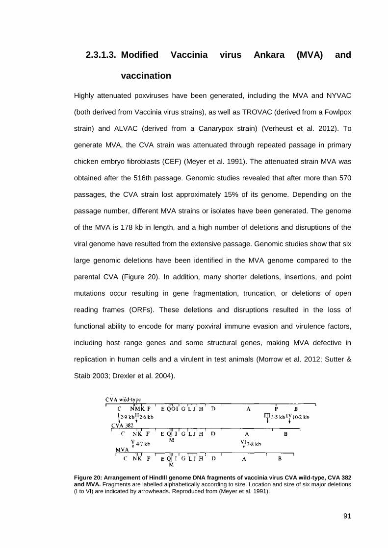
91
Modified Vaccinia virus Ankara (MVA) and 2.3.1.3.
vaccination
Highly attenuated poxviruses have been generated, including the MVA and NYVAC
(both derived from Vaccinia virus strains), as well as TROVAC (derived from a Fowlpox
strain) and ALVAC (derived from a Canarypox strain) (Verheust et al. 2012). To
generate MVA, the CVA strain was attenuated through repeated passage in primary
chicken embryo fibroblasts (CEF) (Meyer et al. 1991). The attenuated strain MVA was
obtained after the 516th passage. Genomic studies revealed that after more than 570
passages, the CVA strain lost approximately 15% of its genome. Depending on the
passage number, different MVA strains or isolates have been generated. The genome
of the MVA is 178 kb in length, and a high number of deletions and disruptions of the
viral genome have resulted from the extensive passage. Genomic studies show that six
large genomic deletions have been identified in the MVA genome compared to the
parental CVA (Figure 20). In addition, many shorter deletions, insertions, and point
mutations occur resulting in gene fragmentation, truncation, or deletions of open
reading frames (ORFs). These deletions and disruptions resulted in the loss of
functional ability to encode for many poxviral immune evasion and virulence factors,
including host range genes and some structural genes, making MVA defective in
replication in human cells and a virulent in test animals (Morrow et al. 2012; Sutter &
Staib 2003; Drexler et al. 2004).
Figure 20: Arrangement of HindIll genome DNA fragments of vaccinia virus CVA wild-type, CVA 382 and MVA. Fragments are labelled alphabetically according to size. Location and size of six major deletions
(I to VI) are indicated by arrowheads. Reproduced from (Meyer et al. 1991).
92
MVA provides the advantages of genetic stability and high-level gene expression as
well as deficient replication in human cells. Thus, depending on the nature of the
inserted target gene, constructs can be handled under biosafety level 1 laboratory
conditions. MVA has an excellent clinical safety record and induces significant humoral
and cellular immune response. It has limited replication in some human cells and is
clearly safe in immunosuppressed patients (Sutter & Staib 2003; Amara et al. 2001;
Blanchard et al. 1998). Studies suggest that the high immunogenicity of MVA vectored
vaccines may result from apoptosis of human DCs upon MVA infection (Verheust et al.
2012).
MVA is widely considered as the vaccinia virus strain of choice in preclinical and
clinical research, in particular in the field of recombinant vectors for both vaccinations
against various pathogens or as a delivery vector for gene therapy. A study showed
that MVA-572 and MVA-I721 strains demonstrate ability to replicate in some human
cell lines (human keratinocyte cell line HaCat, human embryo kidney cell line 293,
human bone osteosarcoma cell line 143B, and human cervix adenocarcinoma cell line
HeLa) and in immunodeficient mice (Suter et al. 2009). However, many MVA-based
vector studies have not indicated any major side effects in preclinical animals or in
human clinical trials undertaken so far. However, the use of MVA and recombinant
MVA vectors must include an assessment of potential risks for human health and the
environment (Verheust et al. 2012).
MVA has the adaptability to express a high level of genes as well as to produce
heterologous proteins. Further, although MVA is characterised by severe growth
deficiency in human cells, it shows good efficacy of recombinant gene expression
(Morrow et al. 2012; Verheust et al. 2012). In 2003, Moorthy et al. reported the first
safety data profile of recombinant MVA immunisation in humans, testing the efficacy of
MVA vaccine against Plasmodium falciparum malaria. Since then, other studies testing
MVA vaccines for prophylaxis or immunotherapy against AIDS and other chronic
infections and cancers have been completed and published (Verheust et al. 2012;
93
Moorthy et al. 2003). As the development of an effective vaccine against HIV is an
urgent need, investigations into the development of an adequate recombinant MVA
vector vaccine against HIV increased. Thus far, many studies have been dedicated to
developing recombinant MVA, producing immunodeficiency virus antigens to elicit
humoral and/or cellular immune responses. Many MVA-based vaccine studies have
successfully reported that both antibody and high-level cellular responses can be
elicited when different SIV or SHIV antigens expressed in model hosts are expressed
by rMVA vaccine candidates. However, further investigation is still needed into the
efficacy of elicitation of the potent immunity and induction of broadly virus-neutralising
antibody responses required to achieve a successful HIV vaccine (Verheust et al.
2012; Schnierle et al. 2007).
Vaccinia Retroviral Hybrid Vector 2.3.1.4.
Retroviral vectors have become important tools in gene therapy as a result of a number
of highly desirable properties, mainly their stable genomic integration. However,
retroviral vectors cannot be grown to high titers and they tend to be unstable and are
sensitive to inactivation by complement when transfused into patients. The search for
more robust retroviral delivery systems has led to the development of hybrid viral
vectors with advantageous properties of another viral vector system.
A hybrid vector can be generated by integrating the retroviral unit into the genome of a
VV. Hence, instead of being delivered by transfection of a proviral plasmid DNA, the
retroviral vector is backpacked onto a carrier virus (MVA) which actively infects cells
with high efficiency and produces the genomic RNA in the cytoplasm. Both replicating
and replication-defective VVs are appropriate as carriers. The approach is depicted in
Figure 48 on page 136.
MVA transcribes its genes, replicates its DNA, and assembles its virions in the
cytoplasm. It provides its own specific transcription apparatus, whereas retrovirus
genomes are transcribed from nuclear proviruses by cellular factors. To serve as an

94
efficient MVA early transcription unit, the proviral retrovirus vector has to be
substantially modified, and is then inserted into the vaccinia DNA genome because of
the cytosolic life cycle of MVA. Cellular promoters and other eukaryotic transcriptional
signals like polyA sites and splicing signals, are not recognised. Gene expression is
controlled solely by virus-specific elements.
Early transcripts are capped mRNAs of discrete length terminating about 50 bp
downstream of a specific viral termination signal (TTTTTNT). Thus, early genes usually
carry no termination signal within the coding region, but in the 3’ untranslated region
(UTR) of a gene. In order to convert the proviral DNA, existing in a proviral plasmid,
into a functioning vaccinia early transcription unit, internal TTTTTNT signals have to be
removed. The vector is cloned downstream of a strong vaccinia early promoter
precisely fused to the retroviral R region, thus replacing the retroviral 5’-U3. A new
transcriptional stop is introduced downstream of the 3’-R region to obtain properly
processed RNA genomes from the vaccinia system (Konetschny et al. 2003).
Generation of recombinant MVA 2.3.1.5.
Currently, the most frequently used method of generating recombinant poxvirus is
allowing homologous DNA recombination in infected cells between a plasmid-based
transfer vector and the virus. Homologous recombination occurs relatively frequently
during poxviral replication. Recombination into specific sites of the MVA genome is
typically directed by a plasmid-based transfer vector encoding homologous sequences
flanking the transgene (Figure 21 and
Figure 22).
An expression cassette including the gene of interest and a vaccinia-specific promoter
is inserted forming a recombinant virus. Multiple cloning sites are encoded, facilitating
the insertion of the foreign gene of choice (Verheust et al. 2012; Schnierle et al. 2007).
A selection marker system is usually inserted to aid the clonal isolation of the
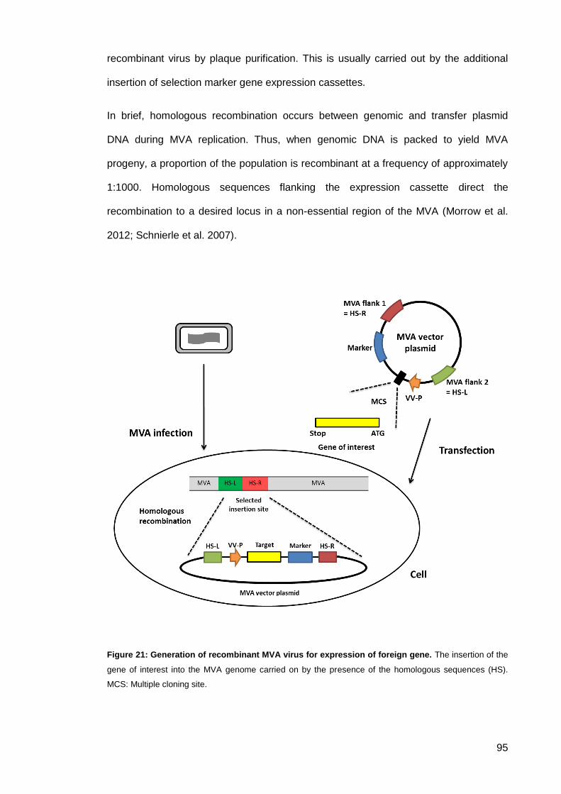
95
recombinant virus by plaque purification. This is usually carried out by the additional
insertion of selection marker gene expression cassettes.
In brief, homologous recombination occurs between genomic and transfer plasmid
DNA during MVA replication. Thus, when genomic DNA is packed to yield MVA
progeny, a proportion of the population is recombinant at a frequency of approximately
1:1000. Homologous sequences flanking the expression cassette direct the
recombination to a desired locus in a non-essential region of the MVA (Morrow et al.
2012; Schnierle et al. 2007).
Figure 21: Generation of recombinant MVA virus for expression of foreign gene. The insertion of the
gene of interest into the MVA genome carried on by the presence of the homologous sequences (HS).
MCS: Multiple cloning site.
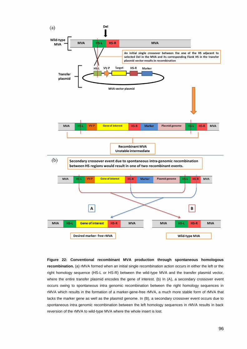
96
Figure 22: Conventional recombinant MVA production through spontaneous homologous
recombination. (a) rMVA formed when an initial single recombination action occurs in either the left or the
right homology sequence (HS-L or HS-R) between the wild-type MVA and the transfer plasmid vector,
where the entire transfer plasmid encodes the gene of interest. (b) In (A), a secondary crossover event
occurs owing to spontaneous intra genomic recombination between the right homology sequences in
rMVA which results in the formation of a marker-gene-free rMVA, a much more stable form of rMVA that
lacks the marker gene as well as the plasmid genome. In (B), a secondary crossover event occurs due to
spontaneous intra genomic recombination between the left homology sequences in rMVA results in back
reversion of the rMVA to wild-type MVA where the whole insert is lost.

97
2.3.1.5.1. Insertion sites
Several regions have been targeted for exogenous DNA insertion. Initially, classical
gene loci encoding for the vaccinia virus proteins thymidine kinase (TK) and the
haemagglutinin gene (HA) were used. However, sites of naturally occurring deletions
formed during the course of MVA attenuation (Del) are now more commonly used.
Moreover, intergenic regions (IGR) have also been used (Morrow et al. 2012; Schnierle
et al. 2007).
Deletion (Del) sites represent non-essential regions in the MVA genome whose
deletion minimally impacts MVA viability in chicken embryo fibroblasts (CEFs). These
regions formed during the process of 570 serial passages on CEFs that transformed
the pathogenic vaccinia virus Ankara strain into MVA (Meyer et al. 1991). Deletion sites
have been routinely used for insertion of recombinant genes to form MVA derived
recombinant vaccines (Drexler et al. 2004). However, the stability of certain transgenes
in Del sites is variable depending on the toxicity of the expressed product during
cellular and viral replication (Wyatt et al. 2008). In some cases, mutations or deletions
that occur in transgenes after several passages of rMVA in culture may result in
reduced or complete loss of expression (Wyatt et al. 2009).
2.3.1.5.2. Marker selection
There are several well-established methods for clonal isolation of rMVA involving the
co-expression of a marker gene. Early techniques relied on insertion of the foreign
gene into the TK locus; then bromodeoxyuridine (BrdU) enrichment selection based on
the TK phenotype was carried out. However, this technique showed some non-specific
isolation. TK mutation from the cytotoxic influence of the BrdU could be detected, and,
as a result, false-positive isolates could be selected. Later, this method was combined
with the simultaneous insertion of the E. coli LacZ gene (Morrow et al. 2012).
The E.coli enzyme β-galactosidase coded in the LacZ gene is one of the most
frequently used marker genes. The LacZ gene has the ability to turn plaques of rMVA
98
from non-stained into blue-stained plaques when located with the synthetic
chromogenic substrate X-gal. The infected cell line containing the active LacZ gene will
code for β-galactosidase, which cleaves the β-glycosidic bond in D-lactose found in X-
gal. When X-gal is cleaved by β-galactosidase, galactose and 5-bromo-4-chloro-3-
hydroxyindole is formed, producing a characteristic blue-coloured plaque (Kremer et al.
2012; Serebriiskii & Golemis 2000). To improve versatility in recombinant isolation,
other genes can be added to the transfer gene vector. Studies have shown that
antibiotic resistance markers such as the E.coli neomycin gene and the xanthine-
guanine phosphoribosyl transferase (gpt) gene, and additional markers can be inserted
such as green fluorescent protein (GFP) or detection of the product of the E.coli β-
glucuronidase A gene (gus). Frequently used for recombinant virus purification is the
E.coli gpt gene encoding the enzyme xanthine guanine phosphoribosyl transferase.
This method allows dominant positive selection for resistance against mycophenolic
acid (MPA). As long as marker genes are not cross-reactive, it is possible to combine
more than one marker gene into one recombinant, to improve the selection. This can
occur under the control of a single vaccinia promoter, as in the case of gpt-gus fusion
(Morrow et al. 2012; Schnierle et al. 2007).
2.3.1.5.3. Promoters
Since the discovery of the first vaccinia virus promoter, the 7.5 kb early/late promoter,
in 1982, significant improvements have been made in designing and synthesising
vaccine promoters. Studies showed that using rational optimisation of promoters
results in enhanced immune responses. Using an optimised promoter in rMVA vaccine
development leads to higher and more efficient expression of deired antigens.
Resulting in a significant enhancement in immune responses as well as potential lower
doses required for vaccination. Vaccinia promoters are active in one of the three
transcriptional stages – early, intermediate or late – and some, such as the 7.5 kb and
H5 promoters, contain both early and late elements in tandem (Morrow et al. 2012).
99
In designing rMVA-based vaccines it is important to achieve high levels of antigen
expression while avoiding conflicting effects. To help reach this goal, assessment of
optimal promoter activity must be undertaken in vitro. It is important to ensure that the
transgene is not located close to the vaccinia virus early transcription termination sites
TTTTTNT. Impairment of gene expression is prevented by excision of non-essential
DNA between the promoter and transcription start site (Morrow et al. 2012). Ideally all
these modifications should be employed simultaneously.
2.3.1.5.4. T7 RNA polymerase hybrid system
In this study all genes of interest were under the control of a T7 promoter. Hence, the
transient cytoplasmic expression system used in this study relies on synthesising the
bacteriophage T7 RNA polymerase in the cytoplasm of the mammalian cells.
Mammalian cells are co-infected with two recombinant vaccinia viruses, one contains
the genes of interest while the other encodes bacteriophage T7 RNA polymerase
(driven by a poxviral promoter, p7.5).
During incubation, the genes of interest are efficiently transcribed by T7 RNA
polymerase and translated in the cytoplasm of the infected cells. The decision to utilise
this system was based on several factors. Such a system will provide a control
mechanism for the expression of transgenes (no expression without co-infection), thus
enhancing the safety profile of the proposed gene therapy candidate. As transgenes
are not expressed during the manufacturing stage, more stable recombinant viruses
are achieved. T7-RNA polymerase known to be extremely promoter specific and only
transcribes genes downstream of a T7-promoter with a very low error rate, this serves
as a desirable characteristic for a viral vector vaccine. The DNA-dependent T7 RNA
polymerase is a very active enzyme that less frequently terminates transcription and is
found to be highly selective for initiation of its own promoter sequences (Tabor 2001;
Elroy-Stein & Moss 2001).
100
2.3.1.5.5. Genes of interest
The final result in this project will be the production of a pseudotype virus like particle
(VLP) encoding IgG1b12 sequence in its core. The VLP is of SIVmac239 origin.
Expression cassettes used contained two common genes (gag, pol) and five additional
accessory genes (tat, rev, vpx, vpr and vif).
Genes encoding the structural proteins of VLPs were organised in three major regions
of the genome: - gag (group specific antigen), internal structural proteins; pol
(polymerase) enzymes and envelope genes were pseudotyped with VSV-G envelope
proteins. The gag gene codes for the Gag polyprotein (matrix, capsid, p2,
nucleocapsid, p1 and p6), which is then processed by the viral protease to generate
proteins associated with the viral mRNA and some may form a part of the viral capsid.
The pol gene (polymerase), translated into a Gag-Pol polypeptide via a ribosomal
frame-shift, codes for the viral enzymes reverse transcriptase RT (p55/p51), integrase
IN (p32), RNase H, and protease PR (p11).
The accessory proteins are essential for viral replication and virion infectivity. Tat
(transactivator of transcription) is a powerful activator of viral gene expression. The
protein binds a structured region present at the 5’ end of the transcribed viral RNAs
and recruits different cellular factors to the viral LTR promoter. Rev (regulator of
expression of virion proteins) binds a highly structured RNA sequence (Rev-responsive
element, RRE), present in correspondence to the env gene and is thus contained as a
potential intron in the fully spliced mRNAs. The role of Rev is to mediate transport of
these RRE-containing mRNAs outside the nucleus.
Vpr (viral protein R) blocks progression of the cell cycle in the infected cells by
accumulating them in the G2-M phase, is incorporated into virions, and contributes to
nuclear transport of the reverse transcribed viral cDNA in the infected cells. Vif (virion
infectivity factor) is essential for proper virion infectivity since it blocks the function of a
cellular enzyme, the RNA deaminase APOBEC, which would otherwise inactivate the
101
viral genome by introducing mutations. Vpx (virion-associated protein) targets a host
restriction factor for proteasomal degradation and is involved in the import of the viral
pre-integration complex into the host nucleus (Collier et al. 2011; Carter & Saunders
2013).
Pseudotyping of lentiviruses is mainly used to expand the host range of retroviral
vectors. The G glycoprotein of the VSV-G is almost always used to replace the Env
proteins of the lentiviruses. VSV-G pseudotyped lentiviral vectors have expanded
tropism that spur applications for gene therapy and can infect most types of cells.
Studies have indicated that pseudotyped lentiviruses are particularly stable (Burns et
al. 1993).
The expression cassette of the gene therapy agent is split into two independent MVA
vectors. It is speculated that the final result should be the insertion of IgG1 b12
antibody genes into the mammalian genome. The first genomic rMVA
(rMVA/IgG1b12/T7) will carry the HIV-neutralising monoclonal antibody (mAb)
sequences as well as the T7 polymerase and will deliver them into the cytoplasm of
mammalian cells. A second packaging rMVA (rMVA/gag/pol/VSV-G: encodes a simian
retroviral particle pseudo-typed with vesicular stomatitis virus glycoprotein) will be used
to facilitate the delivery of the mAb genes into the human genome inside the nucleus.
The second rMVA is a viral vector that expresses heterologous polypeptides capable of
assembling into defective non-self propagating viral particles mimicking the packaging
structure of an SIV. The new non-infectious artificial lentivirus that results from the
hybrid dual-vector system will infect neighbouring cells, penetrate the nucleus and will
insert its genetic materials into the mammalian genome, functioning as a vehicle in this
gene therapy project.
102
Materials and Methods 2.4.
Genetically Modified Organisms approval for this study was obtained for the laboratory
and the project. The lab permissions and the GMO permission were granted by the
University of Manchester safety committee, the GMO Establishment license# 541
(p5603-4).
2.4.1. Molecular Cloning
All the plasmids were designed using Lasergene Genomics Suite (DNASTAR,
Madison, USA) software, as well as NCBI GenBank database. The transfer plasmid
was designed (refer to section 2.5.1) and sent for manufacture to Blue Heron
Biotechnology, Bothell, WA, USA.
For multiplication of plasmid DNA the following E.coli strain was used:
SCS1 Supercompetent Cells were obtained from Stratagene/Agilent Technologies UK
Ltd.,(Wokingham, UK). SCS1 are E.coli cells hation 5.1.1.ve a recA1 endA1 gyrA96 thi-
1 hsdR17 (RК- , MК+) supE44 relA1 genotype and are characterised by endA
(endonuclease) and recA (recombination) deficiency which improves insert stability and
the quality of miniprep DNA.
Plasmid designing 2.4.1.1.
2.4.1.1.1. pLF-IgG1b12-mTK
The transfer plasmid was prepared from a pBR322 backbone. The cloning vector
pBR322 complete sequence was obtained from GenBank: J01749.1. MVA sequences
from the classical insertion site, the thymidine kinase gene locus (TK), were selected
after slight modification to the flank the gene of interest to form the functional cassette.
The gene of interest (IgG1b12) is to be inserted into the TK locus of the MVA strain by
in vitro recombination in primary chicken embryo fibroblasts (pCEFs). Modified TK
sequence was obtained from vaccinia virus strain MVATGN33.1 complete genome,
GenBank: EF675191.1. The CMV promoter and its enhancer were obtained from
103
Promega Corporation. The E.coli xanthine guanine phosphoribosyl transferase (gpt)
gene and the E.coli β-galactosidase lacZ gene sequences were encoded in this
plasmid as a dual marker system. A LacZ sequence was added to the TK gene flanking
regions by traditional cloning. β-galactosidase sequence was obtained from
GenBank:AAA24053.1, while gpt sequences were obtained from E.coli KLY, complete
genome, GenBank: CP008801.1. To drive gpt expression, synthetic E/L promoter
derived from Psc6 was used after reverse complementation of its sequence. The
promoter sequence from Chakrabarti et al, 1997 was used and direct repeats of
repetitive MVA-DNA fragments homologous to the right end of flank1 were added,
allowing for the deletion of the marker expression cassette (gpt and lacZ marker
system) by homologous recombination. The last 50 bp from the right end of flank1
encoding the right TK locus fragment was copied and added before the Reverse
complement pE/L promoter (Figure 28).
The heavy and light sequences of IgG1b12 were retrieved from the crystal structure of
the intact human IgG1b12 (Figure 23). A slight modification was done via codon de-
optimisation and/or nucleotide mutation to breakup any C repetitive sequences, to
disrupt potential kozak consensus and/or to introduce a silent stopping codon. Also,
few amino acids were replaced to fit consensus for IgG1 heavy chain (Figure 24).
Figure 23: Crystal structure of the intact human IgG1b12. (Adapted from:
http://www.ncbi.nlm.nih.gov/Structure/mmdb/mmdbsrv.cgi?uid=1HZH accessed 18/10/17).
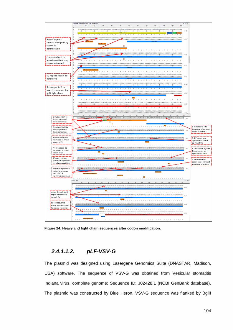
104
Figure 24: Heavy and light chain sequences after codon modification.
2.4.1.1.2. pLF-VSV-G
The plasmid was designed using Lasergene Genomics Suite (DNASTAR, Madison,
USA) software. The sequence of VSV-G was obtained from Vesicular stomatitis
Indiana virus, complete genome; Sequence ID: J02428.1 (NCBI GenBank database).
The plasmid was constructed by Blue Heron. VSV-G sequence was flanked by BglII
105
and PacI. This plasmid was used to replace the Env sequence in already existing
plasmid pCoEnvRev.D4 that was provided by Dr Tom Blanchard.
DNA extraction and purification from bacterial cells 2.4.1.2.
Plasmid DNA was isolated using the QIAprep Spin Miniprep Kit (QIAGEN #27104).
Following the manufacturer instructions, an overnight E.coli culture was pelleted by
centrifugation. Bacterial cells were then lysed by alkaline lysis followed by adsorption of
DNA onto silica membranes in the presence of high salt concentrations. Impurities
were washed with the supplied washing buffer and pure plasmid DNA eluted in a small
volume of elution buffer. The concentration of the plasmid DNA was determined using
the ND1000 NanoDrop Spectrophotometer (Labtech, Uckfield, UK).
Preparation of vector and insert: Restrictions digest 2.4.1.3.
In a total volume of 50μl: 1µg of DNA, 5μl of 10xNEBuffer and 1μl (10unit) enzyme
were mix. Both incubation times and temperature are enzyme dependent.
DNA End Modification: Dephosphorylation 2.4.1.4.
1 µL (1 unit/µL) of Recombinant Shrimp Alkaline Phosphatase (rSAP) (NEB #M0371)
was added to each 1 pmol of DNA (about 1 µg of a 3 kb plasmid). After incubation at
37°C for 30 minutes, the reaction was stopped by heat inactivation at 65°C for 5
minutes.
Agarose gel electrophoresis and gel extraction 2.4.1.5.
2.4.1.5.1. Agarose gel electrophoresis
PCR products and DNA digests were analysed by gel electrophoresis on the basis of
their molecular weight. Fragment sizes were determined in relation to the bands of a
mixture of standard molecules (marker ladder) on the gel. The DNA fragments in
agarose gels were visualised with UV light using SYBR® Safe DNA Gel Stain (Thermo
Fisher Scientific, USA) or Gel Red (Biotium Inc., Hayward, USA) following the
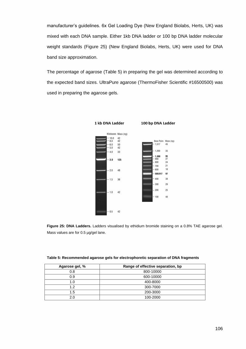
106
manufacturer’s guidelines. 6x Gel Loading Dye (New England Biolabs, Herts, UK) was
mixed with each DNA sample. Either 1kb DNA ladder or 100 bp DNA ladder molecular
weight standards (Figure 25) (New England Biolabs, Herts, UK) were used for DNA
band size approximation.
The percentage of agarose (Table 5) in preparing the gel was determined according to
the expected band sizes. UltraPure agarose (ThermoFisher Scientific #16500500) was
used in preparing the agarose gels.
Figure 25: DNA Ladders. Ladders visualised by ethidium bromide staining on a 0.8% TAE agarose gel.
Mass values are for 0.5 µg/gel lane.
Table 5: Recommended agarose gels for electrophoretic separation of DNA fragments
Agarose gel, % Range of effective separation, bp
0.8 800-10000
0.9 600-10000
1.0 400-8000
1.2 300-7000
1.5 200-3000
2.0 100-2000
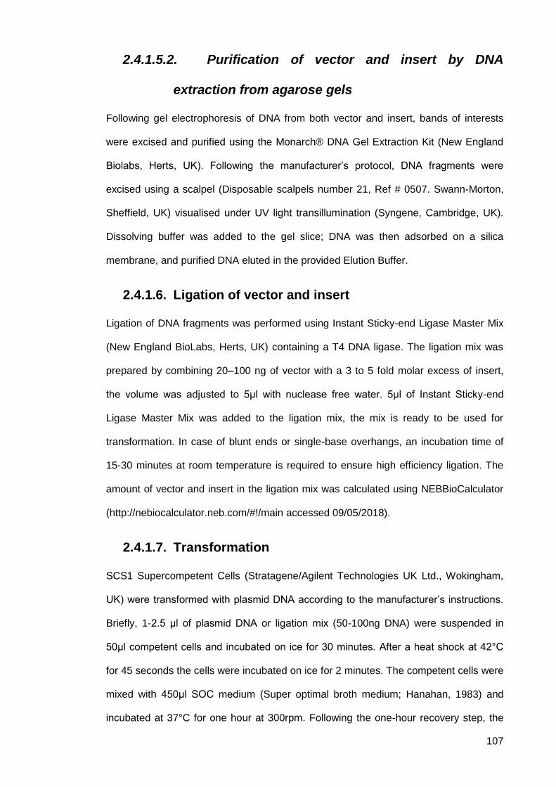
107
2.4.1.5.2. Purification of vector and insert by DNA
extraction from agarose gels
Following gel electrophoresis of DNA from both vector and insert, bands of interests
were excised and purified using the Monarch® DNA Gel Extraction Kit (New England
Biolabs, Herts, UK). Following the manufacturer’s protocol, DNA fragments were
excised using a scalpel (Disposable scalpels number 21, Ref # 0507. Swann-Morton,
Sheffield, UK) visualised under UV light transillumination (Syngene, Cambridge, UK).
Dissolving buffer was added to the gel slice; DNA was then adsorbed on a silica
membrane, and purified DNA eluted in the provided Elution Buffer.
Ligation of vector and insert 2.4.1.6.
Ligation of DNA fragments was performed using Instant Sticky-end Ligase Master Mix
(New England BioLabs, Herts, UK) containing a T4 DNA ligase. The ligation mix was
prepared by combining 20–100 ng of vector with a 3 to 5 fold molar excess of insert,
the volume was adjusted to 5μl with nuclease free water. 5μl of Instant Sticky-end
Ligase Master Mix was added to the ligation mix, the mix is ready to be used for
transformation. In case of blunt ends or single-base overhangs, an incubation time of
15-30 minutes at room temperature is required to ensure high efficiency ligation. The
amount of vector and insert in the ligation mix was calculated using NEBBioCalculator
(http://nebiocalculator.neb.com/#!/main accessed 09/05/2018).
Transformation 2.4.1.7.
SCS1 Supercompetent Cells (Stratagene/Agilent Technologies UK Ltd., Wokingham,
UK) were transformed with plasmid DNA according to the manufacturer’s instructions.
Briefly, 1-2.5 μl of plasmid DNA or ligation mix (50-100ng DNA) were suspended in
50μl competent cells and incubated on ice for 30 minutes. After a heat shock at 42°C
for 45 seconds the cells were incubated on ice for 2 minutes. The competent cells were
mixed with 450μl SOC medium (Super optimal broth medium; Hanahan, 1983) and
incubated at 37°C for one hour at 300rpm. Following the one-hour recovery step, the
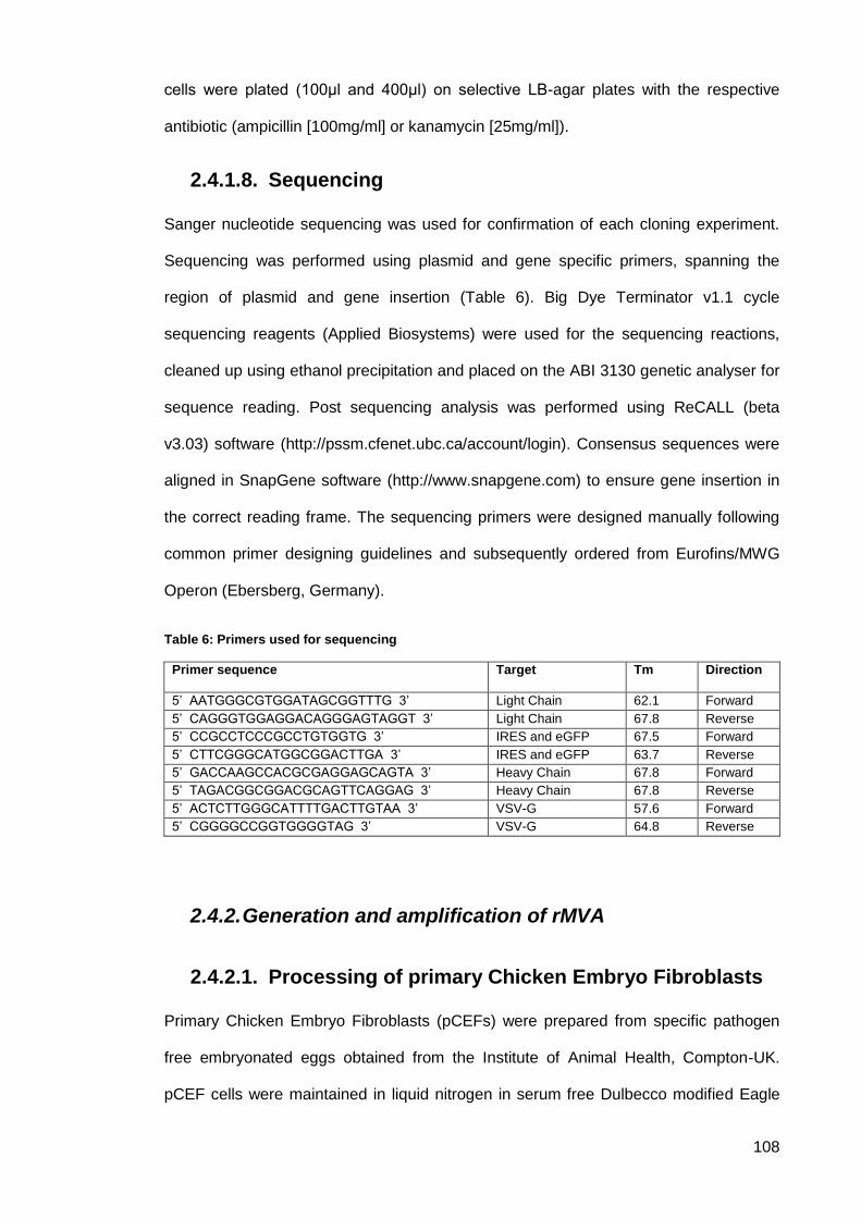
108
cells were plated (100μl and 400μl) on selective LB-agar plates with the respective
antibiotic (ampicillin [100mg/ml] or kanamycin [25mg/ml]).
Sequencing 2.4.1.8.
Sanger nucleotide sequencing was used for confirmation of each cloning experiment.
Sequencing was performed using plasmid and gene specific primers, spanning the
region of plasmid and gene insertion (Table 6). Big Dye Terminator v1.1 cycle
sequencing reagents (Applied Biosystems) were used for the sequencing reactions,
cleaned up using ethanol precipitation and placed on the ABI 3130 genetic analyser for
sequence reading. Post sequencing analysis was performed using ReCALL (beta
v3.03) software (http://pssm.cfenet.ubc.ca/account/login). Consensus sequences were
aligned in SnapGene software (http://www.snapgene.com) to ensure gene insertion in
the correct reading frame. The sequencing primers were designed manually following
common primer designing guidelines and subsequently ordered from Eurofins/MWG
Operon (Ebersberg, Germany).
Table 6: Primers used for sequencing
Primer sequence Target Tm Direction
5’ AATGGGCGTGGATAGCGGTTTG 3’ Light Chain 62.1 Forward
5’ CAGGGTGGAGGACAGGGAGTAGGT 3’ Light Chain 67.8 Reverse
5’ CCGCCTCCCGCCTGTGGTG 3’ IRES and eGFP 67.5 Forward
5’ CTTCGGGCATGGCGGACTTGA 3’ IRES and eGFP 63.7 Reverse
5’ GACCAAGCCACGCGAGGAGCAGTA 3’ Heavy Chain 67.8 Forward
5’ TAGACGGCGGACGCAGTTCAGGAG 3’ Heavy Chain 67.8 Reverse
5’ ACTCTTGGGCATTTTGACTTGTAA 3’ VSV-G 57.6 Forward
5’ CGGGGCCGGTGGGGTAG 3’ VSV-G 64.8 Reverse
2.4.2. Generation and amplification of rMVA
Processing of primary Chicken Embryo Fibroblasts 2.4.2.1.
Primary Chicken Embryo Fibroblasts (pCEFs) were prepared from specific pathogen
free embryonated eggs obtained from the Institute of Animal Health, Compton-UK.
pCEF cells were maintained in liquid nitrogen in serum free Dulbecco modified Eagle
109
medium (DMEM). Each frozen vial containing 1mL of pCEFs (3.5 × 10⁸ cells) was
resuscitated using cell growth medium prepared from DMEM (Lot no. RNBC6720)
containing 10% heat-inactivated foetal calf serum (FCS) and 10 mM L-glutamine into a
tissue culture flask (either T175 or T75). The tissue culture flasks upon seeding the
cells (one frozen vial/flask) were incubated at 37°C in a tissue culture incubator
supplied with 5% CO2 for 2–3 days or until 100% confluency was achieved. For a 175
cm2 flask, cells would usually be ready for cell passage on the third day at a ratio of
1:3, whereas for a 75 cm2 flask, cells would usually be ready for splitting on the second
day at the same ratio. Adherent cells were removed from the culture flask surface by
treatment with TrypZean® Solution, 1x (SIGMA-ALDRICH #T3449). Cells were
incubated with TrypZean for 5 minutes. Trypsinization reaction was stopped by adding
FCS.
Virus stock 2.4.2.2.
The modified vaccinia Ankara (MVA) used in this project was a gift from Professor
Anton Mayr (Ludwigs Maximilian University, Munich, Germany). He provided sealed
vials of wild type MVA (wtMVA) used in the smallpox eradication campaign. The
wtMVA had been passaged twice on pCEFs since then, but not plaque-purified. The
virus then amplified and the titre was determined to be 8.5 x 10⁸ pfu/ µL by plaque
assay (refer to section 2.4.2.3).
2.4.2.2.1. Virus titration
2% Low melting point agarose
5 g Low melting point agarose (Sigma-Aldrich; cat #A9045)
250 mL water (Sigma Aldrich; cat #W3500-500ML
The suspension was autoclaved for 15 minutes at 121°C (Astell, Kent, UK;
Model AMB420BT)
The stock concentration of virus samples was determined using plaque assay in
pCEFs. Tenfold dilutions of the virus-stock sample in DMEM medium were adsorbed
on pCEFs in duplicates using 6-well tissue culture plates. After 3 hours incubation at
110
37C, the cells were overlaid with 2ml/well of pre-warmed (42°C) 2% low melting point
agarose (Sigma-Aldrich; cat #A9045) with 2X MEM (Life Technologies Ltd.; cat
#11935-046) medium and 10% FCS in a 1:1 ratio. The plates were then incubated at
37°C with 5% CO2 and 95% humidity until plaques were visible. To determine the virus
titre only wells with isolated plaques were counted and where the number of plaques
exceeded 20.
2.4.2.2.2. Generation of recombinant MVA
In order to generate recombinant MVA, the transfer plasmid of choice that was
obtained by molecular cloning was transfected into pCEFs, which were then infected
with either wtMVA or rMVA. Transfer vector plasmids were transfected into MVA-
infected cells, and homologous recombination between MVA and plasmid DNA
generated a recombinant virus.
2.4.2.2.3. Transfection of primary chicken embryo
fibroblasts
pCEFs were transfected with endotoxin free plasmid DNA using SuperFect
Transfection Reagent (QIAGEN; cat #301305) following the manufacturer’s guidelines.
On the day of transfection the density of pCEFs was 8x10⁵ cells/well (6-well dish). For
one well 5-8μg of plasmid DNA were mixed with DMEM to make up a final volume of
100μL. Then 10μL of SuperFect transfection reagent was added to the mix and
vortexed for 10 seconds. To allow for transfection-complex formation the solution was
incubated for 10 minutes at room temperature while growth medium was aspirated
from pCEFs. Then, 600μL of growth medium (DMEM + FCS + L-glutamine) was added
to the transfection mix and pipetted onto the pCEF cell layer. The plates were
incubated at 37°C with 5% CO2 and 95% humidity for 3 hours with a rocking step every
15 minutes.
111
2.4.2.2.4. Infection of pCEFs with wildtype MVA or
recombinant MVA
Pre-titrated wtMVA or rMVA stock was thawed and diluted to a multiplicity of infection
(MOI) of 0.5 in 1mL DMEM. Following a three-hour infection step, the transfection mix
was aspirated from the pCEFs cell layer and replaced with 1mL infection mix per well.
The plates were incubated (37°C with 5% CO2 and 95% humidity) for 3 hours and
rocked every 15 minutes. Subsequently the infection mix was aspirated and the cell
layer was covered with 2 mL DMEM+ 10% FCS + 2 mM L-glutamine. The plates were
placed in an incubator for 48 hours. The pCEFs and virus were then harvested using
cell scraper (Falcon, Corning GmbH; Cat # 353086). The cell lysate underwent three
freeze-thaw cycles and was stored at -80°C until further usage.
Plaque assay and rMVA isolation 2.4.2.3.
Primary overlay
2% Low melting point agarose (melted in a microwave)
2X Minimum essential medium eagle (MEM, Life Technologies Ltd.; cat
#11935-046) 10% Fetal Calf Serum (FCS, Biosera, Gentaur Ltd.; cat #FB-
13770/500).
Secondary overlay
2% Low melting point agarose (melted in a microwave)
2X Minimum essential medium eagle (MEM, Life Technologies Ltd.; cat
#11935-046 )
Supplement with 1/1000 volume of 5-Bromo-4-chloro-3-indolyl-β-D-
galactopyranoside (X-gal, Fisher Scientific: cat # BP1615-1)
Virus lysate was homogenised for 10 seconds in a sonicator (Misonix, sonicator 3000
cabinet) at 150 W. 80% confluent pCEFs monolayer (in 6-well dishes) was infected
with variable volumes ranging from 50μL – 200μL of lysate, dilutions were prepared in
DMEM, and incubated for 3 hours at 37°C with 5% CO2 and 95% humidity. The
inoculum was aspirated and the cells were covered with 2 mL/well pre-warmed (42°C)
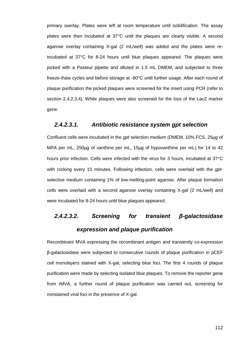
112
primary overlay. Plates were left at room temperature until solidification. The assay
plates were then incubated at 37°C until the plaques are clearly visible. A second
agarose overlay containing X-gal (2 mL/well) was added and the plates were re-
incubated at 37°C for 8-24 hours until blue plaques appeared. The plaques were
picked with a Pasteur pipette and diluted in 1.5 mL DMEM, and subjected to three
freeze-thaw cycles and before storage at -80°C until further usage. After each round of
plaque purification the picked plaques were screened for the insert using PCR (refer to
section 2.4.2.3.4). White plaques were also screened for the loss of the LacZ marker
gene.
2.4.2.3.1. Antibiotic resistance system gpt selection
Confluent cells were incubated in the gpt selection medium (DMEM, 10% FCS, 25µg of
MPA per mL, 250µg of xanthine per mL, 15µg of hypoxanthine per mL) for 14 to 42
hours prior infection. Cells were infected with the virus for 3 hours, incubated at 37C
with rocking every 15 minutes. Following infection, cells were overlaid with the gpt-
selective medium containing 1% of low-melting-point agarose. After plaque formation
cells were overlaid with a second agarose overlay containing X-gal (2 mL/well) and
were incubated for 8-24 hours until blue plaques appeared.
2.4.2.3.2. Screening for transient β-galactosidase
expression and plaque purification
Recombinant MVA expressing the recombinant antigen and transiently co-expression
β-galactosidase were subjected to consecutive rounds of plaque purification in pCEF
cell monolayers stained with X-gal, selecting blue foci. The first 4 rounds of plaque
purification were made by selecting isolated blue plaques. To remove the reporter gene
from rMVA, a further round of plaque purification was carried out, screening for
nonstained viral foci in the presence of X-gal.
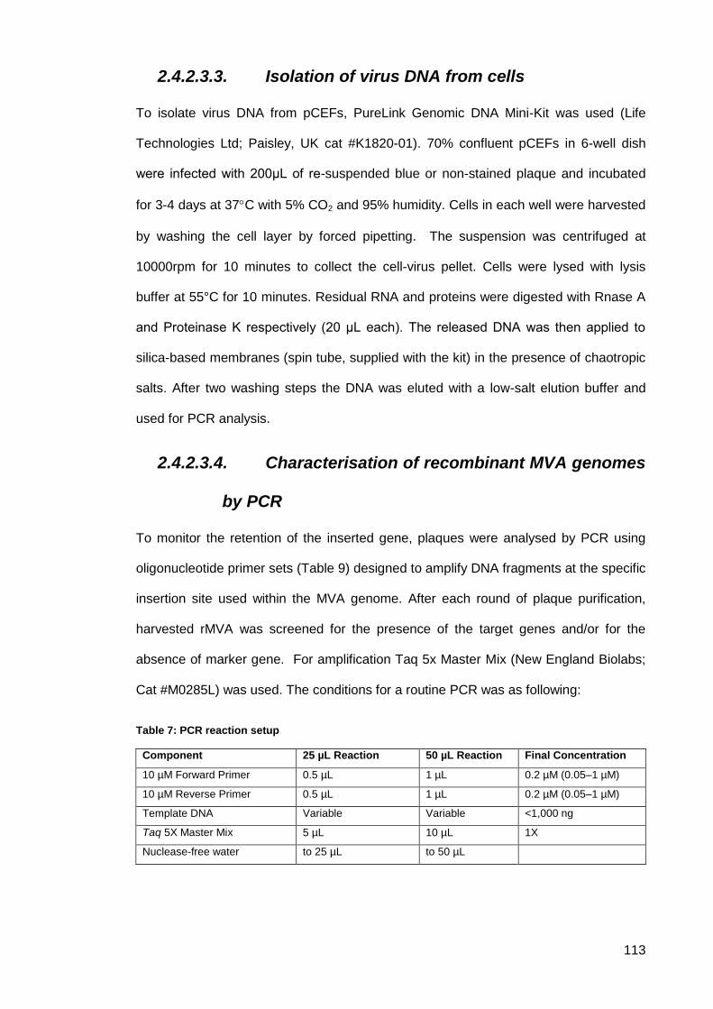
113
2.4.2.3.3. Isolation of virus DNA from cells
To isolate virus DNA from pCEFs, PureLink Genomic DNA Mini-Kit was used (Life
Technologies Ltd; Paisley, UK cat #K1820-01). 70% confluent pCEFs in 6-well dish
were infected with 200μL of re-suspended blue or non-stained plaque and incubated
for 3-4 days at 37C with 5% CO2 and 95% humidity. Cells in each well were harvested
by washing the cell layer by forced pipetting. The suspension was centrifuged at
10000rpm for 10 minutes to collect the cell-virus pellet. Cells were lysed with lysis
buffer at 55°C for 10 minutes. Residual RNA and proteins were digested with Rnase A
and Proteinase K respectively (20 μL each). The released DNA was then applied to
silica-based membranes (spin tube, supplied with the kit) in the presence of chaotropic
salts. After two washing steps the DNA was eluted with a low-salt elution buffer and
used for PCR analysis.
2.4.2.3.4. Characterisation of recombinant MVA genomes
by PCR
To monitor the retention of the inserted gene, plaques were analysed by PCR using
oligonucleotide primer sets (Table 9) designed to amplify DNA fragments at the specific
insertion site used within the MVA genome. After each round of plaque purification,
harvested rMVA was screened for the presence of the target genes and/or for the
absence of marker gene. For amplification Taq 5x Master Mix (New England Biolabs;
Cat #M0285L) was used. The conditions for a routine PCR was as following:
Table 7: PCR reaction setup
Component 25 µL Reaction 50 µL Reaction Final Concentration
10 µM Forward Primer 0.5 µL 1 µL 0.2 µM (0.05–1 µM)
10 µM Reverse Primer 0.5 µL 1 µL 0.2 µM (0.05–1 µM)
Template DNA Variable Variable <1,000 ng
Taq 5X Master Mix 5 µL 10 µL 1X
Nuclease-free water to 25 µL to 50 µL
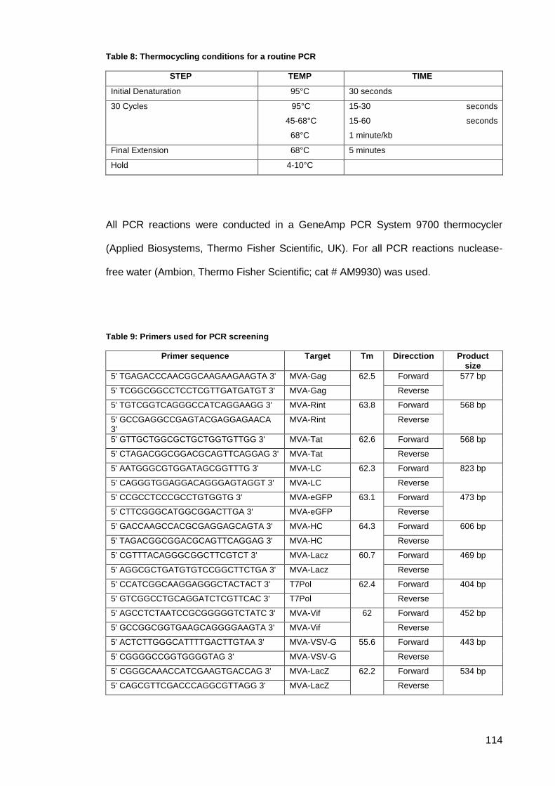
114
Table 8: Thermocycling conditions for a routine PCR
STEP TEMP TIME
Initial Denaturation 95°C 30 seconds
30 Cycles 95°C
45-68°C
68°C
15-30 seconds
15-60 seconds
1 minute/kb
Final Extension 68°C 5 minutes
Hold 4-10°C
All PCR reactions were conducted in a GeneAmp PCR System 9700 thermocycler
(Applied Biosystems, Thermo Fisher Scientific, UK). For all PCR reactions nuclease-
free water (Ambion, Thermo Fisher Scientific; cat # AM9930) was used.
Table 9: Primers used for PCR screening
Primer sequence Target Tm Direcction Product size
5' TGAGACCCAACGGCAAGAAGAAGTA 3' MVA-Gag 62.5 Forward 577 bp
5' TCGGCGGCCTCCTCGTTGATGATGT 3' MVA-Gag Reverse
5' TGTCGGTCAGGGCCATCAGGAAGG 3' MVA-Rint 63.8 Forward 568 bp
5' GCCGAGGCCGAGTACGAGGAGAACA 3'
MVA-Rint Reverse
5' GTTGCTGGCGCTGCTGGTGTTGG 3' MVA-Tat 62.6 Forward 568 bp
5' CTAGACGGCGGACGCAGTTCAGGAG 3' MVA-Tat Reverse
5' AATGGGCGTGGATAGCGGTTTG 3' MVA-LC 62.3 Forward 823 bp
5' CAGGGTGGAGGACAGGGAGTAGGT 3' MVA-LC Reverse
5' CCGCCTCCCGCCTGTGGTG 3' MVA-eGFP 63.1 Forward 473 bp
5' CTTCGGGCATGGCGGACTTGA 3' MVA-eGFP Reverse
5' GACCAAGCCACGCGAGGAGCAGTA 3' MVA-HC 64.3 Forward 606 bp
5' TAGACGGCGGACGCAGTTCAGGAG 3' MVA-HC Reverse
5' CGTTTACAGGGCGGCTTCGTCT 3' MVA-Lacz 60.7 Forward 469 bp
5' AGGCGCTGATGTGTCCGGCTTCTGA 3' MVA-Lacz Reverse
5' CCATCGGCAAGGAGGGCTACTACT 3' T7Pol 62.4 Forward 404 bp
5' GTCGGCCTGCAGGATCTCGTTCAC 3' T7Pol Reverse
5' AGCCTCTAATCCGCGGGGGTCTATC 3' MVA-Vif 62 Forward 452 bp
5' GCCGGCGGTGAAGCAGGGGAAGTA 3' MVA-Vif Reverse
5' ACTCTTGGGCATTTTGACTTGTAA 3' MVA-VSV-G 55.6 Forward 443 bp
5' CGGGGCCGGTGGGGTAG 3' MVA-VSV-G Reverse
5' CGGGCAAACCATCGAAGTGACCAG 3' MVA-LacZ 62.2 Forward 534 bp
5' CAGCGTTCGACCCAGGCGTTAGG 3' MVA-LacZ Reverse
115
2.4.3. Transfection using Lipfectamine 2000
In a 24-well plate, 70-90% confluent cells were transfected with rMVA encoding eGFP.
Four different amounts of Lipofectamine reagent (2, 3, 4 and 5µL) each was diluted in
Opti-MEM medium 50 µL. 5 µg of plasmid DNA was diluted in opti-MEM medium to
give a final volume of 250µL. Diluted DNA was added to diluted Lipofectamine 2000
reagent at 1:1 ratio (50µL:50µL). DNA-lipid complex was incubated for 5 minutes at
room temperature. The complex was then added to the cells and incubated for 6 hours
at 37C. The mixture was then replaced with fresh 10% FCS/DMEM media. Cells were
incubated up to 5 days at 37. Then transfected cells were analysed for fluorescence.
116
Results 2.5.
2.5.1. Design and construction of MVA transfer plasmid
vectors
In all vector design pBR322 was the plasmid of choice. It is a relatively small plasmid
encoding the ampR gene (source plasmid RSF2124) that produces the ampicillin
resistance protein and the tetR gene, which forms the tetracycline resistance protein.
In case of traditional molecular cloning, if a vector is linearised by a single restriction
enzyme or has been cut using two enzymes with compatible ends, a dephosphorylation
step must be carried out to ensure that the vector does not recircularise during ligation.
pLF-IgG1b12-mTK 2.5.1.1.
Several promoters were used in this design. A T7 promoter was placed upstream in the
R region (U5 region + 5’LTR) and the tRNA primer-binding site. The T7 promoter
sequence was obtained from bacteriophage T7 complete genome NCBI Reference
Sequence: NC_001604.1. Human CMV IE gene promoter was added to drive
expression of the monoclonal antibody sequence and the eGFP. The CMV promoter is
one of the strongest mammalian promoters known. It has been frequently used in
several studies for a high level mammalian expression and it is found to be optimal in
driving LC-HC expression. The lacZ sequence wasn’t included in our plasmid design,
as it is already has been designed in the lab.
In this project, combining the heavy and light chains in a single expression cassette
was done using a tri-cistronic setup, where two different IRES elements were included
(Figure 26). IRES elements are used to combine antibody heavy and light chains
together with the marker a reporter gene GFP in a single expression cassette.
Additionally, IRES allows the driving translation initiation of the downstream cistrons
(Jostock 2011).
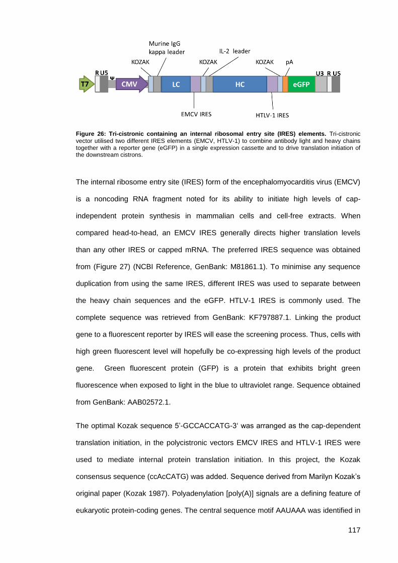
117
Figure 26: Tri-cistronic containing an internal ribosomal entry site (IRES) elements. Tri-cistronic
vector utilised two different IRES elements (EMCV, HTLV-1) to combine antibody light and heavy chains together with a reporter gene (eGFP) in a single expression cassette and to drive translation initiation of the downstream cistrons.
The internal ribosome entry site (IRES) form of the encephalomyocarditis virus (EMCV)
is a noncoding RNA fragment noted for its ability to initiate high levels of cap-
independent protein synthesis in mammalian cells and cell-free extracts. When
compared head-to-head, an EMCV IRES generally directs higher translation levels
than any other IRES or capped mRNA. The preferred IRES sequence was obtained
from (Figure 27) (NCBI Reference, GenBank: M81861.1). To minimise any sequence
duplication from using the same IRES, different IRES was used to separate between
the heavy chain sequences and the eGFP. HTLV-1 IRES is commonly used. The
complete sequence was retrieved from GenBank: KF797887.1. Linking the product
gene to a fluorescent reporter by IRES will ease the screening process. Thus, cells with
high green fluorescent level will hopefully be co-expressing high levels of the product
gene. Green fluorescent protein (GFP) is a protein that exhibits bright green
fluorescence when exposed to light in the blue to ultraviolet range. Sequence obtained
from GenBank: AAB02572.1.
The optimal Kozak sequence 5’-GCCACCATG-3‘ was arranged as the cap-dependent
translation initiation, in the polycistronic vectors EMCV IRES and HTLV-1 IRES were
used to mediate internal protein translation initiation. In this project, the Kozak
consensus sequence (ccAcCATG) was added. Sequence derived from Marilyn Kozak’s
original paper (Kozak 1987). Polyadenylation [poly(A)] signals are a defining feature of
eukaryotic protein-coding genes. The central sequence motif AAUAAA was identified in
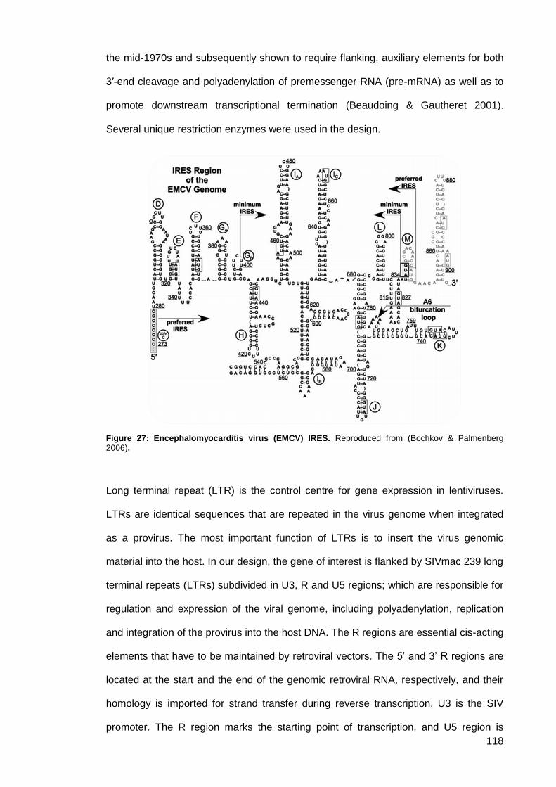
118
the mid-1970s and subsequently shown to require flanking, auxiliary elements for both
3′-end cleavage and polyadenylation of premessenger RNA (pre-mRNA) as well as to
promote downstream transcriptional termination (Beaudoing & Gautheret 2001).
Several unique restriction enzymes were used in the design.
Figure 27: Encephalomyocarditis virus (EMCV) IRES. Reproduced from (Bochkov & Palmenberg 2006).
Long terminal repeat (LTR) is the control centre for gene expression in lentiviruses.
LTRs are identical sequences that are repeated in the virus genome when integrated
as a provirus. The most important function of LTRs is to insert the virus genomic
material into the host. In our design, the gene of interest is flanked by SIVmac 239 long
terminal repeats (LTRs) subdivided in U3, R and U5 regions; which are responsible for
regulation and expression of the viral genome, including polyadenylation, replication
and integration of the provirus into the host DNA. The R regions are essential cis-acting
elements that have to be maintained by retroviral vectors. The 5’ and 3’ R regions are
located at the start and the end of the genomic retroviral RNA, respectively, and their
homology is imported for strand transfer during reverse transcription. U3 is the SIV
promoter. The R region marks the starting point of transcription, and U5 region is

119
critical for reverse transcription. The other key elements are the packaging signal (Ψ)
and the polypurine tract (PPT). The packaging signal, as in many other virus species,
allows RNA genome encapsidation during virion assembly in the cytoplasm. The PPT
element is a key element for reverse transcription. The R region is an important
element in many steps of the lentiviral life cycle, such as transactivation of
transcription, polyadenylation and first-strand transfer during reverse transcription. SIV
mac 239 LTR sequences were retrieved from GenBank: M62835.1. The synthesised
plasmid is shown in diagrammatic form in figure below.
Figure 28: Schematic representation of the transfer plasmid pLF-IgG1b12-mTK. The thymidine
kinase (tk) locus used as an intergenic insertion site for the IgG1b12 expression cassette.
Restriction digestion was used either to screen for positive clones or to prepare DNA
samples for cloning. Both vector and insert from a plasmid source were digested with
appropriate restriction enzymes. Restriction digests were carried out as recommended
by New England BioLabs. Double digests were only performed when both enzymes
showed 75-100% activity in the same buffer. Depending on an enzyme's activity rating
in a non-optimal NEBuffer, numbers of units and/or incubation time were adjusted to
compensate lower rate of cleavage. If there was no buffer in which both restriction
enzymes exhibited >50% activity, a sequential digest was performed. First digestion
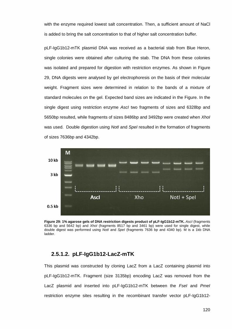
120
with the enzyme required lowest salt concentration. Then, a sufficient amount of NaCl
is added to bring the salt concentration to that of higher salt concentration buffer.
pLF-IgG1b12-mTK plasmid DNA was received as a bacterial stab from Blue Heron,
single colonies were obtained after culturing the stab. The DNA from these colonies
was isolated and prepared for digestion with restriction enzymes. As shown in Figure
29, DNA digests were analysed by gel electrophoresis on the basis of their molecular
weight. Fragment sizes were determined in relation to the bands of a mixture of
standard molecules on the gel. Expected band sizes are indicated in the Figure. In the
single digest using restriction enzyme AscI two fragments of sizes and 6328bp and
5650bp resulted, while fragments of sizes 8486bp and 3492bp were created when XhoI
was used. Double digestion using NotI and SpeI resulted in the formation of fragments
of sizes 7636bp and 4342bp.
Figure 29: 1% agarose gels of DNA restriction digests product of pLF-IgG1b12-mTK. AscI (fragments 6336 bp and 5642 bp) and XhoI (fragments 8517 bp and 3461 bp) were used for single digest, while double digest was performed using NotI and SpeI (fragments 7636 bp and 4340 bp). M is a 1kb DNA
ladder.
pLF-IgG1b12-LacZ-mTK 2.5.1.2.
This plasmid was constructed by cloning LacZ from a LacZ containing plasmid into
pLF-IgG1b12-mTK. Fragment (size 3135bp) encoding LacZ was removed from the
LacZ plasmid and inserted into pLF-IgG1b12-mTK between the FseI and PmeI
restriction enzyme sites resulting in the recombinant transfer vector pLF-IgG1b12-
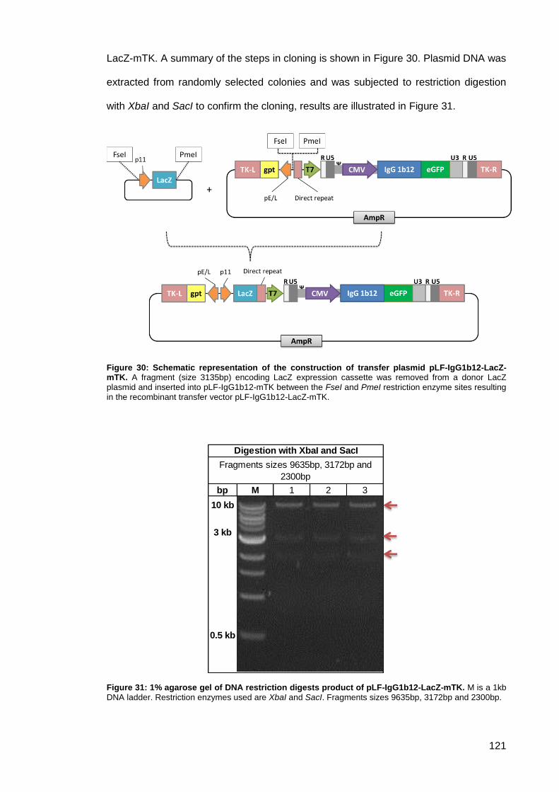
121
LacZ-mTK. A summary of the steps in cloning is shown in Figure 30. Plasmid DNA was
extracted from randomly selected colonies and was subjected to restriction digestion
with XbaI and SacI to confirm the cloning, results are illustrated in Figure 31.
Figure 30: Schematic representation of the construction of transfer plasmid pLF-IgG1b12-LacZ-mTK. A fragment (size 3135bp) encoding LacZ expression cassette was removed from a donor LacZ plasmid and inserted into pLF-IgG1b12-mTK between the FseI and PmeI restriction enzyme sites resulting in the recombinant transfer vector pLF-IgG1b12-LacZ-mTK.
Figure 31: 1% agarose gel of DNA restriction digests product of pLF-IgG1b12-LacZ-mTK. M is a 1kb DNA ladder. Restriction enzymes used are XbaI and SacI. Fragments sizes 9635bp, 3172bp and 2300bp.
bp M 1 2 3
10 kb
3 kb
0.5 kb
Digestion with XbaI and SacI
Fragments sizes 9635bp, 3172bp and
2300bp
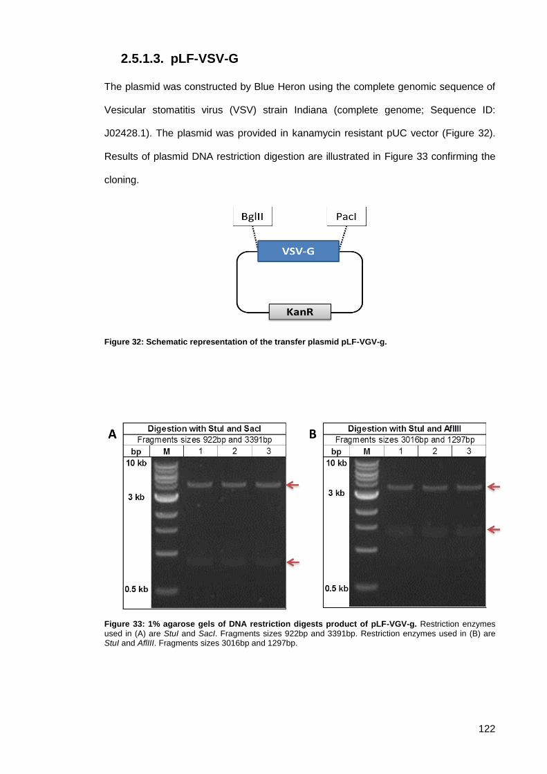
122
pLF-VSV-G 2.5.1.3.
The plasmid was constructed by Blue Heron using the complete genomic sequence of
Vesicular stomatitis virus (VSV) strain Indiana (complete genome; Sequence ID:
J02428.1). The plasmid was provided in kanamycin resistant pUC vector (Figure 32).
Results of plasmid DNA restriction digestion are illustrated in Figure 33 confirming the
cloning.
Figure 32: Schematic representation of the transfer plasmid pLF-VGV-g.
Figure 33: 1% agarose gels of DNA restriction digests product of pLF-VGV-g. Restriction enzymes used in (A) are StuI and SacI. Fragments sizes 922bp and 3391bp. Restriction enzymes used in (B) are StuI and AflIII. Fragments sizes 3016bp and 1297bp.
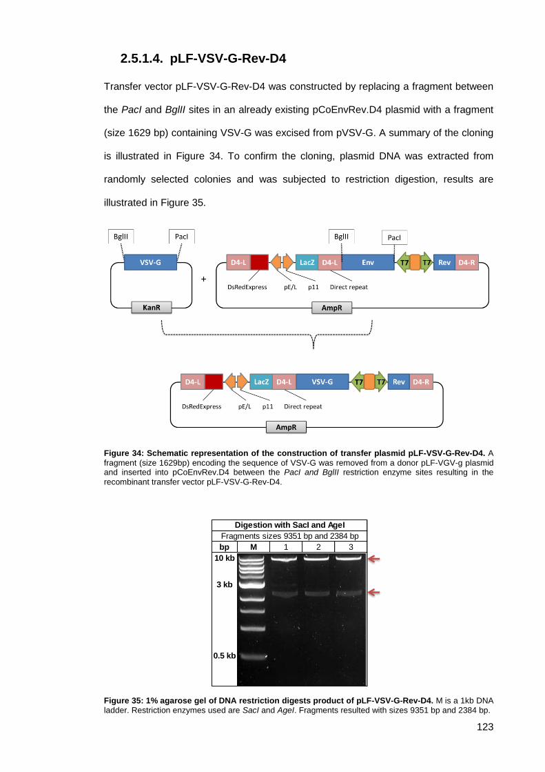
123
pLF-VSV-G-Rev-D4 2.5.1.4.
Transfer vector pLF-VSV-G-Rev-D4 was constructed by replacing a fragment between
the PacI and BglII sites in an already existing pCoEnvRev.D4 plasmid with a fragment
(size 1629 bp) containing VSV-G was excised from pVSV-G. A summary of the cloning
is illustrated in Figure 34. To confirm the cloning, plasmid DNA was extracted from
randomly selected colonies and was subjected to restriction digestion, results are
illustrated in Figure 35.
Figure 34: Schematic representation of the construction of transfer plasmid pLF-VSV-G-Rev-D4. A
fragment (size 1629bp) encoding the sequence of VSV-G was removed from a donor pLF-VGV-g plasmid and inserted into pCoEnvRev.D4 between the PacI and BglII restriction enzyme sites resulting in the
recombinant transfer vector pLF-VSV-G-Rev-D4.
Figure 35: 1% agarose gel of DNA restriction digests product of pLF-VSV-G-Rev-D4. M is a 1kb DNA ladder. Restriction enzymes used are SacI and AgeI. Fragments resulted with sizes 9351 bp and 2384 bp.
bp M 1 2 3
10 kb
3 kb
0.5 kb
Digestion with SacI and AgeI
Fragments sizes 9351 bp and 2384 bp
124
pLF-IgG1b12-LacZ-D4 2.5.1.5.
The modification to the TK locus region during the design of the transfer plasmid was
lethal to the rMVA expressing IgG. Thus, shifting the insertion site of the IgG
expression cassette from TK locus to a different insertion site was fundamental. A
fragment size of 5650bp encoding IgG expression from the TK targeting plasmid pLF-
IgG1b12-LacZ-mTK replaced the expression cassette of pLF-VSV-G-Rev-D4 at the
AscI site, resulting in the formation of pLF-IgG1b12-LacZ-D4 which target Del4. A
summary of the cloning is illustrated in Figure 36. Plasmid DNA was extracted from
randomly selected colonies to confirm the cloning by restriction digest; results are
illustrated in Figure 37.
Figure 36: Schematic representation of the construction of transfer plasmid pLF-IgG1b12-LacZ-D4.
A fragment (size 5650bp) encoding IgG expression from pLF-IgG1b12-LacZ-mTK replaced the expression cassette of pLF-VSV-G-Rev-D4 at the AscI site, resulting in the formation of pLF-IgG1b12-LacZ-D4.
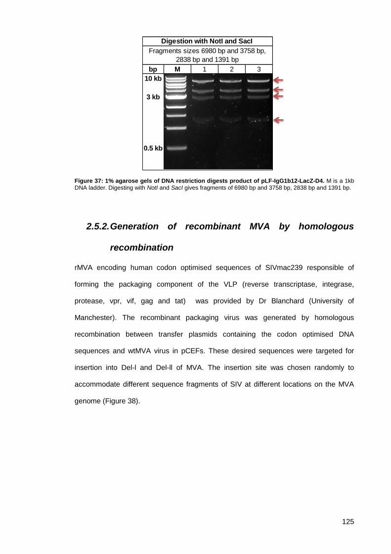
125
Figure 37: 1% agarose gels of DNA restriction digests product of pLF-IgG1b12-LacZ-D4. M is a 1kb DNA ladder. Digesting with NotI and SacI gives fragments of 6980 bp and 3758 bp, 2838 bp and 1391 bp.
2.5.2. Generation of recombinant MVA by homologous
recombination
rMVA encoding human codon optimised sequences of SIVmac239 responsible of
forming the packaging component of the VLP (reverse transcriptase, integrase,
protease, vpr, vif, gag and tat) was provided by Dr Blanchard (University of
Manchester). The recombinant packaging virus was generated by homologous
recombination between transfer plasmids containing the codon optimised DNA
sequences and wtMVA virus in pCEFs. These desired sequences were targeted for
insertion into Del-l and Del-ll of MVA. The insertion site was chosen randomly to
accommodate different sequence fragments of SIV at different locations on the MVA
genome (Figure 38).
bp M 1 2 3
10 kb
3 kb
0.5 kb
Fragments sizes 6980 bp and 3758 bp,
2838 bp and 1391 bp
Digestion with NotI and SacI
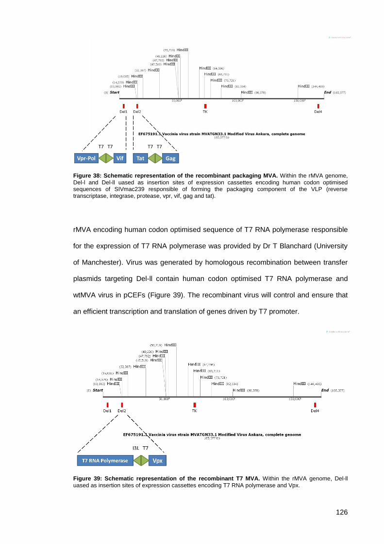
126
Figure 38: Schematic representation of the recombinant packaging MVA. Within the rMVA genome,
Del-l and Del-ll uased as insertion sites of expression cassettes encoding human codon optimised sequences of SIVmac239 responsible of forming the packaging component of the VLP (reverse transcriptase, integrase, protease, vpr, vif, gag and tat).
rMVA encoding human codon optimised sequence of T7 RNA polymerase responsible
for the expression of T7 RNA polymerase was provided by Dr T Blanchard (University
of Manchester). Virus was generated by homologous recombination between transfer
plasmids targeting Del-ll contain human codon optimised T7 RNA polymerase and
wtMVA virus in pCEFs (Figure 39). The recombinant virus will control and ensure that
an efficient transcription and translation of genes driven by T7 promoter.
Figure 39: Schematic representation of the recombinant T7 MVA. Within the rMVA genome, Del-ll
uased as insertion sites of expression cassettes encoding T7 RNA polymerase and Vpx.
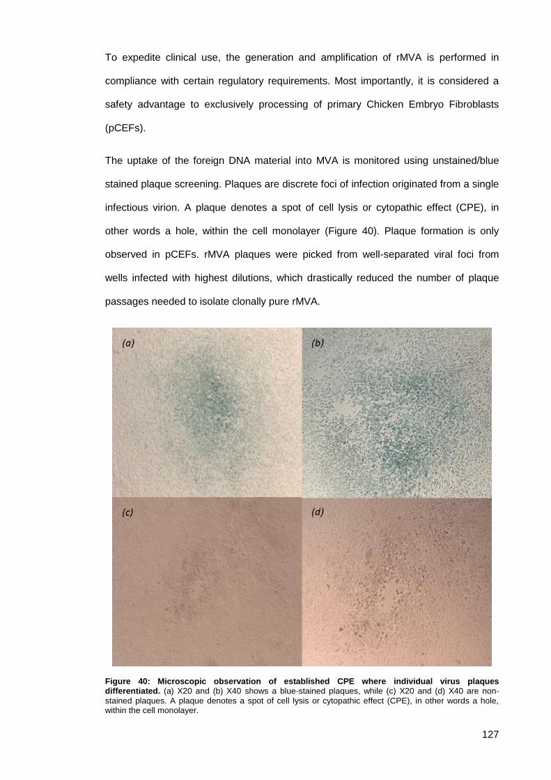
127
To expedite clinical use, the generation and amplification of rMVA is performed in
compliance with certain regulatory requirements. Most importantly, it is considered a
safety advantage to exclusively processing of primary Chicken Embryo Fibroblasts
(pCEFs).
The uptake of the foreign DNA material into MVA is monitored using unstained/blue
stained plaque screening. Plaques are discrete foci of infection originated from a single
infectious virion. A plaque denotes a spot of cell lysis or cytopathic effect (CPE), in
other words a hole, within the cell monolayer (Figure 40). Plaque formation is only
observed in pCEFs. rMVA plaques were picked from well-separated viral foci from
wells infected with highest dilutions, which drastically reduced the number of plaque
passages needed to isolate clonally pure rMVA.
Figure 40: Microscopic observation of established CPE where individual virus plaques differentiated. (a) X20 and (b) X40 shows a blue-stained plaques, while (c) X20 and (d) X40 are non-
stained plaques. A plaque denotes a spot of cell lysis or cytopathic effect (CPE), in other words a hole, within the cell monolayer.
128
Homologous recombination between the transfer plasmid vector and the virus was
visualised in infected pCEF monolayers by the formation of blue plaques in the
presence of X-gal (Figure 41). The primary overlay medium prevents the development
of secondary plaques by forcing the released virus particles from the infected cell to
infect only neighbouring cells. rMVA incorporating and expressing gpt was first
subjected for an initial round of selection in the presence of mycophenolic acid (MPA).
This selection system was carried out only for the first round of plaque purification. It is
based on plaque isolation in the presence of MPA, an inhibitor of purine metabolism.
Recombinant virus incorporating and expressing the E.coli xanthine-guanine
phosphoribosyl transferase gene (gpt), unlike normal gpt(-) virus, can form plaques in
medium containing MPA and the nucleotide precursors xanthine and hypoxanthine.
Figure 41: Blue plaque formation on 6-well dish. (A) A well visualis infected pCEF monolayers with
rMVA through the formation of blue plaques in the presence of X-gal. (B) A well shows uninfected pCEF monolayers.
Four consecutive rounds of blue plaque selection in the presence of X-gal was
conducted, then the plates were screened for non-stained plaques for the loss of LacZ.
A higher dilution of virus inoculum was related to better plaque separation and an
intense blue colour formation. Starting from the fifth round of plaque selection, non-
stained viral foci in the presence of X-gal were selected. The formation of non-stained
plaques resulted from a second intragenomic recombination event where the reporter
gene from rMVA is removed. In regard to a complex vaccine for human use, the
construction of a marker-free variant is very desirable. Plaques were picked only after
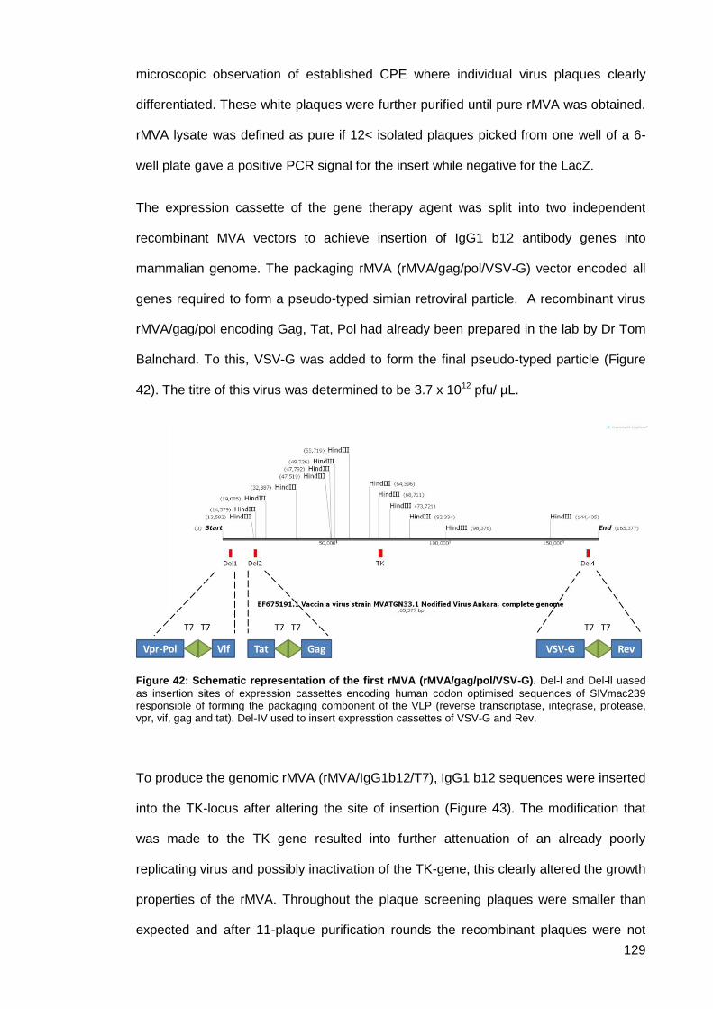
129
microscopic observation of established CPE where individual virus plaques clearly
differentiated. These white plaques were further purified until pure rMVA was obtained.
rMVA lysate was defined as pure if 12< isolated plaques picked from one well of a 6-
well plate gave a positive PCR signal for the insert while negative for the LacZ.
The expression cassette of the gene therapy agent was split into two independent
recombinant MVA vectors to achieve insertion of IgG1 b12 antibody genes into
mammalian genome. The packaging rMVA (rMVA/gag/pol/VSV-G) vector encoded all
genes required to form a pseudo-typed simian retroviral particle. A recombinant virus
rMVA/gag/pol encoding Gag, Tat, Pol had already been prepared in the lab by Dr Tom
Balnchard. To this, VSV-G was added to form the final pseudo-typed particle (Figure
42). The titre of this virus was determined to be 3.7 x 10¹² pfu/ µL.
Figure 42: Schematic representation of the first rMVA (rMVA/gag/pol/VSV-G). Del-l and Del-ll uased
as insertion sites of expression cassettes encoding human codon optimised sequences of SIVmac239 responsible of forming the packaging component of the VLP (reverse transcriptase, integrase, protease, vpr, vif, gag and tat). Del-IV used to insert expresstion cassettes of VSV-G and Rev.
To produce the genomic rMVA (rMVA/IgG1b12/T7), IgG1 b12 sequences were inserted
into the TK-locus after altering the site of insertion (Figure 43). The modification that
was made to the TK gene resulted into further attenuation of an already poorly
replicating virus and possibly inactivation of the TK-gene, this clearly altered the growth
properties of the rMVA. Throughout the plaque screening plaques were smaller than
expected and after 11-plaque purification rounds the recombinant plaques were not

130
detectable. Suggesting that the TK deletion incorporated in the plasmid design
terminated the viral replication.
Figure 43: Schematic representation of the second rMVA (rMVA/IgG1b12/T7). Del-ll uased as
insertion sites of expression cassettes encoding T7 RNA polymerase and Vpx, while TK locus used to insert the IgG1b12 expression cassette.
Cloning the IgG1b12 sequences into a different insertion site was successfully
performed using Del site IV (refer to section 2.5.1.5). This time rMVA encoding T7
polymerase was used to produce the recombinant virus (Figure 44). However, no CPE
was observed when the virus was cultured. Several attempts were carried to form a
successful recombination. However, each time there was no CPE detected which might
indicate the failure of the formation of recombinant virus.
Figure 44: Schematic representation of the second rMVA (rMVA/IgG1b12/T7). Del-ll uased as
insertion sites of expression cassettes encoding T7 RNA polymerase and Vpx, while Del-IV used to insert the IgG1b12 expression cassette.
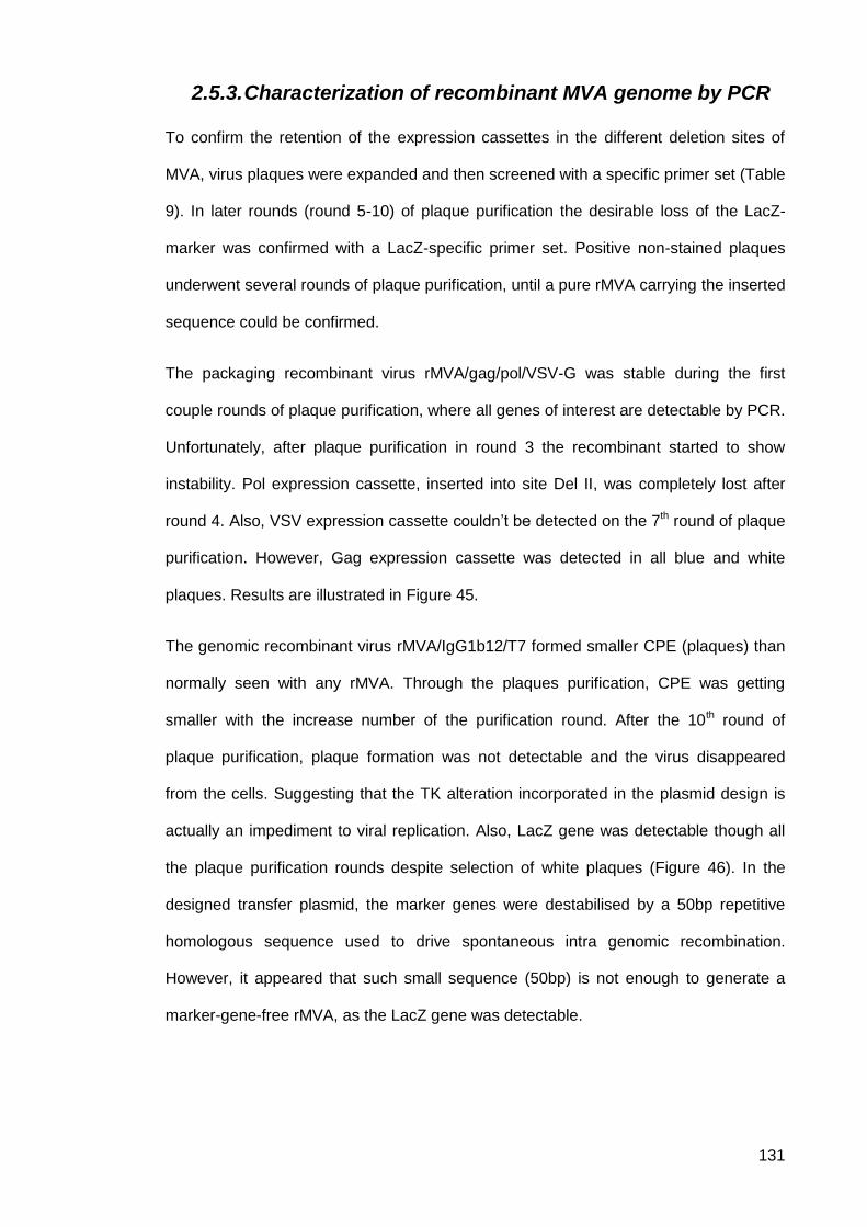
131
2.5.3. Characterization of recombinant MVA genome by PCR
To confirm the retention of the expression cassettes in the different deletion sites of
MVA, virus plaques were expanded and then screened with a specific primer set (Table
9). In later rounds (round 5-10) of plaque purification the desirable loss of the LacZ-
marker was confirmed with a LacZ-specific primer set. Positive non-stained plaques
underwent several rounds of plaque purification, until a pure rMVA carrying the inserted
sequence could be confirmed.
The packaging recombinant virus rMVA/gag/pol/VSV-G was stable during the first
couple rounds of plaque purification, where all genes of interest are detectable by PCR.
Unfortunately, after plaque purification in round 3 the recombinant started to show
instability. Pol expression cassette, inserted into site Del II, was completely lost after
round 4. Also, VSV expression cassette couldn’t be detected on the 7th round of plaque
purification. However, Gag expression cassette was detected in all blue and white
plaques. Results are illustrated in Figure 45.
The genomic recombinant virus rMVA/IgG1b12/T7 formed smaller CPE (plaques) than
normally seen with any rMVA. Through the plaques purification, CPE was getting
smaller with the increase number of the purification round. After the 10th round of
plaque purification, plaque formation was not detectable and the virus disappeared
from the cells. Suggesting that the TK alteration incorporated in the plasmid design is
actually an impediment to viral replication. Also, LacZ gene was detectable though all
the plaque purification rounds despite selection of white plaques (Figure 46). In the
designed transfer plasmid, the marker genes were destabilised by a 50bp repetitive
homologous sequence used to drive spontaneous intra genomic recombination.
However, it appeared that such small sequence (50bp) is not enough to generate a
marker-gene-free rMVA, as the LacZ gene was detectable.
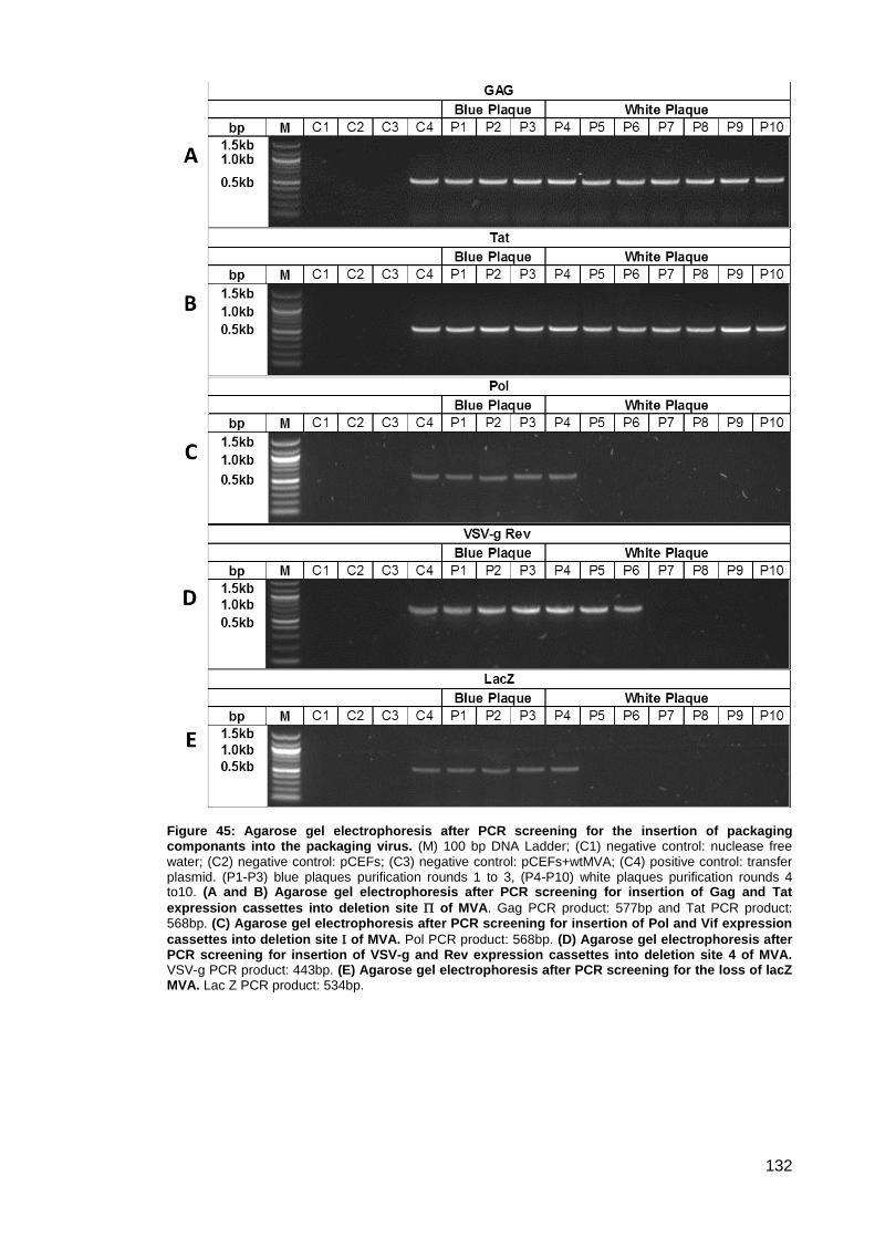
132
Figure 45: Agarose gel electrophoresis after PCR screening for the insertion of packaging componants into the packaging virus. (M) 100 bp DNA Ladder; (C1) negative control: nuclease free
water; (C2) negative control: pCEFs; (C3) negative control: pCEFs+wtMVA; (C4) positive control: transfer plasmid. (P1-P3) blue plaques purification rounds 1 to 3, (P4-P10) white plaques purification rounds 4 to10. (A and B) Agarose gel electrophoresis after PCR screening for insertion of Gag and Tat
expression cassettes into deletion site of MVA. Gag PCR product: 577bp and Tat PCR product: 568bp. (C) Agarose gel electrophoresis after PCR screening for insertion of Pol and Vif expression
cassettes into deletion site of MVA. Pol PCR product: 568bp. (D) Agarose gel electrophoresis after PCR screening for insertion of VSV-g and Rev expression cassettes into deletion site 4 of MVA. VSV-g PCR product: 443bp. (E) Agarose gel electrophoresis after PCR screening for the loss of lacZ MVA. Lac Z PCR product: 534bp.
133
Figure 46: Agarose gel electrophoresis after PCR screening for the insertion of antibody expression cassette into TK of MVA forming the genomic virus. (M) 100 bp DNA Ladder; (C1)
negative control: nuclease free water; (C2) negative control: pCEFs; (C3) negative control: pCEFs+wtMVA; (C4) positive control: transfer plasmid. (P1-P3) blue plaques purification rounds 1 to 3, (P4-P10) white plaques purification rounds 4 to10. (A) Agarose gel electrophoresis after PCR screening for Heavy chain. HC PCR product: 606bp. (B) Agarose gel electrophoresis after PCR screening for Light chain. LC PCR product: 823bp. (C) Agarose gel electrophoresis after PCR screening for eGFP. eGFP PCR product: 473bp. (D) Agarose gel electrophoresis after PCR screening for the loss of lacZ MVA. Lac Z PCR product: 534bp.
2.5.4. Transfection
Vero cells were transfected with pLF-IgG1b12-mTK using Lipofectamine® 2000
reagent which delivers the plasmid DNA with excellent transfection performance. This
step was made to test protein expression, as the IgG sequence together with eGFP
incorporated in our plasmid is driven by CMV promoter. Thus, it is speculated that if the
plasmid was delivered inside the cells proteins will be expressed and green fluorescent
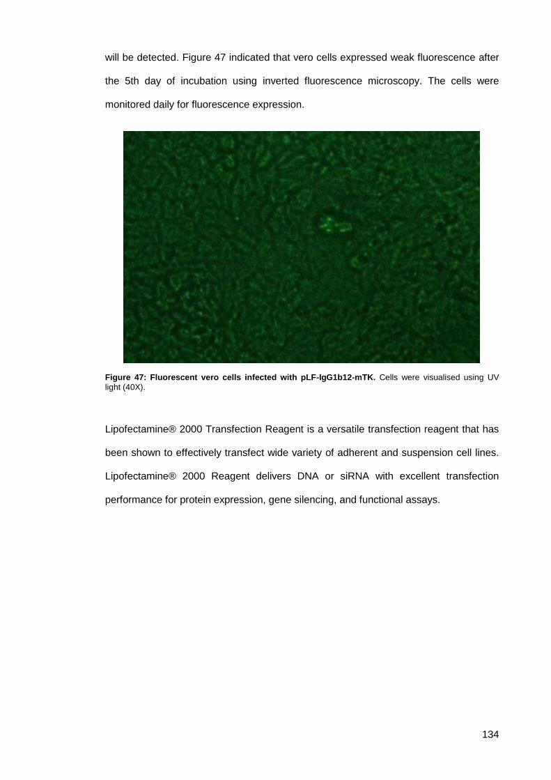
134
will be detected. Figure 47 indicated that vero cells expressed weak fluorescence after
the 5th day of incubation using inverted fluorescence microscopy. The cells were
monitored daily for fluorescence expression.
Figure 47: Fluorescent vero cells infected with pLF-IgG1b12-mTK. Cells were visualised using UV
light (40X).
Lipofectamine® 2000 Transfection Reagent is a versatile transfection reagent that has
been shown to effectively transfect wide variety of adherent and suspension cell lines.
Lipofectamine® 2000 Reagent delivers DNA or siRNA with excellent transfection
performance for protein expression, gene silencing, and functional assays.
135
Discussion and Conclusion 2.6.
This project used a replication-restricted poxvirus split-vector system developed
utilising a Vaccinia Retroviral Hybrid Vector, where MVA is used to deliver SIV like
particles into mammalian cells. The first hybrid dual-vector system was developed to
double the packaging capacity of recombinant adeno-associated virus (rAAV) by
splitting the expression cassette between two independent AAV vectors (Beaudoing &
Gautheret 2001; McClements & MacLaren 2017). Studies indicated that this split vector
system was safe in vivo and stable during vaccine production and probably enabled
higher titres of vaccine.
In this study, MVA was employed as the viral vector to overcome the limited packaging
capacity of AAV and other vectors. The research hypothesis is illustrated in Figure 48.
The recombination between the split-vector recombinants with MVA origin will result in
the formation and the production of simian like particles. The resulted VLPs are
attenuated and thus not fully replication-competent retroviruses. The fact that SIV
replicates and infects several species of African non-human primates, gives the system
an excellent safety profile.
VLPs used encode HIV-neutralising monoclonal antibody sequences into its core to
function as a simian retroviral gene therapy agent. Theses virus particles are pseudo-
typed with vesicular stomatitis virus glycoprotein (VSV-G). Pseudotyping with VSV-G
was performed to expand the retroviral tropism. The replication-restricted poxvirus split-
vector system developed is composed of recombinant MVA, rMVA/gag/pol/VSV-G and
rMVA/IgG1b12/T7 responsible for the production of the packaging component and the
neutralising antibody respectively.
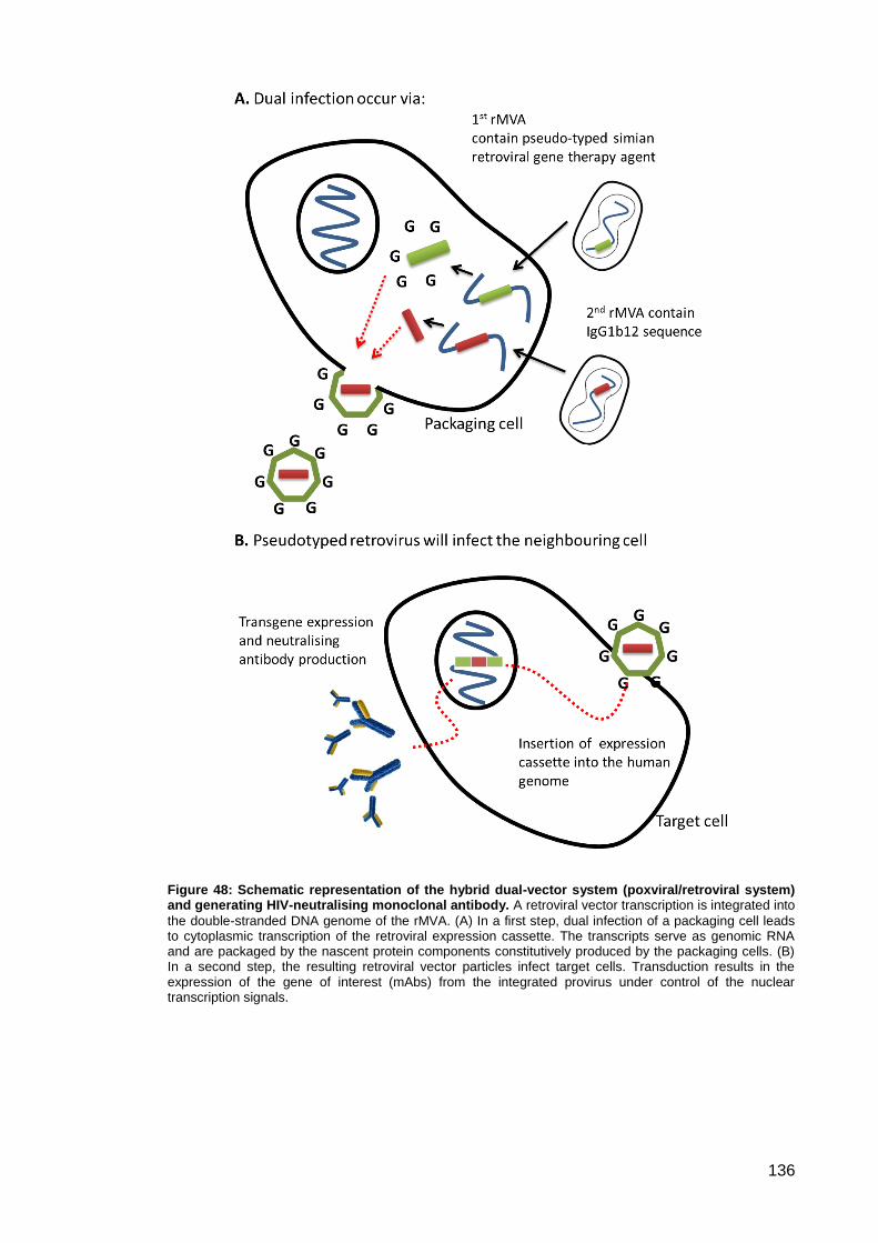
136
Figure 48: Schematic representation of the hybrid dual-vector system (poxviral/retroviral system) and generating HIV-neutralising monoclonal antibody. A retroviral vector transcription is integrated into
the double-stranded DNA genome of the rMVA. (A) In a first step, dual infection of a packaging cell leads to cytoplasmic transcription of the retroviral expression cassette. The transcripts serve as genomic RNA and are packaged by the nascent protein components constitutively produced by the packaging cells. (B) In a second step, the resulting retroviral vector particles infect target cells. Transduction results in the expression of the gene of interest (mAbs) from the integrated provirus under control of the nuclear transcription signals.
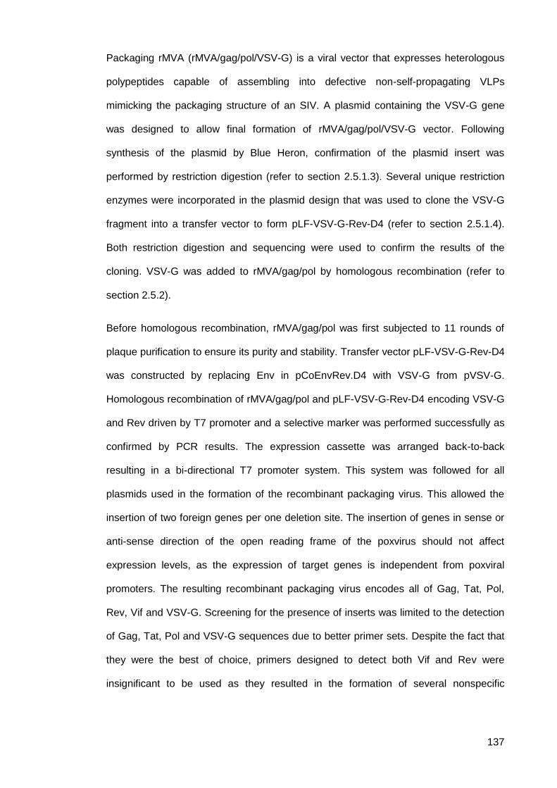
137
Packaging rMVA (rMVA/gag/pol/VSV-G) is a viral vector that expresses heterologous
polypeptides capable of assembling into defective non-self-propagating VLPs
mimicking the packaging structure of an SIV. A plasmid containing the VSV-G gene
was designed to allow final formation of rMVA/gag/pol/VSV-G vector. Following
synthesis of the plasmid by Blue Heron, confirmation of the plasmid insert was
performed by restriction digestion (refer to section 2.5.1.3). Several unique restriction
enzymes were incorporated in the plasmid design that was used to clone the VSV-G
fragment into a transfer vector to form pLF-VSV-G-Rev-D4 (refer to section 2.5.1.4).
Both restriction digestion and sequencing were used to confirm the results of the
cloning. VSV-G was added to rMVA/gag/pol by homologous recombination (refer to
section 2.5.2).
Before homologous recombination, rMVA/gag/pol was first subjected to 11 rounds of
plaque purification to ensure its purity and stability. Transfer vector pLF-VSV-G-Rev-D4
was constructed by replacing Env in pCoEnvRev.D4 with VSV-G from pVSV-G.
Homologous recombination of rMVA/gag/pol and pLF-VSV-G-Rev-D4 encoding VSV-G
and Rev driven by T7 promoter and a selective marker was performed successfully as
confirmed by PCR results. The expression cassette was arranged back-to-back
resulting in a bi-directional T7 promoter system. This system was followed for all
plasmids used in the formation of the recombinant packaging virus. This allowed the
insertion of two foreign genes per one deletion site. The insertion of genes in sense or
anti-sense direction of the open reading frame of the poxvirus should not affect
expression levels, as the expression of target genes is independent from poxviral
promoters. The resulting recombinant packaging virus encodes all of Gag, Tat, Pol,
Rev, Vif and VSV-G. Screening for the presence of inserts was limited to the detection
of Gag, Tat, Pol and VSV-G sequences due to better primer sets. Despite the fact that
they were the best of choice, primers designed to detect both Vif and Rev were
insignificant to be used as they resulted in the formation of several nonspecific
138
fragments and thus cannot be reliable for screening. This is related to the small size of
the sequence of interest, the template.
In the first round of plaque formation, 40 blue-stained plaques were picked for initial
screening. Half of these blue-stained plaques were positive for Gag, Pol and VSV-G.
Out of these, a random blue-stained plaque was selected for further purification. In the
second round 36 blue-stained plaques were picked. 12 were screened but only 7 were
positive. In the third round, 19 blue-stained plaques were selected. 10 plaques were
screened and all were positive for all inserts. This suggests that the blue-stained
plaque selected on round two formed from a stable recombinant virus. In the fourth
round, 20 blue-stained plaques were selected and screened. All of which lost the pol
construct completely, while still encoding Gag, Tat, Rev and VSV-G constructs.
Following several re-attempts and around 60 blue-stained plaques screening, only one
plaque was found to give positive signal for all inserts during the fourth round of
screening. Unfortunately, by the fifth round the Pol construct was again lost. This
indicates that Del site I used in inserting the Pol construct is dramatically unstable.
Insertion of the expression cassette into a different deletion site was a necessary
following step. The resulted recombinant virus (after the loss of Pol) rMVA/gag/VAS-G
was further purified to re-insert the Pol cassette into it targeting different insertion site.
However, during purification Del site IV started to show instability, as VSV-G could not
be detected in all of the screened plaques during plaque purification round 7 (refer to
section 2.5.3). Further work on the recombinant virus was suspended as due to
shortage of pCEFs supply.
For the genomic (rMVA/IgG1b12/T7) recombinant virus, a plasmid-based transfer
vector, encoding monoclonal antibody IgG1b12 expressed under the control of a CMV
promoter flanked by an LTR of simian origin, was designed and the whole expression
cassette was driven by a T7 promoter. Homologous recombination between wtMVA
and the plasmid resulted in the formation of recombinant virus. Detailed mapping of the
transfer plasmid is found in section 2.4.1.1.1. The backbone of all plasmids used was
139
pBR322 which is known to cause less metabolic stress to E.coli due to its low copy
number. This can result in lower DNA yield but enhances the stability of large
recombinant DNA inserts. The TK locus was selected to be the insertion site. Unlike
deletion regions, the TK locus is well characterised to allow genetically stable
integration of foreign genes in many vaccinia strains (Scheiflinger et al. 1996). This will
provide much more stable recombinant virus especially where the insertion gene is of
large size. The gpt and lacZ dual selection system was used to reduce rounds of
purification of the recombinant MVA as well as to increase the specificity of the
selection (Morrow et al. 2012).
Optimised promoters were used in rMVA vaccine development, these can significantly
enhance the immune response and potentially lower the dose required for vectored
immunoprophylaxis. IE promoter was inserted to drive the marker expression. A T7
promoter was placed upstream in the R region (U5 region + 5’LTR) and the tRNA
primer-binding site to drive transcription of the immunoglobulin expression cassette in
the presence of T7 RNA polymerase. CMV IE promoter was added to drive the
expression of the monoclonal antibody sequence and the eGFP after integration in
nuclear DNA. To avoid the resulting transcriptional interference by using multiple
promoters in close proximity, a single strong promoter was used in this study. CMV IE
is known to be optimal in driving LC-HC expression. Protein coding sequences must
contain a translational start codon (ATG) and translational stop codon, plus a Kozak
sequence upstream of the start codon. Thus, Kozak consensus sequences were added
to ensure efficient translation.
In retroviruses, the long terminal repeat (LTR) is the control centre for gene expression.
All of the requisite signals for gene expression are found in the LTRs: Enhancer,
promoter, transcription initiation (capping), transcription terminator and polyadenylation
signal. Expression directed by the viral LTR signals is carried out entirely by host cell
enzymes (RNA pol II, poly A synthetase, guanyl transferase). The enhancer and other
transcription regulatory signals are contained in the U3 region of the 5' LTR, and the
140
TATA box is located roughly 25 bp from the beginning of the R sequence. The
integrated provirus has two LTRs. 5' LTR normally acts as an RNA pol II promoter and
3' LTR acts in transcription termination and polyadenylation. The transcript begins, by
definition, at the beginning of R, it is capped, and proceeds through U5 and the rest of
the provirus, and is usually terminated by the addition of a poly A tract just after the R
sequence in the 3' LTR. In addition, the R region in this plasmid was introduced to
serve as regions of homologous recombination. This would allow the integration of the
IgG1b12 mAb gene sequences into the packaging virus in the presence of T7
polymerase.
To shorten the time required to generate and clone rMVAs and to increase the level of
expression of the gene of interest, we employed a tri-cistronic cassette under the
control of a single promoter. The strategy is based on the use of an internal ribosomal
entry site (IRES) sequence that permits both the gene of interest and the selection
marker to be translated from a single tricistronic mRNA (Borman et al. 1997; Gaines &
Wojchowski 1999). When an IRES element is placed between two genes, the cap-
independent translation of the second gene is mediated. This cap-independent
translation is less efficient than a typical cap-dependent translation, resulting in lower
peptide levels for the second gene. Using such a design enables strict control of the
amounts of light chain (LC) to heavy chain (HC) peptide. It has been found that IRES-
mediated tri-cistronic vector with LC as the first cistron controlled the LC:HC at a 4:1
ratio in all the clones. This design is preferred because excess LC is found to generate
a greater proportion of high mAb-producing clones where the product has a low
aggregation and consistent glycosylation. Green fluorescent protein (GFP) was added
after the mAb sequence to ease the detection of the protein expression.
The screening system relies on the co-production of the reporter enzyme, E.coli β-
galactosidase, which allows screening of the foci of the MVA-infected cells by
colorimetric assays as well as the co-expression of the E.coli gpt gene encoding
xanthine guanine-phosphoribosyl transferase, which allows the selection of resistance
141
against the antimicrobial agent, mycophenolic acid. The gpt and lacZ marking system
is frequently used in recombinant virus purification. This method would allow dominant
positive selection of the resulted rMVA for resistance against mycophenolic acid as well
as blue plaque formation (Morrow et al. 2012; Schnierle et al. 2007; Scheiflinger et al.
1996).
Free plasmid DNA was ordered from Blue Heron to perform transfection. As GFP was
incorporated in our plasmid and driven by CMV promoter, expression of green
fluoresce could be detected if DNA plasmid were introduced to mammalian cells by
transfection. Lipofectamine® 2000 was used as a transfection reagent to couple the
expression plasmid to a cationic lipid or polymer producing a liposome that interacts
with the cell membrane and results in endocytosis of the molecule. Vero cells were
used to perform the transfection. After the fifth day of incubation, weak fluorescence
was detected using an inverted fluorescence microscope. This confirmed that the CMV
promoter expressed the gene of interest if the open reading frame was delivered to the
cell nucleus.
Following receipt of the synthesised plasmid from Blue Heron restriction digestion was
conducted and DNA digests were analysed by gel electrophoresis confirming the
plasmid sequence. LacZ was cloned successfully in the plasmid which was confirmed
by restriction digest and sequencing. Homologous recombination between the transfer
plasmid and wt-MVA was performed to form the genomic rMVA. Following the
formation of the recombinant virus, an initial round of plaque purification indicated that
the resultant virus encoded the gene of interest. Blue-stained plaques were selected for
screening up to round 4 then non-stained plaques were selected for virus purification.
By the 11th round of plaque purification the recombinant virus disappeared from the
cells. LacZ marker gene was detected in both blue and white plaques (refer to section
2.5.3). The modification that was made on the TK gene resulted in further attenuation
of an already poorly replicating virus and probable inactivation of the TK-gene, which
clearly altered the growth properties of the rMVA (Scheiflinger et al. 1996). Thus after
142
11-plaque purification rounds the recombinant plaques were dramatically smaller,
suggesting that the TK deletion incorporated in the plasmid design is actually an
impediment to viral replication. In addition, in the designed plasmid the marker genes
were destabilised by a 50bp repetitive homologous sequence used to drive
spontaneous intra-genomic recombination. However, after the 10th -plaque purification
round it appeared that such a small sequence is not enough to generate a marker-
gene-free rMVA, as the LacZ gene was still detectable. Findings indicated that cloning
of the expression cassette into a different insertion site was necessary, Del IV was
selected. Cloning of IgG1b12 gene into a transfer plasmid targeting D4 was performed
successfully, results are confirmed by restriction digest and sequencing. Unfortunately,
plaques formation couldn’t be detected which suggest that the homologous
recombination was not successful. In such case both T7 polymerase and T7 promoter
are present at the same time. This could possibly lead to the expression of the IgG,
where the mAb could mask and neutralise the virus. Also, the possible IgG expression
might affect the efficiency of the homologous recombination process. Thus result in the
failure of the homologous recombination. Homologous recombination between the
transfer plasmid and wild type MVA may provide the ultimate solution. However, this
means that we will use 3 rMVA to co-infect pCEFs instead of our initial plan of using
dual vector. Unfortunately, this is challenging for the cells where an overdose of MVA is
most likely to be harmful to the cells. However, further work with MVA was precluded
due to unexpected delays in chicken embryo fibroblast supply.
In conclusion, due to the challenges in the above work the whole system had to be
changed. Baculovirus work was suggested and planned.
143
Chapter 3 3.
HIV neutralising antibody delivered by gene therapy
with a hybrid baculovirus/SIV vector
Abstract 3.1.
Baculovirus (BV) (Autographa californica multiple nucleopolyhedrovirus, AcMNPV) is
an insect, enveloped, virus that is often genetically engineered to allow recombinant
protein production in insect cell culture. Conventionally, baculovirus expression
systems (BES) have provided versatile platforms used for production of a wide range of
recombinant proteins including the expression of virus-like particles (VLPs). Self-
assembly of BV produced recombinant viral capsid proteins has allowed the production
of VLPs, which resemble an authentic virion and maintains its original antigenic
composition. VLPs provide potent immunogenicity and safety when used as vaccines
given the fact that they are replication-incompetent virus shells. Baculoviruses are
capable of transducing various types of mammalian cells and thus can serve as a
vehicle to efficiently deliver genes to mammalian cells. However, these transgenes
cannot be expressed unless they are driven by an appropriate promoter such as the
CMV IE promoter (Kataoka et al. 2012). The baculovirus vector “BacMam” has been
successfully used as a transient expression vector for gene delivery in mammalian
cells. The BacMam system combines the advantages of transient viral expression,
ease of generation of protein, and a broad cell tropism; enabling rapid, efficient and
flexible protein expression in various mammalian cells (Kost & Condreay 2002).
The aim of this study was to develop retrovirus like particles to serve as a new gene
therapy carrier system, permitting the insertion of an IgG1 b12 sequence into the
mammalian cell genome with the intention of in vivo production of high titres of anti-HIV
neutralising monoclonal antibody. We used two different BacMam systems to allow the

144
production of VLPs by transducing mammalian cells with different recombinant
baculoviruses (rBVs). VLPs were expressed either under the control of T7 RNA
polymerase system or under the cytomegalovirus immediate early gene promoter.
This proof of principle study not only determines the feasibility of antibody gene
transfer, but also investigates the efficiency of SIV like particle production in
mammalian cells, to support their candidacy as potential vaccines and/or
immunotherapy agents.
145
Objectives 3.2.
This chapter includes two separate studies describing the construction and validation of
two independent BacMam systems for functional gene therapy and pseudotyped
retrovirus production. Generated retroviral particles delivered in vitro provide a proof of
principle to demonstrate production of long-acting immunotherapy vectors for
prevention of HIV infection and treatment of existing HIV infection.
The first BacMam system allowed expression of VLPs by transducing mammalian cells
with five different recombinant baculoviruses, under the control of the T7 RNA
polymerase system. BacMam-Gag/Tat encodes for both the group-specific antigen
(Gag) that forms the main structural proteins, and transactivator (Tat) a positive
regulator of transcription. BacMam-Pol/Vif/Vpr expresses polymerase (Pol), which
codes for reverse transcriptase (Rt), protease (Pro), and integrase (Int); together with
viral infectivity factor (Vif) and the virus protein R (Vpr), both required for virus
replication. To extend the limited cell tropism inherent in SIV, the VLP was
pseudotyped with vesicular stomatitis virus glycoprotein (VSV-G). BacMam-VSV-G/Rev
encodes both VSV-G and regulator of expression of virion (Rev) which is essential for
viral replication. BacMam-IgG1 b12 was designed to encode for the antibody gene
sequence to be packed as a core sequence of VLP. BacMam-T7 is responsible for the
expression of T7 RNA polymerase at high levels.
The second system allowed the production of VLPs by transducing mammalian cells
with four different recombinant baculoviruses, under the control of CMV IE promoter.
BacMam-Gag/Tat encodes for both the group-specific antigen (Gag) that forms the
main structural proteins, and transactivator (Tat) a positive regulator of transcription.
BacMam-Pol/Vif/Vpr expresses polymerase (Pol), which codes for reverse
transcriptase (Rt), protease (Pro) and integrase (Int); together with viral infectivity factor
(Vif) and virus protein R (Vpr), both required for the virus replication. To extend the
limited cell tropism inherant in SIV, the VLP was pseudotyped with vesicular stomatitis
virus glycoprotein (VSV-G). BacMam-VSV-G/Rev encodes both VSV-G and the

146
regulator of expression of virion (Rev) which is essential for viral replication. BacMam-
IgG1 b12 was designed to encode for the antibody gene sequence to be packed as a
core sequence of VLP.
147
Introduction 3.3.
3.3.1. Baculovirus
Baculoviruses are a large group of rod-shaped, enveloped, double-stranded DNA
viruses. Their nucleocapsid has two distinct ends: apical cap end with a small
protuberance and one end blunt (Figure 49). The size of the nucleocapsid varies,
ranging from 250 to 300 nm in length and by 30 to 60 nm in diameter. The
nucleocapsid encloses a circular, super-coiled, double-stranded DNA genome of 80–
180 kb which encodes between 90 and 180 genes. Baculoviruses infect insect larvae
most prominently of the order Lepidoptera. Members of the Baculoviridae family have
been isolated from more than 700 host species. The most extensively studied virus
used in gene expression is Autographa californica multiple nucleopolyhedrovirus
(AcMNPV) (Possee & King 2007; Jarvis 2009)
The BV genome replicates and transcribes in the nuclei of infected host cells.
Baculoviruses have a bi-phasic life cycle producing two types of infectious virual
particles: the budded virion (BV) and the occlusion-derived virion (ODV). The ODV is
involved in virus transmission as it initiates the infection at the larvae midgut of feeding
caterpillars. After this primary infection, the infectious form BV produces systemic
infection in larvae as it is responsible for cell-to-cell transmission within the host and in
cell culture. The two forms of virions are produced at different times and different
subcellular places during the replication cycle (Figure 50). Baculoviruses are produced
during the initial replication when nucleocapsids bud from infected cells. These virions
contain a single nucleocapsid and an envelope derived from the plasma membrane of
the infected cell and contains the viral fusion proteins (GP64 or F protein). The ODVs
are produced during the very late phase of replication and are formed in the nucleus by
envelopement of single or multiple nucleocapsids, which then become incorporated
within the protein matrix forming occlusion bodies (OB) or polyhedra that are released
into the environment upon death of the infected larva. The ODV infect midgut epithelial
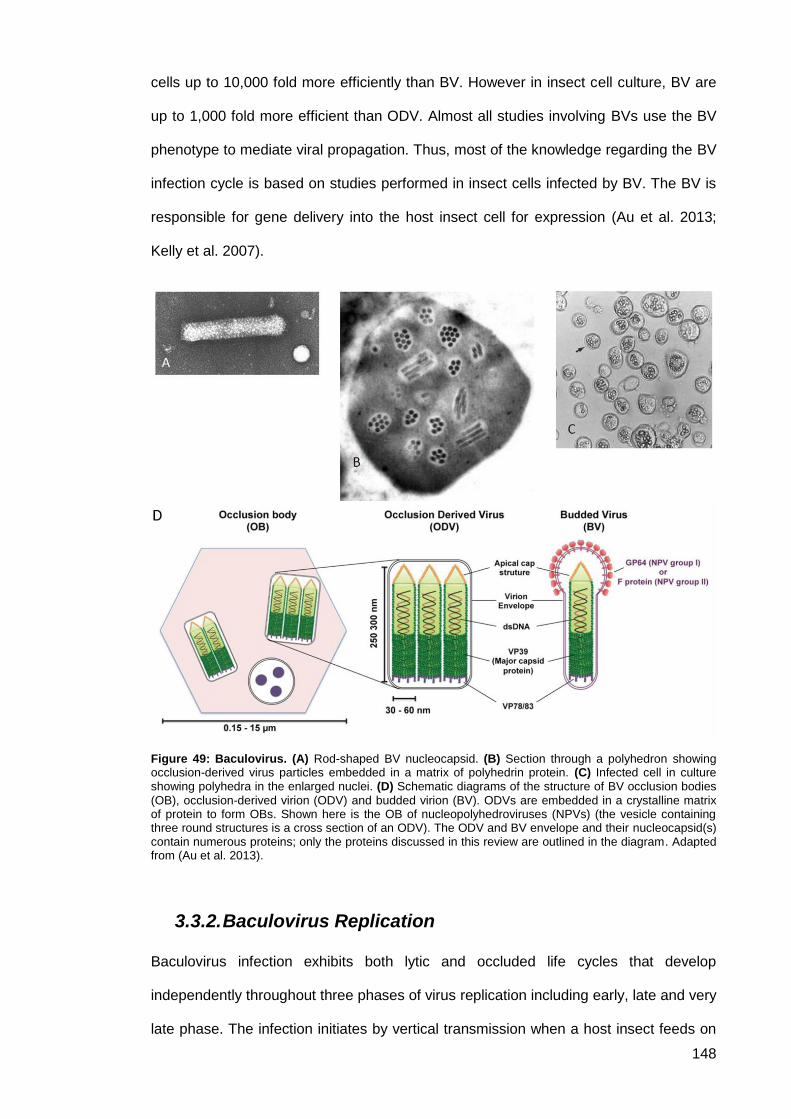
148
cells up to 10,000 fold more efficiently than BV. However in insect cell culture, BV are
up to 1,000 fold more efficient than ODV. Almost all studies involving BVs use the BV
phenotype to mediate viral propagation. Thus, most of the knowledge regarding the BV
infection cycle is based on studies performed in insect cells infected by BV. The BV is
responsible for gene delivery into the host insect cell for expression (Au et al. 2013;
Kelly et al. 2007).
Figure 49: Baculovirus. (A) Rod-shaped BV nucleocapsid. (B) Section through a polyhedron showing occlusion-derived virus particles embedded in a matrix of polyhedrin protein. (C) Infected cell in culture showing polyhedra in the enlarged nuclei. (D) Schematic diagrams of the structure of BV occlusion bodies
(OB), occlusion-derived virion (ODV) and budded virion (BV). ODVs are embedded in a crystalline matrix of protein to form OBs. Shown here is the OB of nucleopolyhedroviruses (NPVs) (the vesicle containing three round structures is a cross section of an ODV). The ODV and BV envelope and their nucleocapsid(s) contain numerous proteins; only the proteins discussed in this review are outlined in the diagram. Adapted from (Au et al. 2013).
3.3.2. Baculovirus Replication
Baculovirus infection exhibits both lytic and occluded life cycles that develop
independently throughout three phases of virus replication including early, late and very
late phase. The infection initiates by vertical transmission when a host insect feeds on
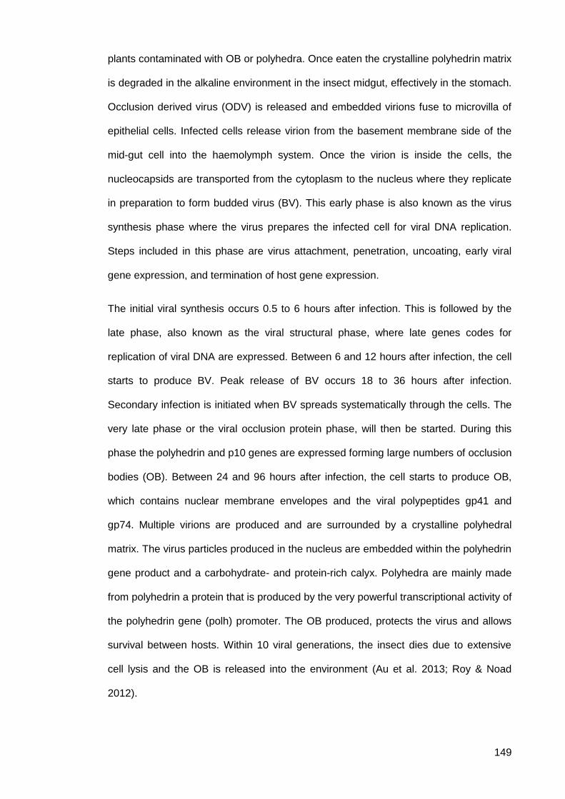
149
plants contaminated with OB or polyhedra. Once eaten the crystalline polyhedrin matrix
is degraded in the alkaline environment in the insect midgut, effectively in the stomach.
Occlusion derived virus (ODV) is released and embedded virions fuse to microvilla of
epithelial cells. Infected cells release virion from the basement membrane side of the
mid-gut cell into the haemolymph system. Once the virion is inside the cells, the
nucleocapsids are transported from the cytoplasm to the nucleus where they replicate
in preparation to form budded virus (BV). This early phase is also known as the virus
synthesis phase where the virus prepares the infected cell for viral DNA replication.
Steps included in this phase are virus attachment, penetration, uncoating, early viral
gene expression, and termination of host gene expression.
The initial viral synthesis occurs 0.5 to 6 hours after infection. This is followed by the
late phase, also known as the viral structural phase, where late genes codes for
replication of viral DNA are expressed. Between 6 and 12 hours after infection, the cell
starts to produce BV. Peak release of BV occurs 18 to 36 hours after infection.
Secondary infection is initiated when BV spreads systematically through the cells. The
very late phase or the viral occlusion protein phase, will then be started. During this
phase the polyhedrin and p10 genes are expressed forming large numbers of occlusion
bodies (OB). Between 24 and 96 hours after infection, the cell starts to produce OB,
which contains nuclear membrane envelopes and the viral polypeptides gp41 and
gp74. Multiple virions are produced and are surrounded by a crystalline polyhedral
matrix. The virus particles produced in the nucleus are embedded within the polyhedrin
gene product and a carbohydrate- and protein-rich calyx. Polyhedra are mainly made
from polyhedrin a protein that is produced by the very powerful transcriptional activity of
the polyhedrin gene (polh) promoter. The OB produced, protects the virus and allows
survival between hosts. Within 10 viral generations, the insect dies due to extensive
cell lysis and the OB is released into the environment (Au et al. 2013; Roy & Noad
2012).
150
Figure 50: Key stages in the infection cycle of AcMNPV. (A) Infection is initiated by the ingestion of a
virus occlusion body (OB). This consists of multiple virus nucleocapsids surrounded by a single lipid envelope (ODV) embedded in a protein matrix formed by the virally encoded polyhedrin protein. (B) The occlusion body is dissolved by the alkaline environment of the insect mid-gut, releasing ODV which initiates a primary infection in the midgut epithelial cells. (C) Virus enters cells and replicates in the nucleus. Two different forms of infectious virus are produced in infected cells. Budded virus (BV) is released at the cell surface and mediates systemic infection of the insect via the tracheal system, and ODV remains embedded in occlusion bodies. (D) Late stages of virus infection trigger liquefaction of the host, releasing the environmentally stable proteinaceous occlusion bodies. Polyhedrin protein is nonessential for the infection of cells in continuous culture in the laboratory and its high level of synthesis makes its promoter ideal for the high-level production of recombinant protein. Reproduced from (Roy & Noad 2012).
Despite extensive studies on the BV life cycle, several steps remains poorly
characterised. The means by which BV delivers its genomic material into the nucleus of
the infected cells, is yet to be defined. Understanding such mechanisms will help to
enhance the effectiveness of using BV in protein expression, gene therapy, and
vaccine production as nuclear importation is a vital event in the BV life cycle.
3.3.3. AcMNPV cell entry
Tjia et al. 1983, first found that BVs can be internalised by mammalian cells and at
least some of the viral DNA reached the nucleus (Tjia et al. 1983). The mechanism of
entry of AcMNPV into cells to infect insect cells and transduce mammalian cells is yet
151
to be fully understood. Cell entry of AcMNPV has been studied mainly in insect cells by
using extracellular BV (Figure 51). Infectious AcMNPV particles, in BV form, enter
susceptible insect cells through receptor-mediated endocytosis. It has been found that
the BV GP64 envelope fusion protein is involved in the initial AcMNPV attachment on
the cell surface; GP64 is acquired when the nucleocapsids bud through the host cell
plasma membrane. Following receptor-mediated internalisation of AcMNPV, GP64
undergoes conformational change into a fusion-competent state at low pH, and the
nucleocapsid is released into the cytoplasm after cell fusion. However, recent studies
suggest that AcMNPV enters cells through direct fusion with the plasma membrane at
low pH (Wang et al. 2014). GP64 is also involved in secretion of viral particles from
infected cells as GP64-null viruses exhibited around 98% reduction in viral budding.
GP64 derived from AcMNPV was shown to induce transduction of mammalian cells by
HaSNPV (Helicoverpa armigera single nucleopolyhedrovirus), a virus that does not
transduce mammalian cells.
The range of mammalian cell types transduced by HaSNPV expressing the GP64 of
AcMNPV was consistent with those transduced by AcMNPV (Lang et al., 2005). Tani et
al. 2003, reported that AcMNPV entry was inhibited when mammalian cells were
treated with purified lipids, and virus entry was significantly low when mutant hamster
cell lines deficient in phospholipid synthesis were used (Tani et al. 2003). Data from
Kaname et al. 2001, suggest that the interaction of GP64 with phospholipids on the cell
surface thus plays an important role in BV entry into mammalian cells, as GP64
interacts with CD55/decay-accelerating factor (DAF) in a lipid raft and confers
resistance to serum inactivation (Tani et al. 2003). The lipid raft is a cholesterol-rich
microdomain on the cell surface that plays crucial roles in signal transduction, protein
sorting, and membrane transport. Although further studies are needed to clarify the
involvement of cholesterol in the lipid raft in the internalisation of BV into mammalian
cells, data from Kataoka et al (2001) suggest that cholesterol participates in the
internalisation of BV but not in the binding to mammalian cells and that BV is
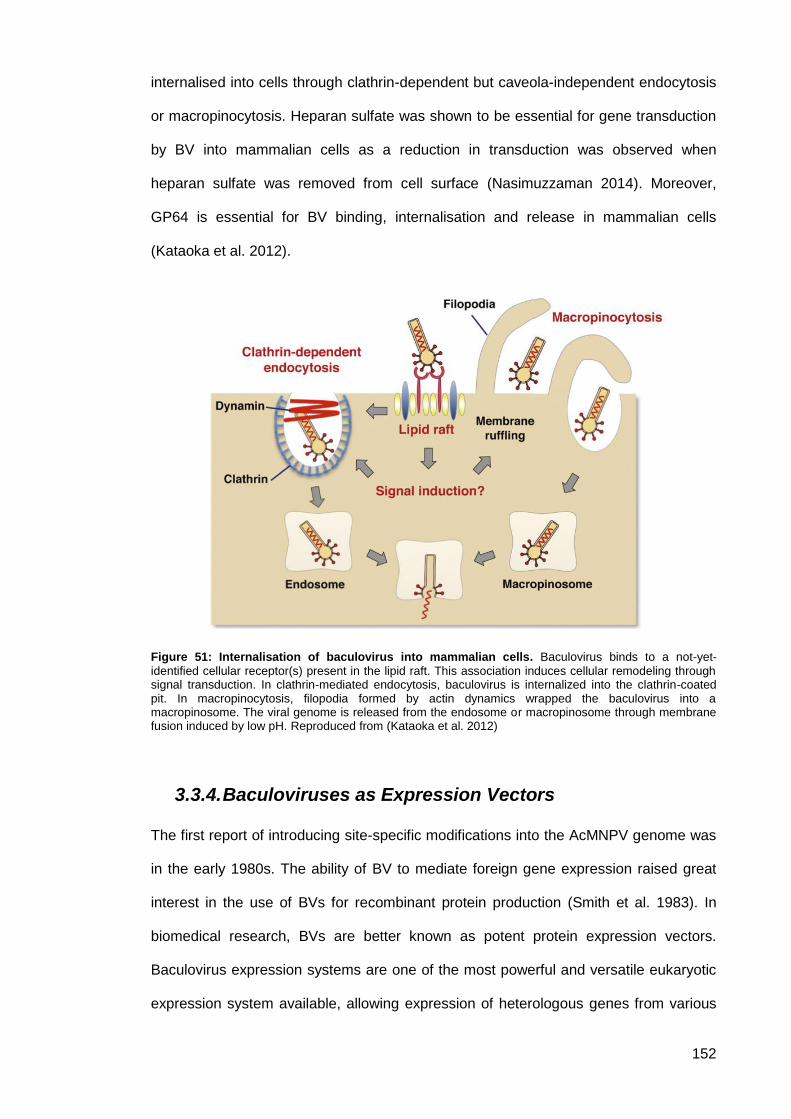
152
internalised into cells through clathrin-dependent but caveola-independent endocytosis
or macropinocytosis. Heparan sulfate was shown to be essential for gene transduction
by BV into mammalian cells as a reduction in transduction was observed when
heparan sulfate was removed from cell surface (Nasimuzzaman 2014). Moreover,
GP64 is essential for BV binding, internalisation and release in mammalian cells
(Kataoka et al. 2012).
Figure 51: Internalisation of baculovirus into mammalian cells. Baculovirus binds to a not-yet-
identified cellular receptor(s) present in the lipid raft. This association induces cellular remodeling through signal transduction. In clathrin-mediated endocytosis, baculovirus is internalized into the clathrin-coated pit. In macropinocytosis, filopodia formed by actin dynamics wrapped the baculovirus into a macropinosome. The viral genome is released from the endosome or macropinosome through membrane fusion induced by low pH. Reproduced from (Kataoka et al. 2012)
3.3.4. Baculoviruses as Expression Vectors
The first report of introducing site-specific modifications into the AcMNPV genome was
in the early 1980s. The ability of BV to mediate foreign gene expression raised great
interest in the use of BVs for recombinant protein production (Smith et al. 1983). In
biomedical research, BVs are better known as potent protein expression vectors.
Baculovirus expression systems are one of the most powerful and versatile eukaryotic
expression system available, allowing expression of heterologous genes from various
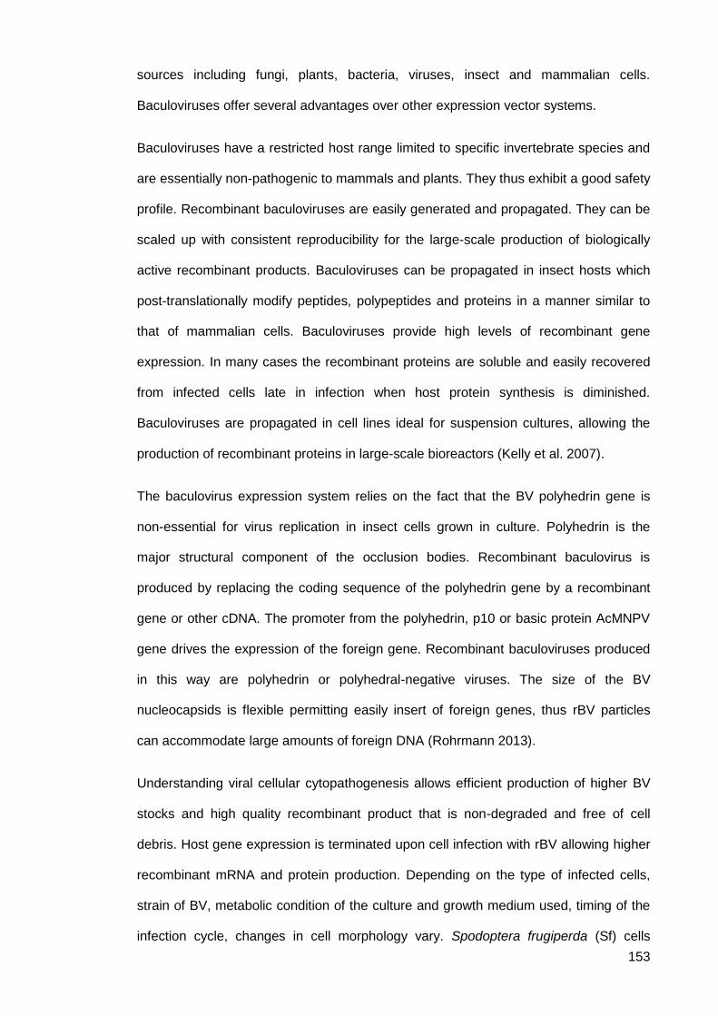
153
sources including fungi, plants, bacteria, viruses, insect and mammalian cells.
Baculoviruses offer several advantages over other expression vector systems.
Baculoviruses have a restricted host range limited to specific invertebrate species and
are essentially non-pathogenic to mammals and plants. They thus exhibit a good safety
profile. Recombinant baculoviruses are easily generated and propagated. They can be
scaled up with consistent reproducibility for the large-scale production of biologically
active recombinant products. Baculoviruses can be propagated in insect hosts which
post-translationally modify peptides, polypeptides and proteins in a manner similar to
that of mammalian cells. Baculoviruses provide high levels of recombinant gene
expression. In many cases the recombinant proteins are soluble and easily recovered
from infected cells late in infection when host protein synthesis is diminished.
Baculoviruses are propagated in cell lines ideal for suspension cultures, allowing the
production of recombinant proteins in large-scale bioreactors (Kelly et al. 2007).
The baculovirus expression system relies on the fact that the BV polyhedrin gene is
non-essential for virus replication in insect cells grown in culture. Polyhedrin is the
major structural component of the occlusion bodies. Recombinant baculovirus is
produced by replacing the coding sequence of the polyhedrin gene by a recombinant
gene or other cDNA. The promoter from the polyhedrin, p10 or basic protein AcMNPV
gene drives the expression of the foreign gene. Recombinant baculoviruses produced
in this way are polyhedrin or polyhedral-negative viruses. The size of the BV
nucleocapsids is flexible permitting easily insert of foreign genes, thus rBV particles
can accommodate large amounts of foreign DNA (Rohrmann 2013).
Understanding viral cellular cytopathogenesis allows efficient production of higher BV
stocks and high quality recombinant product that is non-degraded and free of cell
debris. Host gene expression is terminated upon cell infection with rBV allowing higher
recombinant mRNA and protein production. Depending on the type of infected cells,
strain of BV, metabolic condition of the culture and growth medium used, timing of the
infection cycle, changes in cell morphology vary. Spodoptera frugiperda (Sf) cells
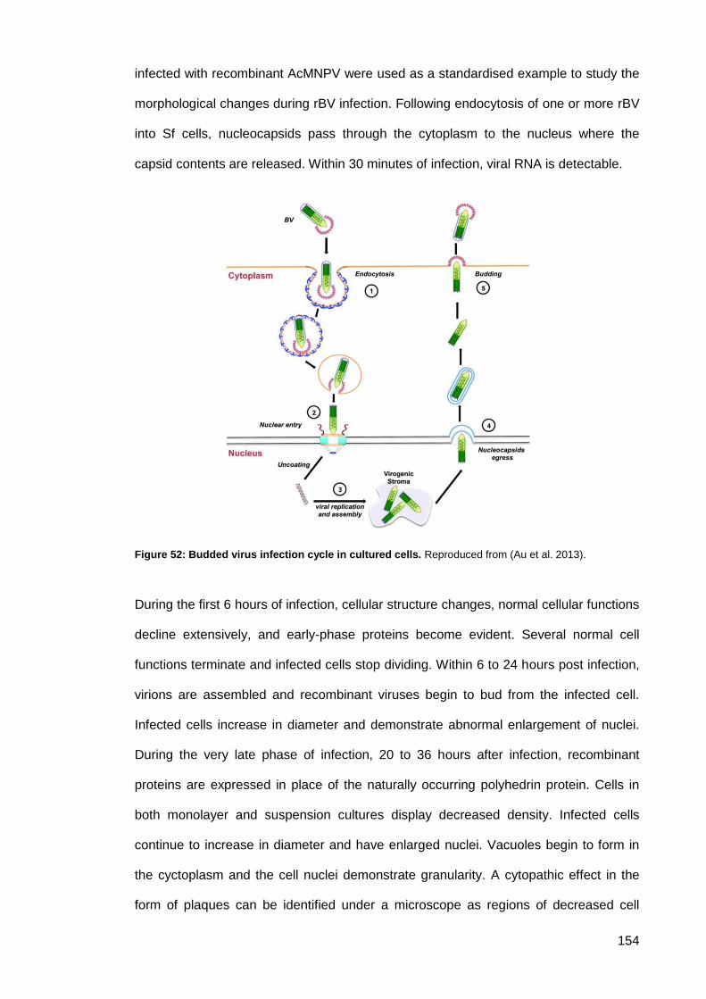
154
infected with recombinant AcMNPV were used as a standardised example to study the
morphological changes during rBV infection. Following endocytosis of one or more rBV
into Sf cells, nucleocapsids pass through the cytoplasm to the nucleus where the
capsid contents are released. Within 30 minutes of infection, viral RNA is detectable.
Figure 52: Budded virus infection cycle in cultured cells. Reproduced from (Au et al. 2013).
During the first 6 hours of infection, cellular structure changes, normal cellular functions
decline extensively, and early-phase proteins become evident. Several normal cell
functions terminate and infected cells stop dividing. Within 6 to 24 hours post infection,
virions are assembled and recombinant viruses begin to bud from the infected cell.
Infected cells increase in diameter and demonstrate abnormal enlargement of nuclei.
During the very late phase of infection, 20 to 36 hours after infection, recombinant
proteins are expressed in place of the naturally occurring polyhedrin protein. Cells in
both monolayer and suspension cultures display decreased density. Infected cells
continue to increase in diameter and have enlarged nuclei. Vacuoles begin to form in
the cyctoplasm and the cell nuclei demonstrate granularity. A cytopathic effect in the
form of plaques can be identified under a microscope as regions of decreased cell
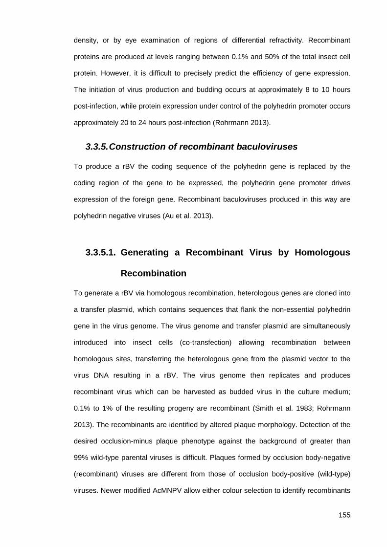
155
density, or by eye examination of regions of differential refractivity. Recombinant
proteins are produced at levels ranging between 0.1% and 50% of the total insect cell
protein. However, it is difficult to precisely predict the efficiency of gene expression.
The initiation of virus production and budding occurs at approximately 8 to 10 hours
post-infection, while protein expression under control of the polyhedrin promoter occurs
approximately 20 to 24 hours post-infection (Rohrmann 2013).
3.3.5. Construction of recombinant baculoviruses
To produce a rBV the coding sequence of the polyhedrin gene is replaced by the
coding region of the gene to be expressed, the polyhedrin gene promoter drives
expression of the foreign gene. Recombinant baculoviruses produced in this way are
polyhedrin negative viruses (Au et al. 2013).
Generating a Recombinant Virus by Homologous 3.3.5.1.
Recombination
To generate a rBV via homologous recombination, heterologous genes are cloned into
a transfer plasmid, which contains sequences that flank the non-essential polyhedrin
gene in the virus genome. The virus genome and transfer plasmid are simultaneously
introduced into insect cells (co-transfection) allowing recombination between
homologous sites, transferring the heterologous gene from the plasmid vector to the
virus DNA resulting in a rBV. The virus genome then replicates and produces
recombinant virus which can be harvested as budded virus in the culture medium;
0.1% to 1% of the resulting progeny are recombinant (Smith et al. 1983; Rohrmann
2013). The recombinants are identified by altered plaque morphology. Detection of the
desired occlusion-minus plaque phenotype against the background of greater than
99% wild-type parental viruses is difficult. Plaques formed by occlusion body-negative
(recombinant) viruses are different from those of occlusion body-positive (wild-type)
viruses. Newer modified AcMNPV allow either colour selection to identify recombinants
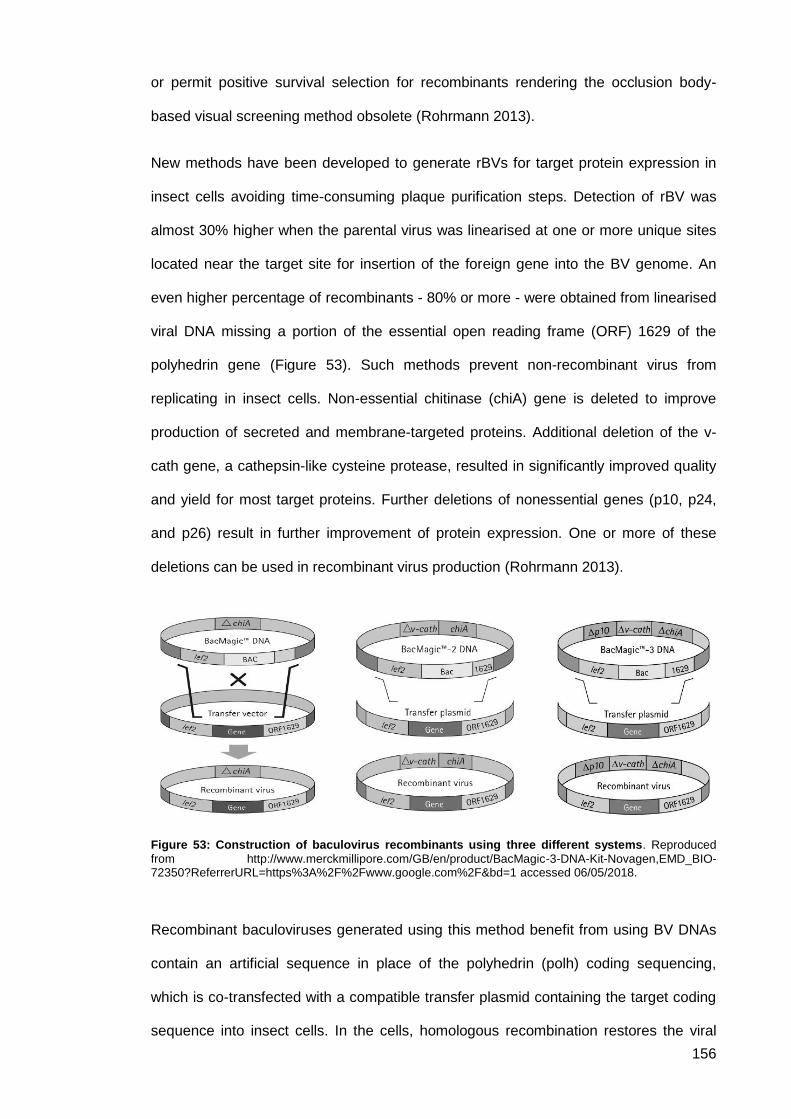
156
or permit positive survival selection for recombinants rendering the occlusion body-
based visual screening method obsolete (Rohrmann 2013).
New methods have been developed to generate rBVs for target protein expression in
insect cells avoiding time-consuming plaque purification steps. Detection of rBV was
almost 30% higher when the parental virus was linearised at one or more unique sites
located near the target site for insertion of the foreign gene into the BV genome. An
even higher percentage of recombinants - 80% or more - were obtained from linearised
viral DNA missing a portion of the essential open reading frame (ORF) 1629 of the
polyhedrin gene (Figure 53). Such methods prevent non-recombinant virus from
replicating in insect cells. Non-essential chitinase (chiA) gene is deleted to improve
production of secreted and membrane-targeted proteins. Additional deletion of the v-
cath gene, a cathepsin-like cysteine protease, resulted in significantly improved quality
and yield for most target proteins. Further deletions of nonessential genes (p10, p24,
and p26) result in further improvement of protein expression. One or more of these
deletions can be used in recombinant virus production (Rohrmann 2013).
Figure 53: Construction of baculovirus recombinants using three different systems. Reproduced
from http://www.merckmillipore.com/GB/en/product/BacMagic-3-DNA-Kit-Novagen,EMD_BIO-72350?ReferrerURL=https%3A%2F%2Fwww.google.com%2F&bd=1 accessed 06/05/2018.
Recombinant baculoviruses generated using this method benefit from using BV DNAs
contain an artificial sequence in place of the polyhedrin (polh) coding sequencing,
which is co-transfected with a compatible transfer plasmid containing the target coding
sequence into insect cells. In the cells, homologous recombination restores the viral
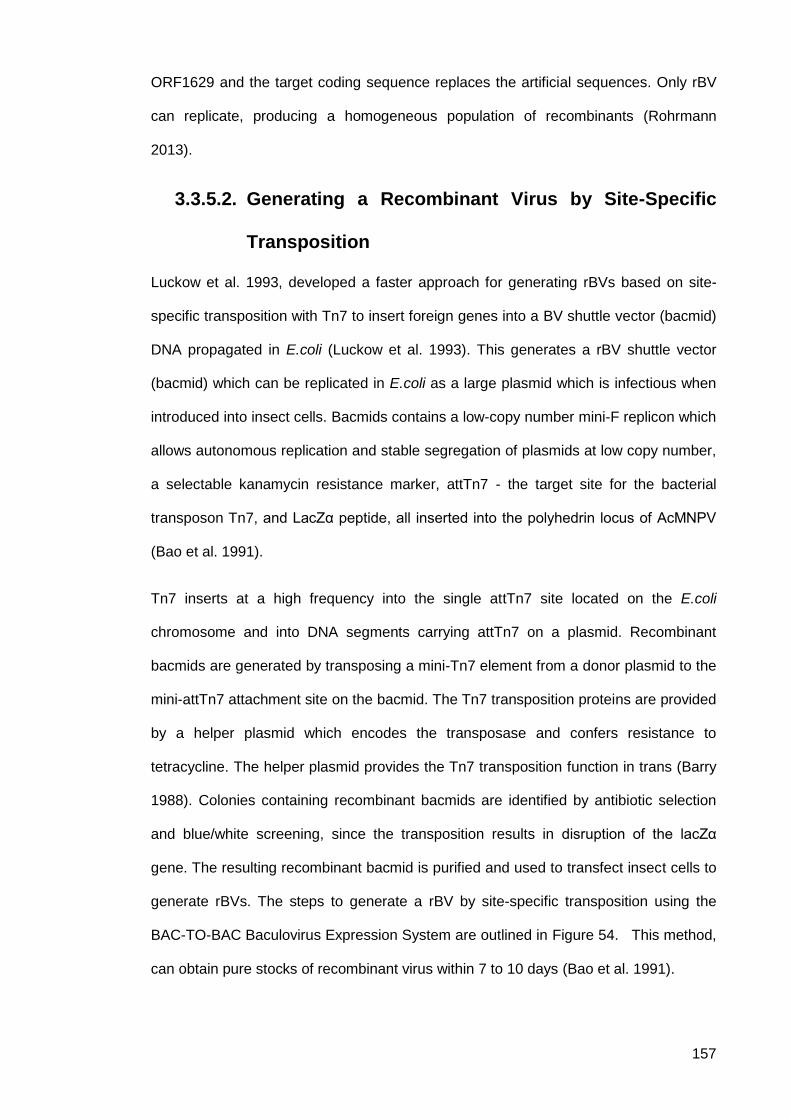
157
ORF1629 and the target coding sequence replaces the artificial sequences. Only rBV
can replicate, producing a homogeneous population of recombinants (Rohrmann
2013).
Generating a Recombinant Virus by Site-Specific 3.3.5.2.
Transposition
Luckow et al. 1993, developed a faster approach for generating rBVs based on site-
specific transposition with Tn7 to insert foreign genes into a BV shuttle vector (bacmid)
DNA propagated in E.coli (Luckow et al. 1993). This generates a rBV shuttle vector
(bacmid) which can be replicated in E.coli as a large plasmid which is infectious when
introduced into insect cells. Bacmids contains a low-copy number mini-F replicon which
allows autonomous replication and stable segregation of plasmids at low copy number,
a selectable kanamycin resistance marker, attTn7 - the target site for the bacterial
transposon Tn7, and LacZα peptide, all inserted into the polyhedrin locus of AcMNPV
(Bao et al. 1991).
Tn7 inserts at a high frequency into the single attTn7 site located on the E.coli
chromosome and into DNA segments carrying attTn7 on a plasmid. Recombinant
bacmids are generated by transposing a mini-Tn7 element from a donor plasmid to the
mini-attTn7 attachment site on the bacmid. The Tn7 transposition proteins are provided
by a helper plasmid which encodes the transposase and confers resistance to
tetracycline. The helper plasmid provides the Tn7 transposition function in trans (Barry
1988). Colonies containing recombinant bacmids are identified by antibiotic selection
and blue/white screening, since the transposition results in disruption of the lacZα
gene. The resulting recombinant bacmid is purified and used to transfect insect cells to
generate rBVs. The steps to generate a rBV by site-specific transposition using the
BAC-TO-BAC Baculovirus Expression System are outlined in Figure 54. This method,
can obtain pure stocks of recombinant virus within 7 to 10 days (Bao et al. 1991).
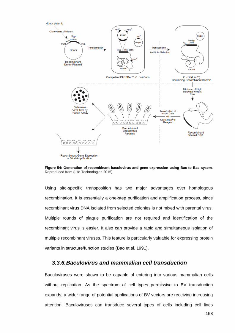
158
Figure 54: Generation of recombinant baculovirus and gene expression using Bac to Bac sysem.
Reproduced from (Life Technologies 2015)
Using site-specific transposition has two major advantages over homologous
recombination. It is essentially a one-step purification and amplification process, since
recombinant virus DNA isolated from selected colonies is not mixed with parental virus.
Multiple rounds of plaque purification are not required and identification of the
recombinant virus is easier. It also can provide a rapid and simultaneous isolation of
multiple recombinant viruses. This feature is particularly valuable for expressing protein
variants in structure/function studies (Bao et al. 1991).
3.3.6. Baculovirus and mammalian cell transduction
Baculoviruses were shown to be capable of entering into various mammalian cells
without replication. As the spectrum of cell types permissive to BV transduction
expands, a wider range of potential applications of BV vectors are receiving increasing
attention. Baculoviruses can transduce several types of cells including cell lines
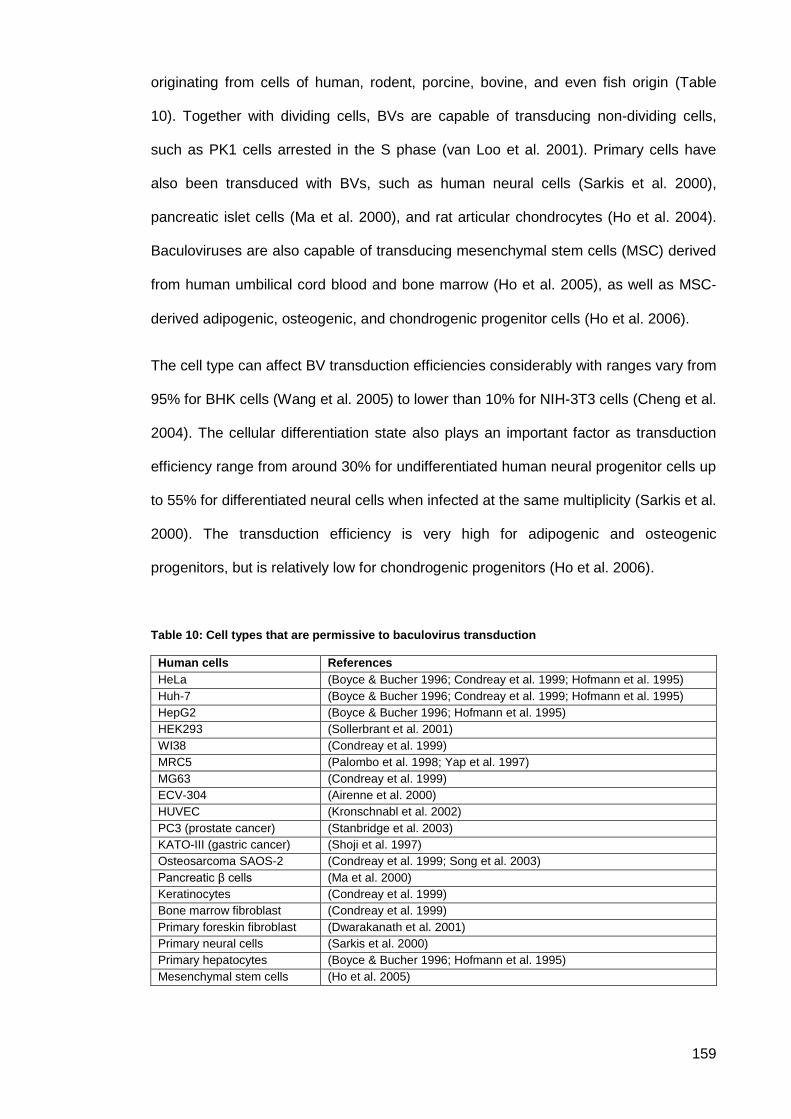
159
originating from cells of human, rodent, porcine, bovine, and even fish origin (Table
10). Together with dividing cells, BVs are capable of transducing non-dividing cells,
such as PK1 cells arrested in the S phase (van Loo et al. 2001). Primary cells have
also been transduced with BVs, such as human neural cells (Sarkis et al. 2000),
pancreatic islet cells (Ma et al. 2000), and rat articular chondrocytes (Ho et al. 2004).
Baculoviruses are also capable of transducing mesenchymal stem cells (MSC) derived
from human umbilical cord blood and bone marrow (Ho et al. 2005), as well as MSC‐
derived adipogenic, osteogenic, and chondrogenic progenitor cells (Ho et al. 2006).
The cell type can affect BV transduction efficiencies considerably with ranges vary from
95% for BHK cells (Wang et al. 2005) to lower than 10% for NIH-3T3 cells (Cheng et al.
2004). The cellular differentiation state also plays an important factor as transduction
efficiency range from around 30% for undifferentiated human neural progenitor cells up
to 55% for differentiated neural cells when infected at the same multiplicity (Sarkis et al.
2000). The transduction efficiency is very high for adipogenic and osteogenic
progenitors, but is relatively low for chondrogenic progenitors (Ho et al. 2006).
Table 10: Cell types that are permissive to baculovirus transduction
Human cells References
HeLa (Boyce & Bucher 1996; Condreay et al. 1999; Hofmann et al. 1995)
Huh-7 (Boyce & Bucher 1996; Condreay et al. 1999; Hofmann et al. 1995)
HepG2 (Boyce & Bucher 1996; Hofmann et al. 1995)
HEK293 (Sollerbrant et al. 2001)
WI38 (Condreay et al. 1999)
MRC5 (Palombo et al. 1998; Yap et al. 1997)
MG63 (Condreay et al. 1999)
ECV-304 (Airenne et al. 2000)
HUVEC (Kronschnabl et al. 2002)
PC3 (prostate cancer) (Stanbridge et al. 2003)
KATO-III (gastric cancer) (Shoji et al. 1997)
Osteosarcoma SAOS-2 (Condreay et al. 1999; Song et al. 2003)
Pancreatic β cells (Ma et al. 2000)
Keratinocytes (Condreay et al. 1999)
Bone marrow fibroblast (Condreay et al. 1999)
Primary foreskin fibroblast (Dwarakanath et al. 2001)
Primary neural cells (Sarkis et al. 2000)
Primary hepatocytes (Boyce & Bucher 1996; Hofmann et al. 1995)
Mesenchymal stem cells (Ho et al. 2005)

160
Baculoviruses can serve as a vehicle to efficiently deliver and express genes to
mammalian cells. However, these transgenes cannot be expressed unless they are
driven by a mammalian promoter such as the CMV IE promoter. Various mammalian
promoters have been cloned into BV vectors to drive gene transcription in mammalian
cells (Table 11). Spenger et al., compared transgenic expression of several promoters
including Simian virus 40 (SV40), CMV, RSV, and a cellular promoter (human ubiquitin
C) in a range of cell types including CHO, COS-1, and HEK293 cells. Results indicated
that CMV and RSV promoters were the most active in all cell lines tested, followed by
the ubiquitin C promoter, while SV40 promoter was the weakest of them all (Spenger et
al. 2004).
Table 11: Promoter used in Baculovirus expression system
Promoter References
Rous sarcoma virus long terminal repeat (RSV-LTR)
promoter
(Boyce & Bucher 1996)
Cytomegalovirus immediate early promoter CMV-IE (Hofmann et al. 1995; Sollerbrant et al.
2001)
Simian virus 40 (SV40) promoter (Spenger et al. 2004)
Hybrid chicken -actin promoter (CAG) (Shoji et al. 1997; Stanbridge et al. 2003)
Hepatitis B virus (HBV) promoter/enhancer (Delaney & Isom 1998)
Human -fetoprotein promoter/enhancer (Park et al. 2007)
Human ubiquitin C promoter (Spenger et al. 2004)
Hybrid neuronal promoter (Wang & Wang 2005)
Drosophila heat shock protein (hsp70) (Viswanathan et al. 2003)
Baculoviruses do not replicate in mammalian cells and thus have an excellent safety
profile combined with being well-tolerated by cells. The Baculovirus vector BacMam
has been successfully used as a transient expression vector in cell based assays, live
cell imaging and stem cell biology research. Because of its ability to transduce
mammalian cells without viral replication, BVs are considered as promising candidates
for gene therapy and vaccine vectors in mammalian cells. The BacMam system
combines the advantages of viral transient expression, ease of generation of protein
and a broad cell tropism; enabling rapid, efficient and flexible protein expression in
various mammalian cells (Dukkipati et al. 2008).
161
To introduce genes into mammalian cells, a standard transduction process is used.
BacMam particles are taken up by endocytosis or macropinocytosis and released for
transcription and expression following migration to the nucleus. Gene expression
begins within 4–6 hours of transduction and is at near maximum level within 24 hours
of transduction. Depending on transduction efficiency, cell type and cell division rate,
the transgene remains detectable from 5 to 14 days (Airenne et al. 2013).
Using BacMam technology for gene expression in mammalian cells offers several
advantages. Baculoviruses can transduce a broad range of cell types, including
primary and stem cells with high transduction efficiency. Recombinant baculoviruses
are capable of expressing large gene inserts in mammalian systems where insert with
more than 38Kbp, and simultaneous delivery of multiple genes has been
demonstrated. Studies indicate that BacMam transduction produces a highly
reproducible and titratable expression. As BVs do not replicate in mammalian cells,
minimal microscopy is needed for observable cytopathic effects. In most cases, BVs
are handled under biosafety level 1, while biosafety level 2 may apply depending on
the transgene expressed (Airenne et al. 2013).
3.3.7. Generation of VLPs using baculovirus expression
Virus-like particles (VLPs) present viral antigens in a more authentic conformation than
monomeric structural proteins. VLPs mimic the structure of virus particles and display
excellent adjuvant properties, being capable of inducing innate and cognate immune
responses. Depending on the biological function of the VLPs different expression
systems can be applied. Baculoviruses can express VLPs in bacteria, yeast, insect and
mammalian cells (Table 11) (Liu et al. 2013). Insect cells have fast specific growth
rates allowing large-scale protein production under controlled culture conditions.
Expression systems like bacteria and yeast achieve higher production scales.
However, those systems are simple and VLPs produced from such systems do not
match the degree of complexity of those produced from insect cells nor mammalian
cells which form much more diverse proteins. Insect cells are relatively easy to culture
162
for those with experience and do not require tedious cell culture adaptation procedures
as in mammalian cells. They can grow in the absence of serum, which is highly
desirable feature when rescuing VLPs from intensive purification processes. Production
yields of insect cells are significantly higher and faster when compared with
mammalian cell-based VLP production. Production processes can be shortened to
approximately 12 weeks from identification of a new strain. VLPs expressed from insect
cells have similar glycosylation patterns similar to those such as wild type influenza
VLPs. The insect expression system offer several advantages over mammalian
expression systems (Liu et al. 2013). However, not all VLP produced in insect cells
produce the correct configuration of protein. This relates to the fact that insect cells
have a different pattern of post-translational modifications than mammalian cells (Davis
& Wood 1995). Insect cells perform simpler post-translational glycosylation based on
enriched mannose (Wu & Passarelli 2010).
The differences in essential mechanisms between insect and mammalian cells, such
as glycosylation and folding of heterologous proteins post translation, may hinder the
formation of VLPs, especially structurally complicated VLPs. Improved insect cells lines
– that perform mammalian-like post-translation modifications – are becoming available.
VLPs induce potent humoral and cellular immune responses as they present high-
density B-cell epitopes responsible of antibody production and intracellular T-cell
epitopes. Recombinant VLPs can serve as an excellent candidate for vaccine and
diagnostic purposes. The underlying mechanism of efficacious prophylactic vaccines is
generation of neutralising antibodies against specific epitopes. In the case of VLPs, this
depends on correct antigen conformation and effective antigen presentation on the
VLP surface (Liu et al. 2013).
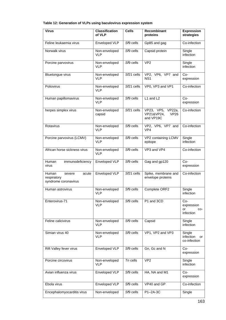
163
Table 12: Generation of VLPs using baculovirus expression system
Virus Classification of VLP
Cells Recombinant proteins
Expression strategies
Feline leukaemia virus Enveloped VLP Sf9 cells Gp85 and gag Co-infection
Norwalk virus Non-enveloped VLP
Sf9 cells Capsid protein Single infection
Porcine parvovirus Non-enveloped VLP
Sf9 cells VP2 Single infection
Bluetongue virus Non-enveloped VLP
Sf21 cells VP2, VP6, VP7 and NS1
Co-expression
Poliovirus Non-enveloped VLP
Sf21 cells VP0, VP3 and VP1 Co-infection
Human papillomavirus Non-enveloped VLP
Sf9 cells L1 and L2 Co-expression
herpes simplex virus Non-enveloped capsid
Sf21 cells VP23, VP5, VP22a, VP21&VP24, VP26 and VP19C
Co-infection
Rotavirus Non-enveloped VLP
Sf9 cells VP2, VP6, VP7 and VP4
Co-infection
Porcine parvovirus (LCMV) Non-enveloped VLP
Sf9 cells VP2 containing LCMV epitope
Single infection
African horse sickness virus Non-enveloped VLP
Sf9 cells VP3 and VP4 Co-infection
Human immunodeficiency virus
Enveloped VLP Sf9 cells Gag and gp120 Co-expression
Human severe acute respiratory syndrome coronavirus
Enveloped VLP Sf21 cells Spike, membrane and envelope proteins
Co-infection
Human astrovirus Non-enveloped VLP
Sf9 cells Complete ORF2 Single infection
Enterovirus-71 Non-enveloped VLP
Sf9 cells P1 and 3CD Co-expression or co-infection
Feline calicivirus Non-enveloped VLP
Sf9 cells Capsid Single infection
Simian virus 40 Non-enveloped VLP
Sf9 cells VP1, VP2 and VP3 Single infection or co-infection
Rift Valley fever virus Enveloped VLP Sf9 cells Gn, Gc and N Co-expression
Porcine circovirus Non-enveloped VLP
Tn cells VP2 Single infection
Avian influenza virus Enveloped VLP Sf9 cells HA, NA and M1 Co-expression
Ebola virus Enveloped VLP Sf9 cells VP40 and GP Co-infection
Encephalomyocarditis virus Non-enveloped Sf9 cells P1–2A-3C Single

164
VLP infection
Rous sarcoma virus Enveloped VLP Tn cells
and SL Gag Single
infection
Japanese encephalitis virus Enveloped VLP Sf9 cells prM and gE Co-infection
Coxsackievirus A16 Non-enveloped VLP
Sf9 cells P1 and 3CD Co-expression
Foot-and-mouth disease virus Non-enveloped VLP
Sf9 cells P1–2A-3C Single infection
Chikungunya virus Enveloped VLP Sf21 cells Structural polyprotein Single infection
Chimeric VLP. Sf: Spodoptera frugiperda; Tn: Trichoplusia ni; LCMV: lymphocytic choriomeningitis virus; ORF: open reading frame; SL: silkworm larvae; VLP: virus-like particle.
Currently, numerous VLP-based vaccine candidates have entered preclinical testing or
clinical trials and many are in the proof-of-concept stage. To date, only a few fully VLP-
based vaccines against viral infections have been licensed for human use (Liu et al.
2013). Recombivax HB® (Merck) and Engerix®-B (GSK) are licensed against hepatitis B virus
(HBV). Both are Non-enveloped VLPs composed of HBV surface antigen (HBsAg)
harvested and purified after being expressed in yeast (Saccharomyces cerevisae)
infected with BV. Both vaccines induce the production of the same antibodies to HBsAg
and have similar immunogenic properties that may allow for interchangeability.
GlaxoSmithKline’s Cervarix™ (GSK) is a VLP-based bivalent human papillomavirus
vaccine against cervical cancer. Composed of HPV16/18 formulated in L1 ASO4
adjuvant and produced using a rBV expression system in an insect cell line. Hecolin®
is a vaccine against hepatitis E virus (HEV) developed by Xiamen Innovax Biotech Co.,
Ltd (China), based on recombinant HEV ORF2 truncated protein HEV 239 (aa368-
aa606) expressed from BV in an E.coli expression system. FluBlok®, a seasonal
influenza subunit vaccine for adults is a recombinant trivalent hemagglutinin (rHA)
vaccine produced in insect cell culture using the BV expression system. It is tailored
annually to provide protection against the latest strains of influenza by containing the
corresponding hemagglutinin (HA) antigens.
165
3.3.8. Immune response and potential as gene therapy
vectors and vaccine vectors
The interest in using BVs as vectors for both in vitro and in vivo gene delivery has
increased. Several studies indicated that BVs can be used for in vivo gene transfers
among different tissue types. Airenne et al., showed that BVs can be used for in vivo
gene transfer in rabbit carotid artery with efficiency comparable to adenoviruses
(Airenne et al. 2000). Huser et al., studied gene transfer of complement-resistant BV
vectors into complement-sufficient neonatal rats by injection into their livers (Hüser et
al. 2001). Studies have been conducted to understand the efficiency of BV gene
delivery to rat brain (Sarkis et al. 2000; Lehtolainen et al. 2002; Wang & Wang 2005)
and several in vivo mouse studies have targetted BV gene transfer in different tissue
types including brain, skeletal muscle, cerebral cortex and testis and liver (Sarkis et al.
2000; Pieroni et al. 2001; Tani et al. 2003; Hoare et al. 2005).
Other advantages include the broad range of mammalian cells capable of transduction;
their nontoxic and non-replicative nature; large packaging capacity; ease of production
and so BVs appear to be a promising tool for gene therapy (Table 13). However, for
BV‐mediated in vivo gene therapy, contact between BVs and serum complement
results in the rapid inactivation of the virus. Sandig et al. 1996, described the potential
limitation caused by the complement system in a systemic or intraportal application as
well as in direct injection of BVs into the rodent liver parenchyma (Sandig et al. 1996).
This phenomenon was not observed when the BV was injected into rodent brain.
Within the brain the complement level may be insufficient to affect gene transfer
(Lehtolainen et al. 2002). Hofmann & Strauss showed that BVs most likely activate the
classical pathway of the C system and assembly of very late C components is required
for inactivation of the vector (Hofmann & Strauss 1998). Hoare et al., further showed
that both classical and alternative pathways are involved in the inactivation and
suggested that naturally occurring IgM antibodies with high affinity for BVs may be
partially responsible for the inactivation (Hoare et al. 2005).
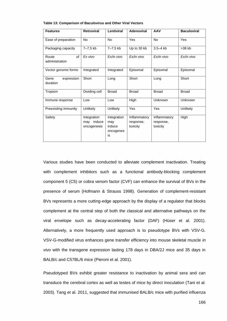
166
Table 13: Comparison of Baculovirus and Other Viral Vectors
Features Retroviral Lentiviral Adenoviral AAV Baculoviral
Ease of preparation No No Yes No Yes
Packaging capacity 7–7.5 kb 7–7.5 kb Up to 30 kb 3.5–4 kb >38 kb
Route of
administration
Ex vivo Ex/in vivo Ex/in vivo Ex/in vivo Ex/in vivo
Vector genome forms Integrated Integrated Episomal Episomal Episomal
Gene expression
duration
Short Long Short Long Short
Tropism Dividing cell Broad Broad Broad Broad
Immune response Low Low High Unknown Unknown
Preexisting immunity Unlikely Unlikely Yes Yes Unlikely
Safety Integration
may induce
oncogenesis
Integration
may
induce
oncogenes
is
Inflammatory
response,
toxicity
Inflammatory
response,
toxicity
High
Various studies have been conducted to alleviate complement inactivation. Treating
with complement inhibitors such as a functional antibody‐blocking complement
component 5 (C5) or cobra venom factor (CVF) can enhance the survival of BVs in the
presence of serum (Hofmann & Strauss 1998). Generation of complement‐resistant
BVs represents a more cutting‐edge approach by the display of a regulator that blocks
complement at the central step of both the classical and alternative pathways on the
viral envelope such as decay‐accelerating factor (DAF) (Hüser et al. 2001).
Alternatively, a more frequently used approach is to pseudotype BVs with VSV-G.
VSV-G‐modified virus enhances gene transfer efficiency into mouse skeletal muscle in
vivo with the transgene expression lasting 178 days in DBA/2J mice and 35 days in
BALB/c and C57BL/6 mice (Pieroni et al. 2001).
Pseudotyped BVs exhibit greater resistance to inactivation by animal sera and can
transduce the cerebral cortex as well as testes of mice by direct inoculation (Tani et al.
2003). Tang et al. 2011, suggested that immunised BALB/c mice with purified influenza
167
VLPs produced from BV-transduced mammalian cells, or directly immunised with
pseudotyped BacMam BV survived without any signs of sickness or weight loss. Both
induced influenza-specific immune responses and provided mice with sterilising
protection against influenza challenge (Tang et al. 2011). Baculoviruses are considered
safe, but more studies are required to confirm their safety for in vivo and ex vivo
applications. Gronowski et al., was the first to study the immune response against BVs
in mammalian cells. He reported that administration of BVs in vitro and in vivo induced
the production of IFN α/β from both human and murine cell lines. This IFN-stimulation
resulted from the live virus and not due to the presence of viral RNA or DNA
(Gronowski et al. 1999). Abe et al., found that BV DNA, not the gp64 glycoprotein,
induced the innate immune system activation and produced IFN‐α via the Toll-like
receptor 9 (TLR9)/MyD88-dependent signaling pathway. The production of
inflammatory cytokines were severely reduced in peritoneal macrophages (PECs) and
splenic CD11c+ dendritic cells (DCs) derived from mice deficient in MyD88 or TLR9
after cultivation with AcMNPV. In contrast, a significant amount of IFN-α was still
detectable in the PECs and DCs of these mice after stimulation with AcMNPV,
suggesting that a TLR9/MyD88-independent signalling pathway might also participate
in the production of IFN-α by AcMNPV (Abe et al. 2005).
It has been suggested that IFN induction requires an interaction between the BV
envelope protein, gp64, and the receptors on the corresponding cell membrane
(Kataoka et al. 2012). This finding provides further emphasis that viral-induced IFN
production is not always dependent on viral replication. Administration of BVs in vivo
protects mice from death when challenged with a lethal dose of FMDV (Molinari et al.
2010). However, the mechanisms by which viruses interact with a host cell and initiate
IFN production are not well understood. Beck et al., demonstrated that upon BV
exposure, production of cytokines in hepatocytes is up-regulated. Baculovirses had an
antagonistic effect on cytochrome P450 (CYP) induction processes and expression
profiles which may alter the detoxification capabilities of the liver (Beck et al. 2000).
168
Airenne et al., found that in vivo administration of BV to rabbit carotid artery resulted in
signs of inflammation (Airenne et al. 2000). Abe et al., inoculated mice with rBV by
intramuscular, intradermal, i.p., and intranasal routes using a rBV expressing
haemagglutinin (HA) of influenza virus under the control of the CAG promoter (Abe et
al. 2003). Innate immune responses and the level of protection were found to depend
on the route of administration. Mice were inoculated with rBV by intramuscular,
intradermal, i.p., and intranasal routes and then were challenged with a lethal dose of
the influenza virus. Higher titers of anti-hemagglutinin Ab resulted from intramuscular or
i.p. immunisation compared to intradermal or intranasal administration. However,
intranasal immunisation conferred sufficient protection from the lethal challenge of the
influenza virus.
Baculoviruses have been found to have immune-stimulatory capacities to promote the
release of inflammatory cytokines such as tumour necrosis factor alpha (TNF-α),
interleukin-6 (IL-6), and IL-12, in a murine macrophage cell line, RAW264.7.
Facciabene et al., have determined the induction of humoral and cell-mediated immune
responses to the E2 glycoprotein of hepatitis C virus (HCV) and to carcinoembryonic
antigen (CEA) in mice vaccinated with rBV vectors expressing the putative structural
component of the HCV virions or the epithelial tumor antigen. Their findings agreed
with the finding that VSV-G‐pseudotyped BVs reduced the minimal dose required to
elicit a measurable T cell response tenfold. This indicates that VSV-G‐pseudotyped
viruses provide more efficient immunogen expression in transduced cells and avoid
complement inactivation (Facciabene et al. 2004).
Although BVs seems to be a promising candidate as a vaccine vector against
infectious diseases as well as vectors for in vivo human gene therapy, further
investigation of the mechanisms of viral uptake and induction of immunity are needed,
particularly in understanding which cytokines are induced by BVs, and how these
cytokines modulate cellular and humoral immunities in vivo.
169
Material and methods: 3.4.
Genetically Modified Organisms approval for this study was obtained for the laboratory
and the project. The lab permissions and the GMO permission were granted by the
University of Manchester safety committee, the GMO Establishment license# 541
(p5603-4).
3.4.1. Virus like particle expression utilising the T7 RNA
Polymerase/Promoter system
BacMam system one allowed expression of VLPs by transducing mammalian cells with
five different rBVs bearing the T7 RNA polymerase system. BacMam-Gag/Tat encodes
for both the group-specific antigen (Gag) that forms the main structural proteins, and
transactivator (Tat) a positive regulator of transcription; BacMam-Pol/Vif/Vpr expresses
polymerase (Pol), encodes reverse transcriptase (Rt), protease (Pro) and integrase
(Int) together with viral infectivity factor (Vif) and virus protein R (Vpr), (required for the
virus replication). To extend the limited cell tropism inherent in SIV, the VLP was
pseudotyped with VSV-G. BacMam-VSV-G/Rev encodes both VSV-G and regulator of
expression of virion (Rev) which is essential for viral replication. BacMam-IgG1b12 was
designed to encode for antibody gene sequence to be packed as a core sequence of
the VLP. Genes of interest in all four rBVs were driven by the T7 promoter in the
presence of the T7 polymerase. The fifth recombinant, BacMam-T7 is responsible for
the expression of T7 RNA polymerase in high levels, protein expression was driven by
CMV IE.
3.4.2. Virus like particle expression using a CMV Promoter
BacMam system allowed expression of VLPs by transducing mammalian cells with
four different rBVs using CMV to drive the expression of all genes of interests.
BacMam-Gag/Tat encodes for both the group-specific antigen (Gag) that forms the
main structural proteins, together with transactivator (Tat) a positive regulator of
transcription. BacMam-Pol/Vif/Vpr expresses polymerase (Pol), which codes for
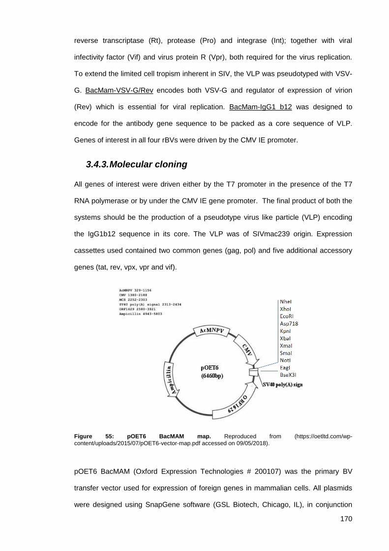
170
reverse transcriptase (Rt), protease (Pro) and integrase (Int); together with viral
infectivity factor (Vif) and virus protein R (Vpr), both required for the virus replication.
To extend the limited cell tropism inherent in SIV, the VLP was pseudotyped with VSV-
G. BacMam-VSV-G/Rev encodes both VSV-G and regulator of expression of virion
(Rev) which is essential for viral replication. BacMam-IgG1 b12 was designed to
encode for the antibody gene sequence to be packed as a core sequence of VLP.
Genes of interest in all four rBVs were driven by the CMV IE promoter.
3.4.3. Molecular cloning
All genes of interest were driven either by the T7 promoter in the presence of the T7
RNA polymerase or by under the CMV IE gene promoter. The final product of both the
systems should be the production of a pseudotype virus like particle (VLP) encoding
the IgG1b12 sequence in its core. The VLP was of SIVmac239 origin. Expression
cassettes used contained two common genes (gag, pol) and five additional accessory
genes (tat, rev, vpx, vpr and vif).
Figure 55: pOET6 BacMAM map. Reproduced from (https://oetltd.com/wp-
content/uploads/2015/07/pOET6-vector-map.pdf accessed on 09/05/2018).
pOET6 BacMAM (Oxford Expression Technologies # 200107) was the primary BV
transfer vector used for expression of foreign genes in mammalian cells. All plasmids
were designed using SnapGene software (GSL Biotech, Chicago, IL), in conjunction
171
with data held in the NCBI GenBank database. pOET6 BacMam transfer vector was
purchased from Oxford Expression Technologies (Oxford, UK). pT7-Vpx_MVA, pPol-
Vpr-Vif_MVA and pGag_Tat_MVA shuttle vectors were provided by Dr Tom Blanchard
University of Manchester. pLF-IgG1b12-LacZ-D4 and pLF-VSV-G-Rev-D4 were
transfer vectors developed in the MVA work (refer to sections 2.5.1.5 and 2.5.1.4)
For amplification of plasmid DNA the following E.coli strains were used:
Either: NovaBlue GigaSingles Competent Cells (71227-3, Novagen.UK). NovaBlue is
a K-12 strain derivative that offers high transformation efficiency, genotype: endA1
hsdR17(rK12– mK12
+) supE44 thi-1 recA1 gyrA96 relA1 lac F'[proA+B+ lacIqZΔM15::Tn10
(TcR)].
Or: NEB 10-beta Competent a derivative of DH10B (NEB #C3019L). It is T1 phage
resistant and endonuclease I (endA1) deficient. NEB 10-beta electrocompetent E.coli
cells are Δ(ara-leu) 7697 araD139 fhuA ΔlacX74 galK16 galE15 e14-
ϕ80dlacZΔM15 recA1 relA1 endA1 nupG rpsL (StrR) rph spoT1 Δ(mrr-hsdRMS-
mcrBC).
DNA extraction and purification from bacterial cells 3.4.3.1.
Plasmid DNA was isolated using the QIAprep Spin Miniprep Kit (QIAGEN #27104).
Following the manufacturer instructions, an overnight E.coli culture was pelleted by
centrifugation. Bacterial cells were then lysed by alkaline lysis followed by adsorption of
DNA onto silica in the presence of high salt. Impurities were washed with the supplied
washing buffer and pure plasmid DNA eluted in a small volume of elution buffer. The
concentration of the plasmid DNA was determined using the ND1000 NanoDrop
Spectrophotometer (Labtech, Uckfield,UK).
172
Preparation of vector and insert: Restriction digest 3.4.3.2.
In a total volume of 50μl: 1µg of DNA, 5μl of 10xNEBuffer and 1μl (10units) enzyme
were mixed. Both incubation times and temperatures were enzyme dependent.
DNA End Modification 3.4.3.3.
3.4.3.3.1. Dephosphorylation
1 µL (1 unit/µL) of recombinant shrimp Alkaline Phosphatase (rSAP) (NEB #M0371)
was added to each pmol of DNA (about 1 µg of a 3 kb plasmid). After incubation at
37°C for 30 minutes, the reaction was stopped by heat inactivation at 65°C for 5
minutes.
3.4.3.3.2. Blunting/End-repair
Quick Blunting Kit (NEB #E1201) was used to fill in (5’3’) and ‘chew back’ (3’5’).
For up to 5µg DNA a total reaction of 25µL was prepared including 5 µL of 10X
Blunting Buffer, 5 µL of 1 mM dNTP Mix and 2 µL of Blunt Enzyme Mix. The reaction
mix was then incubated at room temperature for 30 minutes, followed by an inactivation
step of heating at 70°C for 10 minutes.
PCR Using Q5® High-Fidelity DNA polymerase 3.4.3.4.
Inserts from a PCR product were amplified using Q5 High-Fidelity DNA Polymerase
(NEB # M0491). Conditions for optimal performance were used following the
manufacturer’s structures (Table 14, Table 15 and Table 16). NEB Tm Calculator
(https://tmcalculator.neb.com/#!/main) was used to determine optimal annealing
temperatures.
173
Table 14: Reaction Setup
COMPONENT 25 µL REACTION 50 µL REACTION FINAL
CONCENTRATION
5X Q5
Reaction Buffer
5 µL 10 µL 1X
10 mM dNTPs 0.5 µL 1 µL 200 µM
10 µM Forward Primer 1.25 µL 2.5 µL 0.5 µM
10 µM Reverse Primer 1.25 µL 2.5 µL 0.5 µM
Template DNA variable variable < 1,000 ng
Q5 High-Fidelity DNA
Polymerase
0.25 µL 0.5 µL 0.02 U/µL
5X Q5 High GC Enhancer
(optional)
(5 µL) (10 µL) (1X)
Nuclease-Free Water to 25 µL to 50 µL
Table 15: Thermocycling Conditions for a Routine PCR
STEP TEMP TIME
Initial Denaturation 98°C 30 seconds
25–35 Cycles 98°C
*50–72°C
72°C
5–10 seconds
10–30 seconds
20–30 seconds/kb
Final Extension 72°C 2 minutes
Hold 4–10°C
Table 16: Recommended amounts of DNA template for a 50 µL reaction
DNA AMOUNT
DNA Genomic 1 ng–1 µg
Plasmid or Viral 1 pg–1 ng
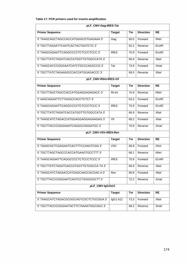
174
Table 17: PCR primers used for inserts amplification
pLF_CMV-Gag-IRES-Tat
Primer Sequence Target Tm Direction RE
5' TAAGCAGCTAGCCACCATGGGCGTGAGAAA 3' Gag 69.5 Forward NheI
5' TGCTTAGAATTCAATCACTACTGGTCTC 3' 62.2 Reverse EcoRI
5' TAAGCAGAATTCAGGCCCCTCTCCCTCCC 3' IRES 70.9 Forward EcoRI
5' TGCTTATCTAGATCACCATGGTTGTGGCCATA 3' 66.9 Reverse XbaI
5' TAAGCACCCGGGAATCATCTGCCCAGGCCG 3' Tat 73.6 Forward XmaI
5' TGCTTATCTAGAAGGCCACCATGGAGACCC 3' 69.5 Reverse XbaI
pLF_CMV-RtInt-IRES-Vif
Primer Sequence Target Tm Direction RE
5' TGCTTAGCTAGCCACCATGGAGGAGAGACC 3' Rt-Int 70.9 Reverse NheI
5' AAGCAGAATTCTTAGGCCACCTCTCT 3' 63.2 Forward EcoRI
5' TAAGCAGAATTCAGGCCCCTCTCCCTCCC 3' IRES 70.9 Forward EcoRI
5' TGCTTATCTAGATCACCATGGTTGTGGCCATA 3' 66.9 Reverse XbaI
5' TAAGCATCTAGACCATGGAGGAGGAGAAGAG 3' Vif 68.2 Forward XbaI
5' TGCTTACCCGGGAAATCAGGCCAGGATGC 3' 70.9 Reverse XmaI
pLF_CMV-VSV-IRES-Rev
Primer Sequence Target Tm Direction RE
5' TAAGCACTCGAGAATCACTTTCCAAGTCGG 3' VSV 66.8 Forward XhoI
5' TGCTTAGCTAGCCCACCATGAAGTGCCTTT 3' 68.1 Reverce NheI
5' TAAGCAGAATTCAGGCCCCTCTCCCTCCC 3' IRES 70.9 Forward EcoRI
5' TGCTTATCTAGATCACCATGGTTGTGGCCA TA 3' 66.9 Reverse XbaI
5' TAAGCATCTAGAACCATGAGCAACCACGAG A 3' Rev 66.8 Forward XbaI
5' TGCTTACCCGGGAATCAGTCCTGGGGGCTT 3' 72.2 Reverce XmaI
pLF_CMV-IgG1b12
Primer Sequence Target Tm Direction RE
5' TAAGCATCTAGACGCGGCAGTCGCTCTGCGGA 3' IgG1 b12 73.3 Forward XbaI
5' TGCTTACCCGGGAATACTTCTAAAATGGCAGC 3' 68.2 Reverce SmaI
175
Agarose gel electrophoresis and gel extraction 3.4.3.5.
3.4.3.5.1. Agarose gel electrophoresis
PCR products and DNA digests were analysed by gel electrophoresis on the basis of
their molecular weight. Fragment sizes were determined in relation to the bands of a
mixture of standard DNA fragments (marker ladder) on the gel. DNA fragments in
agarose gels were visualised with UV light using Invitrogen™ SYBR™ Safe DNA Gel
Stain (ThermoFisher Scientific # S33102) or Gel Red (Biotium Inc., Hayward, USA)
following manufacturer’s guidelines. 6x Gel Loading Dye was mixed with each DNA
sample (NEB # B7024S). Either 1kb DNA ladder or 100 bp DNA ladder molecular
weight standards (Figure 25; NEB #3232L, NEB # 3231L) were used for DNA band
size approximation. The percentage of agarose used in preparing the gel was
determined according to the expected band sizes. The percentage of agarose in
preparing the gel was determined according to the expected band sizes (Table 5).
UltraPure agarose (ThermoFisher Scientific # 16500500) was used in preparing the
agarose gels. PCR products and DNA digests were analysed by gel electrophoresis on
the basis of their molecular weight. Fragment sizes were determined in relation to the
bands of a mixture of standard molecules (marker ladder) on the gel.
3.4.3.5.2. Purification of vector and insert by DNA
extraction from agarose gels
Following gel electrophoresis of DNA from both vector and insert, bands of interests
were excised and purified using the Monarch® DNA Gel Extraction Kit (NEB #T1020S).
Following the manufacturer’s protocol, DNA fragments were excised using a scalpel
(Disposable scalpel number 21, Ref # 0507. Swann-Morton, Sheffield, UK) after
visualization under UV light transillumination (Syngene, Cambridge, UK). Dissolving
buffer was added to the slice; DNA was then adsorbed on a silica membrane, and
purified DNA eluted in the provided Elution Buffer.
176
Ligation of vector and insert 3.4.3.6.
Ligation of DNA fragments was performed using Instant Sticky-end Ligase Master Mix
(NEB #M0370S) containing a T4 DNA ligase. The ligation mix was prepared by
combining 20–100 ng of vector with a 3 to 5 fold molar excess of insert, the volume
was adjusted to 5μl with nuclease free water. 5μl of Instant Sticky-end Ligase Master
Mix was added to the ligation mix.
In the case of blunt ends, ligation was made using the Quick Ligation Kit (NEB,
M2200). The ligation mix was prepared by combining 50 ng of vector with 3 to 5 fold
molar excess of insert DNA, the volume was then adjusted to 10 µL with nuclease-free
water. 10 µL of 2X Quick Ligation Buffer was added together with 1 µL of Quick T4
DNA Ligase followed by thorough mixing. The reaction mix was then centrifuged briefly
and incubated at room temperature (25°C) for 5 minutes.
The amount of vector and insert in the ligation mix was calculated using
NEBBioCalculator (http://nebiocalculator.neb.com/#!/main).
Transformation 3.4.3.7.
Competent Cells were transformed with plasmid DNA according to the manufacturer’s
instructions. Briefly 1-2.5 μl of plasmid DNA or ligation mix (50-100ng DNA) were
suspended in 50μl competent cells and incubated on ice for 5 minutes. After a heat
shock at 42°C for 30 seconds the cells were incubated on ice for two minutes. The
competent cells were mixed with 250μl SOC medium (Super optimal broth medium,
Hanahan, 1983) and incubated at 37°C for one hour at 300rpm. Following the one hour
recovery step, the cells were plated (100μl and 150μl) on selective LB-agar plates with
the respective antibiotic (ampicillin [100mg/ml] or kanamycin [25mg/ml]).
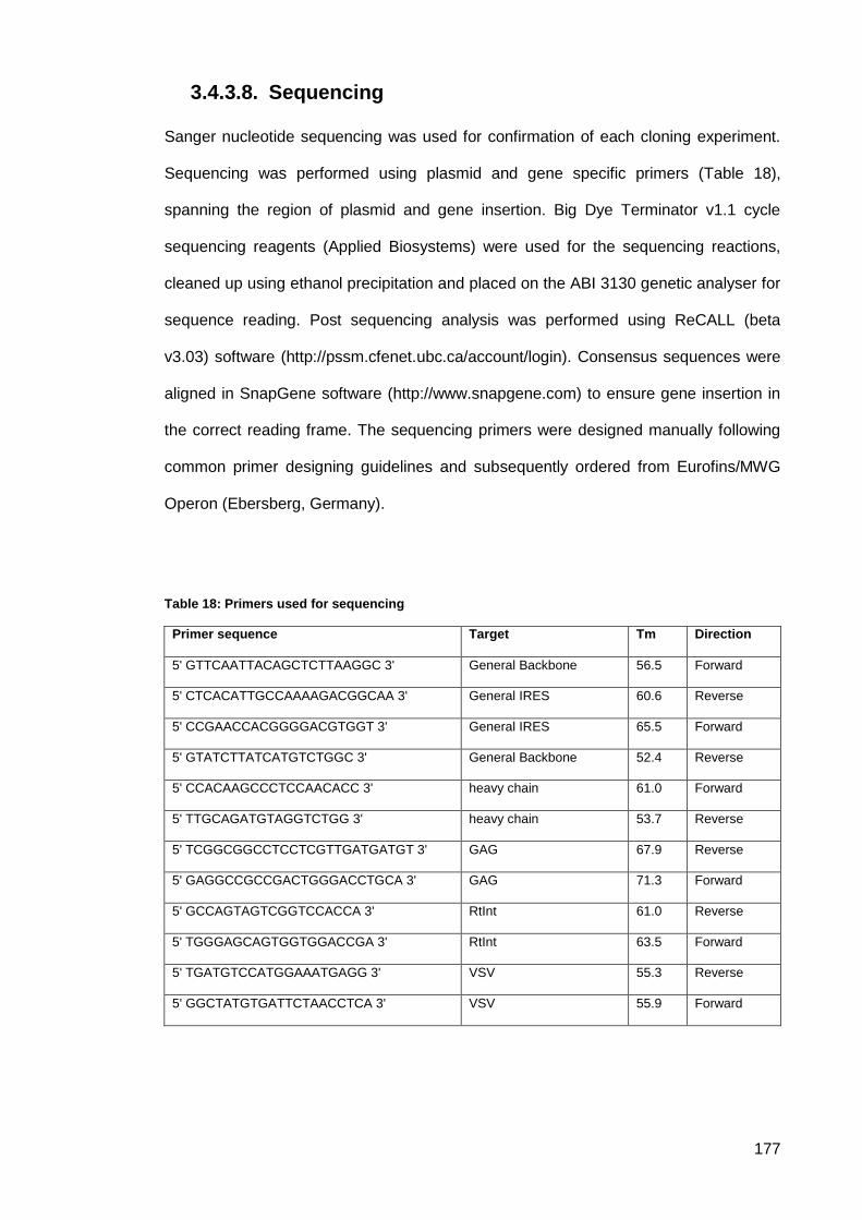
177
Sequencing 3.4.3.8.
Sanger nucleotide sequencing was used for confirmation of each cloning experiment.
Sequencing was performed using plasmid and gene specific primers (Table 18),
spanning the region of plasmid and gene insertion. Big Dye Terminator v1.1 cycle
sequencing reagents (Applied Biosystems) were used for the sequencing reactions,
cleaned up using ethanol precipitation and placed on the ABI 3130 genetic analyser for
sequence reading. Post sequencing analysis was performed using ReCALL (beta
v3.03) software (http://pssm.cfenet.ubc.ca/account/login). Consensus sequences were
aligned in SnapGene software (http://www.snapgene.com) to ensure gene insertion in
the correct reading frame. The sequencing primers were designed manually following
common primer designing guidelines and subsequently ordered from Eurofins/MWG
Operon (Ebersberg, Germany).
Table 18: Primers used for sequencing
Primer sequence Target Tm Direction
5' GTTCAATTACAGCTCTTAAGGC 3' General Backbone 56.5 Forward
5' CTCACATTGCCAAAAGACGGCAA 3' General IRES 60.6 Reverse
5' CCGAACCACGGGGACGTGGT 3' General IRES 65.5 Forward
5' GTATCTTATCATGTCTGGC 3' General Backbone 52.4 Reverse
5' CCACAAGCCCTCCAACACC 3' heavy chain 61.0 Forward
5' TTGCAGATGTAGGTCTGG 3' heavy chain 53.7 Reverse
5' TCGGCGGCCTCCTCGTTGATGATGT 3' GAG 67.9 Reverse
5' GAGGCCGCCGACTGGGACCTGCA 3' GAG 71.3 Forward
5' GCCAGTAGTCGGTCCACCA 3' RtInt 61.0 Reverse
5' TGGGAGCAGTGGTGGACCGA 3' RtInt 63.5 Forward
5' TGATGTCCATGGAAATGAGG 3' VSV 55.3 Reverse
5' GGCTATGTGATTCTAACCTCA 3' VSV 55.9 Forward
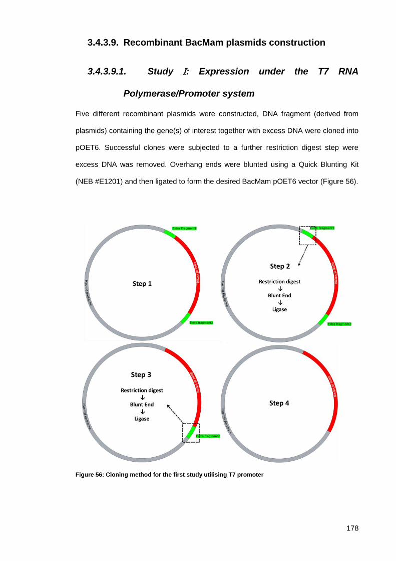
178
Recombinant BacMam plasmids construction 3.4.3.9.
3.4.3.9.1. Study : Expression under the T7 RNA
Polymerase/Promoter system
Five different recombinant plasmids were constructed, DNA fragment (derived from
plasmids) containing the gene(s) of interest together with excess DNA were cloned into
pOET6. Successful clones were subjected to a further restriction digest step were
excess DNA was removed. Overhang ends were blunted using a Quick Blunting Kit
(NEB #E1201) and then ligated to form the desired BacMam pOET6 vector (Figure 56).
Figure 56: Cloning method for the first study utilising T7 promoter
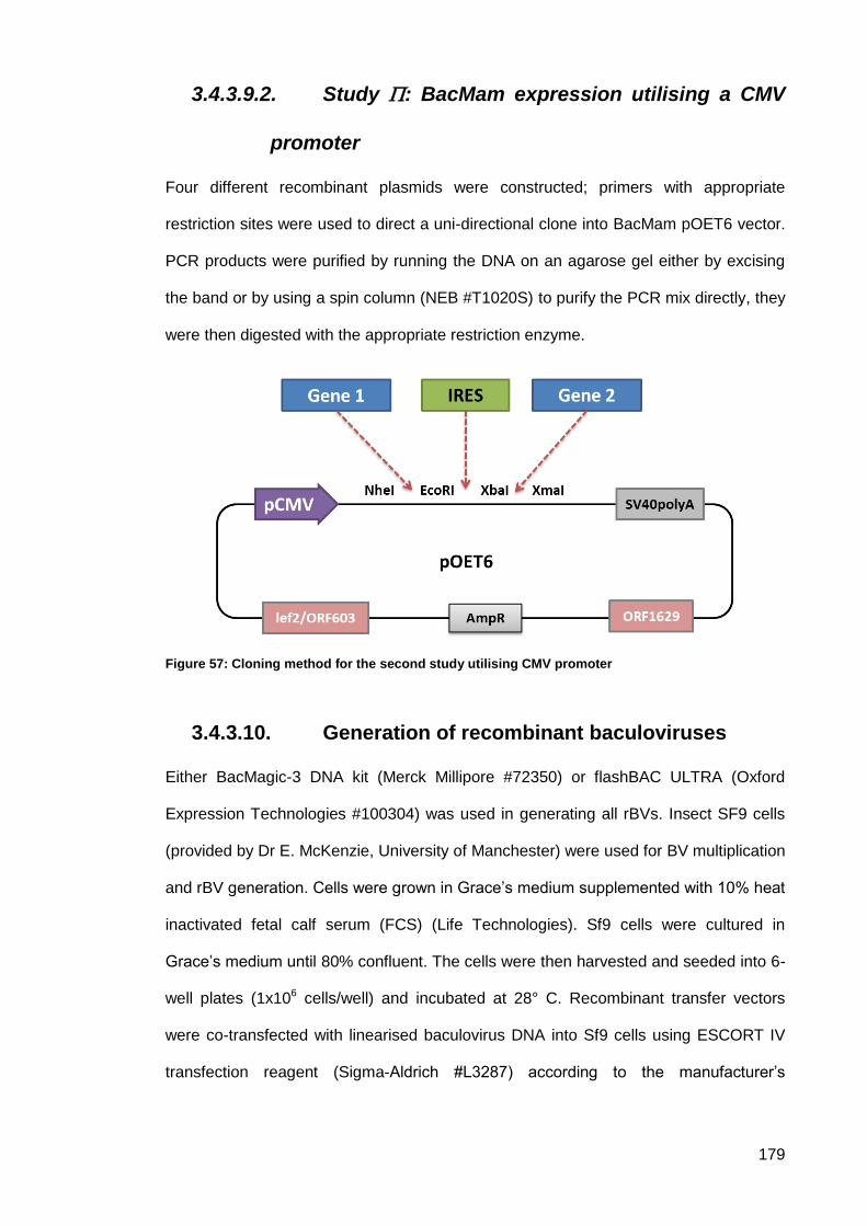
179
3.4.3.9.2. Study : BacMam expression utilising a CMV
promoter
Four different recombinant plasmids were constructed; primers with appropriate
restriction sites were used to direct a uni-directional clone into BacMam pOET6 vector.
PCR products were purified by running the DNA on an agarose gel either by excising
the band or by using a spin column (NEB #T1020S) to purify the PCR mix directly, they
were then digested with the appropriate restriction enzyme.
Figure 57: Cloning method for the second study utilising CMV promoter
Generation of recombinant baculoviruses 3.4.3.10.
Either BacMagic-3 DNA kit (Merck Millipore #72350) or flashBAC ULTRA (Oxford
Expression Technologies #100304) was used in generating all rBVs. Insect SF9 cells
(provided by Dr E. McKenzie, University of Manchester) were used for BV multiplication
and rBV generation. Cells were grown in Grace’s medium supplemented with 10% heat
inactivated fetal calf serum (FCS) (Life Technologies). Sf9 cells were cultured in
Grace’s medium until 80% confluent. The cells were then harvested and seeded into 6-
well plates (1x10⁶ cells/well) and incubated at 28° C. Recombinant transfer vectors
were co-transfected with linearised baculovirus DNA into Sf9 cells using ESCORT IV
transfection reagent (Sigma-Aldrich #L3287) according to the manufacturer’s
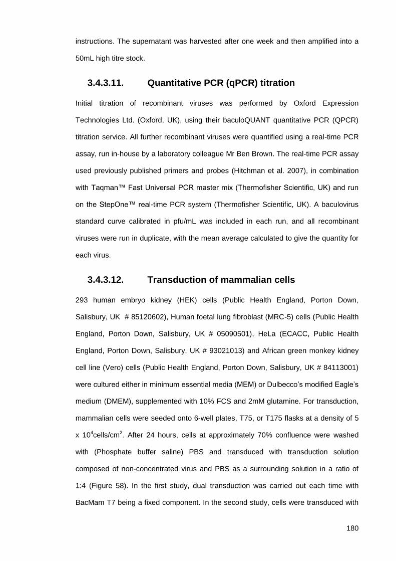
180
instructions. The supernatant was harvested after one week and then amplified into a
50mL high titre stock.
Quantitative PCR (qPCR) titration 3.4.3.11.
Initial titration of recombinant viruses was performed by Oxford Expression
Technologies Ltd. (Oxford, UK), using their baculoQUANT quantitative PCR (QPCR)
titration service. All further recombinant viruses were quantified using a real-time PCR
assay, run in-house by a laboratory colleague Mr Ben Brown. The real-time PCR assay
used previously published primers and probes (Hitchman et al. 2007), in combination
with Taqman™ Fast Universal PCR master mix (Thermofisher Scientific, UK) and run
on the StepOne™ real-time PCR system (Thermofisher Scientific, UK). A baculovirus
standard curve calibrated in pfu/mL was included in each run, and all recombinant
viruses were run in duplicate, with the mean average calculated to give the quantity for
each virus.
Transduction of mammalian cells 3.4.3.12.
293 human embryo kidney (HEK) cells (Public Health England, Porton Down,
Salisbury, UK # 85120602), Human foetal lung fibroblast (MRC-5) cells (Public Health
England, Porton Down, Salisbury, UK # 05090501), HeLa (ECACC, Public Health
England, Porton Down, Salisbury, UK # 93021013) and African green monkey kidney
cell line (Vero) cells (Public Health England, Porton Down, Salisbury, UK # 84113001)
were cultured either in minimum essential media (MEM) or Dulbecco’s modified Eagle’s
medium (DMEM), supplemented with 10% FCS and 2mM glutamine. For transduction,
mammalian cells were seeded onto 6-well plates, T75, or T175 flasks at a density of 5
x 104cells/cm2. After 24 hours, cells at approximately 70% confluence were washed
with (Phosphate buffer saline) PBS and transduced with transduction solution
composed of non-concentrated virus and PBS as a surrounding solution in a ratio of
1:4 (Figure 58). In the first study, dual transduction was carried out each time with
BacMam T7 being a fixed component. In the second study, cells were transduced with
181
either single recombinant BacMam or with all recombinant viruses combined. Cells
were incubated at 27C° for 4-6 hours with mild shaking. Transduction solution was then
replaced with culture medium containing 3 mM sodium butyrate (Sigma-Aldrich #
B5887) as a histone deacetylase (HDAC) inhibitor. After 24 hours of incubation at
37C°, the medium was replaced with fresh medium. Cultures were examined for GFP
expression using fluorescence microscopy. 72 hours post transduction, cells were
collected for analysis.
Figure 58: Transduction of mammalian cells with baculovirus.
Immunohistochemistry 3.4.3.13.
Vero and MRC-5 cells were seeded in 6- well plate at a density of 5 x 10⁴ cells/cm2. 24
hours post seeding, cells at approximately 70% confluence were transduced with dual
BacMam viruses bearing BacMam T7 as a fixed component. Immunohistochemistry
(IHC) was conducted for the first study only. At 24 hours after transduction, cells were
washed and fixed in absolute ethanol for an hour at room temperature. Cells then
washed with water for 5 minutes and immersed in 0.5% hydrogen peroxide in methanol
for 30 minutes, followed by three times washing step each for 2 minutes in Tris-
buffered saline (TBS). Cells were incubated with either anti-gp64 antibody (AcV5) or a
specific primary antibody targeting the protein of interest for 30 minutes at room
temperature (Table 19). Cells were washed thoroughly with TBS and incubated with
182
secondary antibody corresponding with its primary antibody for 30 minutes at room
temperature, a TBS wash was then used to stop the reaction. VECTOR NovaRED
Peroxidase (HRP) Substrate Kit (Vector Laboratories # SK4800) was used. NovaRED
reagents were added to each well until faint red-brown colour developed. Cells were
washed with distilled water before examined under the microscope.
Western Blot Analysis 3.4.3.14.
3.4.3.14.1. Protein lysis
72h post transduction, uninfected (negative control) and infected cell monolayers were
washed twice with cold 1X PBS and were then harvested using a 25 cm cell scraper,
and pelleted by centrifugation at 700g for 5 minutes at 4°C. The supernatant was
discarded and the cell pellet of each sample was re-suspended in 500 μl of lysis buffer
[150 mM NaCl (Fisher Scientific #S/3160/6); 25 mM Tris-HCl (Formedium #TRIS01);
1% Triton-X-100 (Sigma-Aldrich #X100); Proteinase Inhibitor Cocktail 1:5000 (Thermo-
Scientific #78425); Benzonase 1:10,000 (Novagen #70746); 5 mM Imidazole (Sigma
Life Science #I0125-1KG); pH 8.0]. The cell lysate was incubated on ice for 30 minutes
with pipetting up and down ten times every 10 minutes. Subsequently the lysate was
clarified by centrifugation at 15,000g for 30 minutes at 4°C. The resultant lysate (500μl)
containing the soluble protein was carefully transferred into a fresh 1.5 mL Eppendorf
tube and used either immediately or stored at -80 Cº for further use.
3.4.3.14.2. Protein-electrophoresis
For the separation of proteins according to their size, 4-20% Mini-Protean TGX stain
free gels (Bio- Rad, cat #456-8093) were used, according to the manufacturers’
guidelines. Cell lysate and culture supernatant were run for protein detection. Before
loading the gel, samples were mixed in 1:1 ratio with loading buffer [62.5 mM Tris
buffer (pH 6.8) (Formedium #TRIS01); 2% SDS (Fisher Scientific #1261-1680); 25%
Glycerol (Sigma-Aldrich #G5516); 0.01% Bromophenol blue (Sigma-Aldrich #318744)].
Samples were denatured for 5 minutes at 95°C. After a brief pulse centrifugation, 15 μL
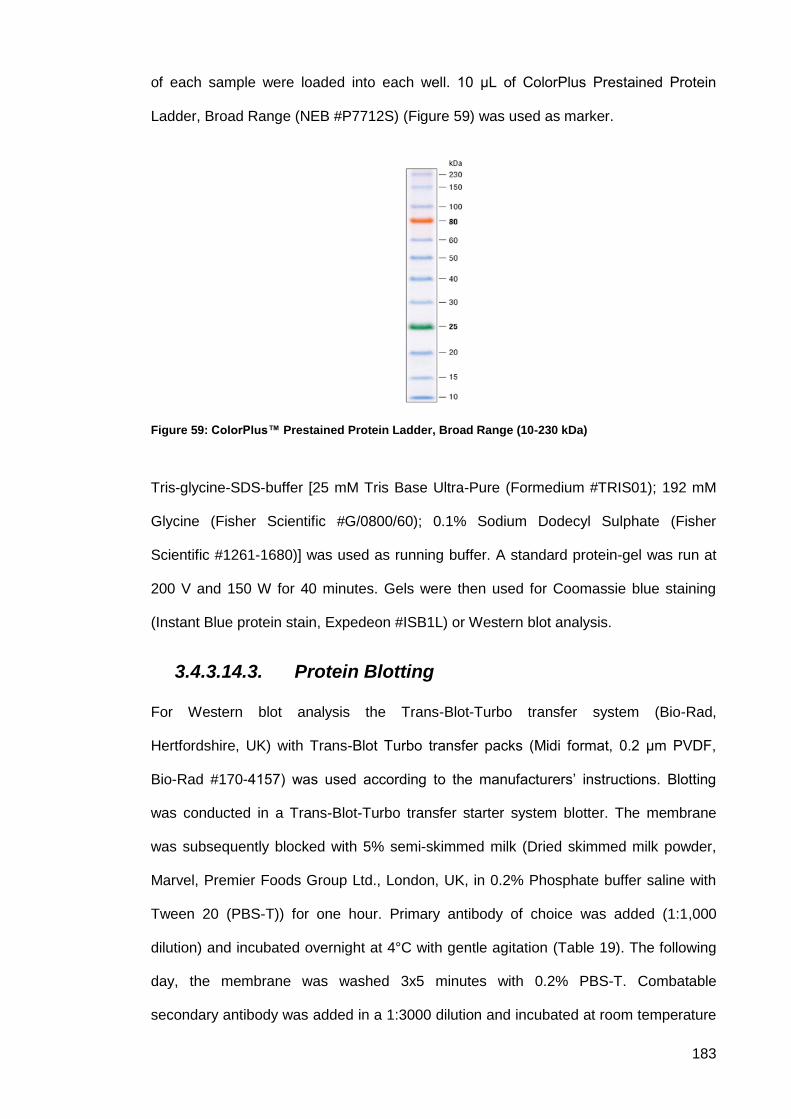
183
of each sample were loaded into each well. 10 μL of ColorPlus Prestained Protein
Ladder, Broad Range (NEB #P7712S) (Figure 59) was used as marker.
Figure 59: ColorPlus™ Prestained Protein Ladder, Broad Range (10-230 kDa)
Tris-glycine-SDS-buffer [25 mM Tris Base Ultra-Pure (Formedium #TRIS01); 192 mM
Glycine (Fisher Scientific #G/0800/60); 0.1% Sodium Dodecyl Sulphate (Fisher
Scientific #1261-1680)] was used as running buffer. A standard protein-gel was run at
200 V and 150 W for 40 minutes. Gels were then used for Coomassie blue staining
(Instant Blue protein stain, Expedeon #ISB1L) or Western blot analysis.
3.4.3.14.3. Protein Blotting
For Western blot analysis the Trans-Blot-Turbo transfer system (Bio-Rad,
Hertfordshire, UK) with Trans-Blot Turbo transfer packs (Midi format, 0.2 μm PVDF,
Bio-Rad #170-4157) was used according to the manufacturers’ instructions. Blotting
was conducted in a Trans-Blot-Turbo transfer starter system blotter. The membrane
was subsequently blocked with 5% semi-skimmed milk (Dried skimmed milk powder,
Marvel, Premier Foods Group Ltd., London, UK, in 0.2% Phosphate buffer saline with
Tween 20 (PBS-T)) for one hour. Primary antibody of choice was added (1:1,000
dilution) and incubated overnight at 4°C with gentle agitation (Table 19). The following
day, the membrane was washed 3x5 minutes with 0.2% PBS-T. Combatable
secondary antibody was added in a 1:3000 dilution and incubated at room temperature
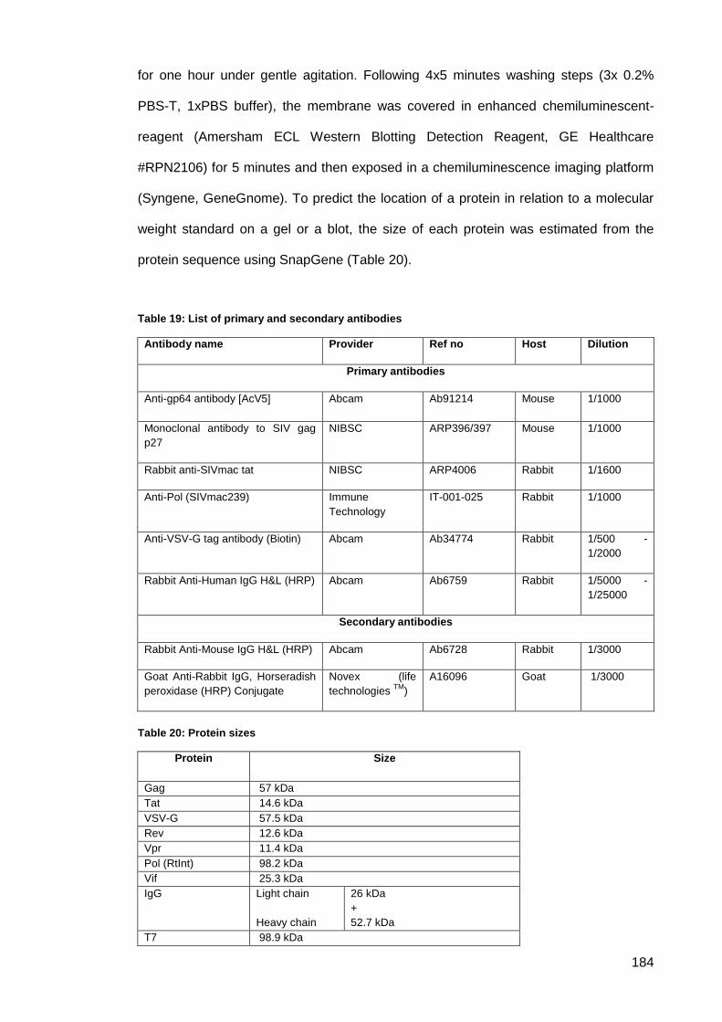
184
for one hour under gentle agitation. Following 4x5 minutes washing steps (3x 0.2%
PBS-T, 1xPBS buffer), the membrane was covered in enhanced chemiluminescent-
reagent (Amersham ECL Western Blotting Detection Reagent, GE Healthcare
#RPN2106) for 5 minutes and then exposed in a chemiluminescence imaging platform
(Syngene, GeneGnome). To predict the location of a protein in relation to a molecular
weight standard on a gel or a blot, the size of each protein was estimated from the
protein sequence using SnapGene (Table 20).
Table 19: List of primary and secondary antibodies
Antibody name Provider Ref no Host Dilution
Primary antibodies
Anti-gp64 antibody [AcV5] Abcam
Ab91214 Mouse 1/1000
Monoclonal antibody to SIV gag
p27
NIBSC ARP396/397 Mouse 1/1000
Rabbit anti-SIVmac tat NIBSC ARP4006 Rabbit 1/1600
Anti-Pol (SIVmac239) Immune
Technology
IT-001-025 Rabbit 1/1000
Anti-VSV-G tag antibody (Biotin) Abcam Ab34774 Rabbit 1/500 -
1/2000
Rabbit Anti-Human IgG H&L (HRP) Abcam Ab6759 Rabbit 1/5000 -
1/25000
Secondary antibodies
Rabbit Anti-Mouse IgG H&L (HRP) Abcam Ab6728 Rabbit 1/3000
Goat Anti-Rabbit IgG, Horseradish
peroxidase (HRP) Conjugate
Novex (life
technologies TM
)
A16096 Goat 1/3000
Table 20: Protein sizes
Protein
Size
Gag 57 kDa
Tat 14.6 kDa
VSV-G 57.5 kDa
Rev 12.6 kDa
Vpr 11.4 kDa
Pol (RtInt) 98.2 kDa
Vif 25.3 kDa
IgG Light chain
Heavy chain
26 kDa
+
52.7 kDa
T7 98.9 kDa
185
Ultracentrifugation and Transmission Electron 3.4.3.15.
Microscopy (TEM)
In the second study, the supernatant from 72 hours post transduction HEK239 cell
culture was collected and centrifuged at 700g for 5 minutes at 4C. The supernatant
was collected and subjected to ultracentrifugation. Beckman Ultra-Clear centrifuge
tubes (Cat # 344057) were sterilised for 15 minutes by exposure to UV light in a
biological safety cabinet. A total of 30 mL viral supernatant was centrifuged at 90,000 g
for 90 minutes at 4°C in a Beckman SW50.1 swinging bucket rotor lined with a
Beckman Ultra-Clear centrifuge tube. To obtain accurate balanced volumes in each
tube, supernatant was loaded while measuring on a weighing balance to ensure that all
tubes would be in similar final volumes. Following centrifugation, the medium was
carefully disposed of into a bleach-filled container. Pellets were gently resuspended in
200µL of PBS by pipetting 20 times using a P200 pipette, care being taken to minimise
the creation of foam. The sample was then sent to the Electron Microscopy Facility,
University of Manchester. The sample was incubated for 1 minute on carbon coated
400 Mesh Grids Copper (Agar scientific # AGS160-4). This was followed by washing 5x
in distilled water to remove phosphate ions, then negatively stained using 1% uranyl
acetate dissolved in distilled water (pH 4.2 to 4.5) for 30 second followed by an air
drying step. The negatively stained specimen was examined using a 20-120 kV FEI
Tecnai G2 Spirit BioTWIN Transmission Electron Microscope. Micrographs were
prepared using an Orius SC1000 CCD camera system.

186
Results 3.5.
3.5.1. Design and construction of BacMam transfer plasmids
Study : T7 RNA Polymearse/Promoter system 3.5.1.1.
3.5.1.1.1. pLF_T7_RNA-Polymerase
A summary of the cloning procedure is shown in Figure 60. DNA plasmid extracted
from randomly selected colonies of transformed E.coli was subjected to restriction
digestion to screen for positive clones (Figure 61, Figure 62 and Figure 63). Insert
positive clones were confirmed by sequencing.
Figure 60: Schematic representation of pLF_T7_RNA-Polymerase cloning steps. A fragment size
4562 bp containing T7 polymerase was excised from pT7-Vpx_MVA and inserted into pOET6 between the Acc65I and NotI sites. The resultant plasmid was then cleaned from excess DNA flanking the T7 polymerase cassette by cutting one end with Acc65I and BstBI, blunting and ligatiion. The second end was cut with AscI and NotI, which was then blunted and ligated to form pT7 RNA polymerase. The T7 polymerase cassette was placed downstream of the CMV promoter.
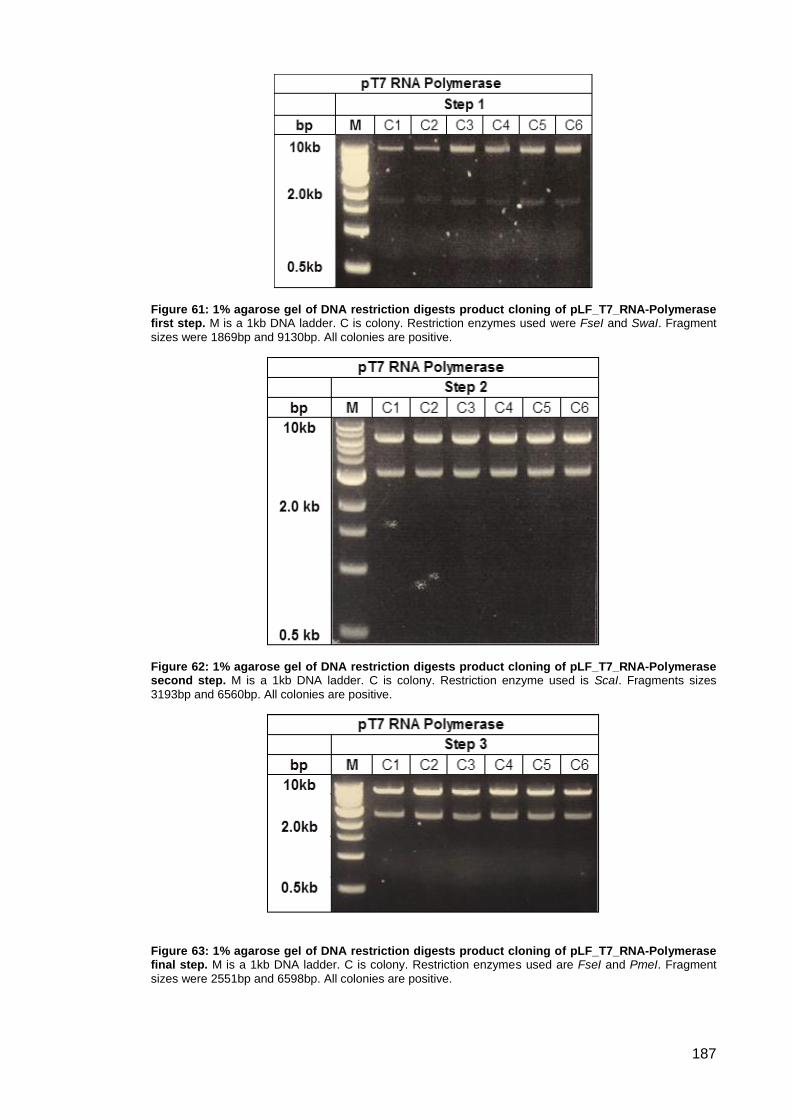
187
Figure 61: 1% agarose gel of DNA restriction digests product cloning of pLF_T7_RNA-Polymerase first step. M is a 1kb DNA ladder. C is colony. Restriction enzymes used were FseI and SwaI. Fragment
sizes were 1869bp and 9130bp. All colonies are positive.
Figure 62: 1% agarose gel of DNA restriction digests product cloning of pLF_T7_RNA-Polymerase second step. M is a 1kb DNA ladder. C is colony. Restriction enzyme used is ScaI. Fragments sizes
3193bp and 6560bp. All colonies are positive.
Figure 63: 1% agarose gel of DNA restriction digests product cloning of pLF_T7_RNA-Polymerase final step. M is a 1kb DNA ladder. C is colony. Restriction enzymes used are FseI and PmeI. Fragment
sizes were 2551bp and 6598bp. All colonies are positive.

188
3.5.1.1.2. pLF_T7_IgGb12-GFP
A summary of the cloning procedure is shown in Figure 64. Plasmid DNA was
extracted from randomly selected colonies and was subjected to restriction digestion
(Figure 65 and Figure 66). Positive clones were confirmed by sequencing.
Figure 64: Schematic representation of pLF_T7_IgGb12-GFP cloning steps. The IgGb12 expression
cassette with fragment size 6306bp encoding EGFP was removed from pIgGb12-GFP_MVA and inserted into pOET6 between the XbaI and HpaI sites. The restriction fragment between PacI and Bgl III was deleted and overhanging sequences were filled-in/removed by blunt end modification to form the recombinant transfer vector pLF_T7_IgGb12-GFP.
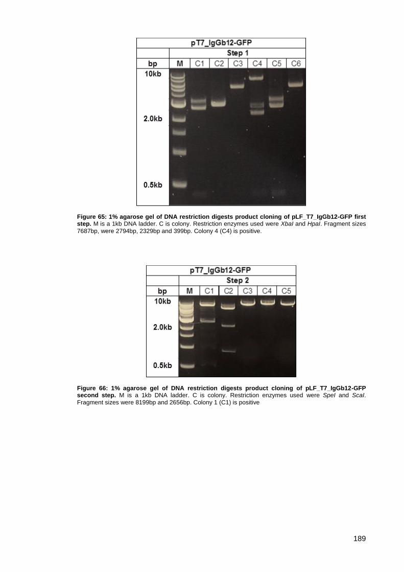
189
Figure 65: 1% agarose gel of DNA restriction digests product cloning of pLF_T7_IgGb12-GFP first step. M is a 1kb DNA ladder. C is colony. Restriction enzymes used were XbaI and HpaI. Fragment sizes
7687bp, were 2794bp, 2329bp and 399bp. Colony 4 (C4) is positive.
Figure 66: 1% agarose gel of DNA restriction digests product cloning of pLF_T7_IgGb12-GFP second step. M is a 1kb DNA ladder. C is colony. Restriction enzymes used were SpeI and ScaI.
Fragment sizes were 8199bp and 2656bp. Colony 1 (C1) is positive
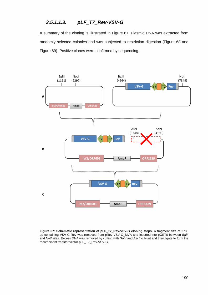
190
3.5.1.1.3. pLF_T7_Rev-VSV-G
A summary of the cloning is illustrated in Figure 67. Plasmid DNA was extracted from
randomly selected colonies and was subjected to restriction digestion (Figure 68 and
Figure 69). Positive clones were confirmed by sequencing.
Figure 67: Schematic representation of pLF_T7_Rev-VSV-G cloning steps. A fragment size of 2785 bp containing VSV-G Rev was removed from pRev-VSV-G_MVA and inserted into pOET6 between BglII and NotI sites. Excess DNA was removed by cutting with SphI and AscI to blunt and then ligate to form the recombinant transfer vector pLF_T7_Rev-VSV-G.
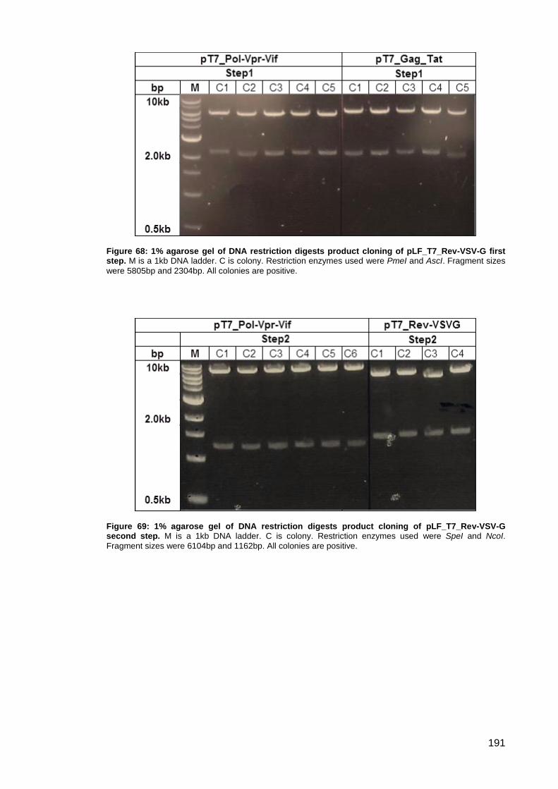
191
Figure 68: 1% agarose gel of DNA restriction digests product cloning of pLF_T7_Rev-VSV-G first step. M is a 1kb DNA ladder. C is colony. Restriction enzymes used were PmeI and AscI. Fragment sizes
were 5805bp and 2304bp. All colonies are positive.
Figure 69: 1% agarose gel of DNA restriction digests product cloning of pLF_T7_Rev-VSV-G second step. M is a 1kb DNA ladder. C is colony. Restriction enzymes used were SpeI and NcoI.
Fragment sizes were 6104bp and 1162bp. All colonies are positive.
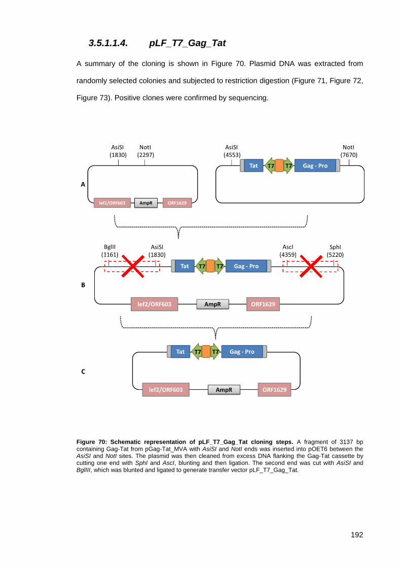
192
3.5.1.1.4. pLF_T7_Gag_Tat
A summary of the cloning is shown in Figure 70. Plasmid DNA was extracted from
randomly selected colonies and subjected to restriction digestion (Figure 71, Figure 72,
Figure 73). Positive clones were confirmed by sequencing.
Figure 70: Schematic representation of pLF_T7_Gag_Tat cloning steps. A fragment of 3137 bp containing Gag-Tat from pGag-Tat_MVA with AsiSI and NotI ends was inserted into pOET6 between the AsiSI and NotI sites. The plasmid was then cleaned from excess DNA flanking the Gag-Tat cassette by cutting one end with SphI and AscI, blunting and then ligation. The second end was cut with AsiSI and BglIII, which was blunted and ligated to generate transfer vector pLF_T7_Gag_Tat.
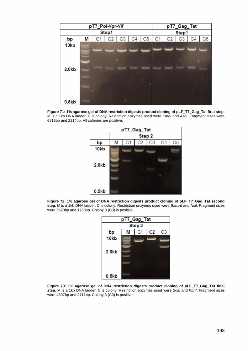
193
Figure 71: 1% agarose gel of DNA restriction digests product cloning of pLF_T7_Gag_Tat first step. M is a 1kb DNA ladder. C is colony. Restriction enzymes used were PmeI and AscI. Fragment sizes were
6816bp and 2314bp. All colonies are positive.
Figure 72: 1% agarose gel of DNA restriction digests product cloning of pLF_T7_Gag_Tat second step. M is a 1kb DNA ladder. C is colony. Restriction enzymes used were BamHI and NsiI. Fragment sizes
were 6520bp and 1753bp. Colony 3 (C3) is positive.
Figure 73: 1% agarose gel of DNA restriction digests product cloning of pLF_T7_Gag_Tat final step. M is a 1kb DNA ladder. C is colony. Restriction enzymes used were ScaI and KpnI. Fragment sizes
were 4897bp and 2711bp. Colony 3 (C3) is positive.
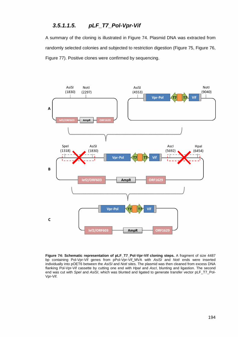
194
3.5.1.1.5. pLF_T7_Pol-Vpr-Vif
A summary of the cloning is illustrated in Figure 74. Plasmid DNA was extracted from
randomly selected colonies and subjected to restriction digestion (Figure 75, Figure 76,
Figure 77). Positive clones were confirmed by sequencing.
Figure 74: Schematic representation of pLF_T7_Pol-Vpr-Vif cloning steps. A fragment of size 4487 bp containing Pol-Vpr-Vif genes from pPol-Vpr-Vif_MVA with AsiSI and NotI ends were inserted individually into pOET6 between the AsiSI and NotI sites. The plasmid was then cleaned from excess DNA flanking Pol-Vpr-Vif cassette by cutting one end with HpaI and AscI, blunting and ligastion. The second end was cut with SpeI and AsiSI, which was blunted and ligated to generate transfer vector pLF_T7_Pol-Vpr-Vif.
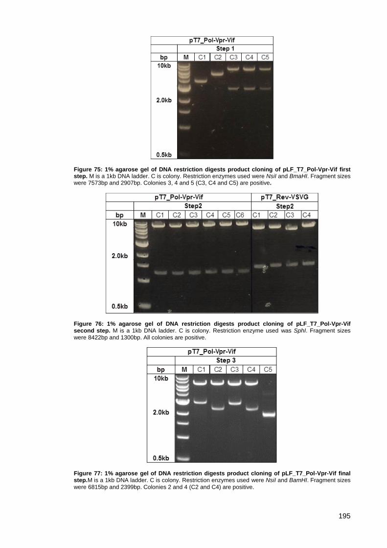
195
Figure 75: 1% agarose gel of DNA restriction digests product cloning of pLF_T7_Pol-Vpr-Vif first step. M is a 1kb DNA ladder. C is colony. Restriction enzymes used were NsiI and BmaHI. Fragment sizes were 7573bp and 2907bp. Colonies 3, 4 and 5 (C3, C4 and C5) are positive.
Figure 76: 1% agarose gel of DNA restriction digests product cloning of pLF_T7_Pol-Vpr-Vif second step. M is a 1kb DNA ladder. C is colony. Restriction enzyme used was SphI. Fragment sizes
were 8422bp and 1300bp. All colonies are positive.
Figure 77: 1% agarose gel of DNA restriction digests product cloning of pLF_T7_Pol-Vpr-Vif final step.M is a 1kb DNA ladder. C is colony. Restriction enzymes used were NsiI and BamHI. Fragment sizes
were 6815bp and 2399bp. Colonies 2 and 4 (C2 and C4) are positive.

196
Study , CMV promoter 3.5.1.2.
3.5.1.2.1. pLF_CMV-Gag-IRES-Tat, pLF_CMV-RtInt-IRES-
Vif and pLF_CMV-VSV-IRES-Rev
To form the pLF_CMV-Gag-IRES-Tat, pLF_CMV-RtInt-IRES-Vif and pLF_CMV-VSV-
IRES-Rev constructs, 3 different genes of interest were inserted into each plasmid.
NheI and EcoRI was used to insert Gag, Rt-Int separately into 2 different pOET6; while
NheI and XhoI used to insert VSV-G into a 3rd different pOET6. EcoRI and XbaI was
used to insert IRES in each of the resulting plasmids. Finally, XmaI and XbaI were
used to insert Tat, Vif and Rev into each plasmid. A summary of the cloning results is
shown in Figure 78. Plasmid DNA was extracted from randomly selected colonies and
subjected to restriction digestion (Figure 79). Positive clones were confirmed by
sequencing.
Figure 78: Schematic representation of pLF_CMV-Gag-IRES-Tat, pLF_CMV-RtInt-IRES-Vif and pLF_CMV-VSV-IRES-Rev cloning steps.
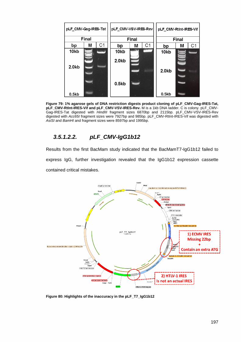
197
Figure 79: 1% agarose gels of DNA restriction digests product cloning of pLF_CMV-Gag-IRES-Tat, pLF_CMV-RtInt-IRES-Vif and pLF_CMV-VSV-IRES-Rev. M is a 1kb DNA ladder. C is colony. pLF_CMV-Gag-IRES-Tat digested with HindIII fragment sizes 6870bp and 2115bp. pLF_CMV-VSV-IRES-Rev digested with Acc65I fragment sizes were 7927bp and 985bp. pLF_CMV-RtInt-IRES-Vif was digested with AsiSI and BamHI and fragment sizes were 8597bp and 1995bp.
3.5.1.2.2. pLF_CMV-IgG1b12
Results from the first BacMam study indicated that the BacMamT7-IgG1b12 failed to
express IgG, further investigation revealed that the IgG1b12 expression cassette
contained critical mistakes.
Figure 80: Highlights of the inaccuracy in the pLF_T7_IgG1b12

198
Sequencing analysis showed that the encephalomyocarditis virus (EMCV) internal
ribosomal entry site (IRES) inserted between the light and heavy chain was missing
22bp and contained an extra ATG (Figure 80). This was likely to effect IRES activity
particularly in bicistronic construct.
Transfer plasmid contains the correct EMCV IRES sequence were designed and
manufactured by Eurofin Genomics, Germany. Unfortunately Eurofin couldn’t generate
the plasmid in one piece. The plasmid was prepared as Fragment A and B and then re-
assembled. Several stages were needed to join Fragment A and B together. First,
Fragment A was cloned into pOET6 between the EcoRI and NheI restriction sites NheI
is located in two locations one of which is directly upstream of EcoRI. Fragment A was
excised by cutting with EcoRI first then with NheI to eliminate the second NheI (Figure
81).
To confirm cloning, plasmid DNA was extracted from randomly selected colonies and
subjected to restriction digestion (Figure 82). Second, primers were designed to
introduce appropriate restriction sites to clone fragment B into pOET6+Fragment A
between EcoRI and XmaI (Figure 83). To confirm cloning, plasmid DNA was extracted
from randomly selected colonies and was subjected to restriction digestion (Figure 84).
Finally, the resulting plasmid pOET6+Fragment A+B contained a gap between
fragment A and B, which would interfere with protein expression. To correct this, the
fragment between XhoI was replaced with the correct one from pIgG+P2A (Figure 87).
pIgG+P2A was constructed by replacing a fragment located between AflII and BstAPI.
This fragment was excised from pCorrect_seq1 and cloned into the pOET6-IgG12
vector (Figure 85). To confirm cloning, plasmid DNA was extracted from randomly
selected colonies and was subjected to restriction digestion (Figure 86 and Figure 88).
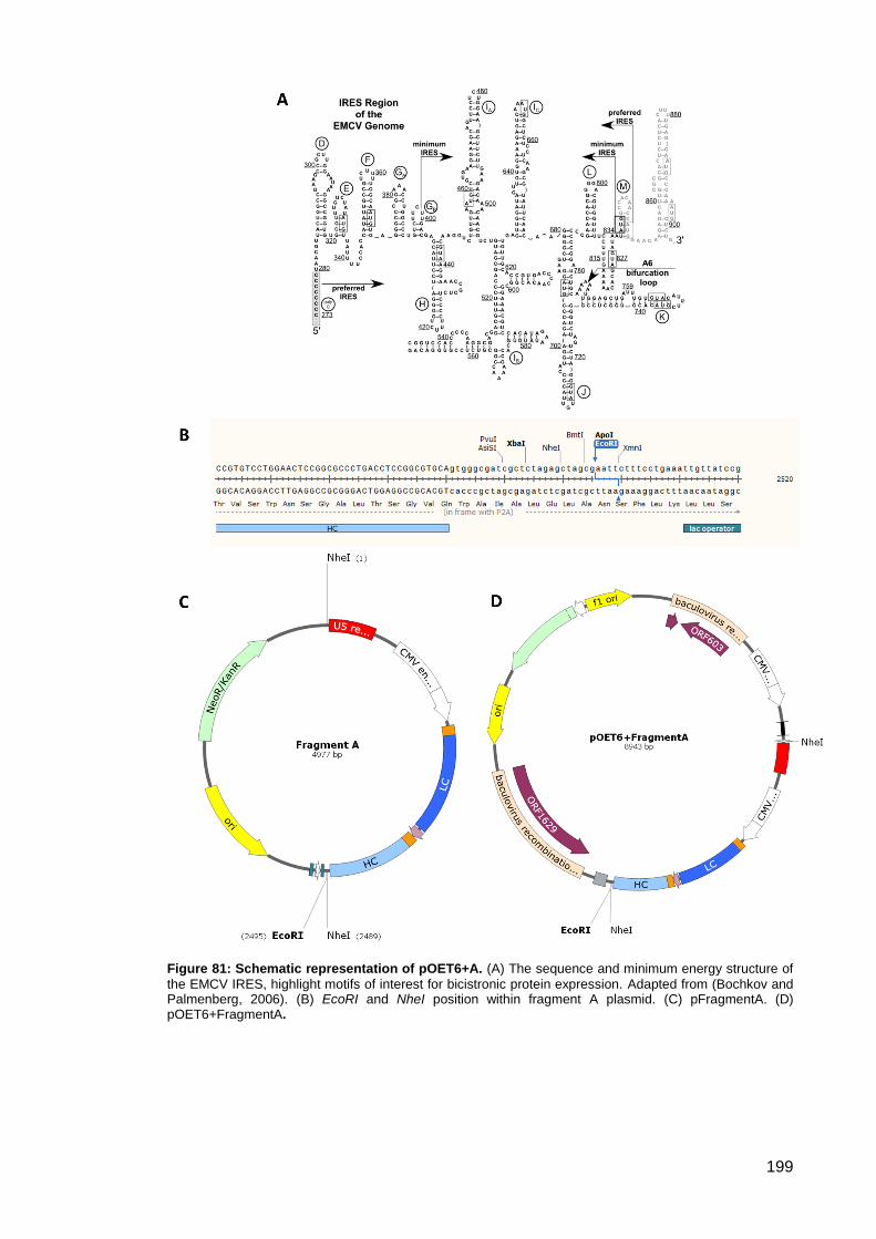
199
Figure 81: Schematic representation of pOET6+A. (A) The sequence and minimum energy structure of
the EMCV IRES, highlight motifs of interest for bicistronic protein expression. Adapted from (Bochkov and Palmenberg, 2006). (B) EcoRI and NheI position within fragment A plasmid. (C) pFragmentA. (D) pOET6+FragmentA.
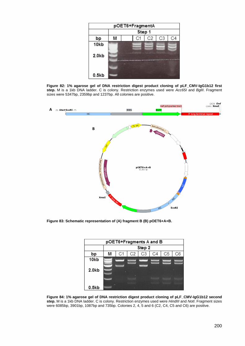
200
Figure 82: 1% agarose gel of DNA restriction digest product cloning of pLF_CMV-IgG1b12 first step. M is a 1kb DNA ladder. C is colony. Restriction enzymes used were Acc65I and BglII. Fragment
sizes were 5347bp, 2359bp and 1237bp. All colonies are positive.
Figure 83: Schematic representation of (A) fragment B (B) pOET6+A+B.
Figure 84: 1% agarose gel of DNA restriction digest product cloning of pLF_CMV-IgG1b12 second step. M is a 1kb DNA ladder. C is colony. Restriction enzymes used were HindIII and NotI. Fragment sizes
were 6085bp, 3901bp, 1087bp and 735bp. Colonies 2, 4, 5 and 6 (C2, C4, C5 and C6) are positive.
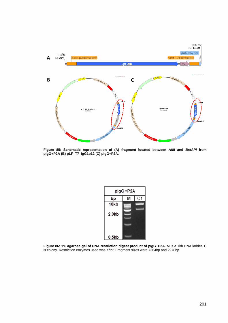
201
Figure 85: Schematic representation of (A) fragment located between AflII and BstAPI from pIgG+P2A (B) pLF_T7_IgG1b12 (C) pIgG+P2A.
Figure 86: 1% agarose gel of DNA restriction digest product of pIgG+P2A. M is a 1kb DNA ladder. C is colony. Restriction enzymes used was XhoI. Fragment sizes were 7364bp and 2978bp.
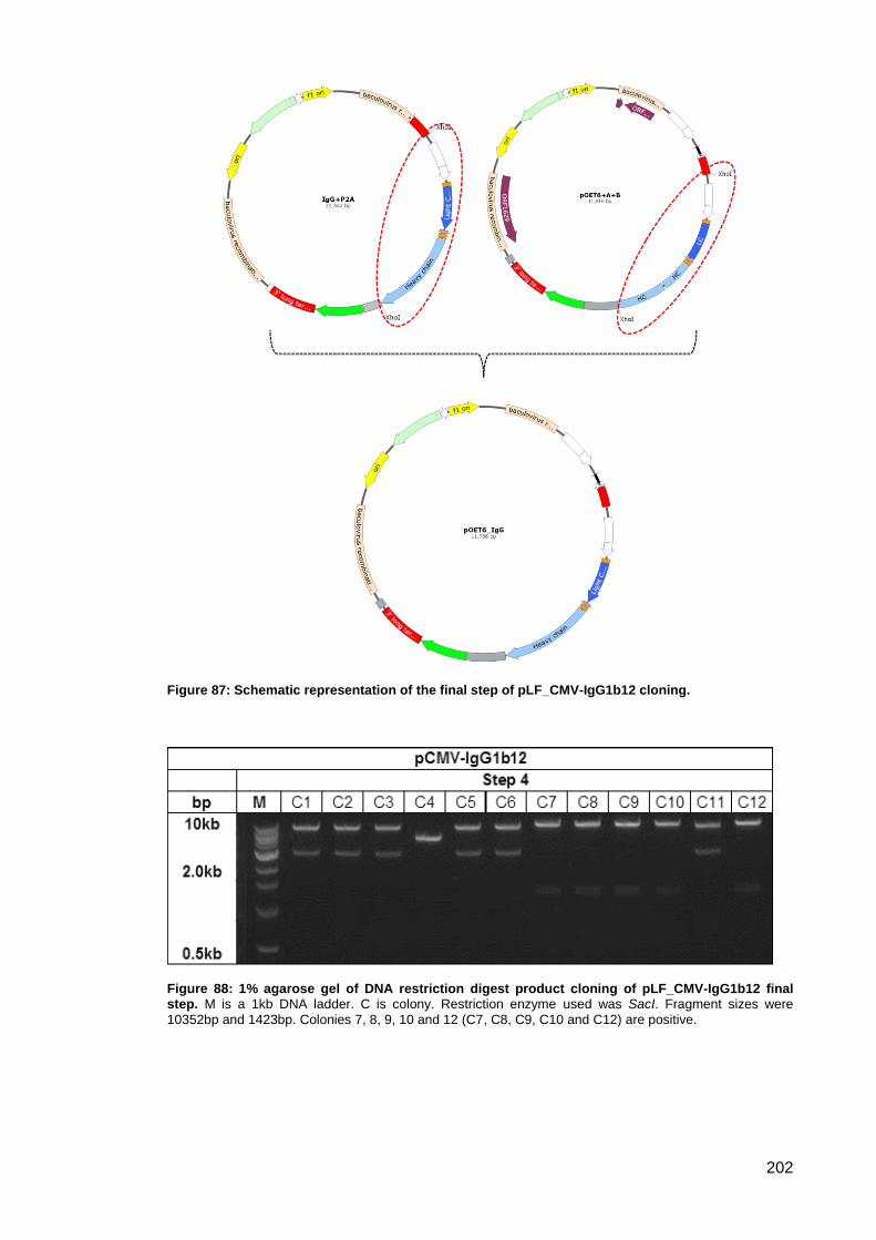
202
Figure 87: Schematic representation of the final step of pLF_CMV-IgG1b12 cloning.
Figure 88: 1% agarose gel of DNA restriction digest product cloning of pLF_CMV-IgG1b12 final step. M is a 1kb DNA ladder. C is colony. Restriction enzyme used was SacI. Fragment sizes were
10352bp and 1423bp. Colonies 7, 8, 9, 10 and 12 (C7, C8, C9, C10 and C12) are positive.
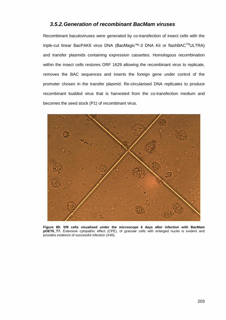
203
3.5.2. Generation of recombinant BacMam viruses
Recombinant baculoviruses were generated by co-transfection of insect cells with the
triple-cut linear BacPAK6 virus DNA (BacMagic™-3 DNA Kit or flashBACTMULTRA)
and transfer plasmids containing expression cassettes. Homologous recombination
within the insect cells restores ORF 1629 allowing the recombinant virus to replicate,
removes the BAC sequences and inserts the foreign gene under control of the
promoter chosen in the transfer plasmid. Re-circularised DNA replicates to produce
recombinant budded virus that is harvested from the co-transfection medium and
becomes the seed stock (P1) of recombinant virus.
Figure 89: Sf9 cells visualised under the microscope 6 days after infection with BacMam pOET6_T7. Extensive cytopathic effect (CPE), of granular cells with enlarged nuclei is evident and
provides evidence of successful infection (X40).
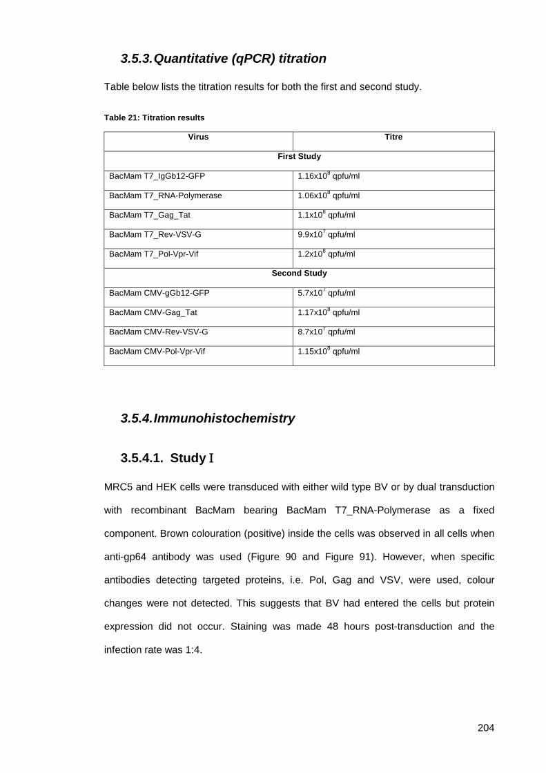
204
3.5.3. Quantitative (qPCR) titration
Table below lists the titration results for both the first and second study.
Table 21: Titration results
Virus Titre
First Study
BacMam T7_IgGb12-GFP 1.16x10⁸ qpfu/ml
BacMam T7_RNA-Polymerase 1.06x10⁸ qpfu/ml
BacMam T7_Gag_Tat 1.1x10⁸ qpfu/ml
BacMam T7_Rev-VSV-G 9.9x10⁷ qpfu/ml
BacMam T7_Pol-Vpr-Vif 1.2x10⁸ qpfu/ml
Second Study
BacMam CMV-gGb12-GFP 5.7x10⁷ qpfu/ml
BacMam CMV-Gag_Tat 1.17x10⁸ qpfu/ml
BacMam CMV-Rev-VSV-G 8.7x10⁷ qpfu/ml
BacMam CMV-Pol-Vpr-Vif 1.15x10⁸ qpfu/ml
3.5.4. Immunohistochemistry
Study 3.5.4.1.
MRC5 and HEK cells were transduced with either wild type BV or by dual transduction
with recombinant BacMam bearing BacMam T7_RNA-Polymerase as a fixed
component. Brown colouration (positive) inside the cells was observed in all cells when
anti-gp64 antibody was used (Figure 90 and Figure 91). However, when specific
antibodies detecting targeted proteins, i.e. Pol, Gag and VSV, were used, colour
changes were not detected. This suggests that BV had entered the cells but protein
expression did not occur. Staining was made 48 hours post-transduction and the
infection rate was 1:4.
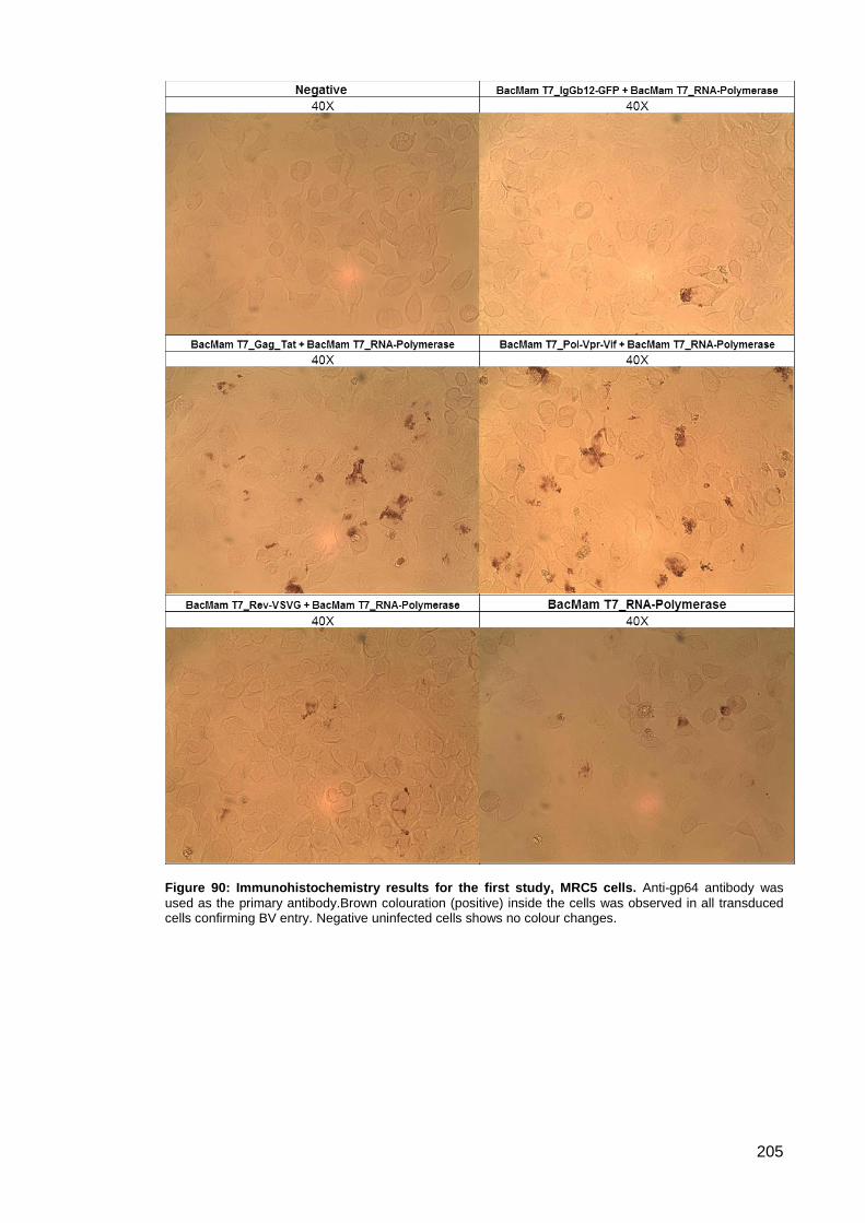
205
Figure 90: Immunohistochemistry results for the first study, MRC5 cells. Anti-gp64 antibody was
used as the primary antibody.Brown colouration (positive) inside the cells was observed in all transduced cells confirming BV entry. Negative uninfected cells shows no colour changes.
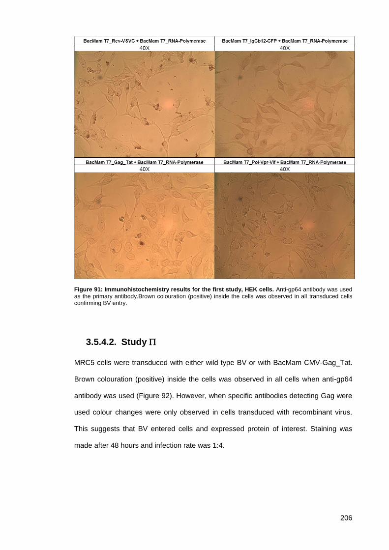
206
Figure 91: Immunohistochemistry results for the first study, HEK cells. Anti-gp64 antibody was used
as the primary antibody.Brown colouration (positive) inside the cells was observed in all transduced cells confirming BV entry.
Study 3.5.4.2.
MRC5 cells were transduced with either wild type BV or with BacMam CMV-Gag_Tat.
Brown colouration (positive) inside the cells was observed in all cells when anti-gp64
antibody was used (Figure 92). However, when specific antibodies detecting Gag were
used colour changes were only observed in cells transduced with recombinant virus.
This suggests that BV entered cells and expressed protein of interest. Staining was
made after 48 hours and infection rate was 1:4.
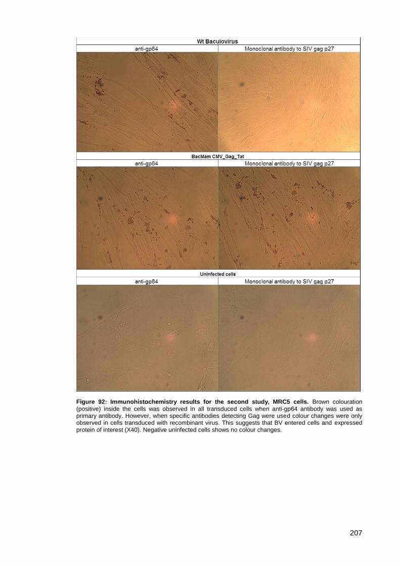
207
Figure 92: Immunohistochemistry results for the second study, MRC5 cells. Brown colouration
(positive) inside the cells was observed in all transduced cells when anti-gp64 antibody was used as primary antibody. However, when specific antibodies detecting Gag were used colour changes were only observed in cells transduced with recombinant virus. This suggests that BV entered cells and expressed protein of interest (X40). Negative uninfected cells shows no colour changes.

208
3.5.5. Green-fluorescent protein (GFP) expression
Green-fluorescent protein (GFP) was incorporated within the expression cassette of
IgG1b12. GFP expression was used to study expression efficiency. The IgG1b12
expression cassette combines the expression of heavy and light chains in a single
cassette together with GFP in a tri-cistronic setup. EMCV IRES elements were used to
combine the reporter gene GFP with antibody sequence. GFP expression proves the
activity of the EMCV IRES integrated into all recombinant BacMam constructs.
Monolayers of VERO and HeLa cells were co-transfected with BacMam CMV-gGb12-
GFP. 1-3 days after transfection, GFP expression was visualised under an inverted
fluorescent microscope (Optika-XDS-3FL). Green fluorescent signal was not clearly
confirmed as observation of all transfected cells shows very week fluorescent signals
(Figure 93). Before transfection, all cells showed negative green flourecent signlas.
Figure 93: Transfection of mammalian cells with BacMam pOET6_IgGb12-GFP recombinant plasmid. (A) Vero cells, Infection rate 1:4, 24 hour post transfection. (B) Hela cells, Infection rate 1:4, 24
hour post transfection.
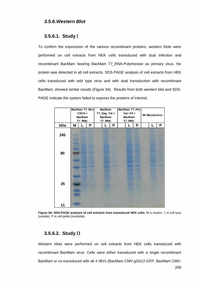
209
3.5.6. Western Blot
Study 3.5.6.1.
To confirm the expression of the various recombinant proteins, western blots were
performed on cell extracts from HEK cells transduced with dual infection and
recombinant BacMam bearing BacMam T7_RNA-Polymerase as primary virus. No
protein was detected in all cell extracts. SDS-PAGE analysis of cell extracts from HEK
cells transduced with wild type virus and with dual transduction with recombinant
BacMam, showed similar results (Figure 94). Results from both western blot and SDS-
PAGE indicate the system failed to express the proteins of interest.
Figure 94: SDS-PAGE analysis of cell extracts from transduced HEK cells. M is marker. L is cell lysis
(soluble). P is cell pellet (insoluble).
Study 3.5.6.2.
Western blots were performed on cell extracts from HEK cells transduced with
recombinant BacMam virus. Cells were either transduced with a single recombinant
BacMam or co-transduced with all 4 rBVs (BacMam CMV-gGb12-GFP, BacMam CMV-
kDa M L P L P L P L P
245
80
25
11
BacMam T7_Rev-
VSVG +
BacMam
T7_RNA-
BacMam
T7_Gag_Tat +
BacMam
T7_RNA-
BacMam T7_Pol-
Vpr-Vif +
BacMam
T7_RNA-
Wt Baculovirus
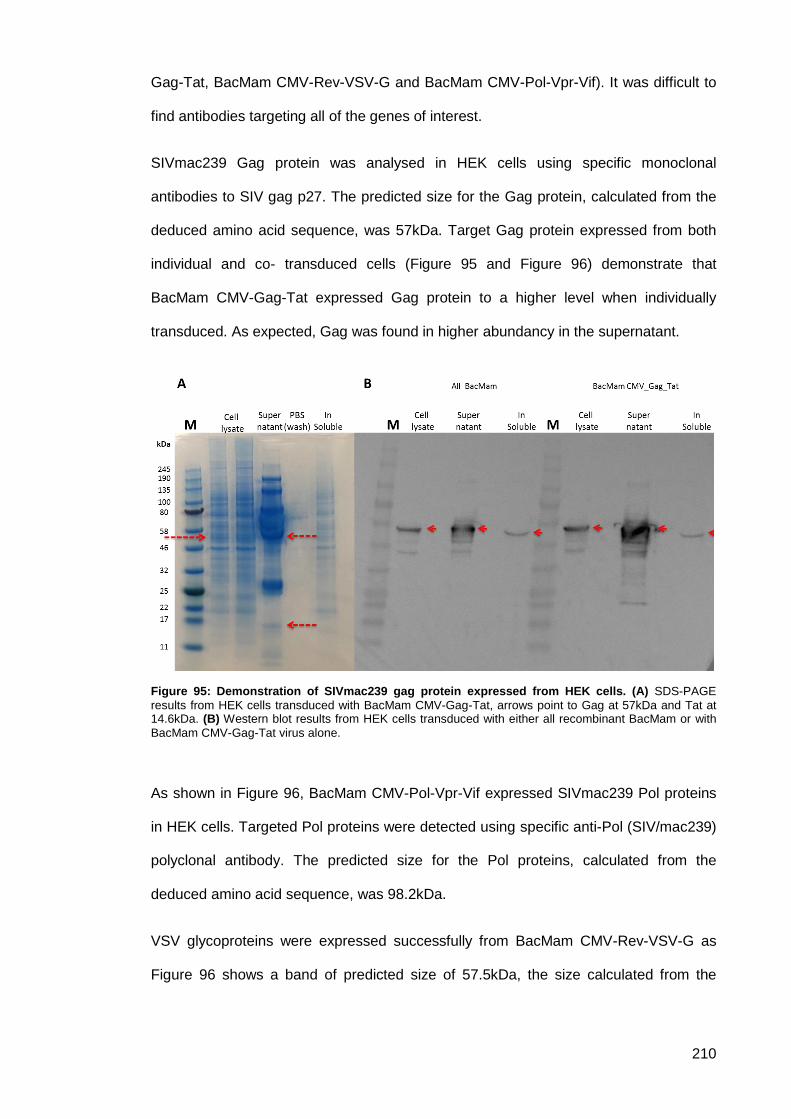
210
Gag-Tat, BacMam CMV-Rev-VSV-G and BacMam CMV-Pol-Vpr-Vif). It was difficult to
find antibodies targeting all of the genes of interest.
SIVmac239 Gag protein was analysed in HEK cells using specific monoclonal
antibodies to SIV gag p27. The predicted size for the Gag protein, calculated from the
deduced amino acid sequence, was 57kDa. Target Gag protein expressed from both
individual and co- transduced cells (Figure 95 and Figure 96) demonstrate that
BacMam CMV-Gag-Tat expressed Gag protein to a higher level when individually
transduced. As expected, Gag was found in higher abundancy in the supernatant.
Figure 95: Demonstration of SIVmac239 gag protein expressed from HEK cells. (A) SDS-PAGE
results from HEK cells transduced with BacMam CMV-Gag-Tat, arrows point to Gag at 57kDa and Tat at 14.6kDa. (B) Western blot results from HEK cells transduced with either all recombinant BacMam or with
BacMam CMV-Gag-Tat virus alone.
As shown in Figure 96, BacMam CMV-Pol-Vpr-Vif expressed SIVmac239 Pol proteins
in HEK cells. Targeted Pol proteins were detected using specific anti-Pol (SIV/mac239)
polyclonal antibody. The predicted size for the Pol proteins, calculated from the
deduced amino acid sequence, was 98.2kDa.
VSV glycoproteins were expressed successfully from BacMam CMV-Rev-VSV-G as
Figure 96 shows a band of predicted size of 57.5kDa, the size calculated from the
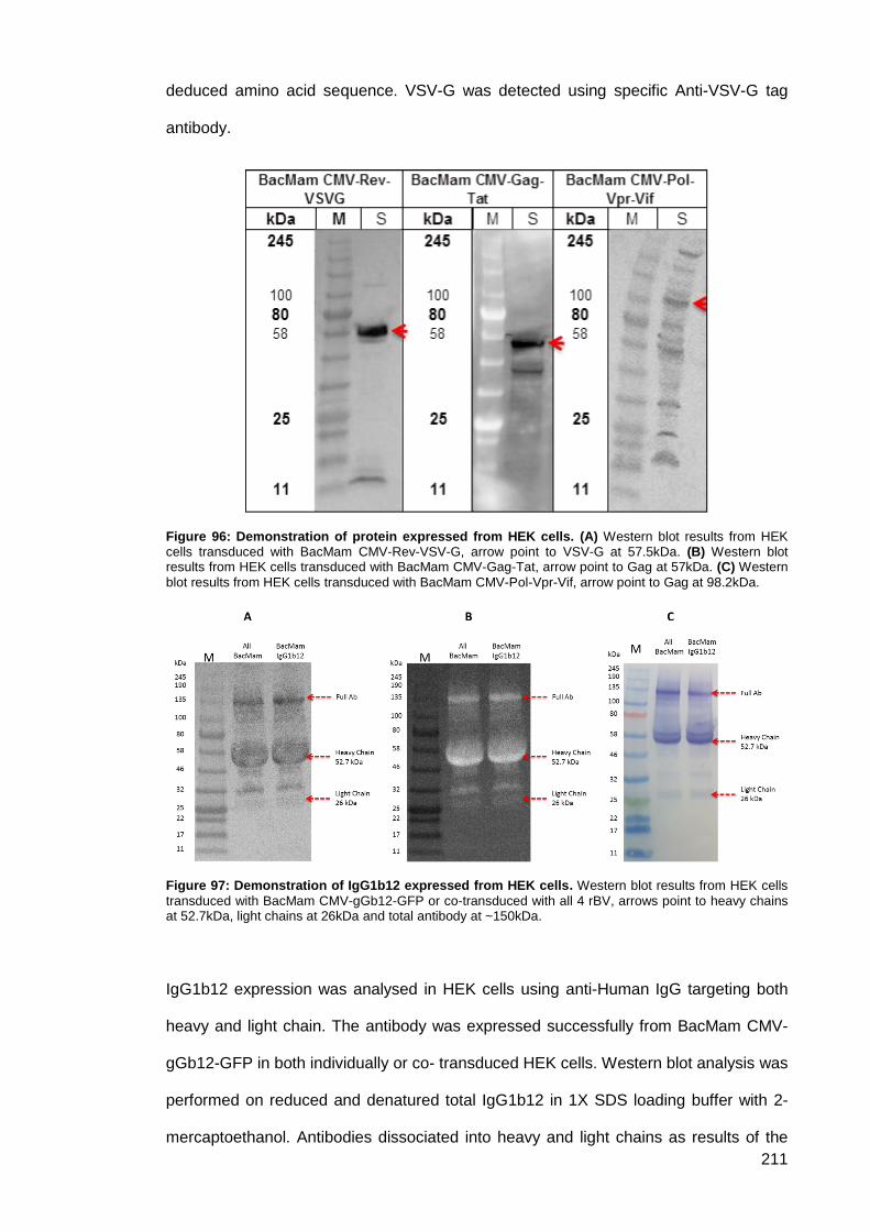
211
deduced amino acid sequence. VSV-G was detected using specific Anti-VSV-G tag
antibody.
Figure 96: Demonstration of protein expressed from HEK cells. (A) Western blot results from HEK cells transduced with BacMam CMV-Rev-VSV-G, arrow point to VSV-G at 57.5kDa. (B) Western blot results from HEK cells transduced with BacMam CMV-Gag-Tat, arrow point to Gag at 57kDa. (C) Western
blot results from HEK cells transduced with BacMam CMV-Pol-Vpr-Vif, arrow point to Gag at 98.2kDa.
Figure 97: Demonstration of IgG1b12 expressed from HEK cells. Western blot results from HEK cells
transduced with BacMam CMV-gGb12-GFP or co-transduced with all 4 rBV, arrows point to heavy chains at 52.7kDa, light chains at 26kDa and total antibody at ~150kDa.
IgG1b12 expression was analysed in HEK cells using anti-Human IgG targeting both
heavy and light chain. The antibody was expressed successfully from BacMam CMV-
gGb12-GFP in both individually or co- transduced HEK cells. Western blot analysis was
performed on reduced and denatured total IgG1b12 in 1X SDS loading buffer with 2-
mercaptoethanol. Antibodies dissociated into heavy and light chains as results of the
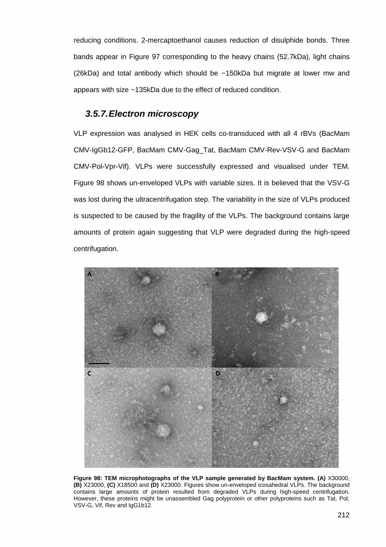
212
reducing conditions. 2-mercaptoethanol causes reduction of disulphide bonds. Three
bands appear in Figure 97 corresponding to the heavy chains (52.7kDa), light chains
(26kDa) and total antibody which should be ~150kDa but migrate at lower mw and
appears with size ~135kDa due to the effect of reduced condition.
3.5.7. Electron microscopy
VLP expression was analysed in HEK cells co-transduced with all 4 rBVs (BacMam
CMV-IgGb12-GFP, BacMam CMV-Gag_Tat, BacMam CMV-Rev-VSV-G and BacMam
CMV-Pol-Vpr-Vif). VLPs were successfully expressed and visualised under TEM.
Figure 98 shows un-enveloped VLPs with variable sizes. It is believed that the VSV-G
was lost during the ultracentrifugation step. The variability in the size of VLPs produced
is suspected to be caused by the fragility of the VLPs. The background contains large
amounts of protein again suggesting that VLP were degraded during the high-speed
centrifugation.
Figure 98: TEM microphotographs of the VLP sample generated by BacMam system. (A) X30000, (B) X23000, (C) X18500 and (D) X23000. Figures show un-enveloped icosahedral VLPs. The background
contains large amounts of protein resulted from degraded VLPs during high-speed centrifugation. However, these proteins might be unassembled Gag polyprotein or other polyproteins such as Tat, Pol, VSV-G, Vif, Rev and IgG1b12.
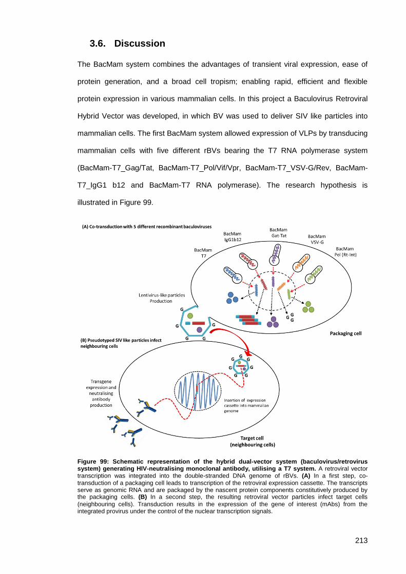
213
Discussion 3.6.
The BacMam system combines the advantages of transient viral expression, ease of
protein generation, and a broad cell tropism; enabling rapid, efficient and flexible
protein expression in various mammalian cells. In this project a Baculovirus Retroviral
Hybrid Vector was developed, in which BV was used to deliver SIV like particles into
mammalian cells. The first BacMam system allowed expression of VLPs by transducing
mammalian cells with five different rBVs bearing the T7 RNA polymerase system
(BacMam-T7_Gag/Tat, BacMam-T7_Pol/Vif/Vpr, BacMam-T7_VSV-G/Rev, BacMam-
T7_IgG1 b12 and BacMam-T7 RNA polymerase). The research hypothesis is
illustrated in Figure 99.
Figure 99: Schematic representation of the hybrid dual-vector system (baculovirus/retrovirus system) generating HIV-neutralising monoclonal antibody, utilising a T7 system. A retroviral vector transcription was integrated into the double-stranded DNA genome of rBVs. (A) In a first step, co-
transduction of a packaging cell leads to transcription of the retroviral expression cassette. The transcripts serve as genomic RNA and are packaged by the nascent protein components constitutively produced by the packaging cells. (B) In a second step, the resulting retroviral vector particles infect target cells
(neighbouring cells). Transduction results in the expression of the gene of interest (mAbs) from the integrated provirus under the control of the nuclear transcription signals.
214
It was speculated that co-transduction of mammalian cells with the five different
recombinants BacMam would result in the formation and the production of simian virus
like particles. The VLPs were designed to encode HIV-neutralising monoclonal
antibody sequences to function as a simian retroviral gene therapy agent. VLPs are
attenuated and thus not fully replication-competent retroviruses. These virus particles
were designed to be pseudo-typed with vesicular stomatitis virus glycoprotein (VSV-G).
The VLPs infect neighbouring cells and insert IgG1b12 sequences into the cells
genome. Neutralising antibody will then be produced from transduced cells.
To generate recombinant BacMam plasmid vectors, a BacMam pCMV-DEST vector
was used. However, all cloning attempt failed to produce a recombinant vector.
Investigation showed that the vector backbone contains ccdB. CcdB codes for the toxic
protein (CcdB) that acts as a DNA gyrase poison, locking the DNA gyrase with broken
double stranded DNA and ultimately causing cell death. Although cloning using
restriction enzyme was possible, the choice of cloning for this plasmid is Gateway®
technology in which insertion completely replaces CcdB with the investigator’s insert of
interest. Traditional cloning failed to remove CcdB, thus cells could not propagate. A
different plasmid was therefore considered. Baculovirus transfer vector pOET6 was
used for construction of pT7_BacMam and to obtain rBVs carrying genes of interests
under the control of the T7 promoter. Construction pT7_BacMam transfer plasmids
using pOET6 was a challenge as all of genes of interests were from MVA work which
used a different set of restriction enzymes. Expression cassettes were extracted from
MVA transfer plasmids with extra sequences flanking the gene of interest. DNA
fragment containing the gene of interest and excess DNA was cloned into pOET6.
Successful clones were subjected to further restriction digests steps, out of frame DNA
was removed forming the desired BacMam pOET6 vector (Figure 56). Generating
BacMam transfer plasmids and construction of recombinant BacMam viruses were
successful and confirmed by restriction digestion, sequencing and qPCR. However,
protein expression was not detected with any of the 5 rBVs. HEK cells were transduced
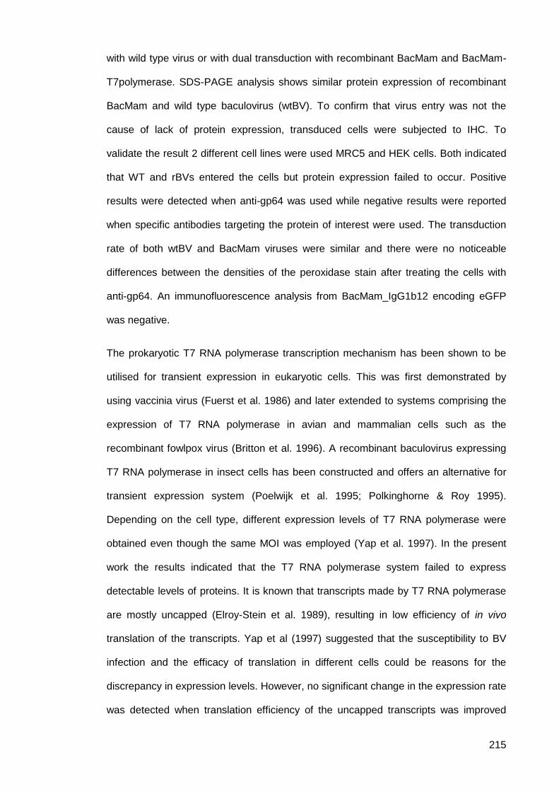
215
with wild type virus or with dual transduction with recombinant BacMam and BacMam-
T7polymerase. SDS-PAGE analysis shows similar protein expression of recombinant
BacMam and wild type baculovirus (wtBV). To confirm that virus entry was not the
cause of lack of protein expression, transduced cells were subjected to IHC. To
validate the result 2 different cell lines were used MRC5 and HEK cells. Both indicated
that WT and rBVs entered the cells but protein expression failed to occur. Positive
results were detected when anti-gp64 was used while negative results were reported
when specific antibodies targeting the protein of interest were used. The transduction
rate of both wtBV and BacMam viruses were similar and there were no noticeable
differences between the densities of the peroxidase stain after treating the cells with
anti-gp64. An immunofluorescence analysis from BacMam_IgG1b12 encoding eGFP
was negative.
The prokaryotic T7 RNA polymerase transcription mechanism has been shown to be
utilised for transient expression in eukaryotic cells. This was first demonstrated by
using vaccinia virus (Fuerst et al. 1986) and later extended to systems comprising the
expression of T7 RNA polymerase in avian and mammalian cells such as the
recombinant fowlpox virus (Britton et al. 1996). A recombinant baculovirus expressing
T7 RNA polymerase in insect cells has been constructed and offers an alternative for
transient expression system (Poelwijk et al. 1995; Polkinghorne & Roy 1995).
Depending on the cell type, different expression levels of T7 RNA polymerase were
obtained even though the same MOI was employed (Yap et al. 1997). In the present
work the results indicated that the T7 RNA polymerase system failed to express
detectable levels of proteins. It is known that transcripts made by T7 RNA polymerase
are mostly uncapped (Elroy-Stein et al. 1989), resulting in low efficiency of in vivo
translation of the transcripts. Yap et al (1997) suggested that the susceptibility to BV
infection and the efficacy of translation in different cells could be reasons for the
discrepancy in expression levels. However, no significant change in the expression rate
was detected when translation efficiency of the uncapped transcripts was improved
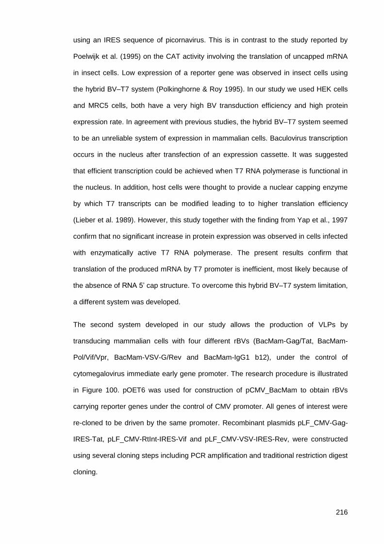
216
using an IRES sequence of picornavirus. This is in contrast to the study reported by
Poelwijk et al. (1995) on the CAT activity involving the translation of uncapped mRNA
in insect cells. Low expression of a reporter gene was observed in insect cells using
the hybrid BV–T7 system (Polkinghorne & Roy 1995). In our study we used HEK cells
and MRC5 cells, both have a very high BV transduction efficiency and high protein
expression rate. In agreement with previous studies, the hybrid BV–T7 system seemed
to be an unreliable system of expression in mammalian cells. Baculovirus transcription
occurs in the nucleus after transfection of an expression cassette. It was suggested
that efficient transcription could be achieved when T7 RNA polymerase is functional in
the nucleus. In addition, host cells were thought to provide a nuclear capping enzyme
by which T7 transcripts can be modified leading to to higher translation efficiency
(Lieber et al. 1989). However, this study together with the finding from Yap et al., 1997
confirm that no significant increase in protein expression was observed in cells infected
with enzymatically active T7 RNA polymerase. The present results confirm that
translation of the produced mRNA by T7 promoter is inefficient, most likely because of
the absence of RNA 5’ cap structure. To overcome this hybrid BV–T7 system limitation,
a different system was developed.
The second system developed in our study allows the production of VLPs by
transducing mammalian cells with four different rBVs (BacMam-Gag/Tat, BacMam-
Pol/Vif/Vpr, BacMam-VSV-G/Rev and BacMam-IgG1 b12), under the control of
cytomegalovirus immediate early gene promoter. The research procedure is illustrated
in Figure 100. pOET6 was used for construction of pCMV_BacMam to obtain rBVs
carrying reporter genes under the control of CMV promoter. All genes of interest were
re-cloned to be driven by the same promoter. Recombinant plasmids pLF_CMV-Gag-
IRES-Tat, pLF_CMV-RtInt-IRES-Vif and pLF_CMV-VSV-IRES-Rev, were constructed
using several cloning steps including PCR amplification and traditional restriction digest
cloning.
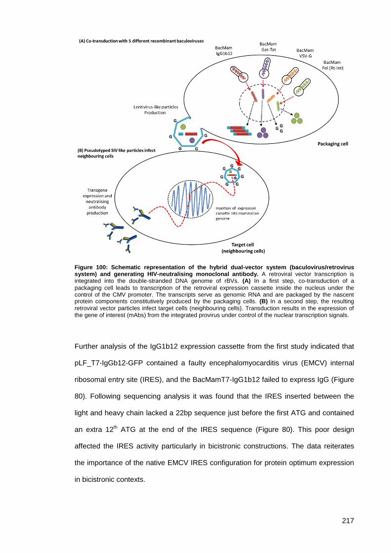
217
Figure 100: Schematic representation of the hybrid dual-vector system (baculovirus/retrovirus system) and generating HIV-neutralising monoclonal antibody. A retroviral vector transcription is integrated into the double-stranded DNA genome of rBVs. (A) In a first step, co-transduction of a
packaging cell leads to transcription of the retroviral expression cassette inside the nucleus under the control of the CMV promoter. The transcripts serve as genomic RNA and are packaged by the nascent protein components constitutively produced by the packaging cells. (B) In a second step, the resulting
retroviral vector particles infect target cells (neighbouring cells). Transduction results in the expression of the gene of interest (mAbs) from the integrated provirus under control of the nuclear transcription signals.
Further analysis of the IgG1b12 expression cassette from the first study indicated that
pLF_T7-IgGb12-GFP contained a faulty encephalomyocarditis virus (EMCV) internal
ribosomal entry site (IRES), and the BacMamT7-IgG1b12 failed to express IgG (Figure
80). Following sequencing analysis it was found that the IRES inserted between the
light and heavy chain lacked a 22bp sequence just before the first ATG and contained
an extra 12th ATG at the end of the IRES sequence (Figure 80). This poor design
affected the IRES activity particularly in bicistronic constructions. The data reiterates
the importance of the native EMCV IRES configuration for protein optimum expression
in bicistronic contexts.
218
The studies pointed to the involvement of secondary structures in the recognition of
functional ribosome entry sites, and the importance of the distance between the
polypyrimidine track and the initiating AUG sequence. In the case of the
Encephalomyocarditis virus-derived IRES, the second AUG located 22 nucleotides
downstream of the UUUCC sequence, present in the poly pyrimidine-rich track at the 3'
of the IRES, has been identified as the authentic viral initiation codon (Kaminski et al.
1994; Ohlmann & Jackson 1999; Jang & Wimmer 1990). The preferred IRES (viral
bases 273-845) for optimum activity is illustrated in Figure 81-A (Bochkov &
Palmenberg 2006). Virus mapping experiments have suggested that ribosome
recognition may actually be enhanced by including the first few codons of the
polyprotein ORF (López de Quinto & Martínez-Salas 1998). The extra length is not
required for ribosome recognition, but it can provide a useful spacer between the A and
B cistrons, allowing the IRES to fold without undue structural constraints. Placing the
IRES in a position too close to the A cistron or immediately adjacent to a 5’cap is likely
to fail or interfere with translation from the upstream ORF (Martin et al. 2006). Studies
have demonstrated that the EMCV IRES predominant AUG (the 11th AUG) cannot be
modified without strongly affecting the overall efficiency of the IRES-dependent
expression (Martin et al. 2006). Placing the IRES immediately after the light chain
sequence and including a 12th ATG in the original (faulty) design resulted into the
formation of an attenuated IRES which was not ideal for maximal protein synthesis.
Including HTLV-1 IRES between the heavy chain and the eGFP in the IgG1b12
cassette was not optimum. An early study of the mechanism of translation initiation of
HTLV-1 mRNA suggested the presence of an IRES within its 5’ UTR. This IRES was
successfully used in the construction of tricistronic viral vectors (Olivares et al. 2014).
However, when these data were re-examined, the existence of an IRES was
dismissed, suggesting that translation initiation of the HTLV-1 mRNA was exclusively
cap dependent. Stringent RNA and protein analyses determined the 5′ UTR of human
T-cell leukemia virus type 1 (HTLV-1) exhibit a 5′ proximal post-transcriptional control
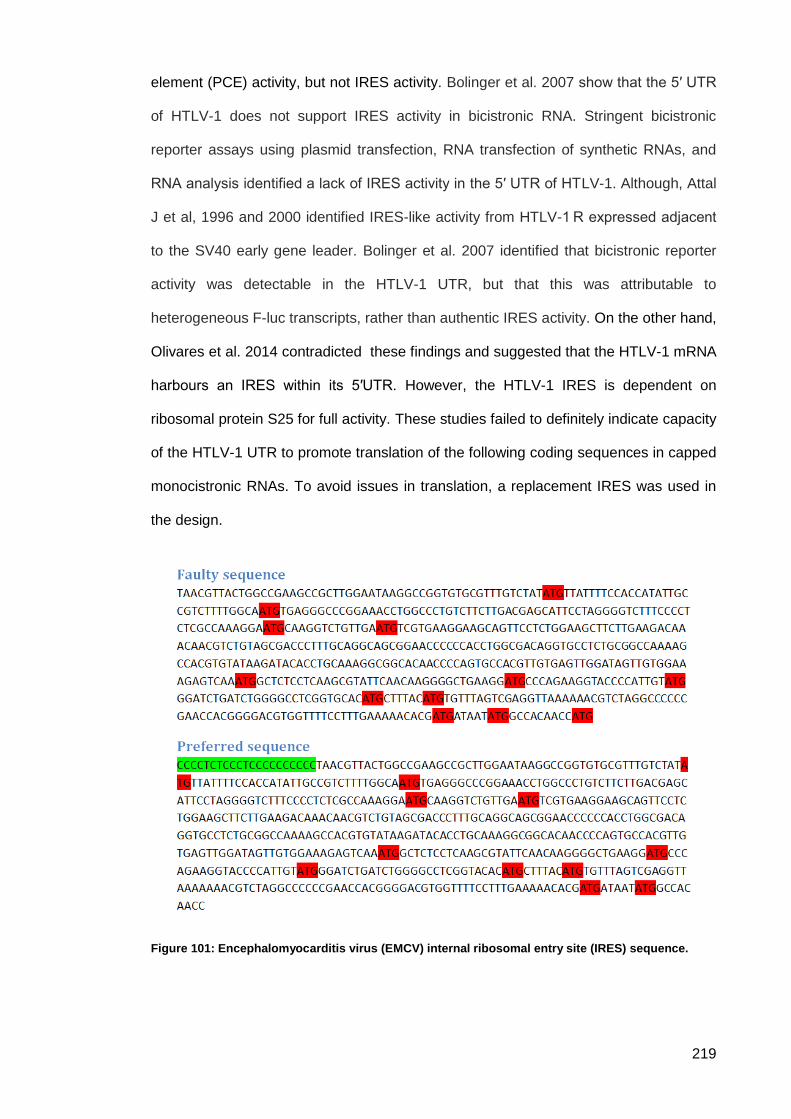
219
element (PCE) activity, but not IRES activity. Bolinger et al. 2007 show that the 5′ UTR
of HTLV-1 does not support IRES activity in bicistronic RNA. Stringent bicistronic
reporter assays using plasmid transfection, RNA transfection of synthetic RNAs, and
RNA analysis identified a lack of IRES activity in the 5′ UTR of HTLV-1. Although, Attal
J et al, 1996 and 2000 identified IRES-like activity from HTLV-1 R expressed adjacent
to the SV40 early gene leader. Bolinger et al. 2007 identified that bicistronic reporter
activity was detectable in the HTLV-1 UTR, but that this was attributable to
heterogeneous F-luc transcripts, rather than authentic IRES activity. On the other hand,
Olivares et al. 2014 contradicted these findings and suggested that the HTLV-1 mRNA
harbours an IRES within its 5′UTR. However, the HTLV-1 IRES is dependent on
ribosomal protein S25 for full activity. These studies failed to definitely indicate capacity
of the HTLV-1 UTR to promote translation of the following coding sequences in capped
monocistronic RNAs. To avoid issues in translation, a replacement IRES was used in
the design.
Figure 101: Encephalomyocarditis virus (EMCV) internal ribosomal entry site (IRES) sequence.
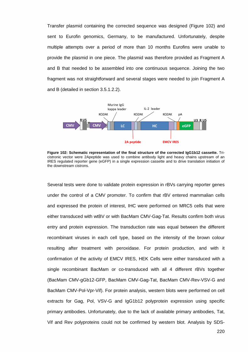
220
Transfer plasmid containing the corrected sequence was designed (Figure 102) and
sent to Eurofin genomics, Germany, to be manufactured. Unfortunately, despite
multiple attempts over a period of more than 10 months Eurofins were unable to
provide the plasmid in one piece. The plasmid was therefore provided as Fragment A
and B that needed to be assembled into one continuous sequence. Joining the two
fragment was not straightforward and several stages were needed to join Fragment A
and B (detailed in section 3.5.1.2.2).
Figure 102: Schematic representation of the final structure of the corrected IgG1b12 cassette. Tri-
cistronic vector were 2Apeptide was used to combine antibody light and heavy chains upstream of an IRES regulated reporter gene (eGFP) in a single expression cassette and to drive translation initiation of the downstream cistrons.
Several tests were done to validate protein expression in rBVs carrying reporter genes
under the control of a CMV promoter. To confirm that rBV entered mammalian cells
and expressed the protein of interest, IHC were performed on MRC5 cells that were
either transduced with wtBV or with BacMam CMV-Gag-Tat. Results confirm both virus
entry and protein expression. The transduction rate was equal between the different
recombinant viruses in each cell type, based on the intensity of the brown colour
resulting after treatment with peroxidase. For protein production, and with it
confirmation of the activity of EMCV IRES, HEK Cells were either transduced with a
single recombinant BacMam or co-transduced with all 4 different rBVs together
(BacMam CMV-gGb12-GFP, BacMam CMV-Gag-Tat, BacMam CMV-Rev-VSV-G and
BacMam CMV-Pol-Vpr-Vif). For protein analysis, western blots were performed on cell
extracts for Gag, Pol, VSV-G and IgG1b12 polyprotein expression using specific
primary antibodies. Unfortunately, due to the lack of available primary antibodies, Tat,
Vif and Rev polyproteins could not be confirmed by western blot. Analysis by SDS-
221
PAGE showed a clear band at the expected size for Tat, suggesting the successful
expression of that protein. For the Vif and Rev polyproteins, SDS-PAGE analysis did
not produce clear bands at the expected size. This suggests either a complete lack of
protein expression, or the expression was at a low level, preventing the detection by
SDS-PAGE. For those proteins that currently lack a commercially available primary
detection antibody, future work could include the production of antibodies for these
proteins. Of the proteins confirmed by WB, the exact location of protein production
varied. Although detected in cell lysate, IgG1b12, Gag and VSV-G were most abundant
extracellularly i.e. in the supernatant. However, Pol was mainly intracellular.
To investigate IgG1b12 expression and integration into mammalian cells, HEK, VERO
and HeLa cells were transduced with BacMam CMV-gGb12-GFP or co-transduced with
all 4 recombinant BacMam viruses. The IgG1b12 expression cassette is composed of
LC, HC and eGFP driven by a CMV promoter and flanked by LTRs. Green fluorescent
signal was observed in all transduced VERO and HeLa cells, proving that the
expression of IgG1b12 was successful. Cell extracts from transduced HEK cells was
subjected to western blot, results indicated that IgG1b12 was detected in both single
and co-transduced cells. Detection of both heavy and light chain antibodies separately
on the SDS-PAGE demonstrated production of the antibody proteins. Results from IHC
together with western blot indicated that IgG1b12 was expressed when cells were
transduced with BacMam CMV-gGb12-GFP alone as well as when co-transduced with
all 4 rBVs. This suggests that antibody secretion occurred without integration of the
antibody expression cassette into the cellular genome, where expression was driven by
CMV promotor. Or perhaps, that the LTRs alone are sufficient for integration regardless
of the presence of Rt, Integrase and protease. However, the evidence so far for a role
of the LTR in integration is circumstantial (Hindmarsh & Leis 1999). Determination of
whether or not the antibody sequence is integrated within the host cell genome is
clearly critical. If stable integration of the antibody gene is not achieved then sustained
intracellular production of antibody will not be accomplished.
222
TEM was used to analyse the morphological aspect of the VLPs and measure the
individual particle size per sample. Virus like particle expression was analysed in HEK
cells co-transduced with all 4 rBVs. Upon expression, the Gag polyprotein of SIV
spontaneously assembles giving rise to enveloped VLPs. The results indicate that the
great majority of Gag present in cell culture supernatants have shown to be correctly
assembled and have been successfully expressed and secreted as VLPs. Figure 98
shows VLPs visualised under TEM. It should be noted that in presence of viral
protease, SIV maturation (process by which the Gag polyprotein is cleaved into its
distinct subunits and the VLP is reorganised) will take place. Viral particles generated
were smaller than the expected mature virion size (110-130 nm) (Gentile et al. 1994).
Configuration of the visualised VLPs presents an angular outline forming icosahedral
structures. The background contains large amounts of protein perhaps suggesting that
the VLPs were degraded during high-speed centrifugation. However, these proteins
might be unassembled Gag polyprotein or other polyproteins such as Tat, Pol, VSV-G,
Vif, Rev and IgG1b12.
Our study confirms that mammalian cells can be transduced with multiple different
rBVs simultaneously. Co-transduction ensures that monocistronic BVs can be easily
and rapidly produced if new genotypes or serotypes appear. In agreement with
previous studies we found that the MOIs do not affect the transduction rate or the
protein expression. The choice of the surrounding solution during transduction has a
considerable effect on transduction efficiency. PBS was used as a surrounding solution
and the volumetric ratio of diluted virus and PBS was 1:4, following recommendation
from Sung et al. 2014.
These proof of principle studies not only establishes the feasibility of antibody gene
transfer, but also investigates the efficiency of SIV like particles production in
mammalian cells using BacMam technology, to support their candidacy as potential
vaccines and/or immunotherapy agents.
223
Future work 3.7.
Successful transduction of various mammalian cells has been demonstrated with
different rBVs. Protein expression and VLP expression was detected. To further
validate these results it will be necessary to confirm that the VLPs contain all of the
desired proteins and the IgG1b12 expression cassette encoded into its core. Following
infection of cells with the VLP confirmation of the integration of the antibody sequence
into the mammalian genome and determination of the site of integration is necessary.
This can be done most efficient by sequencing the entire genome of the VLP infected
host cells. Antibody was detected when cells were transduced with BacMam CMV-
gGb12-GFP only, suggesting that direct transduction rather transduction mediated by
VLPs may allow a more direct route to cellular delivery of the antibody sequence into
the mammalian genome.
The need of the Rt integrase and associated proteins for successful integration would
then be questionable. The possibility that antibody secretion can be carried out without
integration needs to be investigated. A key aspect is to confirm the intake of the
antibody sequence by VLPs and inserting it into the mammalian genome. This can be
investigated by infecting mammalian cells with purified VLPs and monitor the
expression of IgG1b12. Technically if the VLPs encode the antibody expression
cassette within its core, IgG1b12 will be secreted. Whole genome sequencing can be
used to confirm the insertion. We also, need to monitor the expression of the antibody
to examine wither it is a stable intergration and for how long IgG1b12 will be
exbressed. Mok et al. 2007, found that the stability of HIV-1-based vector gene
expression is extremely durable, with over 80% of selected cell clones persistently
expressing transgene at 18 months post infection (Mok et al. 2007).
Analysis of SIV integration shows a striking tendency to integrate within transcription
units (73% of the mapped integration events), but no propensity toward integration in
any specific region of the transcription units (Hematti et al. 2004). The effect of
antibody secretion on cells quality and growing should not be neglected. Also, the
224
neutralising effectivity of the antibody secreted must be determend to validaite the
protection effect of our system. Our current results indicated that IgG1b12 was
detected when cells were transduced with BacMam CMV-gGb12-GFP alone as well as
when co-transduced with all 4 rBV. The possibility of delivering the Ab sequence into
mammalian cells can be accomplished using single BV encoded LRTs must be
investigated.
225
Chapter 4 4.
General discussion and future work
The HIV pandemic continues despite international effort to control it. Progress has
been made in reducing AIDS-related deaths (by nearly 50% since the peak of the
epidemic), but each day more than 5,000 people are newly infected (World Health
Organization Guideline 2016). Development of a vaccine to aid control of the pandemic
has been in progress since 1984 but to date, despite enormous international effort, the
only vaccine trial to show promise (RV144) protected only 31.2% of study participants.
A multiplicity of antiviral drugs for the treatment of HIV infection have been developed
but their effective use relies on complex specialist laboratory support (HIV viral load
testing, antiviral drug resistance testing, therapeutic drug monitoring) and advanced
medical care of patients to manage associated drug toxicities. In addition, high costs of
the drugs place a heavy economic burden on health systems and restrict their
application in the countries where highest rates of HIV infection are seen. Vaccines
have been our most effective means to protect us from most of the world’s deadliest
viral infectious diseases. However, there are many confounding issues that prevent the
production of a sufficiently effective HIV vaccine (Morrow et al. 2012; Esparza 2001).
Challenges include HIV’s extreme antigenic variability; lack of understanding of
immune correlates for protection; limitations of available animal models; and the
constraints associated with the need for multiple large-scale clinical trials in different
parts of the world.
The hyper mutation capability of HIV has proved an, as yet, insurmountable problem in
developing a protective HIV vaccine. However, the discovery of individuals in high
endemicity areas who are resistant to HIV infection, and the recognition of so called
‘elite responders’ to HIV infection (individuals who very effectively suppress HIV
replication after initial infection), led to description of the so-called ‘broadly reactive’
neutralising antibodies. It is believed that elicitation of broadly neutralising antibodies
226
(bNAbs) is a key requirement of a successful HIV vaccine. These antibodies have been
shown capable of neutralising the infectivity of a wide range of HIV strains. Passive
administration of bNAbs has been shown to provide protection from infection after
intravenous, vaginal, rectal, and oral virus challenge in non-human primate models
(Mascola et al. 1999; Hessell et al. 2010; Hessell et al. 2009). Moreover, several
studies have demonstrated that in a non-human primate (macaques) model vaccine-
induced bNAb responses can confer complete protection against homologous SHIV
challenge (Barnett et al. 2008; Bogers et al. 2008; Pal et al. 2006). This suggests that a
vaccine capable of eliciting sufficient levels of bNAb against HIV-1 could prevent the
establishment of infection. In order to be effective, such vaccination would be required
to elicit neutralising antibodies that bind to the highly variable Env glycoprotein and
neutralise a broad range of primary isolates in humans. Animal model studies show
that high serum concentrations of bNAbs are required to provide adequate protection
(Walker & Burton 2010). While passive immunisation may work, its administration
echoes many of the high cost, technological demand, and complex medical support
that are seen in deployment of HIV antiviral drugs. These problems could be
surmounted if the gene for bNAbs could be delivered intracellularly.
The present project used different unique hybrid vectors to insert HIV-neutralising
monoclonal antibody sequences into the human genome using a simian retroviral gene
therapy agent pseudo-typed with vesicular stomatitis virus glycoprotein (VSV-G).
Required genes to form SIV like particles encoding the neutralising antibody sequence
were encoded in earlier developed poxvirus split-vectors or into a baculovirus
expression system. Protein expression from either system was expected to result in the
production of pseudotyped replication incompetent retroviruses like particles. The
retroviral particles were in turn expected to deliver a gene for the expression of HIV
neutralising antibody intracellularly and then mediate its integration into the human
genome. The inserted genetic materials were composed of an expression cassette
carrying a CMV promoter and the neutralising anti-HIV antibody gene with the aim of
227
establishing constant expression of the neutralising monoclonal antibody over a
prolonged period within the cell. The VLPs were intentionally designed to be of a
simian origin to increase the safety profile of the VLPs. Wild type SIV replicates in and
infect several species of non-human primates but does not infect or replicate in
humans (Sakuma & Takeuchi 2012). To add a further level of safety, the VLP genes
were attenuated and lacked the ability to replicate in any type of mammalian cell.
One essential element in any HIV vaccine is to be able to elicit broadly neutralising
antibodies (bNAbs) that can inhibit the infection of diverse HIV strains (Montero et al.
2008; Schiffner et al. 2013). Thus far, VLPs in vaccine production have limited success
in developing a successful vaccine against HIV. Env has been considered as the
principal antigen for HIV VLP vaccine development. However, Env has several features
that limit the accessibility to the neutralising epitopes which strongly affect the
development of bNAbs. The heavy glycosylation resulting in the formation of a glycan
shield on the surface of Env trimer severely reduce the access to protein epitopes (Wei
et al. 2003; Sagar et al. 2006; van Gils et al. 2011; Pancera et al. 2014). Moreover, Env
trimer hides its conserved neutralising sites, such as epitopes in the CD4-binding site
and the membrane-proximal external region (MPER), and only fully exposes them
when bind to CD4 and undergo conformational changes (Pancera et al. 2014; Frey et
al. 2008; Montero et al. 2008). In addition, the low density of native Env spikes
displayed on the viral surface, as well as the contamination with non-functional Env,
also contribute to the inaccessibility of neutralising epitopes (Burton & Mascola 2015;
Klein & Bjorkman 2010; Liu et al. 2008).
Another way of producing HIV VLP is by using Gag polyprotein which is capable of
self-assembly. HIV-1 Gag can serve as a platform for displaying Env antigen as well as
being a promising HIV-1 vaccine component for prophylactic and therapeutic purposes
(Buonaguro et al. 2001; Deml et al. 2005; Rovinski et al. 1995; Sakuragi et al. 2002). It
has been reported that HIV-1 Gag-VLPs were capable of priming significant cellular
responses as well as antibody response with cross-clade in chronically HIV-infected
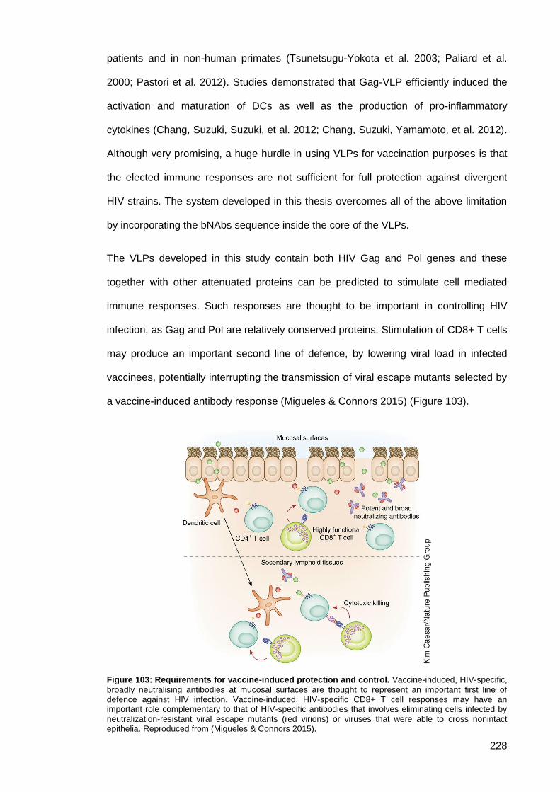
228
patients and in non-human primates (Tsunetsugu-Yokota et al. 2003; Paliard et al.
2000; Pastori et al. 2012). Studies demonstrated that Gag-VLP efficiently induced the
activation and maturation of DCs as well as the production of pro-inflammatory
cytokines (Chang, Suzuki, Suzuki, et al. 2012; Chang, Suzuki, Yamamoto, et al. 2012).
Although very promising, a huge hurdle in using VLPs for vaccination purposes is that
the elected immune responses are not sufficient for full protection against divergent
HIV strains. The system developed in this thesis overcomes all of the above limitation
by incorporating the bNAbs sequence inside the core of the VLPs.
The VLPs developed in this study contain both HIV Gag and Pol genes and these
together with other attenuated proteins can be predicted to stimulate cell mediated
immune responses. Such responses are thought to be important in controlling HIV
infection, as Gag and Pol are relatively conserved proteins. Stimulation of CD8+ T cells
may produce an important second line of defence, by lowering viral load in infected
vaccinees, potentially interrupting the transmission of viral escape mutants selected by
a vaccine-induced antibody response (Migueles & Connors 2015) (Figure 103).
Figure 103: Requirements for vaccine-induced protection and control. Vaccine-induced, HIV-specific,
broadly neutralising antibodies at mucosal surfaces are thought to represent an important first line of defence against HIV infection. Vaccine-induced, HIV-specific CD8+ T cell responses may have an important role complementary to that of HIV-specific antibodies that involves eliminating cells infected by neutralization-resistant viral escape mutants (red virions) or viruses that were able to cross nonintact epithelia. Reproduced from (Migueles & Connors 2015).
229
The use of immunotherapy delivered through genetic manipulation allows in vivo
delivery of broad and potent NAbs without the logistical complexities of traditional
immunotherapy procedures. The concept has been explored in other studies for
example Lewis et al. used an adeno-associated virus (AAV) vector to deliver the IgG1
b12 gene into mouse muscle and found that the antibody molecules imparted
neutralisation activity in sera for over six months (Lewis et al. 2002). This approach
was also tested in a nonhuman primate model by delivering neutralising
immunoadhesins (antigen-binding variable fragment domains of Fabs fused to the Fc
fragment of a rhesus IgG2 molecule) into macaques (Johnson et al. 2009). The
immunoadhesins were expressed in the macaque muscle myofibers, and serum
neutralisation activity was sustained for over one year. More importantly, sterilising
protection against SIV challenge was achieved in six out of nine immunised monkeys,
and all nine monkeys were protected from AIDS. Studies indicated that the human
monoclonal antibody IgG1 b12 can prevent SHIV SF162 infection in rhesus macaques.
However, the dose of antibodies that is required to achieve complete neutralisation was
suggested to be extremely high, which is unlikely to be achieved through current
immunisation techniques (Hessell et al. 2007; Hessell et al. 2009; Moldt et al. 2012;
Parren et al. 2001). Encouraged by these results, we have selected IgG1b12 to be our
bNAb of choice. Evidence from antibody binding to infected cells suggests that b12
may recognise a native conformation of gp120 more effectively than other antibodies
directed against the CD4 binding site (Burton et al. 1994). IgG1b12 can neutralise
almost 50% of clade B viral strains and approximately 30% of non-clade B strains
(Yang & Wang 2014). Moreover, the IgG1b12 antibody was found to neutralise
approximately 35% of circulating isolates (Kwong et al. 2011).
However, with the increase number of studies on bNAb several new broadly
neutralising Abs have shown greater neutralisation potency and breadth than the first
generation of bNAbs (Klein et al. 2013; Yang & Wang 2014; Kwong et al. 2011). One
such antibody the human monoclonal antibody VRC-HIVMAB060-00-AB (VRC01)
230
which has shown considerable promise when used for passive immunisation, providing
immediate protection against HIV infection. Thus for future work VRC01 will be
considered as replacement antibody. Similar to IgG1b12, VRC01 is a bNAb that targets
the CD4-binding site of the HIV envelope glycoprotein. However, VRC01 has a higher
neutralisation effect, it can neutralise up to 90% of a broad panel of 190 group M HIV
envelope pseudotyped viruses with a mean 50% inhibitory concentration (IC50) of 0.33
μg per millilitre (Wu et al. 2010). Passive administration of VRC01, has been shown to
prevent HIV transmission in animal models and is now being tested in clinical trials of
vertical and horizontal transmission in humans (Bar et al. 2016).
Two different approaches were used to express the protein of interests. In the first
scheme, transient gene expression in mammalian cells was based on either
recombinant MVA or recombinant baculovirus synthesising T7 RNA polymerase under
the control of a CMV promoter. The T7 RNA polymerase has strict specificities for
promoter and terminator respectively but can produce high levels of transcriptase
activity. The localisation of T7 RNA polymerase in cytoplasm gives higher levels of
protein expression than nuclear localisation. Yap et al. 1997, found that nuclei from
HepG2 cells infected with baculovirus encoding T7 RNA polymerase exhibited very low
activity which was indistinguishable from that found in uninfected cells (Yap et al.
1997).
Several features make MVA a favourable candidate as an efficient vector for gene
delivery or vaccination. The viral genome has a high capacity (up to 25 kb) for
recombinant DNA insertion. It also contains a strong poxviral promoter that ensures
efficient recombinant DNA expression. Another advantage is the lack of persistence or
genomic integration in the host due to its cytoplasmic replication. Although MVA-572
and MVA-I721 strains have been shown to replicate in some human cell lines and in
immunodeficient mice (Verheust et al. 2012). As a vaccine, MVA has proven safety
and immunogenicity in subjects infected with HIV and MVA based vectors and
vaccines are easily produced. For large-scale manufacture, the MVA vector is
231
generally produced in chick embryo fibroblast (CEF) cells. Recently, AGE1.CR and
EB66 cell lines have been developed for improved MVA production. A serious problem
in this project was a lack of availability of specific pathogen free (SPF) fertile eggs to
allow propagation of MVA. Further problems were encountered with the stability of
genetic insertion in MVA. A deletion site was used for insertion which resulted in
unstable constructs where the gene or genes of interest were repeatedly lost. To
circumvent this problem the use of baculovirus as a gene vector was explored.
Since Hofmann et al. and Boyce and Buchner reported, that recombinant baculoviruses
can enter mammalian cells and express proteins of interest when driven by mammalian
promoters, several studies have deepened our understanding of BacMam technology
and its utility (Hofmann & Strauss 1998; Boyce & Bucher 1996). Different commercially
available systems are available for generation of recombinant baculoviruses. These
gene delivery vectors can be easily manipulated and produced. Baculoviruses have
broad cell type specificity, are suitable for both transient and stable gene transfer, and,
if mass application is envisaged, are cost-effective in comparison to chemical gene
transfer procedures. Recombinant baculoviruses are non-replicative in mammalian
cells and can simultaneously deliver multiple genes. Many commonly used cell lines as
well as primary cultures are efficiently transduced with little to no microscopically
observable cytopathic effect or cytotoxicity. Baculoviruses have the capacity for
encoding significant amounts of additional genetic material within their genome. A
recombinant baculovirus containing up to a 38 kb insert can be generated and
propagated in a genetically stable manner in insect cells. The synthetic methods for
construction of defined genes and their introduction to shuttle plasmids to permit
construction of modified baculoviruses are well established. Surprisingly, publications
reporting application of utilising BacMam technology in immunisation
challenge/experiments against pathogens are still limited.
In the present project transduction of mammalian cells using baculoviruses encoding
defective retrovirus genes, caused the formation of virus like particles. Production and
232
expression of VLPs occur in the targeted cell. Thus glycosylation, protein folding and
particle assembly will not be affected. Also, in comparision with other schemes, such
as production of VLP in bacteria, yeast and insect cells, contamination with foreign
cellular proteins is eliminated. Studies have shown that it is almost impossible to
produce VLPs without contaminant cellular proteins, such virus/host complexes may
produce complex immunogenicity (Cho 2000; Levy 2007).
The VLPs released infected neighbouring cells. In these infected cells reverse
transcription of the genome of the retrovirus construct followed by integration of the
resultant cDNA into the host cell genome brings about production of HIV antibody -
driven by a viral CMV IE promoter- both intra and extra cellularly. This genetic
manipulation could provide a safe, sustainable vaccine to protect those who have not
yet encountered HIV from infection (protective vaccination). Additionally, it holds the
promise of immunotherapy for those already infected with the virus by neutralising the
infectivity of virus released during HIV infection (immunotherapy). This could replace
antiviral drug treatment for the majority of those infected with HIV.
Production of such a novel vaccine/therapeutic would provide an easily delivered and
affordable global treatment. The advantages of this approach are the development of a
vaccine that is stable and easily transported on cold chain transport as baculoviruses
may be freeze dried (a major advantage for developing countries with limited
infrastructure eliminating the need for ‘cold-chain’ transport and storage of vaccine in
zones with high atmospheric temperatures); baculoviruses do not replicate within
human cells thus the problems of pre-existing immunity seen with some gene transfer
vectors (e.g. adenovirus) are avoided; a single dose of the vaccine would result in the
development of long term, stable, production of antibody in vivo; significant therapeutic
application (immunotherapy) in those already infected, as an adjunct, or better
replacement for antiviral chemotherapy (avoiding the necessity for continuous
monitoring of the infection, drug toxicity and the large economic costs of drug therapy);
providing a realistic, affordable tool in the global effort to control the HIV pandemic.
233
One limitation of gene therapy using retroviruses is that the integrase enzyme can
insert the genetic material of the virus in any arbitrary position in the genome of the
host. If genetic material happens to be inserted in the middle of one of the original
genes of the host cell, this gene will be disrupted (insertional mutagenesis). If the gene
happens to be one regulating cell division, uncontrolled cell division (i.e., cancer) can
occur. This problem has recently begun to be addressed by utilising zinc finger
nucleases or by including certain sequences such as the beta-globin locus control
region to direct the site of integration to specific chromosomal sites. Full understanding
of the safety of such process is important. It is known that natural retroviral infection
with HIV produces such direct oncogenesis with exceptional rarity and only in the
context of continuous HIV replication.
As discussed before, results from this project indicate the expression and secretion of
IgG1b12 from transduced cells (refer to section 3.5.6). Since the antibody was detected
when cells were transduced with BacMam CMV-gGb12-GFP only, the possibility that
antibody secretion was carried out without integration should be investigated first. The
resultant antibody titre should be measured together with antibody neutralisation
effectivity. Assessment of IgG1b12 potency and evaluation of its inhibitory activity
should be carried in vitro using TZM-bl cells where the IC90s of IgG1b12 against HIV-1
infection is derived. The effect of antibody secretion on tissue viability should not be
neglected. Transduced cells should be passaged and monitored for any morphological
and functional changes.
To validate our results we have to confirm the integration of the antibody sequence into
the mammalian genome and determane the site of integration. This can be done by
whole human genome sequencing and using other methodologies such as Linear -
amplification mediated PCR (LAM-PCR) to catalogue retroviral insertions. Evidence
from studies concerned with retroviral integration patterns indicated that SIV integration
shows a significant tendency to integrate within transcription units (73% of the mapped
integration events), but no propensity toward integration in any specific region of the
234
transcription units (Hematti et al. 2004). Retroviral gene integration into the host cell
chromosome raises an on going concern of the risk of insertional mutagenesis and
oncogene activation. These fears were particularly raised after early gene therapy
clinical trial-treated patients with gene therapy for X-linked severe combined
immunodeficiency syndrome (SCID). Two out of 11 patients involved in the clinical trail
developed T cell leukaemias 3 years following transplantation of autologous bone
marrow CD34+ cells transduced with a standard retroviral vector expressing the
common γ chain (γc) transgene. However, insertional mutagenesis has not been
observed in replication defective retroviral vector-mediated gene transfer, either in
animal models or in clinical trials (Schmidt et al. 2002; Kiem et al. 2004). Reassuring
results from long term clinical and molecular follow up of over 200 patients enrolled in
clinical trials utilising retroviral vectors to transduce hematopoietic progenitor cells
showed no development of leukaemia (Kiem et al. 2004).
The extended clinical follow-up and molecular analysis of vector insertional clonality
report in nonhuman primates and dogs suggests a nonrandom pattern of insertion is
unlikely to become dominant or be associated with haematologic abnormalities.
Without a transgene to provide an abnormal proliferative or survival signal, the risk of
leukaemogenesis appears to be very low (Moolten & Cupples 1992; Kiem et al. 2004).
Thus it is very unlikely that the development of leukaemia in the children enrolled in the
γC X-SCID trial resulted solely from a single or a few retroviral insertions per cell.
Additional factors are more likely to be related in the development of leukemia in these
patients. The underlying immunodeficiency perhaps resulted in an expanded lymphoid
progenitor target cell population; the therapeutic gene itself, γc, might be a risk factor,
when expressed from a recombinant retroviral vector under the control of the vector's
LTR in patients with X-linked SCID; also, the patients might have expressed a
constitutively activated cytokine receptor transgene that resulted in oncogene activation
(Kohn et al. 2003).

235
In regard to the present BacMam expression system, a simpler route to the desired
vaccine/therapeutic may be possible. Instead of using 4 different baculovirus constructs
to transduce mammalian cells all necessary genes could be inserted within one
baculovirus genome. Further, instead of assembling a defective retrovirus within the
mammalian cell and using this to produce a retrovirus-antibody gene delivery system,
the baculovirus could be used to directly deliver the antibody gene to the host cell
without the intermediary step of production of a defective retrovirus to achieve gene
delivery. The baculovirus would be modified with surface molecules from VSV to
achieve broad cell tropism. Modification of the coat of the baculovirus (pseudotyping) is
readily achieved through addition of VSV glycoprotein genes to the baculovirus
genome using standard molecular biological approaches. Pseudotyping of the
recombinant baculovirus with VSV-G will increase cell tropism as well as gene delivery.
Also, prevent possible virus inactivation by complement reported in vivo studies with
unmodified baculoviruses (Tani et al. 2003; Pieroni et al. 2001; Barsoum et al. 1997),
as envelope modification of the baculovirus can change its immunogenic properties
(Kaikkonen et al. 2011).
To avoid antibody overproduction, integration of a silencing gene alongside the
antibody gene would be used to control any potential for antibody over production.
However, direct in vivo gene transfer has been found to permit high and sustainable
production of circulating ectopic MAb without inducing an anti-idiotypic response
sufficiently robust to exert a neutralising effect (Noël et al. 2002). Thus, such system is
very unlikely to trigger anaphylaxis. Many regulatable systems are currently under
development and some have been used successfully for in vivo preclinical applications
(Goverdhana et al. 2005). The tetracycline regulatory system (Gossen & Bujard 1992)
is the most widely used and versatile system. The Tet-regulatable system can be used
to reduce unwanted gene expression. A chimeric protein incorporating a modified TetR
fused to transcriptional silencing domains of the regulated protein. Co-expression of
both rtTA proteins and regulated protein will produce insufficient heterodimerise due to
236
modifications on the dimerising surfaces of the TetR component (Freundlieb et al.
1999).
The aim of our study is to elicit high titres of HIV neutralising antibodies in vaccinated
individuals, over a prolonged period with a constant protection level. Upon encouraging
results, the study will progress to preclinical pharmacological and toxicological testing.
A pharmacodynamic study of the potential vaccine will be conducted to evaluate
immunogenicity using a humanised mouse model of HIV infection, to provide “proof of
concept” information to support a clinical development plan.
237
Refrences
Abe, T. et al., 2003. Baculovirus induces an innate immune response and confers protection from lethal influenza virus infection in mice. Journal of immunology (Baltimore, Md. : 1950), 171(3), pp.1133–9.
Available at: http://www.ncbi.nlm.nih.gov/pubmed/12874198.
Abe, T. et al., 2005. Involvement of the Toll-like receptor 9 signaling pathway in the induction of innate immunity by baculovirus. Journal of virology, 79(5), pp.2847–58. Available at:
http://www.ncbi.nlm.nih.gov/pubmed/15709004.
Airenne, K.J. et al., 2000. Baculovirus-mediated periadventitial gene transfer to rabbit carotid artery. Gene therapy, 7(17), pp.1499–504. Available at: http://www.nature.com/articles/3301269.
Airenne, K.J. et al., 2013. Baculovirus: an insect-derived vector for diverse gene transfer applications. Molecular therapy : the journal of the American Society of Gene Therapy, 21(4), pp.739–49.
Available at: http://www.ncbi.nlm.nih.gov/pubmed/23439502.
Amara, R.R. et al., 2001. Control of a mucosal challenge and prevention of AIDS by a multiprotein DNA/MVA vaccine. Science (New York, N.Y.), 292(5514), pp.69–74. Available at:
http://www.ncbi.nlm.nih.gov/pubmed/11393868.
Ambrose, Z. et al., 2007. HIV/AIDS: in search of an animal model. Trends in biotechnology, 25(8), pp.333–7. Available at: http://www.ncbi.nlm.nih.gov/pubmed/17574286 [Accessed April 7, 2014].
Antoine, G. et al., 1998. The complete genomic sequence of the modified vaccinia Ankara strain: comparison with other orthopoxviruses. Virology, 244(2), pp.365–96. Available at:
http://www.ncbi.nlm.nih.gov/pubmed/9601507.
Aragón, T.J. et al., 2003. Risks of serious complications and death from smallpox vaccination: a systematic review of the United States experience, 1963-1968. BMC public health, 3, p.26. Available at:
http://www.ncbi.nlm.nih.gov/pubmed/12911836.
Armbruster, C. et al., 2002. A phase I trial with two human monoclonal antibodies (hMAb 2F5, 2G12) against HIV-1. AIDS (London, England), 16(2), pp.227–33.
Armbruster, C. et al., 2004. Passive immunization with the anti-HIV-1 human monoclonal antibody (hMAb) 4E10 and the hMAb combination 4E10/2F5/2G12. The Journal of antimicrobial chemotherapy, 54(5), pp.915–20.
Arthur, L.O. et al., 1998. Chemical inactivation of retroviral infectivity by targeting nucleocapsid protein zinc fingers: a candidate SIV vaccine. AIDS research and human retroviruses, 14 Suppl 3, pp.S311-9.
Available at: http://www.ncbi.nlm.nih.gov/pubmed/9814959.
Au, S., Wu, W. & Panté, N., 2013. Baculovirus nuclear import: open, nuclear pore complex (NPC) sesame. Viruses, 5(7), pp.1885–900. Available at: http://www.ncbi.nlm.nih.gov/pubmed/23881277.
Baba, T.W. et al., 2000. Human neutralizing monoclonal antibodies of the IgG1 subtype protect against mucosal simian-human immunodeficiency virus infection. Nature medicine, 6(2), pp.200–6.
Baba, T.W. et al., 1999. Live attenuated, multiply deleted simian immunodeficiency virus causes AIDS in infant and adult macaques. Nature medicine, 5(2), pp.194–203. Available at: http://www.ncbi.nlm.nih.gov/pubmed/9930868.
Balazs, A.B. et al., 2012. Antibody-based protection against HIV infection by vectored immunoprophylaxis. Nature, 481(7379), pp.81–4. Available at: http://www.pubmedcentral.nih.gov/articlerender.fcgi?artid=3253190&tool=pmcentrez&rendertype=abstract [Accessed March 19, 2014].
Balazs, A.B. & West, A.P., 2013. Antibody gene transfer for HIV immunoprophylaxis. Nature immunology, 14(1), pp.1–5. Available at: http://www.ncbi.nlm.nih.gov/pubmed/23238748 [Accessed April 1, 2014].
Baltimore, D., 1988. Gene therapy. Intracellular immunization. Nature, 335(6189), pp.395–6. Available at: http://www.ncbi.nlm.nih.gov/pubmed/3166513.
Bao, Y. et al., 1991. An improved Tn7-based system for the single-copy insertion of cloned genes into chromosomes of gram-negative bacteria. Gene, 109(1), pp.167–168. Available at: http://linkinghub.elsevier.com/retrieve/pii/037811199190604A.
Bar, K.J. et al., 2016. Effect of HIV Antibody VRC01 on Viral Rebound after Treatment Interruption. New
238
England Journal of Medicine, 375(21), pp.2037–2050. Available at:
http://www.nejm.org/doi/10.1056/NEJMoa1608243.
Barnett, S.W. et al., 2008. Protection of macaques against vaginal SHIV challenge by systemic or mucosal and systemic vaccinations with HIV-envelope. AIDS (London, England), 22(3), pp.339–48. Available at: http://www.ncbi.nlm.nih.gov/pubmed/18195560.
Barry, G.F., 1988. A broad-host-range shuttle system for gene insertion into the chromosomes of gram-negative bacteria. Gene, 71(1), pp.75–84. Available at: http://www.ncbi.nlm.nih.gov/pubmed/2850977.
Barsoum, J. et al., 1997. Efficient transduction of mammalian cells by a recombinant baculovirus having the vesicular stomatitis virus G glycoprotein. Human gene therapy, 8(17), pp.2011–8. Available at: http://www.ncbi.nlm.nih.gov/pubmed/9414250.
Baxby, D., 1996. Poxviruses, Available at: http://www.ncbi.nlm.nih.gov/pubmed/21413326.
Beaudoing, E. & Gautheret, D., 2001. Identification of alternate polyadenylation sites and analysis of their tissue distribution using EST data. Genome research, 11(9), pp.1520–6. Available at:
http://www.ncbi.nlm.nih.gov/pubmed/11544195.
Beck, N., Sidhu, J. & Omiecinski, C., 2000. Baculovirus vectors repress phenobarbital-mediated gene induction and stimulate cytokine expression in primary cultures of rat hepatocytes. Gene Therapy,
7(15), pp.1274–1283. Available at: http://www.nature.com/articles/3301246.
Bennett, M.S. & Akkina, R., 2013. Gene therapy strategies for HIV/AIDS: preclinical modeling in humanized mice. Viruses, 5(12), pp.3119–41. Available at:
http://www.ncbi.nlm.nih.gov/pubmed/24351796 [Accessed March 27, 2014].
Benton, T. et al., 2002. The use of UCOE vectors in combination with a preadapted serum free, suspension cell line allows for rapid production of large quantities of protein. Cytotechnology, 38(1–3), pp.43–6. Available at: http://www.ncbi.nlm.nih.gov/pubmed/19003085.
Berman, P.W. et al., 1990. Protection of chimpanzees from infection by HIV-1 after vaccination with recombinant glycoprotein gp120 but not gp160. Nature, 345(6276), pp.622–5. Available at: http://www.ncbi.nlm.nih.gov/pubmed/2190095.
Bi, J.-X. et al., 2003. Human c-fos promoter mediates high-level, inducible expression in various mammalian cell lines. Biotechnology and bioengineering, 81(7), pp.848–54. Available at: http://www.ncbi.nlm.nih.gov/pubmed/12557318.
Binley, J.M. et al., 2004. Comprehensive Cross-Clade Neutralization Analysis of a Panel of Anti-Human Immunodeficiency Virus Type 1 Monoclonal Antibodies. Journal of Virology, 78(23), pp.13232–13252. Available at: http://jvi.asm.org/cgi/doi/10.1128/JVI.78.23.13232-13252.2004.
Blanchard, T.J. et al., 1998. Modified vaccinia virus Ankara undergoes limited replication in human cells and lacks several immunomodulatory proteins: implications for use as a human vaccine. The Journal of general virology, 79 ( Pt 5), pp.1159–67. Available at: http://www.ncbi.nlm.nih.gov/pubmed/9603331.
Bochkov, Y.A. & Palmenberg, A.C., 2006. Translational efficiency of EMCV IRES in bicistronic vectors is dependent upon IRES sequence and gene location. BioTechniques, 41(3), pp.283–292. Available at: https://www.future-science.com/doi/10.2144/000112243.
Bogers, W.M. et al., 1995. Protection from HIV-1 envelope-bearing chimeric simian immunodeficiency virus (SHIV) in rhesus macaques infected with attenuated SIV: consequences of challenge. AIDS (London, England), 9(12), pp.F13-8. Available at: http://www.ncbi.nlm.nih.gov/pubmed/8605046.
Bogers, W.M.J.M. et al., 2008. Systemic neutralizing antibodies induced by long interval mucosally primed systemically boosted immunization correlate with protection from mucosal SHIV challenge. Virology, 382(2), pp.217–225. Available at: http://dx.doi.org/10.1016/j.virol.2008.09.016.
Bolinger, C. et al., 2007. RNA helicase A interacts with divergent lymphotropic retroviruses and promotes translation of human T-cell leukemia virus type 1. Nucleic Acids Research, 35(8), pp.2629–2642. Available at: https://academic.oup.com/nar/article-lookup/doi/10.1093/nar/gkm124.
Bordería, A. V & Berkhout, B., 2009. Towards a genetic AIDS vaccine. Retrovirology, 6, p.93. Available at: http://www.pubmedcentral.nih.gov/articlerender.fcgi?artid=2766360&tool=pmcentrez&rendertype=abstract [Accessed January 23, 2014].

239
Borman, A.M. et al., 1997. Comparison of picornaviral IRES-driven internal initiation of translation in cultured cells of different origins. Nucleic acids research, 25(5), pp.925–32. Available at: http://www.ncbi.nlm.nih.gov/pubmed/9023100.
Borman, A.M., Deliat, F.G. & Kean, K.M., 1994. Sequences within the poliovirus internal ribosome entry segment control viral RNA synthesis. The EMBO journal, 13(13), pp.3149–57. Available at: http://www.ncbi.nlm.nih.gov/pubmed/8039507.
Boyce, F.M. & Bucher, N.L., 1996. Baculovirus-mediated gene transfer into mammalian cells. Proceedings of the National Academy of Sciences of the United States of America, 93(6), pp.2348–52. Available at: http://www.ncbi.nlm.nih.gov/pubmed/8637876.
Boyer, J.D. et al., 1997. Protection of chimpanzees from high-dose heterologous HIV-1 challenge by DNA vaccination. Nature Medicine, 3(5), pp.526–532. Available at: http://www.nature.com/doifinder/10.1038/nm0597-526.
Breman, J.G. & Arita, I., 1980. The Confirmation and Maintenance of Smallpox Eradication. New England Journal of Medicine, 303(22), pp.1263–1273. Available at: http://www.nejm.org/doi/abs/10.1056/NEJM198011273032204.
Breman, J.G. & Henderson, D.A., 2002. Diagnosis and Management of Smallpox. New England Journal of Medicine, 346(17), pp.1300–1308. Available at: http://www.nejm.org/doi/abs/10.1056/NEJMra020025.
Britton, P. et al., 1996. Expression of bacteriophage T7 RNA polymerase in avian and mammalian cells by a recombinant fowlpox virus. The Journal of general virology, 77 ( Pt 5), pp.963–7. Available at: http://www.ncbi.nlm.nih.gov/pubmed/8609493.
Buchbinder, S.P. et al., 2008. Efficacy assessment of a cell-mediated immunity HIV-1 vaccine (the Step Study): a double-blind, randomised, placebo-controlled, test-of-concept trial. Lancet (London, England), 372(9653), pp.1881–1893. Available at: http://www.ncbi.nlm.nih.gov/pubmed/19012954.
Buonaguro, L. et al., 2001. High efficient production of Pr55(gag) virus-like particles expressing multiple HIV-1 epitopes, including a gp120 protein derived from an Ugandan HIV-1 isolate of subtype A. Antiviral research, 49(1), pp.35–47. Available at: http://www.ncbi.nlm.nih.gov/pubmed/11166859.
Burns, J.C. et al., 1993. Vesicular stomatitis virus G glycoprotein pseudotyped retroviral vectors: concentration to very high titer and efficient gene transfer into mammalian and nonmammalian cells. Proceedings of the National Academy of Sciences of the United States of America, 90(17), pp.8033–7. Available at: http://www.pubmedcentral.nih.gov/articlerender.fcgi?artid=47282&tool=pmcentrez&rendertype=abstract.
Burton, D.R. et al., 1991. A large array of human monoclonal antibodies to type 1 human immunodeficiency virus from combinatorial libraries of asymptomatic seropositive individuals. Proceedings of the National Academy of Sciences of the United States of America, 88(22),
pp.10134–7. Available at: http://www.pubmedcentral.nih.gov/articlerender.fcgi?artid=52882&tool=pmcentrez&rendertype=abstract.
Burton, D.R. et al., 1994. Efficient neutralization of primary isolates of HIV-1 by a recombinant human monoclonal antibody. Science (New York, N.Y.), 266(5187), pp.1024–7. Available at: http://www.ncbi.nlm.nih.gov/pubmed/7973652.
Burton, D.R. & Mascola, J.R., 2015. Antibody responses to envelope glycoproteins in HIV-1 infection. Nature immunology, 16(6), pp.571–6. Available at: http://www.ncbi.nlm.nih.gov/pubmed/25988889.
Burton, D.R. & Parren, P.W., 2000. Vaccines and the induction of functional antibodies: time to look beyond the molecules of natural infection? Nature medicine, 6(2), pp.123–5. Available at: http://www.ncbi.nlm.nih.gov/pubmed/10655088.
Cacciatore, J.J., Chasin, L.A. & Leonard, E.F., 2010. Gene amplification and vector engineering to achieve rapid and high-level therapeutic protein production using the Dhfr-based CHO cell selection system. Biotechnology advances, 28(6), pp.673–81. Available at: http://www.ncbi.nlm.nih.gov/pubmed/20416368.
Calarese, D. a et al., 2003. Antibody domain exchange is an immunological solution to carbohydrate cluster recognition. Science (New York, N.Y.), 300(5628), pp.2065–71. Available at: http://www.ncbi.nlm.nih.gov/pubmed/12829775 [Accessed February 4, 2014].
240
Capistrán, M.A., 2010. A study of latency , reactivation and apoptosis throughout HIV pathogenesis. Mathematical and Computer Modelling, 52(7–8), pp.1011–1015. Available at: http://dx.doi.org/10.1016/j.mcm.2010.03.022.
Carter & Saunders, 2013. Virology: Principles and Applications,
Casimiro, D.R. et al., 2003. Comparative immunogenicity in rhesus monkeys of DNA plasmid, recombinant vaccinia virus, and replication-defective adenovirus vectors expressing a human immunodeficiency virus type 1 gag gene. Journal of virology, 77(11), pp.6305–13. Available at: http://www.ncbi.nlm.nih.gov/pubmed/12743287.
Cavacini, L.A. et al., 1998. Functional and molecular characterization of human monoclonal antibody reactive with the immunodominant region of HIV type 1 glycoprotein 41. AIDS research and human retroviruses, 14(14), pp.1271–80. Available at: http://www.ncbi.nlm.nih.gov/pubmed/9764911.
Chang, M.O., Suzuki, T., Suzuki, H., et al., 2012. HIV-1 Gag-virus-like particles induce natural killer cell immune responses via activation and maturation of dendritic cells. Journal of innate immunity, 4(2), pp.187–200. Available at: http://www.ncbi.nlm.nih.gov/pubmed/21778700.
Chang, M.O., Suzuki, T., Yamamoto, N., et al., 2012. HIV-1 Gag-virus-like particles inhibit HIV-1 replication in dendritic cells and T cells through IFN-α-dependent upregulation of APOBEC3G and 3F. Journal of innate immunity, 4(5–6), pp.579–90. Available at: http://www.ncbi.nlm.nih.gov/pubmed/22739040.
Cheng, T. et al., 2004. A rapid and efficient method to express target genes in mammalian cells by baculovirus. World journal of gastroenterology, 10(11), pp.1612–8. Available at: http://www.ncbi.nlm.nih.gov/pubmed/15162535.
Chhatbar, C. et al., 2011. HIV vaccine: hopes and hurdles. Drug discovery today, 16(21–22), pp.948–56. Available at: http://www.ncbi.nlm.nih.gov/pubmed/21889604 [Accessed January 23, 2014].
Cho, M.W., 2000. Assessment of HIV vaccine development: past, present, and future. Advances in pharmacology (San Diego, Calif.), 49, pp.263–314. Available at: http://www.ncbi.nlm.nih.gov/pubmed/11013767.
Choi, E. et al., 2016. First Phase I human clinical trial of a killed whole ‑ HIV ‑ 1 vaccine : demonstration of
its safety and enhancement of anti ‑ HIV antibody responses. Retrovirology, pp.1–16.
Churchill, D. et al., 2016. British HIV Association guidelines for the treatment of HIV-1-positive adults with antiretroviral therapy 2015. HIV Medicine, 17, pp.s2–s104. Available at: http://doi.wiley.com/10.1111/hiv.12426.
Clackson, T. et al., 1991. Making antibody fragments using phage display libraries. Nature, 352(6336),
pp.624–628. Available at: http://www.nature.com/doifinder/10.1038/352624a0.
Collier, L., Kellam, P. & Oxford, J., 2011. Human virology. 4th ed., Oxford : Oxford University Press.
Condreay, J.P. et al., 1999. Transient and stable gene expression in mammalian cells transduced with a recombinant baculovirus vector. Proceedings of the National Academy of Sciences of the United States of America, 96(1), pp.127–32. Available at: http://www.ncbi.nlm.nih.gov/pubmed/9874783.
Corey, L. et al., 2011. HIV-1 vaccines and adaptive trial designs. Science translational medicine, 3(79), p.79ps13. Available at: http://www.ncbi.nlm.nih.gov/pubmed/21508308.
Corti, D. et al., 2010. Analysis of memory B cell responses and isolation of novel monoclonal antibodies with neutralizing breadth from HIV-1-infected individuals. PloS one, 5(1), p.e8805. Available at: http://www.ncbi.nlm.nih.gov/pubmed/20098712.
Davies, D.R. & Chacko, S., 1993. Antibody structure. Accounts of Chemical Research, 26(8), pp.421–427. Available at: http://www.ncbi.nlm.nih.gov/pubmed/1078259.
Davis, D. et al., 2013. In vitro neutralization of low dose inocula at physiological concentrations of a monoclonal antibody which protects macaques against SHIV challenge. PloS one, 8(8), p.e72702. Available at: http://www.pubmedcentral.nih.gov/articlerender.fcgi?artid=3745472&tool=pmcentrez&rendertype=abstract [Accessed March 6, 2014].
Davis, T.R. & Wood, H.A., 1995. Intrinsic glycosylation potentials of insect cell cultures and insect larvae. In vitro cellular & developmental biology. Animal, 31(9), pp.659–63. Available at:
http://www.ncbi.nlm.nih.gov/pubmed/8564076.
241
Deacon, N.J. et al., 1995. Genomic Structure of an Attenuated Quasi Species of HIV-1 from a Blood Transfusion Donor and Recipients. Science, 270(5238), pp.988–991. Available at: http://www.sciencemag.org/cgi/doi/10.1126/science.270.5238.988.
Delaney, W.E. & Isom, H.C., 1998. Hepatitis B virus replication in human HepG2 cells mediated by hepatitis B virus recombinant baculovirus. Hepatology (Baltimore, Md.), 28(4), pp.1134–46. Available at: http://www.ncbi.nlm.nih.gov/pubmed/9755254.
Deml, L. et al., 2005. Recombinant HIV-1 Pr55gag virus-like particles: potent stimulators of innate and acquired immune responses. Molecular immunology, 42(2), pp.259–77. Available at: http://www.ncbi.nlm.nih.gov/pubmed/15488613.
Desrosiers, R.C. et al., 1989. Vaccine protection against simian immunodeficiency virus infection. Proceedings of the National Academy of Sciences of the United States of America, 86(16), pp.6353–7. Available at: http://www.ncbi.nlm.nih.gov/pubmed/2548210.
Devico, A. et al., 1996. Covalently crosslinked complexes of human immunodeficiency virus type 1 (HIV-1) gp120 and CD4 receptor elicit a neutralizing immune response that includes antibodies selective for primary virus isolates. Virology, 218(1), pp.258–63. Available at: http://www.ncbi.nlm.nih.gov/pubmed/8615032.
Douglas, N.W., Munro, G.H. & Daniels, R.S., 1997. HIV/SIV glycoproteins: structure-function relationships. Journal of molecular biology, 273(1), pp.122–49. Available at:
http://www.pubmedcentral.nih.gov/articlerender.fcgi?artid=3347801&tool=pmcentrez&rendertype=abstract.
Drexler, I., Staib, C. & Sutter, G., 2004. Modified vaccinia virus Ankara as antigen delivery system: how can we best use its potential? Current opinion in biotechnology, 15(6), pp.506–12. Available at: http://www.ncbi.nlm.nih.gov/pubmed/15560976 [Accessed April 1, 2014].
Dukkipati, A. et al., 2008. BacMam system for high-level expression of recombinant soluble and membrane glycoproteins for structural studies. Protein expression and purification, 62(2), pp.160–70. Available at: http://www.ncbi.nlm.nih.gov/pubmed/18782620.
Dull, T. et al., 1998. A third-generation lentivirus vector with a conditional packaging system. Journal of virology, 72(11), pp.8463–71. Available at: http://www.ncbi.nlm.nih.gov/pubmed/9765382%5Cnhttp://www.pubmedcentral.nih.gov/articlerender.fcgi?artid=PMC110254.
Dwarakanath, R.S. et al., 2001. The use of recombinant baculoviruses for sustained expression of human cytomegalovirus immediate early proteins in fibroblasts. Virology, 284(2), pp.297–307. Available at: http://www.ncbi.nlm.nih.gov/pubmed/11384228.
Dyer, W.B. et al., 1999. Strong human immunodeficiency virus (HIV)-specific cytotoxic T-lymphocyte activity in Sydney Blood Bank Cohort patients infected with nef-defective HIV type 1. J Virol, 73(1), pp.436–443. Available at: http://www.ncbi.nlm.nih.gov/entrez/query.fcgi?cmd=Retrieve&db=PubMed&dopt=Citation&list_uids=9847349.
Elroy-Stein, O., Fuerst, T.R. & Moss, B., 1989. Cap-independent translation of mRNA conferred by encephalomyocarditis virus 5’ sequence improves the performance of the vaccinia virus/bacteriophage T7 hybrid expression system. Proceedings of the National Academy of Sciences of the United States of America, 86(16), pp.6126–30. Available at: http://www.ncbi.nlm.nih.gov/pubmed/2548200.
Elroy-Stein, O. & Moss, B., 2001. Gene expression using the vaccinia virus/T7 RNA polymerase hybrid system. Current protocols in molecular biology, Chapter 16, p.Unit16.19. Available at: http://www.ncbi.nlm.nih.gov/pubmed/18265126.
Emini, E.A. et al., 1990. Yeast-expressed p55 precursor core protein of human immunodeficiency virus type 1 does not elicit protective immunity in chimpanzees. AIDS research and human retroviruses, 6(11), pp.1247–50. Available at: http://www.ncbi.nlm.nih.gov/pubmed/2127681.
Ensoli, B. et al., 2016. HIV-Tat immunization induces cross-clade neutralizing antibodies and CD4+ T cell increases in antiretroviral-treated South African volunteers: a randomized phase II clinical trial. Retrovirology, 13(1), p.34. Available at: http://retrovirology.biomedcentral.com/articles/10.1186/s12977-016-0261-1.
Ensoli, B. et al., 2010. Therapeutic Immunization with HIV-1 Tat Reduces Immune Activation and Loss of Regulatory T-Cells and Improves Immune Function in Subjects on HAART K. J. Hasenkrug, ed.
242
PLoS ONE, 5(11), p.e13540. Available at: http://dx.plos.org/10.1371/journal.pone.0013540.
Ensoli, F. et al., 2015. HIV-1 Tat immunization restores immune homeostasis and attacks the HAART-resistant blood HIV DNA: results of a randomized phase II exploratory clinical trial. Retrovirology,
12(1), p.33. Available at: ???
Escors, D. & Breckpot, K., 2010. Lentiviral vectors in gene therapy: their current status and future potential. Archivum immunologiae et therapiae experimentalis, 58(2), pp.107–19. Available at:
http://www.ncbi.nlm.nih.gov/pubmed/20143172.
Esparza, J., 2001. An HIV vaccine : how and when ? Bulletin of the World Health Organization, 79(12), pp.1133–1137.
Facciabene, A., Aurisicchio, L. & La Monica, N., 2004. Baculovirus vectors elicit antigen-specific immune responses in mice. Journal of virology, 78(16), pp.8663–72. Available at:
http://www.ncbi.nlm.nih.gov/pubmed/15280475.
Fang, J. et al., 2005. Stable antibody expression at therapeutic levels using the 2A peptide. Nature biotechnology, 23(5), pp.584–90. Available at: http://www.ncbi.nlm.nih.gov/pubmed/15834403.
Ferrantelli, F., Cafaro, A. & Ensoli, B., 2004. Nonstructural HIV proteins as targets for prophylactic or therapeutic vaccines. Current opinion in biotechnology, 15(6), pp.543–56. Available at: http://www.ncbi.nlm.nih.gov/pubmed/15560981.
Ferrantelli, F. & Ruprecht, R.M., 2002. Neutralizing antibodies against HIV -- back in the major leagues? Current opinion in immunology, 14(4), pp.495–502. Available at: http://www.ncbi.nlm.nih.gov/pubmed/12088685.
Flint et al, 2009. Principles of Virology 3rd ed., Amer Society for Microbiology.
Flynn, N.M. et al., 2005. Placebo-controlled phase 3 trial of a recombinant glycoprotein 120 vaccine to prevent HIV-1 infection. The Journal of infectious diseases, 191(5), pp.654–65. Available at: https://academic.oup.com/jid/article-lookup/doi/10.1086/428404.
Freundlieb, S., Schirra-Müller, C. & Bujard, H., 1999. A tetracycline controlled activation/repression system with increased potential for gene transfer into mammalian cells. The journal of gene medicine, 1(1), pp.4–12.
Frey, G. et al., 2008. A fusion-intermediate state of HIV-1 gp41 targeted by broadly neutralizing antibodies. Proceedings of the National Academy of Sciences of the United States of America, 105(10), pp.3739–44. Available at: http://www.ncbi.nlm.nih.gov/pubmed/18322015.
Fuerst, T.R. et al., 1986. Eukaryotic transient-expression system based on recombinant vaccinia virus that synthesizes bacteriophage T7 RNA polymerase. Proceedings of the National Academy of Sciences of the United States of America, 83(21), pp.8122–6. Available at:
http://www.ncbi.nlm.nih.gov/pubmed/3095828.
Fulginiti, V.A. et al., 2003. Smallpox Vaccination: A Review, Part I. Background, Vaccination Technique, Normal Vaccination and Revaccination, and Expected Normal Reactions. Clinical Infectious Diseases, 37(2), pp.241–250. Available at: https://academic.oup.com/cid/article-lookup/doi/10.1086/375824.
Fuller, D.H., Corb, M.M., et al., 1997. Enhancement of immunodeficiency virus-specific immune responses in DNA-immunized rhesus macaques. Vaccine, 15(8), pp.924–6. Available at: http://www.ncbi.nlm.nih.gov/pubmed/9234549.
Fuller, D.H., Simpson, L., et al., 1997. Gene gun-based nucleic acid immunization alone or in combination with recombinant vaccinia vectors suppresses virus burden in rhesus macaques challenged with a heterologous SIV. Immunology and cell biology, 75(4), pp.389–96. Available at: http://www.ncbi.nlm.nih.gov/pubmed/9315483.
Gaines, P. & Wojchowski, D.M., 1999. pIRES-CD4t, a dicistronic expression vector for MACS- or FACS-based selection of transfected cells. BioTechniques, 26(4), pp.683–8. Available at: http://www.ncbi.nlm.nih.gov/pubmed/10343906.
Gallo, R.C., 1999. Tat as one key to HIV-induced immune pathogenesis and Tat (correction of Pat) toxoid as an important component of a vaccine. Proceedings of the National Academy of Sciences of the United States of America, 96(15), pp.8324–6. Available at:
http://www.ncbi.nlm.nih.gov/pubmed/10411869.

243
Geisse, S., 2009. Reflections on more than 10 years of TGE approaches. Protein expression and purification, 64(2), pp.99–107. Available at: http://www.ncbi.nlm.nih.gov/pubmed/19027070.
Giacca, M., 2010. Gene Therapy, Milano: Springer Milan. Available at:
http://link.springer.com/10.1007/978-88-470-1643-9.
van Gils, M.J. et al., 2011. Longer V1V2 region with increased number of potential N-linked glycosylation sites in the HIV-1 envelope glycoprotein protects against HIV-specific neutralizing antibodies. Journal of virology, 85(14), pp.6986–95. Available at: http://www.ncbi.nlm.nih.gov/pubmed/21593147.
Girard, M.P. et al., 2006. A review of vaccine research and development: the human immunodeficiency virus (HIV). Vaccine, 24(19), pp.4062–81. Available at: http://www.ncbi.nlm.nih.gov/pubmed/16530298 [Accessed January 17, 2014].
Girard, M.P. et al., 2011. Human immunodeficiency virus (HIV) immunopathogenesis and vaccine development: a review. Vaccine, 29(37), pp.6191–218. Available at: http://www.ncbi.nlm.nih.gov/pubmed/21718747 [Accessed January 23, 2014].
Gossen, M. & Bujard, H., 1992. Tight control of gene expression in mammalian cells by tetracycline-responsive promoters. Proceedings of the National Academy of Sciences of the United States of America, 89(12), pp.5547–51.
Goverdhana, S. et al., 2005. Regulatable gene expression systems for gene therapy applications: progress and future challenges. Molecular therapy : the journal of the American Society of Gene Therapy, 12(2), pp.189–211.
Goyvaerts, C. et al., 2013. Targeted Lentiviral Vectors: Current Applications and Future Potential. In Gene Therapy - Tools and Potential Applications. InTech. Available at: http://www.intechopen.com/books/gene-therapy-tools-and-potential-applications/targeted-lentiviral-vectors-current-applications-and-future-potential.
Gray, G.E. et al., 2014. The phase 2b HVTN 503/Phambili study test-of-concept HIV vaccine study, investigating a recombinant adenovirus type 5 HIV gag/pol/nef vaccine in South Africa: unblinded, long-term follow-up. The Lancet. Infectious diseases, 14(5), pp.388–96. Available at:
http://www.ncbi.nlm.nih.gov/pubmed/24560541.
Greene, W.C. & Peterlin, B.M., 2002. Charting HIV’s remarkable voyage through the cell: Basic science as a passport to future therapy. Nature medicine, 8(7), pp.673–80. Available at: http://www.ncbi.nlm.nih.gov/pubmed/12091904.
Greenough, T.C., Sullivan, J.L. & Desrosiers, R.C., 1999. Declining CD4 T-cell counts in a person infected with nef-deleted HIV-1. The New England journal of medicine, 340(3), pp.236–7. Available at: http://www.ncbi.nlm.nih.gov/pubmed/9917232.
Greenwood et al, 2012. Medical microbiology 18th ed., Churchill Livingstone.
Gronowski, A.M. et al., 1999. Baculovirus stimulates antiviral effects in mammalian cells. Journal of virology, 73(12), pp.9944–51. Available at: http://www.ncbi.nlm.nih.gov/pubmed/10559307.
Gulick, R.M. et al., 1997. Treatment with Indinavir, Zidovudine, and Lamivudine in Adults with Human Immunodeficiency Virus Infection and Prior Antiretroviral Therapy. New England Journal of Medicine, 337(11), pp.734–739. Available at: http://www.nejm.org/doi/abs/10.1056/NEJM199709113371102.
Haigwood, N.L. et al., 1999. Protection from pathogenic SIV challenge using multigenic DNA vaccines. Immunology letters, 66(1–3), pp.183–8. Available at: http://www.ncbi.nlm.nih.gov/pubmed/10203053.
Hammer, S.M. et al., 1997. A controlled trial of two nucleoside analogues plus indinavir in persons with human immunodeficiency virus infection and CD4 cell counts of 200 per cubic millimeter or less. AIDS Clinical Trials Group 320 Study Team. The New England journal of medicine, 337(11), pp.725–33. Available at: http://www.ncbi.nlm.nih.gov/pubmed/9287227.
Helio, A. et al., 2013. Lentiviral Gene Therapy Vectors: Challenges and Future Directions. In Gene Therapy - Tools and Potential Applications. InTech. Available at: http://www.intechopen.com/books/gene-therapy-tools-and-potential-applications/lentiviral-gene-therapy-vectors-challenges-and-future-directions.
Hematti et al., 2004. Distinct genomic integration of MLV and SIV vectors in primate hematopoietic stem and progenitor cells. PLoS biology, 2(12), p.e423. Available at: http://www.ncbi.nlm.nih.gov/pubmed/15550989.
244
Herrlich, A. & Mayr, A., 1957. [Smallpox vaccine from tissue culture from a bull’s tongue; at the same time a contribution to the question of culture vaccines]. Archiv fur die gesamte Virusforschung, 7(3), pp.284–96. Available at: http://www.ncbi.nlm.nih.gov/pubmed/13435739.
Hessell, A.J. et al., 2009. Broadly Neutralizing Human Anti-HIV Antibody 2G12 Is Effective in Protection against Mucosal SHIV Challenge Even at Low Serum Neutralizing Titers M. Farzan, ed. PLoS Pathogens, 5(5), p.e1000433. Available at: http://dx.plos.org/10.1371/journal.ppat.1000433.
Hessell, A.J. et al., 2010. Broadly Neutralizing Monoclonal Antibodies 2F5 and 4E10 Directed against the Human Immunodeficiency Virus Type 1 gp41 Membrane-Proximal External Region Protect against Mucosal Challenge by Simian-Human Immunodeficiency Virus SHIVBa-L. Journal of Virology, 84(3), pp.1302–1313. Available at: http://jvi.asm.org/cgi/doi/10.1128/JVI.01272-09.
Hessell, A.J. et al., 2007. Fc receptor but not complement binding is important in antibody protection against HIV. Nature, 449(7158), pp.101–4. Available at: http://www.ncbi.nlm.nih.gov/pubmed/17805298.
Hindmarsh, P. & Leis, J., 1999. Retroviral DNA integration. Microbiology and molecular biology reviews : MMBR, 63(4), p.836–43, table of contents. Available at: http://www.ncbi.nlm.nih.gov/pubmed/10585967.
Hirsch, V.M. et al., 1996. Patterns of viral replication correlate with outcome in simian immunodeficiency virus (SIV)-infected macaques: effect of prior immunization with a trivalent SIV vaccine in modified vaccinia virus Ankara. Journal of virology, 70(6), pp.3741–52. Available at: http://www.ncbi.nlm.nih.gov/pubmed/8648709.
Hitchman, R.B. et al., 2007. Quantitative real-time PCR for rapid and accurate titration of recombinant baculovirus particles. Biotechnology and bioengineering, 96(4), pp.810–4. Available at: http://www.ncbi.nlm.nih.gov/pubmed/16952179.
Ho, Y.-C. et al., 2006. Baculovirus transduction of human mesenchymal stem cell-derived progenitor cells: variation of transgene expression with cellular differentiation states. Gene therapy, 13(20), pp.1471–9. Available at: http://www.ncbi.nlm.nih.gov/pubmed/16763663.
Ho, Y.-C. et al., 2004. Highly efficient baculovirus-mediated gene transfer into rat chondrocytes. Biotechnology and bioengineering, 88(5), pp.643–51. Available at: http://www.ncbi.nlm.nih.gov/pubmed/15472927.
Ho, Y.-C. et al., 2005. Transgene expression and differentiation of baculovirus-transduced human mesenchymal stem cells. The journal of gene medicine, 7(7), pp.860–8. Available at: http://www.ncbi.nlm.nih.gov/pubmed/15712348.
Hoare, J. et al., 2005. Complement inhibition rescued mice allowing observation of transgene expression following intraportal delivery of baculovirus in mice. The journal of gene medicine, 7(3), pp.325–33. Available at: http://www.ncbi.nlm.nih.gov/pubmed/15521052.
Hofmann, C. et al., 1995. Efficient gene transfer into human hepatocytes by baculovirus vectors. Proceedings of the National Academy of Sciences of the United States of America, 92(22), pp.10099–103. Available at: http://www.ncbi.nlm.nih.gov/pubmed/7479733.
Hofmann, C. & Strauss, M., 1998. Baculovirus-mediated gene transfer in the presence of human serum or blood facilitated by inhibition of the complement system. Gene Therapy, 5(4), pp.531–536. Available at: http://www.nature.com/articles/3300607.
Honnen, W.J. et al., 2007. Type-specific epitopes targeted by monoclonal antibodies with exceptionally potent neutralizing activities for selected strains of human immunodeficiency virus type 1 map to a common region of the V2 domain of gp120 and differ only at single positions fro. Journal of virology,
81(3), pp.1424–32. Available at: http://www.ncbi.nlm.nih.gov/pubmed/17121806.
Huang, J. et al., 2012. Broad and potent neutralization of HIV-1 by a gp41-specific human antibody. Nature, 491(7424), pp.406–12. Available at: http://www.ncbi.nlm.nih.gov/pubmed/23151583.
Huber, M. & Trkola, a, 2007. Humoral immunity to HIV-1: neutralization and beyond. Journal of internal medicine, 262(1), pp.5–25. Available at: http://www.ncbi.nlm.nih.gov/pubmed/17598812 [Accessed March 6, 2014].
Hüser, A., Rudolph, M. & Hofmann, C., 2001. Incorporation of decay-accelerating factor into the baculovirus envelope generates complement-resistant gene transfer vectors. Nature Biotechnology, 19(5), pp.451–455. Available at: http://www.nature.com/articles/nbt0501_451.
245
Igarashi, T. et al., 1999. Emergence of a highly pathogenic simian/human immunodeficiency virus in a rhesus macaque treated with anti-CD8 mAb during a primary infection with a nonpathogenic virus. Proceedings of the National Academy of Sciences of the United States of America, 96(24), pp.14049–54. Available at: http://www.ncbi.nlm.nih.gov/pubmed/10570196%5Cnhttp://www.pubmedcentral.nih.gov/articlerender.fcgi?artid=PMC24188.
Jacobson, J.M. et al., 2000. Single-dose safety, pharmacology, and antiviral activity of the human immunodeficiency virus (HIV) type 1 entry inhibitor PRO 542 in HIV-infected adults. The Journal of infectious diseases, 182(1), pp.326–9. Available at: http://www.ncbi.nlm.nih.gov/pubmed/10882617.
Jacobson, J.M. et al., 2004. Treatment of advanced human immunodeficiency virus type 1 disease with the viral entry inhibitor PRO 542. Antimicrobial agents and chemotherapy, 48(2), pp.423–9. Available at: http://www.ncbi.nlm.nih.gov/pubmed/14742190.
Jang, S.K. & Wimmer, E., 1990. Cap-independent translation of encephalomyocarditis virus RNA: structural elements of the internal ribosomal entry site and involvement of a cellular 57-kD RNA-binding protein. Genes & development, 4(9), pp.1560–72. Available at: http://www.ncbi.nlm.nih.gov/pubmed/2174810.
Jarvis, D.L., 2009. Chapter 14 Baculovirus–Insect Cell Expression Systems. In Methods in Enzymology. Elsevier Inc., pp. 191–222. Available at: http://dx.doi.org/10.1016/S0076-6879(09)63014-7.
Jeffs, S.A. et al., 2004. Expression and characterisation of recombinant oligomeric envelope glycoproteins derived from primary isolates of HIV-1. Vaccine, 22(8), pp.1032–46. Available at: http://www.ncbi.nlm.nih.gov/pubmed/15161081.
Joag, S. V et al., 1997. Characterization of the pathogenic KU-SHIV model of acquired immunodeficiency syndrome in macaques. AIDS Res Hum Retroviruses, 13(8), pp.635–645.
Joag, S. V et al., 1996. Chimeric simian/human immunodeficiency virus that causes progressive loss of CD4+ T cells and AIDS in pig-tailed macaques. Journal of virology, 70(5), pp.3189–97. Available at: http://www.pubmedcentral.nih.gov/articlerender.fcgi?artid=190182&tool=pmcentrez&rendertype=abstract.
Joag, S. V et al., 1998. Oral immunization of macaques with attenuated vaccine virus induces protection against vaginally transmitted AIDS. Journal of virology, 72(11), pp.9069–78. Available at: http://www.pubmedcentral.nih.gov/articlerender.fcgi?artid=110324&tool=pmcentrez&rendertype=abstract.
Johnson, P.R. et al., 2009. Vector-mediated gene transfer engenders long-lived neutralizing activity and protection against SIV infection in monkeys. Nature medicine, 15(8), pp.901–6. Available at:
http://www.pubmedcentral.nih.gov/articlerender.fcgi?artid=2723177&tool=pmcentrez&rendertype=abstract [Accessed February 20, 2014].
Johnson, R.P. et al., 1999. Highly attenuated vaccine strains of simian immunodeficiency virus protect against vaginal challenge: inverse relationship of degree of protection with level of attenuation. Journal of virology, 73(6), pp.4952–61. Available at: http://www.pubmedcentral.nih.gov/articlerender.fcgi?artid=112539&tool=pmcentrez&rendertype=abstract.
Johnston, M.I., 2000. The role of nonhuman primate models in AIDS vaccine development. Molecular medicine today, 6(7), pp.267–70. Available at: http://www.ncbi.nlm.nih.gov/pubmed/10859562.
Johnston, M.I. & Fauci, A.S., 2007. An HIV Vaccine — Evolving Concepts. New England Journal of Medicine, 356(20), pp.2073–2081. Available at: http://www.nejm.org/doi/abs/10.1056/NEJMra066267.
Joseph, A. et al., 2010. Inhibition of in vivo HIV infection in humanized mice by gene therapy of human hematopoietic stem cells with a lentiviral vector encoding a broadly neutralizing anti-HIV antibody. Journal of virology, 84(13), pp.6645–53. Available at: http://www.pubmedcentral.nih.gov/articlerender.fcgi?artid=2903239&tool=pmcentrez&rendertype=abstract [Accessed January 23, 2014].
Jostock, T. et al., 2010. Combination of the 2A/furin technology with an animal component free cell line development platform process. Applied microbiology and biotechnology, 87(4), pp.1517–24. Available at: http://www.ncbi.nlm.nih.gov/pubmed/20461511.
Jostock, T., 2011. Expression of Antibody in Mammalian Cells. In pp. 1–24. Available at: http://www.springerlink.com/index/10.1007/978-94-007-1257-7_1.
246
Jostock, T. et al., 2004. Rapid generation of functional human IgG antibodies derived from Fab-on-phage display libraries. Journal of immunological methods, 289(1–2), pp.65–80. Available at: http://www.ncbi.nlm.nih.gov/pubmed/15251413.
Jostock, T. et al., 2001. Soluble gp130 is the natural inhibitor of soluble interleukin-6 receptor transsignaling responses. European journal of biochemistry, 268(1), pp.160–7. Available at: http://www.ncbi.nlm.nih.gov/pubmed/11121117.
Kaikkonen, M.U., Ylä-Herttuala, S. & Airenne, K.J., 2011. How to avoid complement attack in baculovirus-mediated gene delivery. Journal of invertebrate pathology, 107 Suppl, pp.S71-9. Available at: http://www.ncbi.nlm.nih.gov/pubmed/21784233.
Kalams, S. a et al., 2012. Safety and immunogenicity of an HIV-1 gag DNA vaccine with or without IL-12 and/or IL-15 plasmid cytokine adjuvant in healthy, HIV-1 uninfected adults. PloS one, 7(1), p.e29231. Available at: http://www.pubmedcentral.nih.gov/articlerender.fcgi?artid=3252307&tool=pmcentrez&rendertype=abstract [Accessed December 1, 2014].
Kaloff, C.R. & Haas, I.G., 1995. Coordination of immunoglobulin chain folding and immunoglobulin chain assembly is essential for the formation of functional IgG. Immunity, 2(6), pp.629–37. Available at:
http://www.ncbi.nlm.nih.gov/pubmed/7796296.
Kalwy, S., Rance, J. & Young, R., 2006. Toward more efficient protein expression: Keep the message simple. In Molecular Biotechnology.
Kaminski, A., Belsham, G.J. & Jackson, R.J., 1994. Translation of encephalomyocarditis virus RNA: parameters influencing the selection of the internal initiation site. The EMBO journal, 13(7), pp.1673–
81. Available at: http://www.ncbi.nlm.nih.gov/pubmed/8157006.
Kataoka, C. et al., 2012. Baculovirus GP64-Mediated Entry into Mammalian Cells. Journal of Virology, 86(5), pp.2610–2620. Available at: http://jvi.asm.org/cgi/doi/10.1128/JVI.06704-11.
Kelly, B.J., King, L.A. & Possee, R.D., 2007. Introduction to Baculovirus Molecular Biology. In Baculovirus and Insect Cell Expression Protocols. pp. 25–53. Available at: http://link.springer.com/10.1007/978-1-59745-457-5_2.
Kessler, J.A. et al., 1997. Recombinant human monoclonal antibody IgG1b12 neutralizes diverse human immunodeficiency virus type 1 primary isolates. AIDS research and human retroviruses, 13(7),
pp.575–82.
Kiem, H.-P. et al., 2004. Long-term clinical and molecular follow-up of large animals receiving retrovirally transduced stem and progenitor cells: no progression to clonal hematopoiesis or leukemia. Molecular therapy : the journal of the American Society of Gene Therapy, 9(3), pp.389–95. Available at: http://www.ncbi.nlm.nih.gov/pubmed/15006605.
Kirchhoff, F. et al., 1995. Absence of Intact nef Sequences in a Long-Term Survivor with Nonprogressive HIV-1 Infection. New England Journal of Medicine, 332(4), pp.228–232. Available at: http://www.nejm.org/doi/abs/10.1056/NEJM199501263320405.
Klatt, N.R., Silvestri, G. & Hirsch, V., 2012. Nonpathogenic simian immunodeficiency virus infections. Cold Spring Harbor perspectives in medicine, 2(1), p.a007153. Available at: http://www.ncbi.nlm.nih.gov/pubmed/22315718.
Klein, F. et al., 2013. Antibodies in HIV-1 vaccine development and therapy. Science (New York, N.Y.), 341(6151), pp.1199–204. Available at: http://www.ncbi.nlm.nih.gov/pubmed/24031012 [Accessed January 23, 2014].
Klein, J.S. & Bjorkman, P.J., 2010. Few and far between: how HIV may be evading antibody avidity. PLoS pathogens, 6(5), p.e1000908. Available at: http://www.ncbi.nlm.nih.gov/pubmed/20523901.
Klein, M., 2001. Current progress in the development of human immunodeficiency virus vaccines: research and clinical trials. Vaccine, 19(17–19), pp.2210–5. Available at: http://www.ncbi.nlm.nih.gov/pubmed/11257335.
Kohn, D.B., Sadelain, M. & Glorioso, J.C., 2003. Occurrence of leukaemia following gene therapy of X-linked SCID. Nature reviews. Cancer, 3(7), pp.477–88. Available at: http://www.ncbi.nlm.nih.gov/pubmed/12835668.
Konetschny, C. et al., 2003. Generation of transduction-competent retroviral vectors by infection with a single hybrid vaccinia virus. Journal of virology, 77(12), pp.7017–25. Available at:
247
http://www.ncbi.nlm.nih.gov/pubmed/12768020.
Kost, T. a. & Condreay, J.P., 2002. Recombinant baculoviruses as mammalian cell gene-delivery vectors. Trends in Biotechnology, 20(4), pp.173–180. Available at:
http://linkinghub.elsevier.com/retrieve/pii/S0167779901019114.
Kozak, M., 1987. An analysis of 5’-noncoding sequences from 699 vertebrate messenger RNAs. Nucleic acids research, 15(20), pp.8125–48. Available at: http://www.ncbi.nlm.nih.gov/pubmed/3313277.
Kozlowski, P.A. et al., 1994. Contrasting IgA and IgG neutralization capacities and responses to HIV type 1 gp120 V3 loop in HIV-infected individuals. AIDS research and human retroviruses, 10(7), pp.813–22. Available at: http://www.ncbi.nlm.nih.gov/pubmed/7986587.
Kremer, M. et al., 2012. Easy and efficient protocols for working with recombinant vaccinia virus MVA. Methods in molecular biology (Clifton, N.J.), 890, pp.59–92. Available at:
http://www.ncbi.nlm.nih.gov/pubmed/22688761.
Kretzschmar, M. et al., 2006. Frequency of adverse events after vaccination with different vaccinia strains. PLoS medicine, 3(8), p.e272. Available at: http://www.ncbi.nlm.nih.gov/pubmed/16933957.
Kronschnabl, M., Marschall, M. & Stamminger, T., 2002. Efficient and tightly regulated expression systems for the human cytomegalovirus major transactivator protein IE2p86 in permissive cells. Virus research, 83(1–2), pp.89–102. Available at: http://www.ncbi.nlm.nih.gov/pubmed/11864743.
Kushnir, N., Streatfield, S.J. & Yusibov, V., 2012. Virus-like particles as a highly efficient vaccine platform: diversity of targets and production systems and advances in clinical development. Vaccine, 31(1), pp.58–83. Available at: http://www.ncbi.nlm.nih.gov/pubmed/23142589 [Accessed January 29, 2014].
Kwaks, T.H.J. et al., 2003. Identification of anti-repressor elements that confer high and stable protein production in mammalian cells. Nature biotechnology, 21(5), pp.553–8. Available at: http://www.ncbi.nlm.nih.gov/pubmed/12679786.
Kwong, P.D., Mascola, J.R. & Nabel, G.J., 2013. Broadly neutralizing antibodies and the search for an HIV-1 vaccine: the end of the beginning. Nature reviews. Immunology, 13(9), pp.693–701. Available at: http://www.ncbi.nlm.nih.gov/pubmed/23969737 [Accessed January 23, 2014].
Kwong, P.D., Mascola, J.R. & Nabel, G.J., 2011. Rational design of vaccines to elicit broadly neutralizing antibodies to HIV-1. Cold Spring Harbor perspectives in medicine, 1(1), p.a007278. Available at: http://www.pubmedcentral.nih.gov/articlerender.fcgi?artid=3234457&tool=pmcentrez&rendertype=abstract [Accessed March 6, 2014].
Langlois, A.J. et al., 1992. Detection of anti-human cell antibodies in sera from macaques immunized with whole inactivated virus. AIDS research and human retroviruses, 8(9), pp.1641–52. Available at: http://www.ncbi.nlm.nih.gov/pubmed/1457210.
Lehtolainen, P. et al., 2002. Baculoviruses exhibit restricted cell type specificity in rat brain: a comparison of baculovirus- and adenovirus-mediated intracerebral gene transfer in vivo. Gene Therapy, 9(24), pp.1693–1699. Available at: http://www.nature.com/articles/3301854.
Levy, J.A., 2007. HIV and the Pathogenesis of AIDS, Third Edition 3 th., American Society of Microbiology. Available at: http://www.asmscience.org/content/book/10.1128/9781555815653.
Lewis, A.D. et al., 2002. Generation of Neutralizing Activity against Human Immunodeficiency Virus Type 1 in Serum by Antibody Gene Transfer. Journal of Virology, 76(17), pp.8769–8775. Available at: http://jvi.asm.org/cgi/doi/10.1128/JVI.76.17.8769-8775.2002.
Li, J., Menzel, C., et al., 2007. A comparative study of different vector designs for the mammalian expression of recombinant IgG antibodies. Journal of immunological methods, 318(1–2), pp.113–24. Available at: http://www.ncbi.nlm.nih.gov/pubmed/17161420.
Li, J., Zhang, C., et al., 2007. Analysis of IgG heavy chain to light chain ratio with mutant Encephalomyocarditis virus internal ribosome entry site. Protein engineering, design & selection : PEDS, 20(10), pp.491–6. Available at: http://www.ncbi.nlm.nih.gov/pubmed/17951613.
Li, J.T. et al., 1995. Persistent infection of macaques with simian-human immunodeficiency viruses. Journal of Virology, 69(11), pp.7061–7067.
Liang, X. et al., 2005. Vectored Gag and Env but not Tat show efficacy against simian-human immunodeficiency virus 89.6P challenge in Mamu-A*01-negative rhesus monkeys. Journal of
248
virology, 79(19), pp.12321–31. Available at: http://www.ncbi.nlm.nih.gov/pubmed/16160159.
Lieber, A., Kiessling, U. & Strauss, M., 1989. High level gene expression in mammalian cells by a nuclear T7-phase RNA polymerase. Nucleic acids research, 17(21), pp.8485–93. Available at:
http://www.ncbi.nlm.nih.gov/pubmed/2685745.
Life Technologies, 2015. Bac-to-Bac® Baculovirus Expression System.
Liu, F. et al., 2013. Use of baculovirus expression system for generation of virus-like particles: successes and challenges. Protein expression and purification, 90(2), pp.104–16. Available at: http://www.ncbi.nlm.nih.gov/pubmed/23742819.
Liu, J. et al., 2008. Molecular architecture of native HIV-1 gp120 trimers. Nature, 455(7209), pp.109–13. Available at: http://www.ncbi.nlm.nih.gov/pubmed/18668044.
Lohman, B.L. et al., 1994. A partially attenuated simian immunodeficiency virus induces host immunity that correlates with resistance to pathogenic virus challenge. J Virol, 68(0022–538X (Print)), pp.7021–7029.
Longo, O. et al., 2009. Phase I therapeutic trial of the HIV-1 Tat protein and long term follow-up. Vaccine, 27(25–26), pp.3306–3312. Available at: http://linkinghub.elsevier.com/retrieve/pii/S0264410X09000942.
van Loo, N.-D. et al., 2001. Baculovirus Infection of Nondividing Mammalian Cells: Mechanisms of Entry and Nuclear Transport of Capsids. Journal of Virology, 75(2), pp.961–970. Available at: http://jvi.asm.org/cgi/doi/10.1128/JVI.75.2.961-970.2001.
López de Quinto, S. & Martínez-Salas, E., 1998. Parameters influencing translational efficiency in aphthovirus IRES-based bicistronic expression vectors. Gene, 217(1–2), pp.51–6. Available at: http://www.ncbi.nlm.nih.gov/pubmed/9795130.
Lu, W. et al., 2003. Therapeutic dendritic-cell vaccine for simian AIDS. Nature medicine, 9(1), pp.27–32. Available at: http://www.ncbi.nlm.nih.gov/pubmed/12496959.
Luckow, V.A. et al., 1993. Efficient generation of infectious recombinant baculoviruses by site-specific transposon-mediated insertion of foreign genes into a baculovirus genome propagated in Escherichia coli. Journal of virology, 67(8), pp.4566–79. Available at: http://www.ncbi.nlm.nih.gov/pubmed/8392598.
Ma, L. et al., 2000. Baculovirus-mediated gene transfer into pancreatic islet cells. Diabetes, 49(12), pp.1986–91. Available at: http://www.ncbi.nlm.nih.gov/pubmed/11117998.
Maggiorella, M.T. et al., 2004. Long-term protection against SHIV89.6P replication in HIV-1 Tat vaccinated cynomolgus monkeys. Vaccine, 22(25–26), pp.3258–69. Available at: http://www.ncbi.nlm.nih.gov/pubmed/15308348.
Manilla, P. et al., 2005. Regulatory considerations for novel gene therapy products: a review of the process leading to the first clinical lentiviral vector. Human gene therapy, 16(1), pp.17–25. Available at: http://www.ncbi.nlm.nih.gov/pubmed/15703485.
Marasco, W. a, 2005. Therapeutic antibody gene transfer. Nature biotechnology, 23(5), pp.551–2. Available at: http://www.ncbi.nlm.nih.gov/pubmed/15877069.
Marks, J.D. et al., 1991. By-passing immunization. Human antibodies from V-gene libraries displayed on phage. Journal of molecular biology, 222(3), pp.581–97. Available at: http://www.ncbi.nlm.nih.gov/pubmed/1748994.
Marks, J.D. & Bradbury, A., 2004. PCR cloning of human immunoglobulin genes. Methods in molecular biology (Clifton, N.J.), 248, pp.117–34. Available at: http://www.ncbi.nlm.nih.gov/pubmed/14970493.
Martin, P. et al., 2006. Development of a new bicistronic retroviral vector with strong IRES activity. BMC Biotechnology, 6(1), p.4. Available at: http://bmcbiotechnol.biomedcentral.com/articles/10.1186/1472-6750-6-4.
Mascola, J.R., 2003. Defining the protective antibody response for HIV-1. Current molecular medicine, 3(3), pp.209–16. Available at: http://www.ncbi.nlm.nih.gov/pubmed/12699358.
Mascola, J.R. et al., 1996. Immunization with envelope subunit vaccine products elicits neutralizing antibodies against laboratory-adapted but not primary isolates of human immunodeficiency virus type 1. The National Institute of Allergy and Infectious Diseases AIDS Vaccine Evaluati. The Journal of infectious diseases, 173(2), pp.340–8. Available at: http://www.ncbi.nlm.nih.gov/pubmed/8568294.
249
Mascola, J.R. et al., 1999. Protection of Macaques against Pathogenic Simian / Human Immunodeficiency Virus 89 . 6PD by Passive Transfer of Neutralizing Antibodies. , 73(5), pp.4009–4018.
Mascola, J.R. et al., 2000. Protection of macaques against vaginal transmission of a pathogenic HIV-1/SIV chimeric virus by passive infusion of neutralizing antibodies. Nature medicine, 6(2), pp.207–10. Available at: http://www.ncbi.nlm.nih.gov/pubmed/10655111.
Mascola, J.R. & Montefiori, D.C., 2010. The role of antibodies in HIV vaccines. Annual review of immunology, 28, pp.413–44. Available at: http://www.ncbi.nlm.nih.gov/pubmed/20192810.
McClements, M.E. & MacLaren, R.E., 2017. Adeno-associated Virus (AAV) Dual Vector Strategies for Gene Therapy Encoding Large Transgenes. The Yale journal of biology and medicine, 90(4),
pp.611–623. Available at: http://www.ncbi.nlm.nih.gov/pubmed/29259525.
Mercer, A.A., Schmidt, A. & Weber, O. eds., 2007. Poxviruses, Basel: Birkhäuser Basel. Available at:
http://link.springer.com/10.1007/978-3-7643-7557-7.
Meyer, H., Sutter, G. & Mayr, a, 1991. Mapping of deletions in the genome of the highly attenuated vaccinia virus MVA and their influence on virulence. The Journal of general virology, 72 ( Pt 5),
pp.1031–8. Available at: http://www.ncbi.nlm.nih.gov/pubmed/2033387.
Migueles, S.A. & Connors, M., 2015. Success and failure of the cellular immune response against HIV-1. Nature Immunology, 16(6), pp.563–570. Available at: http://www.nature.com/articles/ni.3161.
Mok, H.P., Javed, S. & Lever, A., 2007. Stable gene expression occurs from a minority of integrated HIV-1-based vectors: transcriptional silencing is present in the majority. Gene Therapy, 14(9), pp.741–751. Available at: http://www.nature.com/articles/3302923.
Moldt, B. et al., 2012. A nonfucosylated variant of the anti-HIV-1 monoclonal antibody b12 has enhanced FcγRIIIa-mediated antiviral activity in vitro but does not improve protection against mucosal SHIV challenge in macaques. Journal of virology, 86(11), pp.6189–96. Available at: http://www.ncbi.nlm.nih.gov/pubmed/22457527.
Molinari, P. et al., 2010. Baculovirus treatment fully protects mice against a lethal challenge of FMDV. Antiviral research, 87(2), pp.276–9. Available at: http://www.ncbi.nlm.nih.gov/pubmed/20580746.
Montaño, R.F. & Morrison, S.L., 2002. Influence of the isotype of the light chain on the properties of IgG. Journal of immunology (Baltimore, Md. : 1950), 168(1), pp.224–31. Available at:
http://www.ncbi.nlm.nih.gov/pubmed/11751966.
Montero, M. et al., 2008. The membrane-proximal external region of the human immunodeficiency virus type 1 envelope: dominant site of antibody neutralization and target for vaccine design. Microbiology and molecular biology reviews : MMBR, 72(1), p.54–84, table of contents. Available at: http://www.ncbi.nlm.nih.gov/pubmed/18322034.
Moolten, F.L. & Cupples, L.A., 1992. A model for predicting the risk of cancer consequent to retroviral gene therapy. Human gene therapy, 3(5), pp.479–86. Available at: http://www.ncbi.nlm.nih.gov/pubmed/1420447.
Moorthy, V.S. et al., 2003. Safety and immunogenicity of DNA/modified vaccinia virus ankara malaria vaccination in African adults. The Journal of infectious diseases, 188(8), pp.1239–44. Available at: http://www.ncbi.nlm.nih.gov/pubmed/14551895.
Morrow, W.J.W. et al., 2012. Vaccinology W. J. W. Morrow et al., eds., Oxford, UK: Wiley-Blackwell. Available at: http://doi.wiley.com/10.1002/9781118345313.
Mouquet, H. et al., 2011. Memory B cell antibodies to HIV-1 gp140 cloned from individuals infected with clade A and B viruses. PLoS ONE, 6(9).
Murphey-Corb, M. et al., 1991. Efficacy of SIV/deltaB670 glycoprotein-enriched and glycoprotein-depleted subunit vaccines in protecting against infection and disease in rhesus monkeys. AIDS (London, England), 5(6), pp.655–62. Available at: http://www.ncbi.nlm.nih.gov/pubmed/1883540.
Muster, T. et al., 1993. A conserved neutralizing epitope on gp41 of human immunodeficiency virus type 1. Journal of virology, 67(11), pp.6642–7.
Mutskov, V. & Felsenfeld, G., 2004. Silencing of transgene transcription precedes methylation of promoter DNA and histone H3 lysine 9. The EMBO journal, 23(1), pp.138–49. Available at:
http://www.ncbi.nlm.nih.gov/pubmed/14685282.
Naldini, L. et al., 1996. In Vivo Gene Delivery and Stable Transduction of Nondividing Cells by a Lentiviral
250
Vector. Science, 272(5259), pp.263–267. Available at:
http://www.sciencemag.org/cgi/doi/10.1126/science.272.5259.263.
Nasimuzzaman, M., 2014. Heparan sulfate in baculovirus binding and entry of mammalian cells. Journal of virology, 88(8), pp.4607–8. Available at: http://www.ncbi.nlm.nih.gov/pubmed/24672050.
Niaid.nih.gov, 2014. http://www.niaid.nih.gov/topics/vaccines/underst&ing/pages/typesvaccines.aspx.
Noël, D. et al., 2002. High in vivo production of a model monoclonal antibody on adenoviral gene transfer. Human gene therapy, 13(12), pp.1483–93.
Di Nunzio, F. et al., 2012. HIV-derived vectors for therapy and vaccination against HIV. Vaccine, 30(15), pp.2499–509. Available at: http://www.ncbi.nlm.nih.gov/pubmed/22342915 [Accessed January 23, 2014].
Ohlmann, T. & Jackson, R.J., 1999. The properties of chimeric picornavirus IRESes show that discrimination between internal translation initiation sites is influenced by the identity of the IRES and not just the context of the AUG codon. RNA (New York, N.Y.), 5(6), pp.764–78. Available at: http://www.ncbi.nlm.nih.gov/pubmed/10376876.
Olivares, E. et al., 2014. The 5’ Untranslated Region of the Human T-Cell Lymphotropic Virus Type 1 mRNA Enables Cap-Independent Translation Initiation. Journal of Virology, 88(11), pp.5936–5955. Available at: http://jvi.asm.org/cgi/doi/10.1128/JVI.00279-14.
Pal, R. et al., 2006. Immunization of rhesus macaques with a polyvalent DNA prime/protein boost human immunodeficiency virus type 1 vaccine elicits protective antibody response against simian human immunodeficiency virus of R5 phenotype. Virology, 348(2), pp.341–353. Available at:
http://linkinghub.elsevier.com/retrieve/pii/S0042682205008548.
Paliard, X. et al., 2000. Priming of strong, broad, and long-lived HIV type 1 p55gag-specific CD8+ cytotoxic T cells after administration of a virus-like particle vaccine in rhesus macaques. AIDS research and human retroviruses, 16(3), pp.273–82. Available at: http://www.ncbi.nlm.nih.gov/pubmed/10710215.
Palombo, F. et al., 1998. Site-specific integration in mammalian cells mediated by a new hybrid baculovirus-adeno-associated virus vector. Journal of virology, 72(6), pp.5025–34. Available at: http://www.ncbi.nlm.nih.gov/pubmed/9573272.
Pancera, M. et al., 2014. Structure and immune recognition of trimeric pre-fusion HIV-1 Env. Nature,
514(7523), pp.455–61. Available at: http://www.ncbi.nlm.nih.gov/pubmed/25296255.
Pantophlet, R. & Burton, D.R., 2006. GP120: target for neutralizing HIV-1 antibodies. Annual review of immunology, 24, pp.739–69. Available at: http://www.ncbi.nlm.nih.gov/pubmed/16551265.
Park, E.Y. et al., 2007. Enhanced production of secretory beta1,3-N-acetylglucosaminyltransferase 2 fusion protein into hemolymph of Bombyx mori larvae using recombinant BmNPV bacmid integrated signal sequence. Journal of biotechnology, 129(4), pp.681–8. Available at: http://www.ncbi.nlm.nih.gov/pubmed/17346841.
Parren, P.W. et al., 2001. Antibody protects macaques against vaginal challenge with a pathogenic R5 simian/human immunodeficiency virus at serum levels giving complete neutralization in vitro. Journal of virology, 75(17), pp.8340–7. Available at: http://www.ncbi.nlm.nih.gov/pubmed/11483779.
Parren, P.W.H.I. et al., 2002. Pre- and Postexposure Prophylaxis of Ebola Virus Infection in an Animal Model by Passive Transfer of a Neutralizing Human Antibody. Journal of Virology, 76(12), pp.6408–6412. Available at: http://jvi.asm.org/cgi/doi/10.1128/JVI.76.12.6408-6412.2002.
Pastori, C. et al., 2012. Virus like particle based strategy to elicit HIV-protective antibodies to the alpha-helic regions of gp41. Virology, 431(1–2), pp.1–11. Available at: http://www.ncbi.nlm.nih.gov/pubmed/22658900.
Pegu, A. et al., 2017. Use of broadly neutralizing antibodies for HIV-1 prevention. Immunological reviews, 275(1), pp.296–312. Available at: http://www.ncbi.nlm.nih.gov/pubmed/28133803.
Pieroni, L., Maione, D. & La Monica, N., 2001. In vivo gene transfer in mouse skeletal muscle mediated by baculovirus vectors. Human gene therapy, 12(8), pp.871–81. Available at: http://www.ncbi.nlm.nih.gov/pubmed/11387053.
Pitisuttithum, P. et al., 2006. Randomized, double-blind, placebo-controlled efficacy trial of a bivalent recombinant glycoprotein 120 HIV-1 vaccine among injection drug users in Bangkok, Thailand. The Journal of infectious diseases, 194(12), pp.1661–71. Available at:
251
http://www.ncbi.nlm.nih.gov/pubmed/17109337.
Poelwijk, F. et al., 1995. A hybrid baculovirus-bacteriophage T7 transient expression system. Bio/technology (Nature Publishing Company), 13(3), pp.261–4. Available at:
http://www.ncbi.nlm.nih.gov/pubmed/9634768.
Polkinghorne, I. & Roy, P., 1995. Transient expression in insect cells using a recombinant baculovirus synthesising bacteriophage T7 RNA polymerase. Nucleic acids research, 23(1), pp.188–91.
Available at: http://www.ncbi.nlm.nih.gov/pubmed/7870585.
Posner, M.R. et al., 1991. An IgG human monoclonal antibody that reacts with HIV-1/GP120, inhibits virus binding to cells, and neutralizes infection. Journal of immunology (Baltimore, Md. : 1950), 146(12),
pp.4325–32. Available at: http://www.ncbi.nlm.nih.gov/pubmed/1710248.
Possee, R.D. & King, L.A., 2007. Baculovirus transfer vectors. Methods in molecular biology (Clifton, N.J.),
388, pp.55–76. Available at: http://www.ncbi.nlm.nih.gov/pubmed/17951765.
Purtscher, M. et al., 1994. A broadly neutralizing human monoclonal antibody against gp41 of human immunodeficiency virus type 1. AIDS research and human retroviruses, 10(12), pp.1651–8.
Reimann, K.A. et al., 1996. A chimeric simian/human immunodeficiency virus expressing a primary patient human immunodeficiency virus type 1 isolate env causes an AIDS-like disease after in vivo passage in rhesus monkeys. Journal of virology, 70(10), pp.6922–8.
Rezza, G. et al., 2005. The Presence of Anti‐Tat Antibodies Is Predictive of Long‐Term Nonprogression to
AIDS or Severe Immunodeficiency: Findings in a Cohort of HIV‐1 Seroconverters. The Journal of Infectious Diseases, 191(8), pp.1321–1324. Available at: https://academic.oup.com/jid/article-lookup/doi/10.1086/428909.
Robinson, H.L. & Pertmer, T.M., 2000. DNA vaccines for viral infections: basic studies and applications. Advances in virus research, 55, pp.1–74. Available at: http://www.ncbi.nlm.nih.gov/pubmed/11050940.
Rohrmann, G.F., 2013. Baculovirus expression technology : Theory and application,
Romano, G. et al., 2003. Human immunodeficiency virus type 1 (HIV-1) derived vectors: safety considerations and controversy over therapeutic applications. European journal of dermatology : EJD, 13(5), pp.424–9.
De Rosa, S.C. et al., 2011. HIV-DNA priming alters T cell responses to HIV-adenovirus vaccine even when responses to DNA are undetectable. Journal of immunology (Baltimore, Md. : 1950), 187(6), pp.3391–401. Available at: http://www.ncbi.nlm.nih.gov/pubmed/21844392.
Rossio, J.L. et al., 1998. Inactivation of Human Immunodeficiency Virus Type 1 Infectivity with Preservation of Conformational and Functional Integrity of Virion Surface Proteins. Journal of Virology, 72(10), pp.7992–8001.
Rovinski, B. et al., 1995. Induction of HIV type 1 neutralizing and env-CD4 blocking antibodies by immunization with genetically engineered HIV type 1-like particles containing unprocessed gp160 glycoproteins. AIDS research and human retroviruses, 11(10), pp.1187–95. Available at: http://www.ncbi.nlm.nih.gov/pubmed/8573374.
Roy, P. & Noad, R., 2012. Use of Bacterial Artificial Chromosomes in Baculovirus Research and Recombinant Protein Expression: Current Trends and Future Perspectives. ISRN Microbiology, 2012, pp.1–11. Available at: https://www.hindawi.com/archive/2012/628797/.
Sagar, M. et al., 2006. Human immunodeficiency virus type 1 V1-V2 envelope loop sequences expand and add glycosylation sites over the course of infection, and these modifications affect antibody neutralization sensitivity. Journal of virology, 80(19), pp.9586–98. Available at:
http://www.ncbi.nlm.nih.gov/pubmed/16973562.
Sakuma, R. & Takeuchi, H., 2012. SIV replication in human cells. Frontiers in microbiology, 3, p.162. Available at: http://www.ncbi.nlm.nih.gov/pubmed/22679440.
Sakuragi, S. et al., 2002. HIV type 1 Gag virus-like particle budding from spheroplasts of Saccharomyces cerevisiae. Proceedings of the National Academy of Sciences of the United States of America, 99(12), pp.7956–61. Available at: http://www.ncbi.nlm.nih.gov/pubmed/12060741.
Sanders, R.W. et al., 2002. Differential transmission of human immunodeficiency virus type 1 by distinct subsets of effector dendritic cells. Journal of virology, 76(15), pp.7812–21.
252
Sandig, V. et al., 1996. Gene transfer into hepatocytes and human liver tissue by baculovirus vectors. Human gene therapy, 7(16), pp.1937–45. Available at: http://www.ncbi.nlm.nih.gov/pubmed/8930653.
Sarkis, C. et al., 2000. Efficient transduction of neural cells in vitro and in vivo by a baculovirus-derived vector. Proceedings of the National Academy of Sciences of the United States of America, 97(26), pp.14638–43. Available at: http://www.ncbi.nlm.nih.gov/pubmed/11114190.
Sarngadharan, M.G. et al., 1984. HTLV-III: the etiologic agent of AIDS. Princess Takamatsu symposia, 15, pp.301–8. Available at: http://www.ncbi.nlm.nih.gov/pubmed/6100648.
Sauter, S.L., Rahman, A. & Muralidhar, G., 2005. Non-replicating viral vector-based AIDS vaccines: interplay between viral vectors and the immune system. Current HIV research, 3(2), pp.157–81.
Scheiflinger, F., Falkner, F.G. & Dorner, F., 1996. Evaluation of the thymidine kinase (tk) locus as an insertion site in the highly attenuated vaccinia MVA strain. Archives of virology, 141(3–4), pp.663–9. Available at: http://www.ncbi.nlm.nih.gov/pubmed/8645102.
Schiffner, T., Sattentau, Q.J. & Dorrell, L., 2013. Development of prophylactic vaccines against HIV-1. Retrovirology, 10, p.72. Available at: http://www.ncbi.nlm.nih.gov/pubmed/23866844.
Schlatter, S. et al., 2005. On the optimal ratio of heavy to light chain genes for efficient recombinant antibody production by CHO cells. Biotechnology progress, 21(1), pp.122–33. Available at:
http://www.ncbi.nlm.nih.gov/pubmed/15903249.
Schmidt, M. et al., 2002. Polyclonal long-term repopulating stem cell clones in a primate model. Blood, 100(8), pp.2737–43. Available at: http://www.ncbi.nlm.nih.gov/pubmed/12351380.
Schnierle, B.S., Suezer, Y. & Sutter, G., 2007. Recombinant poxvirus vaccines in biomedical research. In A. A. Mercer, A. Schmidt, & O. Weber, eds. Poxviruses. Basel: Birkhäuser Basel, pp. 311–327.
Available at: https://doi.org/10.1007/978-3-7643-7557-7_15.
Seitz, R., 2016. Human Immunodeficiency Virus (HIV). Transfusion Medicine and Hemotherapy, 43(3), pp.203–222.
Serebriiskii, I.G. & Golemis, E.A., 2000. Uses of lacZ to study gene function: evaluation of beta-galactosidase assays employed in the yeast two-hybrid system. Analytical biochemistry, 285(1), pp.1–15. Available at: http://www.ncbi.nlm.nih.gov/pubmed/10998258.
Sharp, P.M., Shaw, G.M. & Hahn, B.H., 2005. Simian Immunodeficiency Virus Infection of Chimpanzees. Journal of Virology, 79(7), pp.3891–3902. Available at: http://jvi.asm.org/cgi/doi/10.1128/JVI.79.7.3891-3902.2005.
Shibata, R. et al., 1991. Generation of a chimeric human and simian immunodeficiency virus infectious to monkey peripheral blood mononuclear cells. Journal of virology, 65(7), pp.3514–20. Available at:
http://www.pubmedcentral.nih.gov/articlerender.fcgi?artid=241342&tool=pmcentrez&rendertype=abstract.
Shibata, R., Maldarelli, F., et al., 1997. Infection and pathogenicity of chimeric simian-human immunodeficiency viruses in macaques: determinants of high virus loads and CD4 cell killing. The Journal of infectious diseases, 176(2), pp.362–73. Available at: http://www.ncbi.nlm.nih.gov/pubmed/9237701.
Shibata, R., Siemon, C., et al., 1997. Live, attenuated simian immunodeficiency virus vaccines elicit potent resistance against a challenge with a human immunodeficiency virus type 1 chimeric virus. J Virol, 71(11), pp.8141–8148.
Shibata, R. et al., 1999. Neutralizing antibody directed against the HIV–1 envelope glycoprotein can completely block HIV–1/SIV chimeric virus infections of macaque monkeys. Nature Medicine, 5(2), pp.204–210. Available at: http://www.nature.com/articles/nm0299_204.
Shiver, J.W. et al., 2002. Replication-incompetent adenoviral vaccine vector elicits effective anti-immunodeficiency-virus immunity. Nature, 415(6869), pp.331–5. Available at: http://www.ncbi.nlm.nih.gov/pubmed/11797011.
Shiver, J.W. & Emini, E.A., 2004. Recent advances in the development of HIV-1 vaccines using replication-incompetent adenovirus vectors. Annual review of medicine, 55, pp.355–72. Available at: http://www.ncbi.nlm.nih.gov/pubmed/14746526.
Shoji, I. et al., 1997. Efficient gene transfer into various mammalian cells, including non-hepatic cells, by
253
baculovirus vectors. The Journal of general virology, 78 ( Pt 10, pp.2657–64. Available at:
http://www.ncbi.nlm.nih.gov/pubmed/9349488.
Silvera, P. et al., 2002. Outcome of simian-human immunodeficiency virus strain 89.6p challenge following vaccination of rhesus macaques with human immunodeficiency virus Tat protein. Journal of virology, 76(8), pp.3800–9. Available at: http://www.ncbi.nlm.nih.gov/pubmed/11907220.
Simek, M.D. et al., 2009. Human Immunodeficiency Virus Type 1 Elite Neutralizers: Individuals with Broad and Potent Neutralizing Activity Identified by Using a High-Throughput Neutralization Assay together with an Analytical Selection Algorithm. Journal of Virology, 83(14), pp.7337–7348. Available at: http://jvi.asm.org/cgi/doi/10.1128/JVI.00110-09.
Smith, G.E., Fraser, M.J. & Summers, M.D., 1983. Molecular Engineering of the Autographa californica Nuclear Polyhedrosis Virus Genome: Deletion Mutations Within the Polyhedrin Gene. Journal of virology, 46(2), pp.584–93. Available at: http://www.ncbi.nlm.nih.gov/pubmed/16789242.
Sollerbrant, K. et al., 2001. A novel method using baculovirus-mediated gene transfer for production of recombinant adeno-associated virus vectors. The Journal of general virology, 82(Pt 9), pp.2051–60. Available at: http://www.ncbi.nlm.nih.gov/pubmed/11514714.
Song, S.U. et al., 2003. Effective transduction of osteogenic sarcoma cells by a baculovirus vector. The Journal of general virology, 84(Pt 3), pp.697–703. Available at: http://www.ncbi.nlm.nih.gov/pubmed/12604822.
Spenger, A. et al., 2004. Influence of promoter choice and trichostatin A treatment on expression of baculovirus delivered genes in mammalian cells. Protein expression and purification, 38(1), pp.17–23. Available at: http://www.ncbi.nlm.nih.gov/pubmed/15477077.
Stanbridge, L.J., Dussupt, V. & Maitland, N.J., 2003. Baculoviruses as Vectors for Gene Therapy against Human Prostate Cancer. Journal of biomedicine & biotechnology, 2003(2), pp.79–91. Available at: http://www.ncbi.nlm.nih.gov/pubmed/12721513.
Stephenson, K.E. et al., 2012. Preexisting adenovirus seropositivity is not associated with increased HIV-1 acquisition in three HIV-1 vaccine efficacy trials. The Journal of infectious diseases, 205(12), pp.1806–10. Available at: http://www.pubmedcentral.nih.gov/articlerender.fcgi?artid=3415893&tool=pmcentrez&rendertype=abstract [Accessed April 1, 2014].
Stiegler, G. et al., 2001. A potent cross-clade neutralizing human monoclonal antibody against a novel epitope on gp41 of human immunodeficiency virus type 1. AIDS research and human retroviruses, 17(18), pp.1757–65.
Stiegler, G. et al., 2002. Antiviral activity of the neutralizing antibodies 2F5 and 2G12 in asymptomatic HIV-1-infected humans: a phase I evaluation. AIDS (London, England), 16(15), pp.2019–25.
Suen, K.F. et al., 2010. Transient expression of an IL-23R extracellular domain Fc fusion protein in CHO vs. HEK cells results in improved plasma exposure. Protein expression and purification, 71(1), pp.96–102. Available at: http://www.ncbi.nlm.nih.gov/pubmed/20045465.
Sung, L.-Y. et al., 2014. Efficient gene delivery into cell lines and stem cells using baculovirus. Nature protocols, 9(8), pp.1882–99. Available at: http://www.ncbi.nlm.nih.gov/pubmed/25010908 [Accessed July 28, 2016].
Suter, M. et al., 2009. Modified vaccinia Ankara strains with identical coding sequences actually represent complex mixtures of viruses that determine the biological properties of each strain. Vaccine, 27(52), pp.7442–50. Available at: http://www.ncbi.nlm.nih.gov/pubmed/19539582.
Sutjipto, S. et al., 1990. Characterization of monoclonal antibodies that distinguish simian immunodeficiency virus isolates from each other and from human immunodeficiency virus types 1 and 2. The Journal of general virology, 71 ( Pt 1), pp.247–9. Available at: http://www.ncbi.nlm.nih.gov/pubmed/1689369.
Sutter, G. et al., 1994. A recombinant vector derived from the host range-restricted and highly attenuated MVA strain of vaccinia virus stimulates protective immunity in mice to influenza virus. Vaccine, 12(11), pp.1032–40. Available at: http://www.ncbi.nlm.nih.gov/pubmed/7975844.
Sutter, G. & Staib, C., 2003. Vaccinia vectors as candidate vaccines: the development of modified vaccinia virus Ankara for antigen delivery. Current drug targets. Infectious disorders, 3(3), pp.263–71. Available at: http://www.ncbi.nlm.nih.gov/pubmed/14529359.
254
Tabor, S., 2001. Expression using the T7 RNA polymerase/promoter system. Current protocols in molecular biology, Chapter 16, p.Unit16.2. Available at: http://www.ncbi.nlm.nih.gov/pubmed/18265127.
Tang, X.-C., Lu, H.-R. & Ross, T.M., 2011. Baculovirus-produced influenza virus-like particles in mammalian cells protect mice from lethal influenza challenge. Viral immunology, 24(4), pp.311–9. Available at: http://www.ncbi.nlm.nih.gov/pubmed/21830902 [Accessed September 6, 2016].
Tani, H. et al., 2003. In vitro and in vivo gene delivery by recombinant baculoviruses. Journal of virology, 77(18), pp.9799–808. Available at: http://www.ncbi.nlm.nih.gov/pubmed/12941888.
Tjia, S.T., zu Altenschildesche, G.M. & Doerfler, W., 1983. Autographa californica nuclear polyhedrosis virus (AcNPV) DNA does not persist in mass cultures of mammalian cells. Virology, 125(1), pp.107–17. Available at: http://www.ncbi.nlm.nih.gov/pubmed/6402854.
Travers, P., Walport, M. & Capra, J.D., 1999. Immunobiology: The immune system in health and disease,
Trkola, A. et al., 1996. CD4-dependent, antibody-sensitive interactions between HIV-1 and its co-receptor CCR-5. Nature, 384(6605), pp.184–7.
Trkola, A. et al., 2005. Delay of HIV-1 rebound after cessation of antiretroviral therapy through passive transfer of human neutralizing antibodies. Nature medicine, 11(6), pp.615–22.
Tsunetsugu-Yokota, Y. et al., 2003. Yeast-derived human immunodeficiency virus type 1 p55(gag) virus-like particles activate dendritic cells (DCs) and induce perforin expression in Gag-specific CD8(+) T cells by cross-presentation of DCs. Journal of virology, 77(19), pp.10250–9. Available at: http://www.ncbi.nlm.nih.gov/pubmed/12970409.
Veazey, R.S., Marx, P.A. & Lackner, A.A., 2003. Vaginal CD4+ T cells express high levels of CCR5 and are rapidly depleted in simian immunodeficiency virus infection. The Journal of infectious diseases,
187(5), pp.769–76. Available at: http://www.ncbi.nlm.nih.gov/pubmed/12599050.
Verheust, C. et al., 2012. Biosafety aspects of modified vaccinia virus Ankara (MVA)-based vectors used for gene therapy or vaccination. Vaccine, 30(16), pp.2623–32. Available at:
http://www.ncbi.nlm.nih.gov/pubmed/22342706.
Verma, I.M. & Somia, N., 1997. Gene therapy -- promises, problems and prospects. Nature, 389(6648), pp.239–42. Available at: http://www.ncbi.nlm.nih.gov/pubmed/9305836.
Viswanathan, P. et al., 2003. The homologous region sequence (hr1) of Autographa californica multinucleocapsid polyhedrosis virus can enhance transcription from non-baculoviral promoters in mammalian cells. The Journal of biological chemistry, 278(52), pp.52564–71. Available at:
http://www.ncbi.nlm.nih.gov/pubmed/14570875.
Walker, L.M. et al., 2009. Broad and potent neutralizing antibodies from an African donor reveal a new HIV-1 vaccine target. Science (New York, N.Y.), 326(5950), pp.285–9. Available at: http://www.ncbi.nlm.nih.gov/pubmed/19729618.
Walker, L.M. & Burton, D.R., 2010. Rational antibody-based HIV-1 vaccine design: current approaches and future directions. Current opinion in immunology, 22(3), pp.358–66. Available at: http://www.pubmedcentral.nih.gov/articlerender.fcgi?artid=2891291&tool=pmcentrez&rendertype=abstract [Accessed February 20, 2014].
Wang, C.-Y. & Wang, S., 2005. Adeno-associated virus inverted terminal repeats improve neuronal transgene expression mediated by baculoviral vectors in rat brain. Human gene therapy, 16(10), pp.1219–26. Available at: http://www.ncbi.nlm.nih.gov/pubmed/16218783.
Wang, K.-C. et al., 2005. Baculovirus as a highly efficient gene delivery vector for the expression of hepatitis delta virus antigens in mammalian cells. Biotechnology and bioengineering, 89(4), pp.464–73. Available at: http://www.ncbi.nlm.nih.gov/pubmed/15609271 [Accessed July 28, 2016].
Wang, M. et al., 2014. Unraveling the Entry Mechanism of Baculoviruses and Its Evolutionary Implications. Journal of Virology, 88(4), pp.2301–2311. Available at: http://jvi.asm.org/cgi/doi/10.1128/JVI.03204-13.
Wei, X. et al., 2003. Antibody neutralization and escape by HIV-1. Nature, 422(6929), pp.307–12. Available at: http://www.ncbi.nlm.nih.gov/pubmed/12646921.
Wolfe, A.L. et al., 1996. The role of manganese in promoting multimerization and assembly of human immunodeficiency virus type 1 integrase as a catalytically active complex on immobilized long

255
terminal repeat substrates. Journal of virology, 70(3), pp.1424–32. Available at:
http://www.ncbi.nlm.nih.gov/pubmed/8627659.
World Health Organization Guideline, 2016. Consolidated guidelines on the use of antiretroviral drugs for treating and preventing HIV infection: recommendations for a public health approach. World Health Organization, p.155 p.
Wu, W. & Passarelli, A.L., 2010. Autographa californica multiple nucleopolyhedrovirus Ac92 (ORF92, P33) is required for budded virus production and multiply enveloped occlusion-derived virus formation. Journal of virology, 84(23), pp.12351–61. Available at: http://www.ncbi.nlm.nih.gov/pubmed/20861245.
Wu, X. et al., 2010. Rational Design of Envelope Identifies Broadly Neutralizing Human Monoclonal Antibodies to HIV-1. Science, 329(5993), pp.856–861. Available at: http://www.sciencemag.org/cgi/doi/10.1126/science.1187659.
Wyatt, L.S. et al., 2009. Elucidating and minimizing the loss by recombinant vaccinia virus of human immunodeficiency virus gene expression resulting from spontaneous mutations and positive selection. Journal of virology, 83(14), pp.7176–84. Available at: http://www.ncbi.nlm.nih.gov/pubmed/19420086.
Wyatt, L.S. et al., 2008. Enhanced cell surface expression, immunogenicity and genetic stability resulting from a spontaneous truncation of HIV Env expressed by a recombinant MVA. Virology, 372(2),
pp.260–72. Available at: http://www.ncbi.nlm.nih.gov/pubmed/18048074.
Wyatt, R. et al., 1998. The antigenic structure of the HIV gp120 envelope glycoprotein. Nature, 393(6686), pp.705–11. Available at: http://www.ncbi.nlm.nih.gov/pubmed/9641684.
Yang, L. & Wang, P., 2014. Passive Immunization against HIV/AIDS by Antibody Gene Transfer. Viruses, 6(2), pp.428–47. Available at: http://www.ncbi.nlm.nih.gov/pubmed/24473340 [Accessed February 21, 2014].
Yap et al., 1997. A hybrid baculovirus-T7 RNA polymerase system for recovery of an infectious virus from cDNA. Virology, 231(2), pp.192–200. Available at: http://www.ncbi.nlm.nih.gov/pubmed/9168881.
Zhang, H. et al., 2004. Induction of Mucosal and Systemic Neutralizing Antibodies against Human Immunodeficiency Virus Type 1 ( HIV-1 ) by Oral Immunization with Bovine Virus-Like Particles Induction of Mucosal and Systemic Neutralizing Antibodies against Human Immunodeficiency . , 1.
Zhou, T. et al., 2010. Structural basis for broad and potent neutralization of HIV-1 by antibody VRC01. Science (New York, N.Y.), 329(5993), pp.811–7. Available at: http://www.pubmedcentral.nih.gov/articlerender.fcgi?artid=2981354&tool=pmcentrez&rendertype=abstract [Accessed April 4, 2014].
Zhou, T. et al., 2007. Structural definition of a conserved neutralization epitope on HIV-1 gp120. Nature, 445(7129), pp.732–7. Available at: http://www.pubmedcentral.nih.gov/articlerender.fcgi?artid=2584968&tool=pmcentrez&rendertype=abstract [Accessed April 1, 2014].
Zhu, P. et al., 2006. Distribution and three-dimensional structure of AIDS virus envelope spikes. Nature,
441(7095), pp.847–852.
Zolla-Pazner, S., 2004. Identifying epitopes of HIV-1 that induce protective antibodies. Nature reviews. Immunology, 4(3), pp.199–210. Available at: http://www.ncbi.nlm.nih.gov/pubmed/15039757.
Zufferey, R. et al., 1997. Multiply attenuated lentiviral vector achieves efficient gene delivery in vivo. Nature Biotechnology, 15(9), pp.871–875.
Zwick, M.B. et al., 2001. Broadly neutralizing antibodies targeted to the membrane-proximal external region of human immunodeficiency virus type 1 glycoprotein gp41. Journal of virology, 75(22), pp.10892–905.