Evaluation of Different V3 Peptides in an Enzyme Immunoassay for Specific HIV Type 1 Group O...
11
AIDS RESEARCH AM) HUMAN RETROVIRUSES Volume 14, Number 11,1998, pp. 963-972 Mary Ann Liebert, Inc. Evaluation of Different V3 Peptides in an Enzyme Immunoassay for Specific HIV Type 1 Group O Antibody Detection PASCALE ONDOA,' BE'ITY WELEMS,' KATRIEN FRANSEN,' JOHN NKENGASONG,' WOUTER JANSSENS,' LEO J3EYNDRICJSX,' LEOPOLD ZEKENG? PETER NDUMBE? BETTE KORBER? JAAP GOUDSMIT,9 and GUIDO VAN DER GROEN' FRANÇOIS SIMON: SENTOB SARAGOSTI: LUTZ GÜRTLER? J L A R T ~ P E E T E R S ; ~ ABSTRACT Strategies to discriminate group O from group M infections need to be improved. We have developed and evaluated an H N - 1 group O V3 peptide-based enzyme immunoassay (PEW) for specific HIvl group O an- tibody detection among HIV-l-infected patients. Synthetic peptides, derived from the amino acid sequences of the V3 loop of 15 different group O strains and 7 group O consensus sequences, were evaluated in a PEIA against a panel of genetically confirmed group O (n = 33), group M (n = go), and HIV-1 antibody-negative sera (n = 17). The best-performing PEIA(s) were then used to screen 134 sera of European and 336 sera of Cameroonian origin for the presence of anti-HIV-1 group O antibodies. The reactivity of reference ("gold standard") sera to individual peptides in the PEIA resulted in the selection of five different peptides with sen- sitivities (sens), specificities (spec), and test efficiencies (TES) in the range of 90 to 100%. Improvement of the PEIA was obtained with simultaneous reactivity of at least two different peptides in separate wells of an ELISA plate, together with stringent criteria for positivity. We were able to select seven peptide combinations each with a sens, spec, and TE of 96.9,100, and 99.2%, respectively. None of the 134 European and 4 (1.2%) of the 336 Cameroonian samples sera were group O positive in the optimized HIV-1 group O PEM; this was confirmed by the repeated presence of reactives, in agreement with the present knowledge of group O infec- tion distribution. Finally, we were able to develop a strategy with a higher TE (99.2%) than the previously used ANT-70 (98.5%) and ANT-70/MVPSlSO (95.7%). Our results show that optimal specificity rather than optimal sensitivity makes the V3 PEIA a sufficiently accurate epidemiological tool to be useful in estimating specifically group O infection among HIV-l-infected patients. INTRODUCTION though group O infections have been reported from several other countries in Africa, Europe, and the United States? These viruses present a public health challenge, as several commer- Furthermore, an HN-1 subtype B Western blot misses group HE PHYLOGENETIC CLASSIFICATION OF m-1 into group M T and group O is well established.' Geographically,group O cially available serologic assays have failed to detect infections have been documented mainly in Central Africa, al- ~ ~~ ~ ~ 'Institute of Tropical Medicine, 2000 Antwerp, Belgium. 2Laboratoireet Transfusion Sanguine, CHU, Yaoundé, Cameroon, 3Faculty of Medicine, University of Yaoundé I, Yaoundé, Cameroon. 4Laboratoirede Virologie, Hôpital Bichat-Claude Bernard, 75877 Paris, France. 51CGM-INSERMU363, Hôpital Cochin, 75014 Park, France. 6University of Munich, D-80336 Munich, Germany. 7LaboratoireRetrovirus ORSTOM, 34032 Montpellier, France. 8T10 Los Alamos National Laboratory, Los Alamos, New Mexico 87545. %ropean AIDS Vis SerosuxveillanceProgramme and Department of Human Retrovirology, 1105 AZ Amsterdam, The Netherlands. Fonds Documentaire ORSTOM
Transcript of Evaluation of Different V3 Peptides in an Enzyme Immunoassay for Specific HIV Type 1 Group O...
AIDS RESEARCH AM) HUMAN RETROVIRUSES Volume 14, Number 11,1998, pp. 963-972 Mary Ann Liebert, Inc.
Evaluation of Different V3 Peptides in an Enzyme Immunoassay for Specific HIV Type 1 Group O
Antibody Detection
PASCALE ONDOA,' BE'ITY WELEMS,' KATRIEN FRANSEN,' JOHN NKENGASONG,' WOUTER JANSSENS,' LEO J3EYNDRICJSX,' LEOPOLD ZEKENG? PETER NDUMBE?
BETTE KORBER? JAAP GOUDSMIT,9 and GUIDO VAN DER GROEN' FRANÇOIS SIMON: SENTOB SARAGOSTI: LUTZ GÜRTLER? J L A R T ~ P E E T E R S ; ~
ABSTRACT
Strategies to discriminate group O from group M infections need to be improved. We have developed and evaluated an HN-1 group O V3 peptide-based enzyme immunoassay (PEW) for specific HIvl group O an- tibody detection among HIV-l-infected patients. Synthetic peptides, derived from the amino acid sequences of the V3 loop of 15 different group O strains and 7 group O consensus sequences, were evaluated in a PEIA against a panel of genetically confirmed group O (n = 33), group M (n = go), and HIV-1 antibody-negative sera (n = 17). The best-performing PEIA(s) were then used to screen 134 sera of European and 336 sera of Cameroonian origin for the presence of anti-HIV-1 group O antibodies. The reactivity of reference ("gold standard") sera to individual peptides in the PEIA resulted in the selection of five different peptides with sen- sitivities (sens), specificities (spec), and test efficiencies (TES) in the range of 90 to 100%. Improvement of the PEIA was obtained with simultaneous reactivity of at least two different peptides in separate wells of an ELISA plate, together with stringent criteria for positivity. We were able to select seven peptide combinations each with a sens, spec, and TE of 96.9,100, and 99.2%, respectively. None of the 134 European and 4 (1.2%) of the 336 Cameroonian samples sera were group O positive in the optimized HIV-1 group O PEM; this was confirmed by the repeated presence of reactives, in agreement with the present knowledge of group O infec- tion distribution. Finally, we were able to develop a strategy with a higher TE (99.2%) than the previously used ANT-70 (98.5%) and ANT-70/MVPSlSO (95.7%). Our results show that optimal specificity rather than optimal sensitivity makes the V3 PEIA a sufficiently accurate epidemiological tool to be useful in estimating specifically group O infection among HIV-l-infected patients.
INTRODUCTION though group O infections have been reported from several other countries in Africa, Europe, and the United States? These viruses present a public health challenge, as several commer-
Furthermore, an HN-1 subtype B Western blot misses group
HE PHYLOGENETIC CLASSIFICATION OF m-1 into group M T and group O is well established.' Geographically, group O cially available serologic assays have failed to detect infections have been documented mainly in Central Africa, al-
~ ~~ ~ ~
'Institute of Tropical Medicine, 2000 Antwerp, Belgium. 2Laboratoire et Transfusion Sanguine, CHU, Yaoundé, Cameroon, 3Faculty of Medicine, University of Yaoundé I, Yaoundé, Cameroon. 4Laboratoire de Virologie, Hôpital Bichat-Claude Bernard, 75877 Paris, France. 51CGM-INSERM U363, Hôpital Cochin, 75014 Park, France. 6University of Munich, D-80336 Munich, Germany. 7Laboratoire Retrovirus ORSTOM, 34032 Montpellier, France. 8T10 Los Alamos National Laboratory, Los Alamos, New Mexico 87545. %ropean AIDS V i s Serosuxveillance Programme and Department of Human Retrovirology, 1105 AZ Amsterdam, The Netherlands.
Fonds Documentaire ORSTOM
964 ONDOA ET AL.
O infections in about 10% of specimens, thus hampering the confimation of initially reactive HIV antibody results? Com- mercially available HIV antibody screening assays have been updated to correct the lack of sensitivity by the inclusion of group O immunodominant peptides. Still, we need to monitor the spread of HIV-1 group O viruses for seroprevalence esti- mation and further characterization. The latter might be useful in evaluating and possibly improving commercial HIV antibody detection kits.
Previous studies monitoring the prevalence of HIV-1 group O infections made use of an ANT-70 or ANT-7OMVP5 180 V3 peptide-based enzyme immunoassay (PEIA), followed by con- firmation with either a Westem b l ~ t ~ , ~ andlor INNO-LIA HIV- 1 type These strategies are limited. On the one hand, the ANT.-70 and MVP5180 V3 PEIAs yield some false-negative and false-positive HIV-1 group O antibody reactivity? leading to under- or overestimation of the real HIV-1 group O infec- tion seroprevalence. On the other hand, the Westem blot and INNO-LIA type O lack sensitivity andlor specificity as confir- matory assays. The value of reactivity with gp120 of ANT-70 on Westem blot for confimation of W-1 group O infection seems limited? In addition, the INNO-LIA HIV-1 type O re- sults do not always fit with the true genetic character of the virus with which the patient is infected. It was demonstrated that the individuals whose sera react simultaneously with group O and group M peptides in the INNO-LIA HIV-I type O are most often infected with group M virus.1o
Strategies to discriminate group O from group M infections need to be improved. Taken into consideration the great rate of amino acid sequence divergence in the C2V3 region encoded by env, as well as the increase in the number of newly defined HIV-1 group O strains? one can question whether the ANT-70 or ANT-70/MVP5180 V3 PELA is still the most appropriate as- say by which to screen specifically for HIV-1 group O infec- tions among HIV21-infected individuals.
Further studies on gold standard sera (i.e., sera from indi- viduals proven to be infected with either group O or group M viruses, on the basis of sequence and phylogenetic analysis) are necessary to evaluate the intrinsic sensitivity and specificity of the HIV-1 group O PEIA (using V3 or other peptides) and HIV- 1 group O serologic confimatory tests.
In the present study we analyzed to what extent peptides mimicking V3 loop sequences of 15 different group O isolates, as well as 7 consensus peptides, or combinations of these pep- tides, might perform better in predicting group O infections in a group O V3 PEIA.
’
MATERIALS AND METHODS
Sera
Sera, obtained from 33 HIV-positive individuals whose in- fections were confirmed by sequencing and/or phylogenetic analysis to be HIV-1 group O, were tested in PEIAs using 22 different V3 loop peptides to evaluate their intrinsic sensitiv- ity. The sera were collected at the same time the virus was iso- lated from peripheral blood mononuclear cells (FBMCs). These sera included the following (in parentheses is the genome frag- ment used to characterize the strains genetically): ANT.-70 (full
genomic sequence) and VI 1755 (env V3)“; CA9 and VI686 (env gp160, part of pol, and gag p24)lla~l2; 2901194, 2902194, ,
and 320 GA (env gp41 immunodominant region) (L. Gürtler and M. Peeters, personal communications, 1997); and BCFOI, BCF02, BCF03, BCF06, BCF07, BCF08, and BCFll ( e m C2V3 and gag p24).13 The remaining.19 samples (AB193/HA, AB267/HA, AB341/HA, RUD, SBF04, 189GA, 320TCH, 1483195, 4354194, 5778194, 6245194, 6405194, 6599194, 8161194, 8913194, 2045, 2046, 2047, 2048) were all phyloge- netically analyzed using the env C2V3 region (L. Gürtler and F. Simon, personal communications, 1997).
Ninety sera (genetically confirmed as HIV-1 group M) from infected Belgian individuals and 17 sera from HIV-I-seronega- tive Belgian blood donors (negative status determined by Ortho maritan, NJI HIV-I/HIV-2 assay) were used to evaluate the in- trinsic specificity of the HIV-1 group.0 V3 PEIA. Genotyping of the HIV-I group M strains was done by heteroduplex mobil- ity assay and C2V3 sequencing. The subtype distribu- tion was as follows: 25 A, 37 B, 9 C, 11 D, 5 F, 1 G, and 2 H,
The 33 group O- and 90 group M-infected sera, as well as the 17 anti-HIV antibody-negative sera, were used as gold stan- dard sera.
Four hundred and seventy confiied HIV-I antibody posi- tive sera collected in Europe and Cameroon were tested using the optimized HIV-I group O V3 PEIA. The composition of the serum panel was as follows: 82 Cameroonian sera kindly provided by P. Ndumbe and screened with the Vironostika uni- form II plus O kit (Organon Teknika, Durham, NC); 254 sera from HIV-1-infected Cameroonian blood donors, kindly pro- vided by L. Zekeng and screened with ENZYGNOST HIV112, ENZYGNOST HIV-I, Westem blot 1, and Westem blot 2 as- says (Behring, Marburg, Germany) and with an “in-house” Westem blot 5180 produced as described by Gürtler et aZ.15;
and 134 sera from the European AIDS Serosurveillance Pro- gram (EASP, Amsterdam, The Netherlands).16
Peptides
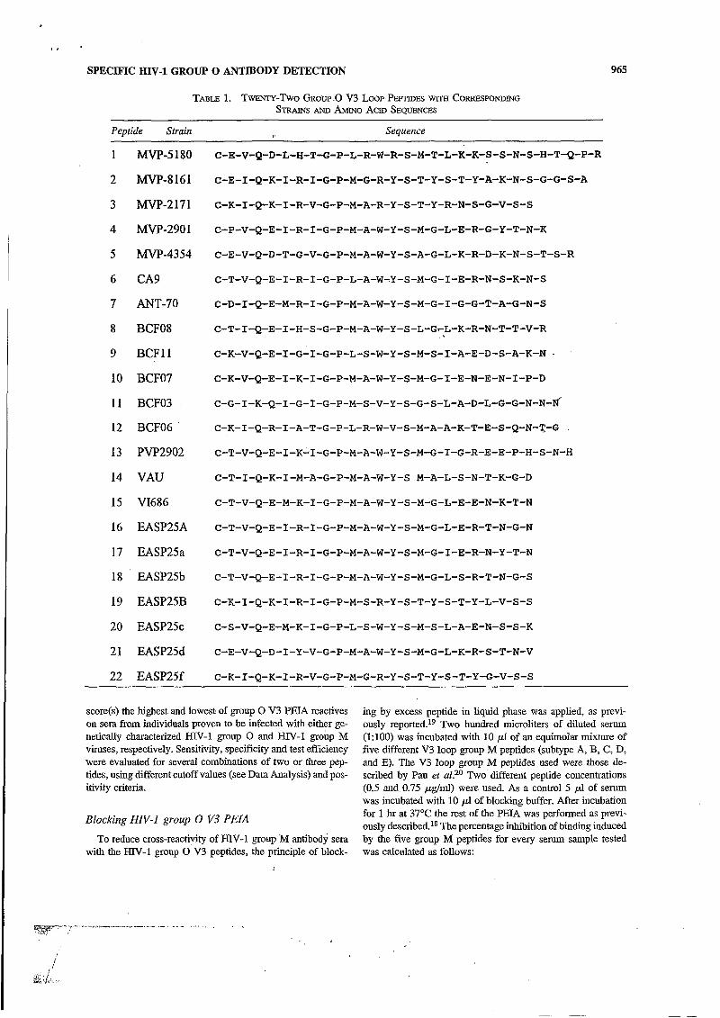
Twenty-two peptides consisting of 24 to 29 amino acid in the V3 region of HIV-1 group O (Table 1) were synthesized by Neosystem (Strasbourg, France) and used as antigens in HIV- 1 group O V3 PEWS. After synthesis, the peptides were >SO% purified by high-performance liquid chromatography analysis. Peptides 1 to 15 are derived from the V3 loop sequences of the isolates indicated. A phenogram analysis was performed on 18 HIV-1 group O V3 amino acid sequences, each 25 amino acids long. Consensus sequences were designed on the basis of ho- mologies among representatives of the clusters in the phenetic tree, as indicated in parentheses: EASP25a (CA9, VI686,
MVP7851, VAU); EASP25c (BCFOI, BCFl1); EASP25d (MVP4354, MVP5180, BCFOI); EASP25f (Mvp2171, MVP8161); EASP25A (EASP25a, EASP25b, EASP25c, EASP25d); EASP25B (EASP25f, BCF03).17 The phenogram .
clusters the peptides according to amino acid similarity.
MVP2901, BCF07, ANT-70); EASP25b (BCFO2, PVP2902,
HW-i group o v3 PEIA The HIV-1 group O V3 PEW was performed as described
previously.1s The criteria for the best group O V3 loop pep- tide(s) to be used in the P E U are defined as the peptide@) that
SPECIFIC HJY-1 GROUP O ANTIBODY DETECTION 965
TABLE 1. TwEN.rr-Two GROW .o v 3 LOOP PEPTWES WITH CORRESPO~ING STRAINS AND AMINO ACID SEQUENCES
r-;? -" -I
i
Peptide Strain Sequence
1 MVP-5180 C-E-V-Q-D-L-H-T-G-P-L-R-W-R-S-M-T-L-K-K-S-S-N-S-H-T-Q-P-R
2 MVP-8161 C-E-I-Q-K-I-R-I-G-P-M-G-R-Y-S-T-Y-S-T-Y-A-K-N-S-G-G-S-A
3 MVP-2171 C-K-I-Q-K-I-R-V-G-P-M-A-R-Y-S-T-Y-R-N-S-G-V-S-s
4 MVP-2901 C-P-V-Q-E-I-R-I-G-P-M-A-W-Y-S-M-G-L-E-R-G-Y-T-N-K
5 MVP-4354 C-E-V-Q-D-T-G-V-G-P-M-A-W-Y-S-A-G-L-K-R-D-K-N-S-T-S-R
6 CA9 C-T-V-Q-E-I-R-I-G-P-L-A-W-Y-S-M-G-I-E-R-N-S-K-N-S
7 ANT-70 C-D-I-Q-E-M-R-I-G-P-M-A-W-Y-S-M-G-I-G-G-T-A-G-N-S
8 BCF08 C-T-I-Q-E-1-H-S-G-P-M-A-W-Y-S-L-GrL-K-R-N-T-T-V-R
9 BCFll C-K-V-Q-E-I-G-I-G-P-L-S-W-Y-S-M-S-1-A-E-D-S-A-K-N .
10 BCFO7 C-K-V-Q-E-I-K-I-G-P-M-A-W-Y-S-M-G-I-E-N-E-N-I-P-D
1 1 BCF03 C-G-I-K-Q-I-G-I-G-P-M-S-V-Y-S-G-S-L-A-D-L-G-G-N-N-~
12 BCF06 C-K-I-Q-R-I-A-T-G-P-L-R-W-V-S-M-A-A-K-T-E-S-Q-N-T-G .
13 PVP2902 C-T-V-Q-E-I-K-.I-G-P-M-A-W-Y-S-M-G-I-G-R-E-E-P-H-S-N-H
14 VAU C-T-I-Q-K-I-M-A-G-P-M-A-W-Y-S M-A-L-S-N-T-K-G-D
15 VI686 C-T-V-Q-E-M-K-I-G-P-M-A-W-Y-S-M-G-L-E-E-N-K-T-N
16 EASP25A C-T-V-Q-E-I-R-I-G-P-M-A-W-Y-S-M-G-L-E-R-T-N-G-N
17 EASP25a C-T-V-Q-E-I-R-I-G-P-M-A-W-Y-S-M-G-I-E-R-N-Y-T-N
18 EASP25b C-T-V-Q-E-I-R-I-G-P-M-A-W-Y-S-M-G-L-S-R-T-N-G-S
19 EASP25B C-K-I-Q-K-I-R-I-G-P-M-S-R-Y-S-T-Y-S-T-Y-L-V-S-s
20 EASP25c C-S-V-Q-E-M-K-I-G-P-L-S-W-Y-S-M-S-L-A-E-N-S-S-K
21 EASP25d C-E-V-Q-D-I-Y-V-G-P-M-A-W-Y-S-M-G-L-K-R-S-T-~-V
22 EASP25f C-K-I-Q-K-I-R-V-G-P-M-G-R-Y-S-T-Y-S-T-Y-G-V-S-S
score(s) the highest and lowest of group O V3 PEL4 reactives on sera from individuals proven to be infected with either ge- netically characterized HIV-I group O and HIV-I group M viruses, respectively. Sensitivity, specificity and test efficiency were evaluated for several combinations of two or three pep- tides, using different cutoff values (see Data Analysis) and pos- itivity criteria.
ing by excess peptide in liquid phase was applied, as previ- ously re~orted. '~ Two hundred microliters of diluted serum (1:lOO) was incubated with 10 pl of an equimolar mixture of five different V3 loop group M peptides (subtype A, €3, C, D, and E). The V3 loop group M peptides used were those de- scribed by Pau et aZJO Two different peptide concentrations (0.5 and 0.75 pg/ml) were used. As a control 5 p l of serum was incubated with 10 pl of blocking buffer. After incubation
Blocking HIV-1 group O V3 PEIA for 1 hr at 37°C the rest of the PEL4 was performed as previ- ously described.*8 The percentage inhibition of binding induced
To reduce cross-reactivity of HN-1 group M antibody sera with the HIV-I group O V3 peptides, the principle of block-
by the five group M peptides for every serum sample tested was calculated as follows:
.-I_...x ._ . . . - , ... , ,

,. . . .
A 25 25
.-
20.. 3 20 -. i!
IS.- 15 -.
1 ' 10 -. '0 . -
A ,, I I
5.-
I 0.5 1 -1.5 -1 4.5 o
y>,':- ,!.' ,: i'
.-
i
i
I
. - o,; i
. . i ._-..,._ "_ .;.. .-&
966 ONDOA ET AL.
(OD without blocking - OD in presence of group M naarde, Gent, Belgium), in which biotinylated V3 peptides peptides)/OD without blocking from different HIV-1 group O and M viruses (consensus
and biotinylated gp41 peptides (O-ANT-70; M-subtype B; Coilfirmatory tests M-subtype D) were applied as a streptavidin complex in par-
INNO-LIA H N - I type O. All of the sera reactive by HIV- allel lines on nylon strips. The INNO-LIA HIV-1 type O as- 1 group O V3 PEU were retested in a line immunoassay for say was performed according to the instructions of the man- the specific detection of antibodies to HIV-1 group O in hu- ufacturer. Positivity criteria were used as described man sera (INNO-LIA HIV-I type O; Innogenetics, Zwij- previously.*
HIV-1 group M, M-Mal, O-ANT-70; O-VI686; 0-MVP5180)
- -51 lag OD
PEPTIDE13
.5 1 las OD
PEPTIDE 5
40
PEPTIDE 7
40
30 + . 30 c
-5 1 las 00
PEPTIDE15
35 'O t
FIG. 1. Log OD distribution curves of the gold standard group O (n = 33), group M (n = go), and HIV antibody-negative (n = 17) sera with the 5 best HIV-1 group O V3 PEIA.
SPECIFIC HI[v-1 GROUP O ANTIBODY DETECTION 967
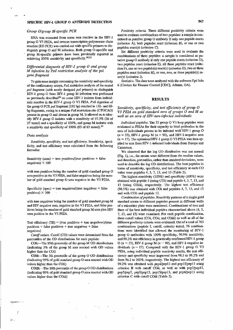
Group Olgroup M-specijc PCR
RNA was extracted from some sera reactive in the HN-1 group O V3 PEIA, and reverse transcriptase;polymerase chain reaction (RT-PCR) was carried out with specific primers to dis- tinguish group O and M infection. Both group O-specific and
’ group M-specific primers have been previously reported as achieving 100% sensitivity and
Direrential diagnosis of HN-I group O and group M infection by Pstl restriction analysis of the pol gene fragment
To gain more insight concerning the sensitivity and specificity of the confirmatory assays, PstI restriction analysis of the nested pol fragment (with newly designed pol primers) to distinguish HIV-1 group O from HIV-1 group M infection was perfomed as previously described1° on some HIV-1 isolates from individ- uals reactive in the HJY-1 group O V3 PEIA. PstI digestion of the group O PCR pol fragment (192 bp) resulted in 132- and 60- bp fragments, owing to a unique Pst1 restriction site (CTGCAG) present in group O and absent in group M. It allowed us to iden- tify HIV-1 group O isolates with a sensitivity of 97.3% (36 of 37 tested) and a specificity of loo%, and group M isolates with a sensitivity and specificity of 100% (63 of 63 tested).l0
Data analysis
Sensitivity, specijicity, and test efficiency. Sensitivity, speci- ficity, and test efficiency were calculated from the following formulas:
Sensitivity (sens) = true positives/(true positives + false negatives) X 100
with true positives being the number of gold standard group O sera positive in the V3 PEIA, and false negatives being the num- ber of gold standard group O sera negative in the V3 PEIA.
Specificity (spec) = true negatives/(true negatives + false positives) X 100
with true negatives being the number of gold standard group M and HIV negative sera, negative in the V3 PEIA, and false pos- itives being the number of gold standard group M sera plus HN sera positive in the V3 PEIA.
Test efficiency (TE) = (true positives + true negatives)/(true positives -I- false positives + true negatives + false negatives)
Cutoff values. Cutoff (CO) values were determined from the percentiles of the OD distributions for each peptide:
Coi:-The 95th percentile of the group M OD distributions (indicating 5% of the group M sera reacted with OD values higher t h q the Coi)
Coi¡:-The 5th percentile of the group O OD distributions (indicating 95% of gold standard group O sera reacted with OD values higher than the COíí)
COi¡i:-The 10th percentile of the group O OD distributions (indicating 90% of gold standard group O sera reacted with OD values higher than the COiii)
Positivity criteria. Three different positivity criteria were used to evaluate combinations of two peptides: a sample is con- sidered as putative group O antibody if only one peptide reacts (criterion A), both peptides react (criterion B), or one or two peptides react(s) (criterion C). Six different positivity criteria were used to evaluate the
combinations of three peptides: a sample is considered as pu- tative group O antibody if only one peptide reacts (criterion D), two peptides react (criterion E), all three peptides react (crite- rionF), one or two peptide(s) react(s) (criterion G), two or three peptides react (criterion H), or one, two, or three peptide@) re- act(s) (criterion I).
Statistics. The data were analyzed with the software Epi-Info 6 (Centers for Disease Control [CDC], Atlanta, GA).
RESULTS
Sensitivity, speCi@city, and test efficiency of group O V3 PEIA on gold standard sera of groups O and M as well as on sera of HN-non-infected individuals
Individual peptides. The 22 group O V3 loop peptides were evaluated in PEIAs for their capacity to bind antibodies in the sera of individuals proven to be infected widEIN-1 group O (n = 33), HIV-1 group M (n = 90), and EIN-1-negative sera (n = 17). The optimized HIV-1 group O V3 PEL4 was then ap- plied to sera from HIV-1-infected individuals from Europe and Cameroon.
We observed that the log OD distribution was not normal (Fig. l), i.e., the means were different from the median values, and therefore, percentiles, rather than standard deviations, were used to descride the log OD distributions. The best peptides in terms of sensitivity, specificity, and test efficiency at each CO value were peptides 4, 5, 7, 13, and 15 (Table 2).
The highest sensitivity (100%) and specificity (100%) were obtained with peptide 5 (using Coi) and peptide 4,5,7,13, and 15 (using COG), respectively., The highest test efficiency (98.5%) was obtained with Coi and peptides 4,7, 13, and 15 and with COii and peptide 13.
Combitution ofpeptides. Reactivity patterns of a single gold standard serum to different peptides present in different wells of a microtiter plate were monitored. Combinations of two and three of the best individual peptides characterized above (4, 5, 7, 13, and 15) were examined. For each peptide combination, three cutoff values (Coi, COii, and COiii) as well as all of the different positivity criteria were evaluated. Out of a total of 765 combinations (peptide I, cutoff, criteria) tested, 76 combina- tions were identified that allowed the monitoring of HIV-1 group O antibodies with 100% specificity, 96.9% sensitivity, and 99.2% test efficiency in genetically confirmed HIV-1 group O (n = 33), HIV-1 group M (n = 90), and HIV-I-negative in- dividuals (n = 17). Compared with the HIV-1 group O V3 PEIA, using individual peptide reactivity results, the test effi- ciency and specifcity were improved from 98.5 to 99.2% and from 96.2 to loo%, respectively. The highest test efficiency of 99.2% was obtained with pe#/pepl3 and pepl5/pepl3 using criterion €3 with cutoff COii, as well as with pep7/pepl3, pep5/pep7, pep5/pepl5, pepWpepl3, and pep4/pepl3 using criterion C with cutoff COiii (Table 2).

968 ONDOA ET AL.
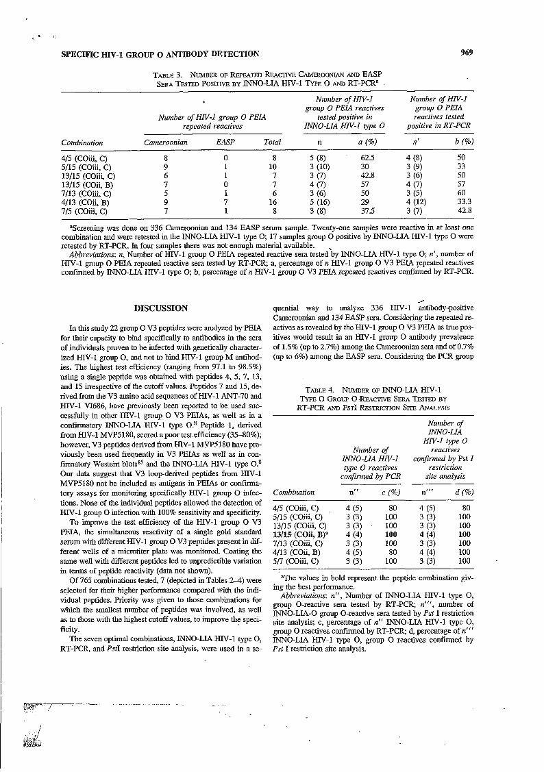
ian and European KIV-1 antibody-positive sera for the pres- ence of HIV-1 group O antibodies. Among 336 HIV-1 anti- body-positive sera from Cameroon, from 5 (1.5%) to 9 (2.7%) were reactive; among 134 EASP sera, from 1 (0.7%) to 7 (6%) were repeatedly reactive in the HIV-1 group O V3 P E L (Table 3). Considering the HIV-1 group O PCR as the gold standard confiiatory test, the best percentage (60%) of confirmed re- peated reactivities was obtained using combination 7/13 (COïi, C), followed by combination 13/15 (COii, B) (57%). Of a to- tal of 470 HIV-1 antibody-positive sera tested, 21 sera (14 Cameroonian, 7 EASP) were repeatedly reactive with at least one of the seven peptide combinations in the HW-1 group O V3 PEIA and were all tested by INNO-LIA HIV-1 type O. None of the EASP sera was c o n f i e d as group O in the INNO-LL4 HIV-1 type O. Only three of seven EASP samples were tested by RT-PCR and confiied as group M infected (for four of them, there was ‘ not enough material available). Four Cameroonian sera were conf i ied as group O, and one as a group O and M dual infection, in the INNO-LIA HIV-1 type O (Table 4). All of the Cameroonian samples were tested by RT-PCR. The four sera found to be group O positive in the INNO-LIA HIV-1 type O were confirmed as group O by RT- PCR (Table 4). The dually reactive sample was confiied as an HIV-1 group M subtype A infection (data not shown). Of the nine group O-negative/group M-positive samples as deter- mined by INNO-LIA HW-1 type O, one was confirmed as a group M infection by RT-PCR; the pol gene of the others could not be amplified. Overall, the RT-PCR confiied that HIV-1 group O antibody prevalence is 1.2% (4 of 336) in the Cameroonian sera, and 0% in the EASP sera.
Blocking HIV-I group O V3 PEIA
Attempts were made to reduce the level of cross-reaction, using methods described by Barin and colleague^.^^ Antibody was diluted in an excess concentration of group M peptides prior to their incubation with solid-phase group O peptides in an attempt to compete out the cross-reactive antibodies. S i x HIV-1 group O and 17 group O V3 PEL4 cross-reacting €IN- 1 group M sera (confirmed with INNO-LIA HN-1 type O and PCR) were retested with the blocking HIV-1 group O V3 PEW, using two different concentrations of group M peptides repre- senting subtypes A, B, C, D, and E. In the Fist experiment, pep- tides 4, 13, and 15 were used in the blocking P E U with group M peptides in the liquid phase at a concentration of 0.5 &nI. No inhibition greater than 50% was observed. The same ex- periment was done with group M peptides at a concentration of 0.75 pg/ml. A 50% inhibition was observed with three cross- reactive group M sera in a pep4/pepl3 blocking P E U and with one group O serum in a pep4 blocking PEIA. No percentage of inhibition higher than 37% was observed in the pep15 block- ing PEL4 (data not shown).
Use of the optimized H N - I group O V3 PEIA to screen HN-infected individuals for H N - I group O iigection
These seven peptide combinations with the use of the ap- propriate criteria and cutoff values resulted in higher test effi- ciencies (99.2% as compared with 98.5%) when reactivities to individual peptides were monitored. Consequently, these seven peptide combination strategies were used to screen Cameroon-
TABLE 2. SENSITIVITY, SPECIFICITY, AND TET EFFTCIENCY OF THE BEST INDIVIDUAL AND SEVEN BEST GROUP 0 v3 PEPTIDE COMBINATIONS IN PEJA”
Senf SpecC TEc SenSc SpecC TEc Senf Spec’ TEc Peptides Coib (%) (%) (%) COiib (%) (%) (%) COiiib (%) (%) (%)
4 0.52 96.9 96.2 98.5 0.68 96.9 98.1 97.8 1.8 90.9 100 97.8 5 0.567 100 96.2 97.1 1.2 96.9 98.1 97.8 2.24 90.9 100 97.8 7 0.902 96.9 96.2 98.5 0.75 96.9 96.2 96.4 1.8 90.9 100 97.8 13 0.225 96.9 96.2 98.5 0.79 96.9 99 98.5 2.3 90.9 100 97.8 15 1.1 96.9 96.2 98.5 1.694 96.9 98.1 97.8 2.8 90.9 100 97.8 4/15 (C)d 0.52/1.1 96.9 93.4, 94.2 0.68/1.694 96.9 96.2 95 1.8/2.86 96.9 100 99.2 5/15 (C) 0.567/1.1 100 94.3 95.7 1.2/1.694 100 97.1 97.8 2.24/2.86 96.9 100 99.2 13/15 (C) 0.225/1.1 96.9 93.4 94.2 0.79/1.694 96.9 97.1 97.1 23/2.86 96.9 100 99.2 13/15 (B)d 0.225/1.1 96.9 99 98.5 0.7911.694 96.9 100 99.2 2.3/2.86 84.8 100 96.4 7/13 (C) 0.902/0.225 96.9 96.2 96.4 0.75/0.79 96.9 96.2 95 1.W2.3 96.9 100 99.2 7/5 (C) 0.902/0.567 100 94.3 95.7 0.75/1.2 100 95.3 96.4 1312.86 96.9 100 99.2 4/13 (B) 0.52/0.225 96.9 100 99.2 0.6810.79 96.9 100 99.2 1.8/2.3 87.8 100 77.9
“When tested on gold standard group O (n = 33), group M (n = 90). and HIV antibody-negative sera (n = 17), using three different cutoff values.
bCOi = 95th percentile of the group M sera OD distribution; COii = 5th percentile of the group O sera OD distribution; COiii = 10th percentile of the group O sera OD distribution.
‘Sens (sensitivity) = true positives (true positives + false negatives) X 100 (with true positives being the number of gold standard group O sera positive in the V3 PEIA, and false negatives being the number of gold standard group O sera negative in the V3 PEIA). Spec (specificity) = true negatives/(true negatives i- false positives) X 100 (with true negatives being the number of gold standard group M sera -I- HN sera negative in the V3 PEIA, and false positives being the number of gold
standard group M sera + HIV sera positive in the V3 PEIA). Test efficiency (TE) = (true positives + true negatives)/(true positives + false positives + true negatives + false negatives).
d(B) = a sample is considered as putative group O if both peptides of the Combination react (C) = a sample is considered as putative group O if at least one peptide of the combination reacts.
The values in bold represent the best performances and the corresponding cut-off obtained with the peptide combinations.
..-
4 1.
vidual peptides. Priority was given to those combinations for which the smallest number of peptides was involved, as well as to those with the highest cutoff values, to improve the speci- ficity.
The seven optimal combinations, IN'NO-LIA HIV-1 type O, RT-PCR, and PstI restriction site analysis, were used in a se- l
SPECIFIC HIV-1 GROUP 0 ANTIBODY DETECTION 969
TABLE 3. NUMBER OF REPEATED REAC~IVE CAMEROONIAN AND EASP SERA TESTED POS^ BY INNO-LIA HIV-1 TYPE O AND RT-PCR' .
Number of HN-I Number of HN-I group O PEIA reactives group O PEIA
tested positive in reactives tested Number of HN-I group O PEIA repeated reactives INNO-LIA HN-I type O positive in RT-PCR
Combination Cameroonian EASP Total n a (%) Il' b (%o)
4/5 (Coiii, C) 8 O 8 5 (8) 62.5 4 (8) 50 33 5/15 (COiii, C) 9 1 10 3 (10) 30 3 (9) 50 13/15 (COiii, C) 6 1 7 3 (7) 42.8 3 (6)
13/15 (COii, B) 7 O 7 4 (7) 57 4 (7) 57 60 7/13 (COiii, C) 5 1 6 3 (6) 50 3 (5) 33.3 4/13 (COii, B) 9 7 16 5 (16) 29 4 (12)
7/5 (COiii, C) 7 1 8 3 (8) 37.5 3 (7) 42.8
aScreening was done on 336 Cameroonian and 134 EASP serum sample. Twenty-one samples were reactive in at least one combination and were retested in the IN'NO-LIA HIV-1 type O; 17 samples group O positive by INNO-LIA HIV-1 type O were retested by RT-PCR. In four samples there was not enough material available.
Abbreviations: n, Number of HIV-1 group O PEIA repeated reactive sera tested'by INNO-LIA HIV-1 type O; n', number of HIV-1 group O PEW repeated reactive sera tested by RT-PCR a, percentage of n HIV-1 group O V3 PEIA Tepeated reactives confmed by INNO-LIA HIV-1 type O; b, percentage of n Mv-I group O V3 PEIA repeated reactives confmed by RT-PCR.
DISCUSSION f
quential way to analyze 336 HIV-1 antibody-positive Cameroonian and 134 EASP sera. Considering the repeated re- actives as revealed by the HIV-1 group O V3 PEIA as true pos- itives would result in an HIV-1 group O antibody prevalence of 1.5% (up to 2.7%) among the Cameroonian sera and of 0.7% (up to 6%) among the EASP sera. Considering the PCR group
TABLE 4. " ~ E R OF I"O-LI.4 W - 1 TYPE 0 GROUP O-REAClTVEi SERA TESTED BY
RT-PCR AND PSTI RESTRICTION SITE ANALYSIS
Number of
HN-I type O
confirmed by Pst I
INNO-LIA
Number of reactives INNO-LIA HN-I type O reactives restriction
confirmed by PCR site analysis
Combination n" c (%) nrr' d (%)
4/5 (COiii, C) . 4 (5) 80 4 (5) 80 5/15 (COiii, C) 3 (3) 100 3 (3) 100 13/15 (COZ, C) 3 (3) 100 3 (3) 100 13/15 (COii, B)a 4 (4) 100 4 (4) 100 7/13 (COiii, C) 3 (3) 100 3 (3) 1 O0 4/13 (COG, B) 4 (5) 80 4 (4) 100 5/7 (COiii, C) 3 (3) 100 3 (3) 100
'The values in bold represent the peptide combination giv- ing the best performance.
Abbreviations: n", Number of "NO-LIA HIV-I type O, group O-reactive sera tested by RT-PCR n'", number of INNO-LIA-O group O-reactive sera tested by Pst I restriction site analysis; c, percentage of i f ' INNO-LIA HIV-1 type O, group O reactives confmed by RT-PCR d, percentage of n r r r INNO-LIA HIV-1 type O, group O reactives confirmed by Pst I restriction site analysis.

970
.I .. ,.I___.i__.. __. .i.. J
ONDOA ET AL.
O with a sensitivity and specificity of 100% as previously re- portedlO*zl as the references test, suggests that HIV-1 group O V3 P E U using combination 13/15 (COE, B) can be considered the best choice. Combination 7/13 (COG, C) yielded even 60% RT-PCR confimed group O V3 PEIA results this combination missed one true positive sample. As such the confirmed preva- lence rate of HN-1 group O antibody positives among the Cameroonian sera tested is 1.2% (4 of 336) and 0% among the EASP sera.
The low group O seroprevalence in Cameroonian and EASP sera, as well as the cross-reactivity of HIV-1 group O V3 pep- tides with group M antibodies, may have contributed to the false positives. Blocking experiments with group M peptides in ex- cess in liquid phase did not really improve the specificity of the V3 PEIA. The reduction in cross-reactivity of a few group M samples with group O peptides was accompanied by a signifi- cant decrease in group O sample OD values in blocking PEJAS. This may suggest that the majority of the cross-reactions are peptide specific and cannot be eliminated by this method. The role of other infections susceptible to cross-reactivity with HIV peptides is not excluded. For instance, Mycobacterium tuber- culosis, herpes simplex, and varicella zoster have been de- scribed to share amino acid motifs with parts of the HIV en- velope
Theoretically, in a situation where 10,000 HIV-positive peo- ple, of which 1% are HIV-1 group O infected, are tested with our seven combinations (sensitivity of 96.9%, specificity of loo%), we expect to find a prevalence rate of 0.97 (3 false neg- atives, O false positives, with a total of 97 initial reactives to be expected). The predicted HN-1 group O prevalence of 0.97% fits well with the real 1% prevalence. In the same situation, us-
' ing ANT-70 alone (sensitivity of 90.9 specificity of 100%) as previously described or combination pep9/pepl3 with an opti- mal sensitivity of '100% and a specificity of 99% (COiii, crite- rion C, data not shown) will give a less accurate prevalence rate of group O hfjxtions of 0.91 and 2%, respectively, instead of 1%. The HIV-1 group O antibody prevalence rates among HIV- positive sera documented so far were low and varied from O to 2%: which is in favor of our test strategy.
Only the H!X1 group O PEIA repeated reactives were rested by INNO-LIA HIV-1 type O and group sO/group M-specific RT-PCR and only the group O RT-PCR-poiitive samples were retested by Pst1 restriction site analysis. The hierarchical test- ing format used in this study is not suitable for intertest com- parison; however, we observed a discrepancy between the INNO-LIA HIV-1 type O and group O-specific RT-PCR re- sule. Of 21 sera (14 Cameroonian, 7 EASP), repeatedly reac- tive with at least one of the seven combinations in the PEIA, 4 were considered HIV-1 group O antibody positive and 1 serum reacted with HIV-1 group O as well as group M peptides in the INNO-LIA HIV-1 type O. The fust four were confirmed by PCR and PstI restriction enzyme analysis. The latter was con- f m e d to be HIV-1 group M infected (subtype A), which sup- ports our previous observations that some dually reactive sera (group M and O) as determined by INNO-LIA HIV-1 type O are not confimed as type M and. O dual infections.'O Peeters et aZ.8 confimed that the majority of individuals whose sera re- act simultaneously with group O and group M V3 peptides in the INNO-LIA HN-1 group O are infected only with an HN-
1 group M virus as indicated by PCR.8 Serological dual group O/group M reactivity may be due to a broad immune response to a single serotype; to an aborted or suppressed group O in- fection in a group M-infected host; or to exposure or infection with a variant containing group O and group M epitopes. In ad- dition, the sensitivity of the PCR pol primers can be questioned, considering the great variability existing within group O, and the relatively small number of group O viruses that have been analyzed to determine the sensitivity and specificity of these primers. 10*21
Exclusive studies of the sensitivity and specificity of the INNO-LIA HIV-1 type O, using sera of genetically proven HIV-1 group O-infected individuals, have not been performed thus far. However, in this study, 14 HIV-1 group O sera of our gold standard panel, 17 sera from this study, and 11 samples (described in Ref. 10) reactive in the group O V3 PEJA were tested in parallel with the JNNO-LIA HIV-1 type O and by RT- PCR. The INNO-LIA HIV-1 type O confiied 25 samples as group O infected (24 were typed as group O by RT-PCR 1 could not be so typed, as its poE gene could not be amplified), 2 samples as group O/group M coinfections (1 was confiied as group O; the second was conf i ied as group M by PCR) and 15 sera as group M infected (the poZ gene of 12 could not be amplified, but 3 were confmed as group M infected by RT- PCR). These results show for thé diagnosis of group O infec- tions (on the basis of the 30 samples from which the pol gene could be amplified), a sensitivity and specificity of the INNO- LIA HIV-1 type O, compared with the group O/group M-spe- cific RT-PCR, of 100% (95% confidence interval, 83.4 to 100%) and 75.0% (95% confidence interval, 21.9 to 98.7%), respectively. This. is in accordance with the findings of others, who have shown that the majority of the INNO-LIA HN-1 type O results so far are confimed by RT-PCR.8*18 Using the INNO- LIA HIV-1 type O on group O V3 PEJA reactive samples im- proves the accuracy of group O infection detection. Besides, the INNO-LIA HIV-1 type O as a confiiatory test requires only serum and does not need an RNA extraction step as is re- quired for PCR, which is logistically more demanding in terms of storage and reagents. For these reasons, this test remains a valuable tool for the diagnosis of an HN-1 group O infection.
Moreover, the INNO-LIA HIV-1 type O scored the serum of a patient infected with HIV-1 BCF03 as HIV-1 group O pos- itive. This serum remained negative by HIV-1 group O V3 PEJA, using our seven best combinations. One can be concerned that screening with two peptides in an ELISA is confiírmed by other peptides on the INNO-LIA HIV-1 type O strip. However,
.the peptides coated on the strips are biotinylated and are pos- sibly of different length than ours. This may influence the an- tibody binding in a positive way. In addition, the result of the INNO-LIA HIV-1 type O is always interpreted by comparing the sample reactivity with group O and group M antibodies, which confers a good specificity to the test.
~ CONCLUSION
To the best Of our knowledge, this is the first time that re- activity of group O peptides against well-characterized group
SPECIFIC HIV-1 GROW O ANTIBODY DETECTION
M and group O samples was studied. We demonstrated that 7 peptide combinations (sensitivity of 96.9%, specificity of loo%, TE of 99.2%) performed better than any of the 22 individual peptides and the previous strategies using -ANT-70 or ANT- 70/MVP5180 in the V3 PEIA. preliminary results from the screening of Cameroonian and European sera demonstrated that the Combination of peptide 13 (PVP2902) and peptide 15 (VI686), followed by confirmation of repeated reactives, may be the best strategy to discriminate between group O and group M infections. Thus far the INNO-LIA HIV-1 type O has proven reliable in confirming a group O ELISA-positive result, but sen- sitivity and specificity analyses have not been performed yet on a sufficiently large panel of genetically confirmed group O and group M isolates. The PCR as a confirmatory assay is preferred but depends on the quantity and quality of nucleic acids in the sample and is logistically more difficult to implement. We sug- gest further specificity and sensitivity studies with larger pan- els of group M and group O sera from different geographic re- gions to improve the perfmance of the HIV-1 group O V3 PEU, to monitor for HIV-1 group O infections among HIV-I- infected individuals.
ACKNOWLEDGMENTS
This work was supported by Grants G 3301.96 and G.0134.97 of the Fonds voor Wetenschappelijk Ondenoek, Brussels and by EC project IC 18-CT96-0110.
We are indebted to the European AIDS Virus Serosurveil- lance Programme (EASP), Contract BMH4-CT96-1559, for the supply of sera and to Dr. E. Saman for the supply of INNO- LIA €€IV-1 type O kits.
We thank Ciska Maeckelbergh for typing the manuscript.
REFERENCES
1.
2.
3.
4.
5.
- 6.
Chameau P, Borman AM, Quillent C, Guetard D, Chamaret S, Co- hen J, Remy G, Montagnier L, and Clavel F Isolation and enve- lope sequence of a highly divergent HIV-I isolate: Definition of a new HN-1 group. Virology 1994;205:247-253. Korber B, Loussert-Ajaka I, Blouin J, and Saragosti S: A compar- ison of HIV-1 group M and group O functional and immunogenic domains in the gag p24 proteins and the C2V3 region of the en- velope protein. In: Human Retroviruses and AIDS 1996 (Myers G, Korber B, Brian F, Kuan-Teh J, Mellors J-W, and Wain-Hobson S, eds.). Los Alamos National Laboratory. Los Alamos, New Mex- ico, 1996, pp. m41-56. Schable C, Zekeng L, Pau CP, Hu D, KaptuC L, Gürtler L, Don- der0 T, TsaguC J-M, Schochetman G, Jaffe H, and George IR: Sen- sitivity of United States HIV antibody tests for detection of HIV- 1 group O infections. Lancet 1994;344:1333-1334. Loussert-Ajaka I, Ly TD, Chaix ML, Ingrand D, Saragosti S, Courouce AM, Brun-Vezinet F, and Simon F HIV-l/HIV-2 seronegativity in HIV-1 subtype O infected patients. Lancet 1994;343:1393-1394. Giirtler LG, Zekeng L, Simon F, Eherle L, Tsague JM, Kaptue L, Brust S, and Knapp S: Reactivity of five anti-HIV-1 subtype O specimens in six different anti-HIV screening ELISAs and three immunoblots. J Viol Methods 1995;51:177-184. Nkengasong JN, Peeters M, vanden Haesevelde M, Musi SS,
971
Willems B, Ndumbe PM, Delaporte E, Perret JL, Piot P, and van den Groen G. Antigenic evidence of the presence of the aberrant HIV-IANT70 virus in Cameroon and Gabon. AIDS 1993; 21536-1538.
7. Peeters M, Lobe V, Nkengasong J, Willems B, Delforge ML, Van Renterghem, Revets H, Sprecher S, and van der Groen G HN-1 group O infection in Belgium. Acta Clin Belg 1995;50(3):171-173.
8. Peeters M, Gueye A, Mboup S, Bibollet-Ruche F, Eukaza E, Mu- langa C, Ouedrago R, Gandji R, Mpele P, Dibanga G, Koumare B, Saidou M, Esu-Williams E, Lombart J-P, Badombena W, Lu0 N, Vanden Haesevelde M, and Delaporte E: Geographical distribution of HIV-1 group O viruses in Africa. AIDS 1997;11:493-498.
9. Mauclere P, Loussert-Ajaka I, Damond F, Fagot P, Souquibres S, Monny Lobé M, Mbopi Keou F-X, Barré-Sinoussi F, Saragosti S, Bxun-Vézinet F, and Simon F Serological and virological charac- terization of HIV-1 group O infections in Cameroon. AIDS 1997;11:445-453.
10. Heyndrickx L, Janssens W, Gürtler L, Zekeng L, Loussert-Ajaka I, Vereecken K, Willems B, Coppens S, Ndumbe P, Fransen K, Saman E, Alary M, and van der Groen G Differential diagnosis of HN type1 gqup O and M infection by polymerase chain reac- tion and PstI restriction analysis of the pol gene fragment. AIDS Res Hum Retroviruses 1998;14(11):973-977. ~
11. Vanden Haesevelde M, Decourt J-L, De Leys R, Vanderborghht B, van der Groen G, van Heuverswijn H, and Saman E: Genomic cloning and complete sequence analysis of a highly divergent African human immunodeficiency virus --ìsolate. J Virol 1994;68:1586-1596.
Ila. Janssens W, Keyndrickx L, Van der Auwem G, er al.: Interpa- tient variability of HIV-1 group O. A D S 1998; submitted
12. Delaporte E, Janssens W, Peeters M, Buve A, Dibanga G, Perret JL, Ditsambou V, Mba m, Courbot MC, Georges A, Bour- geois A, Samb B, Hemel D, Heyndrickx L, Fransen K, van der Groen G, and Larouze B: Epidemiological and molecular charac- teristics of HIV infection in Gabon, 1986-1994. AIDS 1996;
13. Loussert-Ajaka I, Chaix M-L, Korber B, Letourneur F, Gomas E, Allen E, Ly T-D, Brun-Vézinet F, Simmon F, and Saragosti S: Variability of human immunodeficiency virus type 1 group O strains isolated from Cameroonian patients living in France. Vi- rol 1995;69:564@-5649.
14. Fransen K, Buvé A, Nkengasong IN, Laga M, and van der Groen G. Longstanding presence in Belgians of multiple non-B subtypes. Lancet 1996;347:1403.
15. Gürtler LG, Eberle J, Lorbeer B, and Deinhardt F Sensitivity and specificity of commercial ELISA kits for screening anti- LAV/HTL.V-JlI. J Virol Methods 1987;15:11-23.
16. European AIDS Virus Serosurveillance Programme (EASP): An- nual activity report, July 1, 1996-June 31, 1997.
17. Korber BTM, MacInnes K, Smith RF, and Myers G Mutational trends in V3 loop protein sequences observed in different genetic lineages of human immunodeficiency virus type 1. J Virol 1994;68(10):7306744.
18. Peeters M, Nkengasong J, Willems B, Karita E, Delaporte E, Van den Haesevelde, Piot P, and van der Groen G Antibodies to V3 loop peptides derived from chimpanzee lentivirus and the diver- gent HIV-1 ANT-70 isolate in human sera from different geo- graphic regions. AIDS I994;8:1657-1661.
19. Barin F, Lahbabi Y, Buzelay L, Lejeune B, Baillou-Beaufis A, De- nis F, Mathiot C, M’boup s, Vithayasai V, Dietrichu, and Goudeau A Diversity of antibody binding to V3 peptides representing con- sensus sequences of HN type 1 genotype A to E An approach for HIV type 1 serological suhtyping. AIDS Res Hum Retroviruses 1996; 13:127%1289.
20. Pau C-P, Lee-Thomas S, Auwanit W, George JR, Ou C-Y, Parekh
10~903-910.
q.
972
I . . . ., ........ ,'A ,,.<;a '5 . . . . . . . . . . . . . . . . . . ..I .__., ... i ...* i . .---..---~"--.~-~---. ........ -...:.J.
BS, Granade TC, Holloman DL, Phillips S, Schochetman G, Young NL, Takabe Y, Gayle HD, and Weniger B G Highly specific V3 peptide enzyme immun0 assay for serotyping HJY-I specimen from Thailand. AIDS 1993;7:337-340.
21. Janssens W, Fransen K, Loussert-Ajaka I, Heyndrickx L, Ivens T, Eberle J, and Nkengasong J: Diagnosis of €€IV-1 group O infec- tions by polymerase chain reaction. Lancet 1995;346:451-452.
22. Davis D, Chaudhri B, Stephens DM, Came CA, Willers C, and Lachmann PJ: The immunodominance of epitopes within the trans- membrane protein (gp41) of human immunodeficiency virus type
ONDOA E T AL.
1 may be determined by the host's previous exposure to similar epitopes on unrelated antigens. J Gen Virol 1990;1:1975-1983.
Address reprint requests to: Guido van der Groen
Institute of Tropical Medicine Nationalestraat 155
2000 Antwerp, Belgium

Y
O I
20, 1998
,889-2229 -