Osteoblast-Like Cell Behavior on Porous Scaffolds Based on Poly(styrene) Fibers
Impaired cell cycle regulation of osteoblast-related transcriptionfactor Runx2/Cbfa1 in osteosarcoma cells
Inga San Martin1, Nelson Varela1, Marcia Gaete1, Karina Villegas1, Mariana Osorio1, JulioC. Tapia1, Marcelo Antonelli1, Edna Mancilla2, Jane B. Lian3, Janet L. Stein3, Gary SStein3, Andre J. van Wijnen3,*, and Mario Galindo1,*1Program of Cellular and Molecular Biology, Institute of Biomedical Sciences (I.C.B.M.), Facultyof Medicine, University of Chile, Santiago, Chile2Program of Pathophysiology, Institute of Biomedical Sciences (I.C.B.M.), Faculty of Medicine,University of Chile, Santiago, Chile3Department of Cell Biology and Cancer Center, University of Massachusetts Medical School,Worcester, MA 01655-0105
AbstractIn mammals, bone differentiation requires the functional expression of the Runx2/Cbfβheterodimeric complex. Our previous results indicate that Runx2 is also a suppressor of pre-osteoblast proliferation by affecting cell cycle progression at G1. Runx2 levels are cell cycleregulated, oscillating from a maximum during early G1 to a minimum during late G1, S andmitosis phases in proliferating pre-osteoblasts Nevertheless, there is no information concerningCbfβ gene expression during the cell cycle nor on Runx2 cell cycle expression in bone cancercells. We analyzed Runx2 and Cbfβ gene expression during cell cycle progression in the pre-osteoblast MC3T3 and osteosarcoma ROS and SaOS cell lines. The expected reduction of Runx2protein level was observed in MC3T3 cells arrested in late G1 or M phase using mimosine ornocodazole, respectively. However, this reduction was not observed in the cell cycle arrestedosteosarcoma cells. Cbfβ protein levels were not regulated during the cell cycle in pre-osteoblastsand osteosarcoma cells. Using cells synchronized in late G1 and mitosis we found that Runx2levels, but not Cbfβ levels, were cell cycle regulated in MC3T3 osteoblasts. Interestingly, bothfactors showed a constitutively elevated expression throughout the cell cycle in osteosarcomacells. Proteasome inhibition by MG132 prevented cell cycle-dependent downregulation of Runx2protein levels in osteoblasts, but not in osteosarcoma. We propose that Runx2 is involved intumoral osteosarcoma progression. Altogether, deregulated Runx2 expression throughout the cellcycle seems to constitute a central mechanism in the pathogenesis of osteosarcoma.
INTRODUCTIONThe transcription factor Runx2 (run-related transcription factor 2) forms a functionalheterodimeric complex with Cbfβ (core-binding factor β) (Meyers et al., 1993; Speck andStacy, 1995). Cbfβ is a cotranscription factor that does not bind directly to DNA butenhances Runx2 binding to DNA (Ogawa et al., 1993; Speck and Stacy, 1995). Osteogenicdifferentiation induced by Runx2 is dramatically increased by the co-expression of Cbfβ,
* Address Correspondence to: Mario Galindo, I.C.B.M., Faculty of Medicine, University of Chile, Independencia 1027, P.O. Box70061, Santiago, Chile tel: 56-2-9786917 / fax: 56-2- / [email protected]. Andre J. van Wijnen, Department of Cell Biology,University of Massachusetts Medical School, 55 Lake Avenue North, Worcester, MA 01655, USA tel: 508-856-5625 / fax:508-856-6800 / [email protected].
NIH Public AccessAuthor ManuscriptJ Cell Physiol. Author manuscript; available in PMC 2011 March 30.
Published in final edited form as:J Cell Physiol. 2009 December ; 221(3): 560–571. doi:10.1002/jcp.21894.
NIH
-PA Author Manuscript
NIH
-PA Author Manuscript
NIH
-PA Author Manuscript
even though this factor fails to induce osteogenesis on its own (Lien et al., 2007).Furthermore, Runx2 protein is stabilized by its interaction with Cbfβ, hence protecting itfrom ubiquitination and its proteasomal degradation (Huang et al., 2001; Zhao et al., 2003).Thereby, association between Runx2 and Cbfβ is central in the process of bonedifferentiation in vivo, leading to an efficient binding of Runx2 to DNA and the concomitantRunx2 mediated bone phenotypic gene activation(Kundu et al., 2002; Yoshida et al., 2002).
One of the critical steps for normal bone formation is the adequate and strict control of theproliferative expansion of mesenchymal, osteoprogenitor and immature osteoblast cells inresponse to mitogenic signals such as growth factors and cytokines (Liu et al., 2002). Runx2has been found to control osteoblast proliferation, promoting a transition from a proliferativestage to a post proliferative stage prior to osteoblast differentiation. This could be achievedin part through epigenetic mechanisms and partly through the ability of Runx2 to regulatethe expression of cell growth related genes and promote a non- proliferative state (e.g., G0/G1 phase transition and senescence) (Pratap et al., 2003; Galindo et al., 2005; Young et al.,2007a; Young et al., 2007b; Teplyuk et al., 2008; Teplyuk et al., 2009; Zaidi et al., 2007;Kilbey et al., 2007). Accordingly, transient Runx2 overexpression in synchronized cellsdelays cell cycle entry into S phase and significantly decreases cell proliferation in theMC3T3 preosteoblast, Runx2 null calvaria osteoprogenitors, C2C12 pluripotentmesenchymal and IMR-90 fibroblasts cell lines (Pratap et al., 2003; Galindo et al., 2005;Young et al., 2007a; Teplyuk et al., 2008; Teplyuk et al., 2009).
Consequently, Runx2 protein levels are strictly regulated during cell cycle in osteoblastcells. Runx2 protein levels are dramatically elevated in quiescence (G0) or proliferativearrest induced by serum deprivation or by contact inhibition. On the contrary, Runx2 proteindecreases to minimal levels when the cells are stimulated to proliferate, consistent with itsfunction as a negative regulator of cell proliferation (Pratap et al., 2003; Galindo et al., 2005;Galindo et al., 2007). More specifically, Runx2 protein levels decrease at the G1/S transitionand remain low during mitosis, regaining higher levels postmitotically in early G1 (Galindoet al., 2005; Galindo et al., 2007).
Runx2′s proliferative function could be related to the interaction between its carboxylterminal domain and several cofactors that integrate some of the signaling pathways relatedto cell proliferation, such as Smad, Groucho, MAPK, FGF, pRB, c-Jun, c-Fos and TWIST(Nuthall et al., 2002; Thomas et al., 2001; Xiao et al., 2000; Yousfi et al., 2002; Zaidi et al.,2002). Consequently, Runx2 mutants defective for DNA binding or C-terminal generepression/activation functions do not block proliferation (Teplyuk et al., 2008).
The proposed function of Runx2 as a regulator of cell cycle proliferation in bone cells isconsistent with the regulatory function described for other members of the Runx proteinfamily (Pratap et al., 2003; Bae and Choi, 2004; Cameron and Neil, 2004; Miyazono et al.,2004). Runx proteins may function as tumor suppressors or promoters depending on thebiological context (Blyth et al., 2005). Also, Runx2 deficiency has been related to cellimmortalization and tumorigenesis (Zaidi et al., 2007). This is in agreement with earlierstudies involving Runx1 and Runx3, in which inactivating mutations or epigenetic silencingwere associated with hematologic or gastric malignancies. On the other hand, Runx2 hasbeen implicated in cancer progression and bone metastasis in breast and prostate cancer(Blyth et al., 2005). Transient inactivation of Runx2 could promote tumorigenesis, and itssubsequent reactivation could sustain its metastatic phenotype (Khalid et al., 2008; Baniwalet al., 2009).
The observation of elevated levels of Runx2 in OS1 osteosarcoma cells is in contradictionwith the findings of Thomas et al, who suggest that Runx2 protein is negatively regulated in
San Martin et al. Page 2
J Cell Physiol. Author manuscript; available in PMC 2011 March 30.
NIH
-PA Author Manuscript
NIH
-PA Author Manuscript
NIH
-PA Author Manuscript
some types of osteosarcoma (Thomas et al., 2004). Previous studies have shown that ROS17/ 2.8 rat osteosarcoma cells and SaOS-2 human osteosarcoma cells express elevatedRunx2 levels (Galindo et al., 2005). However, there is no systematic study regarding Runx2and Cbfβ expression in osteosarcoma cells nor on their regulation during the cell cycle.
MATERIALS AND METHODSCell Culture and Synchronization
Experiments were performed with the mouse pre-osteoblast cell line MC3T3-E1 andosteosarcoma cells (rat ROS17/2.8 and human SaOS-2 cells). MC3T3-E1 cells weremaintained in αMEM supplemented with 10% fetal bovine serum (FBS) plus 2mM L-glutamine and a penicillin-streptomycin cocktail. ROS17/2.8 cell were cultured in F12supplemented with 5% FBS and SaOS-2 cells in McCoy's supplemented with 15% FBS,plus L-glutamine and antibiotics. Cells were seeded in either 6-well or 100-mm plates at0.08 × 106 cells/well or 0.4 × 10⁶ cells/plate, respectively, and grown in a sub-confluentstate for 24 h until the onset of exponential growth. Cell cultures grown in supplementedmedium were treated with the indicated cell cycle inhibitors to arrest them at different cellcycle stages (Galindo et al., 2005). Cells were treated for 24 h with 400 μM mimosine(Sigma-Aldrich) to arrest cells in the late G1 phase. Cell cycle arrest in mitosis was achievedby nocodazole treatment. Cells grown in medium plus FBS were treated with 100 ng/mlnocodazole (Sigma-Aldrich) for 16 h., followed by shake-off of mitotic cells. Cells arrestedin mitosis (nocodazole) or at late G1 (mimosine) were released by three washes in serum-free medium and stimulated to progress, respectively, to G1 or S phase by the addition offresh medium without drug containing FBS plus 2mM L-glutamine and antibiotics. Afterserum stimulation, cells were harvested at selected time points for Western blot analysis,Northern blot analysis and fluorescence-activated cell sorting (FACS) analysis. Theproteasome inhibitor MG132 (Calbiochem) was used to inhibit cell cycle specificdegradation of proteins. MG132 was prepared as a stock solution of 25 mM in dimethylsulfoxide and administered to the cells after a 1:1,000 dilution (25 μM).
Flow Cytometric AnalysisThe cell distribution at specific cell cycle stages was evaluated by flow cytometry. Cellswere trypsinized, washed with phosphate-buffered saline (PBS), and fixed in 70% ethanol at−20 °C overnight. Then they were treated with RNAse A 10 μg/mL at 37 °C for 15 min.Subsequently, cells were stained with propidium iodide and subjected to FACS analysisbased on DNA content (Teplyuk et al., 2008). Samples (1 × 10⁶ cells) were analyzed usingthe FACStar cell sorter and Consort 30 software (Becton Dickinson).
Western Blot AnalysisRunx2, Cbfβ and cell cycle markers were analyzed by Western blot analysis as describedpreviously (Galindo et al., 2005; Galindo et al., 2007). Briefly, equal amounts of totalcellular protein collected in the presence of the proteasome inhibitor MG132 (Calbiochem)and Complete® cocktail of protease inhibitor (Roche) were resolved in 10% SDS-PAGEand transferred to polyvinylidene difluoride membranes (Immobilon-P; Millipore, Billerica,MA.). Blots were incubated with a 1:2,000 dilution of each primary antibody for 1 h. Rabbitpolyclonal antibodies (Cbfβ, Cdk1, Cdk2, Cdk4, cyclin A and cyclin E), mouse monoclonalantibody (cyclin D1), and goat polyclonal antibodie (actin) were acquired commercially(Santa Cruz Biotechnology, Inc.). Ubiquitin rabbit polyclonal antibody and ubiquitinpurified protein were purchased from Sigma (Sigma Life Science). Runx2-specific mousemonoclonal antibody was the generous gift of Dr. Yoshido Ito (Institute for Molecular andCellular Biology, Singapore). Membranes were then incubated with horseradish peroxidase-conjugated secondary antibodies (Santa Cruz Biotechnology, Inc.) for 1 h. Immunoreactive
San Martin et al. Page 3
J Cell Physiol. Author manuscript; available in PMC 2011 March 30.
NIH
-PA Author Manuscript
NIH
-PA Author Manuscript
NIH
-PA Author Manuscript
protein bands were visualized on a film (BioMax, Kodak) by a chemiluminescence detectionkit (PerkinElmer Life Sciences), and signal intensities were quantitated by densitometry.
Northern Blot AnalysisTotal RNA was isolated from MC3T3, ROS and SaOS cells by using TRIzol reagents(Invitrogen) according to the manufacturer's specifications. Total RNA (20 μg/lane) wasseparated in a 1% agarose-formaldehyde gel, transferred onto Hybond Plus membranes(Amersham Biosciences), and hybridized to probes specific for Runx2, Cbfβ and histoneH4. Hybridization was performed as described previously (Galindo et al., 2005) in thepresence of buffer containing 50% formamide at 42 °C. Then, blots were washedextensively in buffer containing 1X SSC (standard saline citrate) and 0.1% SDS at 55 °C.Data were analyzed after exposure using a Storm 840 PhosphorImager (MolecularDynamics, Inc.). Ethidium bromide staining of the gels was used to assess equal loading andthe RNA quality of samples.
RESULTSRegulation of Runx2 and Cbfβ protein and mRNA levels in preosteblast and osteosarcomacells in late G1 phase and mitosis
In this study, Runx2 and Cbfβ mRNA and protein levels were assessed in the preosteoblast(MC3T3) and osteosarcoma (ROS and SaOS) cell lines arrested in late G1 phase (mimosineblockade) and mitosis (nocodazole blockade). The specific cell cycle markers cyclin D1protein (G1 phase), histone H4 mRNA (S phase) and cyclin A protein (S/G2/Mphase) wereused to confirm late G1 phase and mitotic arrest. Cell cycle blockade at specific phases wasalso corroborated by FACS analysis.
MC3T3 cells arrested in late G1 phase displayed high cyclin D1 protein levels, while lowcyclin A levels were observed (Fig. 1A). Histone H4 mRNA was absent in this cell cyclestage (Fig. 1B). As expected, arresting these cells in mitosis resulted in elevated cyclin D1and cyclin A protein levels, with the absence of histone H4 mRNA (Fig. 1, A and B). On theother hand, the osteosarcoma cell lines arrested in late G1 phase and mitosis exhibited somealtered patterns of cell cycle markers (Fig. 1, D, E, G and H). However, flow cytometryanalysis validated arrest at distinct cell cycle phases in both the preosteoblast andosteosarcoma cell lines (Fig. 1, A, D and G). We confirmed our previous results showingthat Runx2 protein levels were markedly decreased in MC3T3 cells, compared to activelyproliferating asynchronic control cells, when cell cycle arrest was induced in late G1 andmitosis (Fig. 1, B and C). Northern blot data also confirmed that Runx2 mRNA levels weredecreased during late G1 arrest, but elevated in mitosis (Fig. 1B). Contrarily, Cbfβ proteinand mRNA levels were increased in this phase of the cell cycle (Fig. 1, B and C).
Strikingly, ROS and SaOS osteosarcoma cells did not downregulate Runx2 protein levelsduring late G1 phase and mitosis (Fig. 1, E, F, H and I). Runx2 and Cbfβ protein levels wereonly slightly reduced in late G1 and mitosis while their mRNA showed minimal variationsspecific to each cell line.
The results show that while MC3T3 cells display a differential expression of Runx2 andCbfβ in the different cell cycle stages, as expected in normal cells, osteosarcoma cellsexhibit a more constitutive expression of these factors having only minor changes in late G1and mitosis.
San Martin et al. Page 4
J Cell Physiol. Author manuscript; available in PMC 2011 March 30.
NIH
-PA Author Manuscript
NIH
-PA Author Manuscript
NIH
-PA Author Manuscript
Cell cycle regulation of Runx2 and Cbfβ expression in preosteoblast and osteosarcomacells
With the aim of monitoring the expression of Runx2 and its cofactor Cbfβ throughout thecell cycle, we synchronized MC3T3, ROS and SaOS cell lines using mimosine andsubsequently released them from late G1 phase arrest. Cell cycle progression was monitoredby flow cytometric analysis (Fig. 2, A, C and E). Cell cultures arrested in late G1 exhibited ahigh proportion of cells in G1 (70-80%). Cyclin D1 protein levels increased progressivelyduring late G1 (0-9 h) in MC3T3 cells whereas they remained constitutively elevated inosteosarcoma cells (Fig. 2, A, C and E).. Progression beyond the G1/S phase transition wasevident by 9-12 h in preosteoblasts, and it was observed at 0-3 h in ROS cells and at 6-9 h inSaOS cells. The fraction of cells in S phase was dramatically increased in the osteosarcomacell lines, reflected by increased levels of the cell cycle regulatory proteins cyclins E and A,and by the expression of DNA replication-dependent histone H4 gene (Fig. 2). By 18-24 h,all cell lines had progressed into the G2/M phases and subsequently to the postmitotic G1stage (24-30 h), as based on FACS analysis and levels of cell cycle markers, which showeda decline in cyclins E and A, and in H4 gene expression.
Runx2 protein expression was reduced in late G1 (0 h), while Cbfβ protein levels wereelevated at the same time point in MC3T3 cells (Figs. 2B and 3A). As cells progressed intoG1 and S, a transient but discrete increase in Runx2 was observed (3-18 h), followed by amarked increase after the M/G1 phase transition (24 h). High Runx2 levels were maintainedduring early G1 (30 h) reaching a maximum expression at this time. On the other hand, Cbfβprotein levels were constantly elevated displaying only minor variations throughout the cellcycle (Figs. 2B and 3A). On the contrary, Runx2 and Cbfβ protein levels in osteosarcomacells showed much greater stability. In ROS cells Runx2 and Cbfβ protein levels were stablyelevated throughout the cell cycle (Figs. 2D and 3C). Similarly, these two proteins wereelevated in SaOS cells, with a slight increase at the end of S and during the G2/M/G1transition (Figs. 2F and 3E).
Runx2 mRNA levels in synchronized MC3T3 cells were elevated in late G1 (0-6 h),decreasing during the final stages of G1 (6-9 h), reaching their lowest level at the onset of S(12 h) (Figs. 2B and 3B). After this point, Runx2 mRNA levels increased progressivelythroughout the S and G2/M phases (15-24 h) reaching a maximum in early G1 (30 h). CbfβmRNA levels behaved in a similar way to Runx2 throughout the cell cycle in MC3T3 cells(Figs. 2B and 3B).
Runx2 and Cbfβ mRNA levels in synchronized osteosarcoma cells displayed a similarpattern to that observed in preosteoblasts. However their fluctuations during the cell cyclewere smaller (Figs. 2 D and F). Thus, ROS cells exhibit elevated Runx2 and Cbfβ mRNAlevels during late G1 (0 h) which decreased slightly during the G1/S phase transition (3-15 h)(Figs. 2D and 3D). Subsequently, Runx2 mRNA levels increased at the end of S and duringG2/M (18-24 h) reaching maximum levels in early G1 (30 h) (Figs.2D and 3D). However,Cbfβ mRNA levels did not show any major fluctuations during the cell cycle (Figs. 2D and3D). Finally, in SaOS cells, Runx2 and Cbfβ transcript levels fluctuated discretely in parallelduring the cell cycle. Both mRNA levels showed a small decrease in late G1 and early S(0-12 h), increasing progressively during late S, G2/M and early G1 phases, with Cbfβdisplaying only small variations (15-30 h) (Figs. 2F and 3F).
Postmitotic Runx2 and Cbfβ expression in preosteoblast and osteosarcoma cellsOur next step was to assess Runx2 and Cbfβ expression during the M/G1 phase transition inmore detail. To achieve this, osteoblast and osteosarcoma cells were synchronized in mitosisusing nocodazole and were then released into G1. Progression into G1 was monitored by
San Martin et al. Page 5
J Cell Physiol. Author manuscript; available in PMC 2011 March 30.
NIH
-PA Author Manuscript
NIH
-PA Author Manuscript
NIH
-PA Author Manuscript
FACS analysis and expression of specific cell cycle markers. Cell synchronization wasconfirmed by measuring DNA content in the three cell lines during the post-mitoticprogression in early G1 phase (Fig. 4, A, C and E). The data show that 85-91% of cells werearrested in mitosis after nocodazole treatment (0 h). After drug withdrawal and stimulationwith FBS (2-10 h) the percentage of mitotic cells decreased to 25-39%, while the percentageof cells in G1 increased to 46-59%, reflecting the synchronized progression into early G1phase. As expected, cell cycle marker modulations were consistent with this cell cycleprogression. After mitosis, cyclin A protein level decreased abruptly, whereas cyclin Dprotein levels increased progressively, indicating the onset of early G1 (0-10 h) (Fig. 4, A, Cand E). Consistently, cyclin E protein and histone H4 mRNA were absent during this period(Fig. 4).
Reduced Runx2 protein levels observed in MC3T3 cells arrested in mitosis (0 h), increasedacutely at the onset of G1 (2 h). (Fig. 4B and 5A). Subsequently, Runx2 levels remainedhigh, with some fluctuations, as the cells progressed to early G1 (2-10 h). On the other hand,Cbfβ protein levels which were elevated in mitosis decreased slightly post-mitotically (2 h),maintaining constitutively high levels thereafter through early G1 (2-10 h). Interestingly, inboth osteosarcoma cell lines Runx2 and Cbfβ protein levels were highly expressed in mitosis(0 h) (Fig. 4, D and F) In these bone cancer cells, we observed a slight reduction in thelevels of both factors at the onset of G1 (2 h), remaining at constitutively high levels duringprogression to early G1 (2-10 h) (Fig. 5, C and E).
Elevated Runx2 and Cbfβ mRNA levels in mitotic MC3T3 cells (0 h) decreased when cellsprogressed to early G1 (2-4 h) (Figs. 4B and 5B). Runx2 mRNA levels began to increaseagain after 6 h, decreasing slightly by 10 h, while Cbfβ mRNA levels remained consistentlylow during early G1 (4-10 h). In osteosarcoma cells, these mRNAs had a similar temporalexpression pattern during post-mitotic progression in early G1 (Figs. 4, D and F; see alsoFig. 5, D and F). Runx2 and Cbfβ mRNA levels were elevated during mitosis inosteosarcoma cells, similarly to their expression pattern in preosteoblasts. However, theirlevels had a slight decrease during early G1 (2-6 h) and then increased at the end of thisstage (8-10 h) (Fig. 4, D and F; see also Fig. 5, D and F).
Runx2 protein modulation by a ubiquitin proteasome-dependent mechanism during thecell cycle in preosteoblast and osteosarcoma cells
One major mechanism for rapidly modulating protein levels of cell growth regulators atspecific cell cycle stages involves the ubiquitination-dependent degradation of proteinsthrough proteasomal pathways (55-57). Previously, we showed that proteolytic degradationof Runx2 is cell cycle- and proteasome-dependent in MC3T3 cells, and intracellularconcentrations of Runx2 are enhanced by inhibition of the 26 S proteasome (Galindo et al.,2005). To analyze the global activity of the ubiquitin proteasome system in preosteoblastand osteosarcoma cell lines, MG132 induced proteasome inhibition was used. In thepresence of MG132, the levels of endogenous ubiquitinated proteins were significantlyincreased in preosteoblasts, but only modestly in osteosarcoma cells (Fig. 6A). We alsoobserved a reduced level of ubiquitinated proteins during late G1 phase arrest induced bymimosine treatment in osteosarcoma cells, as compared to preosteoblasts (Fig. 6B).However, similar level of ubiquitinated proteins were observed during S phase arrestimposed by the cell cycle blocker hydroxyurea, in both preosteoblast and osteosarcoma cells(Fig. 6C). These results suggest reduced protein ubiquitination and/or proteasomaldegradation activity in osteosarcoma cells.
To determine whether Runx2 is sensitive to proteasomal degradation in a cycle dependentmanner in preosteoblasts and osteosarcoma cells, we examined its protein level at specificcell cycle stages. In fact, MG132 treatment prevented the observed late G1 phase- and S
San Martin et al. Page 6
J Cell Physiol. Author manuscript; available in PMC 2011 March 30.
NIH
-PA Author Manuscript
NIH
-PA Author Manuscript
NIH
-PA Author Manuscript

phase-related decrease of Runx2 protein levels in preosteoblasts (Fig. 6D). Consistently, thecell cycle protein cyclin D1 was elevated in preosteoblasts treated with the proteasomalinhibitor. On the other hand, as expected, we observed relatively constant Runx2 proteinlevels in osteosarcoma cells (Fig. 6, E and F). More importantly, MG132 treatmentminimally increased Runx2 protein levels in ROS and SaOS cells arrested in late G1, withno change in those arrested in S. Altogether these results suggest a non operative orattenuated cell cycle related proteasome-dependent degradation of Runx2 in osteosarcoma.
DISCUSSIONOur results provide novel information on Runx2 and Cbfβ expression during the cell cyclein preosteoblast and osteosarcoma cell lines. Cbfβ protein was found to be constitutively up-regulated throughout the cell cycle in MC3T3, ROS and SaOS cells. Runx2 was negativelyregulated during late G1 phase, G1/S and G2/M phase transitions in MC3T3 cells andrecovery of its protein level was observed postmitotically during early G1, as previouslydescribed. Regarding this negative regulation, previous reports have described a role for theG1 phase progression complex Cdk4/cyclinD1 in Runx2 phosphorylation and its subsequentubiquitination followed by proteasomal degradation (Shen et al., 2006). Additionally, astabilizing effect on Runx2 has been attributed to Cbfβ, since Runx2/Cbfβheterodimerization prevents the ubiquitination of the lysine residues in the Runx2 RHDdomain (Zhao et al., 2003; Huang et al., 2001). Furthermore, there is evidence that the Cbfβpromoter possesses motifs that bind cell cycle regulatory proteins, such as Sp1, Ets and Myc(Hajra and Collins, 1996). It is known that c-myc indirectly stabilizes p53 allowing for thetranscription of p21, which in turn antagonizes the Cdk4/cyclinD1 complex, which controlscell cycle progression through G1. Thus, Runx2 protein stabilization would be coordinatedby the expression of factors that modulate the G1/S transition and also Cbfβ expression.However, we observed Runx2 protein destabilization during progression beyond G1, whenCbfβ protein levels are still high. This finding suggests that degradation of Runx2 by theproteasome required destabilization of the Runx2/Cbfβ interaction is responsible for thedown-regulation of Runx2, rather than Cbfβ degradation during cell cycle progression.
On the contrary, the expression of Runx2 was notoriously stable in the osteosarcoma celllines ROS and SaOS, similarly to the expression of Cbfβ. Thus, we observed a constitutiveand elevated expression of Runx2 protein throughout the cell cycle in osteosarcoma cells.An interesting finding was the disturbed expression of the cell cycle marker cyclin D1 inosteosarcoma cells. In the same way, ROS and SaOS cells showed a disturbance in the cellcycle with accelerated progression to the G1/S transition and a predominant S phase, ascompared to preosteoblasts. Interestingly, we did not observe Runx2 destabilization inosteosarcoma cells although Cdk4/cyclinD1 was expressed at high levels in those cells.Thereby, our data suggest a non operative or attenuated protein ubiquitination and/orproteasomal degradation activity in osteosarcoma cells, although increased Runx2expression rate can not be discarded.
With regards to the Runx2 cell proliferation control properties observed in osteoblasts, it hasbeen hypothesized that Runx2 expression could be frequently silenced in osteosarcomacells, as proposed for other growth suppressing proteins such as pRB and p53 (Nathan et al.,2008). However, these authors reported that elevated Runx2 protein levels were found in theosteosarcoma cell line OS1. In previous studies we have shown that SaOS and ROS cellsexpress clearly detectable levels of Runx2 (Galindo et al., 2005). Furthermore, we havepreviously demonstrated that after mitosis Runx2 protein was distributed equivalently in theprogeny nuclei in ROS cells, which provides a mechanism for the maintenance of Runx2cellular levels and activity after mitosis (Zaidi et al., 2003). In the same way, we have shownthat Runx2 protein is metabolically stable during cell division in SaOS cells, which remains
San Martin et al. Page 7
J Cell Physiol. Author manuscript; available in PMC 2011 March 30.
NIH
-PA Author Manuscript
NIH
-PA Author Manuscript
NIH
-PA Author Manuscript
associated with mitotic chromosomes through sequence-specific DNA binding (Young etal., 2007).
These findings concur with our present study where we describe constitutively elevatedRunx2 levels throughout the ROS and SaOS osteosarcoma cell cycle. Constitutivelyelevated Runx2 expression in osteosarcoma is in agreement with recent studies that revealseveral chromosomal regions that are recurrently amplified in osteosarcoma patients andseveral osteosarcoma cell lines. Specifically, the amplified region 6p12-p21 that containsknown candidate genes including RUNX2, appears as an early marker of genetic changes inosteosarcoma, which in turn is associated with progression and poor prognosis in patientswith this type of cancer. Consequently, these studies show amplification-relatedoverexpression of Runx2 in clinical samples (Lu et al., 2008; Sadikovic et al., 2009). On theother hand, no genetic defects have been described for CBFβ in osteosarcoma yet. However,there is a report of multiple CBFβ gene copies detected by FISH in a single case ofgranulocytic sarcoma associated with myeloid leukemia (Mallo et al., 2007).
Ectopic overexpression of Runx2 revealed an antiproliferative effect of this factor inosteosarcoma cells either in cell culture or in vivo, as well as a proapoptotic function(Thomas et al., 2004; Luo et al., 2008; Eliseev et al., 2008). However, in the current studywe did not observe any growth effect in Runx2 knock down osteosarcoma cells by siRNA(data no shown), which suggests that physiologically elevated levels of Runx2 in these cellsdo not have a role as tumor suppressors. On the other hand, there is evidence that thecanonical wnt pathway is upregulated in osteosarcoma (Hoang et al., 2004; Kansara et al.,2009). This pathway promotes the expression of Runx2 at the onset of osteoblastdifferentiation, but inhibits terminal osseous differentiation that is mediated by this factor(Kehler et al., 2006). These observations are in agreement with the immature bonephenotype described in osteosarcoma cells, which suggests that terminal bone differentiationper se is in opposition to tumorigenesis, as proposed by Thomas and Kansara (2006). Theseprevious observations, together with a recently described role for Runx2 in breast andprostate cancer cell migration and metastasis (Pratap et al., 2005; Pratap et al., 2006; Pratapet al., 2008), place Runx2 and its overexpression as a central area of investigation in bonecancer research.
In conclusion, we found constitutively elevated Runx2 protein levels in the ROS and SaOSosteosarcoma cell lines throughout the cell cycle. In addition, we describe constitutive Cbfβexpression in normal and cancer cells, suggesting that the stabilizing effect exerted by thisprotein on Runx2 could be modulated by other protein complexes. These results, inconjunction with data on the amplification of the Runx2 gene locus in osteosarcoma, suggesta pivotal role for this factor in the pathogenesis of bone cancer.
AcknowledgmentsGrant sponsor: Fondo Nacional de Desarrollo Científico y Tecnológico (FONDECYT) (to M.G.); Grant number:1060772;
Grant sponsor: Fundación Andes (to M.G.); Grant number: C13960/10;
Grant sponsor: Dirección de Investigación, Universidad de Chile (to M.G.); Grant number: REIN 05/4.
REFERENCESBaniwal SK, Khalid O, Sir D, Buchanan G, Coetzee GA, Frenkel B. Repression of Runx2 by
Androgen Receptor (AR) in Osteoblasts and Prostate Cancer Cells: AR Binds Runx2 and Abrogatesits Binding to DNA. Mol Endocrinol. Apr 23.2009 [Epub ahead of print].
San Martin et al. Page 8
J Cell Physiol. Author manuscript; available in PMC 2011 March 30.
NIH
-PA Author Manuscript
NIH
-PA Author Manuscript
NIH
-PA Author Manuscript
Blyth K, Cameron ER, Neil JC. The RUNX genes: gain or loss of function in cancer. Nat Rev Cancer.2005; 5:376–387. [PubMed: 15864279]
Cameron ER, Neil JC. The Runx genes: lineage- specific oncogenes and tumor suppressors.Oncogene. 2004; 23:4308–4314. [PubMed: 15156187]
Eliseev RA, Dong YF, Sampson E, Zuscik MJ, Schwarz EM, O'Keefe RJ, Rosier RN, Drissi MH.Runx2-mediated activation of the Bax gene increases osteosarcoma cell sensitivity to apoptosis.Oncogene. 2008; 27:3605–3614. [PubMed: 18223689]
Galindo M, Kahler RA, Teplyuk NM, Stein JL, Lian JB, Stein GS, Westendorf JJ, van Wijnen AJ. Cellcycle related modulations in Runx2 protein levels are independent of lymphocyte enhancer-bindingfactor 1 (Lef1) in proliferating osteoblasts. J Mol Histol. 2007; 38:501–506. [PubMed: 17885813]
Galindo M, Pratap J, Young DW, Hovhannisyan H, Im HJ, Choi JY, Lian JB, Stein JL, Stein GS, vanWijnen AJ. The bone- specific expression of Runx2 oscillates during the cell cycle to support a G1-related antiproliferative function in osteoblasts. J Biol Chem. 2005; 280:20274–20285. [PubMed:15781466]
Hajra A, Collins FS. Structure of the leukemia-associated human CBF gene. Genomics. 1996; 38:107.[PubMed: 9064279]
Hauang LF, Fukai N, Selby PB, Olsen BR, Mundlos S. Mouse Clavicular Development: analysis ofwild-type and cleidocranial dysplasia mutant mice. Dev. Dyn. 1997; 210:33–40. [PubMed:9286593]
Huang G, Shigesada K, Ito K, Wee HJ, Yokomizo T, Ito Y. Dimerization with PEBP2beta protectsRUNX1/AML1 from ubiquitin-proteasome-mediated degradation. EMBO J. 2001; 20:723–733.[PubMed: 11179217]
Kahler RA, Galindo M, Lian J, Stein GS, van Wijnen AJ, Westendorf JJ. Lymphocyte enhancer-binding factor 1 (Lef1) inhibits terminal differentiation of osteoblasts. J Cell Biochem. 2006;97:969–983. [PubMed: 16267835]
Kansara M, Tsang M, Kodjabachian L, Sims NA, Trivett MK, Ehrich M, Dobrovic A, Slavin J,Choong PF, Simmons PJ, Dawid IB, Thomas DM. Wnt inhibitory factor 1 is epigeneticallysilenced in human osteosarcoma, and targeted disruption accelerates osteosarcomagenesis in mice.J Clin Invest. 2009; 119:837–851. [PubMed: 19307728]
Khalid O, Baniwal SK, Purcell DJ, Leclerc N, Gabet Y, Stallcup MR, Coetzee GA, Frenkel B.Modulation of Runx2 activity by estrogen receptor-alpha: implications for osteoporosis and breastcancer. Endocrinology. 2008; 149:5984–5995. [PubMed: 18755791]
Kilbey A, Blyth K, Wotton S, Terry A, Jenkins A, Bell M, Hanlon L, Cameron ER, Neil JC. Runx2disruption promotes immortalization and confers resistance to oncogene-induced senescence inprimary murine fibroblasts. Cancer Res. 2007; 67:11263–11271. [PubMed: 18056452]
Kundu M, Javed A, Jeon JP, Horner A, Shum L, Eckhaus M, Muenke M, Lian JB, Yang Y, NuckollsGH, Stein GS, Liu PP. Cbf beta interacts with Runx2 and has a critical role in bone development.Nat Genet. 2002; 32:639–644. [PubMed: 12434156]
Lien CY, Lee OK, Su Y. Cbfb enhances the osteogenic differentiation of both human and mousemesenchymal stem cells induced by Cbfa-1 via reducing its ubiquitination-mediated degradation.Stem Cells. 2007; 25:1462–1468. [PubMed: 17379770]
Liu F, Aubin JE, Malaval L. Expression of leukemia inhibitory factor (LIF)/interleukin-6, familycytokines and receptors during in vitro osteogenesis: differential regulation by dexamethasone andLIF. Bone. 2002; 31:212–219. [PubMed: 12110437]
Lu XY, Lu Y, Zhao YJ, Jaeweon K, Kang J, Xiao-Nan L, Ge G, Meyer R, Perlaky L, Hicks J,Chintagumpala M, Cai WW, Ladanyi M, Gorlick R, Lau CC, Pati D, Sheldon M, Rao PH. Cellcycle regulator gene CDC5L, a potential target for 6p12-p21 amplicon in osteosarcoma. MolCancer Res. 2008; 6:937–946. [PubMed: 18567798]
Luo X, Chen J, Song WX, Tang N, Luo J, Deng ZL, Sharff KA, He G, Bi Y, He BC, Bennett E,Huang J, Kang Q, Jiang W, Su Y, Zhu GH, Yin H, He Y, Wang Y, Souris JS, Chen L, Zuo GW,Montag AG, Reid RR, Haydon RC, Luu HH, He TC. Osteogenic BMPs promote tumor growth ofhuman osteosarcomas that harbor differentiation defects. Lab Invest. 2008; 88:1264–1277.[PubMed: 18838962]
San Martin et al. Page 9
J Cell Physiol. Author manuscript; available in PMC 2011 March 30.
NIH
-PA Author Manuscript
NIH
-PA Author Manuscript
NIH
-PA Author Manuscript
Mallo M, Espinet B, Salido M, Ferrer A, Pedro C, Besses C, Pérez-Vila E, Serrano S, Florensa L, SoléF. Gain of multiple copies of the CBFB gene: a new genetic aberration in a case of granulocyticsarcoma. Cancer Genetics and Cytogenetics. 2007; 179:62–65. [PubMed: 17981216]
Meyers S, Lenny N, Hiebert SW. The t (8;21) fusion protein interferes with AML-1b-dependenttranscriptional activation. Mol. Cell. Biol. 1995; 15:1974–1982. [PubMed: 7891692]
Miyazono K, Maeda S, Imamura T. Coordinate regulation of cell growth and differentiation by TGF-beta superfamily and Runx proteins. Oncogene. 2004; 23:4232–4237. [PubMed: 15156178]
Nathan SS, Pereira BP, Zhou YF, Gupta A, Dombrowski C, Soong R, Pho RW, Stein GS, Salto-TellezM, Cool SM, van Wijnen AJ. Elevated expression of Runx2 as a key parameter in the etiology ofosteosarcoma. Mol Biol Rep. 2009; 36:153–158. [PubMed: 18931939]
Nuthall HN, Joachim K, Palaparti A, Stifani S. A role for cell cicle-regulated phosphorilation ingroucho-mediated transcriptional repression. J. Biol. Chem. 2002; 277:51049–51057. [PubMed:12397081]
Ogawa E, Inuzuka M, Murayama M, Satake M, Naito-Fujimoto M, Ito Y, Shigesada K. Molecularcloning and characterization of PEBP2 beta, the heterodimeric partner of a novel Drosophila runt-related DNA binding protein PEBP2 alpha. Virology. 1993; 194:314–331. [PubMed: 8386878]
Pratap J, Galindo M, Zaidi SK, Vraidii D, Bhat BM, Robinson JA, Choi JY, Komori T, Stein JL, LianJB, Stein GS, van Wijnen AJ. Cell growth regulatory role of Runx2 during proliferative expansionof pre-osteoblasts. Cancer Res. 2003; 63:5357–5362. [PubMed: 14500368]
Pratap J, Javed A, Languino LR, van Wijnen AJ, Stein JL, Stein GS, Lian JB. The Runx2 osteogenictranscription factor regulates matrix metalloproteinase 9 in bone metastatic cancer cells andcontrols cell invasion. Mol Cell Biol. 2005; 25:8581–8591. [PubMed: 16166639]
Pratap J, Lian JB, Javed A, Barnes GL, van Wijnen AJ, Stein JL, Stein GS. Regulatory roles of Runx2in metastatic tumor and cancer cell interactions with bone. Cancer Metastasis Rev. 2006; 25:589–600. [PubMed: 17165130]
Pratap J, Wixted JJ, Gaur T, Zaidi SK, Dobson J, Gokul KD, Hussain S, van Wijnen AJ, Stein JL,Stein GS, Lian JB. Runx2 transcriptional activation of Indian Hedgehog and a downstream bonemetastatic pathway in breast cancer cells. Cancer Res. 2008; 68:7795–7802. [PubMed: 18829534]
Sadikovic B, Yoshimoto M, Chilton-Macneill S, Thorner P, Squire JA, Zielenska M. Identification ofInteractive Networks of Gene Expression Associated with Osteosarcoma Oncogenesis byIntegrated Molecular Profiling. Hum Mol Genet. Mar 13.2009 [Epub ahead of print].
Shen R, Wang X, Drissi H, Liu F, O'Keefe R, Chen D. Cyclin D1-CdK4 induce Runx2 Ubiquitinationand degradation. J. Biol. Chem. 2006; 281:16347–16353. [PubMed: 16613857]
Speck NA, Stacy T. A new transcription factor family associated with human leukemias. Crit. Rev.Eukary. Gene Expr. 1995; 5:3337–3364.
Teplyuk NM, Galindo M, Teplyuk VI, Pratap J, Young DW, Lapointe D, Javed A, Stein JL, Lian JB,Stein GS, van Wijnen AJ. Runx2 regulates G protein-coupled signaling pathways to controlgrowth of osteoblast progenitors. J Biol Chem. 2008; 283:27585–27597. [PubMed: 18625716]
Teplyuk NM, Zhang Y, Lou Y, Hawse JR, Hassan MQ, Teplyuk VI, Pratap J, Galindo M, Stein JL,Stein GS, Lian JB, van Wijnen AJ. The osteogenic transcription factor Runx2 controls genesinvolved in sterol/steroid metabolism, including Cyp11a1 in osteoblasts. Mol Endocrinol. Apr2.2009 [Epub ahead of print].
Thomas DM, Carty SA, Piscopo DM, Lee JS, Wang WF, Forrester WC, Hinds PW. Theretinoblastoma protein acts as a transcriptional coactivator required for osteogenic differentiation.Mol. Cell. 2001; 8:303–316. [PubMed: 11545733]
Thomas D, Johnson S, Sims N, Trivett M, Slavin J, Rubin B, Waring P, Mc Arthur G, Walkley C,Holloway A, Diyagama D, Grim J, Clurman B, Bowtell D, Lee J, Gutiérrez G, Piscopo D, Carty S,Hinds P. Terminal osteoblast differentiation, mediated by Runx2 and p27KIP1, is disrupted inosteosarcoma. J Cell Biol. 2004; 167:925–934. [PubMed: 15583032]
Thomas D, Kansara M. Epigenetic modifications in osteogenic differentiation and transformation. JCell Biochem. 2006; 98:757–769. [PubMed: 16598744]
Yoshida CA, Furuichi T, Fujita T, Fukuyama R, Kanatani N, Kobayashi S, Satake M, Takada K,Komori T. Core-binding factor beta interacts with Runx2 and is required for skeletal development.Nat Genet. 2002; 32:633–638. [PubMed: 12434152]
San Martin et al. Page 10
J Cell Physiol. Author manuscript; available in PMC 2011 March 30.
NIH
-PA Author Manuscript
NIH
-PA Author Manuscript
NIH
-PA Author Manuscript
Young DW, Galindo M, Yang X, Underwood J, Pratap J, Imbalzano AN, Penman S, Nickerson JA,Lian JB, Stein JL, van Wijnen AJ, Stein GS. Mitotic occupancy and lineage-specifictranscriptional control of ribosomal RNA genes by the Runx2 transcription factor. Science. 2007a;445:442–445.
Young DW, Hassan MQ, Yang XQ, Galindo M, Javed A, Zaidi SK, Furcinitti P, Lapointe D,Montecino M, Lian JB, Stein JL, van Wijnen AJ, Stein GS. Mitotic retention of gene expressionpatterns by the cell fate-determining transcription factor Runx2. Proc Natl Acad Sci U S A. 2007b;104:3189–3194. [PubMed: 17360627]
Zaidi SK, Sullivan AJ, van Wijnen AJ, Stein JL, Stein GS, Lian JB. Integration of Runx and Smadregulatory signals at transcriptionally active subnuclear sites. Proc. Natl Acad. Sci. USA. 2002;99:8048–8053. [PubMed: 12060751]
Zaidi SK, Young DW, Pockwinse SM, Javed A, Lian JB, Stein JL, van Wijnen AJ, Stein GS. Mitoticpartitioning and selective reorganization of tissue-specific transcription factors in progeny cells.Proc Natl Acad Sci U S A. 2003; 100:14852–14857. [PubMed: 14657346]
Zaidi SK, Pande S, Pratap J, Gaur T, Grigoriu S, Ali SA, Stein JL, Lian JB, van Wijnen AJ, Stein GS.Runx2 deficiency and defective subnuclear targeting bypass senescence to promoteimmortalization and tumorigenic potential. Proc Natl Acad Sci U S A. 2007; 104:19861–19866.[PubMed: 18077419]
Zhao M, Qiao M, Oyajobi BO, Mundy GR, Chen D. E3 ubiquitin ligase Smurf1 mediates core-bindingfactor alpha1/Runx2 degradation and plays a specific role in osteoblast differentiation. J BiolChem. 2003; 278:27939–27944. [PubMed: 12738770]
Xiao G, Jiang D, Thomas P, Benson MD, Guan K, Karsenty G, Franceschi RT. MAPK pathwaysactivate and phosphorilate the osteoblast-specific transcription factor, Cbfa1. J. Biol. Chem. 2000;275:4453–4459. [PubMed: 10660618]
San Martin et al. Page 11
J Cell Physiol. Author manuscript; available in PMC 2011 March 30.
NIH
-PA Author Manuscript
NIH
-PA Author Manuscript
NIH
-PA Author Manuscript
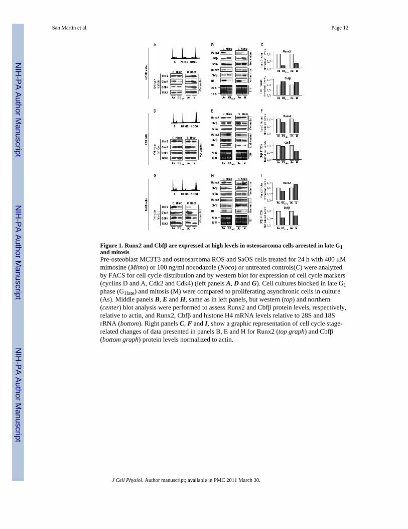
Figure 1. Runx2 and Cbfβ are expressed at high levels in osteosarcoma cells arrested in late G1and mitosisPre-osteoblast MC3T3 and osteosarcoma ROS and SaOS cells treated for 24 h with 400 μMmimosine (Mimo) or 100 ng/ml nocodazole (Noco) or untreated controls(C) were analyzedby FACS for cell cycle distribution and by western blot for expression of cell cycle markers(cyclins D and A, Cdk2 and Cdk4) (left panels A, D and G). Cell cultures blocked in late G1phase (G1late) and mitosis (M) were compared to proliferating asynchronic cells in culture(As). Middle panels B, E and H, same as in left panels, but western (top) and northern(center) blot analysis were performed to assess Runx2 and Cbfβ protein levels, respectively,relative to actin, and Runx2, Cbfβ and histone H4 mRNA levels relative to 28S and 18SrRNA (bottom). Right panels C, F and I, show a graphic representation of cell cycle stage-related changes of data presented in panels B, E and H for Runx2 (top graph) and Cbfβ(bottom graph) protein levels normalized to actin.
San Martin et al. Page 12
J Cell Physiol. Author manuscript; available in PMC 2011 March 30.
NIH
-PA Author Manuscript
NIH
-PA Author Manuscript
NIH
-PA Author Manuscript
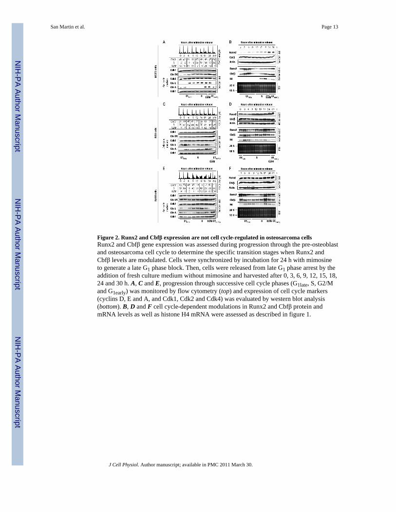
Figure 2. Runx2 and Cbfβ expression are not cell cycle-regulated in osteosarcoma cellsRunx2 and Cbfβ gene expression was assessed during progression through the pre-osteoblastand osteosarcoma cell cycle to determine the specific transition stages when Runx2 andCbfβ levels are modulated. Cells were synchronized by incubation for 24 h with mimosineto generate a late G1 phase block. Then, cells were released from late G1 phase arrest by theaddition of fresh culture medium without mimosine and harvested after 0, 3, 6, 9, 12, 15, 18,24 and 30 h. A, C and E, progression through successive cell cycle phases (G1late, S, G2/Mand G1early) was monitored by flow cytometry (top) and expression of cell cycle markers(cyclins D, E and A, and Cdk1, Cdk2 and Cdk4) was evaluated by western blot analysis(bottom). B, D and F cell cycle-dependent modulations in Runx2 and Cbfβ protein andmRNA levels as well as histone H4 mRNA were assessed as described in figure 1.
San Martin et al. Page 13
J Cell Physiol. Author manuscript; available in PMC 2011 March 30.
NIH
-PA Author Manuscript
NIH
-PA Author Manuscript
NIH
-PA Author Manuscript

Figure 3. Runx2 and Cbfβ protein levels are maintained at high levels during the cell cycle inosteosarcoma cellsRunx2 and Cbfβ gene expression in pre-osteoblast and osteosarcoma cells were quantifiedfrom data shown in figure 2. Graphic representation of cell cycle-related changes in Runx2(closed circles) and Cbfβ (closed squares) protein (A,C and E, left graphs) and mRNA (B,D and F, right graphs) levels after serum stimulation were depicted by continuous lines.The cell cycle markers cyclin D1 (open triangles), E (open diamonds) and A (open squares)proteins and histone H4 mRNA (open circles) levels are depicted by dotted lines todemarcate progression through G1late, S, G2/M and G1early phases. Protein and mRNAvalues were normalized to actin and 28S rRNA, respectively. Cell cycle phases asdetermined by flow cytometry are indicated at the base of the graphs.
San Martin et al. Page 14
J Cell Physiol. Author manuscript; available in PMC 2011 March 30.
NIH
-PA Author Manuscript
NIH
-PA Author Manuscript
NIH
-PA Author Manuscript
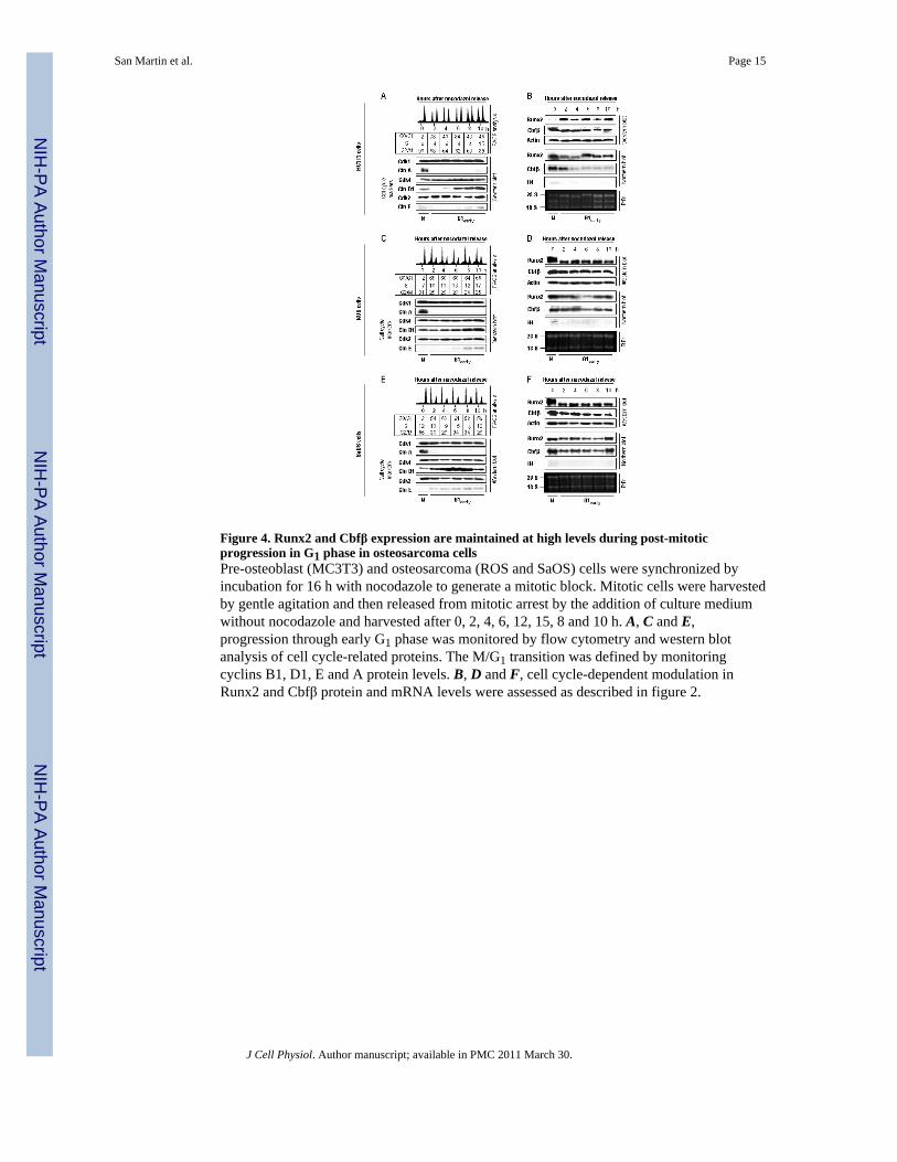
Figure 4. Runx2 and Cbfβ expression are maintained at high levels during post-mitoticprogression in G1 phase in osteosarcoma cellsPre-osteoblast (MC3T3) and osteosarcoma (ROS and SaOS) cells were synchronized byincubation for 16 h with nocodazole to generate a mitotic block. Mitotic cells were harvestedby gentle agitation and then released from mitotic arrest by the addition of culture mediumwithout nocodazole and harvested after 0, 2, 4, 6, 12, 15, 8 and 10 h. A, C and E,progression through early G1 phase was monitored by flow cytometry and western blotanalysis of cell cycle-related proteins. The M/G1 transition was defined by monitoringcyclins B1, D1, E and A protein levels. B, D and F, cell cycle-dependent modulation inRunx2 and Cbfβ protein and mRNA levels were assessed as described in figure 2.
San Martin et al. Page 15
J Cell Physiol. Author manuscript; available in PMC 2011 March 30.
NIH
-PA Author Manuscript
NIH
-PA Author Manuscript
NIH
-PA Author Manuscript

Figure 5. Elevated Runx2 and Cbfβ protein levels observed in mitosis are maintained during G1phase in osteosarcoma cellsRunx2 and Cbfβ gene expression in pre-osteoblast and osteosarcoma cells were quantifiedfrom data shown in figure 4. Graphic representation of cell cycle-related changes in Runx2(closed circles) and Cbfβ (closed squares) protein (A,C and E, left graphs) and mRNA (B,D and F, right graphs) levels after serum stimulation were depicted by continuous lines.The cell cycle markers cyclin D1 (open triangles), A (open squares) and B (open diamonds)proteins and histone H4 mRNA (open circles) levels are depicted by dotted lines todemarcate progression through M/G1 transition, G1early and S phases. Protein and mRNAvalues were normalized to actin and 28S rRNA, respectively. Cell cycle phases asdetermined by flow cytometry are indicated at the base of the graphs.
San Martin et al. Page 16
J Cell Physiol. Author manuscript; available in PMC 2011 March 30.
NIH
-PA Author Manuscript
NIH
-PA Author Manuscript
NIH
-PA Author Manuscript
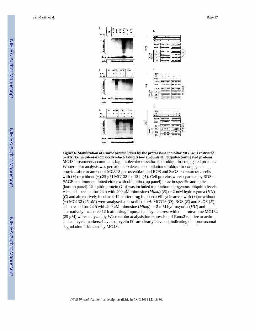
Figure 6. Stabilization of Runx2 protein levels by the proteasome inhibitor MG132 is restrictedto later G1 in osteosarcoma cells which exhibits low amounts of ubiquitin-conjugated proteinsMG132 treatment accumulates high molecular mass forms of ubiquitin-conjugated proteins.Western blot analysis was performed to detect accumulation of ubiquitin-conjugatedproteins after treatment of MC3T3 pre-osteoblast and ROS and SaOS osteosarcoma cellswith (+) or without (−) 25 μM MG132 for 12 h (A). Cell proteins were separated by SDS–PAGE and immunoblotted either with ubiquitin (top panel) or actin specific antibodies(bottom panel). Ubiquitin protein (Ub) was included to monitor endogenous ubiquitin levels.Also, cells treated for 24 h with 400 μM mimosine (Mimo) (B) or 2 mM hydroxyurea (HU)(C) and alternatively incubated 12 h after drug imposed cell cycle arrest with (+) or without(−) MG132 (25 μM) were analysed as described in A. MC3T3 (D), ROS (E) and SaOS (F)cells treated for 24 h with 400 uM mimosine (Mimo) or 2 mM hydroxyurea (HU) andalternatively incubated 12 h after drug imposed cell cycle arrest with the proteasome MG132(25 μM) were analyzed by Western blot analysis for expression of Runx2 relative to actinand cell cycle markers. Levels of cyclin D1 are clearly elevated, indicating that proteasomaldegradation is blocked by MG132.
San Martin et al. Page 17
J Cell Physiol. Author manuscript; available in PMC 2011 March 30.
NIH
-PA Author Manuscript
NIH
-PA Author Manuscript
NIH
-PA Author Manuscript
Copyright © 2022 FDOKUMEN