Carbohydrate nutrition and development of adiposity during adolescence
Upload
independentCategory
view
0download
0
RESEARCH ARTICLE
Osteoblast-Specific Deletion of Pkd2 Leadsto Low-Turnover Osteopenia and ReducedBone Marrow AdiposityZhousheng Xiao1, Li Cao1, Yingjuan Liang1, Jinsong Huang1, Amber RathStern2,3, Mark Dallas2, Mark Johnson2, Leigh Darryl Quarles1*
1. Department of Medicine, University of Tennessee Health Science Center, Memphis, Tennessee, 38165,United States of America, 2. Department of Oral Biology, School of Dentistry, University of Missouri-KansasCity, Kansas City, Missouri, 64108, United States of America, 3. Engineering Systems, Inc., Charlotte, NorthCarolina, 28227, United States of America
Abstract
Polycystin-1 (Pkd1) interacts with polycystin-2 (Pkd2) to form an interdependent
signaling complex. Selective deletion of Pkd1 in the osteoblast lineage reciprocally
regulates osteoblastogenesis and adipogenesis. The role of Pkd2 in skeletal
development has not been defined. To this end, we conditionally inactivated Pkd2 in
mature osteoblasts by crossing Osteocalcin (Oc)-Cre;Pkd2+/null mice with floxed
Pkd2 (Pkd2flox/flox) mice. Oc-Cre;Pkd2flox/null (Pkd2Oc-cKO) mice exhibited decreased
bone mineral density, trabecular bone volume, cortical thickness, mineral apposition
rate and impaired biomechanical properties of bone. Pkd2 deficiency resulted in
diminished Runt-related transcription factor 2 (Runx2) expressions in bone and
impaired osteoblastic differentiation ex vivo. Expression of osteoblast-related genes,
including, Osteocalcin, Osteopontin, Bone sialoprotein (Bsp), Phosphate-regulating
gene with homologies to endopeptidases on the X chromosome (Phex), Dentin
matrix protein 1 (Dmp1), Sclerostin (Sost), and Fibroblast growth factor 23 (FGF23)
were reduced proportionate to the reduction of Pkd2 gene dose in bone of Oc-
Cre;Pkd2flox/+ andOc-Cre;Pkd2flox/null mice. Loss of Pkd2 also resulted in diminished
peroxisome proliferator-activated receptor c (PPARc) expression and reduced bone
marrow fat in vivo and reduced adipogenesis in osteoblast culture ex vivo.
Transcriptional co-activator with PDZ-binding motif (TAZ) and Yes-associated protein
(YAP), reciprocally acting as co-activators and co-repressors of Runx2 and PPARc,
were decreased in bone of Oc-Cre;Pkd2flox/null mice. Thus, Pkd1 and Pkd2 have
coordinate effects on osteoblast differentiation and opposite effects on adipogenesis,
suggesting that Pkd1 and Pkd2 signaling pathways can have independent effects on
mesenchymal lineage commitment in bone.
OPEN ACCESS
Citation: Xiao Z, Cao L, Liang Y, Huang J, SternAR, et al. (2014) Osteoblast-Specific Deletion ofPkd2 Leads to Low-Turnover Osteopenia andReduced Bone Marrow Adiposity. PLoSONE 9(12): e114198. doi:10.1371/journal.pone.0114198
Editor: Carlos M. Isales, Georgia RegentsUniversity, United States of America
Received: August 13, 2014
Accepted: November 4, 2014
Published: December 2, 2014
Copyright: � 2014 Xiao et al. This is an open-access article distributed under the terms of theCreative Commons Attribution License, whichpermits unrestricted use, distribution, and repro-duction in any medium, provided the original authorand source are credited.
Data Availability: The authors confirm that all dataunderlying the findings are fully available withoutrestriction. All relevant data are within the paper.
Funding: This work was supported by grant R01-DK083303 to LDQ from the National Institutes ofHealth. The funders had no role in study design,data collection and analysis, decision to publish, orpreparation of the manuscript.
Competing Interests: The authors have read thejournal’s policy and the authors of this manuscripthave the following competing interests: Dr. AmberRath Stern is employed by a commercial company,Engineering Systems, Inc. This does not alter theauthors’ adherence to PLOS ONE policies onsharing data and materials.
PLOS ONE | DOI:10.1371/journal.pone.0114198 December 2, 2014 1 / 24

Introduction
PKD1 encodes polycystin-1 (PC1), a transmembrane receptor-like protein, which
links environmental clues to intracellular processes regulating cell growth and
development. PKD2 encodes polycystin-2 (PC2), a transient receptor potential
channel. The PC1 binds to PC2 in a ratio of 1 to 3 through their respective C-
terminal coiled-coiled domains to form the functional signaling polycystin
complex in cell surface membranes [1–5]. Typically, the functions of PC1 and PC2
are interdependent and concordant, as evidenced by common Autosomal
Dominant Polycystic Kidney Disease (ADPKD) phenotype caused by inactivation
of either PKD1 or PKD2 [6, 7].
Although the functions of polycystins have largely been derived from the study
of inactivating mutations of either PKD1 or PKD2 in the kidney, Pkd1 and Pkd2
are widely expressed in many tissues and cell types, including the osteoblast
lineage in bone. Recent studies indicate that PC1 (Pkd1) and PC2 (Pkd2) also
form a complex that co-localize to primary cilia in osteoblasts/osteocytes to create
a ‘‘sensor’’ that regulates bone mass [8]. Osteoblast lineage specific deletion of
Pkd1 in mice establishes a direct role for PC1 in regulating both osteoblast
development and transducing the bone response to mechanical loading [8–15].
Indeed, the selective genetic ablation of Pkd1 in osteoblasts and osteocytes results
in osteopenia that is caused by diminished osteoblast-mediated bone formation
and increased bone marrow adipogenesis. Loss of Pkd1 reciprocally decreases
Runx2 and increases expression of PPARc transcription factors that direct the
commitment of mesenchymal stem cells to the osteoblastic and adipocytic
lineages, respectively. Calcium-dependent signal transduction pathways link Pkd1
to Runx2 expression, but the cellular mechanisms mediating the reciprocal
regulation of PPARc have not been defined. The phenotype in the bone specific
Pkd1-deficient mice resembles age-related bone loss, suggesting that under-
standing the function of polycystins in bone may be important in understanding
the pathogenesis and treatment of senile osteoporosis.
Whether loss of Pkd2 function results in a bone phenotype in mice similar to
Pkd1 deficiency has not be investigated. However, recent siRNA mediated knock-
down of Pkd2 in osteoblasts resulted in impaired osteoblasts differentiation in
vitro [16]. In addition, data from a GWAS meta-analyses found that the PKD2
SNP rs12511728 was significantly associated with femoral neck bone mineral
density (BMD)[16] and mutations in PKD2 are associated with abnormal shape of
craniofacial bones in patients with ADPKD [17]. Global homozygous Pkd2null/null
mice die early in utero [18], which precludes assessing the direct effects of Pkd2
on osteoblast function.
To define the osteoblast specific functions of Pkd2, we conditionally inactivated
Pkd2 in postnatal mature osteoblasts. We found that loss of Pkd2 suppressed both
osteoblast-mediated bone formation and adipogenesis, leading to osteopenia and
decreased bone marrow fat. Thus, Pkd1 and Pkd2 have concordant effects on
osteoblastogenesis and opposite effects on adipogenesis, consistent with both
overlapping and independent signaling functions in osteoblasts.
Concordant Regulation of Runx2 and PPARc by PC2
PLOS ONE | DOI:10.1371/journal.pone.0114198 December 2, 2014 2 / 24

Materials and Methods
Animal breeding and genotyping
All animal research was conducted according to guidelines provided by the
National Institutes of Health and the Institute of Laboratory Animal Resources,
National Research Council. The University of Tennessee Health Science Center’s
Animal Care and Use Committee approved all animal studies (Protocol number:
12–160.0). The mice were anesthetized with Ketamine (90 mg/kg) and Xylazine
(10 mg/kg) for a bone densitometry scan, and the mice not useful for
experimental purposes were sacrificed by CO2 inhalation plus cervical dislocation.
We obtained the floxed Pkd2 (in exon 3) mice and heterozygous Pkd2null/+ (in
exon 1) mice from Dr. Guanqing Wu at Vanderbilt University Medical Center
[19] and Osteocalcin (Oc)-Cre mice from Dr. Thomas Clemens at University of
Alabama [20]. These mice were bred and maintained on a C57BL/6J background.
At first, we created double heterozygous Oc-Cre;Pkd2null/+ mice and homozygous
Pkd2flox/flox mice. Then double heterozygous Oc-Cre;Pkd2null/+ mice were mated
with homozygous Pkd2flox/flox mice to generate excised floxed Pkd2 heterozygous
(Oc-Cre;Pkd2flox/+) and null mice (Oc-Cre;Pkd2flox/null or Pkd2Oc-cko), as well as Pkd2
heterozygous mice (Pkd2null/flox) and Oc-Cre negative control mice (Pkd2flox/+,
equivalent to wild-type). These mice were used to collect samples at 6 weeks of age for
phenotypic analysis. For genotyping PCR and Cre-mediated recombination, genomic
DNAs were prepared from tail clips, bone, and other tissue specimens using a Tissue
PCR Kit (Sigma-Aldrich, St. Louis, MO, USA). Mice were genotyped for Oc-Cre
using previously described primers [12], for the Pkd2flox allele using forward primer
59-TCT GAC TTG CAG ACT GTG GG-39 and reverse primer 59-AGG TAG GGG
AAG GTC AGG GTT GG-39 (355 bp product for the Pkd2+ wild-type allele, 575 bp
product for the Pkd2flox floxed allele), for the Pkd2Dflox delta floxed allele using
forward primer 59-AGC TTG GCT GGA CGT AAA-39 and reverse primer 59- AGG
TAG GGG AAG GTC AGG GTT GG-39 (427 bp product for the Pkd2Dflox delta
floxed allele), and for the Pkd2null allele using two forward primers 59-GCG CCG
GCC TAG CTG TCC C-39 and 59-GTG CTA CTT CCA TTT GTC ACG TCC TGC-39
and one reverse primer 59-GTT GTC GCG GCT CCA CG-39 (150 bp product for the
Pkd2+ wild-type allele, 350 bp product for the Pkd2null null allele) as previously
described [19]. All Pkd2 alleles were identified in 2% agarose gels (Figure 1).
Bone densitometry, histomorphometric and micro-CT analysis
Bone mineral density (BMD) of femurs was assessed at 6 weeks of age using a
small animal bone densitometer (Lunar Corp, Madison, WI). Calcein (Sigma-
Aldrich, St. Louise, MO) double labeling of bone and histomorphometric analyses
of periosteal mineral apposition rate (MAR) in tibias were performed using the
osteomeasure analysis system (Osteometrics, Inc., Decatur, GA). The distal
femoral metaphyses were also scanned using a micro-CT 40 scanner (Scanco
Medical AG, Bruttisellen, Switzerland). A 3D images analysis was done to
Concordant Regulation of Runx2 and PPARc by PC2
PLOS ONE | DOI:10.1371/journal.pone.0114198 December 2, 2014 3 / 24

determine bone volume (BV/TV) and cortical thickness (Ct.Th) as previously
described [12, 21].
Three-point bending to failure
Femurs were harvested from freshly euthanized mice. The femurs (1520 slices per
femur) were scanned with a micro-CT 40 scanner (Scanco Medical AG,
Bruttisellen, Switzerland), and saved in a DICOM file format. To characterize the
biomechanical properties of the bones, displacement-controlled three-point
bending to failure tests were conducted on excised femurs ex vivo. For each femur,
all associated soft tissues were removed; the bone was individually wrapped in
saline-soaked gauze, and then stored at 220 C until testing. On the day of testing,
samples were removed from 220 C storage, and allowed to thaw and reach room
temperature. Prior to, and throughout the tests, bone samples were kept hydrated
Figure 1. Osteocalcin(Oc)-Cre-mediated bone specific deletion of Pkd2 from the floxed Pkd2 allele (Pkd2flox). (A) Schematic illustration of wild-type(Pkd2+), null (Pkd2null, deleted Exon 1), and floxed Pkd2 allele before (Pkd2flox) and after deletion (Pkd2Dflox) of the lox P cassette containing Exon 3 via Cre-mediated recombination. (B) Genotype PCR analysis of different tissues that were harvested from 6-week-old Oc-Cre;Pkd2flox/null mice showed bonespecific deletion of the Pkd2 gene. Osteocalcin-Cre-mediated recombination of excised floxed Pkd2 (Pkd2Dflox) allele occurred exclusively in bone, whereasnon-skeletal tissues retained the floxed Pkd2 allele (Pkd2flox). (C and D) Real-time RT-PCR analysis of total Pkd2 and Pkd1 transcripts. Expression of totalPkd2 transcripts was performed using Pkd2- or Pkd1-allele-specific primers as described in Experimental Procedures. The normal Pkd2+ vs cyclophilin A isnormalized to the mean ratio of 5 control mice, which has been set to 1. The percentage of conditional and global deleted transcripts was calculated from therelative levels of the normal Pkd2+ transcripts in different Pkd2 exons. Data are mean ¡S.D. from 6–8 individual mice. Values sharing the same superscriptin C and D are not significantly different at P,0.05.
doi:10.1371/journal.pone.0114198.g001
Concordant Regulation of Runx2 and PPARc by PC2
PLOS ONE | DOI:10.1371/journal.pone.0114198 December 2, 2014 4 / 24

with saline soaked gauze and not allowed to dry out. To allow unbiased
comparison between the samples, consistent span lengths of 6mm and crosshead
displacement rates of 0.1 mm/s were used during testing. The femurs were placed
into the test fixtures so they would be impacted in what would be the anterior to
posterior direction in vivo (ElectroForce 3200, Bose Corp., Minnetonka, MN).
Crosshead displacement and axial load were recorded at a rate of 70 Hz. The
stiffness and ultimate force were calculated from the resulting load versus
displacement curves for each sample. The Young’s modulus (E) for each bone was
calculated using the following equation:
E~SL3
48I
Where S is the stiffness, L is the span length, and I is the area moment of inertia.
The area moment of inertia was calculated across the midspan at the fracture
location using 10 slices midspan of the realigned microCT scans using the BoneJ
plugin for the image-processing program ImageJ (Bone J, NIH, Bethesda, MD,
USA) [8, 22, 23].
Bone microindentation testing (BMT)
The BMT was performed using a microindentation device (ActiveLife Tech, Inc.,
Santa Barbara, CA, USA) as previously described [24–26]. Briefly, the periosteum
of isolated femurs was scratched and a probe assembly placed on the anterior
surface of the mid-femur performed measurements. A 10-cycle indentation with a
maximum 2N force and 2 Hz frequency at a touchdown force of 0.1N and
without any preconditioning was performed and the average value of three
measurements was recorded. The indentation distances were analyzed by specific
software and four parameters were obtained to use as outcome variables:
indentation distance increase (IDI) between the first and last indentation cycle;
total distance between the bone surface and the last indentation cycle (Total ID);
creep indentation distance (Creep ID), the progressive indentation distance
during the stable force phase of the first indentation cycle at the maximum 2N
force; and average unloading slope, the average slope of unloading portion during
three measurements. IDI values were then normalized by the IDI of
polymethylmethacrylate (PMMA) tested with the same probe. Throughout testing
samples were kept moist with 1xPBS.
Detection of bone marrow adipocytes in long bones by oil red O
lipid staining and micro-CT analysis
Whole intact femurs and tibias with encapsulated marrow were dissected from 20
weeks old mice, fixed for 48 hours in phosphate buffered paraformaldehyde,
decalcified in 14% EDTA for two weeks. For oil red O lipid staining, the tibias
were embedded in tissue freezing medium. Cryosectioning was performed on a
frozen sectioning device (Leica, Nussloch, Germany) equipped with a rapid frozen
Concordant Regulation of Runx2 and PPARc by PC2
PLOS ONE | DOI:10.1371/journal.pone.0114198 December 2, 2014 5 / 24

sectioning kit (Instrumedics, Hackensack, NJ). 10 mm thick sections were then
stained with oil red O (ORO) for bone marrow adipocytes as previously described
[9, 12]. Briefly, the sections were rinsed in 60% isopropanol, stained for 20
minutes in 0.5% ORO-isopropanol solution, differentiated in 60% isopropanol,
rinsed in tap water and mounted in glycerin jelly. Sections were examined with an
inverted fluorescent microscope equipped with a digital camera (Leica, Nussloch,
Germany). For detection of bone marrow fat cells by Micro-CT, the femurs were
stained for 2 hours in 2% aqueous osmium tetroxide (OsO4). The Bones were
rinsed in water for 48 hours and then scanned at 6 mm resolution using a micro-
CT 40 scanner, 45 KeVp and 177 mA. Quantification of fat volume, density, and
distribution throughout the marrow was registered to low contrast decalcified
bone as our Lab previously described [9, 12].
Real-time RT-PCR
For quantitative real-time RT2PCR, 1.0 mg total RNA isolated from either the
long bone of 6-week-old mice or 10-days cultured primary osteoblasts in
differentiation media was reverse transcribed as previously described [9, 27]. PCR
reactions contained 100 gg template (cDNA or RNA), 300 gM each forward and
reverse primers, and 1XqPCR Supermix (Bio-Rad, Hercules, CA) in 50 ml. The
threshold cycle (Ct) of tested-gene product from the indicated genotype was
normalized to the Ct for cyclophilin A. Expression of total Pkd2 transcripts was
performed using the following Pkd2-allele-specific primers: In exon 3, forward
primer of normal Pkd2+ transcript: 59-GCA TGA TGA GCT CCA ATG TG-39, and
reverse primer: 59-TCG ACA CTG GGG TGT CTA TG -39. In exon 1, forward
primer of normal Pkd2+ transcript: 59-TTG AGG CAG AGG AGG ATG AC-39,
and reverse primer: 59-CAT CCA TCT CTA CCA CCA TCC-39. The normal
Pkd2+ vs cyclophilin A is normalized to the mean ratio of 5 control mice, which
has been set to 1. The percentage of conditional and global deleted transcripts was
calculated from the relative levels of the normal Pkd2+ transcripts in different Pkd2
exons.
Serum biochemistry
Serum urea nitrogen (BUN) was determined using a BUN diagnostic kit from
Pointe Scientific, Inc. Calcium was measured using a calcium cresolphthalein
complexone kit (Stanbio Laboratories, Boerne, TX, USA) and phosphorus was
measured using the phosphomolybdylate-ascorbic acid method, as previously
described [9, 11, 28]. Serum Osteocalcin levels were measured using a mouse
Osteocalcin EIA kit (Biomedical Technologies Inc. Stoughton, MA, USA). Serum
parathyroid hormone (PTH) levels were measured using the Mouse Intact PTH
ELISA kit (Immutopics, Carlsbad, CA, USA). Serum full-length FGF23 levels were
measured using the FGF23 ELISA kit (Kainos Laboratories, Tokyo, Japan). Serum
OPG and Rank ligand (RankL) were measured using mouse ELISA kits
(Quantikine, R&D Systems, Minneapolis, MN, USA), and serum tartrate resistant
Concordant Regulation of Runx2 and PPARc by PC2
PLOS ONE | DOI:10.1371/journal.pone.0114198 December 2, 2014 6 / 24

acid phosphatase (TRAP) was assayed with a mouse TRAP ELISA kit
(Immunodiagnostic Systems, Fountain Hills, AZ).
Primary osteoblast culture for proliferation, differentiation, and
western blot analysis
Calvaria from E17.5 control and Pkd2-deficient embryos were used to isolate
primary osteoblasts by sequential collagenase digestion at 37 C. To engineer
immortal osteoblast cell lines, isolated primary osteoblasts were infected using a
retroviral vector carrying SV40 large and small T antigen as previously described
[10, 29]. Briefly, cells were grown in 100-mm plates at 50–60% confluence the day
before infection. On the day of infection, the medium was removed and replaced
with medium containing SV40 large and small T antigen-helper-free viral
supernatant in the presence of 4 mg/ml of polybrene (Sigma, St. Louis, MO, USA)
for 48 h. The cells were allowed to recover for 72 h followed by selection with
1 mg/ml puromycin (Sigma-Aldrich) for up to 15 days. The immortalized
osteoblasts were cultured in a-MEM containing 10% FBS and 1% penicillin and
streptomycin (P/S) and characterized following the protocols below. Cell
proliferation was detected by BrdU incorporation assays following the
manufacturer’s directions (QIA58, Calbiochem, Gibbstown, NJ, USA). To induce
differentiation, the immortalized osteoblasts were plated at a density of 26104
cells per well in a 12-well plate and 46104 cells per well in a 6-well plate and
grown up to 21 days in a-MEM containing 10% FBS supplemented with 5 mM b-
glycerophosphate and 25 mg/ml ascorbic acid. ALP activity and Alizarin red-S
histochemical staining for mineralization were performed as previously described
[10, 29]. Total DNA content was measured with a double-strand DNA
quantitation reagent and kit (Molecular Probes, Eugene, OR, USA). Protein
concentrations of the supernatant were determined with a total protein assay kit
(Bio-Rad, Hercules, CA, USA). For signaling mechanism study, the cells were
cultured in the osteogenic differentiation media for 48 hours. Then the cells were
lysed with 150 ml of tissue protein extraction reagent (Pierce Biotechnology,
Rockford, IL, USA) with 1 x Halt protease inhibitor and 1 mM phenylmetha-
nesulfonyl fluoride (PMSF) per well, After three 30-second sonications, total cell
lysates were centrifuged at 13,0006g for 10 minutes and supernatants were stored
at -80 C until use. Protein concentrations of the supernatant were determined
with a total protein assay kit (Bio-Rad, Hercules, CA). Equal quantities of protein
were subjected to 4–12% Bis-Tris gradient Gels (Invitrogen, Carlsbad, CA) and
were analyzed with standard western blot protocols (HRP-conjugated secondary
antibodies from Santa Cruz Biotechnology and ECL chemiluminescent immu-
nodetection system from GE Healthcare Bio-Sciences). Anti-YAP/TAZ (D24E4,
#8418) and Anti-pYAP antibody (Ser127) (D9W2I, #13008) were purchased
from Cell Signaling Technologies (Danvers, MA).Purified mouse anti-TAZ
(560236) was purchased from BD Biosciences (San Jose, CA). Anti-p-TAZ (Ser
89) (sc-17610) and Anti-b-actin (sc-47778) antibodies were from Santa Cruz
Concordant Regulation of Runx2 and PPARc by PC2
PLOS ONE | DOI:10.1371/journal.pone.0114198 December 2, 2014 7 / 24
Biotechnology (Paso Robles, CA). The intensity of bands was quantified using
Image J software (http://rsb.info.nih.gov/ij/).
Transient transfection
The immortalized osteoblasts were cultured in a-MEM containing 10% fetal
bovine serum (FBS) and 1% penicillin/streptomycin (P/S). To examine if PC2
regulates Hippo signaling pathway, a number of 16106 of the immortalized
osteoblasts were transfected with 3.0 mg of 8xGTIIc luciferase reporter (8xGTIIc-
Luc) constructs in combination with 3.0 mg of empty pcDNA3.1 expression
vector, and 0.6 mg of Renilla luciferase-null (RL-null) as internal control plasmid
by electroporation using a cell line optimal transfection kit according to the
manufacturer’s protocol (Amaxa Inc, Gaithersburg, MD). A total of 6.6 mg of
plasmid DNAs was used for each electroporation. The transfected cells were plated
in 12-well plates and harvested in 32 hours after transfection. Cells were lysed in
100 ml of reporter lysis buffer (Promega, Madison, WI). A luciferase assay (20 ml
of cell lysed) was performed using a dual luciferase assay kit (Promega, Madison,
WI), and activity was measured with a tube luminometer (MGM Instruments,
Inc., Hamden, CT).
Statistics
We evaluated differences between groups by one-way analysis of variance. All
values are expressed as means ¡ S.D. All computations were performed using a
commercial biostatistics software (GraphPad Software Inc. La Jolla, CA).
Results
Oc-Cre mediated bone-specific deletion of Pkd2
We studied four genotypes from the breeding strategy, including Oc-Cre;Pkd2flox/null
or Pkd2Oc-cKO, Oc-Cre;Pkd2flox/+, Pkd2flox/null, and Pkd2flox/+. These mice were born at
the expected Mendelian frequency and all genotypes exhibited survival indis-
tinguishable from wild-type mice over the period of study. Oc-Cre expression is
limited to cells of the osteoblast lineage (late osteoblasts.osteocytes) with onset of
expression just before birth [20]. To confirm that the Pkd2 floxed allele in Exon 3 was
selectively deleted in bone, we performed PCR analysis using a combination of
primers that specifically detect floxed Pkd2 alleles (Pkd2flox) and the excised floxed
Pkd2 alleles (Pkd2Dflox) in Oc-Cre;Pkd2flox/+ or Oc-Cre;Pkd1flox/null mice (Figure 1A).
We demonstrated that Oc-Cre-mediated floxed recombination occurred exclusively
in bone, whereas non-skeletal tissues retained the intact floxed Pkd2 alleles (Pkd2flox)
(Figure 1B). The Pkd2null null allele in Exon 1 was used in combination with the Cre-
recombinase deletion of the floxed Pkd2 allele (Pkd2Dflox) in Exon 3 to increase the net
efficiency of Pkd2 inactivation.
The level of the full-length Pkd2 transcripts in Exon 1 and 3 from the femurs of
these four genotypes mice were assessed by real time RT-PCR. As expected, both
Concordant Regulation of Runx2 and PPARc by PC2
PLOS ONE | DOI:10.1371/journal.pone.0114198 December 2, 2014 8 / 24

Pkd2flox/null and Oc-Cre;Pkd2flox/null mice displayed 50% reduction of intact Pkd2
transcripts in Exon 1, whereas Oc-Cre;Pkd2flox/+ and Oc-Cre;Pkd2flox/null mice
exhibited approximately 25% excision of the floxed Exon 3 from Pkd2, indicating
that Oc-Cre mediated bone-specific deletion of the floxed Pkd2 allele is
incomplete (Figure 1C). The combined effect Pkd2null and (Pkd2Dflox) in Oc-
Cre;Pkd2flox/null, however, resulted in a net reduction of intact Pkd2 expression by
,75% in bone (Figure 1C). Thus, there was a progressive reduction of functional
Pkd2 message in these Pkd2 deficient mice, i.e., Pkd2flox/+ (100%), Oc-Cre;Pkd2flox/+
(75%), Pkd2flox/null (50%), and Oc-Cre;Pkd2flox/null (25%) mice (Figure 1C). In
contrast, targeting Pkd2 had no effect on Pkd1 transcript expression (Figure 1D). In
addition, Oc-Cre;Pkd2flox/null, and Oc-Cre;Pkd2flox/+ demonstrated no cyst forma-
tion in the kidney, consistent with the bone specific inactivation of Pkd2 (data not
shown). Pkd2flox/null mice also exhibited no renal cyts, as previously reported [18].
Effects of bone specific deletion of Pkd2 on bone structure
At 6 weeks of age, the gross appearance and body weight of all genotypes were
indistinguishable. Compared to control Pkd2flox/+ mice, conditional heterozygous
Oc-Cre;Pkd2flox/+ mice, which had a 25% reduction in Pkd2 expression, exhibited
no abnormalities in bone mass. However, both global heterozygous Pkd2flox/null
and conditional Pkd2Oc-cKO null mice, which had respective 50% and 75%
reductions in Pkd2 express, were osteopenic, as evidenced by respective 14.4%
verse 9.2% and 12.5% verse 9.2% reductions in BMD in both male and female
adult mice (Figure 2A). Micro-CT analysis revealed that the reduction in bone
mass in both male global heterozygous Pkd2flox/null and conditional Pkd2Oc-cKO
null mice were caused by reductions in trabecular bone volume (19.3% and
30.9%, respectively) and cortical bone thickness (8.4% and 7.3%, respectively)
(Figure 2B). In contrast, conditional heterozygous Oc-Cre;Pkd2flox/+ had no
significant difference in both trabecular and cortical bone compared with control
mice (Figure 2B). Consistent with a low-bone-mass phenotype by BMD and
Micro-CT analysis, we found that loss of Pkd2 in bone from both Pkd2flox/null and
Pkd2Oc-cKO null mice was associated with a decrease in periosteal mineral
apposition rate (MAR, 25.1% and 37.3%, respectively) (Figure 2C), indicating a
significant reduction of bone formation rate in Pkd2 deficient mice.
Effect of conditional deletion of Pkd2 on femur cortical bone
geometry, mechanical and material properties
Conditional deletion of Pkd2 also resulted in site-specific alterations of femur
cortical bone geometry. In this regard, single conditional Oc-Cre;Pkd2flox/+
heterozygous, single global Pkd2flox/null heterozygous, and conditional Pkd2Oc-cKO
null mice showed a gene dose dependent decrease in both total and cortical area at
6 weeks of age when compared with age-matched control Pkd2flox/+ mice
(Table 1), this is in accordance with a significant reduction in moment of inertia,
Ix in these Pkd2 deficient mice (Table 1). Obviously, Pkd2 deficient mice displayed
Concordant Regulation of Runx2 and PPARc by PC2
PLOS ONE | DOI:10.1371/journal.pone.0114198 December 2, 2014 9 / 24

Figure 2. Pkd2 deficiency results in loss of bone mass. (A) Effects of Pkd2 deficiency on bone mineraldensity (BMD) at 6 weeks of age. Compared to control Pkd2flox/+ mice, conditional heterozygous Oc-Cre;Pkd2flox/+ mice exhibited no abnormalities in bone mass. However, both global heterozygous Pkd2flox/null
and conditional Pkd2Oc-cKO null mice were osteopenic, as evidenced by respective 14.4% and 12.5%reduction BMD in male adult mice. (B) Effects of Pkd2 deficiency on structure of femurs. mCT analysis of thedistal femoral metaphyses and midshaft diaphyses revealed that the reduction in bone mass in both global
Concordant Regulation of Runx2 and PPARc by PC2
PLOS ONE | DOI:10.1371/journal.pone.0114198 December 2, 2014 10 / 24

a reduction in the size of the marrow cavity resulting from a smaller midshaft
diameter compared with control mice. To examine whether changes of femoral
bone geometry in the Pkd2 deficient mice may affect bone mechanical properties,
we used these femurs to perform 3-point bending to failure experiments.
Compared with the control mice, global Pkd2flox/null heterozygous and conditional
Pkd2Oc-cKO null mice exhibited a lower maximum force and stiffness in 3-point
bending, but no significant differences in the Young’s modulus (Table 1),
indicating that the changes of bone geometry and bone structure at 6 weeks of age
may preserve the Young’s modulus in global Pkd2flox/null heterozygous and
conditional Pkd2Oc-cKO null mice. There was no difference in these parameters
between single conditional Oc-Cre;Pkd2flox/+ heterozygous mice and control mice
(Table 1).
To assess whether Pkd2 deficiency may have an impact on bone material
properties, we used these femurs to perform reference point indentation (RPI,
micro-indentation) measurements. We found significant differences in several
RPI variables (Table 1) between Pkd2 deficient and control mice. There was a
226%, 82.5%, and 162% increase in IDI, TID, and CID, respectively, between
heterozygous Pkd2flox/null and control mice. A similar 190%, 69%, and 118%
increase in IDI, TID, and CID, respectively, was also observed between Pkd2Oc-cKO
null and control mice. In contrast, there was a 33.3% decrease in the average US in
both global Pkd2flox/null heterozygous and conditional Pkd2Oc-cKO null mice
compared with control mice. However, these parameters were not significantly
different between heterozygous Oc-Cre;Pkd2flox/+ and control mice (Table 1).
These findings that Pkd2 deficiency makes bone more brittle and less stiff.
Pkd2-regulated gene expression in bone
Next, we examined by real-time RT-PCR the expression levels of a panel of
osteoblast lineage-, osteoclast-, chondrocyte-, and adipocyte-related mRNAs from
the femurs of 6-week-old control, heterozygous Pkd2 deficient (Oc-Cre;Pkd2flox/+
and Pkd2flox/null), and Oc-Cre;Pkd2flox/null mice (Table 2). We found that changes
in bone transcripts were more sensitive to reductions in Pkd2 and exhibited a
gene-dose dependent response on osteoblast-related gene transcripts.
We found significant alterations in osteoblast-related gene expression. Bone
derived from heterozygous Oc-Cre;Pkd2flox/+, in spite of having no measurable
change in bone structure and only a 25% reduction in Pkd2 message expression,
heterozygous Pkd2flox/null and conditional Pkd2Oc-cKO null mice were caused by reductions in trabecular bonevolume (19.3% and 30.9%, respectively) and cortical bone thickness (8.4% and 7.3%, respectively). Incontrast, conditional heterozygous Oc-Cre;Pkd2flox/+ had no significant difference in both trabecular andcortical bone compared with control mice. (C) Effects of Pkd2 deficiency on bone formation rate. Both Pkd2flox/null
and Pkd2Oc-cKO null mice exhibited a significant decrease in periosteal mineral apposition rate (MAR, 25.1% and37.3%, respectively), indicating a reduction of bone formation rate in Pkd2 deficient mice. Data represent themean¡S.D. from 6–8 individual mice. Values sharing the same superscript in different group are not significantlydifferent at P,0.05.
doi:10.1371/journal.pone.0114198.g002
Concordant Regulation of Runx2 and PPARc by PC2
PLOS ONE | DOI:10.1371/journal.pone.0114198 December 2, 2014 11 / 24

did have significant reductions in several osteoblast-lineage gene transcripts,
including Osteocalcin, Osteopontin, and Cyclooxygenase 2 (Cox2) mRNA levels
compared to control mice. Bone from heterozygous Pkd1flox/null mice with a 50%
reduction in Pkd2 expression exhibited significant reductions in Runx2-II,
Osteocalcin, Osteopontin, Bsp, Cox2, matrix metalloproteinase 13 (Mmp13), matrix
extracellular phosphoglycoprotein (Mepe), and Sost mRNA levels compared to
control mice (Table 2). Compared with heterozygous Pkd2flox/null mice,
significantly greater reductions of Osteocalcin, Osteopontin, Cox2, Connexin 43
(Cnx43), Rank ligand (RankL), Phex, Dmp1 and Fgf23 were observed in
conditional Pkd2Oc-cKO null mice that had a 75% reduction in Pkd2 expression.
Changes in gene expression in bone correlated with alterations in serum
biomarkers in conditional Pkd2Oc-cKO null mice. In this regard, further evidence
for osteoblast-lineage dysfunction includes reductions in both Fgf23 and RanKL
in serum from conditional Pkd2Oc-cKO null mice at 6 weeks of age (Table 3).
However, no changes of serum phosphorus and calcium levels were observed in
Pkd2Oc-cKO null mice compared to control mice. Also, no significant changes of
serum BUN, PTH, Osteocalcin, and OPG were observed among these four
genotype mice (Table 3).
The Opg/RankL expression ratio was increased in conditional Pkd2Oc-cKO null
mice and was associated with reduced bone expression of Trap and matrix
metalloproteinase 9 (Mmp9), markers of bone resorption (Table 2). Serum levels
of TRAP were also reduced in conditional Pkd2Oc-cKO null mice compared to
control littermates (Table 3), suggesting that Pkd2-mediated bone loss results
from low bone formation rates rather than increased bone resorption. Transcripts
of chondrocyte-related genes did not differ between heterozygous Pkd2 deficient
and Pkd2Oc-cKO null mice (Table 2).
Table 1. Femoral bone geometry, biomechanical, and biomaterial properties in 6-week-old mice.
Parameters Pkd2flox/+ Oc-Cre;Pkd2flox/+ Pkd2flox/null Pkd2Oc-cKO p-value
Cortical bone Geometry
Total area (mm2) 2.5¡0.19a 2.3¡0.17b 2.1¡0.27c 2.1¡0.20c ,0.0001
Cortical area (mm2) 0.61¡0.06a 0.56¡0.06a,b 0.52¡0.09b 0.52¡0.05b 0.0017
Moment of inertia, Ix (mm4) 0.15¡0.04a 0.11¡0.03b 0.10¡0.02b 0.11¡0.03b 0.0007
Biomechanical properties
Maximum force (N) 12.0¡0.97a 10.8¡1.2a,b 8.9¡0.94b 9.4¡1.6b 0.0077
Stiffness (N/mm) 60.2¡9.1a 62.0¡3.4a 46.9¡5.6b 37.1¡8.3b 0.0001
Young’s modulus (Gpa) 2.9¡1.0a 2.9¡0.59a 3.4¡0.52a 2.9¡0.72a 0.5743
Biomaterial properties
IDI (mm) 7.7¡1.6a 9.7¡2.2a 25.1¡3.2b 22.3¡5.3b 0.0053
Total ID (mm) 39.4¡4.5a 47.6¡10.6a 71.9¡7.1b 66.7¡7.7b 0.0015
Creep ID (mm) 4.5¡0.84a 5.3¡0.82a 11.8¡1.3b 9.8¡3.2b 0.0003
Average US (N/mm) 0.18¡0.04a 0.18¡0.02a 0.12¡0.03b 0.12¡0.02b 0.0077
Data are mean ¡S.D. from 6-8 serum samples of 6-week-old individual mice. Values sharing the same superscript are not significantly different at P,0.05.
doi:10.1371/journal.pone.0114198.t001
Concordant Regulation of Runx2 and PPARc by PC2
PLOS ONE | DOI:10.1371/journal.pone.0114198 December 2, 2014 12 / 24

PPARc, an adipocyte transcription factor, and adipocyte markers, including
lipoprotein lipase (Lpl) and adipocyte fatty acid-binding protein 2 (aP2) were
significantly decreased in femurs of in both heterozygous Pkd2flox/null and
conditional Pkd2Oc-cKO null mice (Table 2). Consistent with attenuated
adipogenic markers, bone marrow exhibited an decreased percentage of fat cells in
both Pkd2flox/null and Pkd2Oc-cKO mice at 20 weeks of age, as evidenced by a lower
Table 2. Gene-expression profiles in bone in 6-week-old mice.
Gene Accession no. Oc-Cre;Pkd2flox/+ Pkd2flox/null Oc-Cre; Pkd2flox/null p-value
Osteoblast lineage
Runx2-II NM_009820.5 1.03¡0.23 0.64¡0.14*,# 0.46¡0.28*,# 0.0005
Osteopontin NM_009263.3 0.79¡0.19* 0.71¡0.15* 0.45¡0.15*,#,& ,0.0001
Osteocalcin NM_007541.2 0.76¡0.16* 0.77¡0.16* 0.51¡0.13*,#,& ,0.0001
Bsp NM_008318.3 0.87¡0.12 0.68¡ 0.12* 0.37¡0.24*,# ,0.0001
Cox2 NM_011198.3 0.76¡0.18* 0.77¡0.16* 0.50¡0.15*,#,& 0.0001
Cnx43 NM_010288.3 0.93¡0.23 0.95¡0.18 0.41¡0.18*,#,& ,0.0001
Mmp13 NM_008607.2 0.87¡0.31 0.66¡ 0.21* 0.44¡0.18*,# 0.0004
Opg NM_008764.3 0.98¡0.24 0.87¡0.31 0.84¡0.27 0.5987
RankL NM_011613.3 0.85¡0.18 0.94¡0.19 0.56¡0.21*,#,& 0.0014
Phex NM_011077.2 0.90¡0.41 0.97¡0.39 0.43¡0.14*,#,& 0.0040
Dmp1 NM_016779.2 0.91¡0.27 0.79¡0.23 0.52¡0.21*,#,& 0.0014
Mepe NM_053172.2 1.10¡0.40 0.72¡0.12*,# 0.56¡0.20*,# 0.0017
Sost NM_024449.5 0.98¡0.35 0.68¡0.12*,# 0.58¡0.18*,# 0.0012
Fgf23 NM_022657.4 0.92¡0.33 0.88¡ 0.38 0.63¡0.18* 0.0576
Wnt10b NM_011718.2 0.92¡0.42 0.71¡0.10* 0.35¡0.24*,#,& ,0.0001
Axin2 NM_015732.4 0.94¡0.17 0.72¡0.26* 0.38¡0.13*,#,& ,0.0001
Taz NM_1168281.1 0.75¡0.22* 0.72¡ 0.16* 0.42¡0.24*,#,& 0.0002
Yap NM_009534.3 1.05¡0.11 0.74¡0.17*,# 0.53¡0.34*,# 0.0003
BirC3 NM_007464.3 1.00¡0.18 0.72¡0.18*,# 0.65¡0.18*,# 0.0006
Ctgf NM_010217.2 0.97¡0.33 0.66¡0.18*,# 0.45¡0.21*,# ,0.0001
Inhba NM_008380.1 0.98¡0.21 0.71¡0.16*,# 0.69¡0.11*,# 0.0002
Hprt NM_013556.2 1.07¡0.15 0.95¡0.23 1.08¡0.41 0.7324
Osteoclast
Trap NM_007388.3 0.79¡0.28 0.85¡0.29 0.46¡0.21*,#,& 0.0005
Mmp9 NM_013599.3 0.91¡0.32 0.99¡0.31 0.56¡0.14*,#,& 0.0032
Chondrocyte
Collagen II NM_031163.3 0.91¡0.16 0.90¡0.33 1.29¡0.0.71 0.2935
VegfA NM_009505.4 1.15¡0.21 1.10¡0.29 0.99¡0.37 0.7891
Adipocyte
PPARc NM_009505.4 1.03¡0.32 0.70¡0.15*,# 0.57¡0.17*,# 0.0008
aP2 NM_024406.2 1.16¡0.31 0.98¡0.27 0.64¡0.37*,#,& 0.0157
Lpl NM_008509.2 0.77¡0.22 0.58¡0.27* 0.66¡0.29* 0.0136
Data are mean ¡S.D. from 5–6 tibias of 6-week-old individual mice and expressed as the fold changes relative to the housekeeping gene cyclophilin Asubsequently normalized to control mice. * indicates significant difference from control Pkd2flox/+ mice, # indicates significant difference from Oc-Cre;Pkd2flox/+ mice, & indicates significant difference from Pkd2null/flox mice at p,0.05, respectively.
doi:10.1371/journal.pone.0114198.t002
Concordant Regulation of Runx2 and PPARc by PC2
PLOS ONE | DOI:10.1371/journal.pone.0114198 December 2, 2014 13 / 24

number of adipocytes and fat droplets in decalcified femurs and tibias stained
with Oil Red O (Figure 3A) and Osmium tetroxide (OsO4) (Figure 3B-3C),
indicating an impairment of adipogenesis in Pkd2 deficient mice. Thus, the effect
of selective deletion of Pkd2 in osteoblasts on adipogenesis is opposite to the effect
of Pkd1 deficiency.
Effect of conditional deletion of Pkd2 on osteoblastic function exvivo
We confirmed that there was a gene dose-dependent reduction of Pkd2 transcripts
in osteoblasts derived from control Pkd2flox/+, heterozygous Oc-Cre;Pkd2flox/+ and
Pkd2flox/null, and Pkd2Oc-cKO mice (Figure 4A). To determine the impact of
conditional deleted Pkd2 on osteoblast function ex vivo, we examined cell
proliferation and osteoblastic differentiation and gene expression profiles in
immortalized primary osteoblast cultures derived from control Pkd2flox/+,
heterozygous Oc-Cre;Pkd2flox/+ and Pkd2flox/null, and Pkd2Oc-cKO null mice.
Consistent with defects in bone formation, we found that Pkd2 deficiency
osteoblasts had a higher BrdU incorporation than control osteoblasts, indicating
an increased proliferation rate in these Pkd2 deficiency osteoblasts (Figure 4B). In
addition, both heterozygous Pkd2flox/null and Pkd1Oc-cKO null osteoblasts displayed
impaired osteoblastic differentiation and maturation, as evidenced by lower
alkaline phosphatase activity, diminished calcium deposition in extracellular
matrix, and reduced osteoblastic differentiation markers such as Runx2-II
compared to controls (Figure 4C-4E). In agreement with decreased adipogenic
activity in vivo, the cultured immortalized primary osteoblasts under osteogenic
condition exhibited a markedly decrease of adipocyte markers such as PPARc2 in
both heterozygous Pkd2flox/null and Pkd1Oc-cKO null osteoblast cultures
(Figure 4F), suggesting an impairment of both osteogenesis and adipogenesis in
the Pkd2 deficient osteoblasts.
Table 3. Biochemistry analysis in 6-week-old mice.
Parameters Pkd2flox/+ Oc-Cre;Pkd2flox/+ Pkd2flox/null Pkd2Oc-cKO p-value
BUN (mg/dl) 18¡3.2a 19¡3.8a 18¡2.7a 17¡2.3a 0.5972
FGF23 (pg/ml) 127¡27a 125¡23a 108¡29a 73¡36b 0.0066
PTH (pg/ml) 44¡19a 54¡26a 64¡23a 32¡15a 0.0714
P (mg/dl) 10.3¡0.90a 11.0¡1.01a 11.4¡0.98a 11.1¡0.67a 0.1229
Ca (mg/dl) 8.7¡0.08a 8.7¡0.11a 8.7¡0.17a 8.7¡0.15a 0.4528
Osteocalin (ng/dl) 124¡104a 92¡67a 122¡41a 104¡59a 0.7657
OPG (ng/ml) 3.9¡0.71a 3.9¡0.48a 3.5¡0.95a 3.9¡0.51a 0.5027
RanKL (pg/dl) 83¡14a 81¡21a 77¡20a 54¡17b 0.0346
TRAP (U/L) 19¡1.8a 18¡1.9a 19¡3.7a 15¡1.8b 0.0338
1,25(OH)2D (pg/ml) 59¡8.1a 57¡10.3a 56¡5.4a 55¡11.3a 0.8022
Data are mean ¡S.D. from 6-8 serum samples of 6-week-old individual mice. Values sharing the same superscript are not significantly different at P,0.05.
doi:10.1371/journal.pone.0114198.t003
Concordant Regulation of Runx2 and PPARc by PC2
PLOS ONE | DOI:10.1371/journal.pone.0114198 December 2, 2014 14 / 24

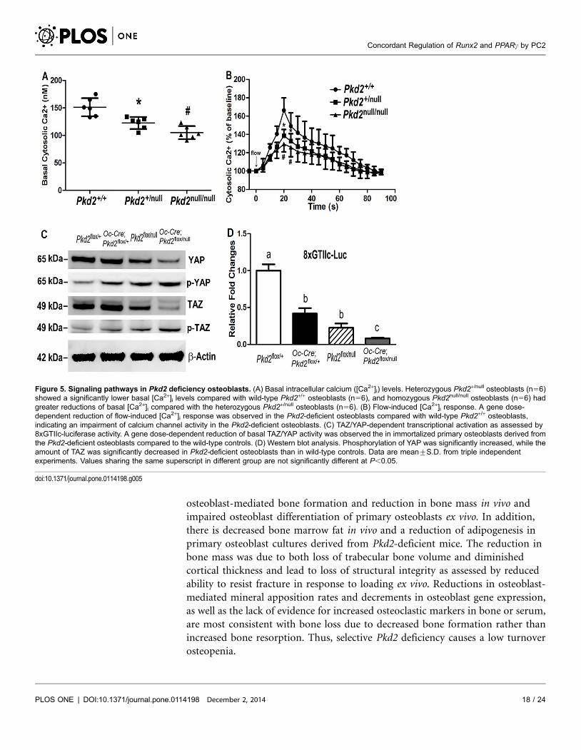
Effects of Pkd2 deficiency on signaling pathways in osteoblasts
We found that Pkd2 deficiency had a gene dose effect on basal intracellular
calcium ([Ca2+]i) concentration and flow-induced intracellular calcium response
in immortalized Pkd2-deficient osteoblasts. In this regard, heterozygous Pkd2null/+
osteoblasts showed a significantly lower basal intracellular calcium ([Ca2+]i)
concentration compared with wild type Pkd2+/+ cells, and homozygous Pkd2null/
null osteoblasts had greater reductions of basal [Ca2+]i compared with the
heterozygous Pkd2null/+ cells (Figure 5A). To study whether PC2-mediated
Figure 3. Pkd2 deficiency impairs adipogenesis in bone. (A) Histology of adipocytes in decalcified tibias.Oil Red O staining showed that the numbers of adipocytes and fat droplets in tibial bone marrow were muchless in 20-week-old Pkd2flox/null and Pkd2Oc-cKO null mice compared with age-matched control Pkd2flox/+ andOc-Cre;Pkd2flox/+ mice. (B) Osmium tetroxide (OsO4) staining of decalcified femurs by mCT analyses. Upperpanel showed the representative images of distal femoral bone marrow by OsO4 staining (yellow). Lowerpanel displayed adipocyte volume/marrow volume (Ad.V/Ma.V, %) and adipocyte number (Ad.N, mm23) bycalculation. Consistent with Oil Red O staining, mCTanalyses showed that adipocyte volume/marrow volume(Ad.V/Ma.V, %) and adipocyte number (Ad.N, mm23) were much lower in the distal femurs from 20-week-oldPkd2flox/null and Pkd2Oc-cKO null mice compared with age-matched control Pkd2flox/+ and Oc-Cre;Pkd2flox/+
mice, indicating an impairment of adipogenesis in the Pkd2 deficient mice. Data represent the mean¡S.D.from 6–8 individual mice. Values sharing the same superscript in different group are not significantly differentat P,0.05.
doi:10.1371/journal.pone.0114198.g003
Concordant Regulation of Runx2 and PPARc by PC2
PLOS ONE | DOI:10.1371/journal.pone.0114198 December 2, 2014 15 / 24

Figure 4. Pkd2 deficiency osteoblasts have a developmental defect in vitro. (A) A real-time RT-PCR analysis of total Pkd2 transcripts in osteoblastcultures. A gene dose-dependent reduction of Pkd2 transcripts was observed in immortalized control and Pkd2-deficient osteiblasts. (B) BrdU incorporation.Primary cultured Pkd2 deficient osteoblasts exhibited a higher BrdU incorporation than control Pkd2flox/+ osteoblasts for 6 hours, indicating increasedproliferation rate in the Pkd2-deficient osteoblasts. (C) ALP activity. Primary cultured Pkd2flox/null and Pkd2Oc-cKO null osteoblasts displayed time-dependentincrements in alkaline phosphatase (ALP) activities during 15 days of culture, but the ALP activity was significantly lower at different time points comparedwith control Pkd2flox/+ and Oc-Cre;Pkd2flox/+ osteoblasts. (D) Quantification of mineralization. Alizarin Red-S was extracted with 10% cetylpyridinium chlorideand quantified as described in Experimental Procedures. Primary cultured Pkd2flox/null and Pkd2Oc-cKO null had time-dependent increments in Alizarin Red-Saccumulation during 22 days of culture, but the accumulation was significantly lower at different time points compared with control Pkd2flox/+ and Oc-Cre;Pkd2flox/+ osteoblasts. (E and F) Gene expression profiles by real-time RT-PCR. 10-days cultured Pkd2flox/null and Pkd2Oc-cKO null osteoblasts inosteogenic differentiation media showed a significant attenuation in both osteogenesis and adipogenesis compared to control Pkd2flox/+ and Oc-Cre;Pkd2flox/+, evidenced by a significant reduction in osteoblastic and adipogenic markers, such as Runx2-II and PPARc2. Data are mean¡S.D. from triplethree independent experiments. Values sharing the same superscript in different group are not significantly different at P,0.05.
doi:10.1371/journal.pone.0114198.g004
Concordant Regulation of Runx2 and PPARc by PC2
PLOS ONE | DOI:10.1371/journal.pone.0114198 December 2, 2014 16 / 24

mechanical flow-induced intracellular calcium level changed, these immortalized
cells were exposed to 6.24 dynes/cm2 pulsatile laminar fluid flow. On fluid
stimulation, we detected an immediate rise in intracellular calcium throughout
the wild type Pkd2+/+ cell population, peaking roughly 10–20 s after stimulation
(Figure 5B). The [Ca2+]i levels then rapidly decreased but were maintained at
moderate levels for 50–60 s before returning to baseline. In contrast, when we
exposed these Pkd2-deficient osteoblasts to an identical flow stimulus, we detected
intermediate calcium response curve in the heterozygous cells and greater
reduction of calcium influx in either the peak or late phase in the homozygous
osteoblasts (Figure 5B). In addition, 100 nM triptolide stimulated normal
calcium influx in wild type Pkd2+/+ cells, moderate calcium signals in
heterozygous Pkd2null/+ cells, and no response in Pkd2 null cells (data not shown)
in the loading chamber, consistent with loss of PC2 abolishes PC2 agonist-
induced calcium response in osteoblasts.
To explore mechanisms of decreased Runx2 and PPARc expression in Pkd2-
deficient mice, we looked for alterations in the Hippo signaling pathway effectors
YAP/TAZ, which play a critical roles in regulating mesenchymal stem cell fate
determination into osteoblasts and adipocytes in response to alterations in
extracellular matrix rigidity and cell shape [30]. We found that loss of Pkd2 in
osteoblasts resulted in significant reduction in TAZ, YAP, and their transcriptional
targets, baculoviral IAP repeat containing 3 (Birc-3), connective tissue growth
factor (Ctgf), and inhibin beta A (Inhba) (Table 2). Western blot analysis revealed
that phosphorylation of YAP and TAZ were significantly increased (indicating
retention of p-YAP and p-TAZ in the cytoplasm) (Figure 5C). We also found that
the total amount of YAP and TAZ were significantly decreased in Pkd2-deficient
osteoblasts (Figure 5C). To examine the effect of Pkd2 inactivation on TAZ/YAP
transcriptional activity, we examined a reporter gene construct with multiple
copies of the TEAD-binding GTIIC (GGAATG) site (8xGTIIC-luciferase). YAP
and TAZ are coactivators of the transcription factor TEAD, which can be used as a
read out of YAP/TAZ activity. GTIIC-luciferase activity was reduced in a Pkd2
gene dose-dependent manner in immortalized primary osteoblasts derived from
the Pkd2-deficient osteoblasts compared to the wild-type controls (Figure 5D).
These findings indicated that loss of PC2 significantly attenuates both YAP and
TAZ components of Hippo signaling pathway in immortalized primary osteoblasts.
In addition, Sost is expressed in osteocytes and regulates bone mass through
Wnt-dependent signaling. We looked for evidence of activation of Wnt signaling
pathway, since Sost message expression was decreased in Pkd2 deficient bone.
However, we observed a decrease in Wnt10b and Axin2, suggesting that loss of
Pkd2 resulted in decreased Wnt signaling, in spite of reductions in Sost.
Discussion
Using an Osteocalcin (Oc) promoter-driven Cre and Pkd2flox/null mouse model, we
demonstrate that osteoblast-specific deletion of Pkd2 results in decreased
Concordant Regulation of Runx2 and PPARc by PC2
PLOS ONE | DOI:10.1371/journal.pone.0114198 December 2, 2014 17 / 24
osteoblast-mediated bone formation and reduction in bone mass in vivo and
impaired osteoblast differentiation of primary osteoblasts ex vivo. In addition,
there is decreased bone marrow fat in vivo and a reduction of adipogenesis in
primary osteoblast cultures derived from Pkd2-deficient mice. The reduction in
bone mass was due to both loss of trabecular bone volume and diminished
cortical thickness and lead to loss of structural integrity as assessed by reduced
ability to resist fracture in response to loading ex vivo. Reductions in osteoblast-
mediated mineral apposition rates and decrements in osteoblast gene expression,
as well as the lack of evidence for increased osteoclastic markers in bone or serum,
are most consistent with bone loss due to decreased bone formation rather than
increased bone resorption. Thus, selective Pkd2 deficiency causes a low turnover
osteopenia.
Figure 5. Signaling pathways in Pkd2 deficiency osteoblasts. (A) Basal intracellular calcium ([Ca2+]i) levels. Heterozygous Pkd2+/null osteoblasts (n56)showed a significantly lower basal [Ca2+]i levels compared with wild-type Pkd2+/+ osteoblasts (n56), and homozygous Pkd2null/null osteoblasts (n56) hadgreater reductions of basal [Ca2+]i compared with the heterozygous Pkd2+/null osteoblasts (n56). (B) Flow-induced [Ca2+]i response. A gene dose-dependent reduction of flow-induced [Ca2+]i response was observed in the Pkd2-deficient osteoblasts compared with wild-type Pkd2+/+ osteoblasts,indicating an impairment of calcium channel activity in the Pkd2-deficient osteoblasts. (C) TAZ/YAP-dependent transcriptional activation as assessed by8xGTIIc-luciferase activity. A gene dose-dependent reduction of basal TAZ/YAP activity was observed the in immortalized primary osteoblasts derived fromthe Pkd2-deficient osteoblasts compared to the wild-type controls. (D) Western blot analysis. Phosphorylation of YAP was significantly increased, while theamount of TAZ was significantly decreased in Pkd2-deficient osteoblasts than in wild-type controls. Data are mean¡S.D. from triple independentexperiments. Values sharing the same superscript in different group are not significantly different at P,0.05.
doi:10.1371/journal.pone.0114198.g005
Concordant Regulation of Runx2 and PPARc by PC2
PLOS ONE | DOI:10.1371/journal.pone.0114198 December 2, 2014 18 / 24

Consistent with PC1 coupling to PC2, both Oc-Cre-mediated reductions of
Pkd1 and Pkd2 transcripts in osteoblasts cause a low bone mass due to decreased
osteoblast-mediated bone formation. However, there are a few notable differences
in the bone phenotype of these conditional Pkd1 and Pkd2 deficient mice,
suggesting that PC1 and PC2 functions are not identical. In this regard, Pkd1
deficiency in osteoblasts resulted in a more robust gene-dose dependent effects
than observed in targeted deletion of Pkd2 in osteoblasts [12]. A greater effect of
PC1 compared to PC2 is also seen in other settings. For example, mutations in
Pkd1 cause a more severe cystic kidney phenotype than mutations in Pkd2 [31]. In
addition, Pkd1 deficient mice have an inverse effect on osteoblastic and adipocytic
differentiation, such that decreased osteoblastic function and osteopenia were
associated with a reciprocal enhancement of adipogensis and increased bone
marrow fat. In contrast, there was a concordant reduction in osteoblastogenesis
and adipogenesis in Pkd2 deficient mice. These differences might reflect the
differential and broader signaling pathways coupled to Pkd1 compared to Pkd2.
Indeed, Pkd1 deficiency resulted in a decrease in Runx2 and an increase in PPARc,
whereas reductions in Pkd2 resulted in parallel reductions of Runx2 and PPARc.
We have previously shown that Pkd1 regulation of bone mass is mediated by
Runx2 [21]. Compound Runx2 and Pkd1 heterozygous mice have additive effects
on reduction of bone mass. Also, Pkd1 effects on Runx2 promoter activity are
mediated through coupling to Pkd2 calcium channel activity and regulation of
calcium signaling pathways. In this regard, Pkd1 deficient osteoblasts had lower
intracellular calcium and Pkd1 responsive enhancer regions of the Runx2
promoter were identified in an area containing AP-1 and NFI binding sites.
shRNA-mediated reductions in PKD1 in MG-63 osteoblasts also reduced
intracellular calcium, attenuated calcium signaling response to shear fluid stress,
and increased cAMP responses [27]. It is likely that reduction in Runx2 message
expression in Pkd2 deficient osteoblasts is related to alterations in similar
intracellular calcium signaling. Indeed, osteoblasts derived from Pkd2-deficient
osteoblasts exhibited lower basal intracellular calcium ([Ca2+]i) and impaired
response to flow-induced intracellular calcium influx, indicating that calcium
channel PC2 is coupled to fluid flow sensing PC1 to response to mechanical
loading osteoblasts.
The differential effects of selective Pkd1 and Pkd2 deficiency in osteoblasts on
bone marrow adipogenesis suggests that PC1 and PC2 signaling can be uncoupled
in bone. This possibility led us to investigate the potential interactions between
polycystins and YAP and TAZ, components of the Hippo signaling pathway. TAZ/
YAP, like polycystins, are regulated by mechanical and cytoskeletal cues [30, 32–
36]. In addition, YAP/TAZ differentially regulates mesenchymal precursors
toward osteoblastic and adipocytic cell fates [32]. Specifically, TAZ acts as a co-
activator of Runx2 and a direct inhibitor of the transcriptional activity of PPARc[37, 38]. Indeed, TAZ overexpression in osteoblasts stimulates Runx2 expression,
osteoblast differentiation and increases bone mass [39]. YAP, on the other hand,
acts as a co-repressor of Runx2 and co-activator of PPARc transcriptional activity
[40–42]. PKA-induced adipogenesis also involves cAMP dependent phosphor-
Concordant Regulation of Runx2 and PPARc by PC2
PLOS ONE | DOI:10.1371/journal.pone.0114198 December 2, 2014 19 / 24

ylation of YAP [43]. Finally, TAZ binds to PC2 leading to its degradation and to
PC1 to both modify its interactions with PC2 and possibly enhance nuclear
translocation and transcriptional functions of the PC1 C-terminal tail [44]. We
observed reductions in both TAZ and YAP in Pkd2-deficient mouse bone. PC-2-
depedendent stimulation of TAZ could work in concert with calcium- and PC-1-
C-terminal tail dependent stimulation of Runx2 to stimulate osteoblastogenesis;
whereas PC-2 stimulation of YAP and promotion of PPARc activity could
stimulate adipogenesis (Figure 6). In this schema, activation of PC-1 results in
stimulation of osteoblastogenesis and inhibition of adipogenesis through
coordinate effects on Runx2 and TAZ signaling. Further studies are needed to
investigate this schema and establish the regulation and functional roles of TAZ
and YAP in polycystin control of osteoblast and adipocyte differentiation.
Interestingly, the reduction in YAP in Pkd2 deficient mice is opposite to the up-
regulation of YAP in ADPKD [45], suggesting differences in tissue specific
regulation of Hippo signaling.
In addition, we observed that both Fgf23 transcripts in bone and serum Fgf23
levels were decreased in Pkd2 deficient mice, consistent with the effects of loss of
Pkd2 to suppress osteoblast/osteocyte functions. Recent studies, however, show
that that serum FGF23 levels are increased in ADPKD patients [46–48] and in a
PKD orthologous mouse model [9]. These results imply that the observed FGF23
elevation is due to effects of chronic kidney disease (CKD) to increase FGF23,
possibly due to systemic and/or local factors that stimulate FGF23 production or
end-organ resistance to FGF23 action in CKD [49]. Alternatively, FGF23 mRNA
and protein expression was detected in cell lining renal cysts, but not in bone of
Figure 6. Schema showing potential interactions between polycystins and Hippo signaling pathwaysin osteoblasts. PC-2 coordinately regulates PPARc and Runx2 to respectively control adipogenesis andosteoblastogenesis. Hippo signaling effectors Yap and Taz are also coordinately regulated by PC-2 as well asother physical forces that act as co-factors for PPARc and Runx2. Inverse effects on osteoblast and adipocytedifferentiation by PC-1 might be explained by uncoupling PC-1 and PC-2 signaling leading to enhancement ofPC-1 C-terminal tail (PC-1-CTT)/Taz signaling and increased Runx2-dependent osteoblastogenesis anddecreased PPARc-mediated adipogenesis.
doi:10.1371/journal.pone.0114198.g006
Concordant Regulation of Runx2 and PPARc by PC2
PLOS ONE | DOI:10.1371/journal.pone.0114198 December 2, 2014 20 / 24

the cy/+ Han:SPRD rat model of PKD; similar finding was also observed in an
inducible Pkd1 knockout mouse model, suggesting ectopic expression of FGF23 in
CKD [49]. The Col4a3 null mouse model of CKD also exhibited increased
circulating FGF23 levels prior to increased expression of FGF23 in bone [50, 51].
Further studies are needed to understand the mechanism leading to increased
FGF23 in CKD, but our studies indicate that specific loss of Pkd2 in bone results
in decreased FGF23 expression, along with other gene products produced by
osteoctyes.
In conclusion, osteoblast-targeted deletion of Pkd2 result in significant bone
loss associated with impaired osteoblastic and adipocytic differentiation.
Reductions in intracellular calcium and alterations in the YAP/TAZ transcrip-
tional modulators of mesenchymal stem cell differentiation are downstream
mediators linking Pkd2 and regulation of Runx2 and PPARc expression. These
findings contrast with prior studies showing that Pkd1 deficiency results in
reciprocal downregulation of Runx2-mediated osteoblastogenesis and upregula-
tion of PPARc-mediated adipogenesis. Thus, Pkd1 and Pkd2 separately regulate
osteogenic and adipogenic pathways. Polycystins in partnership with the Hippo
signaling pathway in osteoblasts may have the fundamental function of
maintaining the osteoblast differentiation state and by regulating adipocytes
differentiation/transdifferentiation in response to environmental factors that
include mechanical loading.
Acknowledgments
We are particularly grateful to Dr. Guanqing Wu at Vanderbilt University Medical
Center for providing the floxed Pkd2 mice. We also thank Active Life Scientific for
their technical support during this study.
Author ContributionsConceived and designed the experiments: ZSX ARS MJ LDQ. Performed the
experiments: ZSX LC YJL JSH MD. Analyzed the data: ZSX JSH ARS. Contributed
reagents/materials/analysis tools: ZSX ARS MJ LDQ. Wrote the paper: ZSX ARS
LDQ.
References
1. Qian F, Germino FJ, Cai Y, Zhang X, Somlo S, et al. (1997) PKD1 interacts with PKD2 through aprobable coiled-coil domain. Nat Genet 16: 179–183.
2. Tsiokas L, Kim E, Arnould T, Sukhatme VP, Walz G (1997) Homo- and heterodimeric interactionsbetween the gene products of PKD1 and PKD2. Proc Natl Acad Sci U S A 94: 6965–6970.
3. Hanaoka K, Qian F, Boletta A, Bhunia AK, Piontek K, et al. (2000) Co-assembly of polycystin-1 and -2produces unique cation-permeable currents. Nature 408: 990–994.
4. Yu Y, Ulbrich MH, Li MH, Buraei Z, Chen XZ, et al. (2009) Structural and molecular basis of theassembly of the TRPP2/PKD1 complex. Proc Natl Acad Sci U S A 106: 11558–11563.
Concordant Regulation of Runx2 and PPARc by PC2
PLOS ONE | DOI:10.1371/journal.pone.0114198 December 2, 2014 21 / 24
5. Zhu J, Yu Y, Ulbrich MH, Li MH, Isacoff EY, et al. (2011) Structural model of the TRPP2/PKD1 C-terminal coiled-coil complex produced by a combined computational and experimental approach. ProcNatl Acad Sci U S A 108: 10133–10138.
6. Nauli SM, Alenghat FJ, Luo Y, Williams E, Vassilev P, et al. (2003) Polycystins 1 and 2 mediatemechanosensation in the primary cilium of kidney cells. Nat Genet 33: 129–137.
7. Nauli SM, Rossetti S, Kolb RJ, Alenghat FJ, Consugar MB, et al. (2006) Loss of polycystin-1 inhuman cyst-lining epithelia leads to ciliary dysfunction. J Am Soc Nephrol 17: 1015–1025.
8. Xiao Z, Dallas M, Qiu N, Nicolella D, Cao L, et al. (2011) Conditional deletion of Pkd1 in osteocytesdisrupts skeletal mechanosensing in mice. FASEB J 25: 2418–2432.
9. Qiu N, Xiao Z, Cao L, David V, Quarles LD (2012) Conditional mesenchymal disruption of pkd1 resultsin osteopenia and polycystic kidney disease. PLoS One 7: e46038.
10. Qiu N, Xiao Z, Cao L, Buechel MM, David V, et al. (2012) Disruption of Kif3a in osteoblasts results indefective bone formation and osteopenia. J Cell Sci 125: 1945–1957.
11. Qiu N, Cao L, David V, Quarles LD, Xiao Z (2010) Kif3a deficiency reverses the skeletal abnormalitiesin Pkd1 deficient mice by restoring the balance between osteogenesis and adipogenesis. PLoS One 5:e15240.
12. Xiao Z, Zhang S, Cao L, Qiu N, David V, et al. (2010) Conditional disruption of Pkd1 in osteoblastsresults in osteopenia due to direct impairment of bone formation. J Biol Chem 285: 1177–1187.
13. Xiao Z, Zhang S, Magenheimer BS, Luo J, Quarles LD (2008) Polycystin-1 regulates skeletogenesisthrough stimulation of the osteoblast-specific transcription factor Runx2-II. J Biol Chem.
14. Xiao Z, Zhang S, Mahlios J, Zhou G, Magenheimer BS, et al. (2006) Cilia-like structures andpolycystin-1 in osteoblasts/osteocytes and associated abnormalities in skeletogenesis and Runx2expression. J Biol Chem 281: 30884–30895.
15. Xiao ZS, Zhang SQ, Magenheimer BS, Calvet JP, Quarles LD (2007) Polycystin-1 slective activationof Runx2-II isoform transcription is mediated through the calcium-PI3K/Akt pathway J Bone Miner Res22: S41.
16. Mesner LD, Ray B, Hsu YH, Manichaikul A, Lum E, et al. (2014) Bicc1 is a genetic determinant ofosteoblastogenesis and bone mineral density. J Clin Invest.
17. Khonsari RH, Ohazama A, Raouf R, Kawasaki M, Kawasaki K, et al. (2013) Multiple postnatalcraniofacial anomalies are characterized by conditional loss of polycystic kidney disease 2 (Pkd2). HumMol Genet 22: 1873–1885.
18. Wu G, Markowitz GS, Li L, D’Agati VD, Factor SM, et al. (2000) Cardiac defects and renal failure inmice with targeted mutations in Pkd2. Nat Genet 24: 75–78.
19. Kim I, Ding T, Fu Y, Li C, Cui L, et al. (2009) Conditional mutation of Pkd2 causes cystogenesis andupregulates beta-catenin. J Am Soc Nephrol 20: 2556–2569.
20. Zhang M, Xuan S, Bouxsein ML, von Stechow D, Akeno N, et al. (2002) Osteoblast-specific knockoutof the insulin-like growth factor (IGF) receptor gene reveals an essential role of IGF signaling in bonematrix mineralization. J Biol Chem 277: 44005–44012.
21. Xiao Z, Zhang S, Magenheimer BS, Luo J, Quarles LD (2008) Polycystin-1 regulates skeletogenesisthrough stimulation of the osteoblast-specific transcription factor RUNX2-II. J Biol Chem 283: 12624–12634.
22. Schneider CA, RasbandWS, Eliceiri KW (2012) NIH Image to ImageJ: 25 years of image analysis. NatMethods 9: 671–675.
23. Doube M, Klosowski MM, Arganda-Carreras I, Cordelieres FP, Dougherty RP, et al. (2010) BoneJ:Free and extensible bone image analysis in ImageJ. Bone 47: 1076–1079.
24. Diez-Perez A, Guerri R, Nogues X, Caceres E, Pena MJ, et al. (2010) Microindentation for in vivomeasurement of bone tissue mechanical properties in humans. J Bone Miner Res 25: 1877–1885.
25. Aref M, Gallant MA, Organ JM, Wallace JM, Newman CL, et al. (2013) In vivo reference pointindentation reveals positive effects of raloxifene on mechanical properties following 6 months oftreatment in skeletally mature beagle dogs. Bone 56: 449–453.
Concordant Regulation of Runx2 and PPARc by PC2
PLOS ONE | DOI:10.1371/journal.pone.0114198 December 2, 2014 22 / 24

26. Gallant MA, Brown DM, Organ JM, Allen MR, Burr DB (2013) Reference-point indentation correlateswith bone toughness assessed using whole-bone traditional mechanical testing. Bone 53: 301–305.
27. Qiu N, Zhou H, Xiao Z (2012) Downregulation of PKD1 by shRNA results in defective osteogenicdifferentiation via cAMP/PKA pathway in human MG-63 cells. J Cell Biochem 113: 967–976.
28. Xiao Z, Awad HA, Liu S, Mahlios J, Zhang S, et al. (2005) Selective Runx2-II deficiency leads to low-turnover osteopenia in adult mice. Dev Biol 283: 345–356.
29. Xiao ZS, Hjelmeland AB, Quarles LD (2004) Selective deficiency of the "bone-related" Runx2-IIunexpectedly preserves osteoblast-mediated skeletogenesis. J Biol Chem 279: 20307–20313.
30. Dupont S, Morsut L, Aragona M, Enzo E, Giulitti S, et al. (2011) Role of YAP/TAZ inmechanotransduction. Nature 474: 179–183.
31. Harris PC, Torres VE (1993) Polycystic Kidney Disease, Autosomal Dominant. In: Pagon RA, AdamMP, Ardinger HH, Bird TD, Dolan CR, et al., editors. GeneReviews(R). Seattle (WA).
32. Low BC, Pan CQ, Shivashankar GV, Bershadsky A, Sudol M, et al. (2014) YAP/TAZ asmechanosensors and mechanotransducers in regulating organ size and tumor growth. FEBS Lett.
33. Halder G, Dupont S, Piccolo S (2012) Transduction of mechanical and cytoskeletal cues by YAP andTAZ. Nat Rev Mol Cell Biol 13: 591–600.
34. Aragona M, Panciera T, Manfrin A, Giulitti S, Michielin F, et al. (2013) A mechanical checkpointcontrols multicellular growth through YAP/TAZ regulation by actin-processing factors. Cell 154: 1047–1059.
35. Gumbiner BM, Kim NG (2014) The Hippo-YAP signaling pathway and contact inhibition of growth.J Cell Sci 127: 709–717.
36. Yu FX, Zhao B, Panupinthu N, Jewell JL, Lian I, et al. (2012) Regulation of the Hippo-YAP pathway byG-protein-coupled receptor signaling. Cell 150: 780–791.
37. Hong JH, Hwang ES, McManus MT, Amsterdam A, Tian Y, et al. (2005) TAZ, a transcriptionalmodulator of mesenchymal stem cell differentiation. Science 309: 1074–1078.
38. Jung H, Lee MS, Jang EJ, Ahn JH, Kang NS, et al. (2009) Augmentation of PPARgamma-TAZinteraction contributes to the anti-adipogenic activity of KR62980. Biochem Pharmacol 78: 1323–1329.
39. Yang JY, Cho SW, An JH, Jung JY, Kim SW, et al. (2013) Osteoblast-targeted overexpression of TAZincreases bone mass in vivo. PLoS One 8: e56585.
40. Zaidi SK, Sullivan AJ, Medina R, Ito Y, van Wijnen AJ, et al. (2004) Tyrosine phosphorylation controlsRunx2-mediated subnuclear targeting of YAP to repress transcription. EMBO J 23: 790–799.
41. Westendorf JJ (2006) Transcriptional co-repressors of Runx2. J Cell Biochem 98: 54–64.
42. Chen SN, Gurha P, Lombardi R, Ruggiero A, Willerson JT, et al. (2014) The hippo pathway isactivated and is a causal mechanism for adipogenesis in arrhythmogenic cardiomyopathy. Circ Res 114:454–468.
43. Yu FX, Zhang Y, Park HW, Jewell JL, Chen Q, et al. (2013) Protein kinase A activates the Hippopathway to modulate cell proliferation and differentiation. Genes Dev 27: 1223–1232.
44. Tian Y, Kolb R, Hong JH, Carroll J, Li D, et al. (2007) TAZ promotes PC2 degradation through aSCFbeta-Trcp E3 ligase complex. Mol Cell Biol 27: 6383–6395.
45. Happe H, van der Wal AM, Leonhard WN, Kunnen SJ, Breuning MH, et al. (2011) Altered Hipposignalling in polycystic kidney disease. J Pathol 224: 133–142.
46. Pavik I, Jaeger P, Ebner L, Poster D, Krauer F, et al. (2012) Soluble klotho and autosomal dominantpolycystic kidney disease. Clin J Am Soc Nephrol 7: 248–257.
47. Pavik I, Jaeger P, Kistler AD, Poster D, Krauer F, et al. (2011) Patients with autosomal dominantpolycystic kidney disease have elevated fibroblast growth factor 23 levels and a renal leak of phosphate.Kidney Int 79: 234–240.
48. Mekahli D, Bacchetta J (2013) From bone abnormalities to mineral metabolism dysregulation inautosomal dominant polycystic kidney disease. Pediatr Nephrol 28: 2089–2096.
Concordant Regulation of Runx2 and PPARc by PC2
PLOS ONE | DOI:10.1371/journal.pone.0114198 December 2, 2014 23 / 24

49. Spichtig D, Zhang H, Mohebbi N, Pavik I, Petzold K, et al. (2014) Renal expression of FGF23 andperipheral resistance to elevated FGF23 in rodent models of polycystic kidney disease. Kidney Int 85:1340–1350.
50. Dai B, David V, Alshayeb HM, Showkat A, Gyamlani G, et al. (2012) Assessment of 24,25(OH)2Dlevels does not support FGF23-mediated catabolism of vitamin D metabolites. Kidney Int 82: 1061–1070.
51. Dai B, David V, Martin A, Huang J, Li H, et al. (2012) A comparative transcriptome analysis identifyingFGF23 regulated genes in the kidney of a mouse CKD model. PLoS One 7: e44161.
Concordant Regulation of Runx2 and PPARc by PC2
PLOS ONE | DOI:10.1371/journal.pone.0114198 December 2, 2014 24 / 24
Copyright © 2022 FDOKUMEN