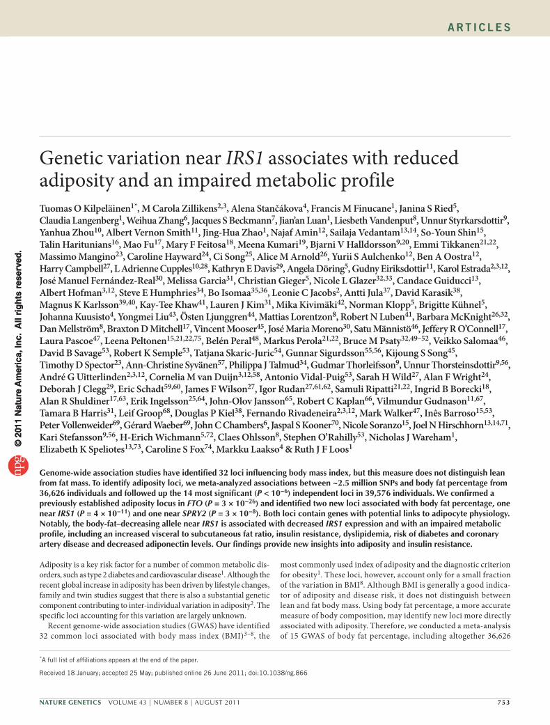
Genetic variation near IRS1 associates with reduced adiposity and an impaired metabolic profile
11
NATURE GENETICS VOLUME 43 | NUMBER 8 | AUGUST 2011 753 Adiposity is a key risk factor for a number of common metabolic dis- orders, such as type 2 diabetes and cardiovascular disease 1 . Although the recent global increase in adiposity has been driven by lifestyle changes, family and twin studies suggest that there is also a substantial genetic component contributing to inter-individual variation in adiposity 2 . The specific loci accounting for this variation are largely unknown. Recent genome-wide association studies (GWAS) have identified 32 common loci associated with body mass index (BMI) 3–8 , the most commonly used index of adiposity and the diagnostic criterion for obesity 1 . These loci, however, account only for a small fraction of the variation in BMI 8 . Although BMI is generally a good indica- tor of adiposity and disease risk, it does not distinguish between lean and fat body mass. Using body fat percentage, a more accurate measure of body composition, may identify new loci more directly associated with adiposity. Therefore, we conducted a meta-analysis of 15 GWAS of body fat percentage, including altogether 36,626 Genetic variation near IRS1 associates with reduced adiposity and an impaired metabolic profile Tuomas O Kilpeläinen 1* , M Carola Zillikens 2,3 , Alena Stančákova 4 , Francis M Finucane 1 , Janina S Ried 5 , Claudia Langenberg 1 , Weihua Zhang 6 , Jacques S Beckmann 7 , Jian’an Luan 1 , Liesbeth Vandenput 8 , Unnur Styrkarsdottir 9 , Yanhua Zhou 10 , Albert Vernon Smith 11 , Jing-Hua Zhao 1 , Najaf Amin 12 , Sailaja Vedantam 13,14 , So-Youn Shin 15 , Talin Haritunians 16 , Mao Fu 17 , Mary F Feitosa 18 , Meena Kumari 19 , Bjarni V Halldorsson 9,20 , Emmi Tikkanen 21,22 , Massimo Mangino 23 , Caroline Hayward 24 , Ci Song 25 , Alice M Arnold 26 , Yurii S Aulchenko 12 , Ben A Oostra 12 , Harry Campbell 27 , L Adrienne Cupples 10,28 , Kathryn E Davis 29 , Angela Döring 5 , Gudny Eiriksdottir 11 , Karol Estrada 2,3,12 , José Manuel Fernández-Real 30 , Melissa Garcia 31 , Christian Gieger 5 , Nicole L Glazer 32,33 , Candace Guiducci 13 , Albert Hofman 3,12 , Steve E Humphries 34 , Bo Isomaa 35,36 , Leonie C Jacobs 2 , Antti Jula 37 , David Karasik 38 , Magnus K Karlsson 39,40 , Kay-Tee Khaw 41 , Lauren J Kim 31 , Mika Kivimäki 42 , Norman Klopp 5 , Brigitte Kühnel 5 , Johanna Kuusisto 4 , Yongmei Liu 43 , Östen Ljunggren 44 , Mattias Lorentzon 8 , Robert N Luben 41 , Barbara McKnight 26,32 , Dan Mellström 8 , Braxton D Mitchell 17 , Vincent Mooser 45 , José Maria Moreno 30 , Satu Männistö 46 , Jeffery R O’Connell 17 , Laura Pascoe 47 , Leena Peltonen 15,21,22,75 , Belén Peral 48 , Markus Perola 21,22 , Bruce M Psaty 32,49–52 , Veikko Salomaa 46 , David B Savage 53 , Robert K Semple 53 , Tatjana Skaric-Juric 54 , Gunnar Sigurdsson 55,56 , Kijoung S Song 45 , Timothy D Spector 23 , Ann-Christine Syvänen 57 , Philippa J Talmud 34 , Gudmar Thorleifsson 9 , Unnur Thorsteinsdottir 9,56 , André G Uitterlinden 2,3,12 , Cornelia M van Duijn 3,12,58 , Antonio Vidal-Puig 53 , Sarah H Wild 27 , Alan F Wright 24 , Deborah J Clegg 29 , Eric Schadt 59,60 , James F Wilson 27 , Igor Rudan 27,61,62 , Samuli Ripatti 21,22 , Ingrid B Borecki 18 , Alan R Shuldiner 17,63 , Erik Ingelsson 25,64 , John-Olov Jansson 65 , Robert C Kaplan 66 , Vilmundur Gudnason 11,67 , Tamara B Harris 31 , Leif Groop 68 , Douglas P Kiel 38 , Fernando Rivadeneira 2,3,12 , Mark Walker 47 , Inês Barroso 15,53 , Peter Vollenweider 69 , Gérard Waeber 69 , John C Chambers 6 , Jaspal S Kooner 70 , Nicole Soranzo 15 , Joel N Hirschhorn 13,14,71 , Kari Stefansson 9,56 , H-Erich Wichmann 5,72 , Claes Ohlsson 8 , Stephen O’Rahilly 53 , Nicholas J Wareham 1 , Elizabeth K Speliotes 13,73 , Caroline S Fox 74 , Markku Laakso 4 & Ruth J F Loos 1 Genome-wide association studies have identified 32 loci influencing body mass index, but this measure does not distinguish lean from fat mass. To identify adiposity loci, we meta-analyzed associations between ~2.5 million SNPs and body fat percentage from 36,626 individuals and followed up the 14 most significant (P < 10 −6 ) independent loci in 39,576 individuals. We confirmed a previously established adiposity locus in FTO (P = 3 × 10 −26 ) and identified two new loci associated with body fat percentage, one near IRS1 (P = 4 × 10 −11 ) and one near SPRY2 (P = 3 × 10 −8 ). Both loci contain genes with potential links to adipocyte physiology. Notably, the body-fat–decreasing allele near IRS1 is associated with decreased IRS1 expression and with an impaired metabolic profile, including an increased visceral to subcutaneous fat ratio, insulin resistance, dyslipidemia, risk of diabetes and coronary artery disease and decreased adiponectin levels. Our findings provide new insights into adiposity and insulin resistance. * A full list of affiliations appears at the end of the paper. Received 18 January; accepted 25 May; published online 26 June 2011; doi:10.1038/ng.866 ARTICLES © 2011 Nature America, Inc. All rights reserved. © 2011 Nature America, Inc. All rights reserved.
-
Upload
independent -
Category
Documents
-
view
2 -
download
0
Transcript of Genetic variation near IRS1 associates with reduced adiposity and an impaired metabolic profile
Nature GeNetics VOLUME 43 | NUMBER 8 | AUGUST 2011 753
Adiposity is a key risk factor for a number of common metabolic disorders, such as type 2 diabetes and cardiovascular disease1. Although the recent global increase in adiposity has been driven by lifestyle changes, family and twin studies suggest that there is also a substantial genetic component contributing to interindividual variation in adiposity2. The specific loci accounting for this variation are largely unknown.
Recent genomewide association studies (GWAS) have identified 32 common loci associated with body mass index (BMI)3–8, the
most commonly used index of adiposity and the diagnostic criterion for obesity1. These loci, however, account only for a small fraction of the variation in BMI8. Although BMI is generally a good indicator of adiposity and disease risk, it does not distinguish between lean and fat body mass. Using body fat percentage, a more accurate measure of body composition, may identify new loci more directly associated with adiposity. Therefore, we conducted a metaanalysis of 15 GWAS of body fat percentage, including altogether 36,626
Genetic variation near IRS1 associates with reduced adiposity and an impaired metabolic profileTuomas O Kilpeläinen1*, M Carola Zillikens2,3, Alena Stančákova4, Francis M Finucane1, Janina S Ried5, Claudia Langenberg1, Weihua Zhang6, Jacques S Beckmann7, Jian’an Luan1, Liesbeth Vandenput8, Unnur Styrkarsdottir9, Yanhua Zhou10, Albert Vernon Smith11, Jing-Hua Zhao1, Najaf Amin12, Sailaja Vedantam13,14, So-Youn Shin15, Talin Haritunians16, Mao Fu17, Mary F Feitosa18, Meena Kumari19, Bjarni V Halldorsson9,20, Emmi Tikkanen21,22, Massimo Mangino23, Caroline Hayward24, Ci Song25, Alice M Arnold26, Yurii S Aulchenko12, Ben A Oostra12, Harry Campbell27, L Adrienne Cupples10,28, Kathryn E Davis29, Angela Döring5, Gudny Eiriksdottir11, Karol Estrada2,3,12, José Manuel Fernández-Real30, Melissa Garcia31, Christian Gieger5, Nicole L Glazer32,33, Candace Guiducci13, Albert Hofman3,12, Steve E Humphries34, Bo Isomaa35,36, Leonie C Jacobs2, Antti Jula37, David Karasik38, Magnus K Karlsson39,40, Kay-Tee Khaw41, Lauren J Kim31, Mika Kivimäki42, Norman Klopp5, Brigitte Kühnel5, Johanna Kuusisto4, Yongmei Liu43, Östen Ljunggren44, Mattias Lorentzon8, Robert N Luben41, Barbara McKnight26,32, Dan Mellström8, Braxton D Mitchell17, Vincent Mooser45, José Maria Moreno30, Satu Männistö46, Jeffery R O’Connell17, Laura Pascoe47, Leena Peltonen15,21,22,75, Belén Peral48, Markus Perola21,22, Bruce M Psaty32,49–52, Veikko Salomaa46, David B Savage53, Robert K Semple53, Tatjana Skaric-Juric54, Gunnar Sigurdsson55,56, Kijoung S Song45, Timothy D Spector23, Ann-Christine Syvänen57, Philippa J Talmud34, Gudmar Thorleifsson9, Unnur Thorsteinsdottir9,56, André G Uitterlinden2,3,12, Cornelia M van Duijn3,12,58, Antonio Vidal-Puig53, Sarah H Wild27, Alan F Wright24, Deborah J Clegg29, Eric Schadt59,60, James F Wilson27, Igor Rudan27,61,62, Samuli Ripatti21,22, Ingrid B Borecki18, Alan R Shuldiner17,63, Erik Ingelsson25,64, John-Olov Jansson65, Robert C Kaplan66, Vilmundur Gudnason11,67, Tamara B Harris31, Leif Groop68, Douglas P Kiel38, Fernando Rivadeneira2,3,12, Mark Walker47, Inês Barroso15,53, Peter Vollenweider69, Gérard Waeber69, John C Chambers6, Jaspal S Kooner70, Nicole Soranzo15, Joel N Hirschhorn13,14,71, Kari Stefansson9,56, H-Erich Wichmann5,72, Claes Ohlsson8, Stephen O’Rahilly53, Nicholas J Wareham1, Elizabeth K Speliotes13,73, Caroline S Fox74, Markku Laakso4 & Ruth J F Loos1
Genome-wide association studies have identified 32 loci influencing body mass index, but this measure does not distinguish lean from fat mass. To identify adiposity loci, we meta-analyzed associations between ~2.5 million SNPs and body fat percentage from 36,626 individuals and followed up the 14 most significant (P < 10−6) independent loci in 39,576 individuals. We confirmed a previously established adiposity locus in FTO (P = 3 × 10⁻²⁶) and identified two new loci associated with body fat percentage, one near IRS1 (P = 4 × 10⁻¹¹) and one near SPRY2 (P = 3 × 10⁻⁸). Both loci contain genes with potential links to adipocyte physiology. Notably, the body-fat–decreasing allele near IRS1 is associated with decreased IRS1 expression and with an impaired metabolic profile, including an increased visceral to subcutaneous fat ratio, insulin resistance, dyslipidemia, risk of diabetes and coronary artery disease and decreased adiponectin levels. Our findings provide new insights into adiposity and insulin resistance.
*A full list of affiliations appears at the end of the paper.
Received 18 January; accepted 25 May; published online 26 June 2011; doi:10.1038/ng.866
A rt i c l e s©
201
1 N
atu
re A
mer
ica,
Inc.
All
rig
hts
res
erve
d.
© 2
011
Nat
ure
Am
eric
a, In
c. A
ll ri
gh
ts r
eser
ved
.

754 VOLUME 43 | NUMBER 8 | AUGUST 2011 Nature GeNetics
A rt i c l e s
individuals of European (n = 29,069) and IndianAsian (n = 7,557) descent and followed up the most significant findings in up to 39,576 European individuals.
RESULTSStage 1 GWAS meta-analysis of body fat percentageWe first performed a metaanalysis for associations of body fat percentage with ~2.5 million genotyped or imputed SNPs from 15 studies, including up to 36,626 individuals of European (n = 29,069) and IndianAsian (n = 7,557) descent (Online Methods and Supplementary Fig. 1). To identify genetic loci that may associate with body fat percentage in European individuals only, we performed an additional metaanalysis of individuals of European descent. We also performed metaanalyses in men (nEuropean = 13,280, nIndianAsian = 6,535) and women (nEuropean = 15,789, nIndianAsian = 1,022) separately to identify sexspecific associations with body fat percentage. Genetic variants in FTO, the fat mass and obesityassociated gene, and near IRS1, the insulin receptor substrate 1 gene, showed genomewide significance (P < 5 × 10⁻⁸) at this stage (Table 1 and Fig. 1). To confirm the loci near IRS1 and in FTO and to identify more adiposity loci, we took forward 14 SNPs representing the 14 most significant and independent loci for which association with body fat percentage reached P < 10−6 in all individuals combined, in Europeans, in men or in women (Supplementary Table 1 and Supplementary Fig. 2). We considered loci to be independent when they were in low linkage disequilibrium (LD) (r2 < 0.3) or were >1 Mb apart.
Stage 2 analyses identify three body fat percentage lociWe examined the associations of the 14 SNPs with body fat percentage in up to 39,576 additional individuals of European descent from 11 studies (stage 2) (Online Methods, Supplementary Table 2 and Supplementary Fig. 1). In a joint metaanalysis of stage 1 and
stage 2 results, 3 of the 14 SNPs reached genomewide significance (P < 5 × 10⁻⁸) for association with body fat percentage (Table 1 and Supplementary Table 3). We confirmed associations for the SNP in FTO (chr16: rs8050136; Pall = 3 × 10⁻²⁶) and for the SNP near IRS1 (chr2: rs2943650; Pall = 4 × 10⁻¹¹), which both reached genomewide significance in stage 1, and identified a third locus near SPRY2, the sprouty homolog 2 gene (chr 13: rs534870; PEuropeans = 3 × 10⁻⁸). The locus near SPRY2 showed association with body fat percentage only in Europeans and not in Indian Asians, whereas the effect sizes for the loci near IRS1 and in FTO were similar in metaanalyses of Europeans only compared to Europeans and Indian Asians combined (Table 1 and Supplementary Table 4). The association of the SNP near IRS1 with body fat percentage was significantly (Psexdifference = 0.02) more pronounced in men (P = 3 × 10⁻¹¹) than in women (P = 9 × 10⁻³) (Table 1 and Supplementary Table 3). Whereas FTO is a wellestablished adiposity gene3,5, the loci near IRS1 and SPRY2 have not been previously implicated in adiposity. Therefore, we focused our followup analyses on the loci near IRS1 and SPRY2 to estimate their impact on related metabolic traits and to gain insight into the potential functional roles of these two new adiposity loci.
Follow up of the locus near IRS1rs2943650 near IRS1 was associated with a 0.16% lower body fat percentage per copy of the major allele. The effect was stronger in men than in women (β = 0.20% and β = 0.06% per allele, respectively). Notably, despite the highly significant associations with body fat percentage, we found no convincing evidence of association between the SNP near IRS1 and BMI (Pall = 0.32, Pmen = 0.16 and Pwomen = 0.79) or other obesityrelated traits (Supplementary Table 5). As BMI represents both fat and lean mass, whereas body fat percentage is a measure of the relative proportion of these two tissues, our observation suggests that the locus near IRS1 specifically influences
table 1 stage 1 and 2 results of sNPs near IRS1 and SPRY2 and in FTO that were associated with body fat percentage at genome-wide significant levels
Locus Meta-analysis
Frequency effect allele
Per allele change in
body fat % βa
Explained variance
(%)
Stage 1 Stage 2 Stage 1+2
n P n P n P
rs2943650 (near IRS1) Chr. 2: 226,814,165 bp Effect allele: T
All individuals 0.64 –0.16 0.03 36,574 7.9 × 10⁻⁹ 39,576 1.9 × 10⁻⁴ 76,150 3.8 × 10⁻¹¹
Europeans 0.64 –0.16 0.03 29,017 3.1 × 10⁻⁶ 39,576 1.9 × 10⁻⁴ 68,593 6.0 × 10⁻⁹
Indian Asians 0.69 NA NA 7,557 2.7 × 10⁻⁴ NA NA NA NA
Men 0.64 –0.20 0.06 19,751 4.1 × 10⁻⁸ 24,047 3.4 × 10⁻⁵ 43,798 2.9 × 10⁻¹¹
European men 0.63 –0.20 0.06 13,216 4.1 × 10⁻⁶ 24,047 3.4 × 10⁻⁵ 37,263 1.8 × 10⁻⁹
Women 0.64 –0.06 0.00 16,823 0.0027 15,529 0.47 32,352 9.0 × 10⁻³
European women 0.63 –0.06 0.00 15,801 0.012 15,529 0.47 31,330 0.024
rs534870 (near SPRY2) Chr. 13: 79,857,208 bp Effect allele: A
All individuals 0.68 –0.14 0.02 36,488 3.2 × 10⁻⁶ 34,342 2.6 × 10⁻³ 70,831 6.5 × 10⁻⁸
Europeans 0.70 –0.14 0.02 28,931 7.9 × 10⁻⁷ 34,342 2.6 × 10⁻³ 63,273 3.2 × 10⁻⁸
Indian Asians 0.68 NA NA 7,557 0.52 NA NA NA NA
Men 0.68 –0.18 0.04 19,726 0.0016 20,537 2.1 × 10⁻³ 40,263 1.1 × 10⁻⁵
European men 0.69 –0.18 0.04 13,190 8.6 × 10⁻⁵ 20,537 2.1 × 10⁻³ 33,727 1.6 × 10⁻⁶
Women 0.69 –0.06 0.01 16,763 9.0 × 10⁻⁴ 13,805 0.34 30,568 2.2 × 10⁻³
European women 0.69 –0.06 0.01 15,741 0.0027 13,805 0.34 29,546 0.0048
rs8050136 (FTO) Chr. 16: 52,373,776 bp Effect allele: C
All individuals 0.60 –0.33 0.11 36,537 3.9 × 10⁻¹⁷ 34,105 4.4 × 10⁻¹¹ 70,642 2.7 × 10⁻²⁶
Europeans 0.59 –0.33 0.11 28,980 4.6 × 10⁻¹⁶ 34,105 4.4 × 10⁻¹¹ 63,085 5.6 × 10⁻²⁵
Indian Asians 0.68 NA NA 7,557 0.011 NA NA NA NA
Men 0.60 –0.29 0.10 19,739 2.5 × 10⁻⁸ 20,624 6.0 × 10⁻⁷ 40,363 1.3 × 10⁻¹³
European men 0.59 –0.29 0.10 13,204 2.1 × 10⁻⁷ 20,624 6.0 × 10⁻⁷ 33,828 1.7 × 10⁻¹²
Women 0.60 –0.39 0.13 16,798 1.2 × 10⁻⁸ 13,481 1.6 × 10⁻⁵ 30,279 1.1 × 10⁻¹²
European women 0.59 –0.39 0.13 15,776 2.2 × 10⁻⁸ 13,481 1.6 × 10⁻⁵ 29,257 2.7 × 10⁻¹²
We defined genome-wide significance as P < 5 × 10⁻⁸. The effect allele for each locus is the body fat percentage decreasing (major) allele. Chromosomal positions are indicated according to build 36 and allele coding based on the positive strand. Chr., chromosome; NA, no individuals available for analysis.aEffect sizes in percentages obtained from stage 2 studies only, which included only individuals of European descent.
© 2
011
Nat
ure
Am
eric
a, In
c. A
ll ri
gh
ts r
eser
ved
.©
201
1 N
atu
re A
mer
ica,
Inc.
All
rig
hts
res
erve
d.

Nature GeNetics VOLUME 43 | NUMBER 8 | AUGUST 2011 755
A rt i c l e s
adiposity, or alternatively, influences fat mass and lean body mass in opposite directions.
rs2943650 is located 500 kb upstream of IRS1, an important mediator of insulin and insulinlike growth factor1 (IGF1) signaling (Fig. 2). Previous GWAS have identified SNPs near IRS1, which are in high LD with rs2943650 (r2 > 0.8 in the HapMap European CEU population), to be associated with various metabolic traits9–11. Notably, although we observed the major allele of rs2943650 to be associated with lower body fat percentage, prior work suggests that the major allele of rs2972146 (r2 = 0.95 with rs2943650) is associated with higher triglycerides and lower highdensity lipoprotein (HDL) cholesterol9, that the major allele of rs2943641 (r2 = 1.00 with rs2943650) is associated with increased insulin resistance and risk of type 2 diabetes10 and that the major allele of rs2943634 (r2 = 0.83 with rs2943650) is associated with increased risk of coronary artery disease11.
To better understand how genetic variation in the locus near IRS1 is associated with both lower body fat percentage and a more adverse metabolic profile, we performed a series of focused followup analyses on the association of rs2943650 with lipid profiles, indices of insulin sensitivity, fat distribution and circulating levels of leptin and adiponectin in the stage 2 studies that had all or some of these traits measured (Online Methods and Supplementary Fig. 1). These analyses confirmed that the bodyfatpercentage–decreasing allele of rs2943650 is associated with higher triglycerides and lower HDL cholesterol levels and with increased insulin resistance, as indicated by the increased ratio of insulin area under the curve (AUC) to glucose AUC and decreased Matsuda12 and Gutt13 insulin sensitivity indexes (Fig. 3 and Supplementary Table 6). Consistent with the sex difference observed for the association of
rs2943650 with body fat percentage, the associations with HDL cholesterol and triglyceride levels were more pronounced in men (n = 9,937 and n = 10,659, respectively) than in women (n = 10,659 and n = 10,848, respectively) (Psexdifference = 0.027 and P = 0.025, respectively) (Fig. 3 and Supplementary Table 6), whereas associations with indices of insulin resistance were similar in both sexes.
To examine whether the association of the locus near IRS1 with body fat percentage is mediated through association with insulin sensitivity, we performed an analysis for body fat percentage adjusted for insulin sensitivity among 6,489 men of the METSIM (Metabolic Syndrome in Men) study (Supplementary Fig. 3). Similarly, to examine whether the association of IRS1 with body fat percentage could explain association with insulin sensitivity, we carried out an analysis for insulin sensitivity adjusted for body fat percentage. Although the effect size for the association of the rs2943650 (near IRS1) major allele with reduced body fat percentage did not significantly (Pdifference = 0.38) change after
18
16
14
12
10
–log
10 P
8
GRIK3
IRS1
SNED1
HTR1A WISP3
MARCH3
GFRA1SPRY2 MC4R
PPP1R3BR1P1L1
FTO
SLC39A14FDFT1
6
4
Chromosome
2
1 2 3 4 5 6 7 8 9 10 11 12 13 14 15 16 17 18 19 21 22
Figure 1 Manhattan plot showing the significance of association with body fat percentage for SNPs in the stage 1 meta-analysis of all individuals (n = 36,626). SNPs are plotted on the x axis according to their position on each chromosome against association with body fat percentage on the y axis (shown as −log10 P). The loci highlighted in blue are the 11 loci that reached an association P < 10−6 in the stage 1 meta-analysis of all individuals, Europeans, men or women and were taken forward for follow-up analyses but did not achieve genome-wide significance (P < 5 × 10⁻⁸) in the meta-analyses combining GWAS and follow-up data. The three loci colored in red are those that reached genome-wide significant association (P < 5 × 10⁻⁸) in the meta-analyses combining GWAS and follow-up data.
10
Plotted SNPs Plotted SNPs Plotted SNPsLocus near IRS1 Locus near SPRY2 Locus in FTO
rs2943650 rs534870 rs80501360.8
r2
0.60.40.2
0.8r2
0.60.40.2
0.8r2
0.60.40.2
rs2972146
rs2943641
rs2943634
–log
10 P
8
6
4
2
226.4 226.6 226.8
Position on chr. 2 (Mb)
227.2 227.4227.0 51.8 52.0 52.2Position on chr. 16 (Mb)
52.6 52.852.4
79.4 79.6 79.8Position on chr. 13 (Mb)
80.2 80.480.0
0
10
–log
10 P
8
6
4
2
0
15
–log
10 P 10
5
0
100
Recom
bination rate (cM/M
b)
80
60
40
20
0
100
Recom
bination rate (cM/M
b)
80
60
40
20
0
100
Recom
bination rate (cM/M
b)
80
60
40
20
0
KIAA1486
RHBDD1
IRS1
SPRY2
CHD9 RBL2
AKTIP
RPGRIP1L
FTO
IRX3
Figure 2 Regional plot of the loci near IRS1, near SPRY2 and in FTO that reached genome-wide significant evidence for association with body fat percentage. The plotted data for the locus near SPRY2 are from the meta-analysis of individuals of European descent only, and the data for the loci near IRS1 and in FTO are from the meta-analysis of all individuals. The rs2943650 (near IRS1), rs534870 (near SPRY2) and rs8050136 (FTO) SNPs that showed the strongest association with body fat percentage are indicated. For the locus near IRS1, rs2972146, rs2943641 and rs2943634, which have been associated with blood levels of HDL cholesterol and triglycerides9, risk of type 2 diabetes10 and risk of coronary artery disease11, respectively, in GWAS meta-analyses, are also indicated. The plot was generated using LocusZoom44 (see URLs).
© 2
011
Nat
ure
Am
eric
a, In
c. A
ll ri
gh
ts r
eser
ved
.©
201
1 N
atu
re A
mer
ica,
Inc.
All
rig
hts
res
erve
d.

756 VOLUME 43 | NUMBER 8 | AUGUST 2011 Nature GeNetics
A rt i c l e s
adjusting for insulin sensitivity, the association with reduced insulin sensitivity became significantly stronger when we adjusted for body fat percentage (Pdifference = 0.035). These observations suggest that the locus near IRS1 may have a primary effect on body fat percentage and that the association with decreased insulin sensitivity is partly mediated by changes in body fat percentage, at least in men.
We next examined whether the concurrent association of the locus near IRS1 with lower body fat percentage and an adverse metabolic profile could be caused by joint associations with body fat distribution. Therefore, we determined associations of rs2943650 with abdominal visceral and subcutaneous fat obtained by computerized tomography in the GWAS metaanalysis data of 10,557 individuals (C.S.F., I.B.B., Y.L. & T.B.H., data not shown). We found that the locus near IRS1 was associated with an adverse distribution of body fat in men, meaning the bodyfatpercentage–decreasing allele reduced subcutaneous fat in men (P = 1.8 × 10⁻³, n = 4,997) but not in women (P = 0.063, n = 5,560), whereas we observed no association with visceral fat in either men (P = 0.95) or in women (P = 0.63). Most evidently, the bodyfat–decreasing allele of the locus near IRS1 was associated with a higher ratio of visceral adipose tissue to subcutaneous adipose tissue in men (P = 6.1 × 10⁻⁶) but not in women (P = 0.31). Our data thus suggest that the locus near IRS1 may associate with a reduced storage of subcutaneous fat in men, which could contribute to the associations of this locus with insulin resistance and dyslipidemia by leading to an ectopic deposition of lipids13.
Having shown association of the locus near IRS1 with the quantity and distribution of body fat and with related metabolic traits, we aimed to determine whether this locus is associated with measures of adipocyte function. Leptin and adiponectin are two hormones (adipokines) produced exclusively in adipose tissue that respond in a reciprocal manner to changes in fat mass and insulin resistance. Higher levels of leptin and lower levels of adiponectin correlate with increased body fat and insulin resistance14. Leptin data was available for 4,641 individuals, and adiponectin data was available for 9,769 individuals participating in our stage 2 metaanalysis (Online Methods and Supplementary Fig. 1). Notably, the bodyfat percentage–decreasing allele was associated with lower adiponectin levels in men (P = 6.1 × 10⁻⁶, n = 8,681), which is in contrast to what we expected given the inverse correlation between body fat percentage and adiponectin levels. We observed no association with lower adiponectin levels in women (n = 1,088), which was significantly different from the association in men (Psexdifference = 0.040) (Fig. 3 and Supplementary Table 6). The association between the locus near IRS1 and leptin levels was not significant, which could be because of low statistical power, as the size of the sample available was small
(Fig. 3 and Supplementary Table 6). Notably, recent studies in leptindeficient (ob/ob) mice have shown that transgenic overexpression of adiponectin permits metabolically healthy expansion of subcutaneous adipose tissue, preventing accumulation of lipids in liver and retaining insulin sensitivity15. Vice versa, we speculate that the lower adiponectin levels, seen in men with the fatpercentage–decreasing allele of the locus near IRS1, might be associated with the lack of ability to expand subcutaneous adipose tissue, leading to a flux of lipids into liver and increased insulin resistance through a lipotoxic mechanism16.
To examine whether rs2943650 modifies the function of IRS1, we studied gene expression profiles within subcutaneous adipose tissue and blood from 604 Icelandic individuals (the deCODE cohort)6,17, within liver (n = 567), subcutaneous (n = 610) and omental (n = 742) adipose tissue from individuals who underwent bariatric surgery18, and within normal cortical brain samples of 193 individuals of European descent19 (Online Methods). We found that the bodyfatpercentage–decreasing allele is associated with lower IRS1 expression in subcutaneous and omental adipose tissue but not in liver, brain or blood (Supplementary Table 7 and Supplementary Fig. 4). The association with reduced expression in subcutaneous and omental adipose tissues seemed more pronounced in men than in women. Previous studies have shown that the bodyfatpercentage–decreasing allele of rs2943641 in the same locus (r2 = 1.0 with rs2943650) is associated with reduced expression of IRS1 protein and reduced insulininduced phosphatidylinositol 3OH kinase activity in skeletal muscle10.
Finally, to determine if there is a sex difference in the adipose tissue expression of IRS1, we analyzed gene expression in isolated adipocytes from male and female mice. The data showed that adipocytes from females express higher levels of Irs1 than adipocytes from males in both visceral and subcutaneous fat depots (Supplementary Fig. 5). We followed up this finding in human adipose tissue and found that the expression of IRS1 was significantly greater in visceral adipose tissue from women (n = 75) than in visceral fat from men (n = 26), whereas we saw no sex difference in IRS1 expression in subcutaneous adipose tissue (Supplementary Fig. 6). The higher basal levels of IRS1 in female adipose tissue could, at least in theory, buffer women against the modest impairment of IRS1 expression associated with genetic variation near IRS1.
Follow up of the locus near SPRY2rs534870, which reached P = 3 × 10⁻⁸ in our metaanalysis of European individuals only, is located 54 kb downstream of SPRY2 with no other genes nearby (Fig. 2). The bodyfat–decreasing (major) allele of rs534870 was associated with a 0.14% decrease in body fat
0.12 All Men Women
0.08
S.d
. cha
nge
per
maj
or (
T)
alle
le o
f rs2
9436
50
0.04
–0.04
–0.08
–0.12HDL cholesterol
Nall = 20,596Nmen = 9,937Nwomen = 10,659
Nall = 20,596Nmen = 9,937Nwomen = 10,659
Nall = 21,168Nmen = 10,320Nwomen = 10,848
Nall = 9,572Nmen = 8,376Nwomen = 1,196
Nall = 13,520Nmen = 11,333Nwomen = 2,187
Nall = 4,641Nmen = 3,530Nwomen = 1,111
Nall = 9,769Nmen = 8,681Nwomen = 1,088
LDL cholesterol Triglycerides Matsuda index Leptin AdiponectinInsAUC/GluAUC
0
Figure 3 Association of the body-fat-percentage–decreasing (T) allele of rs2943650 near IRS1 with blood lipids, insulin sensitivity traits, leptin and adiponectin. The error bars indicate 95% confidence intervals. All traits were inverse normally transformed to approximate normality (mean = 0, s.d. = 1) in men and women separately. All models were adjusted for age and age squared. The numeric values for the associations are presented in supplementary table 6. We found a significant difference between men and women for the levels of HDL cholesterol (P = 0.027), triglycerides (P = 0.025) and adiponectin (P = 0.040). InsAUC/GluAUC, insulin area under the curve (AUC) to glucose AUC ratio.
© 2
011
Nat
ure
Am
eric
a, In
c. A
ll ri
gh
ts r
eser
ved
.©
201
1 N
atu
re A
mer
ica,
Inc.
All
rig
hts
res
erve
d.

Nature GeNetics VOLUME 43 | NUMBER 8 | AUGUST 2011 757
A rt i c l e s
percentage. Unlike for the locus near IRS1, the association was similar in men and women (Psexdifference = 0.62), and we observed no association in Indian Asians (Table 1). We found a modest association for rs534870 with BMI, body weight and risk of obesity in a metaanalysis of all stage 2 studies (Supplementary Table 8). There was no association between the locus near SPRY2 and blood lipids, but we found a nominally significant association between the bodyfatpercentage–decreasing allele and increased insulin sensitivity measured with the Gutt insulin sensitivity index13 (Supplementary Table 8). The association with Gutt index was not significant after adjustment for body fat percentage (P = 0.2). The bodyfatpercentage–decreasing allele of rs534870 was modestly associated with decreased SPRY2 expression in whole blood. In contrast with the locus near IRS1, there was no association between rs534870 and SPRY2 expression in adipose tissue, brain or liver (Supplementary Table 7 and Supplementary Fig. 4).
SPRY2 encodes a negative feedback regulator of the Ras/mitogenactivated protein kinase pathway20. At the cellular level, overexpression of SPRY2 inhibits migration and proliferation of a variety of cell types in response to serum and growth factors21–23. Recent studies have identified Spry1, a homolog of Spry2, as a critical regulator of adipose tissue differentiation in mice24. The loss of Spry1 function resulted in a low bone mass and high body fat phenotype.
Established obesity loci and body fat percentagePrevious GWAS have examined BMI as an index of adiposity3–8, and the recent metaanalysis by the GIANT (Genetic Investigation of Anthropometric Traits) Consortium increased the total number of established BMI susceptibility loci to 32 (ref. 8). The associations of the 32 confirmed BMI loci with body fat percentage were all directionally consistent with the previously established BMI associations (binomial sign test P < 0.0001), and associations for 17 of these loci reached nominal statistical significance (Supplementary Table 9). Our stage 1 sample size of 36,626 individuals was small compared to the GIANT stage 1 metaanalysis of BMI, which included 123,865 individuals, and we thus had insufficient power to confirm all 32 loci as bodyfat–percentage loci. Furthermore, as BMI is a composite trait of fat and lean mass, BMI loci may associate with BMI by increasing fat mass, lean mass or both. Disentangling whether the established BMI loci associate with body fat percentage per se or with body mass overall will require larger sample sizes.
Other GWAS have identified three loci associated with waist circumference25,26 and five loci associated with extreme obesity27,28. Similar to BMI loci, the associations of these eight loci with body fat percentage were directionally consistent with the previously established associations (binomial sign test P = 0.008), and associations for one waist circumference locus and two extreme obesity loci reached nominal significance (Supplementary Table 9).
Apart from GWAS that examined traits related to overall adiposity, recent GWAS studies have identified 14 loci associated with waisttohip ratio adjusted for BMI26,29, a measure of body fat distribution. We found no association between these waisttohip loci and increased body fat percentage (Supplementary Table 9), which is consistent with the observation that these 14 loci are not or only very weakly associated with BMI and likely due to the fact that these loci were identified after accounting for BMI in the analyses.
DISCUSSIONUsing a twostage genomewide association metaanalysis including up to 76,150 individuals, we identified three loci convincingly associated with body fat percentage. Although FTO was previously established as an obesity susceptibility locus3,5, the loci near IRS1 and near
SPRY2 have not previously been identified in the largescale GWAS for BMI8, waist circumference25,26, waisttohip ratio26,29 or extreme obesity27,28, suggesting that these loci have a specific association with body fat percentage.
The locus near IRS1 is associated with lower body fat percentage in men, and more specifically with proportionally less subcutaneous compared to visceral fat. Of particular interest is the pattern of association with other metabolic traits, which was opposite to what would be expected based on the known association between lower body fat percentage and improved metabolic profile. In effect, the fatpercentage–decreasing allele of the locus near IRS1 was associated with higher levels of insulin resistance, an adverse lipid profile and lower levels of adiponectin in men. Furthermore, the fatpercentage–decreasing alleles of SNPs in the locus near IRS1 have previously been associated with increased risk of type 2 diabetes10 and coronary artery disease11.
We, and others10, showed that genetic variation near IRS1 is associated with reduced IRS1 expression in major insulin target tissues, including adipose tissue and muscle, which may explain the association of this locus with increased wholebody insulin resistance and risk of type 2 diabetes. The locus near IRS1 is one of the few loci thought to increase risk of type 2 diabetes through an effect on insulin resistance, whereas other diabetes loci predominantly associate with measures of impaired betacell function10,30. However, the mechanisms linking the locus near IRS1 with type 2 diabetes may be more complex than previously thought. Our data suggest that genetic variation near IRS1 may be associated with a reduced ability to store subcutaneous fat, at least in men, which may partly explain the association with wholebody insulin resistance and dyslipidemia. Adiposetissue insulin sensitivity itself has little impact on wholebody insulin sensitivity, which is largely determined by the liver and muscle31. However, impaired ability of subcutaneous adipose tissue to expand may disrupt insulin signaling in liver and muscle by leading to ectopic deposition of lipids32. Such an indirect mechanism could exacerbate the intrinsic impairment of IRS1 signaling in muscle.
The association of the locus near IRS1 with body fat percentage and with many of the metabolic traits was more pronounced in men than in women. The mechanistic basis for this sexual dimorphism is yet unclear but may be related to the powerful drive to subcutaneous adipogenesis in women compared to men, which may overcome a defect in IRS1 function. Men tend to deposit less subcutaneous and more visceral fat than women33, and IRS1 may thus have a stronger role in the regulation of subcutaneous fat in men. The association of the locus near IRS1 with the expression of IRS1 in subcutaneous adipose tissue was more pronounced in men, indicating that there may be sex differences in the effects of the locus near IRS1 on gene function itself. We also showed a sex difference in both mouse and human adipose tissue expression of IRS1, with adipose tissue from females showing greater IRS1 expression.
IRS1 function has been described in animal models. Knockout of Irs1 in mice leads to hyperinsulinemia and mildtomoderate insulin resistance despite a lean phenotype34,35. IRS2 (ref. 36) and IRS3 (refs. 37,38) partly compensate for the lack of IRS1. Knockout of Irs1 and Irs3 together leads to severe early onset lipoatrophy with marked hyperglycemia, hyperinsulinemia and insulin resistance39. The gene encoding IRS3 is lacking in humans, which may make humans more dependent on IRS1. Data from cell lines of Irs1 knockout animals suggest that Irs1 is involved in adipocyte differentiation40,41. In Irs1 knockout mice, the ability of embryonic fibroblast cells to differentiate into adipocytes is reduced by 60%40. Cells of knockout mice for both
© 2
011
Nat
ure
Am
eric
a, In
c. A
ll ri
gh
ts r
eser
ved
.©
201
1 N
atu
re A
mer
ica,
Inc.
All
rig
hts
res
erve
d.

758 VOLUME 43 | NUMBER 8 | AUGUST 2011 Nature GeNetics
A rt i c l e s
Irs1 and Irs2 are completely unable to differentiate into adipocytes and show a severe reduction in white adipose tissue soon after birth40.
Our second new locus for body fat percentage, near SPRY2, showed association only in Europeans and not in Indian Asians. Different from the locus near IRS1, the association between the bodyfat percentage–decreasing allele of the locus near SPRY2 and insulin resistance was in the expected direction, meaning this allele associated with higher insulin sensitivity, and adjustment for body fat percentage attenuated the association. Similar to the locus near IRS1 (refs. 40,41), the locus near SPRY2 may play a role in regulating adipose tissue differentiation24. Different from the GWAS of BMI, which have mainly established loci mechanistically linked with central nervous system control of appetite and energy expenditure8, our metaanalysis of body fat percentage indicates that loci harboring genes with potential links with adipocyte physiology may also play important roles in the regulation of body adiposity.
Our stage 1 metaanalyses included individuals of European and of Indian Asian descent. The Indian Asian individuals were mainly of north Indian descent (‘ancestral north Indians’, a western Eurasian population) and thus more closely related to Europeans, and, to a lesser extent, to Asians (‘ancestral south Indians’)42. Furthermore, the overall body fat percentage of the Indian Asian sample did not differ from that of individuals of European descent in our study. However, despite some similarities, genetic differences between European and IndianAsian populations remain, and as differences in body composition between both ethnicities have been documented43, we also performed a stage 1 GWAS in Europeans only. Exclusion of the Indian Asians did not affect the associations observed for the locus near IRS1, but it did for the locus near SPRY2. More specifically, the locus near IRS1 was associated with body fat percentage in individuals of European and of IndianAsian descent at stage 1. The association for the locus near SPRY2, however, was only seen in Europeans, whereas we saw no association in Indian Asians. These observations illustrate the value of including the IndianAsian sample, as stratified analyses allowed us to infer the ethnic specificity of the identified loci.
In summary, we identified a locus near IRS1 that is associated with reduced body fat percentage and adipose tissue IRS1 expression in men but also with a combination of adverse metabolic and disease risk traits, including lower levels of subcutaneous fat, increased insulin resistance, dyslipidemia, decreased circulating levels of adiponectin and increased risk of diabetes and coronary artery disease. Furthermore, genetic variation in a locus near SPRY2 associates with body fat percentage in individuals of European descent. Our findings provide new insights into adiposity and insulin resistance.
URLs. LocusZoom44, http://csg.sph.umich.edu/locuszoom/; METAL, http://www.sph.umich.edu/csg/abecasis/Metal/.
METhODSMethods and any associated references are available in the online version of the paper at http://www.nature.com/naturegenetics/.
Note: Supplementary information is available on the Nature Genetics website.
ACKNOWLEDGMENTSA full list of Acknowledgments appears in the Supplementary Note. Funding was provided by Academy of Finland (10404, 124243, 129680, 129494, 141005 and 213506); Agency for Health Care Policy Research (HS06516); Althingi (the Icelandic Parliament); American Heart Association (10SDG269004); AstraZeneca; Baltimore Geriatric Research Education and Clinical Centers; Biocentrum Helsinki Foundation; Biotechnology and Biological Sciences Research Council (G20234); British Heart Foundation (PG/07/133/24260, RG/08/008, SP/04/002, SP/07/007/23671); CamStrad; Cancer Research UK; CedarsSinai Board
of Governors’ Chair in Medical Genetics; Centre for Medical Systems Biology (The Netherlands); Centre Hospitalier Universitaire Vaudois (Lausanne); Croatian Ministry of Science, Education and Sport (19619627662747, 21610803150302 and 30900611942023); Department of Health (UK); Department of Veterans Affairs (USA); Emil and Vera Cornell Foundation; Erasmus Medical Center (Rotterdam); Erasmus University (Rotterdam); European Commission (DG XII, FP7/20072013, FP7KBBE20104266408, HEALTHF22007201681, HEALTHF22008201865GEFOS, HEALTHF42007201413, HEALTHF42007201550, LSHGCT2006018947, LSHGCT200601947, LSHMCT2003503041, LSHMCT2004512013, QLG1CT200101252 and QLG2CT200201254); Finnish Diabetes Foundation; Finnish Diabetes Research Foundation; Finnish Foundation for Cardiovascular Research; Finnish Heart Foundation; Finnish Medical Society; Folkhälsan Research Foundation; Food Standards Agency (UK); Foundation for Life and Health in Finland; German Bündesministerium für Forschung und Technology (01AK803AH and 01IG07015G); German Federal Ministry of Education and Research; German National Genome Research Network (NGFN2 and NGFNPlus: 01GS0823); GiorgiCavaglieri Foundation; GlaxoSmithKline; Göteborg Medical Society; Gyllenberg Foundation; Health and Safety Executive (UK); Health Care Centers in Vasa, Närpes and Korsholm; Hjartavernd (the Icelandic Heart Association); John D. and Catherine T. MacArthur Foundation; Knut and Alice Wallenberg Foundation; Leenaards Foundation; LudwigMaximilians Universität München; Lundberg Foundation; Medical Research Council (UK); Men’s Associates of Hebrew SeniorLife; Ministerio de Ciencia e Innovación (Spain) (SAF2009 and SAF200802073); Ministry for Health, Welfare and Sports (The Netherlands); Ministry of Education (Finland); Ministry of Education, Culture and Science (The Netherlands); Municipal Health Care Center and Hospital in Jakobstad; Municipality of Rotterdam; Närpes Health Care Foundation; National Institute for Health Research (UK); US National Institutes of Health (USA) (AG13196, DK063491, K23DK080145, M01RR00425, N01AG12100, N01AG62101, N01AG62103, N01AG62106, N01HC15103, N01HC25195, N01HC35129, N01HC45133, N01HC55222, N01HC75150, N01 HC85079 through N01HC85086, P30DK072488, R01AG03189001, R01AG18728, R01AG03209801A1, R01AR/AG41398, R01AR046838, R01DK06833603, R01DK075787, R01DK07568102, R01HL03631020A2, R01HL087652, R01HL08770003, R01HL088119, U01HL080295, U01HL72515 and U01HL84756); Netherlands Genomics Initiative/Netherlands Consortium for Healthy Aging (050060810); Netherlands Organisation for Scientific Research (175.010.2005.011 and 91103012); Netherlands Organization for the Health Research and Development; Nordic Center of Excellence in Disease Genetics; Novo Nordisk Foundation; Ollqvist Foundation; Paavo Nurmi Foundation; Perklén Foundation; Petrus and Augusta Hedlunds Foundation; Research Institute for Diseases in the Elderly (01493015; RIDE2); Robert Dawson Evans Endowment; Royal Society (UK); Sahlgrenska Center for Cardiovascular and Metabolic Research (A305:188); Sahlgrenska University Hospital Foundation (ALF/LUA); Science Funding programme (UK); Scottish Executive Health Department; Sigrid Juselius Foundation; State of Bavaria; Stroke Association (UK); Swedish Cultural Foundation in Finland; Swedish Foundation for Strategic Research; Swedish Research Council (K201054X09894193, K201052X20229053 and 20063832); Swedish Strategic Foundation; Swiss Institute of Bioinformatics; Swiss National Science Foundation (3100AO116323/1, 310000112552 and 33CSCO122661); TEKES (1510/31/06); Torsten and Ragnar Söderberg’s Foundation; United Kingdom NIHR Cambridge Biomedical Research Centre; University of Lausanne; University of Maryland General Clinical Research Center (M01 RR 16500); Uppsala University; Västra Götaland Foundation; and Wellcome Trust (077016/Z/05/Z, 084723/Z/08/Z and 091746/Z/10/Z).
AUTHOR CONTRIBUTIONSA full list of author contributions appears in the Supplementary Note.
COMPETING FINANCIAL INTERESTSThe authors declare competing financial interests: details accompany the fulltext HTML version of the paper at http://www.nature.com/naturegenetics/.
Published online at http://www.nature.com/naturegenetics/. Reprints and permissions information is available online at http://www.nature.com/reprints/index.html.
1. Anonymous. Obesity: preventing and managing the global epidemic. Report of a WHO consultation. World Health Organ. Tech. Rep. Ser. 894, 1–253 (2000).
2. Maes, H.H., Neale, M.C. & Eaves, L.J. Genetic and environmental factors in relative body weight and human adiposity. Behav. Genet. 27, 325–351 (1997).
3. Frayling, T.M. et al. A common variant in the FTO gene is associated with body mass index and predisposes to childhood and adult obesity. Science 316, 889–894 (2007).
4. Loos, R.J. et al. Common variants near MC4R are associated with fat mass, weight and risk of obesity. Nat. Genet. 40, 768–775 (2008).
© 2
011
Nat
ure
Am
eric
a, In
c. A
ll ri
gh
ts r
eser
ved
.©
201
1 N
atu
re A
mer
ica,
Inc.
All
rig
hts
res
erve
d.
Nature GeNetics VOLUME 43 | NUMBER 8 | AUGUST 2011 759
A rt i c l e s
5. Scuteri, A. et al. Genome-wide association scan shows genetic variants in the FTO gene are associated with obesity-related traits. PLoS Genet. 3, e115 (2007).
6. Thorleifsson, G. et al. Genome-wide association yields new sequence variants at seven loci that associate with measures of obesity. Nat. Genet. 41, 18–24 (2009).
7. Willer, C.J. et al. Six new loci associated with body mass index highlight a neuronal influence on body weight regulation. Nat. Genet. 41, 25–34 (2009).
8. Speliotes, E.K. et al. Association analyses of 249,796 individuals reveal 18 new loci associated with body mass index. Nat. Genet. 42, 937–948 (2010).
9. Teslovich, T.M. et al. Biological, clinical and population relevance of 95 loci for blood lipids. Nature 466, 707–713 (2010).
10. Rung, J. et al. Genetic variant near IRS1 is associated with type 2 diabetes, insulin resistance and hyperinsulinemia. Nat. Genet. 41, 1110–1115 (2009).
11. Samani, N.J. et al. Genomewide association analysis of coronary artery disease. N. Engl. J. Med. 357, 443–453 (2007).
12. Matsuda, M. & DeFronzo, R.A. Insulin sensitivity indices obtained from oral glucose tolerance testing: comparison with the euglycemic insulin clamp. Diabetes Care 22, 1462–1470 (1999).
13. Gutt, M. et al. Validation of the insulin sensitivity index (ISI(0,120)): comparison with other measures. Diabetes Res. Clin. Pract. 47, 177–184 (2000).
14. Badman, M.K. & Flier, J.S. The adipocyte as an active participant in energy balance and metabolism. Gastroenterology 132, 2103–2115 (2007).
15. Kim, J.Y. et al. Obesity-associated improvements in metabolic profile through expansion of adipose tissue. J. Clin. Invest. 117, 2621–2637 (2007).
16. Virtue, S. & Vidal-Puig, A. It’s not how fat you are, it’s what you do with it that counts. PLoS Biol. 6, e237 (2008).
17. Emilsson, V. et al. Genetics of gene expression and its effect on disease. Nature 452, 423–428 (2008).
18. Zhong, H., Yang, X., Kaplan, L.M., Molony, C. & Schadt, E.E. Integrating pathway analysis and genetics of gene expression for genome-wide association studies. Am. J. Hum. Genet. 86, 581–591 (2010).
19. Myers, A.J. et al. A survey of genetic human cortical gene expression. Nat. Genet. 39, 1494–1499 (2007).
20. Cabrita, M.A. & Christofori, G. Sprouty proteins, masterminds of receptor tyrosine kinase signaling. Angiogenesis 11, 53–62 (2008).
21. Yigzaw, Y., Cartin, L., Pierre, S., Scholich, K. & Patel, T.B. The C terminus of sprouty is important for modulation of cellular migration and proliferation. J. Biol. Chem. 276, 22742–22747 (2001).
22. Lee, C.C. et al. Overexpression of sprouty 2 inhibits HGF/SF-mediated cell growth, invasion, migration, and cytokinesis. Oncogene 23, 5193–5202 (2004).
23. Zhang, C. et al. Regulation of vascular smooth muscle cell proliferation and migration by human sprouty 2. Arterioscler. Thromb. Vasc. Biol. 25, 533–538 (2005).
24. Urs, S. et al. Sprouty1 is a critical regulatory switch of mesenchymal stem cell lineage allocation. FASEB J. 24, 3264–3273 (2010).
25. Heard-Costa, N.L. et al. NRXN3 is a novel locus for waist circumference: a genome-wide association study from the CHARGE Consortium. PLoS Genet. 5, e1000539 (2009).
26. Lindgren, C.M. et al. Genome-wide association scan meta-analysis identifies three loci influencing adiposity and fat distribution. PLoS Genet. 5, e1000508 (2009).
27. Meyre, D. et al. Genome-wide association study for early-onset and morbid adult obesity identifies three new risk loci in European populations. Nat. Genet. 41, 157–159 (2009).
28. Scherag, A. et al. Two new loci for body-weight regulation identified in a joint analysis of genome-wide association studies for early-onset extreme obesity in French and German study groups. PLoS Genet. 6, e1000916 (2010).
29. Heid, I.M. et al. Meta-analysis identifies 13 new loci associated with waist-hip ratio and reveals sexual dimorphism in the genetic basis of fat distribution. Nat. Genet. 42, 949–960 (2010).
30. Grarup, N., Sparso, T. & Hansen, T. Physiologic characterization of type 2 diabetes-related loci. Curr. Diab. Rep. 10, 485–497 (2010).
31. Stumvoll, M. & Jacob, S. Multiple sites of insulin resistance: muscle, liver and adipose tissue. Exp. Clin. Endocrinol. Diabetes 107, 107–110 (1999).
32. Arner, P. Insulin resistance in type 2 diabetes: role of fatty acids. Diabetes Metab. Res. Rev. 18 (Suppl 2) S5–S9 (2002).
33. Wajchenberg, B.L. Subcutaneous and visceral adipose tissue: their relation to the metabolic syndrome. Endocr. Rev. 21, 697–738 (2000).
34. Araki, E. et al. Alternative pathway of insulin signalling in mice with targeted disruption of the IRS-1 gene. Nature 372, 186–190 (1994).
35. Tamemoto, H. et al. Insulin resistance and growth retardation in mice lacking insulin receptor substrate-1. Nature 372, 182–186 (1994).
36. Zhou, L. et al. Insulin receptor substrate-2 (IRS-2) can mediate the action of insulin to stimulate translocation of GLUT4 to the cell surface in rat adipose cells. J. Biol. Chem. 272, 29829–29833 (1997).
37. Kaburagi, Y. et al. Role of insulin receptor substrate-1 and pp60 in the regulation of insulin-induced glucose transport and GLUT4 translocation in primary adipocytes. J. Biol. Chem. 272, 25839–25844 (1997).
38. Smith-Hall, J. et al. The 60 kDa insulin receptor substrate functions like an IRS protein (pp60IRS3) in adipose cells. Biochemistry 36, 8304–8310 (1997).
39. Laustsen, P.G. et al. Lipoatrophic diabetes in Irs1(−/−)/Irs3(−/−) double knockout mice. Genes Dev. 16, 3213–3222 (2002).
40. Miki, H. et al. Essential role of insulin receptor substrate 1 (IRS-1) and IRS-2 in adipocyte differentiation. Mol. Cell. Biol. 21, 2521–2532 (2001).
41. Tseng, Y.H., Kriauciunas, K.M., Kokkotou, E. & Kahn, C.R. Differential roles of insulin receptor substrates in brown adipocyte differentiation. Mol. Cell. Biol. 24, 1918–1929 (2004).
42. Reich, D., Thangaraj, K., Patterson, N., Price, A.L. & Singh, L. Reconstructing Indian population history. Nature 461, 489–494 (2009).
43. Barnett, A.H. et al. Type 2 diabetes and cardiovascular risk in the UK south Asian community. Diabetologia 49, 2234–2246 (2006).
44. Pruim, R.J. et al. LocusZoom: regional visualization of genome-wide association scan results. Bioinformatics 26, 2336–2337 (2010).
1Medical Research Council (MRC) Epidemiology Unit, Institute of Metabolic Science, Cambridge, UK. 2Department of Internal Medicine, Erasmus MC, Rotterdam, The Netherlands. 3Netherlands Genomics Initiative (NGI)-sponsored Netherlands Consortium for Healthy Aging (NCHA), Leiden, The Netherlands. 4Department of Medicine, University of Eastern Finland and Kuopio University Hospital, Kuopio, Finland. 5Institute of Epidemiology, Helmholtz Zentrum München, German Research Center for Environmental Health, Neuherberg, Germany. 6Department of Epidemiology and Biostatistics, School of Public Health, Imperial College London, London, UK. 7Department of Medical Genetics, Lausanne University Hospital, Lausanne, Switzerland. 8Centre for Bone and Arthritis Research, Department of Internal Medicine, Institute of Medicine, Sahlgrenska Academy, University of Gothenburg, Gothenburg, Sweden. 9deCODE Genetics, Reykjavik, Iceland. 10Department of Biostatistics, Boston University School of Public Health, Boston, Massachusetts, USA. 11Icelandic Heart Association, Heart Preventive Clinic and Research Institute, Kopavogur, Iceland. 12Genetic Epidemiology Unit, Department of Epidemiology, Erasmus MC, Rotterdam, The Netherlands. 13Metabolism Initiative and Program in Medical and Population Genetics, Broad Institute, Cambridge, Massachusetts, USA. 14Divisions of Genetics and Endocrinology and Program in Genomics, Children’s Hospital, Boston, Massachusetts, USA. 15Wellcome Trust Sanger Institute, Hinxton, Cambridge, UK. 16Medical Genetics Institute, Cedars-Sinai Medical Center, Los Angeles, California, USA. 17Division of Endocrinology, Diabetes & Nutrition, University of Maryland School of Medicine, Baltimore, Maryland, USA. 18Division of Statistical Genomics, Washington University School of Medicine, St. Louis, Missouri, USA. 19Genetic Epidemiology Group, Department of Epidemiology, University College London, London, UK. 20Reykjavik University, Reykjavik, Iceland. 21Institute for Molecular Medicine Finland FIMM, University of Helsinki, Helsinki, Finland. 22Public Health Genomics, National Institute for Health and Welfare, Helsinki, Finland. 23King’s College London, London, UK. 24MRC Human Genetics Unit, Institute of Genetics and Molecular Medicine, Edinburgh, UK. 25Department of Medical Epidemiology and Biostatistics, Karolinska Institutet, Stockholm, Sweden. 26Department of Biostatistics, University of Washington, Seattle, Washington, USA. 27Centre for Population Health Sciences, The University of Edinburgh Medical School, Edinburgh, UK. 28Framingham Heart Study, Framingham, Massachusetts, USA. 29Touchstone Diabetes Center, Department of Internal Medicine, University of Texas Southwestern Medical Center, Dallas, Texas, USA. 30Department of Diabetes, Endocrinology and Nutrition, Institut d’Investigació Biomédica de Girona, Centro de Investigación Biomédica en Red de Fisiopatología de la Obesidad y Nutrición (CIBEROBN) (CB06/03/0010), Girona, Spain. 31Intramural Research Program, National Institute on Aging, US National Institutes of Health, Bethesda, Maryland, USA. 32Cardiovascular Health Research Unit, University of Washington, Seattle, Washington, USA. 33Department of Medicine, University of Washington, Seattle, Washington, USA. 34Centre for Cardiovascular Genetics, Department of Medicine, University College London, London, UK. 35Folkhälsan Research Centre, Helsinki, Finland. 36Department of Social Services and Health Care, Jakobstad, Finland. 37Population Studies Unit, National Institute for Health and Welfare, Helsinki, Finland. 38Institute for Aging Research, Hebrew SeniorLife and Harvard Medical School, Boston, Massachusetts, USA. 39Department of Clinical Sciences, Lund University, Malmö, Sweden. 40Department of Orthopaedics, Malmö University Hospital, Malmö, Sweden. 41Department of Public Health and Primary Care, Institute of Public Health, University of Cambridge, Cambridge, UK. 42Department of Epidemiology and Public Health, University College London, London, UK. 43Department of Epidemiology and Prevention, Wake Forest University School of Medicine, Winston-Salem, North Carolina, USA. 44Department of Medical Sciences, Uppsala University, Uppsala, Sweden. 45Genetic, Research & Development, GlaxoSmithKline, King of Prussia, Philadelphia, USA. 46Chronic Disease Epidemiology and Prevention Unit, National Institute for Health and Welfare, Helsinki, Finland. 47Institute of Cell & Molecular Biosciences, Newcastle University, Newcastle, UK. 48Instituto de Investigaciones Biomédicas, Alberto Sols, Consejo Superior de Investigaciones Científicas (CSIC) & Universidad Autónoma de Madrid, Madrid, Spain. 49Department of Medicine, University of Washington, Seattle, Washington, USA. 50Department of Epidemiology, University of Washington, Seattle, Washington, USA. 51Department of Health Services, University of Washington, Seattle, Washington, USA. 52Group Health Research Institute, Group Health Cooperative, Seattle, Washington, USA. 53University of Cambridge Metabolic Research Laboratories, Institute of Metabolic Science, Addenbrooke’s Hospital, Cambridge, UK. 54Institute for Anthropological Research, Zagreb, Croatia. 55Department of Endocrinology and Metabolism, University Hospital, Reykjavik, Iceland. 56Faculty of Medicine, University of Iceland, Reykjavik, Iceland. 57Department of Medical Sciences, Molecular Medicine, Science for Life Laboratory, Uppsala
© 2
011
Nat
ure
Am
eric
a, In
c. A
ll ri
gh
ts r
eser
ved
.©
201
1 N
atu
re A
mer
ica,
Inc.
All
rig
hts
res
erve
d.
760 VOLUME 43 | NUMBER 8 | AUGUST 2011 Nature GeNetics
University, Uppsala, Sweden. 58National Genetics Institute (NGI), Centre for Medical Systems Biology (CMSB), Leiden, The Netherlands. 59Pacific Biosciences, Menlo Park, California, USA. 60Sage Bionetworks, Seattle, Washington, USA. 61Croatian Centre for Global Health, University of Split Medical School, Split, Croatia. 62Gen Info Ltd, Zagreb, Croatia. 63Geriatric Research and Education Clinical Center, Veterans Administration Medical Center, Baltimore, Maryland, USA. 64Department of Public Health and Caring Sciences, Uppsala University, Uppsala, Sweden. 65Department of Physiology, Institute of Neuroscience and Physiology, Sahlgrenska Academy, University of Gothenburg, Gothenburg, Sweden. 66Department of Epidemiology and Population Health, Albert Einstein College of Medicine, New York, New York, USA. 67University of Iceland, Reykjavik, Iceland. 68Lund University Diabetes Centre, Department of Clinical Sciences, Lund University, Malmö, Sweden. 69Department of Internal Medicine, Lausanne University Hospital, Lausanne, Switzerland. 70National Heart and Lung Institute, Imperial College London, Hammersmith Hospital, London, UK. 71Department of Genetics, Harvard Medical School, Boston, Massachusetts, USA. 72Institute of Medical Informatics, Biometry and Epidemiology, Ludwig-Maximilians-Universität and Klinikum Großhadern, Munich, Germany. 73Division of Gastroenterology, Massachusetts General Hospital, Boston, Massachusetts, USA. 74National Heart, Lung, and Blood Institute and Harvard Medical School, Boston, Massachusetts, USA. 75Deceased. Correspondence should be addressed to R.J.F.L. ([email protected]).
A rt i c l e s©
201
1 N
atu
re A
mer
ica,
Inc.
All
rig
hts
res
erve
d.
© 2
011
Nat
ure
Am
eric
a, In
c. A
ll ri
gh
ts r
eser
ved
.
Nature GeNeticsdoi:10.1038/NG.866
ONLINE METhODSStudy design. We designed a multistage study (Supplementary Fig. 1) comprising a GWAS metaanalysis for body fat percentage (stage 1) of data of up to 36,626 individuals of European (n = 29,069) or IndianAsian (n = 7,557) descent from 15 studies and selected 14 SNPs with P < 1 × 10⁻⁶ for follow up in stage 2. Stage 2 comprised up to 34,556 additional European individuals from 11 studies. Body fat percentage in stage 1 and stage 2 cohorts was measured either with bioimpedance analysis (BIA) or dual energy Xray absorptiometry (DEXA), as described in the Supplementary Note. To explore the comparability of the methods, we studied the correlation between body fat percentage obtained by DEXA and BIA in the Fenland study (n = 2,535), in which both measurements were taken at the same time point. The Fenland study is a populationbased cohort of European men and women between the ages of 30 and 60 years (Supplementary Note). The Pearson correlation coefficient showed that the DEXA and BIA measurements of body fat percentage are highly correlated (r = 0.92). The correlation between body fat percentage with BMI was moderate (r = 0.62 measured by DEXA and r = 0.58 measured by BIA, respectively).
Metaanalysis of stage 1 and 2 summary statistics identified three loci that reached genomewide significance (P < 5 × 10⁻⁸) for association with body fat percentage. Two of these loci (near IRS1 and near SPRY2) had not been previously identified in genomewide association studies for BMI8, waist circumference25,26, waisttohip ratio26,29 or extreme obesity27,28. Subsequently, we performed a series of focused followup analyses (stage 3) to estimate the impact of these two new established body fat percentage loci and to explore their potential functional roles.
Stage 1 genome-wide association meta-analysis of body fat percentage. Genotyping. The 15 studies included in the stage 1 metaanalysis were genotyped using Affymetrix, Illumina and Perlegen wholegenome genotyping arrays (Supplementary Note). To allow for metaanalysis across different marker sets, imputation of polymorphic HapMap European CEU SNPs was performed using MACH45, IMPUTE46 or BIMBAM47 (Supplementary Note). IndianAsian genotype data in the LOLIPOP study was imputed using pooled haplotypes from all three HapMap populations (CEU, YRI and JPT+CHB). Imputation scores for the successful SNPs were slightly lower for Indian Asians than for Europeans. The IndianAsian GWAS of men genotyped with the Illumina 610K array had an average r2hat of 0.942, whereas the GWAS from the GOOD study, genotyped with the same chip, had an average r2hat of 0.956. In the IndianAsian GWAS genotyped with the Perlegen array, the average r2hat was 0.751, whereas it was 0.851 in the European GWAS sample from the same study, genotyped with the same chip. In previously published data48, comparisons of imputed and experimentally derived genotypes in Indian Asians yielded an estimated imputation error rate of 2.86% per allele and an estimated average r2hat of 89.8%
Association analysis with body fat percentage. Each study performed single marker association analyses with body fat percentage using an additive genetic model implemented in MACH45, Merlin49, SNPTEST46 or PLINK50. Body fat percentage was adjusted for age and age squared and inverse normally transformed to a mean of 0 and an s.d. of 1. Analyses were stratified by sex. To allow for relatedness in the Framingham Heart, Amish HAPI Heart, Family Heart and Erasmus Rucphen studies, regression coefficients were estimated in the context of a variance component model that modeled relatedness in men and women combined with sex as a covariate. In the Twins UK study, only one twin was randomly selected for the analyses from monozygotic twin pairs, whereas from dizygotic twin pairs, both twins were used for the analyses. Association analyses accounted for the relatedness between dizygotic twin pairs.
Before performing metaanalyses on the genomewide association data for the 15 studies, SNPs with poor imputation quality scores (r2hat < 0.3 in MACH, proper_info < 0.4 in IMPUTE or the ratio of observed to expected dosage variance < 0.3 in BIMBAM) were excluded for each study. All individual GWAS were genomic control corrected before metaanalysis. Individual studyspecific genomic control values ranged from 0.979 to 1.052 (Supplementary Note).
Meta-analysis of stage 1 association results. Next, we performed the stage 1 metaanalyses using the inverse variance method, which is based on β coefficients and standard errors from each individual GWAS. The metaanalyses were
performed for all individuals combined, for European individuals, for men and for women using METAL (see URLs). The genomic control values for the metaanalyzed results were 1.074, 1.065, 1.052 and 1.040 in all individuals, European individuals, men and women, respectively (Supplementary Fig. 7).
Selection of SNPs for follow up. Fourteen SNPs, representing the 14 most significant (P < 1 × 10⁻⁶) independent loci in all individuals, Europeans, men or women (Table 1 and Supplementary Fig. 2) were selected for replication analyses (stage 2). Loci were considered independent when they were in low LD (r2 < 0.3) or were >1 Mb apart. SNPs which had been genotyped in less than 50% of the samples and/or that had a minor allele frequency <0.5% were excluded. For some loci, the SNP with the strongest association could not be genotyped for technical reasons and was substituted by a proxy SNP that was in high LD with it (r2 > 0.8) according to the HapMap CEU data. We tested the association of these 14 SNPs in four de novo and seven in silico replication studies in stage 2.
Test for sex difference. The differences between the effect sizes in men and women for the strongest signals were assessed with a t test with additional correction for the correlation between β coefficients in men and women in the GWAS data as follows:
t se sese
= − + −( ) / (( , )
b bb b
men women men women
men women
sqrt corr
2 2
2 mmen womense )
Stage 2 follow up of 14 most significant loci. Samples and genotyping. Directly genotyped data for the 14 SNPs was available from up to 22,485 adults of European ancestry from four studies (Supplementary Note). Samples and SNPs that did not meet the quality control criteria defined by each individual study were excluded. Minimum genotyping quality control criteria were defined as HardyWeinberg equilibrium P > 10−6, call rate >90% and concordance >99% in duplicate samples in each of the followup studies. Association results were also obtained for the 14 most significant SNPs from 10,713 individuals of European ancestry from seven GWAS that had not been included in the stage 1 analyses (Supplementary Note). Studies included between 719 and 3,132 individuals and were genotyped using Affymetrix and Illumina genomewide genotyping arrays. Autosomal HapMap SNPs were imputed using either MACH45 or IMPUTE46. SNPs with poor imputation quality scores from the in silico studies (r2hat < 0.3 in MACH or proper_info < 0.4 in IMPUTE) were excluded.
Association analyses and meta-analysis. We tested the association between the 14 SNPs and body fat percentage in each in silico and de novo stage 2 study separately as described for the stage 1 studies. We subsequently performed a metaanalysis of β coefficients and standard errors from the stage 2 studies using the inverse variance method. Data was available for at least 31,705 individuals for 11 SNPs, except for three SNPs (rs17149412 in FDFT1, rs7736910 near HTR1A and rs7738021 near MARCH3), for which data was only available for ~24,400 individuals because of technical challenges relating to the genotyping and imputation of these SNPs. Next, we performed a metaanalysis of the summary statistics of the stage 1 and stage 2 metaanalyses using the inverse variance method in METAL. Differences in effect sizes between men and women were assessed as in stage 1.
Stage 3 follow up of the loci near IRS1 and SPRY2. Associations with anthropometric traits and obesity measures. The associations of the loci near IRS1 and SPRY2 with BMI, body weight, height, risk of being obese (BMI ≥ 30 kg/m2) and risk of being overweight (BMI ≥ 25 kg/m2) were tested in all stage 2 studies (Supplementary Fig. 1 and Supplementary Note). Additional analyses for waist circumference, hip circumference and waisttohip ratio were performed in three of the stage 2 studies (EPICNorfolk, Fenland and MRC Ely) (Supplementary Fig. 1). The associations of the SNPs with quantitative secondary traits (BMI, body weight, height, waist circumference, hip circumference and waisttohip ratio) were tested with linear regression. The associations with overweight or obese status were assessed using logistic regression. All quantitative traits were analyzed as nontransformed data. All tests assumed an additive genetic model and were stratified by sex, adjusting for age and age squared. Summary statistics (β coefficients and standard errors) were metaanalyzed using the inverse variance method of METAL (see URLs).
Evidence for association of the locus near IRS1 with subcutaneous and visceral fat, obtained by computerized tomography from 10,557 individuals,
© 2
011
Nat
ure
Am
eric
a, In
c. A
ll ri
gh
ts r
eser
ved
.©
201
1 N
atu
re A
mer
ica,
Inc.
All
rig
hts
res
erve
d.

Nature GeNetics doi:10.1038/NG.866
was extracted from another GWAS metaanalysis (C.S.F., I.B.B., Y.L. & T.B.H., data not shown).
Associations with blood lipids. Associations of the loci near IRS1 and SPRY2 with blood levels of HDL cholesterol, triglycerides and lowdensity lipoprotein (LDL) cholesterol were examined in three of the stage 2 studies, including EPICNorfolk, Fenland and MRC Ely (Supplementary Fig. 1). In the EPICNorfolk Study, HDL cholesterol and triglycerides were measured from nonfasting blood, whereas fasting blood samples were available in the MRC Ely and Fenland studies. The concentration of LDL cholesterol was calculated using the Friedewald formula in all three studies. The associations of the SNPs with lipid levels were tested with linear regression in men and women separately, assuming an additive genetic model and adjusting for age, age squared and the use of lipid lowering medication. An inverse normal transformation for lipid levels was performed in men and women separately before the analyses. Summary statistics (β coefficients and standard errors) were pooled using the inverse variance metaanalysis method of METAL (see URLs).
Associations with insulin sensitivity traits. The associations of the locus near IRS1 with insulin sensitivity traits (M/I ratio, with glucose infused (M) derived by the circulating insulin concentration (I); insulin area under the curve (AUC) to glucose AUC ratio; Matsuda insulin sensitivity index12; and Gutt insulin sensitivity index13) were examined in five studies (Metabolic Syndrome in Men (METSIM), MRC Ely, Relationship between insulin sensitivity and cardiovascular disease risk (RISC), Uppsala Longitudinal Study of Men (ULSAM) and Whitehall II), of which three (RISC, ULSAM and Whitehall II) were not part of our stage 2 metaanalysis of body fat percentage (Supplementary Fig. 1). The RISC and ULSAM studies had data from both euglycemichyperinsulinemic clamps and oral glucose tolerance tests (OGTT), whereas the METSIM, MRC Ely and Whitehall II studies had OGTT data only. The M/I ratios were thus only available from the RISC and ULSAM studies, whereas the three other insulin sensitivity traits were available from all five cohorts. The samples and assays used for the measurement of circulating levels of glucose and insulin are shown in the Supplementary Note. The insulin AUC to glucose AUC ratio and Matsuda and Gutt insulin sensitivity indexes were calculated using data from all available measurement time points. As the Whitehall II cohort had only two measurement time points available, the results from Whitehall II were not included in our metaanalysis of the association between the locus near IRS1 and the insulin AUC to glucose AUC ratio.
Individuals were excluded from the analyses on insulin sensitivity traits if they had selfreported or physiciandiagnosed diabetes or were using oral antidiabetic drugs or insulin. The associations of the SNPs near IRS1 with insulin sensitivity traits were tested with linear regression in men and women separately, assuming an additive genetic model and adjusting for age and age squared. All insulin sensitivity traits were inverse normally transformed in men and women separately before the analyses. Summary statistics (β coefficients and standard errors) were pooled using the inverse variance metaanalysis method of METAL (see URLs).
Associations with leptin and adiponectin levels. The associations of the locus near IRS1 with circulating levels of adiponectin were examined in three studies participating in our stage 2 metaanalysis (METSIM, MRC Ely and Osteoporotic Fractures in Men (MrOS) Sweden) and in the RISC study (Supplementary Note). The samples and assays used for the measurement of leptin and adiponectin are listed in the Supplementary Note. The associations were tested using linear regression in men and women separately, assuming an additive genetic model and adjusting for age and age squared. Analyses of leptin levels in the MrOS Study were additionally adjusted for study center. Leptin and adiponectin levels were inverse normal transformed in men and women separately before the analyses. Summary statistics (β coefficients and standard errors) were pooled with the inverse variance metaanalysis method of METAL (see URLs).
Expression quantitative trait loci analyses. The gene expression analyses for the loci near IRS1 and SPRY2 in subcutaneous adipose tissue and in whole blood from Icelandic individuals were carried out as described in detail previously (GEO database: GSE7965 and GPL3991)6,17. In brief, 603 individuals with adipose tissue samples and 745 blood samples were genotyped with the Illumina 317K or 370K chip. The RNA samples were hybridized to a single custom made human array containing 23,720 unique oligonucleotide probes. Association was tested between the SNPs and the mean logarithm (log10) expression ratio (MLR) adjusting for age, sex and age × sex, as well as for
differential cell count in the blood analyses, assuming an additive genetic model and accounting for familial relatedness.
The gene expression analyses for the loci near IRS1 and SPRY2 in liver, subcutaneous and omental fat tissue from American subjects who underwent bariatric surgery have been described in detail previously18. In brief, liver (n = 567), subcutaneous (n = 610) and omental (n = 742) fat tissue were obtained. RNA was extracted and hybridized to a custom Agilent 44,000feature microarray composed of 39,280 oligonucleotide probes targeting transcripts representing 34,266 known and predicted genes. All subjects were genotyped on the Illumina 650Y SNP genotyping arrays. We tested cis associations between each SNP and the adjusted gene expression data using linear regression adjusting for age, race, gender and surgery year.
The expression quantitative trait loci analyses in cortical tissue have also been described in detail previously (GEO database: GSE8919)19. In brief, DNA and RNA of neuropathologically normal cortical brain samples of 193 individuals (mean age 81 years and range 65–100 years) of European descent were isolated. DNA was genotyped using the Affymetrix GeneChip Human Mapping 500K Array Set, and genotypes were imputed using the data from the Phase II HapMap CEU population. RNA expression was assessed with the Illumina Human RefSeq8 Expression BeadChip system. Cis association analyses assumed an additive model and were adjusted for sex and age at death.
Analyses of Irs1 expression in mouse adipocytes. Mice were housed in a temperaturecontrolled environment in groups of two to five at 22–24 °C using a 12 h light, 12 h dark cycle. Some cohorts were singly housed to measure food intake. The mice were fed standard chow (#2916, HarlanTeklad) and water was provided ad libitum. Care of all animals and procedures were approved by the University of Texas Southwestern Medical Center.
The fractionation of stromalvascular cells and adipocytes was performed as described previously51 with slight modifications. Briefly, stromalvascular cells were isolated from pooled white adipose depots (inguinal, perigonadal, retroperitoneal and interscapular (the white adipose tissue juxtaposed to the interscapular brown adipose tissue)) that were explanted and minced into fine pieces (2–5 mm2). The adipose pieces were then digested in adipocyte isolation buffer (100 mM HEPES pH 7.4, 120 mM NaCl, 50 mM KCl, 5 mM glucose, 1 mM CaCl2, 1.5% BSA) containing 1 mg/ml collagenase at 37 °C with constant slow shaking (~120 rpm) for 2 h. During the digestion period, the suspension was triturated several times through a pipet to dissociate the clumps. The suspension was then passed through an 80 mm mesh to remove undigested clumps and debris. The effluent was centrifuged at 500 g for 10 min, and the pellet was washed once in 5 ml PBS. To isolate floating adipocytes, the collagenasetreated mixture was passed through a 210 mm mesh, centrifuged at 500 g for 10 min, and floating adipocytes were collected from the top. Adipocyte gene expression was analyzed by quantitative PCR.
Analyses of IRS1 expression in human adipose tissue. We analyzed 108 (from 29 men and 79 women) subcutaneous and 105 visceral (from 29 men and 76 women) adipose tissue samples from participants with a BMI between 20 kg/m2 and 68 kg/m2 who were recruited at the Endocrinology Service of the Hospital Universitari Dr. Josep Trueta (Girona, Spain). All subjects were of European origin and reported that their body weight had been stable for at least three months before the study. Individuals with liver and renal diseases were excluded. All subjects gave written informed consent. Adipose tissue samples were obtained from subcutaneous and visceral fat depots during elective surgical procedures (cholecystectomy, surgery of abdominal hernia and gastric bypass surgery). All samples were washed, fragmented and immediately flash frozen in liquid nitrogen before storing at −80 °C.
RNA was prepared from the adipose tissue samples using RNeasy Lipid Tissue Mini Kit (QIAGEN). The integrity of each RNA sample was checked by Agilent Bioanalyzer (Agilent Technologies). Total RNA was quantified by a spectrophotometer (GeneQuant, GE Health Care) and reverse transcribed to complementary DNA (cDNA) using a High Capacity cDNA Archive Kit (Applied Biosystems Inc.) according to the manufacturer’s instructions.
Gene expression was assessed by real time PCR using an ABI Prism 7000 Sequence Detection System (Applied Biosystems Inc.) and TaqMan. The following commercially available and prevalidated TaqMan primer and probe sets were used: endogenous control PPIA (4333763, cyclophilin A, Applied Biosystems Inc.) and target gene Insulin Receptor Substrate 1 (IRS1, Hs00178563_m1, Applied Biosystems Inc.). The RTPCR TaqMan reaction was
© 2
011
Nat
ure
Am
eric
a, In
c. A
ll ri
gh
ts r
eser
ved
.©
201
1 N
atu
re A
mer
ica,
Inc.
All
rig
hts
res
erve
d.
Nature GeNeticsdoi:10.1038/NG.866
performed in a final volume of 25 µl. The cycle program consisted of an initial denaturing of 10 min at 95 °C, 40 cycles of 15 s denaturizing phase at 95 °C, and 1 min annealing and extension phase at 60 °C. A threshold cycle (Ct value) was obtained for each amplification curve, and a ∆Ct value was first calculated by subtracting the Ct value for human cyclophilin A (PPIA) RNA from the Ct value for each sample. Fold changes relative to the endogenous control were then determined by calculating 2−∆Ct. The gene expression results are thus expressed as an expression ratio relative to PPIA gene expression according to the manufacturers’ guidelines.
45. Li, Y. & Mach Abecasis, G.R. 1.0: rapid haplotype reconstruction and missing genotype inference. Am. J. Hum. Genet. s79, 2290 (2006).
46. Marchini, J., Howie, B., Myers, S., McVean, G. & Donnelly, P. A new multipoint method for genome-wide association studies by imputation of genotypes. Nat. Genet. 39, 906–913 (2007).
47. Servin, B. & Stephens, M. Imputation-based analysis of association studies: candidate regions and quantitative traits. PLoS Genet. 3, e114 (2007).
48. Chambers, J.C. et al. Common genetic variation near MC4R is associated with waist circumference and insulin resistance. Nat. Genet. 40, 716–718 (2008).
49. Abecasis, G.R., Cherny, S.S., Cookson, W.O. & Cardon, L.R. Merlin—rapid analysis of dense genetic maps using sparse gene flow trees. Nat. Genet. 30, 97–101 (2002).
50. Purcell, S. et al. PLINK: a tool set for whole-genome association and population-based linkage analyses. Am. J. Hum. Genet. 81, 559–575 (2007).
51. Wang, Z.V., Deng, Y., Wang, Q.A., Sun, K. & Scherer, P.E. Identification and characterization of a promoter cassette conferring adipocyte-specific gene expression. Endocrinology 151, 2933–2939 (2010).
© 2
011
Nat
ure
Am
eric
a, In
c. A
ll ri
gh
ts r
eser
ved
.©
201
1 N
atu
re A
mer
ica,
Inc.
All
rig
hts
res
erve
d.