Terminal osteoblast differentiation, mediated by runx2 and p27KIP1, is disrupted in osteosarcoma

Vitamin D Action and Regulation of Bone Remodeling: Suppressionof Osteoclastogenesis by the Mature Osteoblast
Paul A Baldock,1,2 Gethin P Thomas,1,2,3 Jason M Hodge,4 Sara UK Baker,1 Uwe Dressel,5 Peter D O’Loughlin,6
Geoffrey C Nicholson,4 Kathy H Briffa,1,7 John A Eisman,1 and Edith M Gardiner1,5
ABSTRACT: Vitamin D acts through the immature osteoblast to stimulate osteoclastogenesis. Transgenicelevation of VDR in mature osteoblasts was found to inhibit osteoclastogenesis associated with an altered OPGresponse. This inhibition was confined to cancellous bone. This study indicates that vitamin D–mediatedosteoclastogenesis is regulated locally by OPG production in the mature osteoblast.
Introduction: Vitamin D stimulates osteoclastogenesis acting through its nuclear receptor (VDR) in immatureosteoblast/stromal cells. This mobilization of calcium stores does not occur in a random manner, with bonepreferentially removed from cancellous bone. The process whereby the systemic, humoral regulator is targetedto a particular region of the skeleton is unclear.Materials and Methods: Bone resorption was assessed in mice with vitamin D receptor transgenically elevatedin mature osteoblasts (OSVDR). Vitamin D–mediated osteoclastogenesis was examined in vitro using OSVDRosteoblasts and osteoblastic RANKL: osteoprotegerin (OPG) examined in vivo and in vitro after vitamin Dtreatment.Results: Vitamin D–mediated osteoclastogenesis was reduced in OSVDR mice on chow and calcium-restricted diets, with effects confined to cancellous bone. OSVDR osteoblasts had a reduced capacity tosupport osteoclastogenesis in culture. The vitamin D–mediated reduction in OPG expression was reduced inOSVDR osteoblasts in vivo and in vitro, resulting in a reduced RANKL/OPG ratio in OSVDR compared withwildtype, after exposure to vitamin D.Conclusions: Mature osteoblasts play an inhibitory role in bone resorption, with active vitamin D metabolitesacting through the VDR to increase OPG. This inhibition is less active in cancellous bone, effectively targetingthis region for resorption after the systemic release of activated vitamin D metabolites.J Bone Miner Res 2006;21:1618–1626. Published online on July 17, 2006; doi: 10.1359/JBMR.060714
Key words: vitamin D, osteoclastogenesis, mature osteoblast, osteoprotegerin
INTRODUCTION
BONE REMODELING IS the coordinated process that con-tinuously renews small quanta of mineralized tissue
throughout the skeleton to maintain an optimum bonestructure corresponding to mechanical and metabolic de-mands. This apparently simple process makes up the basicmulticellular unit (BMU) of bone remodeling, coordinatedacross multiple axes of regulation to allow the exquisitelevel of control required to respond to specific hormonal or
metabolic requirements while maintaining structural integ-rity. Although the effectors of these changes in bone re-modeling are often circulating factors, such as estrogen and1,25(OH)2D3, the effects can be mediated in a spatiallyrestricted manner. For instance, in situations of calcium de-mand, remodeling is primarily initiated at cancellous sitesreleasing needed calcium but sparing more structurallycritical cortical bone, thus preserving skeletal strength.(1,2)
Similarly, bone loss related to estrogen deficiency, particu-larly in cancellous bone, can be confined to discrete skeletalsites(3) or even discrete trabeculae.(4) Thus, systemic modu-lators of bone remodeling are regulated at a local levelwithin bone tissues.
Initiation of a BMU in site- and rate-specific manners(5)
involves the formation and action of finite osteoclastteams.(6) For example, with dietary calcium deficiency,
Dr Eisman receives research funding from and/or has providedconsultation to Amgen, deCode, Eli Lilly & Co., GE-Lunar, MerckSharp & Dohme, Novartis, Organon, Pfizer, Roche-GSK, Sanofi-Aventis, and Servier. All other authors state that they have noconflicts of interest.
1Bone Research Program, Garvan Institute of Medical Research, St Vincent’s Hospital, Sydney, New South Wales, Australia; 2Theseauthors contributed equally to this work; 3Current address: Centre for Immunology and Cancer Research, University of Queensland,Brisbane, Queensland, Australia; 4Department of Clinical and Biomedical Sciences, The University of Melbourne, Geelong, Victoria,Australia; 5Current address: School of Medicine, University of Queensland, Brisbane, Queensland, Australia; 6Department of ClinicalBiochemistry, Institute of Medical and Veterinary Science, Adelaide, South Australia, Australia; 7Current address: Curtin University ofTechnology, Perth, Australia.
JOURNAL OF BONE AND MINERAL RESEARCH
Volume 21, Number 10, 2006
Published online on July 17, 2006; doi: 10.1359/JBMR.060714
© 2006 American Society for Bone and Mineral Research
1618
whereas the resorptive stimuli are systemically releasedhormones, the control of remodeling occurs at the locallevel. Elevation of PTH in response to calcium stress in-creases circulating 1,25(OH)2D3, which acts on the imma-ture osteoblastic cells to stimulate osteoclastogenesisthrough the RANKL/osteoprotegerin (OPG) regulatorysystem.(7) We previously reported envelope-specific boneremodeling activity in a transgenic mouse model overex-pressing the vitamin D receptor (VDR) specifically in ma-ture cells of the osteoblastic lineage (OSVDR).1,25(OH)2D3 can stimulate both bone formation and re-sorption, regulating bone turnover by acting on both osteo-clastic and osteoblastic cell lineages.(7–12) Consistent withthese pleiotropic effects of vitamin D on bone cells, bothsides of the bone remodeling response were affected inOSDVR mice; however, the effects were location specific,with bone formation and mineral apposition increasedsolely on periosteal surfaces and resorption reduced, spe-cifically in the cancellous compartment.(13) Those findingssuggested that localized regulation of vitamin D action mayoccur through distinct responses in osteoblasts dependenton their stage of differentiation. The antiresorptive pheno-type in these mice was consistent with a vitamin D regu-lated pathway enabling mature osteoblastic cells to inhibitbone resorption and, importantly, acting in this model in alocation-specific manner. To study the nature of this osteo-clastic repression by mature osteoblastic cells further, di-etary calcium restriction was used to induce increased boneturnover in wildtype and OSVDR mice. The osteoclasto-genic response of OSVDR osteoblasts was assessed in vivoand in vitro, and the role of the RANKL/OPG regulatorysystem was examined. This study reveals the in vivo func-tion of a vitamin D–OPG interaction in mature osteoblasticcells acting to reduce the initiation of BMUs in a site-specific manner. Thus, OPG produced by mature cells ofthe osteoblast lineage may function to regulate the spatialcontrol of bone remodeling, providing negative feedback tothe osteoclast stimulatory effects of the immature osteo-blast.
MATERIALS AND METHODS
Transgenic mice
The OSVDR transgenic line OSV3, carrying a single in-sertion of 5–10 copies of the transgene on the inbredFVB/N background as previously described,(13) was usedthroughout. Hemizygous OSV3 mice, bred by mating ho-mozygous males to FVB/N wildtype females, were studied.All studies were carried out with the approval and moni-toring of the Garvan Institute/St Vincent’s Hospital AnimalExperimentation Ethics Committee.
Dietary calcium restriction
Age-matched FVB/N wildtype and OSVDR female micewere housed together in groups of 10 from weaning. Allmice were maintained on standard laboratory chow con-taining 1% calcium, 0.8% total phosphorous, and 1000 IU/kg vitamin D3 (Glen Forrest Stockfeeders, Glen Forrest,Western Australia, Australia). All diets and water were
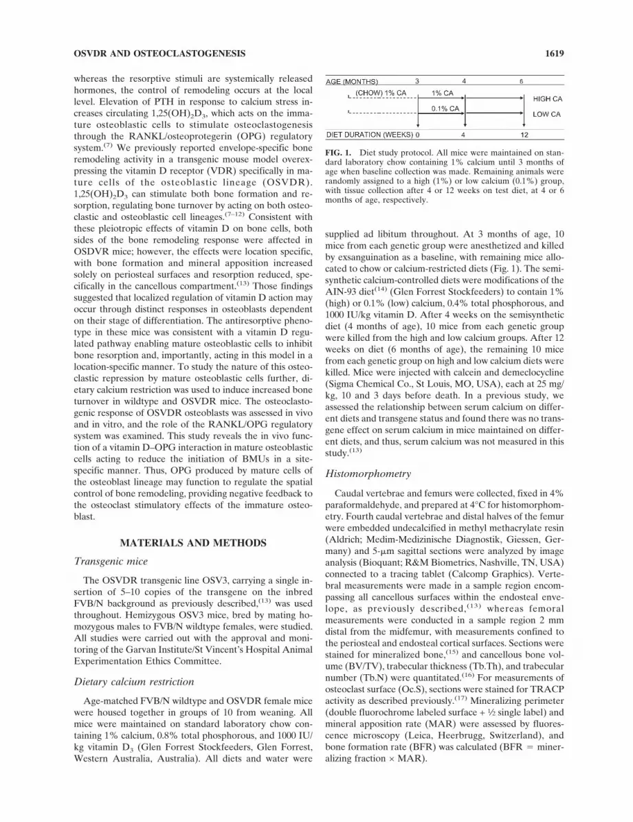
supplied ad libitum throughout. At 3 months of age, 10mice from each genetic group were anesthetized and killedby exsanguination as a baseline, with remaining mice allo-cated to chow or calcium-restricted diets (Fig. 1). The semi-synthetic calcium-controlled diets were modifications of theAIN-93 diet(14) (Glen Forrest Stockfeeders) to contain 1%(high) or 0.1% (low) calcium, 0.4% total phosphorous, and1000 IU/kg vitamin D. After 4 weeks on the semisyntheticdiet (4 months of age), 10 mice from each genetic groupwere killed from the high and low calcium groups. After 12weeks on diet (6 months of age), the remaining 10 micefrom each genetic group on high and low calcium diets werekilled. Mice were injected with calcein and demeclocycline(Sigma Chemical Co., St Louis, MO, USA), each at 25 mg/kg, 10 and 3 days before death. In a previous study, weassessed the relationship between serum calcium on differ-ent diets and transgene status and found there was no trans-gene effect on serum calcium in mice maintained on differ-ent diets, and thus, serum calcium was not measured in thisstudy.(13)
Histomorphometry
Caudal vertebrae and femurs were collected, fixed in 4%paraformaldehyde, and prepared at 4°C for histomorphom-etry. Fourth caudal vertebrae and distal halves of the femurwere embedded undecalcified in methyl methacrylate resin(Aldrich; Medim-Medizinische Diagnostik, Giessen, Ger-many) and 5-�m sagittal sections were analyzed by imageanalysis (Bioquant; R&M Biometrics, Nashville, TN, USA)connected to a tracing tablet (Calcomp Graphics). Verte-bral measurements were made in a sample region encom-passing all cancellous surfaces within the endosteal enve-lope, as previously described,(13) whereas femoralmeasurements were conducted in a sample region 2 mmdistal from the midfemur, with measurements confined tothe periosteal and endosteal cortical surfaces. Sections werestained for mineralized bone,(15) and cancellous bone vol-ume (BV/TV), trabecular thickness (Tb.Th), and trabecularnumber (Tb.N) were quantitated.(16) For measurements ofosteoclast surface (Oc.S), sections were stained for TRACPactivity as described previously.(17) Mineralizing perimeter(double fluorochrome labeled surface + 1⁄2 single label) andmineral apposition rate (MAR) were assessed by fluores-cence microscopy (Leica, Heerbrugg, Switzerland), andbone formation rate (BFR) was calculated (BFR � miner-alizing fraction × MAR).
FIG. 1. Diet study protocol. All mice were maintained on stan-dard laboratory chow containing 1% calcium until 3 months ofage when baseline collection was made. Remaining animals wererandomly assigned to a high (1%) or low calcium (0.1%) group,with tissue collection after 4 or 12 weeks on test diet, at 4 or 6months of age, respectively.
OSVDR AND OSTEOCLASTOGENESIS 1619
Long bone primary cell culture
Femora from 17-day-old mice were removed and cleanedof attached connective tissue and washed in PBS, and theepiphyses were removed. The remaining midshafts wereminced and washed vigorously and repeatedly in PBS toremove marrow cells and transferred to digest mix (1 ml/6femora) of 1 mg/ml collagenase (Boehringer-Mannheim,San Diego, CA, USA), 0.05% trypsin (Commonwealth Se-rum Laboratories, Canberra, Australia), and 0.02% EDTA(ICN, Costa Mesa, CA, USA) in PBS and stirred for 20minutes at 37°C. Cells released from this first digest werediscarded, and cells released from the second 20-minutedigest were harvested and seeded at a density of 104 cells/cm2 into 6-well plates in �MEM (Trace, Sydney, Australia)containing 10% FCS (Gibco, Grand Island, NY, USA), 100U/ml penicillin, 100 mg/ml streptomycin, 40 mg/ml gento-mycin, 20 mM HEPES, and 2 mM glutamine (all Gibco),supplemented with 10 mM �-glycerophosphate and 50 mg/ml ascorbic acid (Sigma) to promote matrix maturation andmineralization. Culture medium was changed after 3 daysand every 2–3 days thereafter. Mineralized cultures (day20) were treated with 10−8 M 1,25(OH)2D3 or 0.1% isopro-panol vehicle and collected 48 h later for gene expressionstudies, as previously described.(18)
Mouse osteoblast/human osteoclast co-cultures
Murine osteoblasts were prepared from femora by serialcollagenase treatments as described above. Cells from thesecond digest were seeded in 75-cm2 flasks at 10,000 cells/cm2. Culture medium was as above but without �-glycero-phosphate and ascorbic acid supplements to prevent min-eralization. Confluence was judged to be at 14 days afterseeding. Osteoblasts were harvested on day 20 (6 days afterconfluence) and co-cultured with osteoclast precursors, asdescribed below. Osteoclast precursors were prepared fromhuman umbilical cord blood mononuclear cells as previ-ously described.(19)
Osteoclast in vitro assays
Mouse osteoblast (2.5 × 10⁴ cells/culture) and osteoclastprecursors (4 × 10⁴ cells/culture) were combined and settledonto 4 × 4-mm sperm whale dentine slices in 96-well tissueculture plates and cultured in 200 �l MEM containing 10%FBS, nonessential amino acids, 50 IU/ml penicillin, 50 �g/ml streptomycin, 2 mM L-glutamine, 25 ng/ml macrophage-colony stimulating factor (M-CSF), and 10−7 M dexameth-asone. After a 2-h settlement, 100 �l of culture supernatantwas removed and replaced with 100 �l of medium contain-ing 1,25(OH)2D3. The cultures were refreshed twice weeklyby replacing additives in one-half volume of medium. Cul-tures were fixed in 1% formalin and reacted for TRACP.The formation of osteoclasts and resorption activity wereassessed as previously described.(19)
Tissue expression
For gene expression analysis, 4-month-old animals weretreated with a single intraperitoneal injection of1,25(OH)2D3 (Calbiochem, La Jolla, CA, USA) at a doseof 2 �g per kg body weight. Femora were collected 6 h later.
Marrow was excluded from the samples by removal of theends of the long bones and flushing the cortical shafts usingPBS to ensure removal of marrow cellular material. Allsamples were snap frozen and stored at −80°C before RNApreparation. RNA was isolated from whole bones or osteo-blastic cell cultures using Trizol reagent (Gibco) with gly-cogen (5 �g/ml) as carrier according to manufacturer pro-tocol. cDNA was generated by reverse transcriptase(Superscript II, Gibco) with random hexamer primers. PCRamplification was carried out with gene-specific primers(Sigma) using Taq polymerase (Amplitaq; Perkin Elmer,Boston, MA, USA). Reactions were carried out in foil-sealed 96-well plates (Thermofast 96; ABGene, Sydney,Australia) on a GeneAmp 9700 machine (Perkin Elmer).For PCR on cDNA from whole bones (OPG, RANKL, andGAPDH), conditions were optimized to produce linear am-plification for the genes. Specific primers and conditionswere as follows: OPG (annealing temperature � 65°C, 31cycles , product 578 bp)—forward, 5 � -TCCTG-GCACCTACCTAAAACAGCA-3� and reverse, 5�-CTACACTCTCGGCATTCACTTTGG-3�; RANKL (an-nealing temperature � 60°C, 37 cycles, product 790 bp)—forward, 5�-GGGAATTACAAAGTGCACCAG-3�
and reverse, 5�-GGTCGGGCAATTCTGAATT-3�;GAPDH (annealing temperature � 65°C, 27 cycles, prod-uct 983 bp)—forward, 5�-GGTCGGTGTGAACG-GATTTGG-3� and reverse, 5�-ATGTAGGCCATGAG-GTCCACC-3�; human VDR (annealing temperature �
65°C, 29 cycles, product 472 bp)—forward, 5�-TCATTCTGACAGATGAGGAAGTGC-3� and reverse,5�-TCCTGGTATCATCTTAGCAAAGCC-3�. For osteo-pontin (OPN) expression in long bones, cDNA levels ofOPN and GAPDH were quantitated by real-time RT-PCRon the Rotor-GeneTM300 Thermal Cycler (Corbett Re-search, Sydney, Australia) using SYBR Premix ExTaq(Takara Bio, Shiga, Japan) according to the manufacturer’sprotocol. Specific primers and conditions were as follows:GAPDH (annealing temperature � 60°C, 45 cycles, prod-uct 451 bp)—forward, 5�-ACCACAGTCCATGCCAT-CAC-3� and reverse, 5�-TCCACCACCCTGTTGCTTA-3�; OPN (annealing temperature � 60°C, 45 cycles, product80 bp)—forward, 5�-GATGCCACAGATGAGGACCTC-3� and reverse, 5�-CTGGGCAACAGGGATGACAT-3�.
For PCR on cDNA from osteoblastic cultures, reactionsfor OPG and RANKL were duplexed with GAPDH afterconditions were optimized to produce linear amplificationfor both genes. For both OPG-GAPDH and RANKL-GAPDH, annealing temperatures were 65°C, with the re-actions run for 29 cycles. Specific primers were the same asfor the bone tissue samples. PCR products were run on1.5% agarose gels and stained with ethidium bromide. TheUV image was captured using the GelDoc apparatus (Bio-Rad, Regents Park, Australia), and the bands were quan-tified using MolecularAnalyst software (BioRad). Data arethe ratio to GAPDH PCR product.
Biochemistry
Serum intact PTH levels were determined by a mouse-specific ELISA (Immunotopics, San Clemente, CA, USA),
BALDOCK ET AL.1620
and 1,25-dihydroxyvitamin D levels were determined byradioimmunoassay after immunoextraction (Immunodiag-nostic Systems, Boldon, UK).
Statistics
Statistical analyses assessing the effect of transgene statusor diet were carried out by one-way ANOVA within diet orage groups separately. After ANOVA, linear contrasts se-lected a priori were used to compare results from the trans-genic line with those from the FVB/N control line, with p <0.05 considered significant (Statview 5.1 Software; AbacusConcepts, Berkeley, CA, USA). Analysis of co-culture datawas also by ANOVA with posthoc linear contrasts (Super-ANOVA Software; Abacus Concepts).
RESULTS
Biochemical response to dietary calcium restriction
in OSVDR mice
Baseline PTH levels did not differ between wildtype andtransgenic mice (Table 1); however, 4 weeks of dietary cal-cium restriction elevated serum PTH in wildtype mice to alevel 60% higher than in OSVDR (p < 0.05). After 12weeks of calcium restriction, wildtype PTH had returned tobaseline (Table 1). In OSVDR mice, calcium restrictionhad no effect on PTH level after either 4 or 12 weeks.Serum 1,25(OH)2D3 was 40% greater in OSVDR micecompared with wildtype mice at baseline and after 12weeks on the high calcium diet (Table 1). Calcium restric-tion resulted in nonsignificant 30% increases in serum1,25(OH)2D3 after both 4 and 12 weeks in wildtype mice,resulting in levels similar to those seen in the OSVDR miceon the high calcium diet.
Effects of age and dietary calcium restriction on
vertebral cancellous bone in OSVDR mice
In wildtype mice fed the high calcium diet, vertebral can-cellous bone volume (Fig. 2A) declined with age between 3and 6 months, consistent with an 80% rise in osteoclastsurface (Fig. 2B) between 4 and 6 months (p � 0.06) andstable BFR (Fig. 2C). Dietary calcium restriction in wild-type mice resulted in significantly more resorption (p <0.02) and a trend to decreased bone volume compared withthe high calcium wildtype group after 4 weeks of treatment,
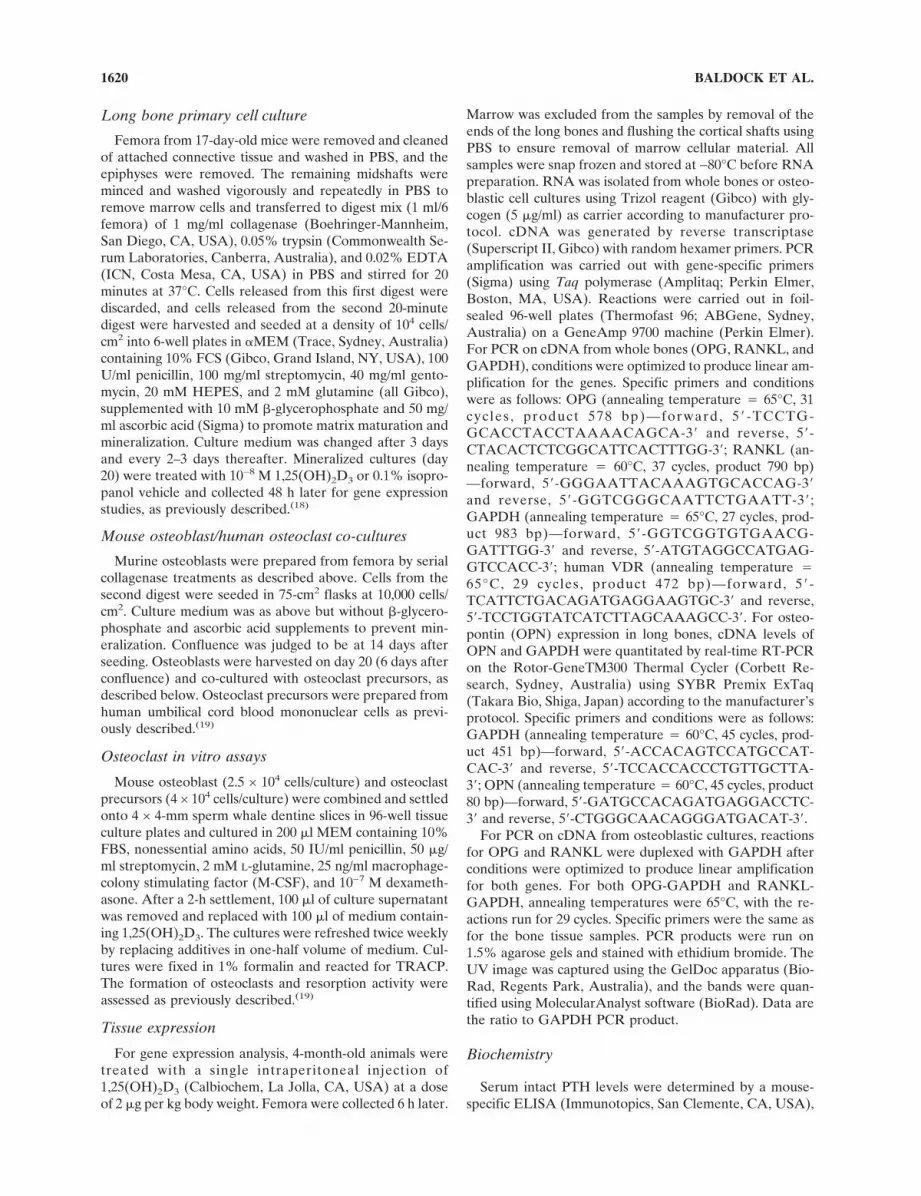
FIG. 2. Effect of elevated osteoblastic VDR on response of ver-tebral cancellous bone to dietary calcium deficiency. (A) Cancel-lous bone volume was elevated in OSVDR (OS) mice after 4 or 12months on high calcium diet (black squares) and after 4 weeks onlow calcium diet (white squares) compared with wildtype (wt)groups fed high and low calcium (black and white diamonds,respectively). (B) Osteoclast surface was significantly lower inOSVDR than wildtype mice after dietary calcium restriction. (C)Bone formation rate in this cancellous compartment was not sig-nificantly different between wildtype and OSVDR in any group.Statistical significance: ap < 0.05 vs. baseline (3 month) within dietgroup, bp < 0.05 vs. 4 months of age within diet group, cp < 0.05 vs.wildtype within diet/age group, dp < 0.05 vs. chow within age/genotype group. Values are mean ± SE, n � 7–12.
TABLE 1. EFFECT OF OSVDR TRANSGENE, DIETARY CALCIUM RESTRICTION AND DURATION OF TREATMENT ON CALCIOTROPIC
HORMONE LEVELS IN SERUM
Baseline 4 weeks 12 weeks
Chow High Low High Low
iPTH (pg/ml)
Wildtype 54 ± 44 41 ± 33 82 ± 27‡ 50 ± 32 54 ± 44
OSVDR 64 ± 30 48 ± 29 52 ± 32 44 ± 35 56 ± 39
1,25(OH)2D3 (pM)*
Wildtype 95 ± 34 85 ± 26 118 ± 38 115 ± 38.3 150 ± 51
OSVDR 130 ± 42† 112 ± 57 114 ± 53 174 ± 53† 178 ± 45
Values are mean ± SD. n � 7–14.
* Significant difference between FVB/N and OSV3 across all nonbaseline groups.† Significant difference between FVB/N and OSV3, within dietary group.‡ Significant difference between high and low, within genetic group.
OSVDR AND OSTEOCLASTOGENESIS 1621
again with stable bone formation throughout the study pe-riod. The diet-induced bone loss after 4 weeks on the lowcalcium diet and the aging-associated loss between 4 and 6months in the high calcium group resulted in equivalentbone volumes for both dietary groups of wildtype mice at 6months reaching equivalent bone parameters (Fig. 2).
Consistent with previous observations,(13) cancellousbone volume was significantly elevated in OSVDR micerelative to wildtype at 4 and 6 months of age. Aging-relatedbone loss was also evident in the transgenics, with the de-cline in bone volume between 3 and 6 months associatedwith a 30% rise in resorption (p � 0.06) between 4 and 6months and a 55% decline in bone formation (p < 0.02).The bone volume difference between wildtype andOSVDR mice fed high calcium persisted throughout thestudy period.
In contrast to wildtype mice, 4 weeks of dietary calciumrestriction in OSVDR did not result in reduced cancel-lous bone volume at 4 months of age. Importantly, boneresorption was not different between high and low calciumOSVDR groups at this time-point, consistent with thetransgene-associated resistance to calcium-induced boneloss previously observed.(13) This resistance did not preventa decline in bone mass, however, because cancellous bonevolume in the OSVDR low calcium group declined to wild-type levels after 12 weeks of diet treatment, in associationwith a nonsignificant trend to increased resorption between4 and 6 months. This trend closely paralleled the change inbones of high calcium–fed OSVDR mice and was accom-panied by a significant and marked decline in bone forma-tion (p < 0.0001). This decline in formation in the OSVDRlow calcium group resulted in a significant difference inbone volume between the OSVDR groups at 6 months.
On femoral cortical surfaces, periosteal osteoclast surfacedeclined with age after baseline in both genotypes, inde-pendent of dietary calcium (Table 2). Conversely, endostealosteoclast surface increased with age between 4 and 6months of age in both genotypes, again independent of di-etary calcium. Importantly, there were no transgene-associated changes in osteoclast surface on cortical bonesurfaces.
Effects of transgenic osteoblast on
osteoclastogenesis in monocyte co-cultures
To elucidate the underlying causes of the reduced resorp-tion seen in transgenic vertebral cancellous bone, the os-
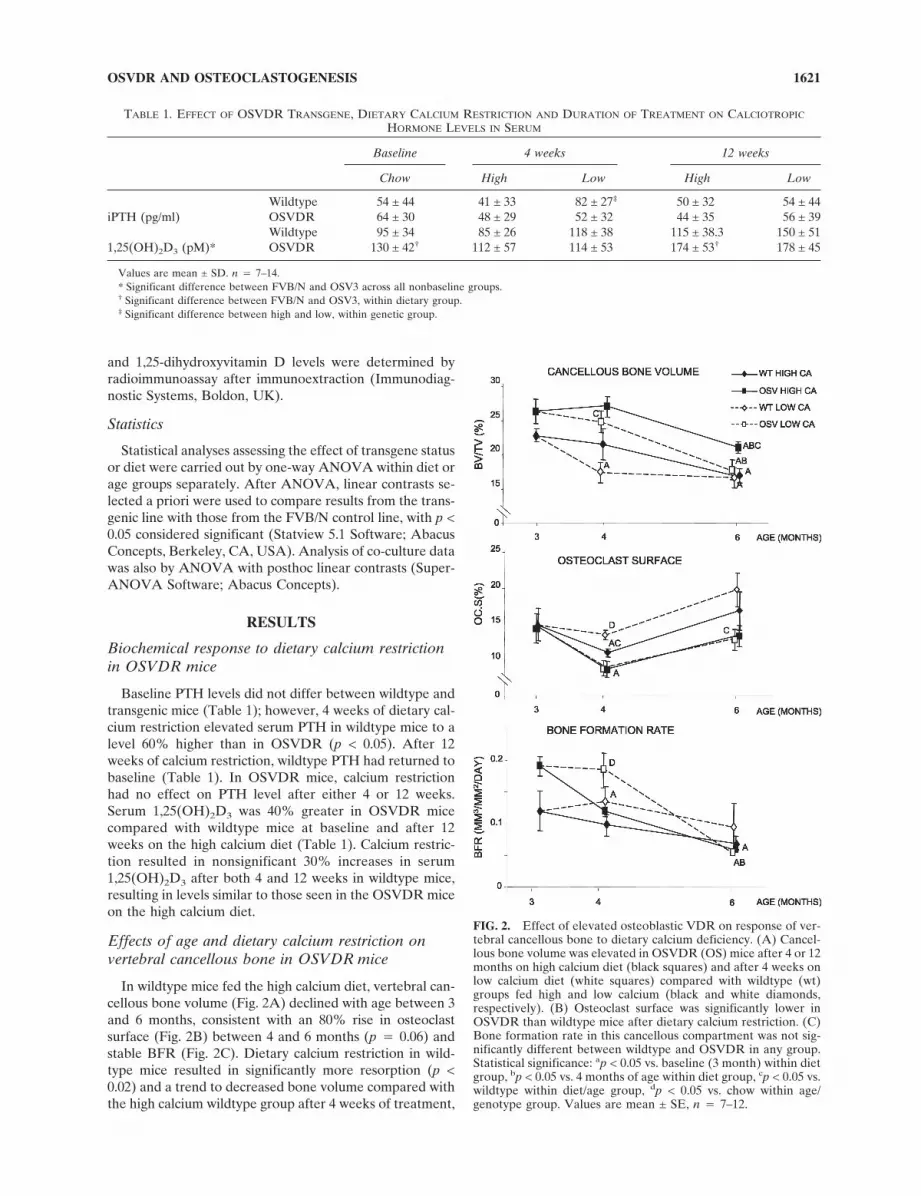
teoclastogenic potential of wildtype and OSVDR osteo-blasts in co-cultures with human monocytes was analyzed(Fig. 3). In the presence of wildtype primary osteoblasts,there was a dose-dependent response to 1,25(OH)2D3 innumbers of TRACP+ multinucleated cells (Fig. 3A) andresorption pit area on bone slices (Fig. 3B). Both osteoclastformation and resorptive activity were increased with1,25(OH)2D3 treatment, with maximal effect at 10−9 and10−10 M, respectively.
FIG. 3. Effect of elevated osteoblastic VDR levels on1,25(OH)2D3-stimulated bone resorption in co-cultures with hu-man cord blood monocytes. (A) Production of TRACP+ multinu-cleated cells in co-cultures with wildtype osteoblasts (wt) rosewith increasing 1,25(OH)2D3 concentration to 10−9 M, reducingthereafter at 10−8 and 10−7 M. TRACP+ cell number in OSVDRcultures was lower than wildtype level in untreated cultures andremained ∼50% lower than wildtype between 10−11 and 10−9 M1,25(OH)2D3, with no transgene-associated reduction evident athigher concentrations. (B) Pit area per bone slice results weresimilar to TRACP+ cell number. ap < 0.05 vs. wildtype, withintreatment concentration. Values are mean ± SE, n � 4.
TABLE 2. EFFECT OF OSTEOBLASTIC VDR OVEREXPRESSION AND DIETARY CALCIUM RESTRICTION ON CORTICAL
OSTEOCLAST SURFACE
Baseline 4 weeks 12 weeks
Chow High Low High Low
Endo Oc.S Wildtype 10.3 ± 3.2 12.2 ± 11.1 15.8 ± 7.9 25.5 ± 10.4* 24.9 ± 3.9*
(%BS) OSVDR 13.9 ± 7.6 14.1 ± 5.4 15.4 ± 5.2 29.4 ± 12.6* 20.1 ± 10.1
Peri Oc.S Wildtype 18.5 ± 5.8 7.2 ± 4.8† 3.3 ± 3.6† 5.3 ± 2.3 5.0 ± 6.7
(%BS) OSVDR 17.2 ± 4.7 6.2 ± 5.7† 5.0 ± 5.0† 4.2 ± 4.0 4.7 ± 4.3
Values are mean ± SD.
* Significant difference between 4 and 12 weeks within dietary group.† Significant difference between baseline and 4 weeks.
BALDOCK ET AL.1622
In monocytes co-cultured with OSVDR osteoblasts, bothparameters were reduced in unstimulated and hormone-treated cultures, with levels of osteoclastogenesis and re-sorption ∼50% lower than in wildtype co-cultures at1,25(OH)2D3 concentrations of 10−9 M or lower (p < 0.05;Fig. 3). Osteoclastogenesis with OSVDR osteoblasts de-creased up to 10−9 M 1,25(OH)2D3 as seen with wildtypeosteoblasts, but at 10−8 M,1,25(OH)2D3 and above did notdiffer from the bone resorption parameters in wildtype co-cultures.
Transgene effects on RANKL and OPG expression
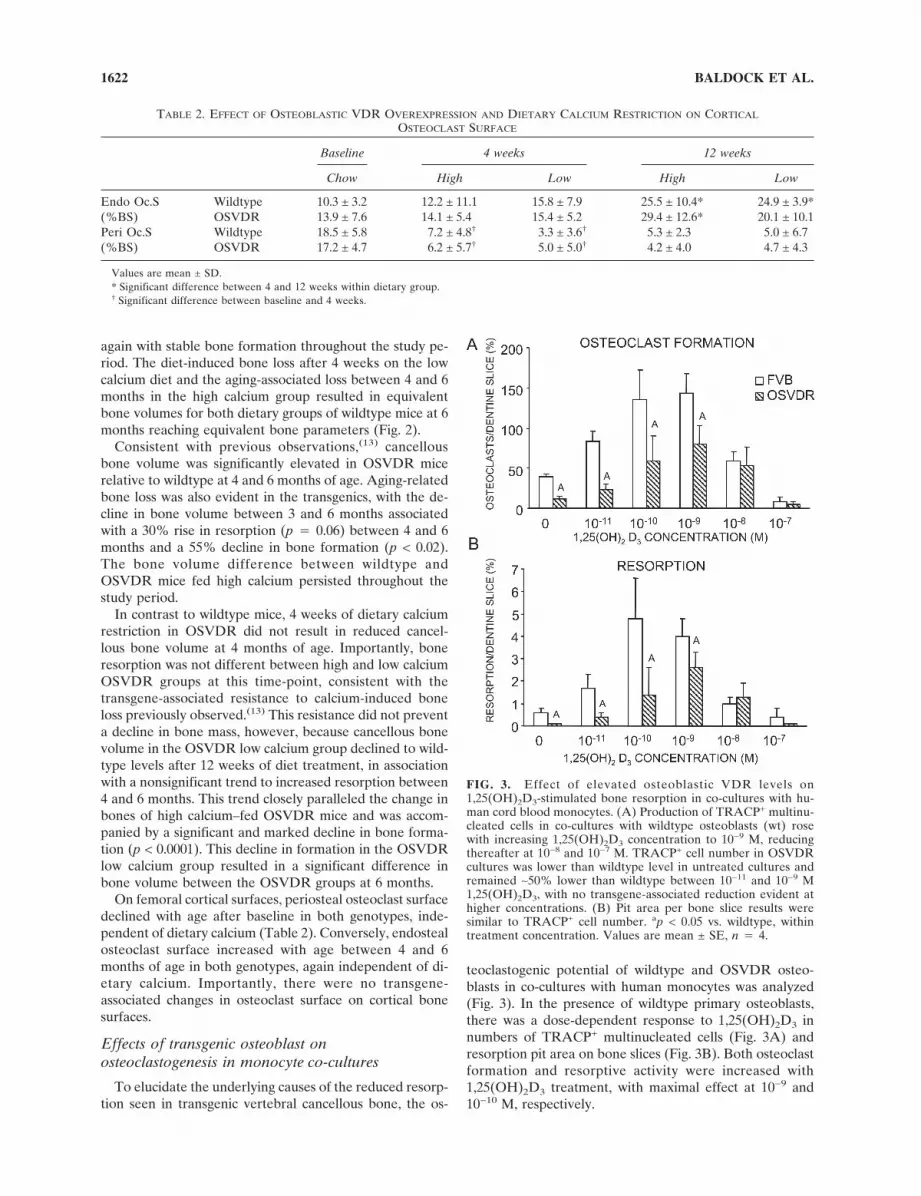
Levels of the principal cytokine mediators of osteoclas-togenesis were assessed. RANKL, but not OPG, expressionwas reduced in long bones of untreated OSVDR mice com-pared with wildtype animals (p < 0.005; Fig. 4), resulting ina lower RANKL/OPG ratio in the OSVDR bones (p <0.05). Treatment with 1,25(OH)2D3 modestly increasedRANKL expression in both wildtype (not significant) andOSVDR (p < 0.05) mice, but levels in treated OSVDRbones remained reduced compared with wildtype. In con-trast, OPG expression after 1,25(OH)2D3 treatment wasreduced in wildtype mice by ∼50% (p < 0.0001; Fig. 4B) butwas not affected by the hormone in OSVDR mice. Thus, inwildtype bones, 1,25(OH)2D3 treatment increased theRANKL/OPG ratio 2.3-fold, but in OSVDR mice treat-ment only increased the ratio 1.3-fold (Fig. 4D). To verifythe enhanced vitamin D responsiveness of the OSVDR os-teoblasts, the expression of osteopontin was also examinedin the long bones (Fig. 4E). Basal levels of OPN mRNAwere 60% higher in OSDVR than in FVB bones (not sig-nificant, p � 0.15). Treatment with 1,25(OH)2D3 increasedOPN expression 4-fold in FVB (p � 0.08). However, in the
OSVDR bones, 1,25(OH)2D3 treatment resulted in a 19-fold elevation in OPN expression (p < 0.05)
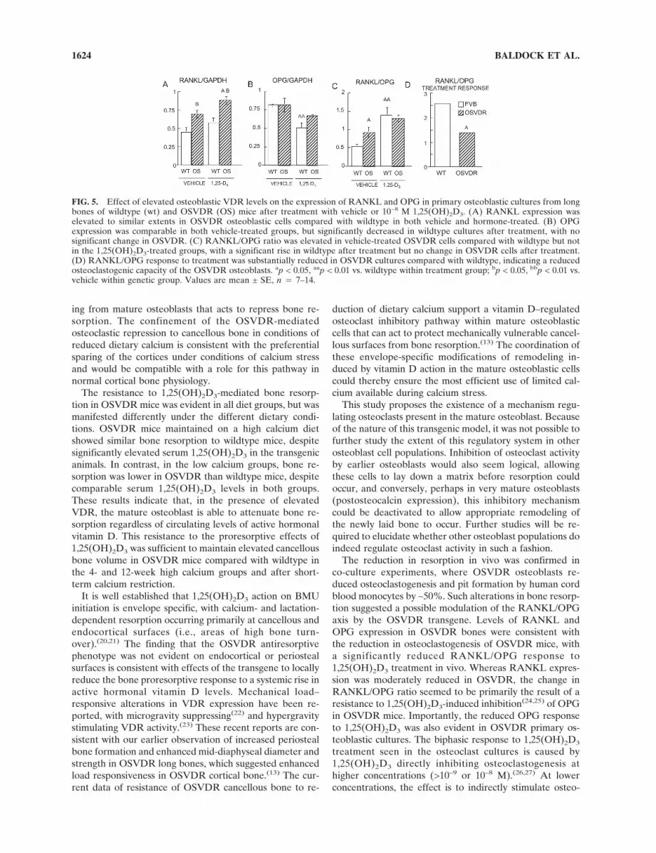
In mature mineralizing long bone primary osteoblasticcultures, in contrast to tissue levels, RANKL expres-sion was higher in both vehicle and 1,25(OH)2D3-treatedOSVDR osteoblasts compared with wildtype (p < 0.01;Fig. 5A). Treatment with 1,25(OH)2D3 increased RANKLexpression in both wildtype and OSVDR cultures 1.2-fold,reaching statistical significance in the OSVDR cultures (p <0.05; Fig. 5A). OPG levels were similar in vehicle-treatedwildtype and OSVDR osteoblasts, and 1,25(OH)2D3 treat-ment reduced OPG expression 1.3-fold in wildtype cultures(p < 0.01) but not in OSVDR cultures (Fig. 5B). Thus, theRANKL/OPG ratio was higher in vehicle-treated OSVDRcultures (p < 0.05) but comparable in 1,25(OH)2D3-treatedwildtype and transgenic cultures (Fig. 5C). Importantly,however, 1,25(OH)2D3 increased the RANKL/OPG ratio2.5-fold in wildtype but only 1.4-fold in OSVDR osteoblas-tic cultures (Fig. 5D).
DISCUSSION
The OSVDR mouse model has for the first time enabledin vivo analysis of the osteoblastic vitamin D response tofocus specifically on the mature osteoblast. Enhanced vita-min D receptor expression specifically in the mature mouseosteoblast was associated with an inhibition of1,25(OH)2D3-mediated bone resorption, specifically oncancellous bone surfaces.(13) This OSVDR antiresorptivephenotype was mainly associated with a cell autonomousosteoblastic effect associated with an altered OPG responseto 1,25(OH)2D3. The findings of this study therefore pro-vide evidence for a vitamin D responsive pathway originat-
FIG. 4. Effect of elevated osteoblasticVDR levels on the expression of RANKLand OPG in the long bones of wildtype (wt)and OSVDR (OS) mice after 1,25(OH)2D3
treatment. (A) RANKL expression was signifi-cantly decreased in vehicle and 1,25(OH)2D3-treated OSVDR cells compared with wild-type. (B) OPG expression was similar in wild-type and OSVDR long bones. In vehicle-treated bones, it was significantly decreased inwildtype but not OSVDR after 1,25(OH)2D3
treatment, with no change in OSVDR. (C)RANKL/OPG ratio was reduced in vehicletreated OSVDR mice compared with wild-type. 1,25(OH)2D3 treatment increasedRANKL/OPG in both groups. (D) RANKL/OPG response to treatment was markedlyreduced in OSVDR bones. (E) Both basallevels and the 1,25(OH)2D3 responsivenessof OPN were enhanced in the OSVDR bones.ap < 0.05, aap < 0.01 vs. wildtype within treat-ment group; bp < 0.05, bbp < 0.01 vs. vehiclewithin genetic group. Values are mean ± SE,n � 7–14.
OSVDR AND OSTEOCLASTOGENESIS 1623
ing from mature osteoblasts that acts to repress bone re-sorption. The confinement of the OSVDR-mediatedosteoclastic repression to cancellous bone in conditions ofreduced dietary calcium is consistent with the preferentialsparing of the cortices under conditions of calcium stressand would be compatible with a role for this pathway innormal cortical bone physiology.
The resistance to 1,25(OH)2D3-mediated bone resorp-tion in OSVDR mice was evident in all diet groups, but wasmanifested differently under the different dietary condi-tions. OSVDR mice maintained on a high calcium dietshowed similar bone resorption to wildtype mice, despitesignificantly elevated serum 1,25(OH)2D3 in the transgenicanimals. In contrast, in the low calcium groups, bone re-sorption was lower in OSVDR than wildtype mice, despitecomparable serum 1,25(OH)2D3 levels in both groups.These results indicate that, in the presence of elevatedVDR, the mature osteoblast is able to attenuate bone re-sorption regardless of circulating levels of active hormonalvitamin D. This resistance to the proresorptive effects of1,25(OH)2D3 was sufficient to maintain elevated cancellousbone volume in OSVDR mice compared with wildtype inthe 4- and 12-week high calcium groups and after short-term calcium restriction.
It is well established that 1,25(OH)2D3 action on BMUinitiation is envelope specific, with calcium- and lactation-dependent resorption occurring primarily at cancellous andendocortical surfaces (i.e., areas of high bone turn-over).(20,21) The finding that the OSVDR antiresorptivephenotype was not evident on endocortical or periostealsurfaces is consistent with effects of the transgene to locallyreduce the bone proresorptive response to a systemic rise inactive hormonal vitamin D levels. Mechanical load–responsive alterations in VDR expression have been re-ported, with microgravity suppressing(22) and hypergravitystimulating VDR activity.(23) These recent reports are con-sistent with our earlier observation of increased periostealbone formation and enhanced mid-diaphyseal diameter andstrength in OSVDR long bones, which suggested enhancedload responsiveness in OSVDR cortical bone.(13) The cur-rent data of resistance of OSVDR cancellous bone to re-
duction of dietary calcium support a vitamin D–regulatedosteoclast inhibitory pathway within mature osteoblasticcells that can act to protect mechanically vulnerable cancel-lous surfaces from bone resorption.(13) The coordination ofthese envelope-specific modifications of remodeling in-duced by vitamin D action in the mature osteoblastic cellscould thereby ensure the most efficient use of limited cal-cium available during calcium stress.
This study proposes the existence of a mechanism regu-lating osteoclasts present in the mature osteoblast. Becauseof the nature of this transgenic model, it was not possible tofurther study the extent of this regulatory system in otherosteoblast cell populations. Inhibition of osteoclast activityby earlier osteoblasts would also seem logical, allowingthese cells to lay down a matrix before resorption couldoccur, and conversely, perhaps in very mature osteoblasts(postosteocalcin expression), this inhibitory mechanismcould be deactivated to allow appropriate remodeling ofthe newly laid bone to occur. Further studies will be re-quired to elucidate whether other osteoblast populations doindeed regulate osteoclast activity in such a fashion.
The reduction in resorption in vivo was confirmed inco-culture experiments, where OSVDR osteoblasts re-duced osteoclastogenesis and pit formation by human cordblood monocytes by ∼50%. Such alterations in bone resorp-tion suggested a possible modulation of the RANKL/OPGaxis by the OSVDR transgene. Levels of RANKL andOPG expression in OSVDR bones were consistent withthe reduction in osteoclastogenesis of OSVDR mice, witha significantly reduced RANKL/OPG response to1,25(OH)2D3 treatment in vivo. Whereas RANKL expres-sion was moderately reduced in OSVDR, the change inRANKL/OPG ratio seemed to be primarily the result of aresistance to 1,25(OH)2D3-induced inhibition(24,25) of OPGin OSVDR mice. Importantly, the reduced OPG responseto 1,25(OH)2D3 was also evident in OSVDR primary os-teoblastic cultures. The biphasic response to 1,25(OH)2D3
treatment seen in the osteoclast cultures is caused by1,25(OH)2D3 directly inhibiting osteoclastogenesis athigher concentrations (>10–9 or 10–8 M).(26,27) At lowerconcentrations, the effect is to indirectly stimulate osteo-
FIG. 5. Effect of elevated osteoblastic VDR levels on the expression of RANKL and OPG in primary osteoblastic cultures from longbones of wildtype (wt) and OSVDR (OS) mice after treatment with vehicle or 10−8 M 1,25(OH)2D3. (A) RANKL expression waselevated to similar extents in OSVDR osteoblastic cells compared with wildtype in both vehicle and hormone-treated. (B) OPGexpression was comparable in both vehicle-treated groups, but significantly decreased in wildtype cultures after treatment, with nosignificant change in OSVDR. (C) RANKL/OPG ratio was elevated in vehicle-treated OSVDR cells compared with wildtype but notin the 1,25(OH)2D3-treated groups, with a significant rise in wildtype after treatment but no change in OSVDR cells after treatment.(D) RANKL/OPG response to treatment was substantially reduced in OSVDR cultures compared with wildtype, indicating a reducedosteoclastogenic capacity of the OSVDR osteoblasts. ap < 0.05, aap < 0.01 vs. wildtype within treatment group; bp < 0.05, bbp < 0.01 vs.vehicle within genetic group. Values are mean ± SE, n � 7–14.
BALDOCK ET AL.1624
clastogenesis through osteoblasts (through the RANKL/OPG system), and it is through this mechanism that we seean effect of the presence of the OSVDR transgene.
Thus, it seems likely that the mature osteoblast acts torestrict osteoclastogenesis by an OPG-related mechanism,which is supported by elevated OPG expression in matureosteoblasts.(18) Such a mechanism would contrast thestrongly RANKL-based stimulation of osteoclastogenesisby the immature osteoblast.(28) Other studies have also sug-gested increased osteoblast maturation can reduce1,25(OH)2D3-regulated osteoclastogenesis in marrow/osteoblast co-culture, although a RANKL-associatedmechanism was postulated rather than OPG.(29)
A recent study has suggested that OPG gene expressionis rapidly and transiently reduced by 1,25(OH)2D3 throughc-jun activity at the activator protein-1 (AP-1) bindingsite.(30) This initial acute reduction in OPG by 1,25(OH)2D3
played an important role in the stimulation of resorption,but chronic 1,25(OH)2D3 exposure rendered the OPG geneinsensitive to repression, thereby giving way to the anabolicphase of the vitamin D effect on bone. The constitutive highlevel of transgenic VDR expression in mature osteoblasticcells(13) in OSVDR mice may thus mimic chronic1,25(OH)2D3 treatment and block repression of OPG ex-pression by c-jun, reducing the catabolic action of vitaminD in the transgenic model.
The findings of this study indicate the presence of a1,25(OH)2D3-regulated pathway in mature osteoblasticcells acting to stabilize OPG expression and thus reduceosteoclastogenesis in vivo. Acting in a site-dependent man-ner, stimulation of this pathway in OSVDR mice reducesresorption of cancellous bone under calcium replete andrestricted conditions. Thus, differential regulation of OPGexpression by 1,25(OH)2D3 in immature versus mature os-teoblasts may confer local protection of bone from the os-teolytic effects of circulating 1,25(OH)2D3. Signals of thisnature may present a negative feedback system, actingthrough the mature osteoblastic cell to exert control overboth the location and frequency of bone remodeling eventsinitiated by immature osteoblastic cells in response to sys-temic signals such as active hormonal vitamin D. These datasupport the existence of distinct and in some ways diversevitamin D regulatory pathways in immature and maturecells of the osteoblastic lineage. These different pathwaysmay be central to the site-specific regulation of bone re-modeling.
ACKNOWLEDGMENTS
The authors thank Dr Julie Ferguson for invaluable vet-erinary advice and the staff of the Garvan Institute Biologi-cal Testing Facility, Dr Pernilla Lundberg, and Dr TedKraegen for critical review of the manuscript. This researchwas supported by National Health and Medical ResearchCouncil Grant 276415.
REFERENCES
1. Shen V, Birchman R, Lindsay R, Dempster DW 1995 Short-term changes in Histomorphometric and biochemical turnover
markers and bone mineral density in oestrogen –and/or dietarycalcium-deficient rats. Bone 16:149–156.
2. Yoshitake K, Yokota K, Kasugai Y, Kagawa M, Sukamoto T,Nakamura T 1999 Effects of 16 weeks of treatment with tibo-lone on bone mass and bone mechanical and Histomorpho-metric indices in mature ovariectomized rats with establishedosteopenia on a low-calcium diet. Bone 25:311–319.
3. Baldock PA, Need AG, Moore RJ, Durbridge TC, Morris HA1999 Discordance between bone turnover and bone loss: Ef-fects of aging and ovariectomy in the rat. J Bone Miner Res14:1442–1448.
4. Thomsen JS, Ebbesen EN, Mosekilde LI 2002 Age-related dif-ferences between thinning of horizontal and vertical trabeculaein human lumbar bone as assessed by a new computerizedmethod. Bone 31:136–142.
5. Burr DB 2002 Targeted and nontargeted bone remodeling.Bone 30:2–4.
6. Parfitt AM 2002 Targeted and nontargeted bone remodeling:Relationship to basic multicellular unit origination and pro-gression. Bone 30:5–7.
7. Suda T, Takahashi N, Udagawa N, Jimi E, Gillespie MT, Mar-tin TJ 1999 Modulation of osteoclast differentiation and func-tion by the new members of the tumor necrosis factor receptorand ligand families. Endocr Rev 20:345–357.
8. Bikle DD, Halloran BP, McGalliard-Cone C, Morey-Holton E1990 Different responses of trabecular and cortical bone to1,25(OH)2D3 infusion. Am J Physiol 259:E715–E722.
9. Marie PJ, Hott M, Garba MT 1985 Contrasting effects of 1,25-dihydroxyvitamin D3 on bone matrix and mineral appositionalrates in the mouse. Metabolism 34:777–783.
10. Suda T, Udagawa N, Nakamura I, Miyaura C, Takahashi N1995 Modulation of osteoclast differentiation by local factors.Bone 17:87S–91S.
11. Suda T, Jimi E, Nakamura I, Takahashi N 1997 Role of 1alpha,25-dihydroxyvitamin D3 in osteoclast differentiation andfunction. Methods Enzymol 282:223–235.
12. Yasuda H, Shima N, Nakagawa N, Yamaguchi K, Kinosaki M,Goto M, Mochizuki SI, Tsuda E, Morinaga T, Udagawa N,Takahashi N, Suda T, Higashio K 1999 A novel molecularmechanism modulating osteoclast differentiation and function.Bone 25:109–113.
13. Gardiner EM, Baldock PA, Thomas GP, Sims NA, HendersonNK, Hollis B, White CP, Sunn KL, Morrison NA, Walsh WR,Eisman JA 2000 Increased formation and decreased resorptionof bone in mice with elevated vitamin D receptor in maturecells of the osteoblastic lineage. FASEB J 14:1908–1916.
14. Reeves PG, Rossow KL, Lindlauf J 1993 Development andtesting of the AIN-93 purified diets for rodents: Results ongrowth, kidney calcification and bone mineralization in ratsand mice. J Nutr 123:1923–1931.
15. Page K 1977 Bone and preparation of bone sections. In Ban-croft JD, Stevens A (eds.) Theory and Practice of HistologicalTechniques. Churchill Livingston, London, UK, pp. 223–248.
16. Parfitt AM, Mathews CH, Villanueva AR, Kleerekoper M,Frame B, Rao DS 1983 Relationships between surface, vol-ume, and thickness of iliac trabecular bone in aging and inosteoporosis. Implications for the microanatomic and cellularmechanisms of bone loss. J Clin Invest 72:1396–1409.
17. Sims NA, White CP, Sunn KL, Thomas GP, Drummond ML,Morrison NA, Eisman JA, Gardiner EM 1997 Human andmurine osteocalcin gene expression: Conserved tissue re-stricted expression and divergent responses to 1,25-dihydroxyvitamin D3 in vivo. Mol Endocrinol 11:1695–1708.
18. Thomas GP, Baker SU, Eisman JA, Gardiner EM 2001 Chang-ing RANKL/OPG mRNA expression in differentiating murineprimary osteoblasts. J Endocrinol 170:451–460.
19. Hodge JM, Kirkland MA, Aitken CJ, Waugh CM, Myers DE,Lopez CM, Adams BE, Nicholson GC 2004 Osteoclastic po-tential of human CFU-GM: Biphasic effect of GM-CSF. JBone Miner Res 19:190–199.
20. Vajda EG, Bowman BM, Miller SC 2001 Cancellous and cor-
OSVDR AND OSTEOCLASTOGENESIS 1625
tical bone mechanical properties and tissue dynamics duringpregnancy, lactation, and postlactation in the rat. Biol Reprod65:689–695.
21. Akesson A, Vahter M, Berglund M, Eklof T, Bremme K,Bjellerup P 2004 Bone turnover from early pregnancy topostweaning. Acta Obstet Gynecol Scand 83:1049–1055.
22. Kumei Y, Morita S, Nakamura H, Katano H, Ohya K,Shimkawa H, Sams CF, Whitson PA 2004 Osteoblast respon-siveness to 1alpha,25-dihydroxyvitamin D3 during spaceflight.Ann N Y Acad Sci 1030:121–124.
23. Morita S, Nakamura H, Kumei Y, Shimokawa H, Ohya K,Shinomiya K 2004 Hypergravity stimulates osteoblast pheno-type expression: A therapeutic hint for disuse bone atrophy.Ann N Y Acad Sci 1030:158–161.
24. Colopy SA, Benz-Dean J, Barrett JG, Sample SJ, Lu Y,Danova NA, Kalscheur VL, Vanderby R Jr, Markel MD, MuirP 2004 Response of the osteocyte syncytium adjacent to anddistant from linear microcracks during adaptation to cyclic fa-tigue loading. Bone 35:881–891.
25. Murakami T, Yamamoto M, Ono K, Nishikawa M, Nagata N,Motoyoshi K, Akatsu T 1998 Transforming growth factor-beta1 increases mRNA levels of osteoclastogenesis inhibitoryfactor in osteoblastic/stromal cells and inhibits the survival ofmurine osteoclast-like cells. Biochem Biophys Res Commun252:747–752.
26. Takasu H, Sugita A, Uchiyama Y, Katagiri N, Okazaki M,Ogata E, Ikeda K 2006 c-Fos protein as a target of anti-osteoclastogenic action of vitamin D, and synthesis of new ana-logs. J Clin Invest 116:528–535.
27. Itonaga I, Sabokbar A, Neale SD, Athanasou NA 1999 1,25-Dihydroxyvitamin D(3) and prostaglandin E(2) act directly oncirculating human osteoclast precursors. Biochem Biophys ResCommun 264:590–595.
28. Grimaud E, Soubigou L, Couillaud S, Coipeau P, Moreau A,Passuti N, Gouin F, Redini F, Heymann D 2003 Receptoractivator of nuclear factor kappaB ligand (RANKL)/osteopro-tegerin (OPG) ratio is increased in severe osteolysis. Am JPathol 163:2021–2031.
29. Deyama Y, Takeyama S, Koshikawa M, Shirai Y, YoshimuraY, Nishikata M, Suzuki K, Matsumoto A 2000 Osteoblastmaturation suppressed osteoclastogenesis in coculture withbone marrow cells. Biochem Biophys Res Commun 274:249–254.
30. Kondo T, Kitazawa R, Maeda S, Kitazawa S 2004 1 alpha,25dihydroxyvitamin D3 rapidly regulates the mouse osteoprote-gerin gene through dual pathways. J Bone Miner Res 19:1411–1419.
Address reprint requests to:Paul A Baldock, PhD
Garvan Institute of Medical Research
384 Victoria Street
Darlinghurst, Sydney, NSW 2010, Australia
E-mail: [email protected]
Received in original form December 13, 2005; revised form April11, 2006; accepted July 12, 2006.
BALDOCK ET AL.1626
Copyright © 2022 FDOKUMEN