The V Protein of Human Parainfluenza Virus 2 Antagonizes Type I Interferon Responses by...
10
The V Protein of Human Parainfluenza Virus 2 Antagonizes Type I Interferon Responses by Destabilizing Signal Transducer and Activator of Transcription 2 Jean-Patrick Parisien,* ,2 Joe F. Lau,* ,2 Jason J. Rodriguez,* ,2 Brian M. Sullivan,* Anne Moscona,† Griffith D. Parks,‡ Robert A. Lamb,§ and Curt M. Horvath* ,1 *Immunobiology Center, and †Department of Pediatrics, Department of Cell Biology/Anatomy, Mount Sinai School of Medicine, New York, New York 10029; ‡Department of Microbiology and Immunology, Wake Forest University School of Medicine, Medical Center Boulevard, Winston–Salem, North Carolina 27157; and §Howard Hughes Medical Institute, Department of Biochemistry, Molecular Biology, and Cell Biology, Northwestern University, Evanston, Illinois 60208-3500 Received December 12, 2000; returned to author for revision January 4, 2001; accepted January 30, 2001 Type I interferon (IFN) induces antiviral responses through the activation of the ISGF3 transcription factor complex that contains the subunit proteins STAT1, STAT2, and p48/ISGF3g/IRF9. The ability of some human paramyxoviruses to overcome IFN actions by specific proteolysis of STAT proteins has been examined. Infection of cells with type 2, but not type 1 or type 3 human parainfluenza virus (HPIV) leads to a loss of cellular STAT2 protein. Expression of a single HPIV2 protein derived from the V open reading frame blocks IFN-dependent transcriptional responses in the absence of other viral proteins. The loss of IFN response is due to V-protein-induced proteolytic degradation of STAT2. Expression of HPIV2 V causes the normally stable STAT2 protein to be rapidly degraded, and this proteolytic activity can be partially alleviated by proteasome inhibition. No V-protein-specific effects on STAT2 mRNA levels were observed. The results indicate that the V protein of HPIV2 is sufficient to recognize and target a specific cellular transcription factor for destruction by cellular machinery. © 2001 Academic Press Key Words: STAT; ISGF3; interferon; paramyxovirus; V protein; HPIV2; proteolysis. INTRODUCTION Cellular response to virus infection involves both in- nate and adaptive host immune functions. Interferons (IFNs) have long been recognized as the mediators of innate antiviral responses. Type I IFNs, IFNa and IFNb, are the principal antiviral cytokines produced by mam- malian cells and function directly on target cells by blocking viral replication (Isaacs and Lindemann, 1957; reviewed in Goodbourn et al., 2000). IFN-dependent interference with viral infection relies on a failure of viral mRNA translation or of viral nucleic acid replication, but the molecular basis for the antiviral state is still not completely understood. It is known that the establishment of the cellular antiviral state requires mRNA and protein synthesis following IFNa treatment. IFNa/b-induced proteins that contribute to the antiviral state are known and these proteins are often the target of virus IFN antagonism (reviewed in Goodbourn et al., 2000). Well-studied examples include activation of such proteins as the double-stranded RNA-dependent protein kinase (protein kinase R), the double-stranded RNA- dependent 29–59 oligoadenylate synthetase–RNAse L system, and the IFN-induced Mx proteins. The effects of IFNa/b in causing the overall antiviral state depend on the synthesis of one or more of these proteins and additional other pathways that have been uncovered in studies of mice deficient in all three of these antiviral effectors (Zhou et al., 1999). Analysis of the promoters of many IFNa/b-stimulated genes revealed a DNA response element specific for Type I IFN induction, the IFN stimulated response ele- ment (ISRE; reviewed in Darnell, 1997; Horvath, 2000). ISGF3 was identified as the protein factor that binds to this response element and was found to contain three proteins from two transcription factor families. Two sub- units are members of the signal transducer and activator of transcription (STAT) family. The IFNa/b-activated factor (ISGF3) contains a heterodimer of STAT1 and STAT2 complexed with a third protein, a member of the inter- feron regulatory factor family referred to as ISGF3g, p48, or IRF9. The IRF9 protein is an essential component of ISGF3 that confers DNA binding specificity to the ISGF3 complex (Levy et al., 1989) and provides specific protein binding recruitment sites for the STAT1 and STAT2 pro- teins (Horvath et al., 1996; Lau et al., 2000; Martinez- Moczygemba et al., 1997). STAT1 and STAT2 are required for ISGF3 signaling, but are not redundant in this pathway. STAT1 protein is a 1 To whom correspondence and reprint requests should be ad- dressed at Box 1630, East Building Room 12-20D, One Gustave L. Levy Place, Mount Sinai School of Medicine, New York, NY 10029. Fax: (212) 849-2525. E-mail: [email protected]. 2 These authors contributed equally to this work. Virology 283, 230–239 (2001) doi:10.1006/viro.2001.0856, available online at http://www.idealibrary.com on 0042-6822/01 $35.00 Copyright © 2001 by Academic Press All rights of reproduction in any form reserved. 230
-
Upload
universityofcalifornia -
Category
Documents
-
view
1 -
download
0
Transcript of The V Protein of Human Parainfluenza Virus 2 Antagonizes Type I Interferon Responses by...
ambr
oastm
dP8
Virology 283, 230–239 (2001)doi:10.1006/viro.2001.0856, available online at http://www.idealibrary.com on
The V Protein of Human Parainfluenza Virus 2 Antagonizes Type I Interferon Responses byDestabilizing Signal Transducer and Activator of Transcription 2
Jean-Patrick Parisien,*,2 Joe F. Lau,*,2 Jason J. Rodriguez,*,2 Brian M. Sullivan,* Anne Moscona,†Griffith D. Parks,‡ Robert A. Lamb,§ and Curt M. Horvath*,1
*Immunobiology Center, and †Department of Pediatrics, Department of Cell Biology/Anatomy, Mount Sinai School of Medicine,New York, New York 10029; ‡Department of Microbiology and Immunology, Wake Forest University School of Medicine,
Medical Center Boulevard, Winston–Salem, North Carolina 27157; and §Howard Hughes Medical Institute, Department of Biochemistry,Molecular Biology, and Cell Biology, Northwestern University, Evanston, Illinois 60208-3500
Received December 12, 2000; returned to author for revision January 4, 2001; accepted January 30, 2001
Type I interferon (IFN) induces antiviral responses through the activation of the ISGF3 transcription factor complex thatcontains the subunit proteins STAT1, STAT2, and p48/ISGF3g/IRF9. The ability of some human paramyxoviruses to overcomeIFN actions by specific proteolysis of STAT proteins has been examined. Infection of cells with type 2, but not type 1 or type3 human parainfluenza virus (HPIV) leads to a loss of cellular STAT2 protein. Expression of a single HPIV2 protein derivedfrom the V open reading frame blocks IFN-dependent transcriptional responses in the absence of other viral proteins. Theloss of IFN response is due to V-protein-induced proteolytic degradation of STAT2. Expression of HPIV2 V causes thenormally stable STAT2 protein to be rapidly degraded, and this proteolytic activity can be partially alleviated by proteasomeinhibition. No V-protein-specific effects on STAT2 mRNA levels were observed. The results indicate that the V protein ofHPIV2 is sufficient to recognize and target a specific cellular transcription factor for destruction by cellular machinery.© 2001 Academic Press
Key Words: STAT; ISGF3; interferon; paramyxovirus; V protein; HPIV2; proteolysis.
M
INTRODUCTION
Cellular response to virus infection involves both in-nate and adaptive host immune functions. Interferons(IFNs) have long been recognized as the mediators ofinnate antiviral responses. Type I IFNs, IFNa and IFNb,
re the principal antiviral cytokines produced by mam-alian cells and function directly on target cells by
locking viral replication (Isaacs and Lindemann, 1957;eviewed in Goodbourn et al., 2000).
IFN-dependent interference with viral infection reliesn a failure of viral mRNA translation or of viral nucleiccid replication, but the molecular basis for the antiviraltate is still not completely understood. It is known that
he establishment of the cellular antiviral state requiresRNA and protein synthesis following IFNa treatment.
IFNa/b-induced proteins that contribute to the antiviralstate are known and these proteins are often the targetof virus IFN antagonism (reviewed in Goodbourn et al.,2000). Well-studied examples include activation of suchproteins as the double-stranded RNA-dependent proteinkinase (protein kinase R), the double-stranded RNA-
1 To whom correspondence and reprint requests should be ad-ressed at Box 1630, East Building Room 12-20D, One Gustave L. Levylace, Mount Sinai School of Medicine, New York, NY 10029. Fax: (212)49-2525. E-mail: [email protected].
2 These authors contributed equally to this work.
a0042-6822/01 $35.00Copyright © 2001 by Academic PressAll rights of reproduction in any form reserved.
230
dependent 29–59 oligoadenylate synthetase–RNAse Lsystem, and the IFN-induced Mx proteins. The effects ofIFNa/b in causing the overall antiviral state depend onthe synthesis of one or more of these proteins andadditional other pathways that have been uncovered instudies of mice deficient in all three of these antiviraleffectors (Zhou et al., 1999).
Analysis of the promoters of many IFNa/b-stimulatedgenes revealed a DNA response element specific forType I IFN induction, the IFN stimulated response ele-ment (ISRE; reviewed in Darnell, 1997; Horvath, 2000).ISGF3 was identified as the protein factor that binds tothis response element and was found to contain threeproteins from two transcription factor families. Two sub-units are members of the signal transducer and activatorof transcription (STAT) family. The IFNa/b-activated factor(ISGF3) contains a heterodimer of STAT1 and STAT2complexed with a third protein, a member of the inter-feron regulatory factor family referred to as ISGF3g, p48,or IRF9. The IRF9 protein is an essential component ofISGF3 that confers DNA binding specificity to the ISGF3complex (Levy et al., 1989) and provides specific proteinbinding recruitment sites for the STAT1 and STAT2 pro-teins (Horvath et al., 1996; Lau et al., 2000; Martinez-
oczygemba et al., 1997).STAT1 and STAT2 are required for ISGF3 signaling, but
re not redundant in this pathway. STAT1 protein is a
a
a
a
Et
wv
231HPIV2 V PROTEIN TARGETS STAT2
component of both the Type I IFN-induced ISGF3 com-plex and the Type II IFN-induced GAF complex andconsequently cells lacking STAT1 have no antiviral re-sponse to either IFN (Horvath and Darnell, 1996). FullIFNa/b antiviral activity requires only that STAT1 bephosphorylated on tyrosine, but either STAT1a or STAT1b(a form of STAT1a lacking the 38-amino-acid transcrip-tional activation domain) will suffice (Horvath and Dar-nell, 1996). These results indicate that while STAT1 isindispensable for ISGF3-induced antiviral effects, STAT2plays a crucial role as the transcription activating proteincomponent in ISGF3. STAT2 is clearly essential for IFNresponses and ISGF3 function, as deficiencies in STAT2can disrupt ISGF3 signaling and lead to decreased ex-pression of antiviral genes (Leung et al., 1995; Qureshi et
l., 1996).As the IFNa/b system represents an early and crucial
step in anti-viral immunity, it is not surprising that manyviruses have evolved strategies to impede the actions ofthis cytokine (reviewed in Goodbourn et al., 2000). The
bility of a virus to antagonize the IFNa/b pathway canhave dramatic consequences for the success of infec-tion, as illustrated in several cases where the IFN antag-onizing genes were deleted to form attenuated viruses(for example, Bergmann et al., 2000; Kitajewski et al.,1986). The virulence of a specific strain can be correlatedwith its susceptibility to the antiviral effects of IFNa/b,
nd evolution of strategies to enhance IFNa/b resistanceare expected to lead to highly infectious viruses and/orpersistent infections. A wide variety of viruses fight theIFNa/b system at a variety of steps, in many cases at anearly point upstream of gene activation, antagonizingboth IFN responses and IFN production (Bergmann et al.,2000; Kitajewski et al., 1986; Talon et al., 2000).
Negative-strand RNA viruses of the family Paramyxo-viridae have evolved specific proteins that directly sup-press IFN signaling by lowering the concentration ofcellular STAT proteins. For example, Sendai virus hasbeen demonstrated recently to block IFN responsesthrough the actions of its C proteins (Garcin et al., 1999).Expression of this single viral protein is sufficient tocause a loss of STAT protein inducibility at the transcrip-tional level. The mechanism of STAT1 transcriptionalsuppression is unknown, but is suspected to involveinteraction of C proteins with cellular factors (Garcin etal., 1999, 2000). A second paramyxovirus, SV5, was dem-onstrated recently to evade IFN antiviral responses byspecifically targeting the transcription factor STAT1 forproteolytic degradation (Didcock et al., 1999). This de-struction of STAT1 was found to be mediated by the soleexpression of a single virus-encoded protein derivedfrom the V open reading frame (Didcock et al., 1999).Subsequent analysis indicated that while mostparamyxoviruses can block IFN signaling, the ability todegrade STAT1 was a unique property of SV5 (Young et
al., 2000). Somewhat surprisingly, it was also found athat in cells infected with human parainfluenza virus 2(HPIV2), STAT1 was not degraded, but rather a loss ofSTAT2 was observed (Young et al., 2000). The mecha-nism of STAT2 degradation or the viral gene responsiblefor catalyzing its destruction was not investigated in thisstudy.
As a first step in understanding the specificity of STATprotein degradation by paramyxoviruses and defining thecellular machinery involved in this process, we investi-gated the ability of the HPIV2 V protein to mediate loss ofSTAT2 protein in the absence of other viral proteins. Ourresults corroborate the finding that infection of culturedcells with HPIV2, but not with HPIV1 or HPIV3, causes adramatic reduction of STAT2 protein steady-state levels.Moreover, we demonstrate that the expression of HPIV2V protein from a cDNA clone is sufficient to abolish IFNresponsive transcriptional activity. Our data illustrate thatexpression of the HPIV2 V protein in cultured cells didnot influence STAT2 mRNA abundance, but instead wasfound to reduce steady-state STAT2 protein levels byincreasing its degradation rate, leading to uncharacter-istic STAT2 instability. Interestingly, this proteolysis isonly partially alleviated by proteasome inhibitors, sug-gesting that alternatives to the ubiquitin–proteasomedegradation system may be responsible for the V proteinactions.
RESULTS
Effects of HPIV infection on steady state STAT1 andSTAT2 protein levels
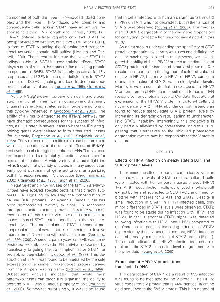
To examine the effects of human parainfluenza viruseson steady-state levels of STAT proteins, cultured cellswere infected with human parainfluenza viruses (HPIV)1–3. At 9 h postinfection, cells were lysed in whole cellextract buffer and subjected to SDS–PAGE and immuno-blotting with antisera for STAT1 and STAT2. Despite asmall reduction in STAT1 in HPIV1-infected cells, onlyminor differences in STAT1 levels were observed. STAT2was found to be stable during infection with HPIV1 andHPIV3. In fact, a stronger STAT2 signal was detectedfollowing infection with HPIV1 and HPIV3 compared touninfected cells, possibly indicating induction of STAT2expression by these viruses. In contrast, HPIV2 infectioncaused a nearly complete loss of STAT2 protein (Fig. 1).This result indicates that HPIV2 infection induces a re-duction in the STAT2 expression level in agreement withthe prior data (Young et al., 2000).
xpression of HPIV2 V protein fromransfected cDNA
The degradation of STAT1 as a result of SV5 infectionas found to be mediated by the V protein. The HPIV2
irus codes for a V protein that is 44% identical in amino
cid sequence to the SV5 V protein. This high degree of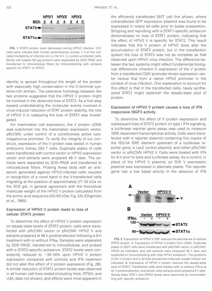
edAiU
tceSdtiasotiftctsp
Er
saIftpvfppg
4S
ts
232 PARISIEN ET AL.
identity is spread throughout the length of the proteinwith especially high conservation in the C-terminal cys-teine-rich domain. The extensive homology between thetwo proteins suggested that the HPIV2 V protein mightbe involved in the observed loss of STAT2. As a first steptoward understanding the molecular events involved invirus-induced reduction of STAT protein stability, the roleof HPIV2 V in catalyzing the loss of STAT2 was investi-gated.
For mammalian cell expression, the V protein cDNAwas subcloned into the mammalian expression vector,pRcCMV, under control of a constitutively active cyto-megalovirus promoter. To verify the integrity of our con-struct, expression of the V protein was tested in humanembryonic kidney 293 T cells. Duplicate plates of cellswere transfected with control vector or HPIV2 expressionvector and extracts were prepared 48 h later. The ex-tracts were separated by SDS–PAGE and transferred tonitrocellulose filters. Probing these blots with an anti-serum generated against HPIV2-infected cells resultedin recognition of a novel band in the V-transfected cellsmigrating at the position of approximately 27–30 kDa onthe SDS gel, in general agreement with the theoreticalmolecular weight of the HPIV2 V protein calculated fromthe amino acid sequence (24,150 kDa; Fig. 2A) (Ohgimotoet al., 1990).
Expression of HPIV2 V protein leads to loss ofcellular STAT2 protein
To determine the effect of HPIV2 V protein expressionon steady-state levels of STAT2 protein, cells were trans-fected with pRcCMV vector or pRcCMV HPIV2 V andextracts prepared at 48 h posttransfection following a 6-htreatment with or without IFNa. Samples were separatedby SDS–PAGE, transferred to nitrocellulose, and probedwith a STAT2-specific antiserum. STAT2 levels were con-sistently reduced to ;30–50% upon HPIV2 V protein
xpression compared with controls and IFN treatmentid not alter this effect of V protein expression (Fig. 2B).similar reduction of STAT2 protein levels was observed
n all human cell lines tested (including Hela, 2fTGH, and
FIG. 1. STAT2 protein level decreases during HPIV2 infection. CV1cells were infected with human parainfluenza viruses 1–3 at the indi-cated multiplicity of infection (m.o.i.) for 9 h. C, control uninfected cells.Whole cell lysates (50 mg protein) were separated by SDS–PAGE andransferred to nitrocellulose filters for immunoblotting with antiserapecific for STAT1 or STAT2.
3A; data not shown), and effects were most apparent in t
he efficiently transfected 293T cell line shown, whereotransfected GFP expression plasmid was found to bexpressed in nearly all cells prior to lysate preparation.tripping and reprobing with a STAT1-specific antiserumemonstrates no loss of STAT1 protein, indicating that
he effect of HPIV2 V is specific for STAT2. This resultndicates that the V protein of HPIV2 does alter theccumulation of STAT2 protein, but in the transfectionystem the loss of STAT2 was not as complete as thatbserved upon HPIV2 virus infection. The difference be-
ween the two systems might reflect fundamental biolog-cal differences inherent in expression of the V proteinrom a transfected CMV promoter-driven expression vec-or versus that from a native HPIV2 promoter in theontext of virus infection. One explanation to account for
his effect is that in the transfected cells, newly synthe-ized STAT2 might replenish the steady-state pool ofrotein.
xpression of HPIV2 V protein causes a loss of IFNesponsive ISGF3 activity
To determine the effect of V protein expression andubsequent loss of STAT2 protein on type I IFN signaling,luciferase reporter gene assay was used to measure
SRE-dependent transcriptional activity. Cells were trans-ected with a reporter plasmid containing five copies ofhe ISG-54 ISRE element upstream of a luciferase re-orter gene, a LacZ control plasmid, and either pRcCMVector or pRcCMV HPIV2 V. Cells were treated with IFNor 6 h prior to lysis and luciferase assay. As a control, inlace of the HPIV2 V plasmid, an SV5 V expressionlasmid was expressed in alternate plates. The reporterene has a low basal activity in the absence of IFN
FIG. 2. Expression of HPIV2 V ORF reduces the abundance of cellularSTAT2 protein. A. Expression of HPIV2 V protein from cDNA. Duplicateplates of 293T cells were transfected with pRcCMV vector or pRcCMV-HPIV2 as indicated, and cell extracts were prepared 48 h later andsubjected to immunoblotting with total HPIV2 antiserum. The positionsof the V protein and a 32-kDa prestained molecular weight marker areindicated. B. Expression of HPIV2 V protein reduces the steady-statelevel of STAT2. Transfected cells were treated with or without IFN-a at
2 h posttransfection, and whole cells extracts were prepared 6 h later.teady-state STAT1 and STAT2 levels were examined by immunoblot-
ing with specific antiserum.
vofvwcp7sd5os
233HPIV2 V PROTEIN TARGETS STAT2
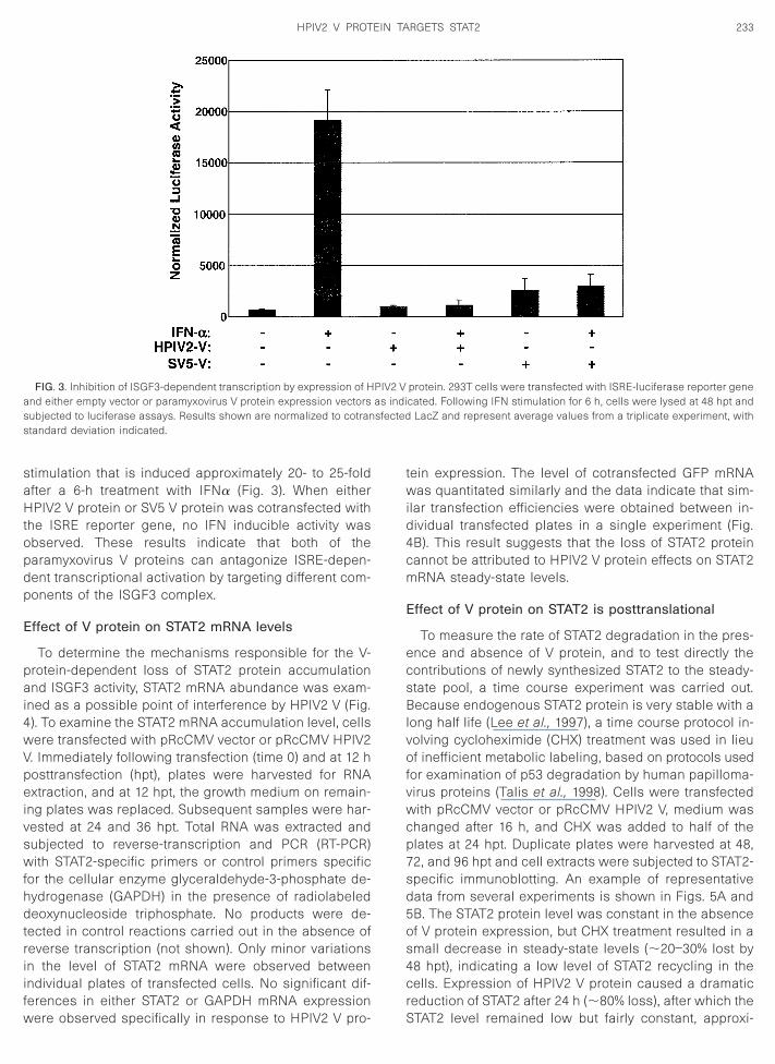
stimulation that is induced approximately 20- to 25-foldafter a 6-h treatment with IFNa (Fig. 3). When eitherHPIV2 V protein or SV5 V protein was cotransfected withthe ISRE reporter gene, no IFN inducible activity wasobserved. These results indicate that both of theparamyxovirus V proteins can antagonize ISRE-depen-dent transcriptional activation by targeting different com-ponents of the ISGF3 complex.
Effect of V protein on STAT2 mRNA levels
To determine the mechanisms responsible for the V-protein-dependent loss of STAT2 protein accumulationand ISGF3 activity, STAT2 mRNA abundance was exam-ined as a possible point of interference by HPIV2 V (Fig.4). To examine the STAT2 mRNA accumulation level, cellswere transfected with pRcCMV vector or pRcCMV HPIV2V. Immediately following transfection (time 0) and at 12 hposttransfection (hpt), plates were harvested for RNAextraction, and at 12 hpt, the growth medium on remain-ing plates was replaced. Subsequent samples were har-vested at 24 and 36 hpt. Total RNA was extracted andsubjected to reverse-transcription and PCR (RT-PCR)with STAT2-specific primers or control primers specificfor the cellular enzyme glyceraldehyde-3-phosphate de-hydrogenase (GAPDH) in the presence of radiolabeleddeoxynucleoside triphosphate. No products were de-tected in control reactions carried out in the absence ofreverse transcription (not shown). Only minor variationsin the level of STAT2 mRNA were observed betweenindividual plates of transfected cells. No significant dif-ferences in either STAT2 or GAPDH mRNA expression
FIG. 3. Inhibition of ISGF3-dependent transcription by expression of Hand either empty vector or paramyxovirus V protein expression vectorssubjected to luciferase assays. Results shown are normalized to cotranstandard deviation indicated.
were observed specifically in response to HPIV2 V pro-
tein expression. The level of cotransfected GFP mRNAwas quantitated similarly and the data indicate that sim-ilar transfection efficiencies were obtained between in-dividual transfected plates in a single experiment (Fig.4B). This result suggests that the loss of STAT2 proteincannot be attributed to HPIV2 V protein effects on STAT2mRNA steady-state levels.
Effect of V protein on STAT2 is posttranslational
To measure the rate of STAT2 degradation in the pres-ence and absence of V protein, and to test directly thecontributions of newly synthesized STAT2 to the steady-state pool, a time course experiment was carried out.Because endogenous STAT2 protein is very stable with along half life (Lee et al., 1997), a time course protocol in-
olving cycloheximide (CHX) treatment was used in lieuf inefficient metabolic labeling, based on protocols used
or examination of p53 degradation by human papilloma-irus proteins (Talis et al., 1998). Cells were transfectedith pRcCMV vector or pRcCMV HPIV2 V, medium was
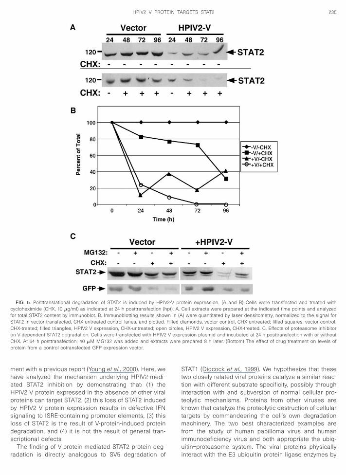
hanged after 16 h, and CHX was added to half of thelates at 24 hpt. Duplicate plates were harvested at 48,2, and 96 hpt and cell extracts were subjected to STAT2-pecific immunoblotting. An example of representativeata from several experiments is shown in Figs. 5A andB. The STAT2 protein level was constant in the absencef V protein expression, but CHX treatment resulted in amall decrease in steady-state levels (;20–30% lost by
48 hpt), indicating a low level of STAT2 recycling in thecells. Expression of HPIV2 V protein caused a dramaticreduction of STAT2 after 24 h (;80% loss), after which the
protein. 293T cells were transfected with ISRE-luciferase reporter genecated. Following IFN stimulation for 6 h, cells were lysed at 48 hpt and
LacZ and represent average values from a triplicate experiment, with
PIV2 Vas indisfected
STAT2 level remained low but fairly constant, approxi-

1dpsdd
2a
234 PARISIEN ET AL.
mately 20–40% of control levels. Addition of CHX re-vealed a rapid loss of STAT2 to near completion by 48hpt. Treatment of cells with IFN did not alter the V proteineffect on STAT2 and neither STAT1 nor STAT3 levels werealtered (not shown). This result indicates that steady-state STAT2 levels are the result of high stability of thebulk of STAT2 with approximately 20–30% of the totalprotein arising due to new protein synthesis. Expressionof HPIV2 V protein results in destabilization of the pre-existing protein but has little effect on new STAT2 proteinsynthesis. These findings indicate that a primary actionof HPIV2 V against STAT2 is posttranslational degrada-tion.
Effect of proteasome inhibitors on HPIV2 V-mediateddegradation
The results indicate that the effects of HPIV2 V proteinon STAT2 were posttranslational. As SV5-mediatedSTAT1 degradation was blocked by proteasome inhibi-tion in previously published experiments (Didcock et al.,
999), it seemed probable that the mechanism of STAT2egradation by HPIV2 might also involve the ubiquitin–roteasome pathway. To test this possibility, a protea-ome inhibitor was used to treat transfected cells toetermine the effect on HPIV2 V protein-mediated STAT2
FIG. 4. Analysis of STAT2 mRNA levels upon V protein expression. Avector or pRcCMV HPIV2 V as indicated. Each lane represents RNAnumbers indicate time (hpt) when RNA was isolated. B. Quantitation alevel of STAT2 normalized to the GAPDH control. The right graph show
egradation (Fig. 5C). When cells expressing HPIV2 V
protein were treated with the proteasome inhibitorMG132, little difference was observed in the STAT2steady-state level at 72 h posttransfection. However, con-current inhibition of new protein synthesis with CHXtreatment revealed that proteasome inhibition partiallypreserved STAT2 protein from the V-protein-mediateddegradation. This result indicates only a minor role forproteasome-mediated degradation in the actions of theHPIV2 V protein against STAT2 and suggests that addi-tional cellular mechanisms might also contribute to the Vprotein’s activity.
DISCUSSION
The anti-viral state induced by IFN is a strong selectivepressure for virus growth and replication. To evade thecellular IFN anti-viral effects, diverse virus types havecreated equally diverse strategies for bypassing thisinnate immune response. The paramyxoviruses SV5 andSendai virus have been recently shown to cause a lossof cellular STAT protein accumulation, but by differentmechanisms involving transcriptional inhibition cata-lyzed by Sendai virus C proteins (Garcin et al., 1999,
000) or posttranslational proteasomal degradation cat-lyzed by SV5 V protein (Didcock et al., 1999). Our finding
that infection of cells with HPIV2 leads to a loss of STAT2
R was carried out using total RNA from cells transfected with emptyd from an individual transfected 60-mm plate. U, untransfected cells;
alization of (A) by phosphorimage analysis. The left graph shows thealized levels of cotransfected GFP mRNA.
. RT-PCisolatend norm
s norm
steady-state protein accumulation is in general agree-
fSCoCp
235HPIV2 V PROTEIN TARGETS STAT2
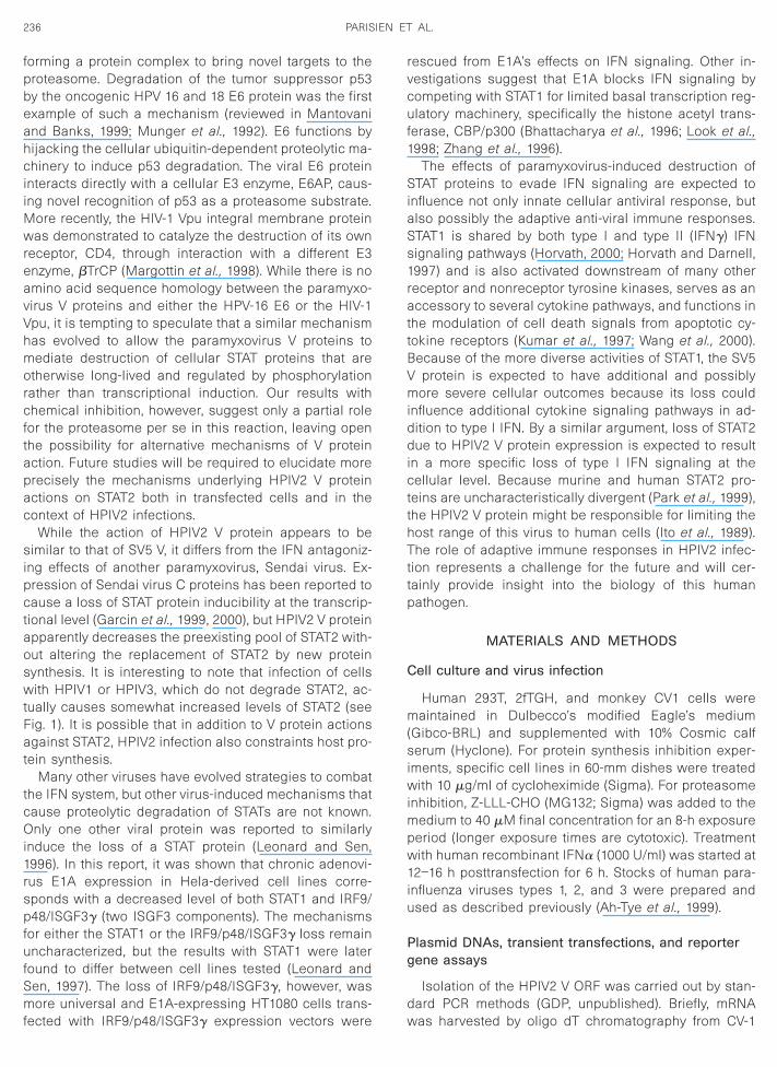
ment with a previous report (Young et al., 2000). Here, wehave analyzed the mechanism underlying HPIV2-medi-ated STAT2 inhibition by demonstrating that: (1) theHPIV2 V protein expressed in the absence of other viralproteins can target STAT2, (2) this loss of STAT2 inducedby HPIV2 V protein expression results in defective IFNsignaling to ISRE-containing promoter elements, (3) thisloss of STAT2 is the result of V-protein-induced proteindegradation, and (4) it is not the result of general tran-scriptional defects.
The finding of V-protein-mediated STAT2 protein deg-
FIG. 5. Posttranslational degradation of STAT2 is induced by HPIVcycloheximide (CHX, 10 mg/ml) as indicated at 24 h posttransfection (hor total STAT2 content by immunoblot. B. Immunoblotting results showTAT2 in vector-transfected, CHX-untreated control lanes, and plotted.HX-treated; filled triangles, HPIV2 V expression, CHX-untreated; openn V-dependent STAT2 degradation. Cells were transfected with HPIV2HX. At 64 h posttransfection, 40 mM MG132 was added and extractsrotein from a control cotransfected GFP expression vector.
radation is directly analogous to SV5 degradation of
STAT1 (Didcock et al., 1999). We hypothesize that thesetwo closely related viral proteins catalyze a similar reac-tion with different substrate specificity, possibly throughinteraction with and subversion of normal cellular pro-teolytic mechanisms. Proteins from other viruses areknown that catalyze the proteolytic destruction of cellulartargets by commandeering the cell’s own degradationmachinery. The two best characterized examples arefrom the study of human papilloma virus and humanimmunodeficiency virus and both appropriate the ubiq-uitin–proteasome system. The viral proteins physically
tein expression. (A and B) Cells were transfected and treated withCell extracts were prepared at the indicated time points and analyzed) were quantitated by laser densitometry, normalized to the signal foriamonds, vector control, CHX-untreated; filled squares, vector control,, HPIV2 V expression, CHX-treated. C. Effects of proteasome inhibitor
ession plasmid and incubated at 24 h posttransfection with or withoutprepared 8 h later. (Bottom) The effect of drug treatment on levels of
2-V propt). A.n in (A
Filled dcirclesV exprwere
interact with the E3 ubiquitin protein ligase enzymes by
aoswtFat
tcOi1rspf
1
SiaS
BVmiddictthTttp
C
m(siw
Pg
d
236 PARISIEN ET AL.
forming a protein complex to bring novel targets to theproteasome. Degradation of the tumor suppressor p53by the oncogenic HPV 16 and 18 E6 protein was the firstexample of such a mechanism (reviewed in Mantovaniand Banks, 1999; Munger et al., 1992). E6 functions byhijacking the cellular ubiquitin-dependent proteolytic ma-chinery to induce p53 degradation. The viral E6 proteininteracts directly with a cellular E3 enzyme, E6AP, caus-ing novel recognition of p53 as a proteasome substrate.More recently, the HIV-1 Vpu integral membrane proteinwas demonstrated to catalyze the destruction of its ownreceptor, CD4, through interaction with a different E3enzyme, bTrCP (Margottin et al., 1998). While there is noamino acid sequence homology between the paramyxo-virus V proteins and either the HPV-16 E6 or the HIV-1Vpu, it is tempting to speculate that a similar mechanismhas evolved to allow the paramyxovirus V proteins tomediate destruction of cellular STAT proteins that areotherwise long-lived and regulated by phosphorylationrather than transcriptional induction. Our results withchemical inhibition, however, suggest only a partial rolefor the proteasome per se in this reaction, leaving openthe possibility for alternative mechanisms of V proteinaction. Future studies will be required to elucidate moreprecisely the mechanisms underlying HPIV2 V proteinactions on STAT2 both in transfected cells and in thecontext of HPIV2 infections.
While the action of HPIV2 V protein appears to besimilar to that of SV5 V, it differs from the IFN antagoniz-ing effects of another paramyxovirus, Sendai virus. Ex-pression of Sendai virus C proteins has been reported tocause a loss of STAT protein inducibility at the transcrip-tional level (Garcin et al., 1999, 2000), but HPIV2 V protein
pparently decreases the preexisting pool of STAT2 with-ut altering the replacement of STAT2 by new proteinynthesis. It is interesting to note that infection of cellsith HPIV1 or HPIV3, which do not degrade STAT2, ac-
ually causes somewhat increased levels of STAT2 (seeig. 1). It is possible that in addition to V protein actionsgainst STAT2, HPIV2 infection also constraints host pro-
ein synthesis.Many other viruses have evolved strategies to combat
he IFN system, but other virus-induced mechanisms thatause proteolytic degradation of STATs are not known.nly one other viral protein was reported to similarly
nduce the loss of a STAT protein (Leonard and Sen,996). In this report, it was shown that chronic adenovi-us E1A expression in Hela-derived cell lines corre-ponds with a decreased level of both STAT1 and IRF9/48/ISGF3g (two ISGF3 components). The mechanisms
or either the STAT1 or the IRF9/p48/ISGF3g loss remainuncharacterized, but the results with STAT1 were laterfound to differ between cell lines tested (Leonard andSen, 1997). The loss of IRF9/p48/ISGF3g, however, wasmore universal and E1A-expressing HT1080 cells trans-
fected with IRF9/p48/ISGF3g expression vectors were wrescued from E1A’s effects on IFN signaling. Other in-vestigations suggest that E1A blocks IFN signaling bycompeting with STAT1 for limited basal transcription reg-ulatory machinery, specifically the histone acetyl trans-ferase, CBP/p300 (Bhattacharya et al., 1996; Look et al.,
998; Zhang et al., 1996).The effects of paramyxovirus-induced destruction of
TAT proteins to evade IFN signaling are expected tonfluence not only innate cellular antiviral response, butlso possibly the adaptive anti-viral immune responses.TAT1 is shared by both type I and type II (IFNg) IFN
signaling pathways (Horvath, 2000; Horvath and Darnell,1997) and is also activated downstream of many otherreceptor and nonreceptor tyrosine kinases, serves as anaccessory to several cytokine pathways, and functions inthe modulation of cell death signals from apoptotic cy-tokine receptors (Kumar et al., 1997; Wang et al., 2000).
ecause of the more diverse activities of STAT1, the SV5protein is expected to have additional and possibly
ore severe cellular outcomes because its loss couldnfluence additional cytokine signaling pathways in ad-ition to type I IFN. By a similar argument, loss of STAT2ue to HPIV2 V protein expression is expected to result
n a more specific loss of type I IFN signaling at theellular level. Because murine and human STAT2 pro-
eins are uncharacteristically divergent (Park et al., 1999),he HPIV2 V protein might be responsible for limiting theost range of this virus to human cells (Ito et al., 1989).he role of adaptive immune responses in HPIV2 infec-
ion represents a challenge for the future and will cer-ainly provide insight into the biology of this humanathogen.
MATERIALS AND METHODS
ell culture and virus infection
Human 293T, 2fTGH, and monkey CV1 cells wereaintained in Dulbecco’s modified Eagle’s medium
Gibco-BRL) and supplemented with 10% Cosmic calferum (Hyclone). For protein synthesis inhibition exper-
ments, specific cell lines in 60-mm dishes were treatedith 10 mg/ml of cycloheximide (Sigma). For proteasome
inhibition, Z-LLL-CHO (MG132; Sigma) was added to themedium to 40 mM final concentration for an 8-h exposureperiod (longer exposure times are cytotoxic). Treatmentwith human recombinant IFNa (1000 U/ml) was started at12–16 h posttransfection for 6 h. Stocks of human para-influenza viruses types 1, 2, and 3 were prepared andused as described previously (Ah-Tye et al., 1999).
lasmid DNAs, transient transfections, and reporterene assays
Isolation of the HPIV2 V ORF was carried out by stan-ard PCR methods (GDP, unpublished). Briefly, mRNA
as harvested by oligo dT chromatography from CV-1
tra(mmtt
Sdwtodrr
A
vimsSdic
237HPIV2 V PROTEIN TARGETS STAT2
cells infected with the Greer strain of HPIV-2. The V ORFwas amplified by RT-PCR using Superscript RT and Taqpolymerase. The following primers were used:
HP2-P59,59-GCGAATTCGTGATCAGATAACCTCCA-
CACCAGAATCATAC-39;SACI-39 vRNA sense,59-GCCGGTACCTTTAAGAGCTCAAT-
GATCTCCTTCACATTCTCCGC-39;
The PCR product was restricted with EcoRI and Asp718and cloned into the same sites of pGEM3 (Promega).Three independent clones were sequenced and found tomatch the published Toshiba strain sequence except fora single nucleotide substitution (an A to G at base 239 onthe published Toshiba sequence (Ohgimoto et al., 1990)).This substitution converts a threonine to an alanine atposition 56, possibly a strain-specific allelic difference. Itmay be relevant that the alanine at position 56 actuallymatches better to the SV5 V protein sequence.
The pGEM3 plasmid containing the Greer strain HPIV2V ORF was the template for a second amplification of theV ORF insert by PCR using Vent Polymerase (NEB, Bev-erly, MA). The following primers were engineered to addflanking NotI and ApaI restriction endonuclease recog-nition sites to the V cDNA:
59-GGGCGGCCGCGAATTCGTGATCAGATAAC-3959-GGGGGCCCAAGAGCTCAATGATCTCC-39.
The resulting PCR product was ligated into a NotI–ApaI restricted pRcCMV vector (Invitrogen) for mamma-lian expression and the resulting construct was verifiedby restriction mapping and nucleotide sequencing.
For transient cDNA transfection, cell monolayers weregrown to 60–80% confluency in 60-mm dishes. Transfec-tion of cells with cDNAs was carried out by standardCaPO4 transfection procedures and assayed at varioustimes after transfection (Ausubel et al., 1994). Transfec-ion of 293T cells by this method is highly efficient,esulting in typical transfection efficiencies of 95–100%s determined by FACS analysis of GFP-transfected cells
JFL and CMH, unpublished). Nonetheless, in all experi-ents, cotransfected controls were included to deter-ine the uniformity and efficiency of the transfection and
o verify that all plates in a given experiment were equallyransfected. For cell lysate analysis, 1 mg of a control
EGFP expression plasmid was included for evaluation oftransfection efficiency by inverted fluorescence micros-copy prior to lysis. For reporter gene assays, 1 mg of acontrol CMV-LacZ plasmid was added as a control forthe transfection procedure and luciferase values werenormalized to b-gal activity. In addition, 5 mg of a reporter
gene containing five copies of the ISG54 ISRE elementupstream of a TATA box and firefly luciferase ORF plas-mid was included. After 16 h, transfection medium wasreplaced with fresh medium supplemented with 10%Cosmic calf serum. Cells were then harvested in anappropriate lysis buffer and analyzed either by SDS–polyacrylamide gel electrophoresis or by measuring lu-ciferase activity according to the manufacturer’s protocol(Promega).
Extracts, antibodies, and immunoblotting
Antiserum specific for STAT1 or STAT2 were obtainedfrom Santa Cruz Biotechnology, antibodies specific forGFP were obtained from Clontech, and an antibody toHPIV2-infected cell extracts was obtained from WhitakerBiochemicals. Cell extracts were prepared as described(Shuai et al., 1994). Cells were washed and scraped intoice-cold PBS and pelleted at 1500 rpm. Subsequent cellpellets were lysed in 2 vol of whole cell extract buffer (50mM Tris, pH 8.0, 280 mM NaCl, 0.5% IGEPAL, 0.2 mMEDTA, 2 mM EGTA, 10% glycerol, 1 mM DTT) supple-mented with a protease inhibitor cocktail (Complete,Boehringer Mannheim) for 15 min. Cellular debris waspelleted at 14,000 rpm and the supernatant was analyzeddirectly or stored at 280°C. Samples were added to
DS–gel loading buffer, boiled for 5 min, and loadedirectly onto SDS–polyacrylamide gels (7%, 12%). Gelsere transferred to nitrocellulose membranes and de-
ected with specific antibodies to the C-terminal portionsf STAT1 and STAT2. Protein–antibody interactions wereetected using secondary antiserum conjugated horse-
adish peroxidase and enhanced chemiluminescenceeagents (NEN Life Sciences).
nalysis of mRNA
RT-PCR reactions were carried out as described pre-iously (Horvath et al., 1996). Total cellular RNA was
solated with Trizol reagent (Gibco-BRL) according to theanufacturer’s instructions. First-strand cDNAs were
ynthesized from DNAse-I-treated total RNA by use ofuperScript II reverse transcriptase (Gibco-BRL) and ran-om antisense primers according to the manufacturer’s
nstruction. Diluted cDNAs were subjected to PCR amplifi-ations with [a-32P]dCTP and primers specific for glycer-
aldehyde-3-phosphatase dehydrogenase (59-GTGAAGG-TCGGAGTCAAC-39 and 59-TGGAATTTGCCATGGGTG-39)GFP 59-ACGTAAACGGCCACAAGTTC-39 and 59-AAG-TCGTGCTGCTTCATGTG-39, or STAT2 (59-GAAGATCT-GAAACTCTCAATGAACTGG-39 and 59-GGGAATTCCC-TAGGGAGCTCTGATGCAGG-39).
PCR-amplified products were separated on a 5% poly-
acrylamide gel, and autoradiography was performed.
G
G
G
H
H
I
I
K
LL
L
L
L
L
238 PARISIEN ET AL.
Samples were quantitated with a Storm phosphorimageanalyzer and ImageQuant software (Molecular Dynam-ics).
ACKNOWLEDGMENTS
We are grateful to John Rassa for his cloning of HPIV2 V cDNAs,Natalia Poltoratskaia for technical assistance with HPIV infections,Chris Cardozo, Sherwin Wilk, and Avrom Caplan for advice on prote-olysis, and Biao He for helpful discussions. This work was supported inpart by the New York City Council Speaker’s Fund for BiomedicalResearch: Toward the Science of Patient Care and a Mount SinaiDean’s Research Incentive Award (to CMH). CMH is the Ira M. Jacob-son, M.D., Liver Scholar of the American Liver Foundation. RAL is anInvestigator of the Howard Hughes Medical Institute.
REFERENCES
Ah-Tye, C., Schwartz, S., Huberman, K., Carlin, E., and Moscona, A.(1999). Virus–receptor interactions of human parainfluenza virusestypes 1, 2 and 3. Microb. Pathog. 27(5), 329–336.
Ausubel, F. M., Brent, R., Kingston, R. E., Moore, D. D., Seidman, J. G.,Smith, J. A., and Struhl, K. (1994). “Current Protocols in MolecularBiology.” Wiley, New York.
Bergmann, M., Garcia-Sastre, A., Carnero, E., Pehamberger, H., Wolff,K., Palese, P., and Muster, T. (2000). Influenza virus NS1 proteincounteracts PKR-mediated inhibition of replication. J. Virol. 74(13),6203–6206.
Bhattacharya, S., Eckner, R., Grossman, S., Oldread, E., Arany, Z.,D’Andrea, A., and Livingston, D. M. (1996). Cooperation of Stat2 andp300/CBP in signalling induced by interferon a. Nature 383(26),344–347.
Darnell, J. E., Jr. (1997). STATs and gene regulation. Science 277,1630–1635.
Didcock, L., Young, D. F., Goodbourn, S., and Randall, R. E. (1999). TheV protein of simian virus 5 inhibits interferon signalling by targetingSTAT1 for proteasome-mediated degradation. J. Virol. 73(12), 9928–9933.
arcin, D., Curran, J., and Kolakofsky, D. (2000). Sendai virus C proteinsmust interact directly with cellular components to interfere withinterferon action. J. Virol. 74(19), 8823–8830.
arcin, D., Latorre, P., and Kolakofsky, D. (1999). Sendai virus C proteinscounteract the interferon-mediated induction of an antiviral state.J. Virol. 73(8), 6559–6565.
oodbourn, S., Didcock, L., and Randall, R. E. (2000). Interferons: Cellsignalling, immune modulation, antiviral response and virus counter-measures. J. Gen. Virol. 81(Pt. 10), 2341–2364.
Horvath, C. M. (2000). STAT proteins and transcriptional responses toextracellular signals. TIBS 25, 496–502.
Horvath, C. M., and Darnell, J. E., Jr. (1996). The antiviral state inducedby alpha interferon and gamma interferon requires transcriptionallyactive Stat1 protein. J. Virol. 70, 647–650.
orvath, C. M., and Darnell, J. E., Jr. (1997). The state of the STATs:Recent developments in the study of signal transduction to thenucleus. Curr. Opin. Cell Biol. 9(2), 233–239.
orvath, C. M., Stark, G. R., Kerr, I. M., and Darnell, J. E., Jr. (1996).Interactions between STAT and non-STAT proteins in the interferonstimulated gene factor 3 transcription complex. Mol. Cell. Biol. 16(12),6957–6964.
saacs, A., and Lindemann, J. (1957). Virus interference. I. The inter-feron. Proc. R. Soc. Lond. B 147, 258–267.
to, Y., Tsurudome, M., Bando, H., Komada, H., and Nishio, M. (1989).Incomplete replication of human parainfluenza virus type 2 in mouse
L929 cells. Arch. Virol. 108(1–2), 137–144.itajewski, J., Schneider, R. J., Safer, B., Munemitsu, S. M., Samuel,C. E., Thimmappaya, B., and Shenk, T. (1986). Adenovirus VAI RNAantagonizes the antiviral action of interferon by preventing acti-vation of the interferon-induced eIF-2 alpha kinase. Cell 45(2),195–200.
Kumar, A., Commane, M., Flickinger, T. W., Horvath, C. M., and Stark,G. R. (1997). Defective tumor necrosis factor-a induced apoptosis inSTAT1-null cells due to low constitutive levels of caspases. Science278, 1630–1632.
au, J. F., and Horvath, C. M. Unpublished observations.au, J. F., Parisien, J. P., and Horvath, C. M. (2000). Interferon regulatory
factor subcellular localization is determined by a bipartite nuclearlocalization signal in the DNA-binding domain and interaction withcytoplasmic retention factors. Proc. Natl. Acad. Sci. USA 97(13),7278–7283.
ee, C. K., Bluyssen, H. A., and Levy, D. E. (1997). Regulation ofinterferon-alpha responsiveness by the duration of Janus kinaseactivity. J. Biol. Chem. 272(35), 21872–21877.
eonard, G. T., and Sen, G. C. (1996). Effects of adenovirus E1A proteinon interferon-signaling. Virology 224(1), 25–33.
eonard, G. T., and Sen, G. C. (1997). Restoration of interferon re-sponses of adenovirus E1A-expressing HT1080 cell lines by overex-pression of p48 protein. J. Virol. 71(7), 5095–5101.
eung, S., Qureshi, S. A., Kerr, I. M., Darnell, J. E., Jr., and Stark, G. R.(1995). Role of STAT2 in the interferon-a/b signaling pathway. Mol.Cell. Biol. 15, 1312–1317.
Levy, D. E., Kessler, D. S., Pine, R. I., and Darnell, J. E., Jr. (1989).Cytoplasmic activation of ISGF3, the positive regulator of interferon-astimulated transcription, reconstituted in vitro. Genes Dev. 3, 1362–1372.
Look, D. C., Roswit, W. T., Frick, A. G., Gris-Alevy, Y., Dickhaus, D. M.,Walter, M. J., and Holtzman, M. J. (1998). Direct suppression ofStat1 function during adenoviral infection. Immunity 9(6), 871–880.
Mantovani, F., and Banks, L. (1999). The interaction between p53 andpapillomaviruses. Semin. Cancer Biol. 9(6), 387–395.
Margottin, F., Bour, S. P., Durand, H., Selig, L., Benichou, S., Richard, V.,Thomas, D., Strebel, K., and Benarous, R. (1998). A novel human WDprotein, h-beta TrCp, that interacts with HIV-1 Vpu connects CD4 tothe ER degradation pathway through an F-box motif. Mol. Cell. 1(4),565–574.
Martinez-Moczygemba, M., Gutch, M. J., French, D. L., and Reich, N. C.(1997). Distinct STAT structure promotes interaction of STAT2 with thep48 subunit of the interferon-alpha-stimulated transcription factorISGF3. J. Biol. Chem. 272(32), 20070–20076.
Munger, K., Scheffner, M., Huibregtse, J. M., and Howley, P. M. (1992).Interactions of HPV E6 and E7 oncoproteins with tumour suppressorgene products. Cancer Surv. 12, 197–217.
Ohgimoto, S., Bando, H., Kawano, M., Okamoto, K., Kondo, K., Tsuru-dome, M., Nishio, M., and Ito, Y. (1990). Sequence analysis of P geneof human parainfluenza type 2 virus: P and cysteine-rich proteins aretranslated by two mRNAs that differ by two nontemplated G residues.Virology 177(1), 116–123.
Park, C., Lecomte, M. J., and Schindler, C. (1999). Murine Stat2 isuncharacteristically divergent. Nucleic Acids Res. 27(21), 4191–4199.
Qureshi, S. A., Leung, S., Kerr, I. M., Stark, G. R., and Darnell, J. E., Jr.(1996). Function of Stat2 protein in transcriptional activation by IFN-a.Mol. Cell. Biol. 16, 288–293.
Shuai, K., Horvath, C. M., Tsai-Huang, L. H., Qureshi, S., Cowburn, D.,and Darnell, J. E., Jr. (1994). Interferon activation of the transcriptionfactor Stat91 involves dimerization through SH2-phosphotyrosyl pep-tide interactions. Cell 76, 821–828.
Talis, A. L., Huibregtse, J. M., and Howley, P. M. (1998). The role of E6AP
in the regulation of p53 protein levels in human papillomavirus
239HPIV2 V PROTEIN TARGETS STAT2
(HPV)-positive and HPV-negative cells. J. Biol. Chem. 273(11), 6439–6445.
Talon, J., Horvath, C. M., Polley, R., Basler, C. F., Muster, T., Palese, P.,and Garcia-Sastre, A. (2000). Activation of interferon regulatory factor3 is inhibited by the influenza A virus NS1 protein. J. Virol. 74(17),7989–7996.
Wang, Y., Wu, T. R., Cai, S., Welte, T., and Chin, Y. E. (2000). Stat1 as acomponent of tumor necrosis factor alpha receptor 1-TRADD signal-ing complex to inhibit NF-kappaB activation. Mol. Cell. Biol. 20(13),4505–4512.
Young, D. F., Didcock, L., Goodbourn, S., and Randall, R. E. (2000).Paramyxoviridae use distinct virus-specific mechanisms to circum-vent the interferon response. Virology 269(2), 383–390.
Zhang, J. J., Vinkemeier, U., Gu, W., Chakravarti, D., Horvath, C. M., andDarnell, J. E., Jr. (1996). Two contact regions between Stat1 andCBP/p300 in interferon g signaling. Proc. Natl. Acad. Sci. USA 93,15092–15096.
Zhou, A., Paranjape, J. M., Der, S. D., Williams, B. R., and Silverman, R. H.(1999). Interferon action in triply deficient mice reveals the existenceof alternative antiviral pathways. Virology 258(2), 435–440.