Dynamics of receptor and protein transducer homodimerisation
11
BioMed Central Page 1 of 11 (page number not for citation purposes) BMC Systems Biology Open Access Research article Dynamics of receptor and protein transducer homodimerisation Julio Vera †1 , Thomas Millat †1 , Walter Kolch 2,3 and Olaf Wolkenhauer* 1 Address: 1 University of Rostock, 18051 Rostock, Germany, 2 The Beatson Institute for Cancer Research, Glasgow G61 1BD, UK and 3 University of Glasgow, Sir Henry Wellcome Functional Genomics Facility, Glasgow, G12 8QQ, UK Email: Julio Vera - [email protected]; Thomas Millat - [email protected]; Walter Kolch - [email protected]; Olaf Wolkenhauer* - [email protected] * Corresponding author †Equal contributors Abstract Background: Signalling pathways are complex systems in which not only simple monomeric molecules interact, but also more complex structures that include constitutive or induced protein assemblies. In particular, the hetero-and homo-dimerisation of proteins is a commonly encountered motif in signalling pathways. Several authors have suggested in recent times that dimerisation relates to a series of physical and biological outcomes used by the cell in the regulation of signal transduction. Results: In this paper we investigate the role of homodimerisation in receptor-protein transducer interactions. Towards this end, mathematical modelling is used to analyse the features of such kind of interactions and to predict the behaviour of the system under different experimental conditions. A kinetic model in which the interaction between homodimers provokes a dual mechanism of activation (single and double protein transducer activation at the same time) is proposed. In addition, we analyse under which conditions the use of a power-law representation for the system is useful. Furthermore, we investigate the dynamical consequences of this dual mechanism and compare the performance of the system in different simulated experimental conditions. Conclusion: The analysis of our mathematical model suggests that in receptor-protein interacting systems with dual mechanism there may be a shift between double and single activation in a way that intense double protein transducer activation could initiate and dominate the signal in the short term (getting a fast intense signal), while single protein activation could control the system in the medium and long term (when input signal is weaker and decreases slowly). Our investigation suggests that homodimerisation and oligomerisation are mechanisms used to enhance and regulate the dynamic properties of the initial steps in signalling pathways. Background The processing of information in living cells is carried out by signal transduction pathways [1]. Through the binding of external ligands to extracellular receptors, the cell can receive signals from its environment and transfer informa- tion into the cell. This information flow is regulated, amplified or modulated by different feedback mecha- nisms and interactions with other pathways (crosstalk). Moreover, signalling pathways are complex systems in which not only simple monomeric molecules interact but also more complex structures that include constitutive or induced protein assemblies [2-4]. In particular, the het- Published: 31 October 2008 BMC Systems Biology 2008, 2:92 doi:10.1186/1752-0509-2-92 Received: 19 December 2007 Accepted: 31 October 2008 This article is available from: http://www.biomedcentral.com/1752-0509/2/92 © 2008 Vera et al; licensee BioMed Central Ltd. This is an Open Access article distributed under the terms of the Creative Commons Attribution License (http://creativecommons.org/licenses/by/2.0 ), which permits unrestricted use, distribution, and reproduction in any medium, provided the original work is properly cited.
Transcript of Dynamics of receptor and protein transducer homodimerisation
BioMed CentralBMC Systems Biology
ss
Open AcceResearch articleDynamics of receptor and protein transducer homodimerisationJulio Vera†1, Thomas Millat†1, Walter Kolch2,3 and Olaf Wolkenhauer*1Address: 1University of Rostock, 18051 Rostock, Germany, 2The Beatson Institute for Cancer Research, Glasgow G61 1BD, UK and 3University of Glasgow, Sir Henry Wellcome Functional Genomics Facility, Glasgow, G12 8QQ, UK
Email: Julio Vera - [email protected]; Thomas Millat - [email protected]; Walter Kolch - [email protected]; Olaf Wolkenhauer* - [email protected]
* Corresponding author †Equal contributors
AbstractBackground: Signalling pathways are complex systems in which not only simple monomericmolecules interact, but also more complex structures that include constitutive or induced proteinassemblies. In particular, the hetero-and homo-dimerisation of proteins is a commonlyencountered motif in signalling pathways. Several authors have suggested in recent times thatdimerisation relates to a series of physical and biological outcomes used by the cell in the regulationof signal transduction.
Results: In this paper we investigate the role of homodimerisation in receptor-protein transducerinteractions. Towards this end, mathematical modelling is used to analyse the features of such kindof interactions and to predict the behaviour of the system under different experimental conditions.A kinetic model in which the interaction between homodimers provokes a dual mechanism ofactivation (single and double protein transducer activation at the same time) is proposed. Inaddition, we analyse under which conditions the use of a power-law representation for the systemis useful. Furthermore, we investigate the dynamical consequences of this dual mechanism andcompare the performance of the system in different simulated experimental conditions.
Conclusion: The analysis of our mathematical model suggests that in receptor-protein interactingsystems with dual mechanism there may be a shift between double and single activation in a waythat intense double protein transducer activation could initiate and dominate the signal in the shortterm (getting a fast intense signal), while single protein activation could control the system in themedium and long term (when input signal is weaker and decreases slowly). Our investigationsuggests that homodimerisation and oligomerisation are mechanisms used to enhance and regulatethe dynamic properties of the initial steps in signalling pathways.
BackgroundThe processing of information in living cells is carried outby signal transduction pathways [1]. Through the bindingof external ligands to extracellular receptors, the cell canreceive signals from its environment and transfer informa-tion into the cell. This information flow is regulated,
amplified or modulated by different feedback mecha-nisms and interactions with other pathways (crosstalk).Moreover, signalling pathways are complex systems inwhich not only simple monomeric molecules interact butalso more complex structures that include constitutive orinduced protein assemblies [2-4]. In particular, the het-
Published: 31 October 2008
BMC Systems Biology 2008, 2:92 doi:10.1186/1752-0509-2-92
Received: 19 December 2007Accepted: 31 October 2008
This article is available from: http://www.biomedcentral.com/1752-0509/2/92
© 2008 Vera et al; licensee BioMed Central Ltd. This is an Open Access article distributed under the terms of the Creative Commons Attribution License (http://creativecommons.org/licenses/by/2.0), which permits unrestricted use, distribution, and reproduction in any medium, provided the original work is properly cited.
Page 1 of 11(page number not for citation purposes)
BMC Systems Biology 2008, 2:92 http://www.biomedcentral.com/1752-0509/2/92
ero- and homo-dimerisation of proteins is a commonlyencountered motif in signalling pathways.
In Klemm [5] the role of dimerisation as a regulatorymechanism in signal transduction is analysed and dis-cussed. Dimerisation is defined as an interaction produc-ing a protein-protein complex composed of two subunits,either identical (homodimerisation) or non-identical(heterodimerisation). The authors argue that dimerisa-tion relates to a series of physical and biological outcomesused by the cell in the regulation of signal transduction.The biophysical outcomes referred to facilitation of prox-imity and orientation in protein interaction, differentialregulation through heterodimerisation, emergence of spa-tio-temporal boundaries, enhanced specificity and regula-tion of monomer-to-dimer transitions. The role ofhomodimeric receptors in the activation and dimerisationof intermediate proteins and transcription factors hasbeen already described in the literature [6-8].
A well-studied example are the JAK/STAT signalling path-ways [9]. In case of the JAK2/STAT5 signalling pathway,the Epo receptor is a preformed inactive dimer in theplasma membrane [10,11]. The binding of Epo results inthe activation of the JAK2 kinase and subsequent phos-phorylation of the cytosolic domain of each Epo receptormonomeric subunit. STAT5 proteins bind to the tyrosinephosphorylated Epo receptor and gets phosphorylation.Afterwards, they dimerised and translocate to the nucleus.The spatial conformation of the receptor as a dimer seemsto indicate that each activated Epo receptor monomercould phosphorylate simultaneously at least one STAT5molecule. The correspondence between the existence of ahomodimer activated receptor and the activation of ahomodimer transduction protein suggests a possible com-plex underlying molecular mechanism for the activationand dimerisation process. Similar behaviour has beenshown in JAK/STAT pathways [12] and other signallingpathways [13-18], suggesting that this homodimer-homodimer interaction could constitute a more generalpattern in cell signalling systems.
The purpose of this work is to use mathematical model-ling to suggest mechanisms of interaction by which thishomodimer-homodimer interaction can occur. In ourwork, two mathematical modelling frameworks are usedand compared. We furthermore investigate the dynamicalconsequences of the interaction mechanisms suggestedand propose general features of an experimental design todiscriminate between the different mechanisms.
Results and discussionMechanistic modellingIn this paper we support the hypothesis that in the trans-duction of signals via homodimeric proteins, the dimeric
nature of the receptor plays an essential role in the fastresponse of the biological system. The surface density ofmany plasma membrane receptors tends to be very low[19]. For a low density of receptors at the plasma mem-brane, active mechanisms for the homodimerisation ofthe cytosolic interacting proteins are required to boost theintracellular response of the system to external stimuli.Towards this end, we propose that the dimeric structure ofthe receptor allows not only the simple activation of amonomer binding protein per receptor but also a simulta-neous coordinated activation of two monomers of thebinding protein, one at each subunit of the receptor. Thecase in which we consider the single interaction betweenone subunit of the receptor and a monomer of the proteintransducer, we call "single protein activation process"with respect to the number of receptor-protein interac-tions. In contrast, two subunits of the same receptor inter-acting simultaneously with two different monomers arereferred to as a "double protein activation process". In thefollowing we are going to assume that higher-order proc-esses, allowing for simultaneous activation of two or moreprotein monomers at the same receptor monomer, are notsignificant. Several studies with the PDGF receptor inwhich all tyrosine phosphorylation sites were mutatedindividually or in combination showed that the signallingwas unchanged when redundant sites were knocked out,suggesting that this assumption is in principle meaningful[20].
The mathematical framework considered describes thetemporal evolution of protein concentrations with cou-pled ordinary differential equations. The rate of change ofmolecular species Xi (number of activated or inactivatedprotein transducer and receptors) is expressed as:
where j are rate coefficients, gjk kinetic orders and ij thestoichiometric coefficients. The stoichiometric coefficientij is positive for products and negative for reactants anddescribes how many molecules of Xi are converted in theconsidered reaction. The rate coefficients contain infor-mation about the physical properties of the reaction, likeactivation energies and internal states [21]. If we assumethat environmental conditions, like temperature, pressureand pH-value do not vary over time, j is a rate constant.The interpretation of the kinetic order gjk depends on thechosen level of description for the biochemical network.When (1) is derived within the framework of statisticalphysics and the complete reaction mechanism is knownand considered, the coefficient gjk has a clear mechanisticmeaning and defines the number of molecules of speciesXi involved on the considered reaction. The kinetic orderhas in this case a positive integer value. In contrast, if we
dXidt
Xij j
jkg
k
jk= ⋅∑ ∏ , (1)
Page 2 of 11(page number not for citation purposes)
BMC Systems Biology 2008, 2:92 http://www.biomedcentral.com/1752-0509/2/92
aggregate elementary reaction steps into a combinedpower-law expression, with the same form of (1), theinterpretation of the coefficient gjk changes and they cantake non-integer values [22-25]. This latter case is investi-gated in the next section.
For the purpose of our investigation, we consider thegeneric case of a diffusive signalling protein (for simplic-ity, named protein, P, in our discussion), which is activatedand dimerised after the interaction with a homodimericactivated receptor (named receptor, R*). In order to focusthe investigation on general design principles underlyingthe homodimer receptor-homodimer transducer proteinclass of interactions, we simplify the mathematical mod-elling of the system, deliberately neglecting details thataccount for differences in the class. The procedure is sim-ilar to the one used in [26].
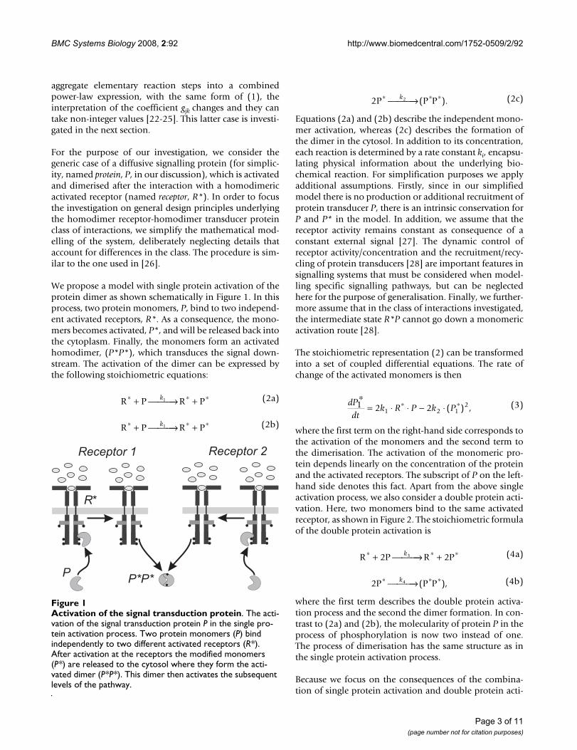
We propose a model with single protein activation of theprotein dimer as shown schematically in Figure 1. In thisprocess, two protein monomers, P, bind to two independ-ent activated receptors, R*. As a consequence, the mono-mers becomes activated, P*, and will be released back intothe cytoplasm. Finally, the monomers form an activatedhomodimer, (P*P*), which transduces the signal down-stream. The activation of the dimer can be expressed bythe following stoichiometric equations:
Equations (2a) and (2b) describe the independent mono-mer activation, whereas (2c) describes the formation ofthe dimer in the cytosol. In addition to its concentration,each reaction is determined by a rate constant ki, encapsu-lating physical information about the underlying bio-chemical reaction. For simplification purposes we applyadditional assumptions. Firstly, since in our simplifiedmodel there is no production or additional recruitment ofprotein transducer P, there is an intrinsic conservation forP and P* in the model. In addition, we assume that thereceptor activity remains constant as consequence of aconstant external signal [27]. The dynamic control ofreceptor activity/concentration and the recruitment/recy-cling of protein transducers [28] are important features insignalling systems that must be considered when model-ling specific signalling pathways, but can be neglectedhere for the purpose of generalisation. Finally, we further-more assume that in the class of interactions investigated,the intermediate state R*P cannot go down a monomericactivation route [28].
The stoichiometric representation (2) can be transformedinto a set of coupled differential equations. The rate ofchange of the activated monomers is then
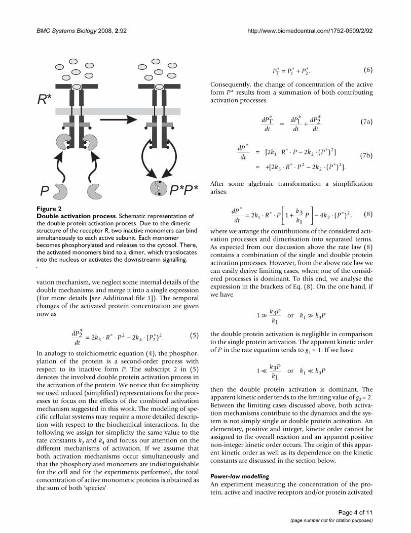
where the first term on the right-hand side corresponds tothe activation of the monomers and the second term tothe dimerisation. The activation of the monomeric pro-tein depends linearly on the concentration of the proteinand the activated receptors. The subscript of P on the left-hand side denotes this fact. Apart from the above singleactivation process, we also consider a double protein acti-vation. Here, two monomers bind to the same activatedreceptor, as shown in Figure 2. The stoichiometric formulaof the double protein activation is
where the first term describes the double protein activa-tion process and the second the dimer formation. In con-trast to (2a) and (2b), the molecularity of protein P in theprocess of phosphorylation is now two instead of one.The process of dimerisation has the same structure as inthe single protein activation process.
Because we focus on the consequences of the combina-tion of single protein activation and double protein acti-
R P R P∗ ∗ ∗+ ⎯ →⎯⎯ +k1 (2a)
R P R P∗ ∗ ∗+ ⎯ →⎯⎯ +k1 (2b)
2P (P P∗ ∗ ∗⎯ →⎯⎯k2 ). (2c)
dPdt
k R P k P1 2 21 2 12
∗= ⋅ ⋅ − ⋅∗ ∗( ) , (3)
R P R P∗ ∗ ∗+ ⎯ →⎯⎯ +2 23k (4a)
2 4P (P P∗ ∗ ∗⎯ →⎯⎯k ), (4b)
Activation of the signal transduction proteinFigure 1Activation of the signal transduction protein. The acti-vation of the signal transduction protein P in the single pro-tein activation process. Two protein monomers (P) bind independently to two different activated receptors (R*). After activation at the receptors the modified monomers (P*) are released to the cytosol where they form the acti-vated dimer (P*P*). This dimer then activates the subsequent levels of the pathway.
Page 3 of 11(page number not for citation purposes)
BMC Systems Biology 2008, 2:92 http://www.biomedcentral.com/1752-0509/2/92
vation mechanism, we neglect some internal details of thedouble mechanisms and merge it into a single expression(For more details [see Additional file 1]). The temporalchanges of the activated protein concentration are givennow as
In analogy to stoichiometric equation (4), the phosphor-ylation of the protein is a second-order process withrespect to its inactive form P. The subscript 2 in (5)denotes the involved double protein activation process inthe activation of the protein. We notice that for simplicitywe used reduced (simplified) representations for the proc-esses to focus on the effects of the combined activationmechanism suggested in this work. The modeling of spe-cific cellular systems may require a more detailed descrip-tion with respect to the biochemical interactions. In thefollowing we assign for simplicity the same value to therate constants k2 and k4 and focuss our attention on thedifferent mechanisms of activation. If we assume thatboth activation mechanisms occur simultaneously andthat the phosphorylated monomers are indistinguishablefor the cell and for the experiments performed, the totalconcentration of active monomeric proteins is obtained asthe sum of both 'species'
Consequently, the change of concentration of the activeform P* results from a summation of both contributingactivation processes
After some algebraic transformation a simplificationarises:
where we arrange the contributions of the considered acti-vation processes and dimerisation into separated terms.As expected from our discussion above the rate law (8)contains a combination of the single and double proteinactivation processes. However, from the above rate law wecan easily derive limiting cases, where one of the consid-ered processes is dominant. To this end, we analyse theexpression in the brackets of Eq. (8). On the one hand, ifwe have
the double protein activation is negligible in comparisonto the single protein activation. The apparent kinetic orderof P in the rate equation tends to g1 = 1. If we have
then the double protein activation is dominant. Theapparent kinetic order tends to the limiting value of g2 = 2.Between the limiting cases discussed above, both activa-tion mechanisms contribute to the dynamics and the sys-tem is not simply single or double protein activation. Anelementary, positive and integer, kinetic order cannot beassigned to the overall reaction and an apparent positivenon-integer kinetic order occurs. The origin of this appar-ent kinetic order as well as its dependence on the kineticconstants are discussed in the section below.
Power-law modellingAn experiment measuring the concentration of the pro-tein, active and inactive receptors and/or protein activated
dPdt
k R P k P2 2 232
4 22
∗= ⋅ ⋅ − ⋅∗ ∗( ) . (5)
P P PT∗ ∗ ∗= +1 2 . (6)
dPdt
dPdt
dPdt
T∗
=∗
+∗
1 2 (7a)
dPdt
k R P k P
k R P k P
∗= ⋅ ⋅ − ⋅
= + ⋅ ⋅ − ⋅
∗ ∗
∗ ∗
[ ( ) ]
[ ( ) ].
2 2
2 2
1 22
32
22
(7b)
dPdt
k R Pkk
P k P∗
= ⋅ ⋅ +⎡
⎣⎢
⎤
⎦⎥ − ⋅∗ ∗2 1 3
141 2
2( ) , (8)
1 31
1 3 k Pk
k k Por
1 31
1 3 k Pk
k k Por
Double activation processFigure 2Double activation process. Schematic representation of the double protein activation process. Due to the dimeric structure of the receptor R, two inactive monomers can bind simultaneously to each active subunit. Each monomer becomes phosphorylated and releases to the cytosol. There, the activated monomers bind to a dimer, which translocates into the nucleus or activates the downstreamn signalling.
Page 4 of 11(page number not for citation purposes)
BMC Systems Biology 2008, 2:92 http://www.biomedcentral.com/1752-0509/2/92
dimers, cannot distinguish between active monomerswhich are produced in a single or double protein activa-tion process. To distinguish the different activation mech-anisms, an indirect method is required, anotherpossibility is to investigate the structure of the protein.However, the detection of two binding sites at the samereceptor, does not guarantee that both sites are usedsimultaneously nor that this is necessarily an effective acti-vation process.
In order to investigate the possibility of two different andsimultaneously acting phosphorylation mechanisms fur-ther, we aggregate both processes introduced in the abovesection into a single contribution. The order of the recep-tor R in the activation process is one, as in the mechanisticmodel (8). However, the contribution of protein P is acombination of the considered single and double activa-tion processes. Since both activation mechanismsdescribed by k1 and k3 are indistinguishable, the estima-tion of these kinetic parameters generates identifiabilityissues. If we consider a situation in which both processescannot be distinguished, a feasible way to reproduce thiscomplexity is the use of a power-law representation,allowing for non-integer kinetic orders. As discussed in[25], power-law models are useful for modelling cellularsignalling when the exact reaction mechanism isunknown or if experimental data are not sufficient. For anexample in cell signalling we refer to [29]: in that work,quantitative time course data were used to identify apower-law model. If we used a power-law term, the pro-duction term (phosphorylation of P) of the new rate equa-tion has the following structure:
V(R*, P) 1·R·Pg. (9)
The apparent kinetic order g, which describes the role ofthe protein, has now a non-integer value between one andtwo. The specific value of g depends on the rate constantsk1, k3 and the concentration of the inactive protein P asshown in the subsection "Mechanistic modelling". Wenote that this apparent kinetic order is only a mathemati-cal analogue to the kinetic order of elementary reactions.It does not provide detailed information about the under-lying mechanisms and hence, it cannot be interpreted inthe same way as the kinetic order of an elementary reac-tion scheme. In addition to the apparent kinetic order g,we also introduce an apparent kinetic constant 1 in thepower-law representation (9). Due to the aggregation ofproduction terms in (9), this apparent kinetic constantdoes not coincide with the kinetic constant in the mecha-nistic rate law (8). Nevertheless, it depends on the kineticconstants k1 and k3 but also on the ratio of efficiency ofsingle and double protein activation processes.
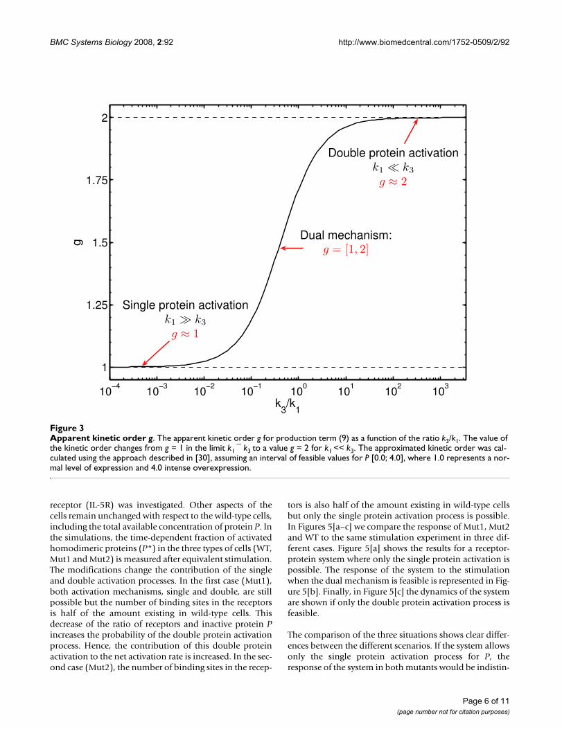
In the considered case, the differences in the efficiency ofthe considered mechanisms are determined by the pro-duction terms of single (3) and double protein activationprocesses (5). In our analysis we neglect the common con-tributions of the activated receptor concentration, R* andconstant prefactors in both terms. As a consequence, theratio of efficiency is mainly determined by the ratio (k3) =k1. A change in this ratio will change the apparent kineticorder g (See Figure 3). We used the method proposed in[30]. As expected, the apparent kinetic order g in Eq. (9)changes its value if the ratio of the phosphorylation mech-anisms is modified. It increases if the contribution of thedouble activation process is augmented in comparison tothe single activation process. As discussed in the subsec-tion "Mechanistic modelling", the kinetic order is limitedby the value g = 2, when the complete phosphorylation ofP is realised by the double protein activation process. Thelower limit is g = 1, when the single activation process isdominant.
In the general case and under the assumption that we donot know the exact relation between the underlying(indistinguishable) processes (encoded by the ratio k3/k1)the power-law representation allows us to reproduceessential dynamical properties of the system, regardlesswhether the ratio is low, medium or high [see AdditionalFile 1]. A power-law model can therefore be used to eluci-date the nature of the underlying mechanism. An esti-mated value for the kinetic order g near one would suggestthat the single protein activation mechanism is the princi-pal or unique contributor to the activation process whilea value near two would indicate the relevance of the dou-ble protein activation process. An intermediate valuewould indicate that the mechanism has a dual nature asdescribed in the previous section.
Dynamical Consequences of the Dual MechanismIn previous sections we assumed that protein P can beactivated by two mechanisms of different order. In thepresent section we discuss how the dynamics of the sys-tem change under different experimental conditions: a)when the activation of a homodimer protein by ahomodimer receptor is proceeded by a dual mechanismas considered above or b) with a simple mechanism, ofsingle or double activation. Towards this end, we comparethe behaviour of wild-type cells to the response of twomutants, as shown in Figure 4. In the first case (Mut1), thedynamics of the receptor recruitment are altered, reducingthe amount of receptors available for activation at theplasma membrane to half. In the second case (Mut2), oneof the monomers in the receptors is constitutively blockedand therefore unable to activate the protein P. The feasi-bility of such experiments was demonstrated by Behr-mann and collaborators [31] in a similar system where theactivation and dimerisation of STAT1 by the interleukin 5
Page 5 of 11(page number not for citation purposes)
BMC Systems Biology 2008, 2:92 http://www.biomedcentral.com/1752-0509/2/92
receptor (IL-5R) was investigated. Other aspects of thecells remain unchanged with respect to the wild-type cells,including the total available concentration of protein P. Inthe simulations, the time-dependent fraction of activatedhomodimeric proteins (P*) in the three types of cells (WT,Mut1 and Mut2) is measured after equivalent stimulation.The modifications change the contribution of the singleand double activation processes. In the first case (Mut1),both activation mechanisms, single and double, are stillpossible but the number of binding sites in the receptorsis half of the amount existing in wild-type cells. Thisdecrease of the ratio of receptors and inactive protein Pincreases the probability of the double protein activationprocess. Hence, the contribution of this double proteinactivation to the net activation rate is increased. In the sec-ond case (Mut2), the number of binding sites in the recep-
tors is also half of the amount existing in wild-type cellsbut only the single protein activation process is possible.In Figures 5[a–c] we compare the response of Mut1, Mut2and WT to the same stimulation experiment in three dif-ferent cases. Figure 5[a] shows the results for a receptor-protein system where only the single protein activation ispossible. The response of the system to the stimulationwhen the dual mechanism is feasible is represented in Fig-ure 5[b]. Finally, in Figure 5[c] the dynamics of the systemare shown if only the double protein activation process isfeasible.
The comparison of the three situations shows clear differ-ences between the different scenarios. If the system allowsonly the single protein activation process for P, theresponse of the system in both mutants would be indistin-
Apparent kinetic order gFigure 3Apparent kinetic order g. The apparent kinetic order g for production term (9) as a function of the ratio k3/k1. The value of the kinetic order changes from g = 1 in the limit k1 ¯ k3 to a value g = 2 for k1 << k3. The approximated kinetic order was cal-culated using the approach described in [30], assuming an interval of feasible values for P [0.0; 4.0], where 1.0 represents a nor-mal level of expression and 4.0 intense overexpression.
10−4
10−3
10−2
10−1
100
101
102
103
1
1.25
1.5
1.75
2
k3/k
1
g
Single protein activationk1 � k3
g ≈ 1
Double protein activationk1 � k3
g ≈ 2
Dual mechanism:g = [1, 2]
Page 6 of 11(page number not for citation purposes)

BMC Systems Biology 2008, 2:92 http://www.biomedcentral.com/1752-0509/2/92
guishable (Figure 5[a]). In contrast, if the system allowsthe dual mechanism, single and double protein activa-tion, the response is different for Mut1 and Mut2 (Figure5[b]). Moreover, on the assumption of an intermediatevalue for the ratio k3 P/k1 in this case, the differences in theresponse between wild-type and mutant cells are moresignificant (Figure 5[b]). Finally, if the activation mecha-nism is a double protein activation process, only Mut1should produce a significant signal after stimulation (Fig-ure 5[c]).
A system, where the stimulation with the same input sig-nal produces identical response in both mutants, wouldnot present the dual mechanism of activation but only thesingle activation. A system where the responses for thesame input signal are differentiated in both mutantswould present the dual mechanism of activation. Finally,a system in which the stimulation of the system producesa signal for the Mut1 but not for the Mut2 (where any
double protein activation process is intentionallyblocked) is a system where only the double protein activa-tion process is possible.
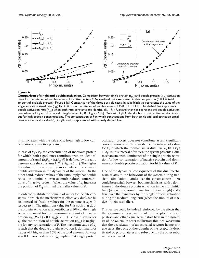
We now discuss under which dynamical conditions thedual mechanism emerge. In the following analysis weassume normalised units for the variables; for which avalue equal to one represents the total amount of proteinP in wild type cells. The most interesting behaviouremerges when the kinetic constant, characterising thedouble protein activation process, is higher than the onefor the single protein activation process (k3 > k1) (Figure6[a]). In this case, the signal rate associated to double pro-tein activation (rDA) dominates for high concentrations ofinactive protein P whereas single activation is moreintense for low P values. Otherwise (k3 k1), single activa-tion rate (rSA) dominates over the whole interval of feasi-ble concentrations for the protein (0.0–1.0). Furthermore,the importance of the double protein activation mecha-
Experimental designFigure 4Experimental design. Experiment proposed to elucidate the activation of homodimer proteins by homodimer receptors. Schematic representation of mutant cells (Mut1 and Mut2) with respect to the wild-type cells (WT).
Time coursesFigure 5Time courses. Hypothetical time courses of the concentration of active protein P* for wild-type (WT) and mutated cells (Mut1, Mut2). The kinetic parameters k1 and k3 are the same for the three configurations (k3/k1 = 0.5). The initial conditions are: P(0) = 1; P* = 0. Figure 5 [a]: response of both mutants when the system is supposed to have only single protein activation. Figure 5 [b]: response of both mutants when a dual mechanism of activation is considered. Figure 5 [c]: response when the mechanism of activation is double protein activation. The configuration of the system for wild-type cells implies an initial inten-sity for the activated receptor R(0) = 1, while in mutant cells R(0) = 0.5.
0 20 40 60 800
0.1
0.2
0.3
time t
P*(
t) (
norm
. uni
ts)
Single protein activation[a]
WTMut 1Mut 2
0 20 40 60 800
0.1
0.2
0.3
time t
P*(
t) (
norm
. uni
ts)
Dual Mechanism[b]
WTMut 1Mut 2
0 20 40 60 800
0.1
0.2
0.3
time t
P*(
t) (
norm
. uni
ts)
Double protein activation[c]
WTMut 1Mut 2
Page 7 of 11(page number not for citation purposes)
BMC Systems Biology 2008, 2:92 http://www.biomedcentral.com/1752-0509/2/92
nism increases with the value of k3 from high to low con-centrations of inactive protein.
In case of k3 > k1, the concentration of inactivate proteinfor which both signal rates contribute with an identicalamount of signal (k1Peq = k3(Peq)2) is defined by the ratiobetween rate the constants k1/k3 (Figure 6[b]). The higherthe value of this ratio is, the more reduced the effect ofdouble activation in the dynamics of the system. On theother hand, reduced values of the ratio imply that doubleactivation dominates even at much reduced concentra-tions of inactive protein. When the value of k3 increasesthe position of Peq is shifted to smaller values of P.
In order to establish the domain of values for the rate con-stants in which the mechanism is effectively dual, we fixan interval of feasible values for the parameter k3 withrespect to k1. The minimum value for k3 is such that dou-ble protein activation rate contributes a 10% of the singleactivation signal for the maximum amount of inactiveprotein: rDA(P = 1) = 0.1·rSA(P = 1.0). Below this value fork3, the contribution of double activation (rDA) is negligi-ble for any concentration of P. The maximum value of k3is such that the double protein activation is dominant forvalues of P higher than 10% of the total amount: Peq = k1/k3 = 0.1. Lower values for Peq implies that single protein
activation process does not contribute at any significantconcentration of P. Thus, we define the interval of valuesfor k3 in which the mechanism is dual like k1/10 k3 10k1. In this interval of values, the system presents a dualmechanism, with dominance of the single protein activa-tion for low concentration of inactive protein and domi-nance of double protein activation for high values of P.
One of the dynamical consequences of this dual mecha-nism relates to the behaviour of the system during tran-sient stimulation. Under certain circumstances therecould be a switch between both mechanisms, with a dom-inance of the double protein activation in the short initialtime (when the amount of inactive protein is high) and atake over the dynamics by the single protein activationduring the medium-long term (when the amount of inac-tive protein is smaller).
This feature could be indeed reinforced by the effects thatthe asymmetric deactivation of the receptor by phos-phatases and other signal terminators have in the dynam-ics of the system. In order to illustrate this idea, we assumethat the deactivation of an activated receptor happens intwo steps: first, one of the subunits of the receptor is deac-tivated by phosphatases and subsequently the other subu-nit is deactivated:
Comparison of single and double activationFigure 6Comparison of single and double activation. Comparison between single protein (rSA) and double protein (rDA) activation rates for the interval of feasible values of inactive protein P. Normalised units were used in this comparison (P = 1 is total amount of available protein). Figure 6 [a]: Comparison of the three possible cases. In solid black we represents the value of the single activation signal rate (rSA) for k1 = 0.3 in the interval of feasible values of P (0.0 P 1.0). The dashed line represents double activation rate (rDA) when both rate constants are identical (k3 = k1). Upward triangles represent the double activation rate when k3 > k1 and downward triangles when k3 <k1. Figure 6 [b]: Only with k3 > k1 the double protein activation dominates but for high protein concentrations. The concentration of P in which contributions from both single and dual activation signal rates are identical is called Peq = k1/k3 and is represented with a finely dashed line.
0 0.2 0.4 0.6 0.8 10
0.05
0.1
0.15
0.2
0.25
P (norm. units)
V(P
) (n
orm
. uni
ts)
rSA
rDA
(k3 = k
1)
rDA
(k3 < k
1)
rDA
(k3 > k
1)
0 0.2 0.4 0.6 0.8 10
0.05
0.1
0.15
0.2
0.25
P (norm. units)
V(P
) (n
orm
. uni
ts)
rSA
Peq
rDA
Dominance of doubleactivation process
Dominance of singleactivation process
[a]
Page 8 of 11(page number not for citation purposes)
BMC Systems Biology 2008, 2:92 http://www.biomedcentral.com/1752-0509/2/92
For simplicity, we suppose that there is no cooperativity inthe receptor deactivation process and therefore both deac-tivation rates are similar (kD1 = kD2). A fully activatedreceptor (R* R* is able to participate in either single ordouble protein activation whereas a partially activatedreceptor (R* R) can only participate in the single activa-tion process. Indeed, the stoichiometry of the activationby the fully activated receptor is not the same for singleand double protein activation: fully activated receptorshave double activation sites for single activation proc-esses. Considering all this, the equation describing thedynamics of protein activation becomes:
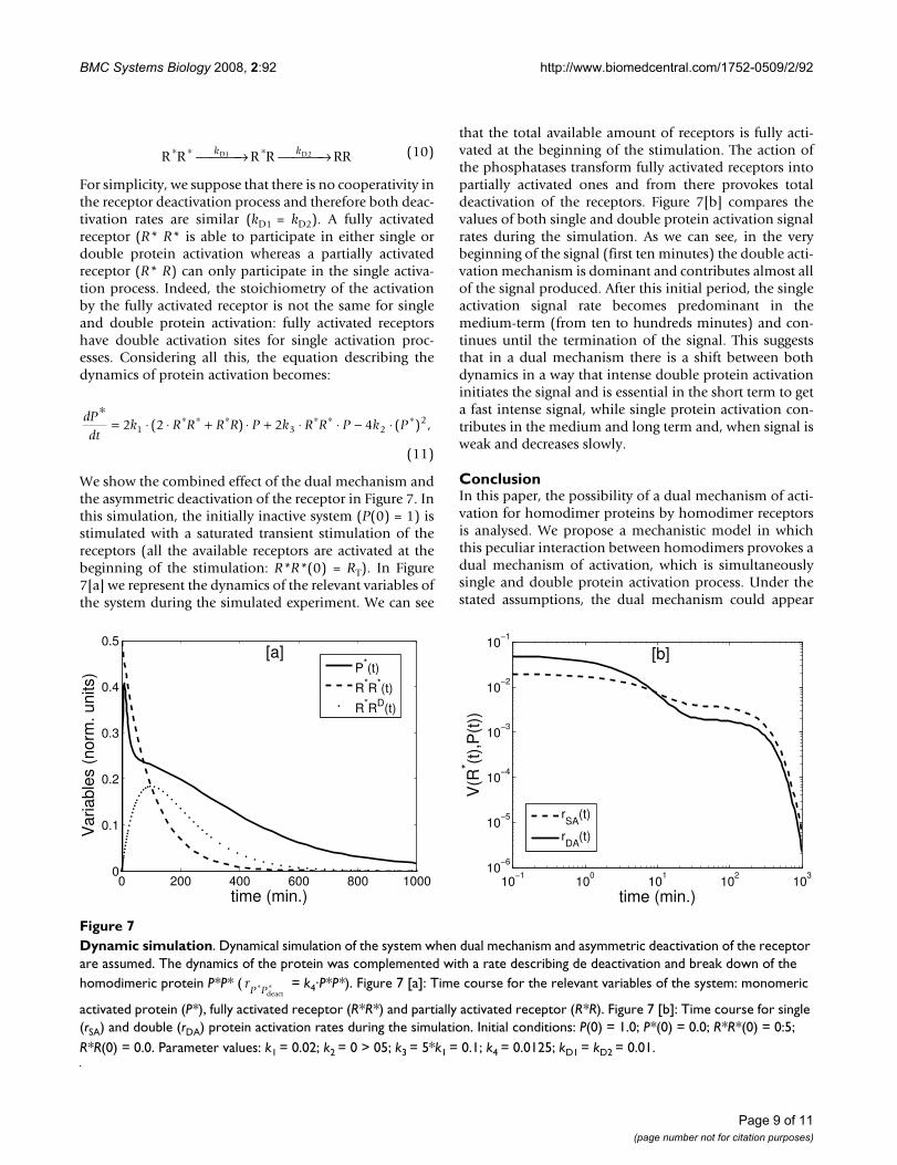
We show the combined effect of the dual mechanism andthe asymmetric deactivation of the receptor in Figure 7. Inthis simulation, the initially inactive system (P(0) = 1) isstimulated with a saturated transient stimulation of thereceptors (all the available receptors are activated at thebeginning of the stimulation: R*R*(0) = RT). In Figure7[a] we represent the dynamics of the relevant variables ofthe system during the simulated experiment. We can see
that the total available amount of receptors is fully acti-vated at the beginning of the stimulation. The action ofthe phosphatases transform fully activated receptors intopartially activated ones and from there provokes totaldeactivation of the receptors. Figure 7[b] compares thevalues of both single and double protein activation signalrates during the simulation. As we can see, in the verybeginning of the signal (first ten minutes) the double acti-vation mechanism is dominant and contributes almost allof the signal produced. After this initial period, the singleactivation signal rate becomes predominant in themedium-term (from ten to hundreds minutes) and con-tinues until the termination of the signal. This suggeststhat in a dual mechanism there is a shift between bothdynamics in a way that intense double protein activationinitiates the signal and is essential in the short term to geta fast intense signal, while single protein activation con-tributes in the medium and long term and, when signal isweak and decreases slowly.
ConclusionIn this paper, the possibility of a dual mechanism of acti-vation for homodimer proteins by homodimer receptorsis analysed. We propose a mechanistic model in whichthis peculiar interaction between homodimers provokes adual mechanism of activation, which is simultaneouslysingle and double protein activation process. Under thestated assumptions, the dual mechanism could appear
R R R R RRD D∗ ∗ ∗⎯ →⎯⎯ ⎯ →⎯⎯k k1 2 (10)
dPdt
k R R R R P k R R P k P∗
= ⋅ ⋅ + ⋅ + ⋅ ⋅ − ⋅∗ ∗ ∗ ∗ ∗ ∗2 2 2 41 3 22( ) ( ) ,
(11)
Dynamic simulationFigure 7Dynamic simulation. Dynamical simulation of the system when dual mechanism and asymmetric deactivation of the receptor are assumed. The dynamics of the protein was complemented with a rate describing de deactivation and break down of the homodimeric protein P*P* ( = k4·P*P*). Figure 7 [a]: Time course for the relevant variables of the system: monomeric
activated protein (P*), fully activated receptor (R*R*) and partially activated receptor (R*R). Figure 7 [b]: Time course for single (rSA) and double (rDA) protein activation rates during the simulation. Initial conditions: P(0) = 1.0; P*(0) = 0.0; R*R*(0) = 0:5; R*R(0) = 0.0. Parameter values: k1 = 0.02; k2 = 0 > 05; k3 = 5*k1 = 0.1; k4 = 0.0125; kD1 = kD2 = 0.01.
0 200 400 600 800 10000
0.1
0.2
0.3
0.4
0.5
time (min.)
Var
iabl
es (
norm
. uni
ts)
P*(t)
R*R*(t)
R*RD(t)
[a]
10−1
100
101
102
103
10−6
10−5
10−4
10−3
10−2
10−1
time (min.)
V(R
* (t),
P(t
))
rSA
(t)
rDA
(t)
[b]
rP P∗ ∗
deact
Page 9 of 11(page number not for citation purposes)
BMC Systems Biology 2008, 2:92 http://www.biomedcentral.com/1752-0509/2/92
essentially as either single or double activation, depend-ing on the ratio between the rate constants of both processand the available amount of inactive protein. For a certaininterval of values for these coefficients, the process canhave an intermediate behaviour, which cannot be reducedto the single or double activation mechanism.
We further analysed a phenomenological power-law rep-resentation, which is able to adequately simulate theresponse of the system. We demonstrated that power-lawrepresentation is useful for cases where experimental dataare available, but which are not sufficient to distinguishand characterise the reaction mechanisms (single or dou-ble activation). The power-law model, together withquantitative experimental data, is an alternative to inves-tigate the structure of a given pathway where the estimatedvalue for the kinetic order is used to decide whether theprocess is mainly of single activation (g 1), double acti-vation (g 2) or dual (intermediate values).
Finally, the consequences of this dual mechanism on thedynamics were investigated through the simulation of dif-ferent experimental conditions. Towards this end, we sim-ulated the behaviour of the system for two mutants: amutant with a reduced number of receptors, and a secondmutant in which one of the subunits of the receptors isblocked for the activation of the studied protein. Only incases in which the system has the dual mechanism of acti-vation both mutants would induce a different response,while a pure double protein activation system would notproduce a significant response to the input signal, whenone of the subunits of the homodimer receptors is consti-tutively blocked for the activation of the studied protein.Furthermore, our analysis suggests that in receptor-pro-tein interacting systems with dual mechanism there maybe an active switch between double and single activationin a way that intense double protein activation could ini-tiate and dominate the signal in the short term (getting afast intense signal), while single protein activation couldcontrol the system in the medium and long term (wheninput signal is weaker and decreases slowly).
Open questions that require attention relate to the under-lying reasons that justify the dual activation mechanism.Mathematical modelling could be used to investigate whyand how this dual mechanism is used by the cell toimprove the performance of monomer-to-dimer transi-tions [5]. Moreover, there is increasing evidence for feed-back inhibitor proteins that bind to activated receptorsand compete with the true effectors for binding, e.g. CIS1,and Mig6 [32-34]. The expression of these feedback inhib-itors is usually induced by the activated receptor. Thus,there may be an implicit switch between first and secondorder processes over the timecourse of stimulation
dependent on feedback inhibitor levels that could beinvestigated in the next future.
Authors' contributionsJV and TM designed the study, set up the mathematicalmodel and performed the calculations concerning theresponsiveness of the system under the supervision of OWFinally, all the authors including WK drafted the manu-script.
Additional material
AcknowledgementsThe authors thank the collaboration of Taesoo Kwon and Ulf Schmitz elab-orating a list of published homod-imer receptor-homodimer transducer protein interactions used in initial versions of this work. This work was sup-ported by the European Commission 6th Framework program and as part of the COSBICS project under contract LSHG-CT-2004-512060 http://www.sbi.uni-rostock.de/cosbics and by the German Federal Ministry of Education and Research (BMBF) through the projects BaCell-SysMo (0313978F) and COSMIC-SysMo (0313981D).
References1. Downward J: The ins and outs of signalling. Nature 2001,
411(6839):759-762.2. Jones N: Transcriptional regulation by dimerization: two
sides to an incestuous relationship. Cell 1990, 61:9-11.3. Jones S, Thornton J: Protein-protein interactions: a review of
protein dimer structures. Prog Biophys Mol Biol 1995, 63:31-65.4. Pereira-Leal J, Levy E, Kamp C, Teichmann S: Evolution of protein
complexes by duplication of homomeric interactions. GenBiol 2007, 8(4):R51.
5. Klemm J, Schreiber S, Crabtree G: Dimerization as a RegulatoryMechanism in Signal Transduction. Annu Rev Immunol 1998,16:569-592.
6. Reich Z, Boniface J, Lyons D, Borochov N, Wachtel E, Davis M: Lig-and-specific oligomerization of T-cell receptor molecules.Nature 1997, 387:617-620.
7. Schlessinger J: Signal transduction by allosteric receptor oli-gomerization. Trends Biol Sci 1988, 13:443-447.
8. Heldin C: Dimerization of cell surface receptors in signaltransduction. Cell 1995, 80:213-223.
9. Ferrajoli A, Faderl S, Ravandi F, Estrov Z: The JAK-STAT pathway:a therapeutic target in hemato-logical malignancies. CurrCancer Drug Targets 2006, 6(8):671-679.
10. Livnah O, Stura E, Middleton S, Jollife L, Wilson I: Crystallographicevidence for preformed dimers of erythropoietin receptorbefore ligand activation. Science 1999, 283(5404):987-990.
11. Remy I, Wilson I, Michnick S: Erythropoietin receptor activationby a ligand-induced conformation change. Science 1999,283:990-993.
Additional file 1On the role of receptor and protein homodimerisation in cell signal-ling Supplementary Material. In the main text the double activation of the receptor is represented by a trimolecular biochemical reaction. The assumptions leading to this simplified representation of receptor activation are explained in detail. Furthermore, the determination of the apparent kinetic order using a power-law approach is discussed here. The supple-mentary material is provided as PDF-file.Click here for file[http://www.biomedcentral.com/content/supplementary/1752-0509-2-92-S1.pdf]
Page 10 of 11(page number not for citation purposes)
http://www.ncbi.nlm.nih.gov/entrez/query.fcgi?cmd=Retrieve&db=PubMed&dopt=Abstract&list_uids=2180585
http://www.ncbi.nlm.nih.gov/entrez/query.fcgi?cmd=Retrieve&db=PubMed&dopt=Abstract&list_uids=2180585
http://www.ncbi.nlm.nih.gov/entrez/query.fcgi?cmd=Retrieve&db=PubMed&dopt=Abstract&list_uids=7746868
http://www.ncbi.nlm.nih.gov/entrez/query.fcgi?cmd=Retrieve&db=PubMed&dopt=Abstract&list_uids=7746868
http://www.ncbi.nlm.nih.gov/entrez/query.fcgi?cmd=Retrieve&db=PubMed&dopt=Abstract&list_uids=9597142
http://www.ncbi.nlm.nih.gov/entrez/query.fcgi?cmd=Retrieve&db=PubMed&dopt=Abstract&list_uids=9597142
http://www.ncbi.nlm.nih.gov/entrez/query.fcgi?cmd=Retrieve&db=PubMed&dopt=Abstract&list_uids=9177351
http://www.ncbi.nlm.nih.gov/entrez/query.fcgi?cmd=Retrieve&db=PubMed&dopt=Abstract&list_uids=9177351
http://www.ncbi.nlm.nih.gov/entrez/query.fcgi?cmd=Retrieve&db=PubMed&dopt=Abstract&list_uids=7834741
http://www.ncbi.nlm.nih.gov/entrez/query.fcgi?cmd=Retrieve&db=PubMed&dopt=Abstract&list_uids=7834741
http://www.ncbi.nlm.nih.gov/entrez/query.fcgi?cmd=Retrieve&db=PubMed&dopt=Abstract&list_uids=9974392
http://www.ncbi.nlm.nih.gov/entrez/query.fcgi?cmd=Retrieve&db=PubMed&dopt=Abstract&list_uids=9974392
http://www.ncbi.nlm.nih.gov/entrez/query.fcgi?cmd=Retrieve&db=PubMed&dopt=Abstract&list_uids=9974392
BMC Systems Biology 2008, 2:92 http://www.biomedcentral.com/1752-0509/2/92
Publish with BioMed Central and every scientist can read your work free of charge
"BioMed Central will be the most significant development for disseminating the results of biomedical research in our lifetime."
Sir Paul Nurse, Cancer Research UK
Your research papers will be:
available free of charge to the entire biomedical community
peer reviewed and published immediately upon acceptance
cited in PubMed and archived on PubMed Central
yours — you keep the copyright
Submit your manuscript here:http://www.biomedcentral.com/info/publishing_adv.asp
BioMedcentral
12. Shuai K, Horvath C, Huang L, Qureshi S, Cowburn D, Darnell J Jr:Interferon activation of the transcription factor Stat91involves dimerization through SH2-phosphotyrosyl peptideinteractions. Cell 1994, 76:821-828.
13. Sakurai H, Nigam S: In vitro branching tubulogenesis: Implica-tions for developmental and cystic disorders, nephronnumber, renal repair, and nephron engineering. Kidney Inter-national 1998, 54:14-26.
14. Jain N, Zhang T, Kee W, Li W, Cao X: Protein kinase C deltaassociates with and phosphorylates Stat3 in an interleukin-6-dependent manner. J Biol Chem 1999, 274(34):24392-400.
15. Moustakas A, Souchelnytskyi S, Heldin C: Smad regulation inTGF- signal transduction. Journal of Cell Science 2001,114:4359-4369.
16. Stamper C, Zhang Y, Tobin J, Erbe D, Ikemizu S, Davis S, Stahl M, See-hra J, Somers W, Mosyak L: Crystal structure of the B7-1/CTLA-4 complex that inhibits human immune responses. Nature2001, 410:608-611.
17. Shao H, Cheng H, Cook R, Tweardy D: Identification and charac-terization of signal transducer and activator of transcription3 recruitment sites within the epidermal growth factorreceptor. Cancer Res 2003, 63(14):3923-30.
18. Schulze W, Deng L, Mann M: Phosphotyrosine interactome ofthe ErbB-receptor kinase family. Mol Sys Biol 2005, 1:2005.0008.
19. Winter S, Howard T, Ware R: Regulation of Expression of theHuman Erythropoietin Receptor Gene. Blood Cell Mol Dis 1996,22(3):214-224.
20. Fambrough D, McClure K, Kazlauskas A, Lander E: Diverse signal-ing pathways activated by growth factor receptors inducebroadly overlapping, rather than independent, sets of genes.Cell 1999, 97:727-741.
21. Hänggi P, Talkner P, Borkovec M: Reaction-rate theory: fiftyyears after Kramers. Rev Mod Phys 1990, 62(2):251-341.
22. Savageau M: Biochemical systems analysis: I. Some mathemat-ical properties of the rate law for the component enzymaticreactions. J Theor Biol 1969, 25:365-369.
23. Savageau M: Biochemical systems analysis: II. Steady statesolutions for an n-poll system using a power-law approxima-tion. J Theor Biol 1969, 25:370-379.
24. Savageau M: Biochemical systems analysis: III. Dynamic solu-tions using a power-law approximation. J Theor Biol 1970,26:215-226.
25. Vera J, Balsa-Canto E, Wellstead P, Banga J, Wolkenhauer O: Power-Law Models of Signal Transduction Pathways. Cell Signal 2007,19(7):1531-41.
26. Barkai N, Leibler S: Robustness in simple biochemical net-works. Nature 1997, 387(6636):913-917.
27. Lauffenburger D, Linderman J: Receptors: Models for binding, trafficing,and signalling Oxford University Press; 1993.
28. Swameye I, Müller T, Timmer J, Sandra O, Klingmüller U: Identifica-tion of nucleocytoplasmic cycling as a remote sensor in cel-lular signaling by data-based modeling. Proc Natl Acad Sci USA2003, 100(3):1028-1033.
29. Vera J, Bachmann PAJ, Becker V, Hormiga J, Torres Darias N, TimmerJ, Klingmuller U, Wolkenhauer O: A systems biology approach toanalyse amplification in the JAK2-STAT5 signalling pathway.BMC Systems Biology 2008, 2:38.
30. Hernandez-Bermejo B, Fairen V, Sorribas A: Power-law modelingbased on least-squares criteria: consequences for systemanalysis and simulation. Math Biosci 2000, 167:87-107.
31. Behrmann I, Janzen C, Gerhartz C, Schmitz-Van de Leur H, HermannsH, Heesel B, Graeve L, Horn F, Tav-ernier J, Heinrich C: A singleSTAT recruitment module in a chimeric cytokine receptorcomplex is sufficient for STAT activation. J Biol Chem 1997,272(8):5269-5274.
32. Yoshimura A: Negative regulation of cytokine signaling. ClinRev Allergy Immunol 2005, 28(3):205-220.
33. Mason J, Morrison D, Basson M, Licht J: Sprouty proteins: multi-faceted negative-feedback regulators of receptor tyrosinekinase signaling. Trends Cell Biol 2006, 16:45-54.
34. Zhang Y, Woude G Vande: Mig-6, signal transduction, stressresponse and cancer. Cell Cycle 2007, 6(5):507-513.
Page 11 of 11(page number not for citation purposes)
http://www.ncbi.nlm.nih.gov/entrez/query.fcgi?cmd=Retrieve&db=PubMed&dopt=Abstract&list_uids=7510216
http://www.ncbi.nlm.nih.gov/entrez/query.fcgi?cmd=Retrieve&db=PubMed&dopt=Abstract&list_uids=7510216
http://www.ncbi.nlm.nih.gov/entrez/query.fcgi?cmd=Retrieve&db=PubMed&dopt=Abstract&list_uids=7510216
http://www.ncbi.nlm.nih.gov/entrez/query.fcgi?cmd=Retrieve&db=PubMed&dopt=Abstract&list_uids=9648059
http://www.ncbi.nlm.nih.gov/entrez/query.fcgi?cmd=Retrieve&db=PubMed&dopt=Abstract&list_uids=9648059
http://www.ncbi.nlm.nih.gov/entrez/query.fcgi?cmd=Retrieve&db=PubMed&dopt=Abstract&list_uids=9648059
http://www.ncbi.nlm.nih.gov/entrez/query.fcgi?cmd=Retrieve&db=PubMed&dopt=Abstract&list_uids=5387046
http://www.ncbi.nlm.nih.gov/entrez/query.fcgi?cmd=Retrieve&db=PubMed&dopt=Abstract&list_uids=5387046
http://www.ncbi.nlm.nih.gov/entrez/query.fcgi?cmd=Retrieve&db=PubMed&dopt=Abstract&list_uids=5387046
http://www.ncbi.nlm.nih.gov/entrez/query.fcgi?cmd=Retrieve&db=PubMed&dopt=Abstract&list_uids=5387047
http://www.ncbi.nlm.nih.gov/entrez/query.fcgi?cmd=Retrieve&db=PubMed&dopt=Abstract&list_uids=5387047
http://www.ncbi.nlm.nih.gov/entrez/query.fcgi?cmd=Retrieve&db=PubMed&dopt=Abstract&list_uids=5387047
http://www.ncbi.nlm.nih.gov/entrez/query.fcgi?cmd=Retrieve&db=PubMed&dopt=Abstract&list_uids=5434343
http://www.ncbi.nlm.nih.gov/entrez/query.fcgi?cmd=Retrieve&db=PubMed&dopt=Abstract&list_uids=5434343
http://www.ncbi.nlm.nih.gov/entrez/query.fcgi?cmd=Retrieve&db=PubMed&dopt=Abstract&list_uids=9202124
http://www.ncbi.nlm.nih.gov/entrez/query.fcgi?cmd=Retrieve&db=PubMed&dopt=Abstract&list_uids=9202124
http://www.ncbi.nlm.nih.gov/entrez/query.fcgi?cmd=Retrieve&db=PubMed&dopt=Abstract&list_uids=9030599
http://www.ncbi.nlm.nih.gov/entrez/query.fcgi?cmd=Retrieve&db=PubMed&dopt=Abstract&list_uids=9030599