Adenosine A2A receptor dynamics studied with the novel fluorescent agonist Alexa488-APEC
17
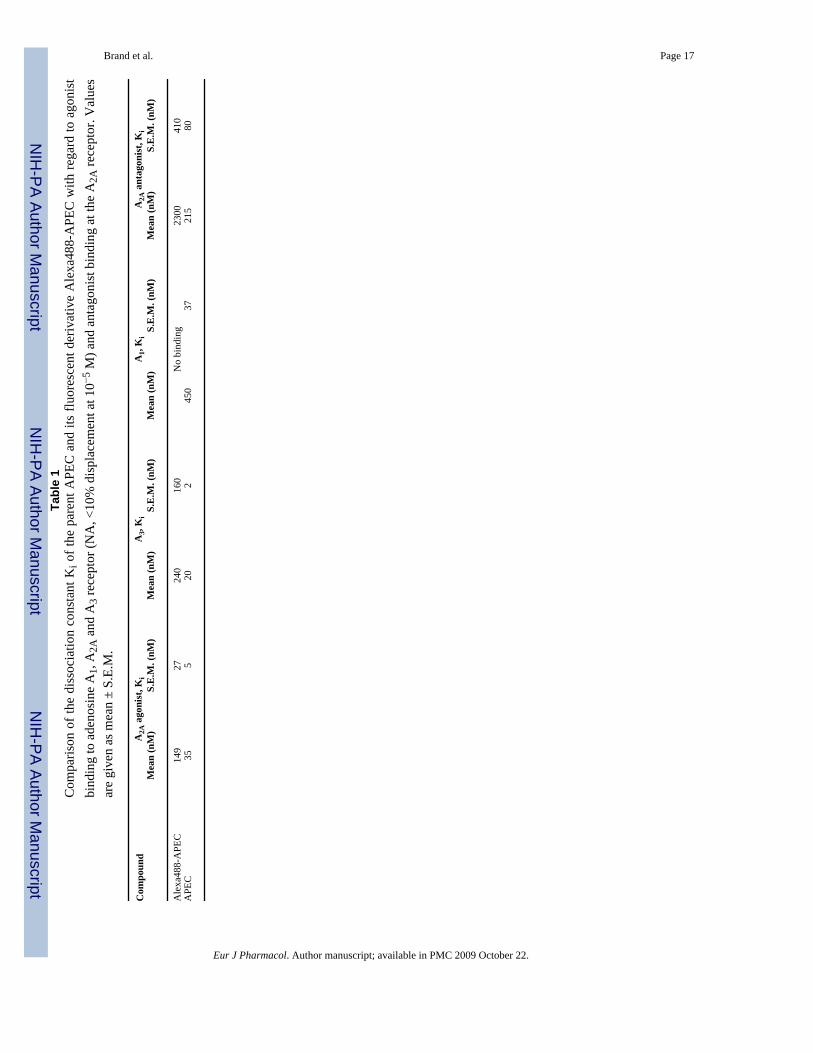
Adenosine A 2A receptor dynamics studied with the novel fluorescent agonist Alexa488-APEC Frank Brand 1 , Athena Klutz 2 , Kenneth A. Jacobson 2 , Bertil B. Fredholm 3 , and Gunnar Schulte 1,4 1 Karolinska Institutet, Dept. Physiology & Pharmacology, Sect. Receptor Biology & Signaling, S-171 77 Stockholm, Sweden 2 NIDDK, National Institutes of Health, Bethesda, MD, USA 3 Karolinska Institutet, Dept. Physiology & Pharmacology, Sect. Molecular Pharmacology, S-171 77 Stockholm, Sweden Abstract G protein-coupled receptors, such as the adenosine A 2A receptor, are dynamic proteins, which undergo agonist-dependent redistribution from the cell surface to intracellular membranous compartments, such as endosomes. In order to study the kinetics of adenosine A 2A receptor redistribution in living cells, we synthesized a novel fluorescent agonist, Alexa488-APEC. Alexa488- APEC binds to adenosine A 2A (K i = 149 ± 27 nM) as well as A 3 receptors (K i = 240 ± 160 nM) but not to adenosine A 1 receptors. Further, we characterized the dose-dependent increase in Alexa488- APEC-induced cAMP production as well as cAMP response element binding (CREB) protein phosphorylation, verifying the ligand’s functionality at adenosine A 2A but not A 2B receptors. In live cell imaging studies, Alexa488-APEC induced adenosine A 2A receptor internalization, which was blocked by the competitive reversible antagonist ZM 241385 and hyperosmolaric sucrose. Further, internalized adenosine A 2A receptors co-localized with clathrin and Rab5, indicating that agonist stimulation promotes adenosine A 2A receptor uptake through a clathrin-dependent mechanism to Rab5-positive endosomes. The basic characterization of Alexa488-APEC provided here showed that it provides a usefultool for tracing adenosine A 2A receptors in vitro. Keywords G protein-coupled receptor; endocytosis; cyclic AMP; CREB; nucleoside; Rab5; clathrin 1. Introduction Adenosine A 2A receptors belong to the family of G protein-coupled receptors (Fredholm et al., 2003). This member of the adenosine receptor family, which consists of adenosine A 1 , A 2A , A 2B , and A 3 receptors (Fredholm et al., 2001), is coupled to stimulatory G proteins of the G s family (Furlong et al., 1992). Thus, receptor stimulation leads to an increase in cAMP 4to whom correspondence should be sent: Gunnar Schulte, PhD, Karolinska Institutet, Dept. Physiology & Pharmacology, Sect. Receptor Biology & Signaling, Nanna Svartz väg 2, S-17177 Stockholm, Sweden, Tel.: +46-8-52487933, Fax.: +46-8-341280, [email protected]. Publisher's Disclaimer: This is a PDF file of an unedited manuscript that has been accepted for publication. As a service to our customers we are providing this early version of the manuscript. The manuscript will undergo copyediting, typesetting, and review of the resulting proof before it is published in its final citable form. Please note that during the production process errors may be discovered which could affect the content, and all legal disclaimers that apply to the journal pertain. NIH Public Access Author Manuscript Eur J Pharmacol. Author manuscript; available in PMC 2009 October 22. Published in final edited form as: Eur J Pharmacol. 2008 August 20; 590(1-3): 36–42. doi:10.1016/j.ejphar.2008.05.036. NIH-PA Author Manuscript NIH-PA Author Manuscript NIH-PA Author Manuscript
Transcript of Adenosine A2A receptor dynamics studied with the novel fluorescent agonist Alexa488-APEC
Adenosine A2A receptor dynamics studied with the novelfluorescent agonist Alexa488-APEC
Frank Brand1, Athena Klutz2, Kenneth A. Jacobson2, Bertil B. Fredholm3, and GunnarSchulte1,41 Karolinska Institutet, Dept. Physiology & Pharmacology, Sect. Receptor Biology & Signaling, S-17177 Stockholm, Sweden2 NIDDK, National Institutes of Health, Bethesda, MD, USA3 Karolinska Institutet, Dept. Physiology & Pharmacology, Sect. Molecular Pharmacology, S-171 77Stockholm, Sweden
AbstractG protein-coupled receptors, such as the adenosine A2A receptor, are dynamic proteins, whichundergo agonist-dependent redistribution from the cell surface to intracellular membranouscompartments, such as endosomes. In order to study the kinetics of adenosine A2A receptorredistribution in living cells, we synthesized a novel fluorescent agonist, Alexa488-APEC. Alexa488-APEC binds to adenosine A2A (Ki = 149 ± 27 nM) as well as A3 receptors (Ki= 240 ± 160 nM) butnot to adenosine A1 receptors. Further, we characterized the dose-dependent increase in Alexa488-APEC-induced cAMP production as well as cAMP response element binding (CREB) proteinphosphorylation, verifying the ligand’s functionality at adenosine A2A but not A2B receptors. In livecell imaging studies, Alexa488-APEC induced adenosine A2A receptor internalization, which wasblocked by the competitive reversible antagonist ZM 241385 and hyperosmolaric sucrose. Further,internalized adenosine A2A receptors co-localized with clathrin and Rab5, indicating that agoniststimulation promotes adenosine A2A receptor uptake through a clathrin-dependent mechanism toRab5-positive endosomes. The basic characterization of Alexa488-APEC provided here showed thatit provides a usefultool for tracing adenosine A2A receptors in vitro.
KeywordsG protein-coupled receptor; endocytosis; cyclic AMP; CREB; nucleoside; Rab5; clathrin
1. IntroductionAdenosine A2A receptors belong to the family of G protein-coupled receptors (Fredholm etal., 2003). This member of the adenosine receptor family, which consists of adenosine A1,A2A, A2B, and A3 receptors (Fredholm et al., 2001), is coupled to stimulatory G proteins ofthe Gs family (Furlong et al., 1992). Thus, receptor stimulation leads to an increase in cAMP
4to whom correspondence should be sent: Gunnar Schulte, PhD, Karolinska Institutet, Dept. Physiology & Pharmacology, Sect. ReceptorBiology & Signaling, Nanna Svartz väg 2, S-17177 Stockholm, Sweden, Tel.: +46-8-52487933, Fax.: +46-8-341280,[email protected]'s Disclaimer: This is a PDF file of an unedited manuscript that has been accepted for publication. As a service to our customerswe are providing this early version of the manuscript. The manuscript will undergo copyediting, typesetting, and review of the resultingproof before it is published in its final citable form. Please note that during the production process errors may be discovered which couldaffect the content, and all legal disclaimers that apply to the journal pertain.
NIH Public AccessAuthor ManuscriptEur J Pharmacol. Author manuscript; available in PMC 2009 October 22.
Published in final edited form as:Eur J Pharmacol. 2008 August 20; 590(1-3): 36–42. doi:10.1016/j.ejphar.2008.05.036.
NIH
-PA Author Manuscript
NIH
-PA Author Manuscript
NIH
-PA Author Manuscript
and phosphorylation of the cAMP-regulated element binding protein (CREB) (Kull et al.,1999; Schulte et al., 2003a). Physiologically, adenosine A2A receptors are involved, e.g., inthe regulation of sleep-wakefulness, locomotion and activity, platelet function, and T cell-dependent responses (Ledent et al., 1997; Jacobson et al., 2006; Fredholm et al., 2007; Huanget al., 2007).
It is widely appreciated that G protein-coupled receptors, such as the adenosine A2A receptor,are dynamic proteins, which traffic throughout membranous compartments of the cell,depending on the receptor’s maturity or activity status (Conn et al., 2007; Xu et al., 2007).Stimulation of receptors exposed on the cell surface can induce receptor desensitization and,consequently, to receptor internalization through different endocytotic pathways leadingprimarily to a reduction in receptor numbers on the cell surface (Wolfe et al., 2007). However,recent evidence emphasizes that the internalized receptors localized in endosomes can alsodirect specific signaling by providing a scaffold for active signaling components (Dewire etal., 2007). In the case of the adenosine A2A receptor, previously described patterns of receptordesensitization indicate that agonist-stimulated adenosine A2A receptors are rapidly andtransiently phosphorylated and desensitized (Palmer et al., 1994). Further investigation showedthat a C-terminal threonine (Thr298) is essential for agonist-induced receptor phosphorylationand short-term desensitization (Palmer et al., 1997). This is a site of phosphorylation throughG protein-coupled receptor kinase-2 (GRK2), which is known to regulate agonist-induceddesensitization of adenosine A2A receptors (Mundell et al., 1997, 1998b). However, short-termadenosine A2A receptor desensitization appears to depend mainly on agonist-induced receptorphosphorylation and not on clathrin-based endocytosis, since hyperosmotic sucrose orconcavalin A treatment did not inhibit agonist-induced desensitization of the adenosine A2Areceptor (Mundell et al., 1998a). Receptor distribution and internalization upon agonisttreatment have not been characterized yet. Many studies addressing receptor dynamics usefluorescently tagged receptors to trace agonist-induced endocytosis. However, since thisapproach requires cell transfection or genetic modification, it is not easily applicable incomplex cellular systems or in vivo. Thus, it is advantageous to detect receptor trafficking bytracing fluorescently labeled agonists, a methodology that has been employed using bothagonist and antagonist as well as small molecule and peptide ligands in various receptorsystems (Daly et al., 2003; Middleton et al., 2005). In order to study adenosine A2A receptordynamics in cell culture, we set out to synthesize a novel fluorescent adenosine A2A receptoragonist, Alexa488-APEC, which we used here to trace adenosine A2A receptors transientlyexpressed in immortalized cell lines over time. We show that this ligand is a moderatelyselective and functional adenosine A2A receptor agonist suitable for live-cell imaging ofstimulation-dependent receptor dynamics. Further, we characterize the internalization pathwayof adenosine A2A receptors as clathrin-dependent uptake into Rab5-positive endosomes.
2. Materials and methods2.1. Material
[125I]N6-(4-Amino-3-iodobenzyl)adenosine-5′-N6-methyluronamide ([125I]-AB-MECA,2000 Ci/mmol) was purchased from GE Healthcare (Piscataway, NJ USA). [3H] 4-(2-[7-Amino-2-(2-furyl)[1,2,4]triazolo[2,3-a][1,3,5]triazin-5ylamino]ethyl)phenol ([3H]ZM241385, 17 Ci/mmol) was purchased from Tocris (Bristol, UK). [3H]2-[p-(2-carboxyethyl)phenylethylamino]-5′-N6-ethylcarboxyamido-adenosine ([3H] CGS21680, 47 Ci/mmol), [3H]2-chloro-N6-cyclopentyladenosine ([3H]CCPA, 42.6 Ci/mmol) and adenosine 3′, 5′ cyclicmonophosphate ([2,8-3H]cAMP, 27.4 Ci/mmol) were purchased from Perkin Elmer (Waltham,MA, USA). NECA (5′-N-ethylcarboxamidoadenosine), rabbit anti-FLAG (F7425), andforskolin were purchased from Sigma-Aldrich (St. Louis, MO, USA). ZM214,385 was fromTocris. Alexa Fluor® 488 carboxylic acid, succinimidyl ester (mixed isomers) was obtained
Brand et al. Page 2
Eur J Pharmacol. Author manuscript; available in PMC 2009 October 22.
NIH
-PA Author Manuscript
NIH
-PA Author Manuscript
NIH
-PA Author Manuscript
from Invitrogen (Carlsbad, CA, USA). 2-[4-(2-aminoethylaminocarbonylethyl)phenylethylamino]-5′-N-ethylcarboxamidoadenosine (APEC) bistrifluoroacetic acid wasprovided by NIMH Chemical Synthesis and Drug Supply Program. Rabbit anti-Ser133-P-CREB and mouse anti-clathrin (3F133) were from Upstate Biotechnology (Cambridge, UK)and Abcam (Hampshire, UK), respectively. Other chemicals were from standard commercialsources and of analytical grade. Plasmids coding for FLAG (N- terminal)-A2A and A2A-CFP(C-terminal) fusion proteins, both constructs lacking the most C terminal 49 aa, were generousgifts from Dr. C. Hoffmann, Würzburg University, Germany. Rab5-GFP was a gift from Dr.P.D. Stahl, Washington University, School of Medicine, St. Louis, MO, USA (Roberts et al.,1999).
2.2. Synthesis of Alexa488-APECTwo milligrams of the Alexa Fluor 488 active ester (3.1 μmol, 16 mM) was dissolved in 0.2ml of dimethyl sulfoxide (DMSO). APEC bistrifluoroacetic acid salt (2.5 mg, 3.2 μmol) wasadded to this solution to achieve a final theoretical concentration of 16 mM, followed by theaddition of 5 μl of triethylamine (35.9 μmol). The reaction was covered and stirred for 1 h. Theproduct was purified using an Agilent HPLC (Santa Clara, CA, USA) equipped with aPhenomenex Luna 5 μ Reverse Phase C18 analytical column (250 × 4.6 mm; Torrance, CA,USA), with acetonitrile:water mixtures at a flow rate of 2 ml/min and the following gradient:0 min, 30% acetonitrile; 6.5 min 50% acetonitrile; 9 min, 90% acetonitrile; 10 min, 30%acetonitrile. The compound eluted at a retention time of 4.2 min. After lyophilization, 2.24 mg(2.12 μmol) of chromatographically homogeneous product was recovered for a 68% yield. Themass spectrum was taken using a Waters LCT Premier mass spectrometer. The only majorpeak had a molecular weight of 1056.2614, consistent with the expected molecular weight ofthe product, Alexa488-APEC, 1056.2616 (C46H46N11O15S2). For thin layer chromatographyAlexa488-APEC solutions were separated on a silica gel 60 thin layer chromatography plate(Merck, Whitehouse Station, NJ, USA) using acetonitrile: water (7:1) as solvent. For furtherdetail on the path of synthesis, structure and analysis by mass spectroscopy, see supplementalmaterial Fig. S1.
2.3. Competitive ligand bindingCompetitive radioligand binding assays were done as described previously (Perreira et al.,2005). Human A1 and A3ARs were stably expressed in Chinese hamster ovary (CHO) cells,while the human A2AAR was stably expressed in HEK293 cells. Cell membrane suspensionswere prepared as described (Gao et al., 2004).
Stock solutions of APEC bistrifluoroacetic acid and Alexa488-APEC in DMSO (5 mM) wereprepared and were stable when stored frozen. For the binding determination, each tubecontained 50 μl of the appropriate radioligand (A1: [3H]CCPA, final conc. 1.0 nM; A2A agonist:[3H]CGS21680, final conc. 10 nM; A2A antagonist: [3H] ZM 241385, final conc. 2 nM; A3:[125I]AB-MECA, final conc. 0.5 nM), 50 μl of APEC bistrifluoroacetic acid or Alexa488-APEC in increasing concentrations, and 100 μl of cell membrane suspension expressing theappropriate receptor diluted with Tris-HCl buffer (50 mM, pH 7.4) containing 10 mMMgCl2. Non-specific binding was determined using 10 μM NECA. The tubes were incubatedat 25°C for 1 h and then filtered through Whatman GF/B filters under reduced pressure usingan MT-24 cell harvester (Brandell, Gaithersburg, MD, USA). Filters were washed rapidly threetimes with ice-cold buffer. For A1 and A2A assays, after the filters were washed, they wereplaced in scintillation vials containing 5 ml of Hydrofluor scintillation buffer and counted usinga Perkin Elmer Liquid Scintillation Analyzer. For A3 assays, the filters were counted using aPackard Cobra II gamma counter. The Ki values were determined using Prism software(GraphPad, San Diego, CA, USA) for all assays.
Brand et al. Page 3
Eur J Pharmacol. Author manuscript; available in PMC 2009 October 22.
NIH
-PA Author Manuscript
NIH
-PA Author Manuscript
NIH
-PA Author Manuscript
2.4. Cell cultureCHO and HEK293 cells were grown in DMEM/F12 (1:1), 10% FBS, 2 mM L-glutamine, 50U/ml penicillin/streptomycin and DMEM, 10% FBS, 2 mM L-glutamine, 50 U/ml penicillin/streptomycin, respectively. Cells were sub-cultured three times a week. Transient transfectionswere done according to the manufacturer’s recommendations with Lipofectamine 2000(Invitrogen, Carlsbad, CA, USA). For live-cell imaging or immunocytochemistry, cells wereseeded on collagen-coated (0.1% solution, Sigma-Aldrich, St. Louis, MO, USA) glass-bottom(MatTek corporation, Ashland, MA, USA) dishes or coverslips, respectively.
2.5. cAMP and P-CREB measurementsDeterminations of cAMP levels and CREB phosphorylation were done as previously described(Schulte et al., 2000, 2003b). Rabbit anti-Ser133-P-CREB was used at a 1:2000 dilution.Graphical and statistical analysis of the results was done with GraphPad Prism5 (nonlinearregression). Statistical significance was analysed with a two-tailed t-test (Fig. 2).
2.6. Live-cell imagingCells were seeded into 35 mm diameter glass bottom dishes and transfected the next day. Formicroscopy, we used cells 20–48 h posttransfection. Cells were washed in Hank’s BufferedSalt Solution (HBSS) before addition of fluorescent ligands. After 10 min incubation with theligand, cells were washed three times in HBSS and investigated under the laser scanningmicroscope (LSM510 Meta, Zeiss, Jena, Germany). In order to control for and to avoid opticalbleed-through between Alexa488 and CFP filter, settings were optimized, and controlexperiments with untagged receptors and single transfections were performed (not shown).
2.7. ImmunocytochemistryFor immunocytochemistry HEK293 cells were transiently transfected with adenosine FLAG-A2A receptors and fixed in 4% paraformaldehyde (10 min at room temperature) 24 hposttransfection. Detection of the FLAG epitope was done as described before (Bryja et al.,2007). Visual evaluation was done on a LSM510 Meta confocal system.
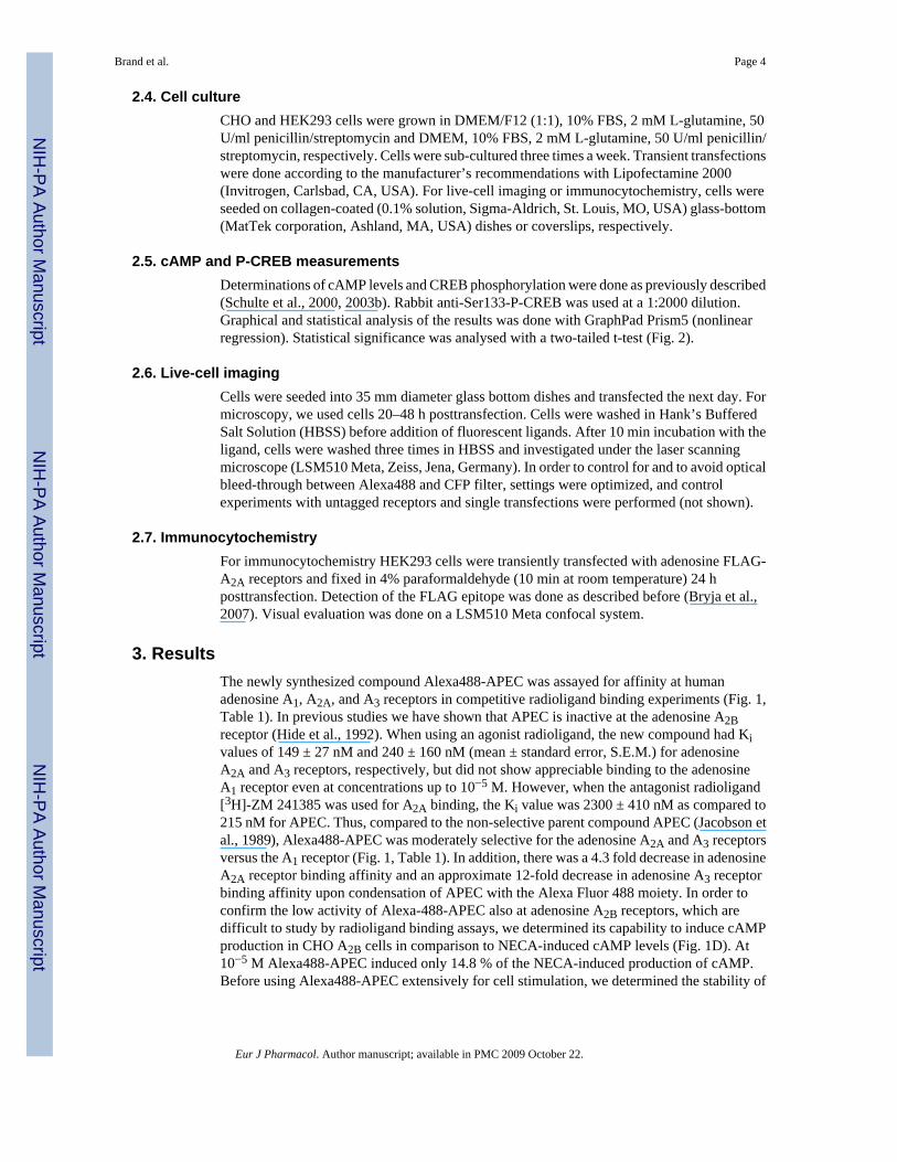
3. ResultsThe newly synthesized compound Alexa488-APEC was assayed for affinity at humanadenosine A1, A2A, and A3 receptors in competitive radioligand binding experiments (Fig. 1,Table 1). In previous studies we have shown that APEC is inactive at the adenosine A2Breceptor (Hide et al., 1992). When using an agonist radioligand, the new compound had Kivalues of 149 ± 27 nM and 240 ± 160 nM (mean ± standard error, S.E.M.) for adenosineA2A and A3 receptors, respectively, but did not show appreciable binding to the adenosineA1 receptor even at concentrations up to 10−5 M. However, when the antagonist radioligand[3H]-ZM 241385 was used for A2A binding, the Ki value was 2300 ± 410 nM as compared to215 nM for APEC. Thus, compared to the non-selective parent compound APEC (Jacobson etal., 1989), Alexa488-APEC was moderately selective for the adenosine A2A and A3 receptorsversus the A1 receptor (Fig. 1, Table 1). In addition, there was a 4.3 fold decrease in adenosineA2A receptor binding affinity and an approximate 12-fold decrease in adenosine A3 receptorbinding affinity upon condensation of APEC with the Alexa Fluor 488 moiety. In order toconfirm the low activity of Alexa-488-APEC also at adenosine A2B receptors, which aredifficult to study by radioligand binding assays, we determined its capability to induce cAMPproduction in CHO A2B cells in comparison to NECA-induced cAMP levels (Fig. 1D). At10−5 M Alexa488-APEC induced only 14.8 % of the NECA-induced production of cAMP.Before using Alexa488-APEC extensively for cell stimulation, we determined the stability of
Brand et al. Page 4
Eur J Pharmacol. Author manuscript; available in PMC 2009 October 22.
NIH
-PA Author Manuscript
NIH
-PA Author Manuscript
NIH
-PA Author Manuscript
the compound under cell culture conditions. In culture with CHO cells over several days, nodegradation products were detectable in the medium by thin layer chromatography (not shown).
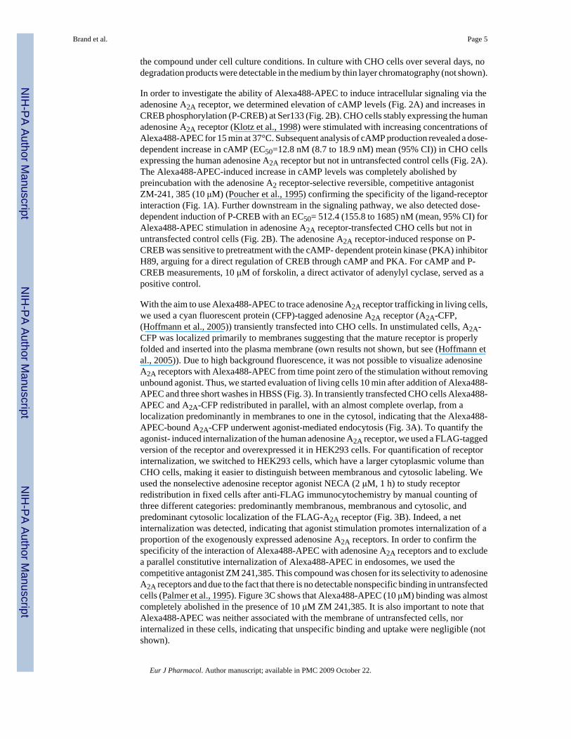
In order to investigate the ability of Alexa488-APEC to induce intracellular signaling via theadenosine A2A receptor, we determined elevation of cAMP levels (Fig. 2A) and increases inCREB phosphorylation (P-CREB) at Ser133 (Fig. 2B). CHO cells stably expressing the humanadenosine A2A receptor (Klotz et al., 1998) were stimulated with increasing concentrations ofAlexa488-APEC for 15 min at 37°C. Subsequent analysis of cAMP production revealed a dose-dependent increase in cAMP (EC50=12.8 nM (8.7 to 18.9 nM) mean (95% CI)) in CHO cellsexpressing the human adenosine A2A receptor but not in untransfected control cells (Fig. 2A).The Alexa488-APEC-induced increase in cAMP levels was completely abolished bypreincubation with the adenosine A2 receptor-selective reversible, competitive antagonistZM-241, 385 (10 μM) (Poucher et al., 1995) confirming the specificity of the ligand-receptorinteraction (Fig. 1A). Further downstream in the signaling pathway, we also detected dose-dependent induction of P-CREB with an EC50= 512.4 (155.8 to 1685) nM (mean, 95% CI) forAlexa488-APEC stimulation in adenosine A2A receptor-transfected CHO cells but not inuntransfected control cells (Fig. 2B). The adenosine A2A receptor-induced response on P-CREB was sensitive to pretreatment with the cAMP- dependent protein kinase (PKA) inhibitorH89, arguing for a direct regulation of CREB through cAMP and PKA. For cAMP and P-CREB measurements, 10 μM of forskolin, a direct activator of adenylyl cyclase, served as apositive control.
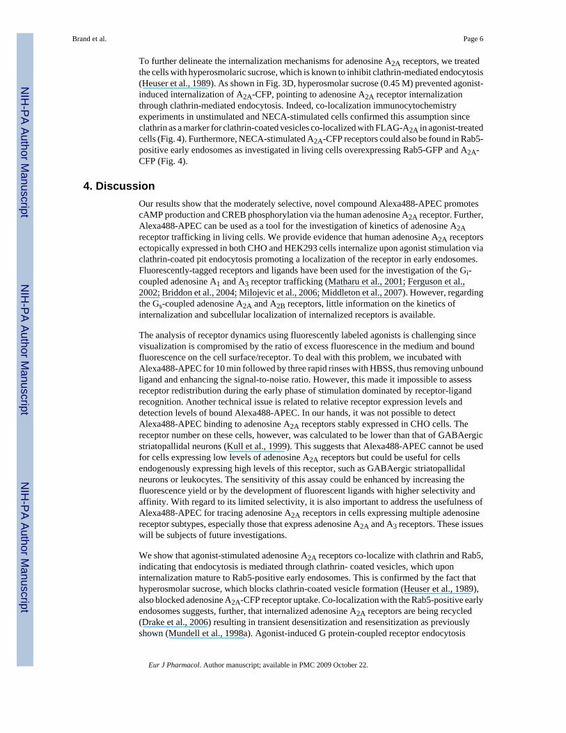
With the aim to use Alexa488-APEC to trace adenosine A2A receptor trafficking in living cells,we used a cyan fluorescent protein (CFP)-tagged adenosine A2A receptor (A2A-CFP,(Hoffmann et al., 2005)) transiently transfected into CHO cells. In unstimulated cells, A2A-CFP was localized primarily to membranes suggesting that the mature receptor is properlyfolded and inserted into the plasma membrane (own results not shown, but see (Hoffmann etal., 2005)). Due to high background fluorescence, it was not possible to visualize adenosineA2A receptors with Alexa488-APEC from time point zero of the stimulation without removingunbound agonist. Thus, we started evaluation of living cells 10 min after addition of Alexa488-APEC and three short washes in HBSS (Fig. 3). In transiently transfected CHO cells Alexa488-APEC and A2A-CFP redistributed in parallel, with an almost complete overlap, from alocalization predominantly in membranes to one in the cytosol, indicating that the Alexa488-APEC-bound A2A-CFP underwent agonist-mediated endocytosis (Fig. 3A). To quantify theagonist- induced internalization of the human adenosine A2A receptor, we used a FLAG-taggedversion of the receptor and overexpressed it in HEK293 cells. For quantification of receptorinternalization, we switched to HEK293 cells, which have a larger cytoplasmic volume thanCHO cells, making it easier to distinguish between membranous and cytosolic labeling. Weused the nonselective adenosine receptor agonist NECA (2 μM, 1 h) to study receptorredistribution in fixed cells after anti-FLAG immunocytochemistry by manual counting ofthree different categories: predominantly membranous, membranous and cytosolic, andpredominant cytosolic localization of the FLAG-A2A receptor (Fig. 3B). Indeed, a netinternalization was detected, indicating that agonist stimulation promotes internalization of aproportion of the exogenously expressed adenosine A2A receptors. In order to confirm thespecificity of the interaction of Alexa488-APEC with adenosine A2A receptors and to excludea parallel constitutive internalization of Alexa488-APEC in endosomes, we used thecompetitive antagonist ZM 241,385. This compound was chosen for its selectivity to adenosineA2A receptors and due to the fact that there is no detectable nonspecific binding in untransfectedcells (Palmer et al., 1995). Figure 3C shows that Alexa488-APEC (10 μM) binding was almostcompletely abolished in the presence of 10 μM ZM 241,385. It is also important to note thatAlexa488-APEC was neither associated with the membrane of untransfected cells, norinternalized in these cells, indicating that unspecific binding and uptake were negligible (notshown).
Brand et al. Page 5
Eur J Pharmacol. Author manuscript; available in PMC 2009 October 22.
NIH
-PA Author Manuscript
NIH
-PA Author Manuscript
NIH
-PA Author Manuscript
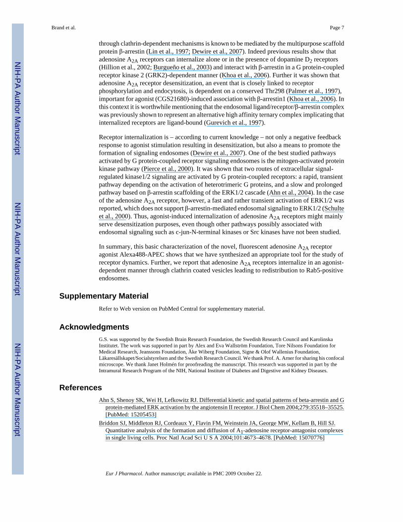
To further delineate the internalization mechanisms for adenosine A2A receptors, we treatedthe cells with hyperosmolaric sucrose, which is known to inhibit clathrin-mediated endocytosis(Heuser et al., 1989). As shown in Fig. 3D, hyperosmolar sucrose (0.45 M) prevented agonist-induced internalization of A2A-CFP, pointing to adenosine A2A receptor internalizationthrough clathrin-mediated endocytosis. Indeed, co-localization immunocytochemistryexperiments in unstimulated and NECA-stimulated cells confirmed this assumption sinceclathrin as a marker for clathrin-coated vesicles co-localized with FLAG-A2A in agonist-treatedcells (Fig. 4). Furthermore, NECA-stimulated A2A-CFP receptors could also be found in Rab5-positive early endosomes as investigated in living cells overexpressing Rab5-GFP and A2A-CFP (Fig. 4).
4. DiscussionOur results show that the moderately selective, novel compound Alexa488-APEC promotescAMP production and CREB phosphorylation via the human adenosine A2A receptor. Further,Alexa488-APEC can be used as a tool for the investigation of kinetics of adenosine A2Areceptor trafficking in living cells. We provide evidence that human adenosine A2A receptorsectopically expressed in both CHO and HEK293 cells internalize upon agonist stimulation viaclathrin-coated pit endocytosis promoting a localization of the receptor in early endosomes.Fluorescently-tagged receptors and ligands have been used for the investigation of the Gi-coupled adenosine A1 and A3 receptor trafficking (Matharu et al., 2001; Ferguson et al.,2002; Briddon et al., 2004; Milojevic et al., 2006; Middleton et al., 2007). However, regardingthe Gs-coupled adenosine A2A and A2B receptors, little information on the kinetics ofinternalization and subcellular localization of internalized receptors is available.
The analysis of receptor dynamics using fluorescently labeled agonists is challenging sincevisualization is compromised by the ratio of excess fluorescence in the medium and boundfluorescence on the cell surface/receptor. To deal with this problem, we incubated withAlexa488-APEC for 10 min followed by three rapid rinses with HBSS, thus removing unboundligand and enhancing the signal-to-noise ratio. However, this made it impossible to assessreceptor redistribution during the early phase of stimulation dominated by receptor-ligandrecognition. Another technical issue is related to relative receptor expression levels anddetection levels of bound Alexa488-APEC. In our hands, it was not possible to detectAlexa488-APEC binding to adenosine A2A receptors stably expressed in CHO cells. Thereceptor number on these cells, however, was calculated to be lower than that of GABAergicstriatopallidal neurons (Kull et al., 1999). This suggests that Alexa488-APEC cannot be usedfor cells expressing low levels of adenosine A2A receptors but could be useful for cellsendogenously expressing high levels of this receptor, such as GABAergic striatopallidalneurons or leukocytes. The sensitivity of this assay could be enhanced by increasing thefluorescence yield or by the development of fluorescent ligands with higher selectivity andaffinity. With regard to its limited selectivity, it is also important to address the usefulness ofAlexa488-APEC for tracing adenosine A2A receptors in cells expressing multiple adenosinereceptor subtypes, especially those that express adenosine A2A and A3 receptors. These issueswill be subjects of future investigations.
We show that agonist-stimulated adenosine A2A receptors co-localize with clathrin and Rab5,indicating that endocytosis is mediated through clathrin- coated vesicles, which uponinternalization mature to Rab5-positive early endosomes. This is confirmed by the fact thathyperosmolar sucrose, which blocks clathrin-coated vesicle formation (Heuser et al., 1989),also blocked adenosine A2A-CFP receptor uptake. Co-localization with the Rab5-positive earlyendosomes suggests, further, that internalized adenosine A2A receptors are being recycled(Drake et al., 2006) resulting in transient desensitization and resensitization as previouslyshown (Mundell et al., 1998a). Agonist-induced G protein-coupled receptor endocytosis
Brand et al. Page 6
Eur J Pharmacol. Author manuscript; available in PMC 2009 October 22.
NIH
-PA Author Manuscript
NIH
-PA Author Manuscript
NIH
-PA Author Manuscript
through clathrin-dependent mechanisms is known to be mediated by the multipurpose scaffoldprotein β-arrestin (Lin et al., 1997; Dewire et al., 2007). Indeed previous results show thatadenosine A2A receptors can internalize alone or in the presence of dopamine D2 receptors(Hillion et al., 2002; Burgueño et al., 2003) and interact with β-arrestin in a G protein-coupledreceptor kinase 2 (GRK2)-dependent manner (Khoa et al., 2006). Further it was shown thatadenosine A2A receptor desensitization, an event that is closely linked to receptorphosphorylation and endocytosis, is dependent on a conserved Thr298 (Palmer et al., 1997),important for agonist (CGS21680)-induced association with β-arrestin1 (Khoa et al., 2006). Inthis context it is worthwhile mentioning that the endosomal ligand/receptor/β-arrestin complexwas previously shown to represent an alternative high affinity ternary complex implicating thatinternalized receptors are ligand-bound (Gurevich et al., 1997).
Receptor internalization is – according to current knowledge – not only a negative feedbackresponse to agonist stimulation resulting in desensitization, but also a means to promote theformation of signaling endosomes (Dewire et al., 2007). One of the best studied pathwaysactivated by G protein-coupled receptor signaling endosomes is the mitogen-activated proteinkinase pathway (Pierce et al., 2000). It was shown that two routes of extracellular signal-regulated kinase1/2 signaling are activated by G protein-coupled receptors: a rapid, transientpathway depending on the activation of heterotrimeric G proteins, and a slow and prolongedpathway based on β-arrestin scaffolding of the ERK1/2 cascade (Ahn et al., 2004). In the caseof the adenosine A2A receptor, however, a fast and rather transient activation of ERK1/2 wasreported, which does not support β-arrestin-mediated endosomal signaling to ERK1/2 (Schulteet al., 2000). Thus, agonist-induced internalization of adenosine A2A receptors might mainlyserve desensitization purposes, even though other pathways possibly associated withendosomal signaling such as c-jun-N-terminal kinases or Src kinases have not been studied.
In summary, this basic characterization of the novel, fluorescent adenosine A2A receptoragonist Alexa488-APEC shows that we have synthesized an appropriate tool for the study ofreceptor dynamics. Further, we report that adenosine A2A receptors internalize in an agonist-dependent manner through clathrin coated vesicles leading to redistribution to Rab5-positiveendosomes.
Supplementary MaterialRefer to Web version on PubMed Central for supplementary material.
AcknowledgmentsG.S. was supported by the Swedish Brain Research Foundation, the Swedish Research Council and KarolinskaInstitutet. The work was supported in part by Alex and Eva Wallström Foundation, Tore Nilsons Foundation forMedical Research, Jeanssons Foundation, Åke Wiberg Foundation, Signe & Olof Wallenius Foundation,Läkaresällskapet/Socialstyrelsen and the Swedish Research Council. We thank Prof. A. Arner for sharing his confocalmicroscope. We thank Janet Holmén for proofreading the manuscript. This research was supported in part by theIntramural Research Program of the NIH, National Institute of Diabetes and Digestive and Kidney Diseases.
ReferencesAhn S, Shenoy SK, Wei H, Lefkowitz RJ. Differential kinetic and spatial patterns of beta-arrestin and G
protein-mediated ERK activation by the angiotensin II receptor. J Biol Chem 2004;279:35518–35525.[PubMed: 15205453]
Briddon SJ, Middleton RJ, Cordeaux Y, Flavin FM, Weinstein JA, George MW, Kellam B, Hill SJ.Quantitative analysis of the formation and diffusion of A1-adenosine receptor-antagonist complexesin single living cells. Proc Natl Acad Sci U S A 2004;101:4673–4678. [PubMed: 15070776]
Brand et al. Page 7
Eur J Pharmacol. Author manuscript; available in PMC 2009 October 22.
NIH
-PA Author Manuscript
NIH
-PA Author Manuscript
NIH
-PA Author Manuscript
Bryja V, Gradl D, Schambony A, Arenas E, Schulte G. beta-Arrestin is a necessary component of Wnt/beta-catenin signaling in vitro and in vivo. Proc Natl Acad Sci U S A 2007;104:6690–6695. [PubMed:17426148]
Burgueño J, Blake DJ, Benson MA, Tinsley CL, Esapa CT, Canela EI, Penela P, Mallol J, Mayor F, LluisC, Franco R, Ciruela F. The adenosine A2A receptor interacts with the actin-binding protein alpha-actinin. J Biol Chem 2003;278:37545–37552. [PubMed: 12837758]
Conn PM, Ulloa-Aguirre A, Ito J, Janovick JA. G protein-coupled receptor trafficking in health anddisease: lessons learned to prepare for therapeutic mutant rescue in vivo. Pharmacol Rev 2007;59:225–250. [PubMed: 17878512]
Daly CJ, McGrath JC. Fluorescent ligands, antibodies, and proteins for the study of receptors. PharmacolTher 2003;100:101–118. [PubMed: 14609715]
Dewire SM, Ahn S, Lefkowitz RJ, Shenoy SK. beta-Arrestins and Cell Signaling. Annu Rev Physiol2007;69:483–510. [PubMed: 17305471]
Drake MT, Shenoy SK, Lefkowitz RJ. Trafficking of G protein-coupled receptors. Circ Res 2006;99:570–582. [PubMed: 16973913]
Ferguson G, Watterson KR, Palmer TM. Subtype-specific regulation of receptor internalization andrecycling by the carboxyl-terminal domains of the human A1 and rat A3 adenosine receptors:consequences for agonist-stimulated translocation of arrestin3. Biochemistry 2002;41:14748–14761.[PubMed: 12475223]
Fredholm BB, Chern Y, Franco R, Sitkovsky M. Prog. Neurobiol 2007;83:263–276.Fredholm BB, Cunha RA, Svenningsson P. Pharmacology of adenosine A2A receptors and therapeutic
applications. Curr Top Med Chem 2003;3:413–426. [PubMed: 12570759]Fredholm BB, IJzerman AP, Jacobson KA, Klotz KN, Linden J. International Union of Pharmacology.
XXV Nomenclature and classification of adenosine receptors. Pharmacol Rev 2001;53:527–552.[PubMed: 11734617]
Furlong TJ, Pierce KD, Selbie LA, Shine J. Molecular characterization of a human brain adenosine A2receptor. Brain Res Mol Brain Res 1992;15:62–66. [PubMed: 1331670]
Gao ZG, Mamedova LK, Chen P, Jacobson KA. 2-Substituted adenosine derivatives: affinity and efficacyat four subtypes of human adenosine receptors. Biochem Pharmacol 2004;68:1985–1993. [PubMed:15476669]
Gurevich VV, Pals-Rylaarsdam R, Benovic JL, Hosey MM, Onorato JJ. Agonist-receptor-arrestin, analternative ternary complex with high agonist affinity. J Biol Chem 1997;272:28849–28852.[PubMed: 9360951]
Heuser JE, Anderson RG. Hypertonic media inhibit receptor-mediated endocytosis by blocking clathrin-coated pit formation. J Cell Biol 1989;108:389–400. [PubMed: 2563728]
Hide I, Padgett WL, Jacobson KA, Daly JW. A2A adenosine receptors from rat striatum and ratpheochromocytoma PC12 cells: characterization with radioligand binding and by activation ofadenylate cyclase. Mol Pharmacol 1992;41:352–359. [PubMed: 1311411]
Hillion J, Canals M, Torvinen M, Casado V, Scott R, Terasmaa A, Hansson A, Watson S, Olah ME,Mallol J, Canela EI, Zoli M, Agnati LF, Ibanez CF, Lluis C, Franco R, Ferre S, Fuxe K.Coaggregation, cointernalization, and codesensitization of adenosine A2A receptors and dopamineD2 receptors. J Biol Chem 2002;277:18091–18097. [PubMed: 11872740]
Hoffmann C, Gaietta G, Bünemann M, Adams SR, Oberdorff-Maass S, Behr B, Vilardaga JP, Tsien RY,Ellisman MH, Lohse MJ. A FlAsH-based FRET approach to determine G protein-coupled receptoractivation in living cells. Nature Methods 2005;2:171–176. [PubMed: 15782185]
Huang ZL, Urade Y, Hayaishi O. Prostaglandins and adenosine in the regulation of sleep and wakefulness.Curr Opin Pharmacol 2007;7:33–38. [PubMed: 17129762]
Jacobson KA, Gao ZG. Adenosine receptors as therapeutic targets. Nat Rev Drug Discov 2006;5:247–264. [PubMed: 16518376]
Jacobson KA, Pannell LK, Ji XD, Jarvis MF, Williams M, Hutchison AJ, Barrington WW, Stiles GL.Agonist derived molecular probes for A2 adenosine receptors. J Mol Recognit 1989;2:170–178.[PubMed: 2561548]
Brand et al. Page 8
Eur J Pharmacol. Author manuscript; available in PMC 2009 October 22.
NIH
-PA Author Manuscript
NIH
-PA Author Manuscript
NIH
-PA Author Manuscript
Khoa ND, Postow M, Danielsson J, Cronstein BN. Tumor necrosis factor-alpha prevents desensitizationof Gαs-coupled receptors by regulating GRK2 association with the plasma membrane. MolPharmacol 2006;69:1311–1319. [PubMed: 16385076]
Klotz KN, Hessling J, Hegler J, Owman C, Kull B, Fredholm BB, Lohse MJ. Comparative pharmacologyof human adenosine receptor subtypes -characterization of stably transfected receptors in CHO cells.Naunyn Schmiedebergs Arch Pharmacol 1998;357:1–9. [PubMed: 9459566]
Kull B, Ferré S, Arslan G, Svenningsson P, Fuxe K, Owman C, Fredholm BB. Reciprocal interactionsbetween adenosine A2A and dopamine D2 receptors in Chinese hamster ovary cells co-transfectedwith the two receptors. Biochem Pharmacol 1999;58:1035–1045. [PubMed: 10509756]
Ledent C, Vaugeois JM, Schiffmann SN, Pedrazzini T, El Yacoubi M, Vanderhaeghen JJ, Costentin J,Heath JK, Vassart G, Parmentier M. Aggressiveness, hypoalgesia and high blood pressure in micelacking the adenosine A2a receptor. Nature 1997;388:674–678. [PubMed: 9262401]
Lin FT, Krueger KM, Kendall HE, Daaka Y, Fredericks ZL, Pitcher JA, Lefkowitz RJ. Clathrin-mediatedendocytosis of the beta-adrenergic receptor is regulated by phosphorylation/dephosphorylation ofbeta-arrestin1. J Biol Chem 1997;272:31051–31057. [PubMed: 9388255]
Matharu AL, Mundell SJ, Benovic JL, Kelly E. Rapid agonist-induced desensitization and internalizationof the A2B adenosine receptor is mediated by a serine residue close to the COOH terminus. J BiolChem 2001;276:30199– 30207. [PubMed: 11333255]
Middleton RJ, Briddon SJ, Cordeaux Y, Yates AS, Dale CL, George MW, Baker JG, Hill SJ, Kellam B.New fluorescent adenosine A1-receptor agonists that allow quantification of ligand-receptorinteractions in microdomains of single living cells. J Med Chem 2007;50:782–793. [PubMed:17249651]
Middleton RJ, Kellam B. Fluorophore-tagged GPCR ligands. Curr Opin Chem Biol 2005;9:517–525.[PubMed: 16125436]
Milojevic T, Reiterer V, Stefan E, Korkhov VM, Dorostkar MM, Ducza E, Ogris E, Boehm S, FreissmuthM, Nanoff C. The ubiquitin-specific protease Usp4 regulates the cell surface level of the A2Areceptor. Mol Pharmacol 2006;69:1083–1094. [PubMed: 16339847]
Mundell SJ, Benovic JL, Kelly E. A dominant negative mutant of the G protein-coupled receptor kinase2 selectively attenuates adenosine A2 receptor desensitization. Mol Pharmacol 1997;51:991–998.[PubMed: 9187265]
Mundell SJ, Kelly E. The effect of inhibitors of receptor internalization on the desensitization andresensitization of three Gs-coupled receptor responses. Br J Pharmacol 1998a;125:1594–1600.[PubMed: 9884089]
Mundell SJ, Luty JS, Willets J, Benovic JL, Kelly E. Enhanced expression of G protein-coupled receptorkinase 2 selectively increases the sensitivity of A2A adenosine receptors to agonist-induceddesensitization. Br J Pharmacol 1998b;125:347–356. [PubMed: 9786508]
Palmer TM, Gettys TW, Jacobson KA, Stiles GL. Desensitization of the canine A2a adenosine receptor:delineation of multiple processes. Mol Pharmacol 1994;45:1082–1094. [PubMed: 8022402]
Palmer TM, Poucher SM, Jacobson KA, Stiles GL. 125I-4-(2-[7-amino- 2-[2-furyl][1,2,4]triazolo[2,3-a][1,3,5] triazin-5-yl-amino]ethyl)phenol, a high affinity antagonist radioligand selective for the A2aadenosine receptor. Mol Pharmacol 1995;48:970–974. [PubMed: 8848012]
Palmer TM, Stiles GL. Identification of an A2a adenosine receptor domain specifically responsible formediating short-term desensitization. Biochemistry 1997;36:832–838. [PubMed: 9020781]
Perreira M, Jiang JK, Klutz AM, Gao ZG, Shainberg A, Lu C, Thomas CJ, Jacobson KA. “Reversine”and its 2-substituted adenine derivatives as potent and selective A3 adenosine receptor antagonists.J Med Chem 2005;48:4910–4918. [PubMed: 16033270]
Pierce KL, Maudsley S, Daaka Y, Luttrell LM, Lefkowitz RJ. Role of endocytosis in the activation ofthe extracellular signal-regulated kinase cascade by sequestering and nonsequestering G protein-coupled receptors. Proc Natl Acad Sci U S A 2000;97:1489–1494. [PubMed: 10677489]
Poucher SM, Keddie JR, Singh P, Stoggall SM, Caulkett PW, Jones G, Coll MG. The in vitropharmacology of ZM 241385, a potent, non-xanthine A2a selective adenosine receptor antagonist.Br J Pharmacol 1995;115:1096–1102. [PubMed: 7582508]
Roberts RL, Barbieri MA, Pryse KM, Chua M, Morisaki JH, Stahl PD. Endosome fusion in living cellsoverexpressing GFP-rab5. J Cell Sci 1999;112:3667–3675. [PubMed: 10523503]
Brand et al. Page 9
Eur J Pharmacol. Author manuscript; available in PMC 2009 October 22.
NIH
-PA Author Manuscript
NIH
-PA Author Manuscript
NIH
-PA Author Manuscript
Schulte G, Fredholm BB. Human adenosine A1, A2A, A2B, and A3 receptors expressed in Chinesehamster ovary cells all mediate the phosphorylation of extracellular-regulated kinase 1/2. MolPharmacol 2000;58:477–482. [PubMed: 10953039]
Schulte G, Fredholm BB. Signalling from adenosine receptors to mitogen-activated protein kinases. CellSignal 2003a;15:813–827. [PubMed: 12834807]
Schulte G, Fredholm BB. The Gs-coupled adenosine A2B receptor recruits divergent pathways to regulateERK1/2 and p38. Exp Cell Res 2003b;290:168–176. [PubMed: 14516797]
Wolfe BL, Trejo J. Clathrin-dependent mechanisms of G protein-coupled receptor endocytosis. Traffic2007;8:462–470. [PubMed: 17376169]
Xu ZQ, Zhang X, Scott L. Regulation of G protein-coupled receptor trafficking. Acta Physiol2007;190:39–45.
Brand et al. Page 10
Eur J Pharmacol. Author manuscript; available in PMC 2009 October 22.
NIH
-PA Author Manuscript
NIH
-PA Author Manuscript
NIH
-PA Author Manuscript
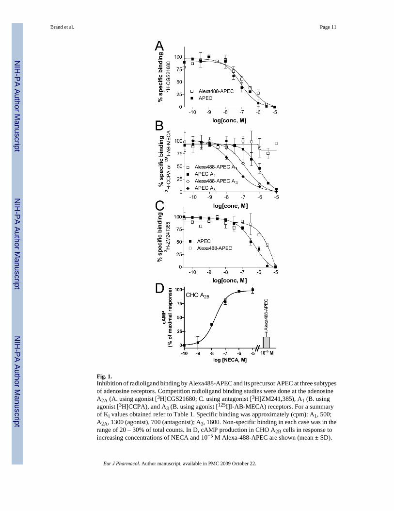
Fig. 1.Inhibition of radioligand binding by Alexa488-APEC and its precursor APEC at three subtypesof adenosine receptors. Competition radioligand binding studies were done at the adenosineA2A (A. using agonist [3H]CGS21680; C. using antagonist [3H]ZM241,385), A1 (B. usingagonist [3H]CCPA), and A3 (B. using agonist [125I]I-AB-MECA) receptors. For a summaryof Ki values obtained refer to Table 1. Specific binding was approximately (cpm): A1, 500;A2A, 1300 (agonist), 700 (antagonist); A3, 1600. Non-specific binding in each case was in therange of 20 – 30% of total counts. In D, cAMP production in CHO A2B cells in response toincreasing concentrations of NECA and 10−5 M Alexa-488-APEC are shown (mean ± SD).
Brand et al. Page 11
Eur J Pharmacol. Author manuscript; available in PMC 2009 October 22.
NIH
-PA Author Manuscript
NIH
-PA Author Manuscript
NIH
-PA Author Manuscript
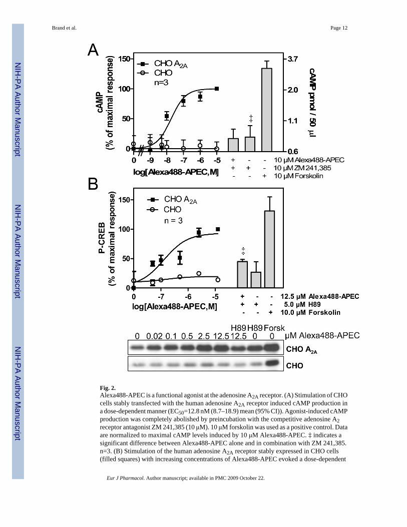
Fig. 2.Alexa488-APEC is a functional agonist at the adenosine A2A receptor. (A) Stimulation of CHOcells stably transfected with the human adenosine A2A receptor induced cAMP production ina dose-dependent manner (EC50=12.8 nM (8.7–18.9) mean (95% CI)). Agonist-induced cAMPproduction was completely abolished by preincubation with the competitive adenosine A2receptor antagonist ZM 241,385 (10 μM). 10 μM forskolin was used as a positive control. Dataare normalized to maximal cAMP levels induced by 10 μM Alexa488-APEC. ‡ indicates asignificant difference between Alexa488-APEC alone and in combination with ZM 241,385.n=3. (B) Stimulation of the human adenosine A2A receptor stably expressed in CHO cells(filled squares) with increasing concentrations of Alexa488-APEC evoked a dose-dependent
Brand et al. Page 12
Eur J Pharmacol. Author manuscript; available in PMC 2009 October 22.
NIH
-PA Author Manuscript
NIH
-PA Author Manuscript
NIH
-PA Author Manuscript
increase in CREB phosphorylation (P-CREB), which was sensitive to PKA inhibition by H89.‡ indicates a significant difference between Alexa488-APEC alone and in combination withH89. Forskolin (10 μM) was used as a positive control. Results are normalized to maximalcAMP levels induced by 12.5 μM Alexa488-APEC. The lower panel shows representativeimmunoblots of P-CREB in CHO A2A as well as untransfected CHO cells (n=3). UntransfectedCHO cells (open circles) did not respond to Alexa488-APEC. n=3 (mean ± SD).
Brand et al. Page 13
Eur J Pharmacol. Author manuscript; available in PMC 2009 October 22.
NIH
-PA Author Manuscript
NIH
-PA Author Manuscript
NIH
-PA Author Manuscript
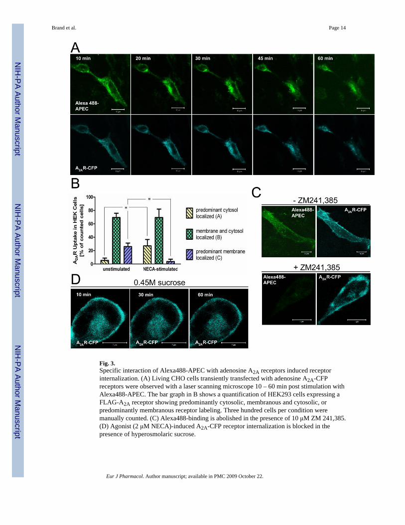
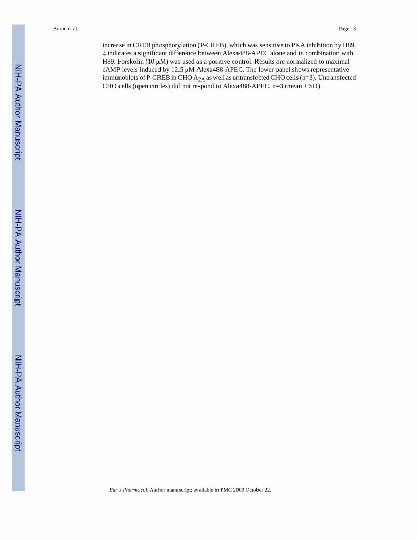
Fig. 3.Specific interaction of Alexa488-APEC with adenosine A2A receptors induced receptorinternalization. (A) Living CHO cells transiently transfected with adenosine A2A-CFPreceptors were observed with a laser scanning microscope 10 – 60 min post stimulation withAlexa488-APEC. The bar graph in B shows a quantification of HEK293 cells expressing aFLAG-A2A receptor showing predominantly cytosolic, membranous and cytosolic, orpredominantly membranous receptor labeling. Three hundred cells per condition weremanually counted. (C) Alexa488-binding is abolished in the presence of 10 μM ZM 241,385.(D) Agonist (2 μM NECA)-induced A2A-CFP receptor internalization is blocked in thepresence of hyperosmolaric sucrose.
Brand et al. Page 14
Eur J Pharmacol. Author manuscript; available in PMC 2009 October 22.
NIH
-PA Author Manuscript
NIH
-PA Author Manuscript
NIH
-PA Author Manuscript
Fig. 4.Agonist-stimulated adenosine A2A receptors internalize through clathrin-dependentendocytosis and localize to early endosomes. FLAG-A2A receptors were expressed in HEK293cells for immunocytochemistry using an anti-FLAG (Cy3, red) and anti-clathrin (Cy2, green)antibody. The upper panels show co- localization of FLAG-A2A and clathrin in fixed,unstimulated cells and after 1 h stimulation with NECA (1 μM). The lower panels show co-localization of A2A-CFP with Rab5-GFP. Living HEK293 cells co-transfected with A2A-CFP(red) and Rab5-GFP (green) are shown before and after 1 h stimulation with 1 μM NECA. n=3.Arrows indicate some of the Rab5-positive and adenosine A2A receptor-containing vesicles,
Brand et al. Page 15
Eur J Pharmacol. Author manuscript; available in PMC 2009 October 22.
NIH
-PA Author Manuscript
NIH
-PA Author Manuscript
NIH
-PA Author Manuscript

which were identified by live cell imaging as newly formed vesicles. Yellow color indicatesco-localization.
Brand et al. Page 16
Eur J Pharmacol. Author manuscript; available in PMC 2009 October 22.
NIH
-PA Author Manuscript
NIH
-PA Author Manuscript
NIH
-PA Author Manuscript
NIH
-PA Author Manuscript
NIH
-PA Author Manuscript
NIH
-PA Author Manuscript
Brand et al. Page 17Ta
ble
1C
ompa
rison
of t
he d
isso
ciat
ion
cons
tant
Ki o
f the
par
ent A
PEC
and
its
fluor
esce
nt d
eriv
ativ
e A
lexa
488-
APE
C w
ith re
gard
to a
goni
stbi
ndin
g to
ade
nosi
ne A
1, A
2A a
nd A
3 rec
epto
r (N
A, <
10%
dis
plac
emen
t at 1
0−5 M
) and
ant
agon
ist b
indi
ng a
t the
A2A
rece
ptor
. Val
ues
are
give
n as
mea
n ±
S.E.
M.
Com
poun
dA
2A a
goni
st, K
iA
3, K
iA
1, K
iA
2A a
ntag
onis
t, K
iM
ean
(nM
)S.
E.M
. (nM
)M
ean
(nM
)S.
E.M
. (nM
)M
ean
(nM
)S.
E.M
. (nM
)M
ean
(nM
)S.
E.M
. (nM
)
Ale
xa48
8-A
PEC
149
2724
016
0N
o bi
ndin
g23
0041
0A
PEC
355
202
450
3721
580
Eur J Pharmacol. Author manuscript; available in PMC 2009 October 22.