NF-κB Is Activated in CD4+ iNKT Cells by Sickle Cell Disease and Mediates Rapid Induction of...
9
NF-kB Is Activated in CD4 + iNKT Cells by Sickle Cell Disease and Mediates Rapid Induction of Adenosine A 2A Receptors Gene Lin 1. , Joshua J. Field 2. , Jennifer C. Yu 1 , Ruey Ken 1 , Donna Neuberg 3 , David G. Nathan 3 , Joel Linden 1 * 1 Division of Inflammation Biology, La Jolla Institute for Allergy and Immunology, La Jolla, California, United States of America, 2 Medical College of Wisconsin, Milwaukee, Wisconsin, United States of America, 3 Dana-Farber Cancer Institute, Boston, Massachusetts, United States of America Abstract Reperfusion injury following tissue ischemia occurs as a consequence of vaso-occlusion that is initiated by activation of invariant natural killer T (iNKT) cells. Sickle cell disease (SDC) results in widely disseminated microvascular ischemia and reperfusion injury as a result of vaso-occlusion by rigid and adhesive sickle red blood cells. In mice, iNKT cell activation requires NF-kB signaling and can be inhibited by the activation of anti-inflammatory adenosine A 2A receptors (A 2A Rs). Human iNKT cells are divided into subsets of CD4+ and CD4- cells. In this study we found that human CD4+ iNKT cells, but not CD4- cells undergo rapid NF-kB activation (phosphorylation of NF-kB on p65) and induction of A 2A Rs (detected with a monoclonal antibody 7F6-G5-A2) during SCD painful vaso-occlusive crises. These findings indicate that SCD primarily activates the CD4+ subset of iNKT cells. Activation of NF-kB and induction of A 2A Rs is concordant, i.e. only CD4+ iNKT cells with activated NF-kB expressed high levels of A 2A Rs. iNKT cells that are not activated during pVOC express low levels of A 2A R immunoreactivity. These finding suggest that A 2A R transcription may be induced in CD4+ iNKT cells as a result of NF- kB activation in SCD. In order to test this hypothesis further we examined cultured human iNKT cells. In cultured cells, blockade of NF-kB with Bay 11–7082 or IKK inhibitor VII prevented rapid induction of A 2A R mRNA and protein upon iNKT activation. In conclusion, NF-kB-mediated induction of A 2A Rs in iNKT cells may serve as a counter-regulatory mechanism to limit the extent and duration of inflammatory immune responses. As activated iNKT cells express high levels of A 2A Rs following their activation, they may become highly sensitive to inhibition by A 2A R agonists. Citation: Lin G, Field JJ, Yu JC, Ken R, Neuberg D, et al. (2013) NF-kB Is Activated in CD4 + iNKT Cells by Sickle Cell Disease and Mediates Rapid Induction of Adenosine A 2A Receptors. PLoS ONE 8(10): e74664. doi:10.1371/journal.pone.0074664 Editor: Arun Rishi, Wayne State University, United States of America Received April 22, 2013; Accepted August 4, 2013; Published October 4, 2013 Copyright: ß 2013 Lin et al. This is an open-access article distributed under the terms of the Creative Commons Attribution License, which permits unrestricted use, distribution, and reproduction in any medium, provided the original author and source are credited. Funding: Supported by grants RC2 HL101367, R34 HL108757 and R01HL095704 from the National Institutes of Health. The funders had no role in study design, data collection and analysis, decision to publish, or preparation of the manuscript. Competing Interests: The authors have declared that no competing interests exist. * E-mail: [email protected] . These authors contributed equally to this work. Introduction Reperfusion injury following tissue ischemia is initiated by the activation of iNKT cells [1–3]. Widely disseminated ischemia- reperfusion injury is a manifestation of HbSS sickle cell disease that is caused by a homozygous point mutation in the ß-globin gene. The mutation promotes deoxyhemoglobin polymerization, formation of rigid sickled RBCs and production of large numbers of adhesive reticulocytes [4]. Tissue damaging vaso-occlusion in SCD has been viewed as resulting from obstruction of small blood vessels by sickled RBCs [5]. The clinical course of SCD is characterized by exacerbations that cause sudden painful vaso- occlusive crises (pVOC) and sometimes life-threatening episodes of acute chest syndrome (ACS). Recently, a modified paradigm has emerged suggesting that the clinical manifestations of SCD occur in part as a consequence of white cell activation [6]. As in ischemia-reperfusion injury, in NY1DD mice with SCD the activation of iNKT cells in response to tissue ischemia initiates an inflammatory cascade [7]. Poor lung function in SCD mice is ameliorated by iNKT cell depletion, by blockade of CD1d- restricted signaling [7], or by stimulation of anti-inflammatory A 2A R receptors that are induced in SCD mice and that inhibit iNKT cell activation [8]. The A 2A R is one of a family of four G protein coupled adenosine receptors (A 1 ,A 2A ,A 2B and A 3 ), that is expressed on most leukocytes and platelets and when activated exerts generally anti-inflammatory effects [9]. We have shown previously that pVOC in SCD patients results in the appearance of iNKT cells with high expression of activated NF-kB and cells that express high levels of anti-inflammatory A 2A Rs. In prior studies we did not determine if the expression of activation markers occurs on the same or different cells than those that express high levels of A 2A Rs. Since A 2A R activation inhibits iNKT cell activation [10] we reasoned that the iNKT cells that are not activated may express high levels of A 2A Rs. Here we demonstrate that NF-kB activation; T-bet induction, A 2A R induction and cytokine production are all largely concordant (i.e. in the same cells) and occurs in a subset of CD4+ iNKT cells. The activation of cultured human iNKT cells results in induction of A 2A R mRNA and protein expression that is blocked by NF-kB inhibitors. The findings suggest that A 2A Rs are PLOS ONE | www.plosone.org 1 October 2013 | Volume 8 | Issue 10 | e74664
-
Upload
independent -
Category
Documents
-
view
4 -
download
0
Transcript of NF-κB Is Activated in CD4+ iNKT Cells by Sickle Cell Disease and Mediates Rapid Induction of...

NF-kB Is Activated in CD4+ iNKT Cells by Sickle CellDisease and Mediates Rapid Induction of Adenosine A2A
ReceptorsGene Lin1., Joshua J. Field2., Jennifer C. Yu1, Ruey Ken1, Donna Neuberg3, David G. Nathan3,
Joel Linden1*
1Division of Inflammation Biology, La Jolla Institute for Allergy and Immunology, La Jolla, California, United States of America, 2Medical College of Wisconsin, Milwaukee,
Wisconsin, United States of America, 3Dana-Farber Cancer Institute, Boston, Massachusetts, United States of America
Abstract
Reperfusion injury following tissue ischemia occurs as a consequence of vaso-occlusion that is initiated by activation ofinvariant natural killer T (iNKT) cells. Sickle cell disease (SDC) results in widely disseminated microvascular ischemia andreperfusion injury as a result of vaso-occlusion by rigid and adhesive sickle red blood cells. In mice, iNKT cell activationrequires NF-kB signaling and can be inhibited by the activation of anti-inflammatory adenosine A2A receptors (A2ARs).Human iNKT cells are divided into subsets of CD4+ and CD4- cells. In this study we found that human CD4+ iNKT cells, butnot CD4- cells undergo rapid NF-kB activation (phosphorylation of NF-kB on p65) and induction of A2ARs (detected with amonoclonal antibody 7F6-G5-A2) during SCD painful vaso-occlusive crises. These findings indicate that SCD primarilyactivates the CD4+ subset of iNKT cells. Activation of NF-kB and induction of A2ARs is concordant, i.e. only CD4+ iNKT cellswith activated NF-kB expressed high levels of A2ARs. iNKT cells that are not activated during pVOC express low levels ofA2AR immunoreactivity. These finding suggest that A2AR transcription may be induced in CD4+ iNKT cells as a result of NF-kB activation in SCD. In order to test this hypothesis further we examined cultured human iNKT cells. In cultured cells,blockade of NF-kB with Bay 11–7082 or IKK inhibitor VII prevented rapid induction of A2AR mRNA and protein upon iNKTactivation. In conclusion, NF-kB-mediated induction of A2ARs in iNKT cells may serve as a counter-regulatory mechanism tolimit the extent and duration of inflammatory immune responses. As activated iNKT cells express high levels of A2ARsfollowing their activation, they may become highly sensitive to inhibition by A2AR agonists.
Citation: Lin G, Field JJ, Yu JC, Ken R, Neuberg D, et al. (2013) NF-kB Is Activated in CD4+ iNKT Cells by Sickle Cell Disease and Mediates Rapid Induction ofAdenosine A2A Receptors. PLoS ONE 8(10): e74664. doi:10.1371/journal.pone.0074664
Editor: Arun Rishi, Wayne State University, United States of America
Received April 22, 2013; Accepted August 4, 2013; Published October 4, 2013
Copyright: � 2013 Lin et al. This is an open-access article distributed under the terms of the Creative Commons Attribution License, which permits unrestricteduse, distribution, and reproduction in any medium, provided the original author and source are credited.
Funding: Supported by grants RC2 HL101367, R34 HL108757 and R01HL095704 from the National Institutes of Health. The funders had no role in study design,data collection and analysis, decision to publish, or preparation of the manuscript.
Competing Interests: The authors have declared that no competing interests exist.
* E-mail: [email protected]
. These authors contributed equally to this work.
Introduction
Reperfusion injury following tissue ischemia is initiated by the
activation of iNKT cells [1–3]. Widely disseminated ischemia-
reperfusion injury is a manifestation of HbSS sickle cell disease
that is caused by a homozygous point mutation in the ß-globin
gene. The mutation promotes deoxyhemoglobin polymerization,
formation of rigid sickled RBCs and production of large numbers
of adhesive reticulocytes [4]. Tissue damaging vaso-occlusion in
SCD has been viewed as resulting from obstruction of small blood
vessels by sickled RBCs [5]. The clinical course of SCD is
characterized by exacerbations that cause sudden painful vaso-
occlusive crises (pVOC) and sometimes life-threatening episodes of
acute chest syndrome (ACS). Recently, a modified paradigm has
emerged suggesting that the clinical manifestations of SCD occur
in part as a consequence of white cell activation [6]. As in
ischemia-reperfusion injury, in NY1DD mice with SCD the
activation of iNKT cells in response to tissue ischemia initiates an
inflammatory cascade [7]. Poor lung function in SCD mice is
ameliorated by iNKT cell depletion, by blockade of CD1d-
restricted signaling [7], or by stimulation of anti-inflammatory
A2AR receptors that are induced in SCD mice and that inhibit
iNKT cell activation [8].
The A2AR is one of a family of four G protein coupled
adenosine receptors (A1, A2A, A2B and A3), that is expressed on
most leukocytes and platelets and when activated exerts generally
anti-inflammatory effects [9]. We have shown previously that
pVOC in SCD patients results in the appearance of iNKT cells
with high expression of activated NF-kB and cells that express
high levels of anti-inflammatory A2ARs. In prior studies we did not
determine if the expression of activation markers occurs on the
same or different cells than those that express high levels of A2ARs.
Since A2AR activation inhibits iNKT cell activation [10] we
reasoned that the iNKT cells that are not activated may express
high levels of A2ARs. Here we demonstrate that NF-kB activation;
T-bet induction, A2AR induction and cytokine production are all
largely concordant (i.e. in the same cells) and occurs in a subset of
CD4+ iNKT cells. The activation of cultured human iNKT cells
results in induction of A2AR mRNA and protein expression that is
blocked by NF-kB inhibitors. The findings suggest that A2ARs are
PLOS ONE | www.plosone.org 1 October 2013 | Volume 8 | Issue 10 | e74664

induced as a consequence of iNKT cell activation and may serve
to limit the duration of their activation.
Materials and Methods
All research involving human participants and the content of
written informed consent forms were approved by the institutional
review boards of the Medical College of Wisconsin and the La
Jolla Institute for Allergy and Immunology. Consent forms signed
by study participants are on file.
Collection and processing of bloodVenous blood was obtained from adult patients, ages 18 to
60 years, with HbSS/HbSb-thalassemia0 at Froedtert Hospital/
Medical College of Wisconsin following informed consent. Paired
samples separated by at least 30 days were collected from the same
patient. Vaso-occlusive pain crisis was defined as an episode of
pain related to SCD in the extremities, back, abdomen, chest or
head lasting at least 2 hours and leading to a hospitalization [11].
Participants were determined to be at steady state when they were
reporting no more than baseline pain and were at least 2 weeks
from a hospitalization or emergency department visit for any
reason.
Flow Cytometry and statisticsRBCs in 0.3 ml blood were lysed (Biolegend) and remaining
cells were washed with cold phosphate-buffered solution, pH 7.2
(PBS) containing 2 mM EDTA, resuspended in cold FACS
staining buffer (PBS, 1%BSA, 1% human AB serum, 0.1%
sodium azide) and incubated on ice for 10 minutes prior to
staining. Remaining cells were incubated for 40 minutes at 4uCwith fixable LIVE/DEAD stain to identify dead cells (Invitrogen)
and then with fluorophore-conjugated antibodies directed against
surface markers. Cells were washed twice with cold PBS, fixed,
and resuspended in fixation/permeabilization buffer (BD biosci-
ences) for 20 minutes at 4uC. After fixation, cells were washed
twice with cold permeabilization buffer and incubated for
45 minutes at 4uC with fluorophore-conjugated antibodies specific
for intracellular antigens. Cells were then washed with cold
permeabilization buffer, fixed with 1% paraformaldehyde for
15 minutes at 4uC, washed with cold PBS, and resuspended in
0.3 ml of FACS staining buffer. The stained samples were stored
at 4uC in the dark until flow cytometric analysis.
Invariant NKT cells were identified as live, CD19- (Invitrogen,
SJ25-C1), CD3+ (Invitrogen & BD biosciences, UCHT1), and
Valpha24-Jalpha18 TCR + (eBioscience, 6B11) cells and their
CD4 phenotype was determined with anti-human CD4 antibody
(BD biosciences, RPA-T4). For some experiments T cells were
stained with biotinylated anti-Va24 antibodies (Beckman Coulter
IM2027)/brilliant violet streptavidin (Biolegend) that detect ,1%
of peripheral T cells. Staining with 6B11 identifies the iNKT
subset of all Valpha24+ cells. Conventional T cells were identified
as live, CD19-, CD3+, and 6B11- cells. The active phosphorylated
form of p65 NF-kB was identified with anti-phospho-NF-kB p65
(Cell Signaling, 93H1). The human adenosine A2A receptor was
detected with anti-human receptor antibody 7F6-G5-A2 [12,13]
(Santa Cruz Biotech) conjugated to Alexa Fluor 647 (Invitrogen).
IL-4, IFN-gamma, CD69 and the transcription factor T-bet were
detected with anti-human IL-4 (BD, 8D4-8), anti human IFN-c(eBioscience, 4S. B3) anti-human CD69 (BD, FN50) and anti
mouse/human T-bet (BD, O4-46) antibodies, respectively. Flow
cytometry was performed using a LSRII (BD biosciences) and data
analysis performed using FlowJo software (Tree star). Data derived
from patients sampled twice, once during pVOC and once at
steady state were analyzed by the paired t-test.
Human iNKT cell culture and activationHuman iNKT cell lines were generated from peripheral blood
mononuclear cells (PBMCs) isolated from normal donor blood
using a ficoll density gradient. A total of 120 million PBMCs were
cultured for 12–14 days in culture medium (45% AIM V, Life
Technologies, 50% RPMI 1640, Gibco, and 5% human AB
serum, GemCell) supplemented with 100 IU/ml IL-2 (NCI) at
and 100 ng/ml alpha-Galactosylceramide (alphaGalCer) (Funa-
Figure 1. Painful vaso-occlusive crisis (pVOC) in SCD patients are associated with increases in the percentage of cytokine-positiveiNKT cells. Blood was sampled from the same adult SCD patients during a pVOC and again at steady state. A) Immunostaining to detect iNKT cellsand intracellular cytokines. iNKT cells (CD3+/6B11+, solid lines) and conventional T cells (CD3+/6B11-, dotted lines) in individual blood samples wereidentified by flow cytometry. The percentage of iNKT cells is calculated relative to the total CD3+ T cell population. B) Paired responses in 8 patientsshowing the percentage of iNKT cells that express high levels of IFN-gamma or IL-4 at steady state, during no more than typical pain, and during apVOC. P values were calculated using one-tailed paired Student’s T tests.doi:10.1371/journal.pone.0074664.g001
Adenosine A2AR Induction in iNKT Cells
PLOS ONE | www.plosone.org 2 October 2013 | Volume 8 | Issue 10 | e74664

koshi, KRN7000). Cells were stained with Live/Dead Aqua dye,
anti-CD19, anti-CD3, and 6B11 antibodies and expanded iNKT
cells were sorted using a BD FACSAria. iNKT cell lines were
maintained with periodic restimulation by co-culturing a 1:5 ratio
of iNKT cells and c-irradiated (4000 Rads) alphaGalCer (100 ng/
ml) pulsed PBMCs. At the time of their use, iNKT cell lines were
.97% pure as determined by flow cytometry with anti-CD3 and
6B11 antibodies. iNKT cells were incubated with vehicle, 1, 10 or
100 mM Bay 11–7082 ((E)-3-[(4-methylphenylsulfonyl]-2- prope-
nenitrile), or 20 mM IKK inhibitor VII (Calbiochem) for
30 minutes prior to activation produced by seeding cells into
wells coated with anti-CD3 antibody (1 mg/ml) (clone OKT3,
eBioscience) or PBS and centrifuging them at 2006 g for
2 minutes. At various time after activation, iNKT cells were
harvested, immunostained to detect surface and intracellular
markers, and then analyzed by flow cytometry.
Quantitative real-time PCRPurified human iNKT cells were harvested and lysed with RLT
Plus lysis buffer. RNA was purified with Qiagen Allprep DNA/
RNA Micro columns as described by manufacturer. cDNA was
synthesized from RNA samples with a QuantiTect Reverse
Transcription Kit as described by the manufacturer. Quantitative
real-time PCR was performed using TaqMan Gene Expression
assays and measured with a Roche 480 Light-Cycler. Relative
RNA expression for A2AR, INFgamma, T-bet, and TNFalpha
were normalized to RNA Polymerase IIA, set at 100.
Results
pVOC is associated with iNKT cell activationUpon activation a subset of iNKT cells rapidly produce
cytokines including INF-c and IL-4 and begin to proliferate
[14]. CD1d-restricted lipid antigens are presented to a subset of
NKT cells that have receptors composed of an invariant
Valpha14-Jalpha18 chain and a restricted repertoire of ß-chains
[15]. iNKT cells in blood from SCD patients can be detected with
fluorescent CD1d tetramers loaded with lipid antigens that bind to
the invariant receptor. The human invariant receptor is also
recognized by antibody 6B11 that binds to an invariant region on
the Valpha14-Jalpha18 chain [16]. Pilot experiments using blood
from controls and HbSS SCD patients demonstrate that 6B11 [16]
can be used reliably to detect iNKT cells in SCD patients by flow
cytometry.
iNKT cell numbers are known to be increased in the blood of
ambulatory patients with SCD compared to African American
controls [7]. In the current study we examined for the first time
paired blood samples taken at least four weeks apart from eight
individual patients with HbSS, once during an acute pVOC, and
once at steady state in the absence of more than typical pain. The
mean age of patients providing paired samples was 2768 years.
With one exception, hydroxyurea was prescribed to all partici-
pants. Median time from hospital admission to sample collection
during pVOC was 3 days. Consistent with expected clinical
changes that occur during pVOC compared to steady state,
patients showed increased pain scores (3 vs. 7, P=0.01; 0= no
pain to 10= worst pain) and decreased hemoglobin (9 vs. 7 g/dL,
P,0.01) at the time of sample collection during pVOC. Figure 1A
shows an example of the effects of pVOC on cells as assessed by
Figure 2. Pain crises in SCD patients cause activation of NF-kB and elevated expression of A2ARs in circulating iNKT cells. A) Diagramdepicting the molecular events leading to activation of NF-kB to regulate transcription. The dashed box depicts phospho-Ser536 on the p65 subunitof NF-kB (p-NF-kB) that is used as an activation marker. B) Diagram depicting the structure of the heptahelical A2AR located on the cell surface andthe epitope on the third intracellular loop that is recognized by the 7F6-G5-A2 anti-A2AR monoclonal antibody. C) Flow cytometric analysis of p-NF-kB and the A2AR in iNKT cells (solid lines) and conventional T cells (dashed lines) of typical blood samples from a SCD subject at steady state andduring a painful vaso-occlusive crisis. D) Paired responses in 8 patients showing the percentage of iNKT cells that have high p-NF-kB or A2ARexpression at steady state and during a pain crisis. P values are based on one-tailed paired Student’s T tests.doi:10.1371/journal.pone.0074664.g002
Adenosine A2AR Induction in iNKT Cells
PLOS ONE | www.plosone.org 3 October 2013 | Volume 8 | Issue 10 | e74664

flow cytometry. Cells that appear in the CD3+/6B11+ gate are
defined as iNKT cells. We identified two distinct populations of
iNKT cells in human blood with relatively low or high expression
of IFN-gamma or IL-4. pVOC significantly increased the
percentage of iNKT cells expressing high levels of both cytokines
(Figure 1B). These findings confirm a characteristic feature of
iNKT cells that distinguishes them from most conventional T cells;
they produce both Th1 cytokines such as IFN-gamma, and Th2
cytokines such as IL-4 [17]. The findings demonstrate that in 8 of
8 patients examined acute pVOC was accompanied by a rapid
increase in the percentage of iNKT cells in the circulation that are
activated to produce cytokines.
Enhanced concordant expression of activated NF-kB andA2ARs in sickle iNKT cellsWe next sought to determine if increased iNKT cell cytokine
production in HbSS SCD patients occurs concordantly in cells
with activated NF-kB, a known major proximal regulator of iNKT
cell cytokine production [18]. We showed previously that on
average, NF-kB is more activated in iNKT cells of patients during
pVOC than in normal controls or in steady state SCD patients not
experiencing pVOC [19]. As illustrated in Figure 2A, the
activation of NF-kB is controlled by Ik-kinase which catalyzes
the rapid phosphorylation, ubiquination and proteolysis of the
inhibitory subunit, IkB [20]. Once IkB dissociates, an active p50-
p65 dimer of NF-kB can be phosphorylated at several sites,
including Ser-526 of p65, and the dimer can translocate to the
nucleus where it regulates transcription. An antibody that
recognizes phospho-Ser-526 on p65 (p-NF-kB) was used to detect
NF-kB activation by flow cytometry in iNKT cells. We also
examined A2AR expression in single human iNKT cells as
determined by immunofluorescence using a monoclonal antibody
(7F6-G5-A2) that sensitively detects an epitope (SQPLPGER)
localized to the A2AR third intracellular loop (Figure 2B). This was
the most sensitive monoclonal antibody produced by immuniza-
tion of mice with the purified full length recombinant human
A2AR as the antigen, and has been used extensively for
immunohistochemical localization of A2ARs in the CNS [13,21].
We showed previously that this Ab detects A2AR immunoreactivity
in a permeabilized subset of cytokine-producing human CD3+ T
cells, but not B cells [12]. In order to evaluate the ability of the
antibody to detect A2ARs in iNKT cells by flow cytometry, we first
confirmed that it detects recombinant human A2ARs stably
transfected into HEK cells. The anti-A2AR antibody detected
receptors in permeabilized, but not intact cells, consistent with
localization of the antibody epitope on an intracellular-facing
receptor domain (Figure 2B). Control experiments were performed
to establish that storage and shipment of blood does not affect the
expression in iNKT cells of p-NF-kB or the A2AR. We found that
p-NF-kB and A2AR immunoreactivity are both elevated in iNKT
cells of SCD patients during acute pVOC (Figure 2, C and D).
The percentage of iNKT cells in the blood of steady state SCD
patients expressing p-NF-kB or A2ARs was highly variable,
reflecting large differences among patients. However, all patients
responded to a pVOC with increases in the percentages of iNKT
cells expressing p-NF-kB and A2ARs. The magnitude of the
difference in anti-A2AR immunofluorescence intensity in activated
vs. non-activated iNKT cells is well over 10-fold (Figure 2C),
indicative of strong A2AR induction upon iNKT cell activation.
Figure 3. Sickle cell disease causes NF-kB activation and enhances A2AR expression only in 6B11+/Valpha24+ iNKT cells. A) iNKT cellsin SCD patients identified as CD3+/6B11+ (solid lines) are positive for Valpha24 and partially positive for high p-NF-kB and high A2AR expression.Conventional 6B11- T cells are indicated with dashed lines in all panels. B,C) Among Valpha24+ T cells, only iNKT cells that are also positive for 6B11express high levels of p-NF-kB and A2ARs.doi:10.1371/journal.pone.0074664.g003
Adenosine A2AR Induction in iNKT Cells
PLOS ONE | www.plosone.org 4 October 2013 | Volume 8 | Issue 10 | e74664

Among lymphocytes in the blood of SCD patients, only iNKT
cells express high levels of p-NF-kB and A2AR immunoreactivity
whereas conventional T cells express only low levels (Figures 2). T
cells that are Valpha24+ include all iNKT cells as well as a small
fraction of conventional T cells. A comparison of Valpha24+iNKT cells that are positive for 6B11 (Figure 3) with the subset of
conventional T cells that are positive for Valpha24+ but negative
for 6B11 confirms that even among Valpha24+ T cells, only the
subset of 6B11+ cells express high levels of p-NF-kB and A2ARs.
These findings support the conclusion that tissue injury in SCD
generates lipid antigens that are uniquely capable of activating the
invariant TCRs found on iNKT cells but not conventional T cells.
Figure 4 A,B shows high concordance among iNKT cells that
express high levels of IL-4, A2ARs and IFN-gamma. Concordance
between p-NF-kB and A2ARs and between IL-4 and IFN-gamma
is also illustrated in the dot plots of Figure 4C which reveal
predominantly cells that are double negative or douple positive for
p-NF-kB/A2AR or IL-4/IFN-gamma, single positive iNKT cells
are rarely observed. These findings suggest that A2ARs are induced
as a consequence of iNKT cell activation.
NF-kB activation is triggered by the degradation of the IkBinhibitory subunit (Figure 2A). As expected, an increase in p-NF-
kB was associated with a decrease in IkB expression in activated
iNKT cells (Figure 4D). Among circulating iNKT cells, most are
CD4+, but some are CD4-. We noticed that iNKT cells that are
activated by SCD (p-NF-kB-high and IkB-low) are also CD4+;thus CD4+ iNKT cells appear to be particularly sensitive to
activation by SCD (Figure 4D).
In order to determine if activation of human iNKT cells rapidly
induces A2AR mRNA and protein we expanded human iNKT
cells in culture. After 13 days in culture, reactivation of these cells
by plate-bound anti-CD3 antibody produced a rapid transient
induction of A2AR mRNA, as well as transcripts for the Th1
transcription factor, T-bet, and Th1 cytokines IFN-gamma and
TNFalpha (Figure 5A). The induction of A2AR mRNA and other
factors were inhibited by pretreating cells for 30 min before their
activation with the NF-kB inhibitor Bay 11–7082 (Figure 5B).
Figure 4. High concordance of cytokine, p-NF-kB and A2AR expression in iNKT cells from SCD patients during pVOC. iNKT cells (CD3+6B11+) were gated based on low or high expression of p-NF-kB or IL-4. A) High p-NF-kB expressing iNKT cells (solid lines) are associated with highimmunostaining for the A2AR, T-bet and CD4. B) High IL-4 expressing iNKT cells are associated with high immunostaining for the A2AR, IFN-gammaand CD4. C) Dot plots illustrate that most cells dually stained for p-NF-kB and A2ARs are either double positive or double negative. Most cells duallystained for IL-4 and IFN-gamma are either double positive or double negative. D) Among iNKT cells, only the CD4+ subsets are activated. The samesubset of CD4+ iNKT cells that express high levels of p-NF-kB also express low levels of IkBalpha (arrow).doi:10.1371/journal.pone.0074664.g004
Adenosine A2AR Induction in iNKT Cells
PLOS ONE | www.plosone.org 5 October 2013 | Volume 8 | Issue 10 | e74664

Inhibition of A2AR mRNA production at 2 hours by Bay 11–7082
suggests that NF-kB is a direct activator of A2AR transcription.
We next examined the effects of NF-kB inhibitors on the
expression of A2AR protein (immunoreactivity) and other activa-
tion markers on cultured human iNKT cells as determined by
FACS immunofluorescence. Activation of iNKT cells for 24 h
with plate-bound anti-CD3 antibody resulted in the appearance of
some iNKT cells with low expression of CD3 and the invariant
receptors recognized by 6B11 (Figure 6A), probably due to down-
regulation of these molecules. Activation also resulted in produc-
tion of an increase in the fluorescence intensity on iNKT cells of
antibodies detecting phospho-NF-kB, A2ARs, T-bet and CD69
(Figure 6B) that was prevented by pretreatment of cells with 1 mMBay 11–7082 or 20 mM IKK inhibitor VII (Figure 6C). The doses
shown were selected on the basis of pilot dose-ranging experi-
ments. Excessively high doses of NF-kB inhibitors resulted in
iNKT cell apoptosis. The findings suggest that the expression of
the A2AR as well as T-bet and CD69 all depend on transcription
that is controlled by NF-kB.
Discussion
iNKT cells are activated by ischemia-reperfusion injury of liver
[10], heart [22,23] kidney [3]. Recent mouse studies revealed that
generalized inflammation in SCD also is precipitated in large part
by the activation of CD1d-restricted iNKT cells [7]. These data
suggest that ischemic tissue injury as a result of pVOC triggers
sterile activation of innate immunity that is propagated by
activation of iNKT cells [10]. Consistent with these findings,
previous studies have demonstrate that in addition to RBC
pathology, SCD is associated with white cell and platelet activation
that contribute to vascular inflammation and vaso-occlusion [24–
27]. It has not been clear how this inflammation is initiated or
propagated to different cell types. The findings of the current study
indicate that pVOC in SCD patients is consistently associated with
rapid iNKT cell activation. Since A2AR activation inhibits iNKT
cell activation [10,19] we reasoned that iNKT cells with high
A2AR expression would be resistant to activation. Contrary to this
expectation, we found a high degree of overlap between NF-kBactivation and high A2AR expression in individual iNKT cells.
These finding suggest that A2ARs are elevated as a consequence of
iNKT cells activation, and may serve to inhibit their activation
over time.
In people, iNKT cells are divided into CD4+ and CD4-
(primarily CD4/CD8 double negative) characterized as Th0/
helper and Th1/effector phenotypes, respectively [28,29]. The
data show that CD4+ iNKT cells of the helper phenotype are
preferentially activated as a result of SCD. This may occur
because CD4 engagement by co-receptors on APCs potentiates
iNKT cell activation [30]. The findings suggest that the activation
of invariant TCRs by host antigens requires CD4 co-stimulation to
preferentially activate the CD4+ subset of iNKT cells. Activation
Figure 5. Induction of transcripts for the A2AR, INF-gamma, T-bet and TNFalpha in activated human iNKT cells is decreased by theNF-kB inhibitor Bay 11–7082 (Bay). (A) Time course of expression of mRNAs for the A2AR, INF-gamma, T-bet, and TNFalpha following activationof cultured human iNKT cells. (B) Relative mRNA at 2 hours in iNKT cells preincubated for 30 minutes with 0, 10 or 100 mM of Bay. The dashed line ineach plot designates expression of RNA Polymerase IIA. P values were calculated by ANOVA and Dunnett’s multiple comparison test, N = 3. * P,0.05,** P,0.01, *** P,0.001. The results are typical of duplicate experiments.doi:10.1371/journal.pone.0074664.g005
Adenosine A2AR Induction in iNKT Cells
PLOS ONE | www.plosone.org 6 October 2013 | Volume 8 | Issue 10 | e74664

of CD4+ iNKT cells results in phosphorylation on Ser-536 of the
p65 subunit of NF-kB and transcription of IFN-gamma and IL-4.
Prior studies in mice indicated that NF-kB plays an essential role
in the activation of iNKT cells; iNKT cell ontogeny and activation
requires signal processing by NF-kB [18]. The administration of
NF-kB inhibitors to SCD mice has been shown to prevent
ischemia/reperfusion-mediated activation of mononuclear and
endothelial cells [31]. In the current study we show that although a
variable percentage of iNKT cells in the circulation of SCD
patients are activated at steady state, the activated percentage was
increased in 8 of 8 patients during pVOC. These findings suggest
that as in mice, the CD1d-restricted NF-kB-dependent activationof iNKT cells in SCD patients orchestrates an inflammatory
cascade that contributes to pVOC and acute chest syndrome.
CD1d-restricted activation of iNKT cells can occur in response
to lipid antigens that are produced by various pathogens.
However, CD1d-restricted iNKT cell activation may be triggered
by autologous host lipid antigens such as ß-D-glucopyranosylcer-
amide [32]. Once activated, iNKT cells produce IFN-c that can
stimulate parenchymal cells to produce IFN-gamma-inducible
chemokines that are chemotactic to other leukocytes [7]. IFN-
gamma also stimulates APCs to enhance the release of cytokines
such as IL-12 and IL-18 that can directly amplify iNKT cell
activation [33]. Even weak TCR-mediated activation sensitizes
iNKT cells to these cytokines [34]. Inflammation and RBC-
medicated vaso-occlusion may trans-activate platelets and neutro-
phils [27] to propagate additional inflammation and vaso-
occlusion. Other disease processes that cause ischemic tissue
injury may also produce rapid iNKT cell activation noted here
during acute pVOC of SCD. Theses include other vaso-occlusive
diseases such as myocardial infarction and stroke, tissue trans-
plantation and peripheral vascular disease.
The activated fraction of human iNKT cells was found to
express much higher A2AR immunoreactivity than the non-
activated fraction of iNKT cells or conventional T cells [13,21].
High A2AR immunoreactivity was also found on a small
percentage of iNKT cells in SCD blood at steady state, but this
percentage is significantly increased in iNKT cells of SCD patients
during pVOC. Hence, like p-NF-kB, A2AR immunoreactivity is a
biomarker of iNKT cell activation. As with human neutrophils
[35] and macrophages [36], the effect of A2AR activation on T
cells [37–39] and iNKT cells [10] is to inhibit inflammation
predominantly by elevating cyclic AMP and activating protein
kinase A. This counteracts NF-kB activation in part by inhibiting
proximal events involved in TCR-mediated signaling transduction
[40]. In the case of macrophages, blockade of NF-kB downstream
of TLR stimulation has been shown to attenuate the induction of
A2ARs [36]. In the current study we found a high concordance
among iNKT cells expressing high levels of p-NF-kB and high
levels of the A2AR. This is consistent with the idea that A2AR
induction may be downstream of NF-kB activation and serves as a
counter-regulatory mechanism to limit inflammation. We also
found that other activation markers are concordant with NF-kBactivation and high A2AR expression in iNKT cells. These include
IFN-gamma, the cardinal Th1 inflammatory cytokine, T-bet, the
master Th1 transcription factor and the Th2 cytokine, IL-4. The
production of IL-4 may be significant because it could contribute
to airway hypersensitivity responses that are common in SCD
children [41–43]. In order to confirm a role for NF-kB in
regulating A2AR transcription, we demonstrated that NF-kBinhibitors prevent induction of A2AR mRNA expression and A2AR
Figure 6. Increase in protein immunoreactivity of phospho-NF-kB (p65), A2AR, T-bet and CD69 upon activation of human iNKT cellsis attenuated by NF-kB inhibitors Bay 11–7082 (Bay, 1 mM) or IKK inhibitor VII (IKK, 20 mM). Cultured human iNKT cells were incubatedwith vehicle or NF-kB inhibitors for 30 minutes prior to activation with plate-bound anti-CD3 antibody or PBS. Following incubation for 24 hours,iNKT cells were harvested, immunostained to detect surface and intracellular markers, then analyzed by flow cytometry. (A) Expression on stimulatedor unstimulated iNKT cells of CD3 and 6B11, used as markers of iNKT cells. (B) Fluorescence intensity of unstimulated and stimulated iNKT cellscultured in the absence or presence of 1 mM Bay. (C) Mean fluorescence intensity (MFI) of phospho-NF-kB, A2AR, T-bet, and CD69 in unstimulated andstimulated iNKT cells cultured in the absence and presence of 1 mM Bay or 20 mM IKK inhibitor 7. The results of typical of triplicate experiments.doi:10.1371/journal.pone.0074664.g006
Adenosine A2AR Induction in iNKT Cells
PLOS ONE | www.plosone.org 7 October 2013 | Volume 8 | Issue 10 | e74664

receptor expression (as estimated by immunofluorescence) upon
activation of cultured human iNKT cells.
Conclusions
The results of this study suggest that A2ARs are strongly induced
as a result of iNKT cell activation. Similar induction of A2ARs has
been noted after activation of macrophages by endotoxin [36] or
activation of conventional T cells [39,44]. Activation of cultured
human iNKT cells was found to produce a rapid induction of
A2AR mRNA and protein that could be blocked by inhibitors of
NF-kB. An increase in receptor expression is known to increase
the functional potency of agonists [45]. These findings suggest that
induction of A2AR receptor expression is downstream of NF-kBactivation, and that A2AR induction may be generally used by
immune cells to limit the extent and duration of inflammatory
responses.
Acknowledgments
The authors thank Cheryl Kim and Kurt Van Gunst of the La Jolla
Institute for Allergy and Immunology (LIAI) Imaging Facility for their
assistance with Flow Cytometry, and Dr. Mitch Kronenberg of the LIAI
for helpful discussions relating to iNKT cells.
Author Contributions
Conceived and designed the experiments: JL JF GL RK. Performed the
experiments: GL JY RK. Analyzed the data: GL JY RK DN. Wrote the
paper: JL JF DGN.
References
1. Cao Z, Yuan Y, Jeyabalan G, Du Q, Tsung A, et al. (2009) Preactivation of
NKT cells with alpha-GalCer protects against hepatic ischemia-reperfusion
injury in mouse by a mechanism involving IL-13 and adenosine A2A receptor.
Am J Physiol Gastrointest Liver Physiol 297: G249–258.
2. Lappas CM, Day YJ, Marshall MA, Engelhard VH, Linden J (2006) Adenosine
A2A receptor activation reduces hepatic ischemia reperfusion injury by
inhibiting CD1d-dependent NKT cell activation. J Exp Med 203: 2639–2648.
3. Li L, Huang L, Sung SS, Lobo PI, Brown MG, et al. (2007) NKT cell activation
mediates neutrophil IFN-gamma production and renal ischemia-reperfusion
injury. Journal of immunology 178: 5899–5911.
4. Brittain JE, Han J, Ataga KI, Orringer EP, Parise LV (2004) Mechanism of
CD47-induced alpha4beta1 integrin activation and adhesion in sickle reticulo-
cytes. The Journal of biological chemistry 279: 42393–42402.
5. Hebbel RP, Moldow CF, Steinberg MH (1981) Modulation of erythrocyte-
endothelial interactions and the vasocclusive severity of sickling disorders. Blood
58: 947–952.
6. Platt OS (2000) Sickle cell anemia as an inflammatory disease. The Journal of
clinical investigation 106: 337–338.
7. Wallace KL, Marshall MA, Ramos SI, Lannigan JA, Field JJ, et al. (2009) NKT
cells mediate pulmonary inflammation and dysfunction in murine sickle cell
disease through production of IFN-gamma and CXCR3 chemokines. Blood
114: 667–676.
8. Wallace KL, Linden J (2010) Adenosine A2A receptors induced on iNKT and
NK cells reduce pulmonary inflammation and injury in mice with sickle cell
disease. Blood.
9. Linden J (2012) Role of adenosine in response to vascular inflammation.
Arterioscler Thromb Vasc Biol 32: 843–844.
10. Lappas CM, Day YJ, Marshall MA, Engelhard VH, Linden J (2006) Adenosine
A2A receptor activation reduces hepatic ischemia reperfusion injury by
inhibiting CD1d-dependent NKT cell activation. The Journal of experimental
medicine 203: 2639–2648.
11. Platt OS, Thorington BD, Brambilla DJ, Milner PF, Rosse WF, et al. (1991)
Pain in sickle cell disease. Rates and risk factors. N Engl J Med 325: 11–16.
12. Koshiba M, Rosin DL, Hayashi N, Linden J, Sitkovsky MV (1999) Patterns of
A2A extracellular adenosine receptor expression in different functional subsets of
human peripheral T cells. Flow cytometry studies with anti-A2A receptor
monoclonal antibodies. Mol Pharmacol 55: 614–624.
13. Rosin DL, Robeva A, Woodard RL, Guyenet PG, Linden J (1998)
Immunohistochemical localization of adenosine A2A receptors in the rat central
nervous system. The Journal of comparative neurology 401: 163–186.
14. Bessoles S, Fouret F, Dudal S, Besra GS, Sanchez F, et al. (2008) IL-2 triggers
specific signaling pathways in human NKT cells leading to the production of
pro- and anti-inflammatory cytokines. J Leukoc Biol 84: 224–233.
15. Kronenberg M, Engel I (2007) On the road: progress in finding the unique
pathway of invariant NKT cell differentiation. Current opinion in immunology
19: 186–193.
16. Exley MA, Hou R, Shaulov A, Tonti E, Dellabona P, et al. (2008) Selective
activation, expansion, and monitoring of human iNKT cells with a monoclonal
antibody specific for the TCR alpha-chain CDR3 loop. European journal of
immunology 38: 1756–1766.
17. O’Reilly V, Zeng SG, Bricard G, Atzberger A, Hogan AE, et al. (2011) Distinct
and overlapping effector functions of expanded human CD4+, CD8alpha + and
CD4-CD8alpha- invariant natural killer T cells. PloS one 6: e28648.
18. Stanic AK, Bezbradica JS, Park JJ, Van Kaer L, Boothby MR, et al. (2004)
Cutting edge: the ontogeny and function of Va14Ja18 natural T lymphocytes
require signal processing by protein kinase C theta and NF-k B. Journal of
immunology 172: 4667–4671.
19. Field JJ, Lin G, Okam MM, Majerus E, Keefer J, et al. (2013) Sickle cell vaso-
occlusion causes activation of iNKT cells that is decreased by the adenosine A2A
receptor agonist regadenoson. Blood.
20. Hayden MS, Ghosh S (2008) Shared principles in NF-kB signaling. Cell 132:
344–362.
21. Hettinger BD, Lee A, Linden J, Rosin DL (2001) Ultrastructural localization of
adenosine A2A receptors suggests multiple cellular sites for modulation ofGABAergic neurons in rat striatum. The Journal of comparative neurology 431:
331–346.
22. Glover DK, Ruiz M, Takehana K, Petruzella FD, Rieger JM, et al. (2007)Cardioprotection by adenosine A2A agonists in a canine model of myocardial
stunning produced by multiple episodes of transient ischemia. American journalof physiology Heart and circulatory physiology 292: H3164–3171.
23. Yang Z, Day YJ, Toufektsian MC, Xu Y, Ramos SI, et al. (2006) Myocardial
infarct-sparing effect of adenosine A2A receptor activation is due to its action onCD4+ T lymphocytes. Circulation 114: 2056–2064.
24. Belcher JD, Bryant CJ, Nguyen J, Bowlin PR, Kielbik MC, et al. (2003)Transgenic sickle mice have vascular inflammation. Blood 101: 3953–3959.
25. Belcher JD, Mahaseth H, Welch TE, Vilback AE, Sonbol KM, et al. (2005)
Critical role of endothelial cell activation in hypoxia-induced vasoocclusion intransgenic sickle mice. American journal of physiology Heart and circulatory
physiology 288: H2715–2725.
26. Belcher JD, Marker PH, Weber JP, Hebbel RP, Vercellotti GM (2000) Activated
monocytes in sickle cell disease: potential role in the activation of vascular
endothelium and vaso-occlusion. Blood 96: 2451–2459.
27. Polanowska-Grabowska R, Wallace K, Field JJ, Chen L, Marshall MA, et al.
(2010) P-selectin-mediated platelet-neutrophil aggregate formation activatesneutrophils in mouse and human sickle cell disease. Arteriosclerosis, thrombosis,
and vascular biology 30: 2392–2399.
28. Gumperz JE, Miyake S, Yamamura T, Brenner MB (2002) Functionally distinct
subsets of CD1d-restricted natural killer T cells revealed by CD1d tetramer
staining. The Journal of experimental medicine 195: 625–636.
29. Lee PT, Benlagha K, Teyton L, Bendelac A (2002) Distinct functional lineages
of human V(alpha)24 natural killer T cells. The Journal of experimentalmedicine 195: 637–641.
30. Thedrez A, de Lalla C, Allain S, Zaccagnino L, Sidobre S, et al. (2007) CD4
engagement by CD1d potentiates activation of CD4+ invariant NKT cells.Blood 110: 251–258.
31. Kollander R, Solovey A, Milbauer LC, Abdulla F, Kelm RJ Jr., et al. (2010)Nuclear factor-k B (NFkB) component p50 in blood mononuclear cells regulates
endothelial tissue factor expression in sickle transgenic mice: implications for the
coagulopathy of sickle cell disease. Translational research: the journal oflaboratory and clinical medicine 155: 170–177.
32. Brennan PJ, Tatituri RV, Brigl M, Kim EY, Tuli A, et al. (2011) Invariantnatural killer T cells recognize lipid self antigen induced by microbial danger
signals. Nature immunology 12: 1202–1211.
33. Bourgeois E, Van LP, Samson M, Diem S, Barra A, et al. (2009) The pro-Th2cytokine IL-33 directly interacts with invariant NKT and NK cells to induce
IFN-gamma production. European journal of immunology 39: 1046–1055.
34. Wang X, Bishop KA, Hegde S, Rodenkirch LA, Pike JW, et al. (2012) Human
invariant natural killer T cells acquire transient innate responsiveness via histoneH4 acetylation induced by weak TCR stimulation. The Journal of experimental
medicine 209: 987–1000.
35. Sullivan GW, Rieger JM, Scheld WM, Macdonald TL, Linden J (2001) CyclicAMP-dependent inhibition of human neutrophil oxidative activity by substituted
2-propynylcyclohexyl adenosine A(2A) receptor agonists. British journal ofpharmacology 132: 1017–1026.
36. Murphree LJ, Sullivan GW, Marshall MA, Linden J (2005) Lipopolysaccharide
rapidly modifies adenosine receptor transcripts in murine and humanmacrophages: role of NF-kB in A(2A) adenosine receptor induction. The
Biochemical journal 391: 575–580.
37. Lappas CM, Rieger JM, Linden J (2005) A2A adenosine receptor induction
inhibits IFN-gamma production in murine CD4+ T cells. Journal of
immunology 174: 1073–1080.
Adenosine A2AR Induction in iNKT Cells
PLOS ONE | www.plosone.org 8 October 2013 | Volume 8 | Issue 10 | e74664
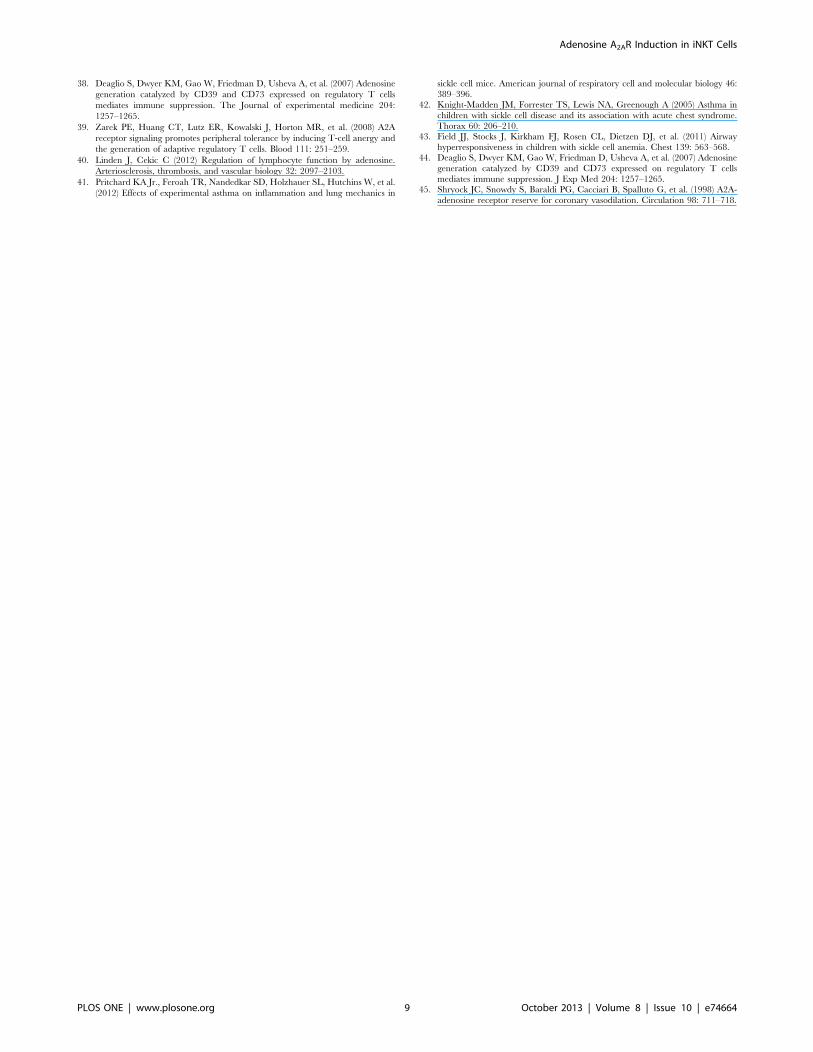
38. Deaglio S, Dwyer KM, Gao W, Friedman D, Usheva A, et al. (2007) Adenosine
generation catalyzed by CD39 and CD73 expressed on regulatory T cells
mediates immune suppression. The Journal of experimental medicine 204:
1257–1265.
39. Zarek PE, Huang CT, Lutz ER, Kowalski J, Horton MR, et al. (2008) A2A
receptor signaling promotes peripheral tolerance by inducing T-cell anergy and
the generation of adaptive regulatory T cells. Blood 111: 251–259.
40. Linden J, Cekic C (2012) Regulation of lymphocyte function by adenosine.
Arteriosclerosis, thrombosis, and vascular biology 32: 2097–2103.
41. Pritchard KA Jr., Feroah TR, Nandedkar SD, Holzhauer SL, Hutchins W, et al.
(2012) Effects of experimental asthma on inflammation and lung mechanics in
sickle cell mice. American journal of respiratory cell and molecular biology 46:
389–396.42. Knight-Madden JM, Forrester TS, Lewis NA, Greenough A (2005) Asthma in
children with sickle cell disease and its association with acute chest syndrome.
Thorax 60: 206–210.43. Field JJ, Stocks J, Kirkham FJ, Rosen CL, Dietzen DJ, et al. (2011) Airway
hyperresponsiveness in children with sickle cell anemia. Chest 139: 563–568.44. Deaglio S, Dwyer KM, Gao W, Friedman D, Usheva A, et al. (2007) Adenosine
generation catalyzed by CD39 and CD73 expressed on regulatory T cells
mediates immune suppression. J Exp Med 204: 1257–1265.45. Shryock JC, Snowdy S, Baraldi PG, Cacciari B, Spalluto G, et al. (1998) A2A-
adenosine receptor reserve for coronary vasodilation. Circulation 98: 711–718.
Adenosine A2AR Induction in iNKT Cells
PLOS ONE | www.plosone.org 9 October 2013 | Volume 8 | Issue 10 | e74664