Calcium-mediated modulation of the quaternary structure and function of adenosine A2A–dopamine D2...
11
Calcium-mediated modulation of the quaternary structure and function of adenosine A 2A -dopamine D 2 receptor heteromers Sergi Ferré a , Amina S. Woods a , Gemma Navarro b , Marisol Aymerich c , Carme Lluís b , and Rafael Franco b,c Sergi Ferré: [email protected]; Amina S. Woods: [email protected]; Gemma Navarro: [email protected]; Marisol Aymerich: [email protected]; Carme Lluís: [email protected]; Rafael Franco: [email protected] a National Institute on Drug Abuse, IRP, NIH, DHHS, 251 Bayview Blvd, Baltimore, MD 21224, USA b Institut d”investigacions Biomediques Agusti Pi I Sunyer, CIBERNED, Department of Biochemistry and Molecular Biology, Faculty of Biology, University of Barcelona, Diagonal 645, Barcelona 08028, Spain c CIMA Neurociencias. Avda Pio XII 55, Pamplona 31008, Spain Abstract The adenosine A 2A -dopamine D 2 receptor heteromer is one of the most studied receptor heteromers. It has important implications for basal ganglia function and pathology. Recent studies using Bioluminescence and Sequential Resonance Energy Transfer techniques shed light on the role of Ca 2+ in the modulation of the quaternary structure of the A 2A -D 2 receptor heteromer, which was found to depend on the binding of calmodulin (CaM) to the carboxy terminus of the A 2A receptor in the A 2A -D 2 receptor heteromer. Importantly, the changes in quaternary structure correlate with changes in function. A Ca 2+ /CaM-dependent modulation of MAPK signaling upon agonist treatment could only be observed in cells expressing A 2A -D 2 receptor heteromers. These studies provide a first example of a Ca 2+ -mediated modulation of the quaternary structure and function of a receptor heteromer. Introduction Ca 2+ plays an important role in the physiology of higher order organisms and is involved in the regulation of many cellular events. Various stimuli, such as membrane depolarization or binding of ligands to plasma transmembrane receptors trigger Ca 2+ -channel opening, which results in a significant influx of Ca 2+ into the cytosol. Then, Ca 2+ -binding proteins act as sensors and mediators of the initial Ca 2+ signal. CaM is a small (17 kDa), highly conserved, soluble, intracellular, acidic Ca 2+ -binding protein that is considered to be a major transducer of Ca 2+ -mediated signals in mammalian cells [1]. A series of recent studies have revealed that CaM can directly interact with intracellular domains of G protein-coupled receptors (GPCRs). CaM’s roles in the interaction with GPCR function are very diverse. It was initially found that CaM binds to the carboxy-terminus of metabotropic glutamate receptors (mGlu 5 and mGlu 7 receptors) altering their phosphorylation [2,3]. CaM has also been shown to bind to the third intracellular loop (3IL) of the μ opioid receptor and the dopamine D 2 receptor at overlapping Correspondence to Sergi Ferré; phone: 1-443-740-2647; fax: 443-740-2816; [email protected]. Publisher's Disclaimer: This is a PDF file of an unedited manuscript that has been accepted for publication. As a service to our customers we are providing this early version of the manuscript. The manuscript will undergo copyediting, typesetting, and review of the resulting proof before it is published in its final citable form. Please note that during the production process errors may be discovered which could affect the content, and all legal disclaimers that apply to the journal pertain. NIH Public Access Author Manuscript Curr Opin Pharmacol. Author manuscript; available in PMC 2011 February 1. Published in final edited form as: Curr Opin Pharmacol. 2010 February ; 10(1): 67. doi:10.1016/j.coph.2009.10.002. NIH-PA Author Manuscript NIH-PA Author Manuscript NIH-PA Author Manuscript
Transcript of Calcium-mediated modulation of the quaternary structure and function of adenosine A2A–dopamine D2...

Calcium-mediated modulation of the quaternary structure andfunction of adenosine A2A-dopamine D2 receptor heteromers
Sergi Ferréa, Amina S. Woodsa, Gemma Navarrob, Marisol Aymerichc, Carme Lluísb, andRafael Francob,cSergi Ferré: [email protected]; Amina S. Woods: [email protected]; Gemma Navarro: [email protected];Marisol Aymerich: [email protected]; Carme Lluís: [email protected]; Rafael Franco: [email protected] National Institute on Drug Abuse, IRP, NIH, DHHS, 251 Bayview Blvd, Baltimore, MD 21224, USAb Institut d”investigacions Biomediques Agusti Pi I Sunyer, CIBERNED, Department of Biochemistryand Molecular Biology, Faculty of Biology, University of Barcelona, Diagonal 645, Barcelona 08028,Spainc CIMA Neurociencias. Avda Pio XII 55, Pamplona 31008, Spain
AbstractThe adenosine A2A-dopamine D2 receptor heteromer is one of the most studied receptor heteromers.It has important implications for basal ganglia function and pathology. Recent studies usingBioluminescence and Sequential Resonance Energy Transfer techniques shed light on the role ofCa2+ in the modulation of the quaternary structure of the A2A-D2 receptor heteromer, which wasfound to depend on the binding of calmodulin (CaM) to the carboxy terminus of the A2A receptor inthe A2A-D2 receptor heteromer. Importantly, the changes in quaternary structure correlate withchanges in function. A Ca2+/CaM-dependent modulation of MAPK signaling upon agonist treatmentcould only be observed in cells expressing A2A-D2 receptor heteromers. These studies provide a firstexample of a Ca2+-mediated modulation of the quaternary structure and function of a receptorheteromer.
IntroductionCa2+ plays an important role in the physiology of higher order organisms and is involved inthe regulation of many cellular events. Various stimuli, such as membrane depolarization orbinding of ligands to plasma transmembrane receptors trigger Ca2+-channel opening, whichresults in a significant influx of Ca2+ into the cytosol. Then, Ca2+-binding proteins act assensors and mediators of the initial Ca2+ signal. CaM is a small (17 kDa), highly conserved,soluble, intracellular, acidic Ca2+-binding protein that is considered to be a major transducerof Ca2+-mediated signals in mammalian cells [1]. A series of recent studies have revealed thatCaM can directly interact with intracellular domains of G protein-coupled receptors (GPCRs).CaM’s roles in the interaction with GPCR function are very diverse. It was initially found thatCaM binds to the carboxy-terminus of metabotropic glutamate receptors (mGlu5 and mGlu7receptors) altering their phosphorylation [2,3]. CaM has also been shown to bind to the thirdintracellular loop (3IL) of the μ opioid receptor and the dopamine D2 receptor at overlapping
Correspondence to Sergi Ferré; phone: 1-443-740-2647; fax: 443-740-2816; [email protected]'s Disclaimer: This is a PDF file of an unedited manuscript that has been accepted for publication. As a service to our customerswe are providing this early version of the manuscript. The manuscript will undergo copyediting, typesetting, and review of the resultingproof before it is published in its final citable form. Please note that during the production process errors may be discovered which couldaffect the content, and all legal disclaimers that apply to the journal pertain.
NIH Public AccessAuthor ManuscriptCurr Opin Pharmacol. Author manuscript; available in PMC 2011 February 1.
Published in final edited form as:Curr Opin Pharmacol. 2010 February ; 10(1): 67. doi:10.1016/j.coph.2009.10.002.
NIH
-PA Author Manuscript
NIH
-PA Author Manuscript
NIH
-PA Author Manuscript

regions required for Gi/o protein activation, thereby compromising the receptor Gi/o protein-mediated activation [4–6•]. Furthermore, CaM can be directly involved in receptor signaling,as in the recently described G protein-independent and arrestin-dependent MAPK activationby the 5-HT2C receptor, which depends on the direct interaction of CaM with the carboxy-terminus of the receptor [7].
The adenosine A2A-dopamine D2 receptor heteromer is one of the most studied receptorheteromers [8•]. In the brain, A2A and D2 receptors are highly expressed in one type of striatalneuron, the GABAergic enkephalinergic neuron [9,10]. This type of neuron constitutes almosthalf the neuronal population in the striatum and its malfunction plays a key role in thepathogenesis of basal ganglia disorders (such as Parkinson’s disease and Huntington’s chorea)and most probably in obsessive-compulsive disorders, schizophrenia and drug addiction [11].The A2A-D2 receptor heteromer plays an important role in the modulation of the activity ofthe GABAergic enkephalinergic neuron [9–12••] and therefore could be targeted in thedevelopment of drugs for these neuropsychiatric disorders. Here we review recent data thatdemonstrates that calmodulin (CaM) oligomerizes with the A2A-D2 receptor heteromer andmodulates its function in a Ca2+-dependent manner.
Electrostatic interactions involved in CaM binding to A2A and D2 receptorsIt has been suggested that the binding sites of CaM in different proteins are often formed byamphiphilic domains, with hydrophobic residues interspersed with several positively chargedones [1]. Based on these characteristics, the introduction of the human A2A receptor sequencein the Internet-based search program “Calmodulin Target Data Base” (Cancer Institute,Ontario, 2002; http://calcium.uhnres.utoronto.ca/ctdb/ctdb/sequence.html) disclosed theexistence of a very likely binding site in the proximal portion of the carboxy-terminus of theA2A receptor. The sequence, 291RIREFRQTFR300, contains several arginines (Arg; positivelycharged residues), 1 Isoleucine and 2 Phenylalanine residues (hydrophobic residues). In fact,in vitro, this A2A receptor epitope formed multiple stable non-covalent complexes with CaM[13•]. However, a more detailed analysis indicated the particular involvement of electrostaticinteractions. A close look at the amino acid sequence of CaM shows that 32.5% of its residuescontain a negative charge [13•]. Furthermore, CaM has several serine (Ser) and threonine (Thr)residues susceptible of phosphorylation by casein kinases located in the vicinity of acidicresidues [13•]. A mass spectrometric analysis of an enzymatic digest of the CaM-A2A receptorepitope non-covalent complexes indicated the involvement of strong electrostatic interactionsbetween the acid motifs of CaM and the Arg residues (particularly the RIR sequence) of theA2A receptor epitope [13•]. A proteomics approach demonstrated the existence of CaM-A2Areceptor interactions in rat brain tissue and a direct interaction between both proteins wasdemonstrated by BRET experiments in transfected mammalian cells [14•]. An importantnegative control was that the A2A receptor mutated in its putative CaM binding motif locatedin the carboxi-terminus (with the sequence 291AIREFAQTFA300) did not interact with CaM[14•].
Electrostatic interactions also play a key role in the binding of CaM to the D2 receptor. A massspectrometric analysis indicated that CaM binds to an Arg-rich epitope from the amino-terminal part of the long 3IL of the D2 receptor [13•]. Importantly, the Arg-rich D2 receptormotif is also involved with the activation of Gi/o protein and with the heteromerization withadenosine A2A receptor (see below). Binding of CaM to the Arg-rich motif does not disturbGi/o recognition but impedes the D2 receptor-induced activation of Gα and this effect is Ca+
dependent [6•]. Thus, Ca2+/CaM blocks Gi/o protein activation by D2 receptor in a non-competitive manner [6•]. But since the Arg-rich motif of the D2 receptor is also involved inA2A-D2 receptor heteromerization, it was interesting to know if CaM could also bind to theD2 receptor in the A2A-D2 receptor heteromer.
Ferré et al. Page 2
Curr Opin Pharmacol. Author manuscript; available in PMC 2011 February 1.
NIH
-PA Author Manuscript
NIH
-PA Author Manuscript
NIH
-PA Author Manuscript

Intermolecular interactions in the A2A-D2 receptor heteromerA2A-D2 receptor heteromerization has been demonstrated in mammalian transfected cells withco-immunoprecipitation and fluorescence and bioluminescence resonance energy transfertechniques (FRET and BRET, respectively) (reviewed in ref. [8•]). By using computerizedmodeling, pull-down techniques and mass spectrometric analysis, it was shown that A2A-D2receptor heteromerization depends on an electrostatic interaction between the Arg-rich epitopelocated in the amino-terminal portion of the 3IL of the D2 receptor and a single phosphategroup from a casein kinase phosphorylable Ser localized in the distal portion of the carboxy-terminus of the A2A receptor [15]. Studies in vitro with peptides corresponding to both epitopesdemonstrated that the Arg-phosphate interaction possesses a “covalent-like” stability. Hence,these bonds could withstand fragmentation by mass spectrometric collision-induceddissociation at energies similar to those that fragment covalent bonds [16]. The Arg-phosphateelectrostatic interaction between epitopes located in intracellular domains is obviously not theonly interaction responsible for A2A-D2 receptor heteromerization. Thus, a significant but notcomplete reduction of BRET is observed when transfected cells express mutated D2 receptorsthat lack the key amino acids involved in the Arg-phosphate interaction [15], indicating thatother receptor domains are also involved. Most probably, intramembrane domains play animportant role in A2A-D2 receptor heteromerization, as it has been demonstrated for otherGPCR homomers and heteromers [17,18]. Nevertheless, the significant modification of BRETwith mutated receptors indicates that the Arg-phosphate interaction is necessary to provide thefinal quaternary structure of the heteromer, which in fact determines its function. Patch-clampexperiments in identified GABAergic enkephalinergic neurons demonstrated that disruptionof the Arg-phosphate interaction in A2A-D2 receptor heteromers (by intracellular addition ofsmall peptides with the same sequence than the receptor epitopes involved in the Arg-phosphateinteraction) completely eliminates the ability of the A2A receptor to antagonistically modulatethe D2 receptor-mediated inhibition of neuronal excitability [12••].
The just mentioned antagonistic interaction between A2A and D2 receptors is most probablyrelated to the existence of an allosteric modulation in the A2A-D2 receptor heteromer. Thiskind of allosteric modulation, in which binding of a ligand to one of the receptor units in thereceptor heteromer changes the binding properties of another receptor unit seems to be acommon biochemical characteristic of receptor heteromers [19,20•]. An antagonistic A2A-D2 receptor interaction has been demonstrated in many different membrane preparations fromdifferent transfected mammalian cells and from rat and human striatal tissues and implies theability of A2A receptor stimulation to change the binding characteristics (decrease the affinity)of the D2 receptor for agonists [8•,10,19]. In experiments with chimeric D1/D2 receptors, theA2A receptor could still modulate the binding characteristics of a D2 receptor with the 6th
transmembrane domains of the D1 receptor, but the modulation disappeared with a D2 receptorwith the 3IL and 5th and 6th transmembrane domains of the D1 receptor [21]. These resultsindicate that the epitope (or epitopes) of the D2 receptor involved in the antagonistic A2A-D2receptor interaction might be located somewhere in the 3IL or 5th transmembrane. Experimentsusing more detailed mutants are in progress to demonstrate that the epitope, in fact, correspondsto the Arg-rich domain of the 3IL.
In addition to the allosteric modulation in the A2A-D2 receptor heteromer, A2A and D2 receptorscan also interact at the second messenger level, and this has been well demonstrated both incell culture and in the brain. In this case, however, it is the stimulation of D2 receptor thatcounteracts the effects of A2A receptor stimulation. A2A receptor, through its coupling toGs/olf proteins, can potentially stimulate adenyl-cyclase, with phosphorylation of several PKAsubstrates, such as DARPP-32, CREB and AMPA receptors and the consequent increase inthe expression of different genes, such as c-fos or preproenkephalin in the GABAergicenkephalinergic neuron [8•,9,10,19]. D2 receptor, on the other hand, can potentially couple to
Ferré et al. Page 3
Curr Opin Pharmacol. Author manuscript; available in PMC 2011 February 1.
NIH
-PA Author Manuscript
NIH
-PA Author Manuscript
NIH
-PA Author Manuscript

Gi/o proteins and counteract the ability of A2A receptor stimulation to signal through the cAMP/PKA cascade [8•,9,10,19]. It might sound counterintuitive, but both types of antagonisticA2A-D2 receptor interactions coexist in the same cells and, in fact, they do coexist in theGABAergic enkephalinergic neurons [8•,9,10,19]. Thus, co-stimulation of A2A and D2receptors implies a simultaneous A2A receptor-mediated inhibition of the D2 receptor-mediatedmodulation of neuronal excitability and a D2 receptor-mediated inhibition of the A2A receptor-mediated modulation of gene expression, which provides a clear example of a functionaldissociation between neuronal excitability and gene expression. This apparently incompatiblecoexistence of reciprocal antagonistic A2A-D2 receptor interactions could be explained by thepresence in the same cell of A2A and D2 receptors forming and not forming A2A-D2 receptorheteromers. The antagonistic A2A-D2 receptor interaction at the second messenger leveldepends on the activation of Gi/o proteins, which requires the Arg-rich domain not beinginteracting with either CaM or the A2A receptor.
Role of CaM in A2A-D2 receptor heteromer functionThe existence of CaM-A2A-D2 receptor oligomerization has been detected by the recentlyintroduced Sequential-BRET-FRET (SRET) technique, which allows the identification ofoligomers formed by three different proteins [22••,23]. In SRET2, the oxidation of a RenillaLuciferase (RLuc) substrate by an RLuc fusion protein triggers the excitation of the acceptorGFP2 by BRET2 and subsequent energy transfer to the acceptor YFP by FRET [22••].SRET2 was obtained with the fusion proteins A2ARluc, CaMGFP2 and D2YFP when addingDeepBlueC as Rluc substrate, indicating that the two acceptor/donor pairs, A2ARluc-CaMGFP2 and CaMGFP2-D2YFP were well oriented at distances of less than 10 nM [14•](Figure 1). But, how are these three proteins arranged? We know that CaM binds to A2A andD2 receptors when not forming heteromers. Does CaM bind to both receptors in the A2A-D2receptor heteromer? Does CaM disrupt the heteromerization or does it change the quaternarystructure of the A2A-D2 receptor heteromer and therefore its function? Which Ca2+-dependendfunctional changes does CaM convey in the A2A-D2 receptor heteromer?
As mentioned above, the Arg-rich domain of the amino-terminal portion of the 3IL of the D2receptor is a binding site for both CaM and for a phosphorylated Ser localized in the distalportion of the carboxy-terminus of the A2A receptor. BRET competition experimentsdemonstrated that increasing the expression of the D2 receptor does not modify the binding ofCaM to the A2A receptor. Also, increasing the expression of CaM does not modify A2A-D2receptor heteromerization. In contrast, increasing amounts of A2A receptor led to significantreduction in the BRET signal due to the interaction between the D2 receptor and CaM [14•].Overall these results indicate that CaM cannot compete with the A2A receptor for its bindingto the D2 receptor but that binds to the A2A receptor in the A2A-D2 receptor heteromer.
The increase in intracellular Ca2+ (with ionomycin treatment) in cells expressing A2A andD2 receptors produced modifications in the BRET signal due to the interaction between A2Aand D2 receptors [14•], which implies alterations in the quaternary structure of the A2A-D2receptor heteromer. Importantly, Ca2+ exerted a selective modulation of A2A-D2 receptorheteromer-mediated activation of MAPK pathway. First, in cells transfected with either A2Aor D2 receptors, agonist-induced activation of MAPK pathway (ERK1/2 phosphorylation) wasnot modified by increasing intracellular Ca2+ (Figure 2a,b). At least for the D2 receptor, thissuggests that MAPK signaling, in HEK-293T cells [6•,14•], is not dependent on the activationof Gi/o protein, since it has been previously shown that the Gi/o protein-dependent signalingefficiency of the D2 receptor is regulated (decreased) by a rise in Ca2+ via CaM [6•]. Second,in cells expressing both receptors, Ca2+ significantly decreased A2A receptor agonist-inducedERK1/2 phosphorylation (Figure 2c) whereas it significantly increased D2 receptor agonist-induced ERK1/2 phosphorylation [14•] (Figure 2d). Third, co-activation of A2A and D2
Ferré et al. Page 4
Curr Opin Pharmacol. Author manuscript; available in PMC 2011 February 1.
NIH
-PA Author Manuscript
NIH
-PA Author Manuscript
NIH
-PA Author Manuscript
receptors in cells co-expressing both receptors led to the same qualitative response thanactivation of A2A receptors alone, with a significant Ca2+-dependent decrease of agonist-induced ERK1/2 phosphorylation [14•] (Figure 2e). The effects of Ca2+ were dependent onthe intracellular levels of CaM [14•], indicating that CaM selectively transduces Ca2+-dependent changes of MAPK signaling in the A2A-D2 receptor heteromer. In summary, theresults indicate that in the absence of Ca2+ the A2A-D2 receptor heteromer produces verysimilar effects on MAPK signaling upon stimulation of one of both receptor units, whilebinding of Ca2+ to CaM produces a very different qualitative response, reducing and enhancingthe effects of the stimulation of A2A and D2 receptors, respectively. Additionally, with co-activation of the A2A receptor, the ability of Ca2+ to increase the effect of D2 receptorstimulation on ERK1/2 phosphorylation is lost. This could be related to the allosteric interactionin the A2A-D2 receptor heteromer, with A2A receptor stimulation antagonizing the effects ofD2 receptor stimulation (Figure 2e).
We still need to determine which molecular mechanisms are involved in the Ca2+/CaM-modulated, A2A-D2 receptor heteromer-mediated MAPK signaling. Which are the G proteinsinvolved? Recent studies strongly suggest that the minimal signal unit for class A GPCRs iscomposed of two receptors and a single G protein and that probably applies to both GPCRhomomers and heteromers [24••]. We then need to figure out which is the G protein that bindsto the A2A-D2 receptor heteromer and if it changes in the presence of CaM. We alreadymentioned that D2 receptor signals through MAPK by a Gi/o protein-independent mechanismin cells only expressing D2 receptors and, most probably, Gi/o proteins are not involved eitherin MAPK signaling by the A2A-D2 receptor heteromer. Thus, the Arg-rich domain of the D2receptor required for Gi/o protein activation is bound to the C-terminus of the A2A receptor inthe A2A-D2 receptor heteromer (see above). Also, we have preliminary data suggesting thatthe Gs/olf/PKA pathway, commonly used by the A2A receptor, is not involved either. One openpossibility is the involvement of Gq, as it has been shown for the D1-D2 receptor heteromer[25]. But it is also probable we are dealing with a CaM-dependent, G protein-independentMAPK activation, as recently reported for the 5-HT2c receptor [7].
ConclusionsReceptor heteromers constitute an expanding new area of research which is changing previousconcepts of receptor pharmacology. A receptor heteromer is defined as a macromolecularcomplex composed of at least two (functional) receptor units with biochemical properties thatare demonstrably different from those of its individual components [20•]. This definitionunderscores the fact that the receptor heteromer constitutes a new functional unit and apreviously unforeseen number of emerging properties have already been described for severaldifferent receptor heteromers. Those properties include changes in ligand recognition,allosteric interactions, G-protein coupling switching and changes in the pattern of activationof signaling pathways and receptor internalization [20•].
Here we reviewed a new biochemical property of the receptor heteromer formed by adenosineA2A and dopamine D2 receptors, which has important implications for basal ganglia functionand dysfunction [9–11]. This property consists of a specific Ca2+-mediated modulation ofMAPK signaling, which depends on the binding of CaM to the proximal portion of the carboxy-terminus of the A2A receptor in the A2A-D2 receptor heteromer. This is associated with changesin the quaternary structure of the A2A-D2 receptor heteromer, as demonstrated by RETtechniques. Experiments with mutated receptors are in progress to determine which receptordomains are key determinants for the establishment of the appropriate Ca2+/CaM-dependentfunctional and structural changes of the A2A-D2 receptor heteromers.
Ferré et al. Page 5
Curr Opin Pharmacol. Author manuscript; available in PMC 2011 February 1.
NIH
-PA Author Manuscript
NIH
-PA Author Manuscript
NIH
-PA Author Manuscript

AcknowledgmentsWork supported by the intramural funds of the National Institute on Drug Abuse, the Spanish Ministry of Educationand Science (grant SAF2006-05481)
References1. Vetter SW, Leclerc E. Novel aspects of calmodulin target recognition and activation. Eur J Biochem
2003;270:404–414. [PubMed: 12542690]2. Minakami R, Jinnai N, Sugiyama H. Phosphorylation and calmodulin binding of the metabotropic
glutamate receptor subtype 5 (mGluR5) are antagonistic in vitro. J Biol Chem 1997;272:20291–20298.[PubMed: 9242710]
3. Nakajima Y, Yamamoto T, Nakayama T, Nakanishi S. A relationship between protein kinase Cphosphorylation and calmodulin binding to the metabotropic glutamate receptor subtype 7. J BiolChem 1999;274:27573–27577. [PubMed: 10488094]
4. Wang D, Sadée W, Quillan JM. Calmodulin binding to G protein-coupling domain of opioid receptors.J Biol Chem 1999;274:22081–22088. [PubMed: 10419536]
5. Wang D, Tolbert LM, Carlson KW, Sadée W. Nuclear Ca2+/calmodulin translocation activated bymu-opioid (OP3) receptor. J Neurochem 2000;74:1418–1425. [PubMed: 10737597]
6 •. Bofill-Cardona E, Kudlacek O, Yang Q, Ahorn H, Freissmuth M, Nanoff C. Binding of calmodulinto the D2-dopamine receptor reduces receptor signaling by arresting the G protein activation switch.J Biol Chem 2000;275:32672–32680. This study clearly demonstrates that the D2 receptor bindsCaM in the N-terminal part of the third intracellular loop, which allows intracellular Ca2+ tomodulate a Gi/o protein-dependent D2 receptor-mediated signaling. [PubMed: 10926927]
7. Labasque M, Reiter E, Becamel C, Bockaert J, Marin P. Physical interaction of calmodulin with the5-hydroxytryptamine2C receptor C-terminus is essential for G protein-independent, arrestin-dependent receptor signaling. Mol Biol Cell 2008;19:4640–4650. [PubMed: 18768750]
8 •. Ferré S, Quiroz C, Woods AS, Cunha R, Popoli P, Ciruela F, Lluis C, Franco R, Azdad K, SchiffmannSN. An update on adenosine A2A-dopamine D2 receptor interactions: implications for the functionof G protein-coupled receptors. Curr Pharm Des 2008;14:1468–74. An updated review about themultiple interactions between A2A and D2 receptors with reference to receptor heteromerizationand to the apparently incompatible co-existence of reciprocal interactions, which seem to dependon the co-existence of separate pools of A2A and D2 receptors forming and not forming A2A-D2receptor heteromers. [PubMed: 18537670]
9. Schiffmann SN, Fisone G, Moresco R, Cunha RA, Ferré S. Adenosine A2A receptors and basal gangliaphysiology. Prog Neurobiol 2007;83:277–292. [PubMed: 17646043]
10. Ferré S, Agnati LF, Ciruela F, Lluis C, Woods AS, Fuxe K, Franco R. Neurotransmitter receptorheteromers and their integrative role in ‘local modules’: the striatal spine module. Brain Res Rev2007;55:55–67. [PubMed: 17408563]
11. Fuxe K, Ferré S, Genedani S, Franco R, Agnati LF. Adenosine receptor-dopamine receptorinteractions in the basal ganglia and their relevance for brain function. Physiol Behav 2007;92:210–217. [PubMed: 17572452]
12 ••. Azdad K, Gall D, Woods AS, Ledent C, Ferré S, Schiffmann SN. Dopamine D2 and adenosineA2A receptors regulate NMDA-mediated excitation in accumbens neurons through A2A-D2receptor heteromerization. Neuropsychopharmacology 2009;34:972–986. Patch-clampexperiments in identified GABAergic enkephalinergic neurons which show the complete disruptionof the A2A receptor-mediated modulation of D2 receptor function with the application of peptideswith the same sequence than epitopes involved in A2A-D2 receptor heteromerization. This studyprovides strong evidence in situ for a specific functional property of the A2A-D2 receptor heteromerin the brain. [PubMed: 18800071]
13 •. Woods AS, Marcellino D, Jackson SN, Franco R, Ferré S, Agnati LF, Fuxe K. How calmodulininteracts with the adenosine A(2A) and the dopamine D(2) receptors. J Proteome Res 2008;7:3428–3434. This study with mass spectrometry techniques demonstrates for the first time an importantrole of electrostatic interactions in the intermolecular interactions between CaM and epitopes of theA2A and D2 receptors. [PubMed: 18590318]
Ferré et al. Page 6
Curr Opin Pharmacol. Author manuscript; available in PMC 2011 February 1.
NIH
-PA Author Manuscript
NIH
-PA Author Manuscript
NIH
-PA Author Manuscript

14 •. Navarro G, Aymerich MS, Marcellino D, Cortes A, Casado V, Mallol J, Canela EI, Agnati LF,Woods AS, Fuxe K, et al. Interactions between calmodulin, adenosine A2A and dopamine D2receptors. J Biol Chem. 2009 By using Bioluminescence and Sequential Resonance Energy Transfertechniques, evidence is provided for oligomerization of CaM, A2A and D2 receptors. The studyalso provides an example of re-organization of signaling protein molecules as a consequence ofreceptor heteromerization and of specific (Ca2+-dependent) signaling by the receptor heteromer.10.1074/jbc.M109.034231
15. Ciruela F, Burgueno J, Casado V, Canals M, Marcelino D, Goldberg SR, Bader M, Fuxe K, AgnatiLF, Lluis C, et al. Combining Mass spectrometry and pull-down techniques for the study of receptorheteromerization. Direct epitope-epitope electrostatic interactions between adenosine A2A anddopamine D2 receptors. Anal Chem 2004;76:5354–5363. [PubMed: 15362892]
16. Woods AS, Ferré S. The amazing stability of the arginine-phosphate electrostatic interaction. JProteome Res 2005;4:1397–1402. [PubMed: 16083292]
17. Guo W, Urizar E, Kralikova M, Mobarec JC, Shi L, Filizola M, Javitch JA. Dopamine D2 receptorsform higher order oligomers at physiological expression levels. EMBO J 2008;27:2293–2304.[PubMed: 18668123]
18. González-Maeso J, Ang RL, Yuen T, Chan P, Weisstaub NV, López-Giménez JF, Zhou M, OkawaY, Callado LF, Milligan G, et al. Identification of a serotonin/glutamate receptor complex implicatedin psychosis. Nature 2008;452:93–97. [PubMed: 18297054]
19. Ferré S, Ciruela F, Woods AS, Lluis C, Franco R. Functional relevance of neurotransmitter receptorheteromers in the central nervous system. Trends Neurosci 2007;30:440–446. [PubMed: 17692396]
20 •. Ferré S, Baler R, Bouvier M, Caron MG, Devi LA, Durroux T, Fuxe K, George SR, Javitch JA,Lohse MJ, et al. Building a new conceptual framework for receptor heteromers. Nat Chem Biol2009;5:131–134. Most recent recommendations for nomenclature of receptor heteromers andcriteria for their identification in native tissues. Some definitions are also established, includingallosteric interaction in the receptor heteromer, which is described as an intermolecular interactionby which binding of a ligand to one of the receptor units in the receptor heteromer changes thebinding properties of another receptor unit. [PubMed: 19219011]
21. Torvinen M, Kozell LB, Neve KA, Agnati LF, Fuxe K. Biochemical identification of the dopamineD2 receptor domains interacting with the adenosine A2A receptor. J Mol Neurosci 2004;24:173–180. [PubMed: 15456930]
22 ••. Carriba P, Navarro G, Ciruela F, Ferré S, Casadó V, Agnati L, Cortés A, Mallol J, Fuxe K, CanelaEI, et al. Detection of heteromerization of more than two proteins by sequential BRET-FRET. NatMethods 2008;5:727–733. New method to determine the existence of three interacting proteins inthe plasma membrane of living mammalian cells. First demonstration of heterotrimerization ofdifferent GPCRs, A2A, D2 and cannabinoid CB1 receptors. [PubMed: 18587404]
23. Cabello N, Gandía J, Bertarelli DC, Watanabe M, Lluís C, Franco R, Ferré S, Luján R, Ciruela F.Metabotropic glutamate type 5, dopamine D2 and adenosine A2a receptors form higher-orderoligomers in living cells. J Neurochem 2009;109:1497–1507. [PubMed: 19344374]
24 ••. Han Y, Moreira IS, Urizar E, Weinstein H, Javitch JA. Allosteric communication betweenprotomers of dopamine class A GPCR dimers modulates activation. Nat Chem Biol 2009;5:688–695. This is and elegant study that shows that the minimal signaling unit of the D2 receptor is areceptor homomer with two protomers and a single G protein. It also shows that such a dimer ismaximally activated by agonist binding to a single protomer and that agonist binding to the secondprotomer inhibits signaling. [PubMed: 19648932]
25. Rashid AJ, O’Dowd BF, Verma V, George SR. Neuronal Gq/11-coupled dopamine receptors: anuncharted role for dopamine. Trends Pharmacol Sci 2007;28:551–555. [PubMed: 17950471]
Ferré et al. Page 7
Curr Opin Pharmacol. Author manuscript; available in PMC 2011 February 1.
NIH
-PA Author Manuscript
NIH
-PA Author Manuscript
NIH
-PA Author Manuscript

Figure 1. SRET for CaM, A2A receptor and D2 receptor in living cellsSRET saturation curves performed in HEK-293 cells expressing A2A-Rluc (0.75 μg of cDNA),CaMGFP2 (0.6 μg of cDNA) and increasing amounts of D2YFP (0.5 to 5 μg of the cDNA).Net SRET was obtained by monitoring the YFP fluorescence emission after DeepBlueCaddition, with subtraction of the value obtained with cells expressing the same amount ofA2A-Rluc and CaMGFP2. Significant net SRET was detected for A2ARluc-CaMGFP2-D2YFPcoupling, while negligible or linear net SRET was obtained in cells expressing equivalentamounts (equivalent fluorescence and luminescence units) of A2ARluc, CaMGFP2 and5HT2BYFP (red curve), or A1RRluc, CaMGFP2 and D2RYFP (blue curve) as negative controls.Values, expressed as net SRET, represent means ± S.E.M. of 5 independent experiments
Ferré et al. Page 8
Curr Opin Pharmacol. Author manuscript; available in PMC 2011 February 1.
NIH
-PA Author Manuscript
NIH
-PA Author Manuscript
NIH
-PA Author Manuscript

performed in triplicate. At the top, scheme depicting the expressed proteins in the SRET assay(modified from ref. [14•]).
Ferré et al. Page 9
Curr Opin Pharmacol. Author manuscript; available in PMC 2011 February 1.
NIH
-PA Author Manuscript
NIH
-PA Author Manuscript
NIH
-PA Author Manuscript

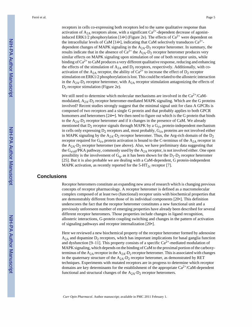
Figure 2. Selective Ca2+/CaM-mediated modulation of A2A-D2 receptor heteromer functionScheme showing the motifs of the C-terminus of the A2A receptor and the 3IL of the D2 receptorinvolved in the binding of CaM and in A2A-D2 receptor heteromerizacion, as well as theselective Ca2+/CaM-mediated modulation of MAPK activation in the A2A-D2 receptorheteromer (see text). (a, b) When not forming heteromers, CaM binds to the proximal portionof the carboxy-terminus of the A2A receptor and to the Arg-rich domain localized in the amino-terminus of the 3IL of the D2 receptor. Under these conditions the effects of the stimulation ofeither receptor on MAPK activation does not change in the presence or absence of Ca2+. (c–e) When forming heteromers, the distal portion of the A2A receptor binds to the Arg-richdomain of the D2 receptor, which cannot bind to CaM. In the A2A-D2 receptor heteromer, thebinding of Ca2+ to CaM modifies its quaternary structure and decreases the effects ofstimulation of A2A receptor (c) and increases the effects of stimulation of the D2 receptor (d)on MAPK activation. Co-activation of A2A and D2 receptors in cells co-expressing bothreceptors led to the same qualitative response than activation of A2A receptors alone, probablydue to the ability of A2A receptor stimulation to antagonize allosterically the effects inducedby D2 receptor stimulation in the A2A-D2 receptor heteromer (e). Receptors are shown as
Ferré et al. Page 10
Curr Opin Pharmacol. Author manuscript; available in PMC 2011 February 1.
NIH
-PA Author Manuscript
NIH
-PA Author Manuscript
NIH
-PA Author Manuscript

monomers for the sake of simplicity, but they are most probably forming homomers when notforming heteromers. Also for schematic purposes, the main portion of each receptor unit isrepresented by a sphere and only the long 3IL of the D2 receptor and the long tail of the A2Areceptor are explicitly shown.
Ferré et al. Page 11
Curr Opin Pharmacol. Author manuscript; available in PMC 2011 February 1.
NIH
-PA Author Manuscript
NIH
-PA Author Manuscript
NIH
-PA Author Manuscript


![In vivo vulnerability to competition by endogenous dopamine: Comparison of the D2 receptor agonist radiotracer (-)-N-[11C]propyl-norapomorphine ([11C]NPA) with the D2 receptor antagonist](https://static.fdokumen.com/doc/165x107/631cb975a1cc32504f0c9f3c/in-vivo-vulnerability-to-competition-by-endogenous-dopamine-comparison-of-the-d2-1675155902.jpg)