Gi protein coupling to adenosine A1-A2A receptor heteromers in human brain caudate nucleus
10
See discussions, stats, and author profiles for this publication at: https://www.researchgate.net/publication/44606994 Gi protein coupling to adenosine A1-A2A receptor heteromers in human brain caudate nucleus Article in Journal of Neurochemistry · August 2010 DOI: 10.1111/j.1471-4159.2010.06810.x · Source: PubMed CITATIONS 6 READS 22 11 authors, including: Some of the authors of this publication are also working on these related projects: Desarrollos contamétricos View project Sergio Barrondo Universidad del País Vasco / Euskal Herriko U… 14 PUBLICATIONS 162 CITATIONS SEE PROFILE Josefa Mallol University of Barcelona 155 PUBLICATIONS 4,984 CITATIONS SEE PROFILE Enric I. Canela University of Barcelona 219 PUBLICATIONS 6,010 CITATIONS SEE PROFILE Joan Sallés Universidad del País Vasco / Euskal Herriko U… 69 PUBLICATIONS 669 CITATIONS SEE PROFILE All content following this page was uploaded by Luis F. Callado on 29 September 2014. The user has requested enhancement of the downloaded file. All in-text references underlined in blue are added to the original document and are linked to publications on ResearchGate, letting you access and read them immediately.
Transcript of Gi protein coupling to adenosine A1-A2A receptor heteromers in human brain caudate nucleus

Seediscussions,stats,andauthorprofilesforthispublicationat:https://www.researchgate.net/publication/44606994
GiproteincouplingtoadenosineA1-A2Areceptorheteromersinhumanbraincaudatenucleus
ArticleinJournalofNeurochemistry·August2010
DOI:10.1111/j.1471-4159.2010.06810.x·Source:PubMed
CITATIONS
6
READS
22
11authors,including:
Someoftheauthorsofthispublicationarealsoworkingontheserelatedprojects:
DesarrolloscontamétricosViewproject
SergioBarrondo
UniversidaddelPaísVasco/EuskalHerrikoU…
14PUBLICATIONS162CITATIONS
SEEPROFILE
JosefaMallol
UniversityofBarcelona
155PUBLICATIONS4,984CITATIONS
SEEPROFILE
EnricI.Canela
UniversityofBarcelona
219PUBLICATIONS6,010CITATIONS
SEEPROFILE
JoanSallés
UniversidaddelPaísVasco/EuskalHerrikoU…
69PUBLICATIONS669CITATIONS
SEEPROFILE
AllcontentfollowingthispagewasuploadedbyLuisF.Calladoon29September2014.
Theuserhasrequestedenhancementofthedownloadedfile.Allin-textreferencesunderlinedinblueareaddedtotheoriginaldocument
andarelinkedtopublicationsonResearchGate,lettingyouaccessandreadthemimmediately.

,1 , ,1 ,
, ,
*Biochemistry and Molecular Biology Department, University of Barcelona, Centro de Investigacion Biomedica en Red de
Enfermedades Neurodegenerativas, CIBERNED, Barcelona, Spain
�Department of Pharmacology (Facultad de Farmacia), University of the Basque Country, Vitoria-Gasteiz, Spain
�Centro de Investigacion Biomedica en Red de Salud Mental, CIBERSAM, Spain
§Department of Pharmacology, University of the Basque Country, Leioa, Bizkaia, Spain
Adenosine was one of the first neuromodulators/neurotrans-mitters for which different receptor subtypes were reported asbeing coexpressed in a given cell (Rees et al. 2003; Ferre et al.2007b; Sebastiao and Ribeiro 2009). Coexpression of receptorsubtypes coupled to different G proteins appears to be acommon feature for an ever increasing number of both types offirst messengers, hormones and neurotransmitters. From thephenomenological detection of coexpression of differentreceptors for the same messenger a huge amount of work isrequired to understand the signaling mechanistic interrela-tionships arising form this relevant piece of information.
Adenosine in striatum regulates dopamine neurotransmis-sion by means of adenosine A1 receptors coupled to Gi/o
proteins, which are widely distributed in the CNS, and A2A
receptors coupled to Gs/olf proteins, which are concentrated instriatum and scarcely present in other brain areas (Sebastiaoand Ribeiro 2009). In the striatum the two receptors are
found coexpressed in as high as 61% of presynapticterminals from cortical glutamatergic neurons (Ciruela et al.2006) and using integrated anatomical, electrophysiological,and biochemical approaches, it has been demonstrated thatpre-synaptic A2A receptors are preferentially localized incortical glutamatergic terminals that contact striatal neuronsof the direct pathway, where they exert a selective modula-tion of corticostriatal neurotransmission (Quiroz et al. 2009).
Received March 15, 2010; revised manuscript received April 29, 2010;accepted May 4, 2010.Address correspondence and reprint requests to Dr. Rafael Franco,
CIMA, Molecular Neuroscience, Neurosciences Division, Universidadde Navarra, Avda Pio XII 55, Pamplona 31008, Spain.E-mail: [email protected] authors contributed equally to this study.Abbreviations used: ADA, adenosine deaminase; DPCPX, 8-cyclo-
pentyl-1,3-dipropylxantine; R-PIA, R-phenylisopropyladenosine.
Abstract
Pharmacological characterization of adenosine A1 and A2A
receptors in human brain caudate nucleus membranes led to
non-cooperative binding of radiolabelled ligands. In human
caudate nucleus but not in cortex, the agonist binding to A1
receptors was modulated by the agonist binding to A2A
receptors indicating a functional negative cross-talk. Accord-
ingly, the A1 receptor-activation-mediated Gi-dependent gua-
nosine 5¢-o-(3-[35S]thio-triphosphate) binding was modulated
by agonist binding to A2A receptors. A2A receptors occupation
led to a decrease in the potency of A1 receptor agonists.
These results indicate that A1 but not A2A receptors activation,
likely occurring at low adenosine concentrations, engages a
Gi-mediated signaling; however, when both receptors are
occupied by adenosine, there is an A2A receptor-mediated
impairment of Gi-operated transducing units. These findings
are relevant to get insight into the complex relationships de-
rived from co-expression of multiple neurotransmitter/neuro-
modulator receptors subtypes that individually are coupled to
different G proteins. A further finding was the demonstration
that the A2A receptor agonist, CGS 21680, at high concen-
trations able to significantly bind to the A1 receptor, behaved
as a partial agonist of the later receptor. This fact might be
taken into account when characterizing CGS 21680 actions in
human cells expressing A1 receptors when the compound is
used at micromolar concentrations.
Keywords: adenosine receptors, cell signaling, human brain,
ligand binding, receptor heteromers.
J. Neurochem. (2010) 114, 972–980.
JOURNAL OF NEUROCHEMISTRY | 2010 | 114 | 972–980 doi: 10.1111/j.1471-4159.2010.06810.x
972 Journal Compilation � 2010 International Society for Neurochemistry, J. Neurochem. (2010) 114, 972–980� 2010 The Authors

Therefore, striatal adenosine is also regulating glutamatergicneurotransmission of the direct pathway by means ofterminals in which the two receptors are present. Becauseof the strength of the A1–A2A receptor interaction measuredby biophysical techniques in heterologous cell systems(Ciruela et al. 2006) it is likely that A1 and A2A receptorspresent in the same neuron and in the same location wouldform heteromers. In fact, heteromerization of adenosine A1
and A2A receptors has been described in the cell surface ofcotransfected cells by means of biochemical techniques andby bioluminescence resonance energy transfer (Ciruela et al.2006; Ferre et al. 2007a). In rodent striatum heteromerizationis suspected in basis of cross-regulation in the binding ofagonists; both in transfected cells and in rat striatum thebinding of agonists to A2A leads to a decrease in the bindingof agonists to A1 receptors (Ciruela et al. 2006). Therefore, anegative cross-talk is expected in regions having cells co-expressing those receptors but not in regions such as cerebralcortex where the expression of the A2A is low and thereforeno heteromerization is expected. It is therefore evident thatadenosine neuroregulation is more complex than expectedand its understanding requires the knowledge of thefunctional consequences of the cross-talk established be-tween A1 and A2A receptors. Evidently adenosine may not besimultaneously increasing cAMP levels via A2A receptorsand decrease them via A1 receptors. Functional consequencesare expected that may directly be mediated by A1–A2A
heteromers or indirectly by the participation of othercoupling proteins. Consequences at early signaling eventsmust be identified to understand, from a molecular point ofview, differences in adenosine-driven signaling in cellscoexpressing A1 and A2A receptors versus cells expressingeither receptor individually. The present study confirms anegative cross-talk in agonist binding in human caudatenucleus, but more importantly, [35S]GTPcS binding assaysprovided evidence for relevant consequences in Gi-proteincoupling derived from the interreceptor cross-modulation inhuman caudate nucleus.
Materials and methods
MaterialsGuanosine 5¢-o-(3-[35S]thio-triphosphate) ([35S]GTPcS, 1000–
1400 Ci/mmol) was purchased from Perkin-Elmer (Barcelona,
Spain). Protease inhibitor cocktail, R-phenylisopropyladenosine(R-PIA), ZM 241385, 8-cyclopentyl-1,3-dipropylxantine (DPCPX)
and 2-p-(2-carboxyethyl)phenethylamino-5¢-N-ethylcarboxamido
adenosine HCl (CGS 21680) were obtained from Sigma Chemical
Company (St. Louis, MO, USA). [3H]R-PIA (30.5 Ci/mmol) was
from Moravek Biochemicals Inc. (Brea, CA, USA). [3H]ZM 241385
was from NEN Perkin Elmer (Wellesley, MA, USA). Adenosine
deaminase (ADA, EC 3.5.4.4) was supplied by Roche (Basel,
Switzerland). Ecoscint H scintillation cocktail was purchased from
National Diagnostics (Atlanta, GA, USA).
Human brain samplesHuman brain samples were obtained at autopsy in the Basque
Institute of Legal Medicine (Bilbao, Spain) from male subjects
without a history of neuropathological or psychiatric disorders and
who had died suddenly, mainly by car accidents. Toxicological
screening was negative for all these subjects and brain samples were
histologically determined as normal. Samples from the frontal cortex
and the caudate nucleus of each subject were dissected at the time of
autopsy, stored at )70�C until assay and encoded in order to protect
the identity of the subject. The time interval between death and
autopsy (postmortem delay at 4�C) was 26 ± 4 h.
All tissue samples were collected in accordance with protocols
approved by the Human Studies Committee of each of the
institutions involved.
Membrane preparation and protein determinationMembrane suspensions from human brain caudate nucleus were
prepared as described previously (Casado et al. 1990). Tissue was
disrupted with a Polytron homogenizer (PTA 20 TS rotor, setting 3;
Kinematica, Basel, Switzerland) for three 5-s-periods in 10 volumes
of 50 mM Tris–HCl buffer, pH 7.4, containing a protease inhibitor
cocktail (1/1000). After eliminating cell debris by centrifugation at
1000 g, membranes were obtained by centrifugation at 105 000 g(40 min, 4�C) and the pellet was resuspended and recentrifuged
under the same conditions. Membranes were stored at )80�C,washed once more as described above and resuspended in 50 mM
Tris–HCl buffer for immediate use. Protein was quantified by the
bicinchoninic acid method (Pierce Chemical Co., Rockford, IL,
USA) using bovine serum albumin dilutions as standard.
Radioligand binding assaysSaturation experiments were performed by incubating brain mem-
brane suspensions (0.3 mg of protein/mL) with increasing concen-
trations of the A1 receptor agonist [3H]R-PIA (triplicates of 13
different concentrations, from 0.05 nM to 17 nM) at 25�C in
50 mM Tris–HCl buffer, pH 7.4, containing 10 mM MgCl2 and
0.2 U/mL of ADA, and allowing sufficient time to achieve
equilibrium for the lowest radioligand concentration (5 h). For
competition experiments, membrane suspensions (0.3 mg of pro-
tein/mL) were incubated for 2 h at 25�C in 50 mM Tris–HCl buffer,
pH 7.4, containing 10 mM MgCl2 and 0.2 U/mL of ADA with
0.8 nM of the A2A receptor antagonist [3H]ZM 241385 or 0.44 nM
of the A1 receptor agonist [3H]R-PIA and increasing concentrations
of ZM 241385 or increasing concentrations of the A2A receptor
agonist, CGS 21680 (triplicates of 11–14 different concentrations,
from 0.03 nM to 150 lM). Non-specific binding for A1 receptor
was determined in the presence of 50 lM R-PIA and non-specific
binding for A2A receptor was determined in the presence of 50 lMCGS 21680. In all cases, free and membrane-bound ligand were
separated by rapid filtration of 500 lL aliquots in a cell harvester
(Brandel, Gaithersburg, MD, USA) through Whatman GF/C filters
embedded in 0.3% polyethylenimine that were subsequently
washed for 5 s with 5 mL of ice-cold Tris–HCl buffer. The filters
were incubated with 10 mL of Ecoscint H scintillation cocktail
(National Diagnostics) overnight at 20�C and radioactivity counts
were determined using a Tri-Carb 1600 scintillation counter
(PerkinElmer, Boston, MA, USA) with an efficiency of 62% (Sarrio
et al. 2000).
� 2010 The AuthorsJournal Compilation � 2010 International Society for Neurochemistry, J. Neurochem. (2010) 114, 972–980
Adenosine A1–A2A receptor heteromers in human brain caudate nucleus | 973

Agonist-induced [35S]GTPcS binding to human brain membranesThe [35S]GTPcS binding assays were performed following the
procedure described elsewhere for human and rat brain membranes
(Gonzalez-Maeso et al. 2000; Barrondo and Salles 2009). Briefly,
membranes from human brain cortex and caudate nucleus were
thawed, and incubated at 30�C for 2 h in [35S]GTPcS-incubationbuffer (0.5 nM [35S]GTPcS, 1 mM EGTA, 3 mM MgCl2, 100 mM
NaCl, 0,2 mM dithiothreitol, 50 lM GDP, and 50 mM Tris–HCl,
pH 7.4). The adenosine A1 receptor agonist R-PIA or the adenosine
A2A receptor agonist CGS 21680 (10)10–10)4 M, eight concentra-
tions in both cases) were added to determine receptor-stimulated
[35S]GTPcS binding. Non-specific binding was defined in the
presence of 10 lM unlabelled GTPcS. Also, [35S]GTPcS binding
assays stimulated by CGS 21680 were performed in the presence of
50 nM DPCPX (a selective A1 receptor antagonist). Basal binding
was assumed to be the specific [35S]GTPcS binding in the absence
of agonist. To determine the effect of CGS 21680 on A1-stimulated
[35S]GTPcS binding, human brain membranes from cortex and
caudate nucleus were incubated, under standard conditions for
[35S]GTPcS binding assays, adding increasing concentrations of R-
PIA (10)11–10)4 M) in the absence and in the presence of different
concentrations of CGS 21680 (200 nM, 500 nM, 2 lM and
20 lM). The reactions were terminated by rapid vacuum and
filtration through Whatman GF/C glass fiber filters and the
remaining bound radioactivity was measured by liquid scintillation
spectrophotometry.
Data analysisRadioligand saturation or competition curves were analyzed by
nonlinear regression using the commercial Grafit curve-fitting
software (Erithacus Software, Surrey, UK), by fitting the binding
data to the mechanistic two-state dimer receptor model (Franco
et al. 2005, 2006; Casado et al. 2007). To calculate the macroscopic
equilibrium dissociation constant the following equation for a
saturation binding experiment corresponding to a non-cooperative
agonist binding to A1 receptors was considered:
Aspecific bound ¼ ð4KDAAþ 2A2ÞRT=ð4KDA2 þ 4KDAAþ A2Þ ð1Þ
where A represents the radioligand ([3H]R-PIA) concentration, RT isthe total amount of receptor dimers and KDA is the macroscopic
dissociation constants describing the binding of the first ligand
molecule to the dimeric receptor.
To calculate the macroscopic equilibrium dissociation constant
for the ligand binding to A1 or A2A receptors, the following equation
for a competition binding experiment corresponding to a non-
cooperative binding for both the radioligand and the competitor was
considered as described previously (Casado et al. 2007, 2009a,b):
Abound ¼ð4KDAAþ 2A2 þ 4KDAAB=KDABÞRT=ð4KDA2
þ 4KDAAþ A2 þ 4KDAAB=KDAB þ 4KDA2B=KDB
þ KDA2B2=KDB
2Þ þ Anon�specific bound ð2Þ
where A represents the radioligand concentration, RT is the total
amount of receptor dimers and KDA is the macroscopic dissociation
constants describing the binding of the first radioligand molecule (A)to the dimeric receptor; B represents the assayed competing
compound concentration and KDB is the equilibrium dissociation
constants of the first binding of competitor B; KDAB can be
described as a hybrid equilibrium radioligand/competitor dissocia-
tion constant, which is the dissociation constant of B binding to a
receptor dimer semi-occupied by A; Anon-specific bound represent the
non-specific binding of radioligand A. To calculate the macroscopic
equilibrium dissociation constant for the antagonist binding to A2A
receptors, competition binding experiments using [3H]ZM 241385
as radioligand and ZM 241385 as competitor were performed. In
this case (see Casado et al. 2007, 2009a,b) when the radioligand andcompetitor are the same molecule and binding is non-cooperative,
eqn (2) simplifies to:
Abound ¼ð4KDAAþ 2A2 þ ABÞRT=ð4KDA2 þ 4KDAAþ A2
þ ABþ 4KDABþ B2Þ þ Anon�specific bound ð3Þ
The IC50 values (IC50F and IC50S) in A1 and A2A receptor cross-
talk experiments were calculated using the following equation:
Abound ¼RF � IC50F
IC50F þ Bþ RS � IC50S
IC50S þ Bþ Anon�specific bound ð4Þ
where RF and RS are the specific [3H]R-PIA binding for, respec-
tively, the first and the second component of the biphasic curve.
Goodness of fit was tested according to reduced v2 value given bythe nonlinear regression program. The test of significance for two
different model population variances was based upon the F-
distribution. Using this F test, a probability greater than 95%
(p < 0.05) was considered the criterion to select a more complex
model (cooperativity or biphasic competition curves) over the
simplest one (non-cooperativity or monophasic competition curves).
In all cases, a probability of less than 70% (p > 0.30) resulted when
one model was not significantly better than the other. Results are
given as parameter values ± SEM of three to five independent
experiments.
For analysis of data from [35S]GTPcS assays, individual R-PIA
concentration-response curves, in the absence and the presence of a
fixed concentration of CGS 21680, were fitted by nonlinear
regression to the four parameter Hill equation, using PRISM 4.0
(GraphPad Software, San Diego, CA, USA):
E ¼ Basalþ Emax � Basal=1þ 10ðlog EC50 � log½A�ÞnH ð5Þ
where E denotes effect, log [A] the logarithm of the concentration of
agonist, nH the midpoint slope, log EC50 the logarithm of the
midpoint location parameter, and Emax and Basal the upper and
lower asymptotes, respectively. When required, simultaneous
model-fitting with parameter-sharing across datasets was performed
using PRISM 4. The statistical significance of the differences
between the means was evaluated by unpaired two-tailed Student’s
t-test. The level of significance was set at p < 0.05. The statistical
significance of the differences between log EC50 values was
analyzed by unpaired two-tailed Student’s t-test, because it has
been previously demonstrated that parameters such as EC50 and
affinity constants obtained experimentally are normally distributed
after logarithmic transformation (Fleming et al. 1972; Chistopoulos1998).
Journal Compilation � 2010 International Society for Neurochemistry, J. Neurochem. (2010) 114, 972–980� 2010 The Authors
974 | V. Casado et al.

Results
Pharmacological characterization of human brain A1 andA2A receptorsA1 receptor pharmacological characterization was achievedby means of saturation experiments. Saturation curves wereobtained using caudate nucleus membranes and increasingconcentrations of the selective A1 receptor radioligandagonist, [3H]R-PIA, in the presence of 0.2 U/mL adenosinedeaminase, which degrades endogenous adenosine andenhances agonist binding (Gracia et al. 2008). The bindingof R-PIA in the presence of adenosine deaminase is non-cooperative. Fitting data of the simple saturation curve(Fig. 1) to eqn 1 led to an equilibrium constant KDA =0.26 ± 0.04 nM (mean ± SEM of five separate experi-ments). A2A receptor pharmacological characterization wasachieved by means of competition curve analysis. In braincaudate nucleus membranes, competition experiments wereperformed in the presence of adenosine deaminase using aconstant concentration (0.8 nM) of a selective A2A receptorantagonist, [3H]ZM 241385, and increasing concentrations ofunlabelled ZM 241385 or of a selective A2A receptor agonist,CGS 21680. As expected, the competition curve for theantagonist was monophasic (Fig. 2a), which reflects non-cooperative binding. Data were fitted to eqn 3 and theequilibrium constant for antagonist binding KDA = 0.38 ±0.07 nM (mean ± SEM of three separate experiments) wasobtained. The competition curve for CGS 21680 as displacer(Fig. 2b) was non-cooperative but biphasic as we previouslyreported using sheep striatal membranes (Casado et al.2009a). The resulting equilibrium constants from fitting datato eqn 2 (considering a KDA = 0.38 nM) were KDB = 30 ±10 nM and KDAB = 8 ± 5 nM (mean ± SEM of threeseparate experiments). Overall these results indicate that the binding of these
selective agonists or antagonists to human brain A1 or A2A
receptors is non-cooperative and easily characterizable bymeans of classical pharmacological tools.
A1 and A2A receptor cross-talk detected bypharmacological assaysThe effect of agonist binding to A2A receptors on the agonistbinding to A1 receptor was first investigated using humancaudate nucleus membranes. The 0.44 nM [3H]R-PIA bind-ing to A1 receptors in the presence of increasing concentra-tions of an A2A receptor agonist, CGS 21680, or an A2A
receptor antagonist, ZM 241385, was performed as indicatedin Materials and Methods. The agonist binding to A1
receptors diminished upon increasing CGS 21680 concen-trations (Fig. 3a). The competition curve was biphasicbecause of the fact that two qualitatively different effectswere achieved: one was achieved by CGS 21680 binding tothe A2A receptor and another when this compound binds tothe A1 receptor, i.e. at elevated CGS 21680 concentrations.The two IC50 values resulting from fitting data to eqn 4 were:
0.6
0.4
0.2
Sp
ecif
ic [
3 H]R
-PIA
bin
din
g(p
mo
l/mg
pro
tein
)
15105
Free [[3H]R-PIA] (nM)
Fig. 1 Saturation curves for [3H]R-PIA binding. Human brain caudate
nucleus membranes (0.3 mg protein/mL) were incubated with
increasing [3H] R-PIA concentrations (13 different concentrations be-
tween 0.05 and 17 nM) in the presence of 0.2 U/mL ADA as indicated
in Materials and Methods. Specific binding data were fitted to eqn 1
and data are mean ± SEM of a representative experiment (n = 5)
performed in triplicates.
1.2(a)
(b)
0.8
0.4
[3H
]ZM
241
385
bin
din
g(p
mo
l/mg
pro
tein
)
1 2
–6–8–10Log [ZM 241385] (M)
1.2
0.8
0.4
[3H
]ZM
241
385
bin
din
g(p
mo
l/mg
pro
tein
)–4–6–8–10
Log [CGS 21680] (M)
Fig. 2 Competition experiments of radiolabelled A2A receptor antag-
onist versus increasing concentrations of the A2A receptor ligands.
Competition experiments of 0.8 nM [3H] ZM 241385 versus increasing
concentrations of unlabeled ZM 241385 (a) or increasing concentra-
tions of CGS 21680 (b) were performed (as indicated in Material and
Methods) using human brain caudate nucleus membranes (0.3 mg
protein/mL) and 0.2 U/mL of ADA. Binding data were fitted to eqn 3 (a)
or eqn 2 (b) and data are mean ± SEM of a representative experiment
(n = 3) performed in triplicates.
� 2010 The AuthorsJournal Compilation � 2010 International Society for Neurochemistry, J. Neurochem. (2010) 114, 972–980
Adenosine A1–A2A receptor heteromers in human brain caudate nucleus | 975

40 ± 20 nM and 1.8 ± 0.3 lM (mean ± SEM of threedifferent assays). The lowest IC50 value (IC50F) correspondsto the A2A receptor-mediated reduction in the affinity of theA1 receptor for the agonist (a negative cross-talk effect).From this IC50F value a 55% of R-PIA affinity reduction wasevaluated. Note that the IC50F value (40 nM) is similar to theequilibrium constant for the binding of CGS 21680 to A2A
receptors and that at 40 nM CGS 21680 is occupying lessthan 2% of A1 receptors. The second IC50 value (IC50S)corresponding to the competition between CGS 21680 andR-PIA for the binding to the A1 receptor. At this second IC50
value, that is, at 1.8 lM CGS 21680, the agonist is able tosignificantly bind to A1 receptors and the resulting equilib-rium constant is 0.46 ± 0.05 lM, which lies in the range ofthe affinity of CGS 21680 for the A1. In summary, thebiphasic curve is characterized by two very different IC50
values and the significant finding is that the binding of anagonist to the A2A receptor in human caudate nucleus isnegatively affecting (by reducing the affinity) the agonistbinding to A1 receptors. This negative cross-talk wasobserved using A2A agonists but not antagonists. In fact
the binding of [3H]R-PIA was not affected by the A2A
receptor antagonist ZM 241385 at concentrations where theantagonist binds to the A2A receptor (Fig. 3b). Accordingly,the competition curve using ZM 241385 as displacer wasmonophasic (Fig. 3b) showing an IC50 value in the lM rangein agreement to the expected affinity of ZM 241385 for theA1 receptors. The resulting equilibrium constant from fittingdata to eqn 2 (considering a KDA for [3H]R-PIA binding of0.26 nM) was 0.77 ± 0.06 lM (mean ± SEM of threedifferent assays). On the other hand, the activation of A1
receptors by increasing R-PIA concentrations (0.1–100 nM)did not modify the 20 nM [3H]CGS 21680 binding tocaudate nucleus membranes (results not shown). This meansthat the binding characteristics of A2A receptors are pre-served in the A1–A2A heteromer, whereas the bindingcharacteristics of A1 receptors become controlled by A2A
receptors in the heteromer.To further confirm the validity of our approach to detect
the A1–A2A cross-talk, the 0.44 nM [3H]R-PIA binding to A1
receptors was performed using human brain cortical mem-branes, which have a very low expression of A2A receptors,in the presence of increasing concentrations of the A2A
receptor agonist, CGS 21680 (Fig. 4). As expected becauseof the imbalance between A2A and A1 receptor expression inbrain cortex, the A1 receptor agonist binding was not affectedby CGS 21680 binding to A2A receptors. The competitioncurve was monophasic (Fig. 4) and the resulting equilibriumconstant from fitting data to eqn 2 was 0.22 ± 0.02 lM(mean ± SEM of three different assays), in the range of theaffinity of CGS 21680 for the A1 receptor. Therefore the A1–A2A receptor cross-talk in binding is observed in humanbrain zones in which A1 and A2A receptors are expected to bein the same neurons but not in brain areas such as cortex withsignificant A1 but low A2A receptor expression.
0.6
(a)
(b)
0.4
0.2
[3H
]R-P
IA b
ind
ing
(pm
ol/m
g p
rote
in)
[3H
]R-P
IA b
ind
ing
(pm
ol/m
g p
rote
in)
–4–6–8–10
Log [CGS 21680] (M)
0.6
0.4
0.2
–4–6–8–10
Log [ZM 241385] (M)
Fig. 3 Competition experiments of radiolabeled A1 receptor agonist
versus increasing concentrations of the A2A receptor ligands. Com-
petition experiments of 0.44 nM [3H]R-PIA versus increasing con-
centrations of the A2A receptor agonist, CGS 21680 (a), or the A2A
receptor antagonist, ZM 241385 (b), were performed (as indicated in
Materials and Methods) using human brain caudate nucleus mem-
branes (0.3 mg protein/mL) and 0.2 U/mL of ADA. Binding data were
fitted to eqn 4 and data are mean ± SEM of a representative experi-
ment (n = 3) performed in triplicates.
0.8
0.6
0.4
0.2[3 H]R
-PIA
bin
din
g(p
mo
l/mg
pro
tein
)
–4–6–8–10
Log [CGS 21680] (M)
Fig. 4 Competition experiments of radialabeled A1 receptor agonist
versus increasing concentrations of the A2A receptor agonist in human
brain cortical membranes. Competition experiments of 0.44 nM [3H]R-
PIA versus increasing concentrations of the A2A receptor agonist, CGS
21680, were performed (as indicated in Materials and Methods) using
human brain cortical membranes (0.3 mg protein/mL) and 0.2 U/mL of
ADA. Binding data were fitted to eqn 2 and data are mean ± SEM of a
representative experiment (n = 3) performed in triplicates.
Journal Compilation � 2010 International Society for Neurochemistry, J. Neurochem. (2010) 114, 972–980� 2010 The Authors
976 | V. Casado et al.

Effect of CGS 21680 on R-PIA-induced [35S]GTPcS binding[35S]GTPcS binding assays were performed with theadenosine A1 selective agonist R-PIA and the adenosineA2A selective agonist CGS 21680, in human caudate nucleusmembranes where the two receptors are co-expressed(Ciruela et al. 2006). As shown in Fig. 5, both agonistswere able to stimulate [35S]GTPcS binding in a concentra-tion-dependent manner and the EC50 values resulting formfitting data to eqn 5 were 77.9 ± 0.9 nM and 19.2 ± 0.4 lM(mean ± SEM of three different assays) for R-PIA and CGS21680, respectively. Thus, R-PIA-stimulated [35S]GTPcSbinding with 250-fold higher potency than CGS 21680.Additionally, the efficacy of CGS 21680 to stimulate[35S]GTPcS binding was only a 63% of that achieved byR-PIA (Fig. 5). Taking into account the selectivities,calculated from the affinity ratios obtained in radioligandbinding assays, CGS 21680, at the highest concentrationused, is also activating the A1 receptor. In order to directlydemonstrate the ability of CGS 21680 to stimulate[35S]GTPcS binding through the interaction with the A1
receptor, additional concentration-response curves werecarried out in the presence of the selective A1 receptorantagonist, DPCPX (50 nM). As shown in the inset ofFig. 5, CGS 21680 was not able to stimulate the[35S]GTPcS binding in the presence of 50 nM DPCPX.According to these results [35S]GTPcS binding assays canbe used as a selective measure of A1 receptor activationunder the applied conditions.
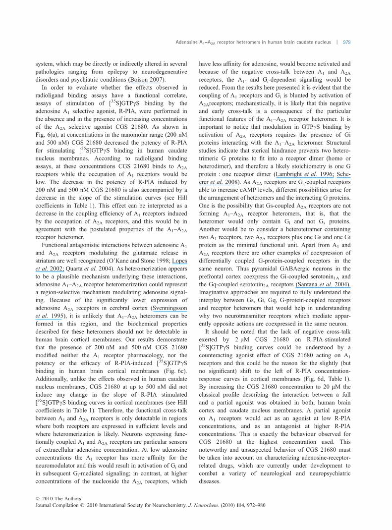
Next, we tried to evaluate the putative functional cross-talkbetween adenosine A1 and A2A receptors. To this aim, R-PIA-induced [35S]GTPcS binding was investigated in thepresence of different concentrations of CGS 21680 (200 nM,500 nM, 2 lM and 20 lM) in both human brain cortex andcaudate nucleus membranes. In human caudate nucleusmembranes, the presence of CGS 21680 below 2 lMconcentration induced a dose-dependent shift to the right ofR-PIA stimulated [35S]GTPcS binding curves without mod-ifying the Emax values (Fig. 6a, Table 1). These resultsindicate a decrease in the coupling efficiency of A1 receptorsresulting from occupation of A2A receptors. Furthermore,200 nM and 500 nM CGS 21680 caused the flattening of theR-PIA concentration-response curves (Hill coefficients < 1),indicating that CGS 21680, by binding to A2A receptors,induced some negative cross-talk for A1 adenosine receptoractivation (Table 1). Higher concentrations did not inducefurther displacement (to the right) of the curve. In thepresence of 20 lM CGS 21680 the shape of the full agonist(R-PIA) concentration-response curve was modified accord-ing to a pattern of a partial agonist, with an increase in basalbinding of 21 ± 5% (Fig. 6b and Table 1). In order toexamine the specificity of the functional cross-talk betweenA1 and A2A receptors, similar experiments were performed inhuman brain cortical membranes. Because of the much lowerexpression of A2A receptors in cerebral cortex than in caudatenucleus, human brain cortical membranes can be used asnegative control for this functional cross-talk. Unlike theeffects observed using caudate nucleus membranes, thepresence of CGS 21680 (200 nM and 500 nM) did notproduce any effects on R-PIA-stimulated [35S]GTPcS bind-ing curves (Fig. 6c). Moreover, no alterations on Hillcoefficient for these curves were observed (Table 1). Athigher concentrations of CGS 21680 (2 lM and 20 lM) theeffects on R-PIA-stimulated [35S]GTPcS binding curveswere similar to those detected using caudate nucleusmembranes (Fig. 6d and Table 1).
Discussion
One important and differential feature of the binding ofagonists to adenosine A1 receptors in membranes fromhuman brain (in the presence of adenosine deaminase) is thatit is of non-complex fashion; in terms of communicationbetween potentially different binding sites the binding can bereferred as to be non-cooperative. Similar results are obtainedin membranes from calf brain (Garritsen et al. 1990) whereasthe binding in brain membranes from rodent and otherspecies display complex (or cooperative) binding (Williamsand Risley, 1980; Casado et al. 1990). This differentpharmacology must be taken into account as extrapolationof pharmacological data obtained in rodent to humans mustbe done with due caution. Whenever possible studiesconcerning A1 receptors should be performed using human
Fig. 5 Stimulation of [35S]GTPcS binding by R-PIA and CGS 21680.
[35S]GTPcS binding was performed using human brain caudate nu-
cleus membranes. Both, R-PIA (d) and CGS 21680 (s) were assayed
from 0.1 nM to 0.1 mM and non-specific binding was determined in the
presence of 10 lM unlabeled GTPcS. Points and data are mean ±
SEM from three different experiments performed in duplicate. Data
were normalized using the Bottom and Top values of R-PIA stimulated
[35S]GTPcS binding curves (4021 ± 246 and 10046 ± 277 cpm,
respectively). In the inset is represented CGS 21680 stimulated
[35S]GTPcS binding curves in the absence (s) and in the presence ( )
of 50 nM DPCPX.
� 2010 The AuthorsJournal Compilation � 2010 International Society for Neurochemistry, J. Neurochem. (2010) 114, 972–980
Adenosine A1–A2A receptor heteromers in human brain caudate nucleus | 977

samples or samples from other mammals containing an A1
receptor with closer pharmacological resemblance to thehuman receptor. In case of using heterologous systems theyshould express the human receptor and, preferably, anypartner receptor (human) that may be coexpressed in the finaltarget tissue. Coexpressing partner receptors is recommendedwhen using heterologous models because of the fact thatcompounds may disclose different qualitative and/or quan-titative pharmacology when assayed for the same receptorbut in different heteromeric assemblies (Franco et al. 2010).Thus, here slight differences have been detected in theaffinity of CGS 21680 binding to cortical A1 receptors or tocaudate nucleus A1 receptor (KD1 of 0.22 ± 0.02 lM or0.46 ± 0.05 lM, respectively). This is probably because of adifferent composition of the complex/heteromer containingA1 receptors in the two brain areas, i.e. in cortex there should
be a high proportion of A1 receptors not interacting with A2A
receptors, but a high proportion of A1–A2A heteromers mayexist in caudate nucleus being the presence of A2A receptorsable to modulate the affinity of A1 receptors.
In the present study, [35S]GTPcS binding assays have beenused to evaluate the functional cross-talk between adenosineA1 and A2A receptors. It should be borne in mind that[35S]GTPcS binding assays in native tissues are only usefulfor Gi/o coupled receptors (Gonzalez-Maeso et al. 2000). AsA1 and A2A receptors are coupled to Gi/o and Gs/olf proteins,respectively (Dunwiddie and Masino 2001), [35S]GTPcSbinding can be used as a specific measure of A1 receptoractivation. In fact, in human brain caudate nucleus mem-branes the A2A selective agonist CGS 21680 stimulated[35S]GTPcS binding with an EC50 value of 20 lM (Fig. 5).According to the affinities of A1 and A2A receptors for CGS21680, no [35S]GTPcS binding stimulation is observed atconcentrations in the nanomolar range at which the 90% ofA2A saturation is reached and little binding to A1 receptorsoccurs. At micromolar concentrations the relative occupationof A1 receptor is significant and the [35S]GTPcS bindingstimulation observed at 20 lM CGS 21680 is measurable. Afurther proof that [35S]GTPcS binding stimulation observedat 20 lM CGS 21680 is mediated by A1 receptor is that no[35S]GTPcS binding stimulation was obtained with CGS21680 when the experiments were performed in the presenceof a selective A1 antagonist (50 nM DPCPX). Therefore,under our experimental conditions the [35S]GTPcS binding isonly stimulated by A1 receptor activation and no significantresponses were observed by A2A receptor activation. Ourresults also show that [35S]GTPcS binding assays represent agood technique to measure adenosine A1 receptor activationin postmortem human tissues, and this could be interestingfor future studies evaluating the dysfunction of the adenosine
Fig. 6 Effects of CGS 21680 on R-PIA-
induced stimulated [35S]GTPcS binding.
Human brain caudate nucleus (a and b) and
cortical (c and d) membranes were used.
(a) and (c) represent R-PIA-stimulated
[35S]GTPcS binding curves in the absence
(d) and in the presence of 200 nM ( ) and
500 nM ( ) CGS 21680. (b) and (d) repre-
sent R-PIA-stimulated [35S]GTPcS binding
in the absence (d) and in the presence of
2 lM (h) and 20 lM (D) CGS 21680.
The EC50 values and the Hill coefficient
obtained for these curves are shown in
Table 1. Points are mean ± SEM from at
least three different experiments performed
in duplicate. Percentages were obtained by
normalizing data with the Top and Bottom
values estimated by non-linear regression
analysis.
Table 1 Effect of CGS 21680 on R-PIA stimulated [35S]GTPcS
binding in human brain cortex and caudate nucleus membranes
CGS 21680 (lM)
Caudate nucleus Cortex
EC50 (nM) nH EC50 (nM) nH
0 78 ± 9 0.93 ± 0.09 61 ± 8 0.9 ± 0.1
0.2 163 ± 7** 0.56 ± 0.08* 94 ± 7 0.8 ± 0.2
0.5 199 ± 7** 0.7 ± 0.1* 97 ± 8 0.77 ± 0.09
2 54 ± 8 1.1 ± 0.2 42 ± 6 0.9 ± 0.3
20 121 ± 60 0.71 ± 0.30 189 ± 6.0** 0.47 ± 0.17*
Parameter values obtained by fitting data from Fig. 6 to eqn 5. Data
values are mean ± SEM of at least three independent experiments
performed in triplicate. *p < 0.05 different from unity. **p < 0.01 when
compared with respective control values (unpaired two-tailed Stu-
dent’s t-test).
Journal Compilation � 2010 International Society for Neurochemistry, J. Neurochem. (2010) 114, 972–980� 2010 The Authors
978 | V. Casado et al.
system, which may be directly or indirectly altered in severalpathologies ranging from epilepsy to neurodegenerativedisorders and psychiatric conditions (Boison 2007).
In order to evaluate whether the effects observed inradioligand binding assays have a functional correlate,assays of stimulation of [35S]GTPcS binding by theadenosine A1 selective agonist, R-PIA, were performed inthe absence and in the presence of increasing concentrationsof the A2A selective agonist CGS 21680. As shown inFig. 6(a), at concentrations in the nanomolar range (200 nMand 500 nM) CGS 21680 decreased the potency of R-PIAfor stimulating [35S]GTPcS binding in human caudatenucleus membranes. According to radioligand bindingassays, at these concentrations CGS 21680 binds to A2A
receptors while the occupation of A1 receptors would below. The decrease in the potency of R-PIA induced by200 nM and 500 nM CGS 21680 is also accompanied by adecrease in the slope of the stimulation curves (see Hillcoefficients in Table 1). This effect can be interpreted as adecrease in the coupling efficiency of A1 receptors inducedby the occupation of A2A receptors, and this would be inagreement with the postulated properties of the A1–A2A
receptor heteromer.Functional antagonistic interactions between adenosine A1
and A2A receptors modulating the glutamate release instriatum are well recognized (O’Kane and Stone 1998; Lopeset al. 2002; Quarta et al. 2004). As heteromerization appearsto be a plausible mechanism underlying these interactions,adenosine A1–A2A receptor heteromerization could representa region-selective mechanism modulating adenosine signal-ing. Because of the significantly lower expression ofadenosine A2A receptors in cerebral cortex (Svenningssonet al. 1995), it is unlikely that A1–A2A heteromers can beformed in this region, and the biochemical propertiesdescribed for these heteromers should not be detectable inhuman brain cortical membranes. Our results demonstratethat the presence of 200 nM and 500 nM CGS 21680modified neither the A1 receptor pharmacology, nor thepotency or the efficacy of R-PIA-induced [35S]GTPcSbinding in human brain cortical membranes (Fig. 6c).Additionally, unlike the effects observed in human caudatenucleus membranes, CGS 21680 at up to 500 nM did notinduce any change in the slope of R-PIA stimulated[35S]GTPcS binding curves in cortical membranes (see Hillcoefficients in Table 1). Therefore, the functional cross-talkbetween A1 and A2A receptors is only detectable in regionswhere both receptors are expressed in sufficient levels andwhere heteromerization is likely. Neurons expressing func-tionally coupled A1 and A2A receptors are particular sensorsof extracellular adenosine concentration. At low adenosineconcentrations the A1 receptor has more affinity for theneuromodulator and this would result in activation of Gi andin subsequent Gi-mediated signaling; in contrast, at higherconcentrations of the nucleoside the A2A receptors, which
have less affinity for adenosine, would become activated andbecause of the negative cross-talk between A1 and A2A
receptors, the A1- and Gi-dependent signaling would bereduced. From the results here presented it is evident that thecoupling of A1 receptors and Gi is blunted by activation ofA2Areceptors; mechanistically, it is likely that this negativeand early cross-talk is a consequence of the particularfunctional features of the A1–A2A receptor heteromer. It isimportant to notice that modulation in GTPcS binding byactivation of A2A receptors requires the presence of Giproteins interacting with the A1–A2A heteromer. Structuralstudies indicate that sterical hindrance prevents two hetero-trimeric G proteins to fit into a receptor dimer (homo orheterodimer), and therefore a likely stoichiometry is one Gprotein : one receptor dimer (Lambright et al. 1996; Sche-erer et al. 2008). As A2A receptors are Gs-coupled receptorsable to increase cAMP levels, different possibilities arise forthe arrangement of heteromers and the interacting G proteins.One is the possibility that Gs-coupled A2A receptors are notforming A1–A2A receptor heteromers, that is, that theheteromer would only contain Gi and not Gs proteins.Another would be to consider a heterotetramer containingtwo A1 receptors, two A2A receptors plus one Gs and one Giprotein as the minimal functional unit. Apart from A1 andA2A receptors there are other examples of coexpression ofdifferentially coupled G-protein-coupled receptors in thesame neuron. Thus pyramidal GABAergic neurons in theprefrontal cortex coexpress the Gi-coupled serotonin1A andthe Gq-coupled serotonin2A receptors (Santana et al. 2004).Imaginative approaches are required to fully understand theinterplay between Gs, Gi, Gq, G-protein-coupled receptorsand receptor heteromers that would help in understandingwhy two neurotransmitter receptors which mediate appar-ently opposite actions are coexpressed in the same neuron.
It should be noted that the lack of negative cross-talkexerted by 2 lM CGS 21680 on R-PIA-stimulated[35S]GTPcS binding curves could be understood by acounteracting agonist effect of CGS 21680 acting on A1
receptors and this could be the reason for the slightly (butno significant) shift to the left of R-PIA concentration-response curves in cortical membranes (Fig. 6d, Table 1).By increasing the CGS 21680 concentration to 20 lM theclassical profile describing the interaction between a fulland a partial agonist was obtained in both, human braincortex and caudate nucleus membranes. A partial agoniston A1 receptors would act as an agonist at low R-PIAconcentrations, and as an antagonist at higher R-PIAconcentrations. This is exactly the behaviour observed forCGS 21680 at the highest concentration used. Thisnoteworthy and unsuspected behavior of CGS 21680 mustbe taken into account on characterizing adenosine-receptor-related drugs, which are currently under development tocombat a variety of neurological and neuropsychiatricdiseases.
� 2010 The AuthorsJournal Compilation � 2010 International Society for Neurochemistry, J. Neurochem. (2010) 114, 972–980
Adenosine A1–A2A receptor heteromers in human brain caudate nucleus | 979

Acknowledgements
We acknowledge the technical help obtained from Jasmina Jimenez
(Molecular Neurobiology laboratory, Barcelona University). This
study was supported by Grants from Spanish Ministerio de Ciencia
y Tecnologıa SAF2009-07276, SAF2008-03229-E and SAF2008-
00146 and grant 060110 from Fundacio La Marato de TV3, and by
grants from Basque Government (GIC07/101-IT-199-07, GIC07/
131-IT-261-07 and S-PE08UN29), MICINN (SAF2009-08460), the
Instituto de Salud Carlos III, Centro de Investigacion Biomedica en
Red de Salud Mental, CIBERSAM and EDRF.
References
Barrondo S. and Salles J. (2009) Allosteric modulation of 5-HT1Areceptors by zinc: binding studies. Neuropharmacology 56, 455–462.
Boison D. (2007) Adenosine as a modulator of brain activity. Drug NewsPerspect. 20, 607–611.
Casado V., Cantı C., Mallol J., Canela E. I., Lluıs C. and Franco R.(1990) Solubilization of A1 adenosine receptor from pig brain.Characterization and evidence of the role of the cell membrane onthe coexistence of the high and low-affinity states. J. Neurosci. Res.26, 461–473.
Casado V., Cortes A., Ciruela F., Mallol J., Ferre S., Lluıs C., CanelaE. I. and Franco R. (2007) Old and new ways to calculate theaffinity of agonists and antagonists interacting with G protein-coupled monomeric and dimeric receptors: the receptor-dimercooperativity index. Pharmacol. Ther. 116, 343–354.
Casado V., Cortes A., Mallol J., Perez-Capote K., Ferre S., Lluis C.,Franco R. and Canela E. I. (2009a) G protein-coupled receptorshomomers and heteromers: a better choice as targets for drugdevelopment than G protein-coupled receptors monomers?. Phar-macol. Ther. 124, 248–257.
Casado V., Ferrada C., Bonaventura J., Gracia E., Mallol J., Canela E. I.,Lluıs C., Cortes A. and Franco R. (2009b) Useful pharmacologicalparameters for G-protein-coupled receptor homodimers obtainedfrom competition experiments. Agonist-antagonist binding modu-lation. Biochem. Pharmacol. 78, 1456–1463.
Chistopoulos A. (1998) Assessing the distribution of parameters inmodels of ligand-receptor interaction: to log or not to log. TrendsPharmacol. Sci. 19, 351–357.
Ciruela F., Casado V., Rodrigues R. J. et al. (2006) Presynaptic controlof striatal glutamatergic neurotransmission by adenosine A1-A2Areceptor heteromers. J. Neurosci. 26, 2080–2087.
Dunwiddie T. V. and Masino S. A. (2001) The role and regulation ofadenosine in the central nervous system. Ann. Rev. Neurosci. 24,31–55.
Ferre S., Ciruela F., Quiroz C. et al. (2007a) Adenosine receptor heter-omers and their integrative role in striatal function. Scientific WorldJ. 7, 74–85.
Ferre S., Agnati L. F., Ciruela F., Lluis C., Woods A. S., Fuxe K. andFranco R. (2007b) Neurotransmitter receptor heteromers and theirintegrative role in ‘local modules’: the striatal spine module. BrainRes. Rev. 55, 55–67.
Fleming W. W., Westfall D. P., De La Lande J. S. and Jellet L. B. (1972)Log-normal distribution of equieffective doses of norepinephrineand acetylcholine in several tissues. J. Pharmacol. Exp. Ther. 181,339–345.
Franco R., Casado V., Mallol J., Ferre S., Fuxe K., Cortes A., Ciruela F.,Lluıs C. and Canela E. I. (2005) Dimer-based model for heptas-panning membrane receptors. Trends Biochem. Sci. 30, 360–366.
Franco R., Casado V., Mallol J. et al. (2006) The two-state dimerreceptor model: a general model for receptor dimers. Mol. Phar-macol. 69, 1905–1912.
Franco R., Canela E. I., Casado V. and Ferre S. (2010) Platforms for theidentification of GPCR targets, and of orthosteric and allostericmodulators. Expert Opin. Drug Discov. 5, 391–403.
Garritsen A., Ijzerman A. P., Beukers M. W., Cragoe Jr E. J. andSoudijn W. (1990) Interaction of amiloride and its analogues withadenosine A1 receptors in calf brain. Biochem. Pharmacol. 40,827–834.
Gonzalez-Maeso J., Rodriguez-Puertas R., Gabilondo A. M. and MeanaJ. J. (2000) Characterization of receptor-mediated [35S]GTPcSbinding to cortical membranes from post-mortem human brain.Eur. J. Pharmacol. 390, 25–36.
Gracia E., Cortes A., Meana J. J. et al. (2008) Human adenosinedeaminase as an allosteric modulator of human A1 adenosinereceptor: abolishment of negative cooperativity for [3H]R-PIAbinding to the caudate nucleus. J. Neurochem. 107, 161–170.
Lambright D. G., Sondek J., Bohm A., Skiba N. P., Hamm H. E. andSigler P. B. (1996) The 2.0 A crystal structure of a heterotrimeric Gprotein. Nature 379, 311–319.
Lopes L. V., Cunha R. A., Kull B., Fredholm B. B. and Ribeiro J. A.(2002) Adenosine A2A receptor facilitation of hippocampal syn-aptic transmission is dependent on tonic A1 receptor inhibition.Neuroscience 112, 319–329.
O’Kane E. M. and Stone T. W. (1998) Interaction between adenosine A1
and A2 receptor-mediated responses in the rat hippocampus invitro. Eur. J. Pharmacol. 362, 17–25.
Quarta D., Borycz J., Solinas M. et al. (2004) Adenosine receptor-mediated modulation of dopamine release in the nucleus accum-bens depends on glutamate neurotransmission and N-methyl-D-aspartate receptor stimulation. J. Neurochem. 91, 873–880.
Quiroz C., Lujan R., Uchigashima M. et al. (2009) Key modulatoryrole of presynaptic adenosine A2A receptors in cortical neuro-transmission to the striatal direct pathway. Scientific World J. 9,1321–1344.
Rees D. A., Scanlon M. F. and Ham J. (2003) Adenosine signallingpathways in the pituitary gland: one ligand, multiple receptors.J. Endocrinol. 177, 357–364.
Santana N., Bortolozzi A., Serrats J., Mengod G. and Artigas F. (2004)Expression of serotonin1A and serotonin2A receptors in pyramidaland GABAergic neurons of the rat prefrontal cortex. Cereb. Cortex14, 1100–1109.
Sarrio S., Casado V., Escriche M., Ciruela F., Mallol J., Canela E. I.,Lluıs C. and Franco R. (2000) The heat shock cognate proteinhsc73 assembles with A(1) adenosine receptors to form func-tional modules in the cell membrane. Mol. Cell. Biol. 20, 5164–5174.
Scheerer P., Park J. H., Hildebrand P. W., Kim Y. J., Krauss N., ChoeH. W., Hofmann K. P. and Ernst O. P. (2008) Crystal structure ofopsin in its G-protein-interacting conformation. Nature 455, 497–502. Comment in Nature (2008) 455, 473–474.
Sebastiao A. M. and Ribeiro J. A. (2009) Adenosine receptors andthe central nervous system. Handb. Exp. Pharmacol. 193, 471–534.
Svenningsson P., Nomikos G. G. and Fredholm B. B. (1995) Biphasicchanges in locomotor behavior and in expression of mRNA forNGFI-A and NGFI-B in rat striatum following acute caffeineadministration. J. Neurosci. 15, 7612–7624.
Williams M. and Risley E. A. (1980) Biochemical characterizationof putative central purinergic receptors by using 2-chloro[3H]adenosine, a stable analog of adenosine. Proc. Natl. Acad. Sci. USA77, 6892–6896.
Journal Compilation � 2010 International Society for Neurochemistry, J. Neurochem. (2010) 114, 972–980� 2010 The Authors
980 | V. Casado et al.