Aberrant A2A receptor function in peripheral blood cells in Huntington's disease
Upload
independentCategory
view
0download
0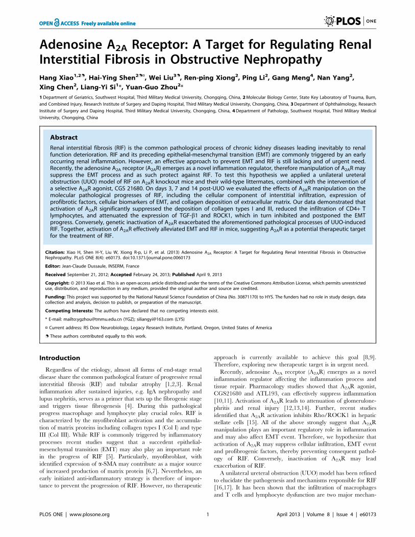
Adenosine A2A Receptor: A Target for Regulating RenalInterstitial Fibrosis in Obstructive NephropathyHang Xiao1,2., Hai-Ying Shen2.¤, Wei Liu3., Ren-ping Xiong2, Ping Li2, Gang Meng4, Nan Yang2,
Xing Chen2, Liang-Yi Si1*, Yuan-Guo Zhou2*
1Department of Geriatrics, Southwest Hospital, Third Military Medical University, Chongqing, China, 2Molecular Biology Center, State Key Laboratory of Trauma, Burn,
and Combined Injury, Research Institute of Surgery and Daping Hospital, Third Military Medical University, Chongqing, China, 3Department of Ophthalmology, Research
Institute of Surgery and Daping Hospital, Third Military Medical University, Chongqing, China, 4Department of Pathology, Southwest Hospital, Third Military Medical
University, Chongqing, China
Abstract
Renal interstitial fibrosis (RIF) is the common pathological process of chronic kidney diseases leading inevitably to renalfunction deterioration. RIF and its preceding epithelial-mesenchymal transition (EMT) are commonly triggered by an earlyoccurring renal inflammation. However, an effective approach to prevent EMT and RIF is still lacking and of urgent need.Recently, the adenosine A2A receptor (A2AR) emerges as a novel inflammation regulator, therefore manipulation of A2AR maysuppress the EMT process and as such protect against RIF. To test this hypothesis we applied a unilateral ureteralobstruction (UUO) model of RIF on A2AR knockout mice and their wild-type littermates, combined with the intervention ofa selective A2AR agonist, CGS 21680. On days 3, 7 and 14 post-UUO we evaluated the effects of A2AR manipulation on themolecular pathological progresses of RIF, including the cellular component of interstitial infiltration, expression ofprofibrotic factors, cellular biomarkers of EMT, and collagen deposition of extracellular matrix. Our data demonstrated thatactivation of A2AR significantly suppressed the deposition of collagen types I and III, reduced the infiltration of CD4+ Tlymphocytes, and attenuated the expression of TGF-b1 and ROCK1, which in turn inhibited and postponed the EMTprogress. Conversely, genetic inactivation of A2AR exacerbated the aforementioned pathological processes of UUO-inducedRIF. Together, activation of A2AR effectively alleviated EMT and RIF in mice, suggesting A2AR as a potential therapeutic targetfor the treatment of RIF.
Citation: Xiao H, Shen H-Y, Liu W, Xiong R-p, Li P, et al. (2013) Adenosine A2A Receptor: A Target for Regulating Renal Interstitial Fibrosis in ObstructiveNephropathy. PLoS ONE 8(4): e60173. doi:10.1371/journal.pone.0060173
Editor: Jean-Claude Dussaule, INSERM, France
Received September 21, 2012; Accepted February 24, 2013; Published April 9, 2013
Copyright: � 2013 Xiao et al. This is an open-access article distributed under the terms of the Creative Commons Attribution License, which permits unrestricteduse, distribution, and reproduction in any medium, provided the original author and source are credited.
Funding: This project was supported by the National Natural Science Foundation of China (No. 30871170) to HYS. The funders had no role in study design, datacollection and analysis, decision to publish, or preparation of the manuscript.
Competing Interests: The authors have declared that no competing interests exist.
* E-mail: mailto:[email protected] (YGZ); [email protected] (LYS)
¤ Current address: RS Dow Neurobiology, Legacy Research Institute, Portland, Oregon, United States of America
. These authors contributed equally to this work.
Introduction
Regardless of the etiology, almost all forms of end-stage renal
disease share the common pathological feature of progressive renal
interstitial fibrosis (RIF) and tubular atrophy [1,2,3]. Renal
inflammation after sustained injuries, e.g. IgA nephropathy and
lupus nephritis, serves as a primer that sets up the fibrogenic stage
and triggers tissue fibrogenesis [4]. During this pathological
progress macrophage and lymphocyte play crucial roles. RIF is
characterized by the myofibroblast activation and the accumula-
tion of matrix proteins including collagen types I (Col I) and type
III (Col III). While RIF is commonly triggered by inflammatory
processes recent studies suggest that a succedent epithelial-
mesenchymal transition (EMT) may also play an important role
in the progress of RIF [5]. Particularly, myofibroblast, with
identified expression of a-SMA may contribute as a major source
of increased production of matrix protein [6,7]. Nevertheless, an
early initiated anti-inflammatory strategy is therefore of impor-
tance to prevent the progression of RIF. However, no therapeutic
approach is currently available to achieve this goal [8,9].
Therefore, exploring new therapeutic target is in urgent need.
Recently, adenosine A2A receptor (A2AR) emerges as a novel
inflammation regulator affecting the inflammation process and
tissue repair. Pharmacology studies showed that A2AR agonist,
CGS21680 and ATL193, can effectively suppress inflammation
[10,11]. Activation of A2AR leads to attenuation of glomerulone-
phritis and renal injury [12,13,14]. Further, recent studies
identified that A2AR activation inhibits Rho/ROCK1 in hepatic
stellate cells [15]. All of the above strongly suggest that A2AR
manipulation plays an important regulatory role in inflammation
and may also affect EMT event. Therefore, we hypothesize that
activation of A2AR may suppress cellular infiltration, EMT event
and profibrogenic factors, thereby preventing consequent pathol-
ogy of RIF. Conversely, inactivation of A2AR may lead
exacerbation of RIF.
A unilateral ureteral obstruction (UUO) model has been refined
to elucidate the pathogenesis and mechanisms responsible for RIF
[16,17]. It has been shown that the infiltration of macrophages
and T cells and lymphocyte dysfunction are two major mechan-
PLOS ONE | www.plosone.org 1 April 2013 | Volume 8 | Issue 4 | e60173

isms contributing to the UUO-induced RIF model [18,19]. In this
model, at the cellular level, tubular dilatation leads the tubular
epithelia to lose their epithelial characteristics and acquire
mesenchymal traits such as a-SMA expression and actin re-
organization. At molecular level, TGF-b1 plays a key role in EMT
via activation of its downstream Rho/ROCK signaling pathway
[20].
Using the experimental UUO-induced RIF mouse model, the
present study was aimed to evaluate the modulatory effect of
A2AR-based manipulation on several aspects of RIF progression,
including interstitial lymphocyte infiltration, cellular biomarkers of
EMT, expression of the profibrogenic factor TGF-b1 and its
downstream Rho/ROCK1 pathway, as well as the consequent
extracellular matrix accumulation.
Materials and Methods
AnimalsAll animal experiments were conducted under approval of the
Institutional Animal Care and Use Committee of Third Military
Medical University (TMMU) and performed under the supervi-
sion of the facility veterinary staff. Mice used in the present study,
i.e., genetic A2AR knockout (KO) mice (A2AR2/2) and their wild-
type (WT) controls (A2AR+/+) were bred at the Animal Care
Center of the Research Institute of Surgery of TMMU after being
imported from Boston University School of Medicine as previously
described [21]. Mice were maintained in a pathogen-free,
humidity- and temperature-controlled environment with 12 h
light-dark cycles and free access to food and drinking water. The
A2AR KO mice and their WT littermates were randomly
designated into six experimental groups (see Table 1) according
to the involvement of UUO procedure and CGS treatment. Ten
mice from each experimental group were sacrificed at the designed
experimental time-points, i.e., day 3, 7, and 14 after UUO. Mouse
kidneys were harvested for the following imunohistochemistry
evaluations.
Unilateral ureteral obstruction (UUO) modelMice (20–25 g weight) were subjected to the UUO procedure
under anesthesia as previously described [22] with modifications.
All surgical procedures were performed under an operating
microscope. Briefly, mice were first anesthetized with sodium
pentobarbital (50 mg/kg, i.p.). After a left flank incision was taken,
the left ureter was exposed, ligated with 6–0 silk sutures at two
points, and cut between the two ligatures. Lastly, the peritoneal
membrane and skin were sutured. Sham surgery was performed as
control by following all steps of UUO-procedure except ligation
and cut of ureter.
Drug treatmentPharmacological activation of A2AR was induced by daily
systemic administration of the selective A2AR agonist, CGS 21680
(Tocris, Cat# 1063, 0.4 mg/kg i.p.) from day 1 after UUO
through the designed experimental time-points, i.e., day 3, 7, and
14 after UUO, when mice were scarified and their kidneys were
harvested.
Reverse transcription quantitative real-time PCR (RT-qPCR)Total RNA extraction of renal sample was conducted using
a total RNA extraction kit (BioFlux, Cat# BSC52S1) and the
reverse transcription reaction was performed using SYBR Premix
Ex Taq kit (DRR041A, Dalian, China), according to the
manufacturer’s instructions. Then qPCR was performed to
quantify the expression level of A2AR, TGF-b1, and ROCK1
mRNAs using SYBR Premix Ex Taq kit (DRR041A, Dalian,
China) and a qPCR reaction thermal cycle of 40 cycles of 95uC(30 s), 58uC (30 s), and 70uC (30 s). The glyceraldehyde 3-
phosphate dehydrogenase (GAPDH) was used as an internal
control to normalize RT-qPCR readout. Gene mRNA expression
levels were calculated relative to the expression level of GAPDH.
All primers used (as shown in Table 2) were synthetized by Takara
(Dalian, China).
Histopathology and ImmunohistochemistryHematoxylin and eosin (H&E) staining and all immunohistos-
tainings were performed on 4 mm-thick paraffin-embedded slice of
kidney using a similar procedure as previously described [23]. The
antigen retrieval process was performed by pressure cooking. The
following primary antibodies and corresponding dilutions were
used: anti-A2AR (ab115250, 1:200, Abcam, Cambridge, MA
USA), anti-CD3 (ab5690, 1:100, Abcam), anti-CD4 (ab51312,
1:100, Abcam), anti-CD8 (MA1-70041, 1:50, Pierce), anti-Foxp3
(ab54501, 1:200, Abcam), anti-CD11b (ab52478, 1:100, Abcam),
anti-CD68 (ab955, 1:100, Abcam), anti-F4/80 (14-4801, 1:50,
Ebioscience), anti-collagen I (ab34710, 1:400, Abcam), anti-
collagen III (ab7778, 1:400, Abcam), anti-a-SMA (ab7817, 1:50,
Abcam). All the quantitative morphological analyses were
performed by separate investigator who was blinded to the
treatment of samples. Positive stained cells and/or area were
assessed and expressed as integrated optical density (IOD) or area.
Three sections of each mouse kidney were measured, and 10
random fields or appointed area around vessels were chosen and
calculated under magnification of 100x (for CD4) or 400x (for Col
I, Col III and CD4+CD25+Foxp3+ Treg cell). The IOD or
positive area was acquired by the Image-Pro Plus 6.0 program
(Media Cybernetics, Bethesda, MD, USA).
Table 1. Experimental groups.
group A2AR UUO CGS
WT+Sham + 2 2
WT+UUO+Veh + + 2
WT+UUO+CGS + + +
KO+Sham 2 2 2
KO+UUO+Veh 2 + 2
KO+UUO+CGS 2 + +
doi:10.1371/journal.pone.0060173.t001
Table 2. Primers used for reverse transcription quantitativereal-time PCR.
Gene Primer Sequence
A2AR forward 59-ccattcgccatcaccatcag-39
reverse 59-cgtcaccaagccattgtacc-39
ROCK1 forward 59-acaccagaaggagctgaatgac-39
reverse 59-ccgcaactgctcaatatcactc-39
TGF-b1 forward 59-tatttggagcctggacacacag-39
reverse 59-cgtagtagacgatgggcagtg-39
doi:10.1371/journal.pone.0060173.t002
Adenosine A2AR and Renal Interstitial Fibrosis
PLOS ONE | www.plosone.org 2 April 2013 | Volume 8 | Issue 4 | e60173
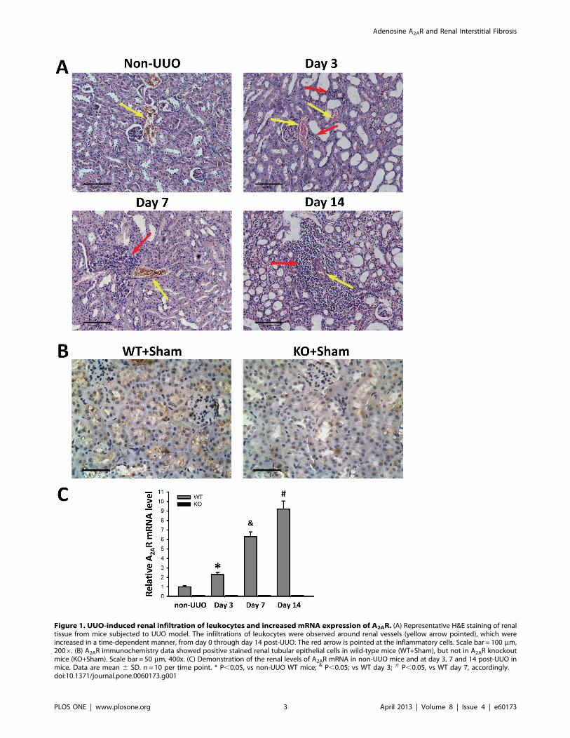
Figure 1. UUO-induced renal infiltration of leukocytes and increased mRNA expression of A2AR. (A) Representative H&E staining of renaltissue from mice subjected to UUO model. The infiltrations of leukocytes were observed around renal vessels (yellow arrow pointed), which wereincreased in a time-dependent manner, from day 0 through day 14 post-UUO. The red arrow is pointed at the inflammatory cells. Scale bar = 100 mm,2006. (B) A2AR immunochemistry data showed positive stained renal tubular epithelial cells in wild-type mice (WT+Sham), but not in A2AR knockoutmice (KO+Sham). Scale bar = 50 mm, 400x. (C) Demonstration of the renal levels of A2AR mRNA in non-UUO mice and at day 3, 7 and 14 post-UUO inmice. Data are mean 6 SD. n = 10 per time point. * P,0.05, vs non-UUO WT mice; & P,0.05; vs WT day 3; # P,0.05, vs WT day 7, accordingly.doi:10.1371/journal.pone.0060173.g001
Adenosine A2AR and Renal Interstitial Fibrosis
PLOS ONE | www.plosone.org 3 April 2013 | Volume 8 | Issue 4 | e60173

Western blotThe Western blot was performed according as previously
described [24] with modification. Briefly, mouse kidneys were first
homogenized in tissue protein extraction reagent (Thermo
scientific, cat# MD156494) with a protease inhibitor cocktail
(Thermo scientific, cat# ME156994) according to the manufac-
turer’s instructions. Forty mg of protein extracts from each sample
were loaded on and separated by 10% SDS-PAGE, then
transferred onto nitrocellulose membrane. The blots were probed
overnight at 4uC with primary antibodies against E-cadherin
(ab76055, 1:1000, Abcam), a-SMA (ab7817, 1:200, Abcam), and
b-actin (a2228, 1:2000, Sigma-Aldrich), respectively, followed by
the respective horseradish peroxidase-linked secondary antibody
(a4416, 1:5000, Sigma-Aldrich). Horseradish peroxidase activity
was visualized via an enhanced chemiluminescence kit (20-500-
120, Biolind, Israel). Images were scanned and processed for
densitometric quantification by the Image analysis program
(Labworks 4.0, UVP).
Statistical analysesThe data are expressed as mean 6 SD. Statistical analysis was
performed using one-way ANOVA followed by Bonferroni post hoc
comparisons. P,0.05 was considered statistical significance.
Results
1. A2AR activation attenuated collagen deposition inmatrix accumulationTo evaluate the effect of A2AR on renal fibrosis, we applied the
UUO model to mice combined with A2AR agonist CGS21680 and
genetic A2AR inactivation (as aforementioned paradigm in
Methods). Pathology assessment using H&E staining and im-
munohistostaining of Col I and Col III deposition were evaluated
at day 3, 7, and 14 after UUO. Our H&E data demonstrated the
successfulness of UUO modeling with featured pathological
changes, e.g. progressively aggravated tubular dilatation and
leukocytes infiltration (Figure 1 A). Our A2AR immunochemistry
data demonstrated that positive stained renal tubular epithelial
cells were seen in WT mice (WT+Sham), but devoid in KO mice
(KO+Sham) (Figure 1B). Furthermore, we used RT-qPCR to
detect the temporal changes of A2AR mRNA expression in the
progress of UUO-induced RIF. We showed that the mRNA level
of A2AR was significantly increased at day 3 through day 14 post-
UUO, in a time-dependent manner. WT mice in WT+UUO+Vehgroup displayed an increase of 156%, 529% and 816% at day 3, 7
and 14, respectively, compared to non-UUO mice (F = 541.22,
P,0.05, n= 10 per time point, Figure 1C). Conversely, A2AR
mRNA level in A2AR KO (KO+UUO+Veh) mice remained under
Figure 2. A2AR activity affected UUO-induced deposition of collagen I. (A) Representative immunohistochemistry of renal collagen I (Col I)from the A2AR KO and WT mice, at day 3, 7 and 14 post-UUO or sham surgery (Sham), following treatment of CGS21680 (CGS) or vehicle (Veh). Scalebar = 50 mm, 4006. (B) Demonstration of Col I deposition in the post-UUO WT animals received treatment of vehicle (WT+UUO+Veh) or A2AR agonistCGS21680 (WT+UUO+CGS), and in the A2AR post-UUO KO mice received treatment of vehicle (KO+UUO+Veh), or CGS21680 (KO+UUO+CGS), at day 3,7 and 14 post-UUO, along with that in sham control animals (WT+Sham and KO+Sham)(n = 10 per group). Data are mean 6 SD. * P,0.05 betweentwo compared groups; NS, no significance.doi:10.1371/journal.pone.0060173.g002
Adenosine A2AR and Renal Interstitial Fibrosis
PLOS ONE | www.plosone.org 4 April 2013 | Volume 8 | Issue 4 | e60173

a detectable threshold from day 1 throughout day 14 post-UUO
(Figure 1C).
Further, immunohistochemistry staining showed that starting at
day 3, Col I and Col III progressively increased along with
interstitial accumulation of extracellular matrix (ECM) in mouse
kidneys from WT+UUO+Veh group (Figure 2 and 3), indicating
the establishment of the UUO-induced RIF model. Importantly,
quantitative morphometric analysis demonstrated that the deposi-
tions of Col I and Col III were both significantly reduced in the
A2AR agonist-treated WT+UUO+CGS group (a reduction of
40.6% and 55.9% at day 3, a reduction of 50.3% and 64.9% at
day 7, respectively), compared to WT+UUO+Veh group (P,0.05,
n = 10 per groups, Figure 2 and 3). Conversely, genetic in-
activation of A2AR significantly exacerbated collagen deposition in
A2AR KO (KO+UUO+Veh) mice, showing an increased Col I
and Col III levels (by 39.6% and 57.1% at day 3, 29.5% and
31.7% at day 7, vs WT+UUO+Veh group, respectively, P,0.05,
n = 10 per groups, Figure 2 and 3). Noteworthy, genetic A2AR
inactivation-induced exacerbation of collagen deposition was not
affected by CGS treatment in KO+UUO+CGS group, showing
significantly increased renal Col I and Col III levels, compared to
WT+UUO+CGS group (P,0.05, n= 10 per groups, Figure 2 and
3).
Importantly, A2AR agonist CGS21680 treatment (in
WT+UUO+CGS group) reversed deposition of collagens at day
3 and day 7 post-UUO, compared to WT+UUO+Veh group
(P,0.05, vs n = 10 per groups, Figure 2 and 3). However, this
inhibitory effect of CGS21680 was blunt at day 14 post-UUO,
showing that the expression level of Col I and Col III in
CGS21680-treated (WT+UUO+CGS) group were similar to that
in other groups (P.0.05, n = 10 per group, Figure 2 and 3).
Together, A2AR activation by CGS21680 resulted in suppression
of collagen deposition at early post-UUO stage, i.e., at day 3 and
day 7, but not at later post-UUO stage (day 14). Nevertheless,
activation of A2AR effectively attenuated and postponed the
progression of RIF whereas inactivation of A2AR exacerbated the
RIF process.
2. A2AR activation inhibited UUO-induced changes on E-cadherin and SMATo evaluate A2AR-mediated effects on the EMT process we
detected the expression levels of a-SMA (the myofibroblast
marker) and E-cadherin (the epithelial marker) that indicate the
transdifferentiation status of epithelial to myofibroblast. Western
blot assay showed that at day 3 no expression difference of a-SMA
and E-cadherin was found between each of the UUO groups and
Figure 3. A2AR activity affected UUO-induced deposition of collagen III. (A) Representative immunohistochemistry of Collagen III) (Col III)from the A2AR KO and WT mice, at day 3, 7 and 14 post-UUO or Sham, following treatment of CGS21680 (CGS) or vehicle (Veh). Scale bar = 50 mm,4006. (B) Demonstration of Col III) deposition in the post-UUO WT animals received treatment of vehicle (WT+UUO+Veh) or A2AR agonist CGS21680(WT+UUO+CGS), and in the A2AR post-UUO KO mice received treatment of vehicle (KO+UUO+Veh), or CGS21680 (KO+UUO+CGS), at day 3, 7 and 14post-UUO, along with that in sham control animals (WT+Sham and KO+Sham)(n = 10 per group). Data are mean 6 SD. * P,0.05 between twocompared groups; NS, no significance.doi:10.1371/journal.pone.0060173.g003
Adenosine A2AR and Renal Interstitial Fibrosis
PLOS ONE | www.plosone.org 5 April 2013 | Volume 8 | Issue 4 | e60173
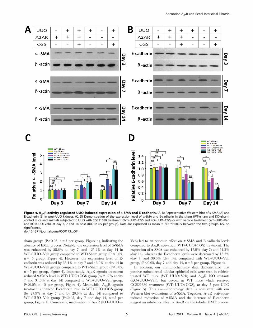
sham groups (P.0.05, n = 5 per group, Figure 4), indicating the
absence of EMT process. Notably, the expression level of a-SMA
was enhanced by 58.6% at day 7, and 125.2% at day 14 in
WT+UUO+Veh group compared to WT+Sham group (P ,0.05,
n = 5 group, Figure 4). However, the expression level of E-
cadherin was reduced by 35.4% at day 7 and 43.0% at day 14 in
WT+UUO+Veh groups compared to WT+Sham group (P,0.05,
n = 5 per group, Figure 4). Importantly, A2AR agonist treatment
reduced a-SMA level in WT+UUO+CGS group (by 21.7% at day
7 and 31.3% at day 14) compared to WT+UUO+Veh group,
P,0.05, n= 5 per group, Figure 4). Meanwhile, A2AR agonist
treatment enhanced E-cadherin level in WT+UUO+CGS group
(by 27.9% at day 7 and by 20.6% at day 14) compared to
WT+UUO+Veh group (P,0.05, day 7 and day 14, n = 5 per
group, Figure 4). Conversely, inactivation of A2AR (KO+UUO+-
Veh) led to an opposite effect on a-SMA and E-cadherin levels
compared to A2AR activation (WT+UUO+CGS) treatment. The
expression of a-SMA was enhanced by 17.9% (day 7) and 54.2%
(day 14), whereas the E-cadherin levels were decreased by 15.7%
(day 7) and 39.6% (day 14), compared with WT+UUO+Vehgroup, (P,0.05, day 7 and day 14, n = 5 per group, Figure 4).
In addition, our immunochemistry data demonstrated that
positive stained renal tubular epithelial cells were seen in vehicle-
treated WT mice (WT+UUO+Veh) and A2AR KO mutants
(KO+UUO+Veh), but devoid in WT mice which received
CGS21680 treatment (WT+UUO+CGS), at day 7 post-UUO
(Figure 5). This immunohistology data is consistent with our
Western blot evaluations of a-SMA. Together, A2AR activation-
induced reduction of a-SMA and the increase of E-cadherin
suggest an inhibitory effect of A2AR on the tubular EMT process.
Figure 4. A2AR activity regulated UUO-induced expression of a-SMA and E-cadherin. (A, B) Representative Western blot of a-SMA (A) andE-cadherin (B) in post-UUO kidneys. (C, D) Demonstration of the expression level of a-SMA and E-cadherin in the sham (WT+sham and KO+sham)control mice and animals subjected to UUO with CGS21680 treatment (WT+UUO+CGS and KO+UUO+CGS) or with vehicle treatment (WT+UUO+Vehand KO+UUO+Veh), at day 3, 7 and 14 post-UUO (n= 5 per group). Data are expressed as mean 6 SD. *P,0.05 between the two groups. NS, nosignificance.doi:10.1371/journal.pone.0060173.g004
Adenosine A2AR and Renal Interstitial Fibrosis
PLOS ONE | www.plosone.org 6 April 2013 | Volume 8 | Issue 4 | e60173

3. A2AR activation attenuated the expression ofprofibrotic mediatorsTo mechanistically evaluate the A2AR modulation on RIF, we
detected the mRNA expression of two crucial profibrotic
mediators, TGF-b1 and ROCK1 using RT-qPCR. We showed
that the expression level of TGF-b1 mRNA was significantly
increased at day 3 through day 14 in WT+UUO+Veh group (an
increase of 411%, 789% and 833% at day 3, 7 and 14 respectively)
compared to WT+Sham control group (P,0.05, n= 10 per group,
Figure 6). Importantly, A2AR agonist treatment attenuated the
increase of TGF-b1 mRNA expression in WT+UUO+CGS
group, leading to a decrease of 60.9% (P,0.05) and 30.0%
(P,0.05) at day 3 and day 7, respectively, vs. WT+UUO+Vehgroup (n= 10 per group). Conversely, genetic inactivation of
A2AR in KO+UUO+Veh group led to an additional enhancement
in mRNA expression of TGF-b1, by 39.1% (day 3) and 37.5%
(day 7) compared to WT+UUO+Veh group, correspondingly
(P,0.05, n = 10 per group, Figure 6). Noteworthy, A2AR
activation-mediated inhibitory effect on TGF-b1 expression was
blunt at 14 day post-UUO, with no difference compared to other
UUO groups (P.0.05 Figure 6), suggesting that the A2AR
activation-induced suppression on TGF-b1 expression occurred
at early but not later post-UUO stage.
Furthermore, RT-qPCR data showed that the expression level
of ROCK1 mRNA was significantly enhanced in kidneys from
WT+UUO+Veh mice, leading to an increase of 122%, 289% and
400%, at day 3, 7, and 14, correspondently, compared to
WT+Sham group (P,0.05, n= 10 per group, Figure 6). The
increase of ROCK1 mRNA shared a similar post-UUO expres-
sion pattern of TGF-b1 mRNA. Importantly, the increased
expression of ROCK1 was suppressed in CGS21680-treated
WT+UUO+CGS animals, showing a reduction of 35.0% (day 3)
and 25.7% (day 7) vs. WT+UUO+Veh (P,0.05, n = 10 per group,
Figure 6). In contrast, genetic inactivation of A2AR (in
KO+UUO+Veh group) led to an exacerbated enhancement of
ROCK1 level, by 30.0% (day 3) and 17.1% (day 7) vs.
WT+UUO+Veh (P,0.05, day 3; P,0.05, day 7; n= 10 per
group, Figure 6). Interestingly, the A2AR effect on ROCK1
expression was also noticed only at day 3 and day 7, but not on
day 14, post-UUO (P.0.05, Figure 6). Together, these findings
revealed that A2AR activation inhibited expression of TGF-b1 and
its downstream factor, ROCK1.
4. Suppression on T lymphocyte infiltration contributesto A2AR-mediated renal protection against RIFInfiltration of T lymphocyte, a key cellular inflammatory
response, plays a crucial role in the initiation of EMT and RIF.
Thus we examined renal T lymphocyte infiltration post-UUO
Figure 5. A2AR activation inhibited UUO-induced EMT process.Representative immunohistochemistry staining of a-SMA in mice at day7 post-UUO. The a-SMA, as the marker for myofibroblast (red arrow),was positively stained on the renal tubular epithelial cells in WT(WT+UUO+Veh) and A2AR KO (KO+UUO+Veh) mice whereas treatmentof CGS21680 reduced positive staining of a-SMA in WT+UUO+CGS mice.Scale bar = 50 mm, 400x.doi:10.1371/journal.pone.0060173.g005
Figure 6. A2AR activity affected UUO-induced mRNA expression of TGF-b1 and ROCK1. Demonstration of the mRNA expression levels ofTGF-b1 (A) and ROCK1 (B) in the sham (WT+sham and KO+sham) control mice and animals subjected to UUO with CGS21680 treatment(WT+UUO+CGS and KO+UUO+CGS) or with vehicle treatment (WT+UUO+Veh and KO+UUO+Veh), at day 3, 7 and 14 post-UUO. (n = 10 per group).Data are mean 6 SD. *P,0.05, between compared groups; NS, no significance.doi:10.1371/journal.pone.0060173.g006
Adenosine A2AR and Renal Interstitial Fibrosis
PLOS ONE | www.plosone.org 7 April 2013 | Volume 8 | Issue 4 | e60173
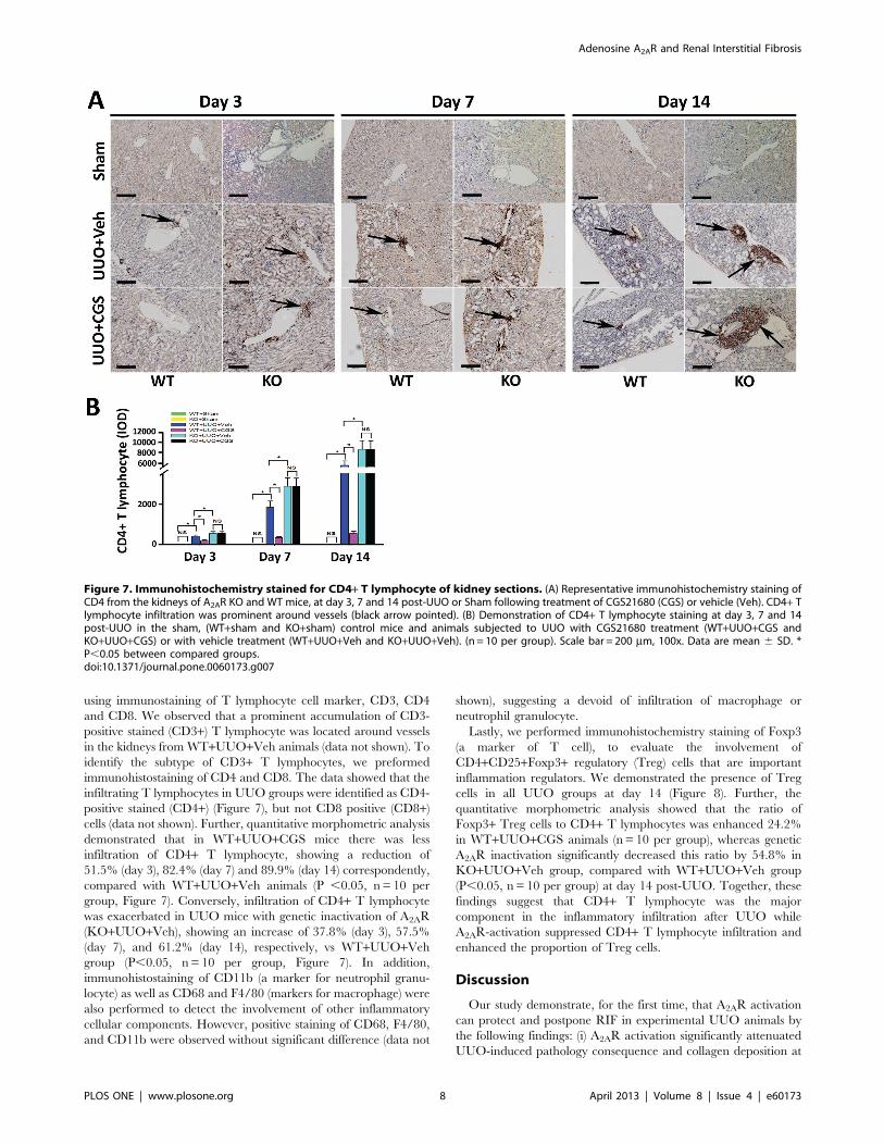
using immunostaining of T lymphocyte cell marker, CD3, CD4
and CD8. We observed that a prominent accumulation of CD3-
positive stained (CD3+) T lymphocyte was located around vessels
in the kidneys from WT+UUO+Veh animals (data not shown). To
identify the subtype of CD3+ T lymphocytes, we preformed
immunohistostaining of CD4 and CD8. The data showed that the
infiltrating T lymphocytes in UUO groups were identified as CD4-
positive stained (CD4+) (Figure 7), but not CD8 positive (CD8+)cells (data not shown). Further, quantitative morphometric analysis
demonstrated that in WT+UUO+CGS mice there was less
infiltration of CD4+ T lymphocyte, showing a reduction of
51.5% (day 3), 82.4% (day 7) and 89.9% (day 14) correspondently,
compared with WT+UUO+Veh animals (P ,0.05, n= 10 per
group, Figure 7). Conversely, infiltration of CD4+ T lymphocyte
was exacerbated in UUO mice with genetic inactivation of A2AR
(KO+UUO+Veh), showing an increase of 37.8% (day 3), 57.5%
(day 7), and 61.2% (day 14), respectively, vs WT+UUO+Vehgroup (P,0.05, n= 10 per group, Figure 7). In addition,
immunohistostaining of CD11b (a marker for neutrophil granu-
locyte) as well as CD68 and F4/80 (markers for macrophage) were
also performed to detect the involvement of other inflammatory
cellular components. However, positive staining of CD68, F4/80,
and CD11b were observed without significant difference (data not
shown), suggesting a devoid of infiltration of macrophage or
neutrophil granulocyte.
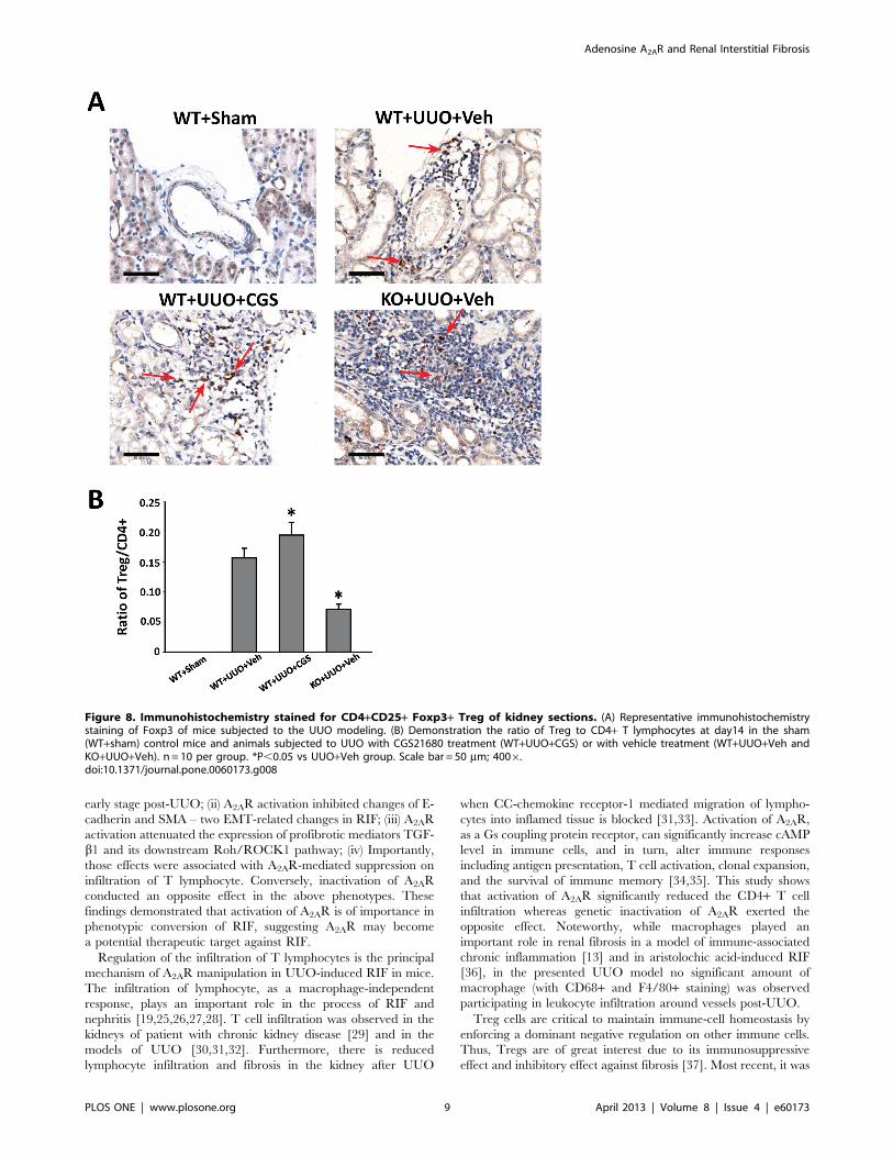
Lastly, we performed immunohistochemistry staining of Foxp3
(a marker of T cell), to evaluate the involvement of
CD4+CD25+Foxp3+ regulatory (Treg) cells that are important
inflammation regulators. We demonstrated the presence of Treg
cells in all UUO groups at day 14 (Figure 8). Further, the
quantitative morphometric analysis showed that the ratio of
Foxp3+ Treg cells to CD4+ T lymphocytes was enhanced 24.2%
in WT+UUO+CGS animals (n = 10 per group), whereas genetic
A2AR inactivation significantly decreased this ratio by 54.8% in
KO+UUO+Veh group, compared with WT+UUO+Veh group
(P,0.05, n= 10 per group) at day 14 post-UUO. Together, these
findings suggest that CD4+ T lymphocyte was the major
component in the inflammatory infiltration after UUO while
A2AR-activation suppressed CD4+ T lymphocyte infiltration and
enhanced the proportion of Treg cells.
Discussion
Our study demonstrate, for the first time, that A2AR activation
can protect and postpone RIF in experimental UUO animals by
the following findings: (i) A2AR activation significantly attenuated
UUO-induced pathology consequence and collagen deposition at
Figure 7. Immunohistochemistry stained for CD4+ T lymphocyte of kidney sections. (A) Representative immunohistochemistry staining ofCD4 from the kidneys of A2AR KO and WT mice, at day 3, 7 and 14 post-UUO or Sham following treatment of CGS21680 (CGS) or vehicle (Veh). CD4+ Tlymphocyte infiltration was prominent around vessels (black arrow pointed). (B) Demonstration of CD4+ T lymphocyte staining at day 3, 7 and 14post-UUO in the sham, (WT+sham and KO+sham) control mice and animals subjected to UUO with CGS21680 treatment (WT+UUO+CGS andKO+UUO+CGS) or with vehicle treatment (WT+UUO+Veh and KO+UUO+Veh). (n = 10 per group). Scale bar = 200 mm, 100x. Data are mean 6 SD. *P,0.05 between compared groups.doi:10.1371/journal.pone.0060173.g007
Adenosine A2AR and Renal Interstitial Fibrosis
PLOS ONE | www.plosone.org 8 April 2013 | Volume 8 | Issue 4 | e60173
early stage post-UUO; (ii) A2AR activation inhibited changes of E-
cadherin and SMA – two EMT-related changes in RIF; (iii) A2AR
activation attenuated the expression of profibrotic mediators TGF-
b1 and its downstream Roh/ROCK1 pathway; (iv) Importantly,
those effects were associated with A2AR-mediated suppression on
infiltration of T lymphocyte. Conversely, inactivation of A2AR
conducted an opposite effect in the above phenotypes. These
findings demonstrated that activation of A2AR is of importance in
phenotypic conversion of RIF, suggesting A2AR may become
a potential therapeutic target against RIF.
Regulation of the infiltration of T lymphocytes is the principal
mechanism of A2AR manipulation in UUO-induced RIF in mice.
The infiltration of lymphocyte, as a macrophage-independent
response, plays an important role in the process of RIF and
nephritis [19,25,26,27,28]. T cell infiltration was observed in the
kidneys of patient with chronic kidney disease [29] and in the
models of UUO [30,31,32]. Furthermore, there is reduced
lymphocyte infiltration and fibrosis in the kidney after UUO
when CC-chemokine receptor-1 mediated migration of lympho-
cytes into inflamed tissue is blocked [31,33]. Activation of A2AR,
as a Gs coupling protein receptor, can significantly increase cAMP
level in immune cells, and in turn, alter immune responses
including antigen presentation, T cell activation, clonal expansion,
and the survival of immune memory [34,35]. This study shows
that activation of A2AR significantly reduced the CD4+ T cell
infiltration whereas genetic inactivation of A2AR exerted the
opposite effect. Noteworthy, while macrophages played an
important role in renal fibrosis in a model of immune-associated
chronic inflammation [13] and in aristolochic acid-induced RIF
[36], in the presented UUO model no significant amount of
macrophage (with CD68+ and F4/80+ staining) was observed
participating in leukocyte infiltration around vessels post-UUO.
Treg cells are critical to maintain immune-cell homeostasis by
enforcing a dominant negative regulation on other immune cells.
Thus, Tregs are of great interest due to its immunosuppressive
effect and inhibitory effect against fibrosis [37]. Most recent, it was
Figure 8. Immunohistochemistry stained for CD4+CD25+ Foxp3+ Treg of kidney sections. (A) Representative immunohistochemistrystaining of Foxp3 of mice subjected to the UUO modeling. (B) Demonstration the ratio of Treg to CD4+ T lymphocytes at day14 in the sham(WT+sham) control mice and animals subjected to UUO with CGS21680 treatment (WT+UUO+CGS) or with vehicle treatment (WT+UUO+Veh andKO+UUO+Veh). n = 10 per group. *P,0.05 vs UUO+Veh group. Scale bar = 50 mm; 4006.doi:10.1371/journal.pone.0060173.g008
Adenosine A2AR and Renal Interstitial Fibrosis
PLOS ONE | www.plosone.org 9 April 2013 | Volume 8 | Issue 4 | e60173

reported that Tregs’ negative regulation on immune cells was
mediated by A2AR activation whereas deletion of A2AR abolished
Tregs’ regulatory effect [38]. These reports support our findings
that A2AR activation by CGS21680 significantly increased the
ratio of Tregs to CD4+ T lymphocytes, whereas this ratio was
significantly decreased in A2AR KO mutants post-UUO. Thus,
regulation of Tregs recruitment and (CD4+) T lymphocyte
infiltration acts as underlying mechanism of A2AR-mediated
effects against RIF.
Another important finding in this study is that A2AR could affect
EMT-related changes in E-cadherin and SMA. While more direct
evidence and evaluations are needed in human studies, EMT is
recently proposed as a crucial mechanism in RIF [5]. During the
EMT process, renal tubular epithelial cell lost the E-cadherin
phenotype and acquire the myofibroblast phenotype a-SMA. Our
findings demonstrated that activation of A2AR restored expression
level of a-SMA and E-cadherin to a basal level in sham animals
(Figure 4). Though indirectly based on Western blot reflecting total
renal tissue rather than TECs-specific on-site changes, this finding
indicates that A2AR activation maintained intrinsic phenotypes of
epithelia and myofibroblast, i.e., inhibited the process of EMT. To
find the mechanism by which A2AR affects EMT, we demon-
strated that activation of A2AR significantly reduced the expression
of TGF-b1, a key profibrotic mediator in EMT, along with
ROCK1, the regulatory protein in the TGF-b1 downstream
pathway Rho/ROCK signaling. In UUO, the enhanced TGF-b1may (i) act as a mitogenic factor to affect collagen synthesis and (ii)
facilitate the EMT process [6,39]. Importantly, activation of A2AR
restored both aforementioned consequences of TGF-b1 post-
UUO. Furthermore, this study showed that TGF-b1-mediated
EMT is regulated by the Rho/ROCK-dependent signaling
pathway [20], and the ROCK pathways play an important role
in RIF and phenotypic modulation of epithelial cells [40,41].
While ROCK has two types (ROCK1 and ROCK2), the ROCK1
is predominantly expressed in the kidney and regulates cell
adhesion, chemotaxis and contraction, as well as epithelial
differentiation [42]. Meanwhile, E-cadherin, not only as a marker,
is also the most important component for maintaining the integrity
and polarity of epithelial cells [43]. The loss of E-cadherin
expression in the renal tubular epithelial cells will lead to a loss of
cell-cell adhesion facilitating the renal tubular epithelial cells enter
the renal interstitium. Studies showed that E-cadherin is regulated
by ROCK1 [42,44], moreover, a-SMA as the important structure
protein of the myofibroblast, is also influenced by the Rho/ROCK
signaling pathway [45]. Importantly, the Rho/ROCK-1 pathway
is closely linked to adenosine activity [15]. Thus activation of
A2AR may, via ROCK1, regulate cell adhesion of tubular
epithelial cells and the EMT process. Further evidence is needed
to address this potential mechanism.
The increase of A2AR after UUO may account for a compen-
satory protective mechanism. In line with our finding, Lee et al
also demonstrated this phenomena [46]. However, it is still unclear
whether the increased A2AR mRNA attribute to inherent renal
cells or immigrated cells, e.g., bone marrow-derived cells [47], via
inflammatory processes post-UUO. Our study suggested that the
effect on T lymphocyte infiltration contribute to A2AR-mediated
protection against RIF. Noteworthy, some of the A2AR activation
resulting effects on post-UUO animals were blunt in the late stage
after UUO with unknown reason. This may be due to the severity
of phenotypes in late post-UUO stage and the progressive
aggravation in pathology unless the pathogenic factors of tubular
obstruction might have been removed. The severe pathology
changes in late post-UUO stage might be irreversible; however, we
also noted the suppressive effect of A2AR activation on expression
of TGF-b1 was devoid at day 14 after UUO. This may be due to
a redundant mechanism between ROCK1 and TGF-b1, for
instance a down-regulation of ROCK1 by A2AR agonist may
result in an increased expression of TGF-b1 [48]. To clarify the
noticed phenomenon, using a model with mild severity or slow
progress may help on this point in the future.
In summary, this study for the first time demonstrates the
beneficial effect of A2AR activation in preventing the progression
of RIF in the UUO animal model. This provides a novel
therapeutic strategy against renal interstitial fibrosis by targeting
the adenosine A2A receptor.
Acknowledgments
The authors thank Dr. Jiang-Fan Chen for kindly providing A2AR
knockout mutant mice.
Author Contributions
Conceived and designed the experiments: YGZ LYS HYS. Performed the
experiments: HX HYS WL. Analyzed the data: HX HYS YGZ.
Contributed reagents/materials/analysis tools: RPX PL GM NY XC.
Wrote the paper: HX HYS YGZ.
References
1. Isaka Y, Takahara S, Imai E (2008) Chronic deteriorating renal function and
renal fibrosis. Contrib Nephrol 159: 109–121.
2. Pannarale G, Carbone R, Del Mastro G, Gallo C, Gattullo V, et al. (2010) The
aging kidney: structural changes. J Nephrol 23 Suppl 15: S37–40.
3. Strutz F (2001) Potential methods to prevent interstitial fibrosis in renal disease.
Expert Opin Investig Drugs 10: 1989–2001.
4. Liu Y (2011) Cellular and molecular mechanisms of renal fibrosis. Nat Rev
Nephrol 7: 684–696.
5. Mucsi I, Rosivall L (2007) Epithelial-mesenchymal transition in renal tubular
cells in the pathogenesis of progressive tubulo-interstitial fibrosis. Acta Physiol
Hung 94: 117–131.
6. Iwano M (2010) EMT and TGF-beta in renal fibrosis. Front Biosci (Schol Ed) 2:
229–238.
7. Zeisberg M, Neilson EG (2010) Mechanisms of tubulointerstitial fibrosis. J Am
Soc Nephrol 21: 1819–1834.
8. Tan X, Li Y, Liu Y (2007) Therapeutic role and potential mechanisms of active
Vitamin D in renal interstitial fibrosis. J Steroid Biochem Mol Biol 103: 491–
496.
9. Hao S, He W, Li Y, Ding H, Hou Y, et al. (2011) Targeted inhibition of beta-
catenin/CBP signaling ameliorates renal interstitial fibrosis. J Am Soc Nephrol
22: 1642–1653.
10. Mazzon E, Esposito E, Impellizzeri D, R Di Paola R, Melani A, et al. (2011)
CGS 21680, an agonist of the adenosine (A2A) receptor, reduces progression of
murine type II collagen-induced arthritis. J Rheumatol 38: 2119–2129.
11. Impellizzeri D, Di Paola R, Esposito E, Mazzon E, Paterniti I, et al. (2011) CGS
21680, an agonist of the adenosine (A2A) receptor, decreases acute lung
inflammation. Eur J Pharmacol 668: 305–316.
12. Ferenbach DA, Hughes J (2011) Adenosine A(2A) agonists as therapy for
glomerulonephritis. Kidney Int 80: 329–331.
13. Garcia GE, Truong LD, Chen JF, Johnson RJ, Feng L (2011) Adenosine A(2A)
receptor activation prevents progressive kidney fibrosis in a model of immune-
associated chronic inflammation. Kidney Int 80: 378–388.
14. Zhang L, Yang N, Wang S, Huang B, Li F, et al. (2011) Adenosine 2A receptor
is protective against renal injury in MRL/lpr mice. Lupus 20: 667–677.
15. Sohail MA, Hashmi AZ, Hakim W, Watanabe A, Zipprich A, et al. (2009)
Adenosine induces loss of actin stress fibers and inhibits contraction in hepatic
stellate cells via Rho inhibition. Hepatology 49: 185–194.
16. Klahr S, Morrissey J (2002) Obstructive nephropathy and renal fibrosis.
Am J Physiol Renal Physiol 283: F861–875.
17. Vaughan ED Jr, Marion D, Poppas DP, Felsen D (2004) Pathophysiology of
unilateral ureteral obstruction: studies from Charlottesville to New York. J Urol
172: 2563–2569.
18. Harris RC, Neilson EG (2006) Toward a unified theory of renal progression.
Annu Rev Med 57: 365–380.
19. Tapmeier TT, Fearn A, Brown K, Chowdhury P, Sacks SH, et al. (2010) Pivotal
role of CD4+ T cells in renal fibrosis following ureteric obstruction. Kidney Int
78: 351–362.
Adenosine A2AR and Renal Interstitial Fibrosis
PLOS ONE | www.plosone.org 10 April 2013 | Volume 8 | Issue 4 | e60173

20. Tian YC, Fraser D, Attisano L, Phillips AO (2003) TGF-beta1-mediated
alterations of renal proximal tubular epithelial cell phenotype. Am J PhysiolRenal Physiol 285: F130–142.
21. Chen JF, Huang Z, Ma J, Zhu J, Moratalla R, et al. (1999) A(2A) adenosine
receptor deficiency attenuates brain injury induced by transient focal ischemia inmice. J Neurosci 19: 9192–9200.
22. Moriyama T, Kawada N, Ando A, Yamauchi A, Horio M, et al. (1998) Up-regulation of HSP47 in the mouse kidneys with unilateral ureteral obstruction.
Kidney Int 54: 110–119.
23. Mao H, Li Z, Zhou Y, Zhuang S, An X, et al. (2008) HSP72 attenuates renaltubular cell apoptosis and interstitial fibrosis in obstructive nephropathy.
Am J Physiol Renal Physiol 295: F202–214.24. Wu G, Tu Y, Jia R (2010) The influence of fasudil on the epithelial-
mesenchymal transdifferentiation of renal tubular epithelial cells from diabeticrats. Biomed Pharmacother 64: 124–129.
25. Zheng G, Wang Y, Mahajan D, Qin X, Alexander SI, et al. (2005) The role of
tubulointerstitial inflammation. Kidney Int Suppl: S96–100.26. Tipping PG, Holdsworth SR (2006) T cells in crescentic glomerulonephritis.
J Am Soc Nephrol 17: 1253–1263.27. Strutz F, Neilson EG (1994) The role of lymphocytes in the progression of
interstitial disease. Kidney Int Suppl 45: S106–110.
28. Yang N, Isbel NM, Nikolic-Paterson DJ, Li Y, Ye R, et al. (1998) Localmacrophage proliferation in human glomerulonephritis. Kidney Int 54: 143–
151.29. Robertson H, Ali S, McDonnell BJ, Burt AD, Kirby JA (2004) Chronic renal
allograft dysfunction: the role of T cell-mediated tubular epithelial tomesenchymal cell transition. J Am Soc Nephrol 15: 390–397.
30. Vielhauer V, Anders HJ, Mack M, Cihak J, Strutz F, et al. (2001) Obstructive
nephropathy in the mouse: progressive fibrosis correlates with tubulointerstitialchemokine expression and accumulation of CC chemokine receptor 2- and 5-
positive leukocytes. J Am Soc Nephrol 12: 1173–1187.31. Eis V, Luckow B, Vielhauer V, Siveke JT, Linde Y, et al. (2004) Chemokine
receptor CCR1 but not CCR5 mediates leukocyte recruitment and subsequent
renal fibrosis after unilateral ureteral obstruction. J Am Soc Nephrol 15: 337–347.
32. Kitagawa K, Wada T, Furuichi K, Hashimoto H, Ishiwata Y, et al. (2004)Blockade of CCR2 ameliorates progressive fibrosis in kidney. Am J Pathol 165:
237–246.33. Anders HJ, Vielhauer V, Frink M, Linde Y, Cohen CD, et al. (2002) A
chemokine receptor CCR-1 antagonist reduces renal fibrosis after unilateral
ureter ligation. J Clin Invest 109: 251–259.34. Lukashev D, Ohta A, Apasov S, Chen JF, Sitkovsky M (2004) Cutting edge:
Physiologic attenuation of proinflammatory transcription by the Gs protein-coupled A2A adenosine receptor in vivo. J Immunol 173: 21–24.
35. Sitkovsky M, Lukashev D, Deaglio S, Dwyer K, Robson SC, et al. (2008)
Adenosine A2A receptor antagonists: blockade of adenosinergic effects and T
regulatory cells. Br J Pharmacol 153 Suppl 1: S457–464.
36. Pozdzik AA, Salmon IJ, Husson CP, Decaestecker C, Rogier E, et al. (2008)
Patterns of interstitial inflammation during the evolution of renal injury in
experimental aristolochic acid nephropathy. Nephrol Dial Transplant 23: 2480–
2491.
37. Tang TT, Yuan J, Zhu ZF, Zhang WC, Xiao H, et al. (2012) Regulatory T cells
ameliorate cardiac remodeling after myocardial infarction. Basic Res Cardiol
107: 232.
38. Ohta A, Kini R, Subramanian M, Madasu M, Sitkovsky M (2012) The
development and immunosuppressive functions of CD4(+) CD25(+) FoxP3(+)regulatory T cells are under influence of the adenosine-A2A adenosine receptor
pathway. Front Immunol 3: 190.
39. Zhou B, Buckley ST, Patel V, Liu Y, Luo J, et al. (2012) Troglitazone attenuates
TGF-beta1-induced EMT in alveolar epithelial cells via a PPARgamma-
independent mechanism. PLoS One 7: e38827.
40. Masszi A, Di Ciano C, Sirokmany G, Arthur WT, Rotstein OD, et al. (2003)
Central role for Rho in TGF-beta1-induced alpha-smooth muscle actin
expression during epithelial-mesenchymal transition. Am J Physiol Renal
Physiol 284: F911–924.
41. Prakash J, de Borst MH, Lacombe M, Opdam F, Klok PA, et al. (2008)
Inhibition of renal rho kinase attenuates ischemia/reperfusion-induced injury.
J Am Soc Nephrol 19: 2086–2097.
42. Kalaji R, Wheeler AP, Erasmus JC, Lee SY, Endres RG, et al. (2012) ROCK1
and ROCK2 regulate epithelial polarisation and geometric cell shape. Biol Cell.
43. Baum B, Georgiou M (2011) Dynamics of adherens junctions in epithelial
establishment, maintenance, and remodeling. J Cell Biol 192: 907–917.
44. Otsu K, Kishigami R, Fujiwara N, Ishizeki K, Harada H (2011) Functional role
of Rho-kinase in ameloblast differentiation. J Cell Physiol 226: 2527–2534.
45. Bhowmick NA, Ghiassi M, Bakin A, Aakre M, Lundquist CA, et al. (2001)
Transforming growth factor-beta1 mediates epithelial to mesenchymal transdif-
ferentiation through a RhoA-dependent mechanism. Mol Biol Cell 12: 27–36.
46. Lee J, Hwang L, Ha H (2012) Adenosine receptors are up-regulated in unilateral
ureteral obstructed rat kidneys. Transplant Proc 44: 1166–1168.
47. Day YJ, Huang L, McDuffie MJ, Rosin DL, Ye H, et al. (2003) Renal protection
from ischemia mediated by A2A adenosine receptors on bone marrow-derived
cells. J Clin Invest 112: 883–891.
48. Fu P, Liu F, Su S, Wang W, Huang XR, et al. (2006) Signaling mechanism of
renal fibrosis in unilateral ureteral obstructive kidney disease in ROCK1
knockout mice. J Am Soc Nephrol 17: 3105–3114.
Adenosine A2AR and Renal Interstitial Fibrosis
PLOS ONE | www.plosone.org 11 April 2013 | Volume 8 | Issue 4 | e60173
Copyright © 2022 FDOKUMEN