Familial membranous subaortic stenosis: Review of familial inheritance patterns and a case report
Upload
independentCategory
view
1download
0
Article
Coexistence of Different Circulating Anti-PodocyteAntibodies in Membranous Nephropathy
Corrado Murtas,* Maurizio Bruschi,† Giovanni Candiano,† Gabriella Moroni,‡ Riccardo Magistroni,§ Andrea Magnano,|
Francesca Bruno,¶ Antonella Radice,** Luciana Furci,§ Lucia Argentiero,| Maria Luisa Carnevali,| Piergiorgio Messa,‡
Francesco Scolari,†† Renato Alberto Sinico,** Loreto Gesualdo,¶ Fernando C. Fervenza,‡‡ Landino Allegri,|
Pietro Ravani,§§ and Gian Marco Ghiggeri*
SummaryBackground and objectives The discovery of different podocyte autoantibodies in membranous nephropathy(MN) raises questions about their pathogenetic and clinical meaning. This study sought to define antibodyisotypes and correlations; to compare levels in MN, other glomerulonephritides, and controls; and to determinetheir association with clinical outcomes.
Design, setting, participants, & measurements Serum IgG1, IgG3, and IgG4 against aldose reductase (AR), SOD2,and a-enolase (aENO)weremeasured at diagnosis in 186 consecutiveMN patients, in 96 proteinuric controls (36with FSGS, and 60 with IgA nephropathy), and in 92 healthy people recruited in four Italian nephrology units.Anti-phospholipase A2 receptor (PLA2r) and anti-neutral endopeptidase (NEP) IgG4 were titrated in the samespecimens. Association with 1-year follow-up clinical parameters was studied in 120 patients.
Results IgG4 was the most common isotype for all antibodies; IgG1 and IgG3 were nearly negligible. IgG4 levelswere positive in a significant proportion ofMNpatients (AR, 34%; SOD2, 28%;aENO, 43%). Antibody titerswerehigher in MN than in healthy and pathologic controls (P,0.005). Anti-NEP IgG4 did not differ from normalcontrols (P=0.12). Anti-PLA2r IgG4 was detected in 60% of patients and correlated with anti-AR, anti-SOD2, andanti-aENO IgG4 (P,0.001). In MN patients negative for the whole antibody panel (20%), 1-year proteinuriawas lower compared with patients with at least one antibody positivity (P,0.05).
Conclusions Our data suggest that IgG4 is the prevalent isotype for antibodies against cytoplasmic antigens ofpodocytes (AR, SOD2, aENO). Their levels were higher than in other proteinuric glomerulonephritides andin normal controls and were correlated with anti-PLA2r. Only baseline negativity for all known antibodiespredicted lower 1-year proteinuria.
Clin J Am Soc Nephrol 7: 1394–1400, 2012. doi: 10.2215/CJN.02170312
IntroductionMembranous nephropathy (MN) is a leading cause ofnephrotic syndrome in adults (1). Glomerular damageis produced by the deposition of subepithelial im-mune deposits that consist mainly of IgG4 and C5b-9(2,3). The definition of causative autoantibodies andtheir renal targets is essential to understand the mech-anisms of disease development and progression.
Recent findings indicate that more than one podocyteprotein may act as an autoantigen in human MN (4).Identified antigens are membrane proteins, such as phos-pholipase A2 receptor (PLA2r) (5) and neutral endopep-tidase (NEP) (6), and components of the cytoplasm, suchas aldose reductase (AR), SOD2, and a-enolase (aENO)(7,8). Circulating levels of the corresponding autoanti-body might be used as biomarker of disease activity.
With the exception of a report of maternal anti-NEPantibodies in antenatal MN (6,9), data on autoantibod-ies are limited to anti-PLA2r antibodies. Anti-PLA2rIgG4 have been described as the largely predominant
circulating and glomerular isotype in MN patients (5).They seem specific (89%) for idiopathic MN (10–12)and can be utilized as support to exclude secondaryMN (10). However, several MN patients are anti-PLA2r negative (30%–50% according to case series)whereas in others, anti-PLA2r positivity persists afterresponse to therapy (11,13). Data on anti-PLA2r donot exclude the presence of other circulating autoan-tibodies such as anti-cytoplasmic antigens of podocytes,whose serum levels might be detected and might beuseful in clinical practice.To test this hypothesis, we measured the levels of
antibodies against both membrane and cytoplasmicautoantigens in patients with and without MN. Ourobjectives were as follows: (1) to define the prevalentisotype for each anti-cytoplasmic antibody and the spec-trum of MN patients in respect to positivity or negativ-ity for the whole panel of currently known antibodies;(2) to compare their levels in participants with MN ver-sus patients suffering from other proteinuric glomerular
*Division ofNephrology, Dialysis,and Transplantation,and †Laboratory onPathophysiology ofUremia, IstitutoGiannina Gaslini,Genoa, Italy; ‡Division ofNephrology andDialysis,IRCCS FondazioneOspedale Maggiore,Mangiagalli, ReginaElena, Milan, Italy;§Department ofNephrology, Universityof Modena, Modena,Italy; |Department ofClinical Medicine,Nephrology, and HealthSciences, University ofParma, Parma, Italy;¶Division of Nephrology,University of Bari, Bari,Italy; **Division ofNephrology and Sectionof Clinical Immunology,San Carlo Hospital,Milan, Italy; ††Division ofNephrology, Universityof Brescia andMontichiari Hospital,Brescia, Italy; ‡‡Divisionof Nephrology andHypertension, MayoClinic, Rochester,Minnesota; and§§Division ofNephrology, Universityof Calgary, Calgary,Alberta, Canada
Correspondence: Dr.Gian Marco Ghiggeri,Division of Nephrology,Dialysis, andTransplantation,Laboratory onPathophysiology ofUremia, Istituto GianninaGaslini, Largo G. Gaslini5, 16148 Genova, Italy,and Dr. Pietro Ravani,Division of Nephrology,University of Calgary,29th St NW (r C210N),Calgary, AB, Canada.Email: [email protected] [email protected]
www.cjasn.org Vol 7 September, 20121394 Copyright © 2012 by the American Society of Nephrology

diseases or normal controls; and (3) to ascertain whethera relationship exists between the type and level of theseantibodies and clinical outcome in individuals with MN.
Materials and MethodsAssembled CohortWe conducted a retrospective study at four Italian
nephrology centers in Parma, Modena, Milano, and Bari.We recruited patients with newly diagnosed MN or otherproteinuric nephropathies and normal controls over 2 years(2008–2010). Ethical approval to test antibody levels wasobtained from the Giannina Gaslini Institute Ethics Com-mittee. The study was registered in the EudraCT registry(EudraCT 2011–003942–41).MN Patients. We recruited 186 consecutive patients
with idiopathic MN, who gave informed consent to havethe serum antibody levels titrated at the time of renal biopsy,before any therapy was started (Table 1). At the time enroll-ment was closed, 120 patients had completed a follow-up of12 months or longer, during which they had received differ-ent therapy schemes, mainly the Ponticelli schedule (Supple-mental Table 1). Criteria for enrollment were as follows:histologically proven MN; negative tests for serum autoanti-bodies (antinuclear antibody, ANCA), cryoglobulins, andviral markers (hepatitis B surface antigen, HIV); absence ofany clinical suspicion of secondary MN; or absence of anyprevious immunosuppressive treatment.Proteinuric Controls. Ninety-two patients with different
nephropathies were recruited at the same institutions: 32patients with FSGS and 60 with IgA nephropathy (IgAN).Diagnosis was always based on histologic criteria. To pro-vide similar clinical conditions to MN patients, FSGS spec-imens were all collected during a relapse of nephroticproteinuria. IgAN patients had to present with proteinuria.0.3 g/d and to be free of any immunosuppressive ther-apy at the time of serum collection (Table 1).Normal Controls. Serum was obtained from 96 normal
controls recruited at the same institutions. They consistedof normal blood donors who had at least one normalurinalysis and serum tests in the prior 6 months (Table 1).
Assays for AutoantibodiesAnti-AR, Anti-SOD2, and Anti-aENO. Circulating IgG1,
IgG3, and IgG4 levels against AR, SOD2, and aENO in sera
were determined with dot blot utilizing recombinant pro-teins fixed to nitrocellulose as antigens, as previously de-scribed (7,8). Details of the method and examples ofvariable positivity are given in the Supplemental Methodsand Supplemental Figure 1. Antibody positivity was definedas a serum level exceeding the 95th percentile of levelstitrated in normal controls.Anti-PLA2r. Circulating anti-PLA2r IgG4 antibodies
were titrated by Western blot against podocyte proteinextracts (kindly offered by Dr. Saleem, University of Bristol,Bristol, UK) previously separated in gradient monodimen-sional electrophoresis (14) and then incubated with serum.The technique is described in detail in the SupplementalMethods. Anti-PLA2r autoantibodies were also evaluatedin a random subsample of MN patients (n=73) by indirectimmunofluorescence with a commercially available test,according to the manufacturer’s instructions (Euroimmun,Lubeck, Germany) (15). Details of the method are given inthe Supplemental Methods and Supplemental Table 2.Anti-NEP. Circulating IgG4 antibodies against NEP
were assessed by dot blot utilizing recombinant NEP asfixed antigen. The assay is based on the same techniquedescribed for anti-AR, anti-SOD2, and anti-aENO, and isreported in the Supplemental Methods.
Statistical AnalysesData were described using frequencies, means, and SDs,
as appropriate. Natural log transformation was performedon non-normally distributed variables before using linearmodels. Due to the presence of several “zeros,” baselinecorrelations were performed using the Spearman rank cor-relation method for non-normally distributed variables;natural log transformation was performed after adding0.01 to each variable value. The Mann–Whitney U testwas used to compare non-normally distributed variables.Separate univariable logistic models were used with pres-ence of MN versus other nephropathies or controls asbinary outcomes to estimate the area under the receiveroperating characteristic (ROC) curves. Logistic regres-sion models of 1-year proteinuria were built on log2-transformed antibody values adjusting for baseline levelsof log2-proteinuria. Two models were built with the dichot-omous outcome defined as complete or partial remission(proteinuria ,0.3 or ,3.5 g/d). Linear regression models
Table 1. Clinical characteristics of patients and controls enrolled in this study
MN (n=186) FSGS (n=32) IgAN (n=60) Normal (n=96)
Male sex 121 (65) 19 (59) 38 (63) 56 (58)Age (yr) 59616 1863 4064 49610RaceCaucasian 184 (99) 32 (100) 56 (93) 96 (100)other 2 (1) 4 (7)
Diabetes 24 (13) 0 (0) 4 (7) 0 (0)Serum creatinine (mg/dl) 1.1 (0.3–6) 0.6 (0.3–1) 0.9 (0.5–1.3) 0.9 (0.6–1.2)Proteinuria (g/d) 5.8 (0.3–28) 6.0 (3.6–12.4) 1.55 (0.9–3.4) 0
Data are presented as n (%) or mean6 SD, or as median (range) for those with non-normal distribution. Clinical data were collected atthe time of serumsample collection. Proteinuria in normal controlswas tested byurine dipstick.MN,membranous nephropathy; IgAN,IgA nephropathy.
Clin J Am Soc Nephrol 7: 1394–1400, September, 2012 Anti-Podocyte Antibodies in Membranous Nephropathy, Mutas et al. 1395
of log2-proteinuria adjusted for baseline proteinuria werealso built on each antibody level. Similarly, linear modelswere used to study the associations between levels of anti-bodies and serum albumin. Models with only one antibodyat a time were built for reasons of colinearity. Model as-sumptions and goodness of fit were verified looking at for-mal tests and graphical tests based on residuals. Allstatistical analyses were performed with STATA/MP 12.1software (StataCorp, College Station, TX).
ResultsPatients with MNwere prevalently male (65%) and had a
mean age of 59616 years. All were proteinuric at the timeof enrollment and had variable creatinine levels (Table 1).Patients with FSGS (59% male) had proteinuria .3.5 g/d,had normal renal function in all cases, and were younger(age 1863 years). IgAN patients (63% male; 4064 years)had proteinuria 0.9–3.4 g/d and normal renal function.Normal controls were prevalently male (58%) with amean age of 49610 years (Table 1).
Antibody Levels in MN and in OtherGlomerulonephritidesAnti-AR, Anti-SOD2, and Anti-aENO Antibody Isotypes/
Serum Levels. Circulating anti-AR, anti-SOD2, and anti-aENO isotypes (IgG1, IgG3, and IgG4) and levels were de-termined by dot blot in the serum of 186 MN patients atdiagnosis (Figure 1). Isotype characterization of each spec-ificity indicated that IgG4 is the predominant IgG subclass.Anti-AR and anti-SOD2 IgG1 and IgG3 were, in fact, onlysporadically positive (,3%). Anti-aENO IgG1–3 were in-stead increased in a small but significant proportion of MNpatients (13% IgG1 and 7% IgG3) (Supplemental Figure 2).In patients with other nephropathies, serum levels of each
IgG4 antibody were found to be lower than in normal con-trols; one IgAN patient presented an isolated anti-aENOpositivity (Figure 1). The percentage of MN patients withIgG4 positive levels was highly significant (AR, 34%; SOD,26%; aENO, 43%; P,0.001 for all) (Supplemental Figure 3).Comparison of levels confirmed significantly higher circu-lating levels of anti-AR, anti-SOD2, and anti-aENO IgG4 inMN patients compared with either normal participants orother nephropathies (Table 2). The area under the ROCcurves for each antibody was significantly greater in MNpatients compared with patients with other nephropathiesor normal participants, although to a lesser extent (Figure 2).Anti-PLA2r and Anti-NEP Antibodies. Anti-PLA2r
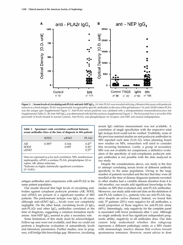
IgG4 was detected by Western blot in 111 MN patients(Figure 3), giving a final estimate of 60% of patients beingpositive (Supplemental Figure 3), confirming previousstudies (5,10). In a random portion of MN sera (73 pa-tients), anti-PLA2r total IgG as determined by indirect im-munofluorescence (12,15) confirmed results of Westernblot titration (Spearman analysis, r=0.91; P,0.001) (Sup-plemental Table 2). Anti-NEP IgG4 serum levels were de-termined with dot blot analysis. The results shown inFigure 3 indicate a minor percentage (17%) of MN patientswith anti-NEP positivity. NEP serum levels were nothigher in MN patients than in normal serum (P=0.12)(Table 2).Multiple Positivity. Many MN patients presented con-
comitant high serum levels of more than one antibody.Anti-PLA2r serum levels correlated with all the otherantibodies and anti-AR correlated with anti-SOD2 (Table 3)(P,0.001 in all cases). Simultaneous multiple positivity ornegativity for the antibody panel was tested in the wholecohort of patients: 19 sera (10%) were completely positiveand 37 (20%) were negative. Most patients (70%) presentedintermediate positivity for one, two, or three antibodies
Figure 1. | Serum antibodies against cytoplasmic antigens of podocytes are increased in a significant portion of MN patients. CirculatingIgG4 (A) anti-AR, (B) anti-SOD2, and (C) anti-aENO in MN and control populations are shown. In all cases, we utilized a technique based ondot blot analysis with recombinant protein linked to nitrocellulose as an antigen (Supplemental Figure 1). Results are given as chemi-luminescence OD arbitrary units that corresponds to one unit of signal intensity of chemiluminescence detected by VersaDoc and computedwith QuantyOne software (Bio-Rad). The horizontal line is set at the 95th percentile of levels titrated in normal controls. AR, aldose reductase;aENO, a-enolase; MN, membranous nephropathy.
1396 Clinical Journal of the American Society of Nephrology
(Table 4). Of the 75 anti-PLA2r–negative patients, 38 (51%)were positive for at least one other antibody (Supplemen-tal Figure 4).
Clinical CorrelationsThere was no relationship between antibody levels and
proteinuria, renal function, and histologic stage at baseline.No histologic or clinical characteristics distinguished anti-PLA2r–positive and anti-PLA2r–negative patients. Antibodylevels failed to predict the probability to reach complete(,0.3 g/d) or partial (, 3.5 g/d) remission after 1 year.When proteinuria was modeled as a continuous variable,only anti-AR IgG4 levels predicted 1-year proteinuria. Sim-ilarly, only anti-aENO IgG4 significantly predicted serumalbumin levels at 12 months (Supplemental Table 3).Finally, 27 MN patients who were negative for all
antibodies completed the 12-month follow-up (Table 4).Although treatment did not differ, they presented a mildtendency to have a better outcome. In fact, compared withMN patients who were positive for at least one antibody,completely negative patients had significantly lower 1-yearproteinuria (Mann–Whitney test, P=0.03), at 0.33 g/d (inter-quartile range, 0.1–1.8) versus 1.50 g/d (interquartile range,
0.2–4.2). A borderline significantly higher remission rate wasalso present in negative patients (48% versus 27%; P=0.07).
DiscussionThe recent discovery that podocyte proteins are targets of
circulating antibodies in human MN represented a break-through in the research on MN pathogenetic mechanisms(5,7,8). However, the implication of more than one antigenin the formation of subepithelial immune deposits raisesquestions about the clinical significance of each autoanti-body. Thus far, four podocyte antigens have been studiedin primary MN: PLA2r was the first discovered (anddeeply investigated) (5,11,13), and AR, SOD2, and aENOwere subsequently identified (7,8). Our study was plannedto bridge the gap of knowledge on antibodies against neo-expressed cytoplasm antigens (i.e., anti-AR, anti-SOD2,and anti-aENO). In fact, there is little information in theliterature about their serum levels and their correlationwith anti-PLA2r. Moreover, the evaluation of a potential as-sociation of serum antibody levels (including anti-PLA2r)with clinical outcomes requires studies in large cohorts ofpatients. Our strategy was therefore based on the concomi-tant determinations of serum levels of anti-cytoplasmic
Table 2. Levels of circulating antibodies in MN patients, controls, and other nephropathies
MN Controls FSGS IgAN
Anti-AR 37.90 (7.12–121.90) 29.67 (9.39–47.36)a 6.38 (3.43–9.15b 9.39 (4.41–14.46)b
Anti-SOD2 84.34 (35.51–223.60) 33.92 (4.11–62.23)b 6.89 (4.16–9.69)b 9.09 (3.93–12.99)b
Anti-aENO 110.40 (27.84–237.70) 47.80 (31.09–67.38)b 5.40 (3.29–9.43)b 9.31 (5.06–18.19)b
Anti-NEP 53.52 (3.52–211.20) 32.42 (14.95–54.73) 4.08 (2.08–6.92)b 7.05 (3.67–11.39)b
Anti-PLA2r 40.26 (13.12–96.76) Undetectable —— ——
Data are expressed as chemiluminescence OD arbitrary units and presented as median (interquartile range). Only data of 111 positivepatients were reported for anti-PLA2r. MN, membranous nephropathy; IgAN, IgA nephropathy; AR, aldose reductase; aENO,a-enolase; NEP, neutral endopeptidase; PLA2r, phospholipase A2 receptor.aA highly statistically significant difference with MN level (two tailed Mann–Whitney U test). P=0.004.bA highly statistically significant difference with MN level (two tailed Mann–Whitney U test). P,0.001.
Figure 2. | ROC curves. The area under the ROC curves for each antibody was significantly greater in MN patients compared with (A) patientswith other nephropathies or (B) normal participants, although to a lesser extent. ROC, receiver operating characteristic; MN, membranousnephropathy.
Clin J Am Soc Nephrol 7: 1394–1400, September, 2012 Anti-Podocyte Antibodies in Membranous Nephropathy, Mutas et al. 1397
antigen antibodies and comparisons with anti-PLA2r in thesame patient population.Our results showed that high levels of circulating anti-
bodies against cytoplasm podocyte proteins (AR, SOD2,and aENO) are present in a significant number of MNpatients. The predominant isotype was IgG4 in all cases,although anti-aENO IgG1–3 levels were not completelynegligible. On the other hand, circulating levels of IgG4
anti-PLA2r and other IgG4 antibodies correlated at thetime of diagnosis, suggesting a common formation mech-anism. Anti-NEP IgG4 seemed to play a secondary role.Some limitations of this study must be acknowledged.
Follow-up sera were not available, and thus we could notperform a lengthwise evaluation of autoantibody levelsand laboratory parameters. Further studies, now in prog-ress, will bridge this knowledge gap. Moreover, circulating
serum IgG subclass measurement was not available. Acorrelation of single specificities with the respective totalIgG isotype level could not be verified. Truthfully, none ofthe previous seminal studies on anti-podocyte antibodies inMN reported such data (5,10–12); when planning brandnew studies on MN, researchers will need to considerthis recurring limitation. Lastly, a group of secondaryMN was not available for comparison; a definitive evalu-ation of the specificity of anti-cytoplasmic podocyte anti-gen antibodies is not possible with the data analyzed inour study.Despite the considerations above, our study is the first
to attempt correlating serum levels of different antibodyspecificity in the same population. Owing to the largenumber of patients recruited and the fact that they were allenrolled at the time of disease diagnosis (patients recruitedin other studies had a variable disease duration from 4 to144 months), our results add significant knowledge to thestudies on MN that evaluated only anti-PLA2r antibodies.Moreover, our study adds relevant data on the definition ofanti-PLA2r outliers (i.e., patients who are anti-PLA2r neg-ative despite an active disease). Indeed, we found thatonly 37 patients (20%) were negative for all antibodies, asmall proportion of those negative for anti-PLA2r alone(40%). Interestingly, only negativity for the complete panelis associated with lower proteinuria after 1 year. Althoughno single antibody level has significant independent prog-nostic ability, negativity to all antibodies does. Our datacannot help understand whether this subgrouprepresents a different pathologic entity or a MN clusterwith immunologic inactive disease that evolves towardspontaneous remission. However, recent advice to dose
Figure 3. | Serum levels of circulating anti-PLA2r and anti-NEP IgG4. (A) Anti-PLA2rwas revealed utilizing aWestern blot assaywith podocyteextracts as a fixed antigen. PLA2r was previously recognized by specific antibodies in the area of the gel between 116 and 220 kDwhere PLA2rwas the unique spot (Supplemental Figure 1). Anti-PLA2r serum positivity was validated with a semiquantitative immunofluorescence test(Supplemental Table 2). (B) Anti-NEP IgG4 was determined with dot blot analysis (Supplemental Figure 1). The horizontal line is set at the 95thpercentile of levels titrated in normal controls. Anti-PLA2r, anti-phospholipase A2 receptor; anti-NEP, anti-neutral endopeptidase.
Table 3. Spearman’s rank correlation coefficient betweenserum antibodies titers at the time of diagnosis in MN patients
SOD2 aENO PLA2r
AR 0.385a 0.166 0.47a
SOD2 0.097 0.36a
aENO 0.37a
Data are expressed as r for each correlation. MN, membranousnephropathy; aENO, a-enolase; PLA2r, phospholipase A2 re-ceptor; AR, aldose reductase.aIndicates an highly statistically significant correlation(P,0.001).
1398 Clinical Journal of the American Society of Nephrology

anti-PLA2r to define MN activity (16,17) and thus to drivethe therapeutic approach is challenged by the results of thisstudy. Further data from different MN cohorts are neededto confirm the advantage of the use of the complete panelof antibodies to individualize the therapeutic approach.A few considerations on MN pathogenesis emerge from
this study. The first consideration is the temporal sequenceof autoantibody production. Although this should be provenin an experimental model, we propose that the temporalappearance of detectable antibodies may follow a commonmechanism. Although we cannot evaluate the timing ofantibody production in the preclinical phase, it is possiblethat podocyte overexpression and de-localization of SOD2and AR may represent an antioxidant response precedingthe humoral immune response. In Heymann nephritis(18,19), podocyte-produced oxygen radicals in the presenceof C5b-9 mediate glomerular damage (20,21). In this light,anti-SOD2 and anti-AR antibodies should follow a first au-toimmune phase. However, autoantibody formation in MNmay require a more complex and disease-specific mecha-nism than a generic podocyte injury, as confirmed by theminimal antibody serum levels in FSGS, the prototype ofpodocytopathy. In fact, we showed a lower circulatinglevel of anti-AR, anti-SOD2, and anti-aENO in IgAN andFSGS compared with normal controls (as confirmed by thebetter performance of ROC curves comparing MN withother nephropathies). These data suggest an effect of pro-teinuria and/or minor total Ig serum levels in loweringanti-cytoplasmic podocyte antigen antibodies and supportthe concept that the production of such antibodies is adisease-specific mechanism in MN.A second consideration concerns the role of a2enolase.
Circulating anti-aENO IgG has been reported in variousautoimmune diseases (22–24). When characterized, theprevalent isotype was of the IgG1 and IgG3 subclasses alsoin previous reports on MN patients (25), whereas anti-aENOIgG4 seems to be more specific for MN (8). Because the shiftfrom IgG1 to IgG4 formation is a slow process that requires acomplex machinery involving T helper-2, B cell activation,and IL-3/13/10 (26), the prevalence of IgG4 in the serumand within the glomeruli of MN patients suggests that theisotype switch may be a specific mechanism of the diseaserather than a marker of inflammation. Because our data donot allow any conclusion on the role of anti-aENO IgG1 andIgG3 in MN, we cannot exclude that they are also involvedin immune-deposit formation and podocyte damage.
In conclusion, our study demonstrates that all recentlydescribed serum anti-podocyte antibodies are increased inMN at diagnosis. Although no strong association withclinical outcome was found for any single autoantibody,follow-up proteinuria is lower in patients who are negativefor all antibodies. A panel including all antibodies is thereforethe most promising biomarker to be tested and utilized inprospective studies.Coexistence of autoantibodies suggests a complex path-
ogenetic pathway that involves different podocyte targets.New experimental models are needed to elucidate the ap-pearance time and the role of each anti-podocyte antibodyin MN development and progression.
AcknowledgmentsThis study was investigator initiated and driven. The authors
thank Dr. Saleem (University of Bristol, Bristol, UK) for supplyinghuman conditionally immortalized podocyte cell lines, as well asProfessor Gianni Cappelli for helpful discussion.Istituto Giannina Gaslini provided financial and logistic support
for this trial. This workwas also supported by the ItalianMinistry ofHealth “Ricerca Corrente,” the Renal Child Foundation, FondazioneMara Wilma e Bianca Querci (“Ruolo dello stress reticolare nellaprogressione del danno renale e tumorale” project), and FondazioneLa Nuova Speranza (“Progetto integrato per la definizione deimeccanismi implicati nella glomerulosclerosi focale”).Part of these results were presented in abstract form at the Eighth
International Congress on Autoimmunity, May 8–13, 2012, Granada,Spain.
DisclosuresNone.
References1. Wasserstein AG: Membranous glomerulonephritis. J Am Soc
Nephrol 8: 664–674, 19972. Glassock RJ: The pathogenesis of idiopathic membranous
nephropathy: A 50-year odyssey. Am J Kidney Dis 56: 157–167,2010
3. Makker SP, Tramontano A: Idiopathicmembranous nephropathy:An autoimmune disease. Semin Nephrol 31: 333–340, 2011
4. Murtas C, Bruschi M, Carnevali ML, Petretto A, Corradini E,Prunotto M, Candiano G, degl’Innocenti ML, Ghiggeri GM,Allegri L: In vivo characterization of renal auto-antigens involvedin human auto-immune diseases: The case of membranousglomerulonephritis. Proteomics Clin Appl 5: 90–97, 2011
5. Beck LH Jr, Bonegio RG, Lambeau G, Beck DM, Powell DW,Cummins TD, Klein JB, Salant DJ: M-type phospholipase A2
Table 4. Subgroup analysis of MN patients by number of baseline antibody positivity
Number of Positive Autoantibodies 4 3 2 1 0
In 186 patients 19 (10) 25 (13) 55 (30) 50 (27) 37 (20)In 120 patients 10 (8) 18 (15) 36 (30) 29 (24) 27 (23)Proteinuria T0 (g/d) 3.91 (2.8–6.5) 4.35 (2.8–8.6) 6.00 (4.3–9.2) 4.74 (2.7–7.3) 5.90 (3.6–7.8)Proteinuria T12 (g/d) 1.24 (0.2–2.2) 2.85 (0.5–6.8) 1.38 (0.1–4.4) 1.10 (0.4–3.2) 0.33 (0.1–1.8)Complete remission 3 (30) 4 (22) 12 (33) 6 (21) 13 (48)
Data are presented as n (%) or median (interquartile range). Autoantibodies considered are against PLA2r, AR, SOD2, and aENO.Clinical data reported are referred to the group of patients who completed the 1-year clinical follow-up (120 patients). Complete re-mission is defined as proteinuria,0.3 g/d.MN,membranous nephropathy; T0, proteinuria at diagnosis; T12, proteinuria after 1 year offollow-up; PLA2r, phospholipase A2 receptor; AR, aldose reductase; aENO, a-enolase.
Clin J Am Soc Nephrol 7: 1394–1400, September, 2012 Anti-Podocyte Antibodies in Membranous Nephropathy, Mutas et al. 1399

receptor as target antigen in idiopathic membranous nephropa-thy. N Engl J Med 361: 11–21, 2009
6. Debiec H, Guigonis V, Mougenot B, Decobert F, Haymann JP,Bensman A, Deschenes G, Ronco PM: Antenatal membranousglomerulonephritis due to anti-neutral endopeptidase anti-bodies. N Engl J Med 346: 2053–2060, 2002
7. Prunotto M, Carnevali ML, Candiano G, Murtas C, Bruschi M,Corradini E, Trivelli A,Magnasco A, Petretto A, Santucci L,MatteiS, Gatti R, Scolari F, Kador P, Allegri L, Ghiggeri GM: Autoim-munity in membranous nephropathy targets aldose reductaseand SOD2. J Am Soc Nephrol 21: 507–519, 2010
8. Bruschi M, Carnevali ML, Murtas C, Candiano G, Petretto A,Prunotto M, Gatti R, Argentiero L, Magistroni R, Garibotto G,Scolari F, Ravani P, Gesualdo L, Allegri L, Ghiggeri GM: Directcharacterization of target podocyte antigens and auto-antibodiesin human membranous glomerulonephritis: Alfa-enolase andborderline antigens. J Proteomics 74: 2008–2017, 2011
9. Debiec H, Nauta J, Coulet F, van der Burg M, Guigonis V,Schurmans T, de Heer E, Soubrier F, Janssen F, Ronco P: Roleof truncating mutations in MME gene in fetomaternalalloimmunisation and antenatal glomerulopathies. Lancet 364:1252–1259, 2004
10. Qin W, Beck LH Jr, Zeng C, Chen Z, Li S, Zuo K, Salant DJ, Liu Z:Anti-phospholipase A2 receptor antibody in membranousnephropathy. J Am Soc Nephrol 22: 1137–1143, 2011
11. Beck LH Jr, Fervenza FC, Beck DM, Bonegio RG, Malik FA,Erickson SB, Cosio FG, Cattran DC, Salant DJ: Rituximab-induceddepletion of anti-PLA2R autoantibodies predicts response inmembranous nephropathy. J Am Soc Nephrol 22: 1543–1550,2011
12. Hoxha E, Harendza S, Zahner G, Panzer U, SteinmetzO, FechnerK, Helmchen U, Stahl RA: An immunofluorescence test forphospholipase-A₂-receptor antibodies and its clinical usefulnessin patients with membranous glomerulonephritis. Nephrol DialTransplant 26: 2526–2532, 2011
13. Murtas C, Ravani P,GhiggeriGM:New insights intomembranousglomerulonephritis: From bench to bedside. Nephrol DialTransplant 26: 2428–2430, 2011
14. Laemmli UK: Cleavage of structural proteins during theassembly of the head of bacteriophage T4. Nature 227:680–685, 1970
15. Debiec H, Ronco P: PLA2R autoantibodies and PLA2R glomer-ular deposits in membranous nephropathy. N Engl J Med 364:689–690, 2011
16. Herrmann SM, Sethi S, Fervenza FC: Membranous nephropathy:The start of a paradigm shift. Curr Opin Nephrol Hypertens 21:203–210, 2012
17. Couser WG: Basic and translational concepts of immune-mediatedglomerular diseases. J Am Soc Nephrol 23: 381–399, 2012
18. Adler S, Huang H: Oxidant stress in kidneys of spontaneouslyhypertensive rats involves both oxidase overexpression and lossof extracellular superoxide dismutase. Am J Physiol RenalPhysiol 287: F907–F913, 2004
19. Heymann W, Lund HZ, Hackel DB: The nephrotic syndrome inrats; with special reference to the progression of the glomerularlesion and to the use of nephrotoxic sera obtained from ducks.J Lab Clin Med 39: 218–224, 1952
20. Adler S, Baker PJ, Pritzl P, Couser WG: Detection of terminalcomplement components in experimental immune glomerularinjury. Kidney Int 26: 830–837, 1984
21. Haas M, Kerjaschki D, Mayer G: Lipid-lowering therapy in mem-branous nephropathy. Kidney Int Suppl 71: S110–S112, 1999
22. Migliorini P, Pratesi F, Bongiorni F, Moscato S, Scavuzzo M,Bombardieri S: The targets of nephritogenic antibodies in sys-temic autoimmune disorders.Autoimmun Rev 1: 168–173, 2002
23. Moodie FD, Leaker B, Cambridge G, Totty NF, Segal AW:Alpha-enolase: A novel cytosolic autoantigen in ANCA posi-tive vasculitis. Kidney Int 43: 675–681, 1993
24. Terrier B, Degand N, Guilpain P, Servettaz A, Guillevin L,Mouthon L: Alpha-enolase: A target of antibodies in infectiousand autoimmune diseases. Autoimmun Rev 6: 176–182, 2007
25. Wakui H, Imai H, Komatsuda A, Miura AB: Circulating anti-bodies against alpha-enolase in patients with primary membra-nous nephropathy (MN). Clin Exp Immunol 118: 445–450, 1999
26. Aalberse RC, Stapel SO, Schuurman J, Rispens T: Immunoglob-ulin G4: An odd antibody. Clin Exp Allergy 39: 469–477, 2009
Received: March 1, 2012 Accepted: June 5, 2012
C.M. and M.B. contributed equally to this work.
Published online ahead of print. Publication date available at www.cjasn.org.
This article contains supplemental material online at http://cjasn.asnjournals.org/lookup/suppl/doi:10.2215/CJN.02170312/-/DCSupplemental.
1400 Clinical Journal of the American Society of Nephrology
Copyright © 2022 FDOKUMEN