Preventable hospitalization and access to primary health care in an area of Southern Italy

Brief Communication
Recurrent 2,8-Dihydroxyadenine Nephropathy: A Rarebut Preventable Cause of Renal Allograft Failure
M. Zaidan1,2,3,y, R. Palsson4,5,y, E. Merieau6,E. Cornec-Le Gall7, A. Garstka8, U. Maggiore9,P. Deteix10, M. Battista11, E.-R. Gagne12,I. Ceballos-Picot2,13, J.-P. Duong Van Huyen2,14,C. Legendre1,2,3, M. Daudon15,y,V. O. Edvardsson4,5,16,y
and B. Knebelmann1,2,3,*,y
1Department of Nephrology-Transplantation, NeckerHospital, APHP, Paris, France2Paris Descartes University, Sorbonne Paris Cite, Paris,France3Departement Biologie cellulaire, INSERM U1151, InstitutNecker Enfants Malades, Paris, France4Division of Nephrology, Internal Medicine Services,Landspitali—The National University Hospital of Iceland,Reykjavik, Iceland5Faculty of Medicine, School of Health Sciences,University of Iceland, Reykjavik, Iceland6Department of Nephrology, Tours University Hospital,Tours, France7Department of Nephrology, Cavale Blanche Hospital,CHRU de Brest, Brest, France8Department of Nephrology, Huriez Hospital, CHRU deLille, Lille, France9Department of Nephrology, Parma University Hospital,Parma, Italy10Department of Nephrology, Gabriel-Montpied Hospital,CHU de Clermont-Ferrand, Clermont-Ferrand, France11Department of Clinical and Experimental Medicine,Nephrology and Transplantation and InternationalResearch Centre Autoimmune Diseases (IRCAD) of theAmedeo Avogadro University, Maggiore Hospital, Novara,Italy12Division of Nephrology, CHUS, FMSS, SherbrookeUniversity, Sherbrooke, QC, Canada13Department of Metabolic Biochemistry, NeckerHospital, APHP, Paris, France14Department of Pathology, Necker Hospital, APHP,Paris, France15Department of Clinical Physiology, Tenon Hospital,APHP, Paris, France16Division of Pediatric Nephrology, Children’s MedicalCenter, Landspitali—The National University Hospital ofIceland, Reykjavik, Iceland�Corresponding author: Bertrand Knebelmann,[email protected] authors contributed equally to the work.
Adenine phosphoribosyltransferase (APRT) deficiencyis a rare autosomal recessive enzyme defect of purine
metabolism that usually manifests as 2,8-dihydroxya-denine (2,8-DHA) nephrolithiasis and more rarelychronic kidney disease. The disease is most oftenmisdiagnosed and can recur in the renal allograft. Weanalyzed nine patients with recurrent 2,8-DHA crystal-line nephropathy, in all of whom the diagnosis hadbeen missed prior to renal transplantation. Thediagnosis was established at a median of 5 (range1.5–312) weeks following the transplant procedure.Patients had delayed graft function (n¼ 2), acute-on-chronic (n¼5) or acute (n¼1) allograft dysfunction,whereas one patient had normal graft function at thetime of diagnosis. Analysis of allograft biopsiesshowed birefringent 2,8-DHA crystals in renal tubularlumens, within tubular epithelial cells and interstitium.Fourier transformed infrared microscopy confirmedthe diagnosis in all cases, whichwas further supportedby 2,8-DHA crystalluria, undetectable erythrocyteAPRT enzyme activity, and genetic testing. Withallopurinol therapy, the allograft function improved(n¼ 7), remained stable (n¼ 1) or worsened (n¼ 1). Atlast follow-up, two patients had experienced allograftloss and five had persistent chronic allograft dysfunc-tion. 2,8-DHA nephropathy is a rare but underdiag-nosed and preventable disorder that can recur in therenal allograft and may lead to allograft loss.
Abbreviations: 2,8-DHA, 2,8-dihydroxyadenine; APRT,adenine phosphoribosyltransferase; CKD, chronic kid-neydisease; eGFR, estimatedglomerular filtration rate;ESRD, end-stage renal disease
Received 14 May 2014, revised 16 June 2014 andaccepted for publication 01 July 2014
Introduction
Adenine phosphoribosyltransferase (APRT) deficiency is a
rare autosomal recessive inherited disorder of purine
metabolism. In the absence of APRT, adenine is oxidized
by xanthine dehydrogenase to 2,8-dihydroxyadenine (2,8-
DHA),which is excreted in the urine (Figure 1). Because 2,8-
DHA is poorly soluble at any physiological pH, 2,8-DHA
crystals form in the urine, resulting in recurrent 2,8-DHA
nephrolithiasis, and less commonly, crystalline nephropa-
thy (1–4). APRT deficiency is frequently misdiagnosed,
owing to the absence of specific manifestations and lack of
awareness of the disease among physicians. When
untreated, the disease can result in chronic kidney disease
(CKD) that can progress to end-stage renal disease (ESRD),
American Journal of Transplantation 2014; 14: 2623–2632Wiley Periodicals Inc.
�C Copyright 2014 The American Society of Transplantationand the American Society of Transplant Surgeons
doi: 10.1111/ajt.12926
2623

and may recur after renal transplantation. To date, only a
few cases of recurrent 2,8-DHA nephropathy have been
reported (5–13). In the present retrospective study, we
analyzed the presenting clinical features and outcome of
nine patients who displayed 2,8-DHA nephropathy follow-
ing renal transplantation.
Methods
Study population
Nine patients from seven different institutions and with documented
recurrent 2,8-DHA allograft crystalline nephropathy were identified through
search of the Necker Hospital database (Paris, France), which is a referral
center for nephrolithiasis and purine metabolic disorders, including two
previously reported patients (14,15). Patient care and conduct of the study
complied with good clinical practice and the Declaration of Helsinki and
Istanbul guidelines.
Baseline characteristics of patients
Clinical and laboratory data at the time of diagnosis and during follow-up
were obtained from the medical records. GFR was estimated according to
the four-variable MDRD formula (16).
Laboratory methods and genetic testing
Kidney biopsy specimens were processed according to standard techni-
ques, stained with hematoxylin and eosin and Masson’s trichrome, and
analyzed by light and polarized light microscopy. Crystals in the renal tissue
were further characterized using Fourier transformed infrared microscopy,
as described previously (17). The diagnosis of 2,8-DHA crystalline
nephropathy was established in all patients by the detection of 2,8-DHA
crystals in the renal allograft and/or urine. APRT enzyme activity assay and/or
genetic testing were performed to confirm APRT deficiency in most
patients. Crystalluria assessment was performed as previously repor-
ted (18,19). APRT enzyme activity was measured in erythrocyte lysates
using radiolabeled 14C-adenine in a chromatographic assay (3). Mutation
analysis was performed using polymerase chain reaction amplification and
sequencing of theAPRT gene after obtaining written informed consent from
the patients (3).
Statistical analysis
Descriptive analyses are provided as median values and range for
continuous variables, and percentages for categorical variables.
Results
Nine patients with recurrent 2,8-DHA crystalline nephropa-
thy were identified, including four women and five men, all
of whom were of European ancestry. Patients’ clinical and
laboratory characteristics are detailed in Table 1.Median age
at the onset of ESRD was 43 (range 25–65) years, and 49
(range 28–67) years at the diagnosis of APRT deficiency. All
nine patients had a past history of CKD, which had been
attributed toobstructive uropathy andnephrolithiasis-related
Figure 1: Metabolic pathways for the disposal of adenine in humans. Adenine phosphoribosyltransferase (APRT) deficiency causes
2,8-dihydroxyadenine (2,8-DHA) accumulation, leading to nephrolithiasis and crystalline nephropathy. In the absence of APRT activity,
adenine cannot be converted to adenosine. Adenine is metabolized through an alternative pathway where it is oxidized by xanthine
dehydrogenase (XDH) to 2,8-DHA via the generation of an intermediate compound, 8-hydroxyadenine. Because 2,8-DHA is insoluble at any
physiological urine pH, it forms 2,8-DHA crystals eventually leading to 2,8-DHA nephrolithiasis and/or crystalline nephropathy. ADA,
adenosine deaminase; AMP, adenosine monophosphate; HGPRT, hypoxanthine-guanine phosphoribosyltransferase; IMP, inosine
monophosphate; PNP, purine nucleoside phosphorylase; PRPP, 5-phosphoribosyl-1-pyrophosphate.
Zaidan et al
2624 American Journal of Transplantation 2014; 14: 2623–2632

chronic tubulointerstitial nephritis in three (33%) cases, to
hypertensive nephrosclerosis in one (11%), and to CKD of
unknown cause in five (56%) patients. None had been
diagnosed with APRT deficiency before the recurrence in
the renal allograft. The diagnosis was made following the
second renal transplant in twopatients.One had lost the first
allograft because of an acute torsion of the graft vein, shortly
after the transplant surgery. The other one had allograft loss
because of disease recurrence, which had been initially
missed. Five (55.6%) patients had a past history of
nephrolithiasis, with the first episode occurring before the
age of 20 years in four cases. However, none had analysis
of kidney stone. The median delay between the first
stone event and diagnosis of APRT deficiency was 30
(range 11–52) years.
Age at kidney transplantation was 46 (range 28–67) years.
All patients, except one, received a deceased donor kidney.
After induction therapy, maintenance immunosuppression
included prednisone, a calcineurin inhibitor, and mycophe-
nolate mofetil, or azathioprine in one case. APRT deficiency
was diagnosed with a median delay of 5 (range 1.5–312)
weeks posttransplant. The median serum creatinine and
estimated GFR (eGFR) at diagnosis were 366mmol/L
(range 109–676) and 14mL/min/1.73m2 (range 8–45),
respectively. Two patients experienced delayed graft
function and underwent early allograft biopsy. One patient
with normal graft function had urine microscopy shortly
after the transplantation because of a past history of
nephrolithiasis. Crystalluria showed 2,8-DHA crystals.
Renal allograft biopsy then confirmed the recurrence of
2,8 DHA nephropathy. Four patients had a long diagnostic
delay, ranging from 72 to 312 weeks after transplantation.
Two had experienced delayed graft function, but no early
biopsy had been performed because of spontaneous and
partial improvement of allograft function. All four patients
then developed chronic allograft dysfunction. They were
initially diagnosed with oxalate, urate or undetermined
crystalline nephropathy. The diagnosis of 2,8-DHAnephrop-
athy was later established in the context of acute
deterioration of allograft function.
Table 1: Clinical and laboratory characteristics at diagnosis
Pt
Demographic data
Order of
renal Tx
History of
nephrolithiasis1Suspected
cause of CKD
Delay of
diagnosis
after Tx
(weeks)
Renal manifestations
Age Gender Origin HTN Pu/Hu/Lu
sCr at
diagnosis
(mmol/L)
Graft
dysfunction
1 28 F France 1st þ (11) CTIN/nephrolithiasis,
solitary kidney
5 þ �/�/NA 150 AGD
2 41 M France 1st � Undetermined 144 þ þ/þ/� 248 A/CGD
3 48 M Italy 2nd þ (30) Undetermined 72 � �/þ/� 283 A/CGD
4 48 M France 2nd � Oxalate nephropathy 1.5 þ þ/þ/þ 676/hemodialysis DGF
5 51 M Canada 1st þ (43) Undetermined 312 � �/�/NA 366 A/CGD
6 49 F Italy 1st � Undetermined 156 � �/þ/� 430 A/CGD
7 58 F France 1st þ (13) CTIN/nephrolithiasis 1.5 þ �/þ/� 109 –
8 64 M France 1st � Undetermined 4 þ þ/�/� 600/hemodialysis DGF
9 67 F Italy 1st þ (52) Hypertensive
nephrosclerosis
3 þ �/�/NA 442/hemodialysis A/CGD
Pt, patient; F, female;M,male; Tx, transplant; CKD, chronic kidney disease; CTIN, chronic tubulointerstitial nephropathy; HTN, hypertension;
Pu/Hu/Lu, proteinuria, hematuria, leukocyturia; sCr, serum creatinine; NA, not available; AGD, acute graft dysfunction; A/CGD, acute-on-
chronic graft dysfunction; DGF, delayed graft function.1Delay between the first kidney stone episode and diagnosis (years). Conversion factor from mmol/L to mg/dL¼0.0113.
Figure 2: Renal allograft biopsy findings viewed by conven-
tional light microscopy. Low magnification view showing focal
deposition of crystals in the allograft parenchyma together with
diffuse inflammatory interstitial infiltrates and varying degrees of
interstitial fibrosis and tubular atrophy (A). Deposits of 2,8-DHA
crystals within tubular lumens forming spherical aggregates,
causing tubular obstruction with foreign-body type reaction (B).
Small needle-shaped and irregular crystals locatedwithin the tubular
epithelial cells (C). Small spherical to large crystal aggregates in the
tubular lumen and within tubular epithelial cells (D).
Recurrent 2,8-Dihydroxyadenine Nephropathy
2625American Journal of Transplantation 2014; 14: 2623–2632

Examination of the renal allograft biopsy specimens
revealed tubulointerstitial injury with no obvious glomerular
lesions and only mild vascular lesions. No patient had
evidence of acute or chronic allograft rejection, nor of drug
toxicity. The diagnosis of oxalate nephropathy was initially
suggested in four patients (44.4%), whereas that of 2,8-
DHA crystalline nephropathy was suspected in only one
patient. Examination by light microscopy revealed varying
degrees of interstitial fibrosis, tubular atrophy and acute
tubular necrosis. Interstitial inflammatory infiltrate was a
common feature (Figure 2A). The appearance of the crystal
deposits for each patient is detailed in Table 2. Yellow-
brownish crystals were predominantly located within the
tubular lumens and in the cytoplasmof tubular epithelial cells
(Figure 2B–D). Intraluminal crystals formed spherical aggre-
gates of different sizes, plugging tubular lumens (Figure 2B
and D). A foreign-body type reaction surrounding the crystal
aggregateswasobserved in thebiopsies from three patients
(Figure 2B). Polarized light microscopy showed the crystals
to be strongly birefringent, demonstrating a radial orientation
with a variable appearance, including needle-, ring- and
spherically-shaped aggregates (Figure 3A–D). The so-called
Maltese cross pattern was observed within the tubular
lumens in only two cases (Figure 3A, inset). All renal allograft
biopsy specimens were analyzed by Fourier transformed
infrared spectroscopy,which confirmed the presenceof 2,8-
DHA crystals. Native kidney or previous allograft biopsies
Table
2:Biopsyfindingsanddiagnosticmethods
Pt
Crystals
on
previous
biopsy
Initialdiagnosis
on
thecurrentbiopsy
Tubulointerstitial
lesions
Distributionof
crystaldeposits
Foreign-body
reaction
Crystalappearanceby
regularlightmicroscopy
Crystalappearanceby
polarizedlightmicroscopy
Diagnosticmethods
ATN
IF/TA
Interstitial
infiltrates
Tubular
lumens
Tubular
cells
Interstitium
Irregular
aggregates
Ring
form
ations
Spherical
Birefringence
Needle
shape
Radial
orientation
Maltese
Cross
FTIR
Crystalluria
APRT
activity
1þ/
GB
Oxalate
CN
��
þþ
þ�
�þ
�þ
þþ
þ�
þ/GB
NA
0%
2þ/
GB
Oxalate
CN
�þ
�þ
þ�
�þ
��
þþ
��
þ/GB
NA
0%
3þ/
GB
Undeterm
inedCN
þþ
þþ
þþ
�þ
þþ
þ�
þþ
þ/GB
þNA
4NA
Oxalate
CN
þþ
�þ
þþ
�þ
��
þþ
��
þ/GB
NA
0%
5þ/
NK
Oxalate
CN
þþ
þþ
þþ
þþ
�þ
þ�
��
þ/GB
NA
0%
6þ/
GB
Urate
CN
��
þþ
þþ
þþ
þþ
þ�
þþ
þ/GB
þNA
7NA
2,8-DHACN
��
þþ
þ�
�þ
��
þþ
��
þ/GB
þ0%
8þ/
NK
Undeterm
inedCN
þþ
�þ
þþ
þþ
þþ
þþ
þ�
þ/GB
þ0%
9NA
Undeterm
inedCN
��
�þ
þþ
�þ
��
þþ
þ�
þ/GB
NA
0%
Pt,patient;GB,graftbiopsy;NK,nativekidney;CN,crystallinenephropathy;ATN,acute
tubularnecrosis;IF/TA,interstitialfibrosisandtubularatrophy;NA,notavailable(notperform
ed);
FTIR,Fouriertransform
edinfraredspectroscopy.
Figure 3: Renal allograft biopsy findings viewed by polarized
light microscopy. The crystals are highly birefringent and of
variable size and appearance. Crystals precipitates within the
tubular lumens (A), forming spherical and irregular aggregates. Very
small needle-shaped crystal deposits located within the tubular
epithelial cells (A, arrows). Crystals exhibiting a typical birefringent
Maltese cross pattern are rarely observed within the tubular
lumens (A, inset). Spherical (B) and ring-like (C) crystal aggregates
composed of radially oriented crystals. Lower magnification
showing small birefringent crystals in only some foci of the graft
parenchyma, and very small crystals diffusely interspersed within
the renal interstitium yielding a stardust-like appearance (D).
Zaidan et al
2626 American Journal of Transplantation 2014; 14: 2623–2632

were available for six patients, and showed similar findings,
particularly crystal deposits.
The diagnosis of APRT deficiency was further supported by
the detection of 2,8-DHA crystals in urine samples from
four patients. Crystalluria revealed round and reddish-
brown crystals when examined by light microscopy, with a
characteristic central Maltese cross pattern observed by
polarized light. Erythrocyte APRT activity was undetectable
in seven tested patients. Genetic analysis was carried out
in six patients (Table 3) and revealed homozygous or
compound heterozygous APRTmutations in most cases. A
single allelic mutation was identified in three patients
despite sequencing of the entire coding region and the
intron–exon junctions of the APRT gene.
All patients received allopurinol as first line therapy with an
initial dose ranging from 100 to 300mg/day. The allopurinol
dose was increased in four patients, and up to 400mg/day
in one case, in order to achieve the complete disappearance
of 2,8-DHA crystals from the urine. One patient received
febuxostat (80mg/day), a nonpurine selective inhibitor of
xanthine dehydrogenase, as a maintenance therapy.
Patients were also advised to increase water intake and
to avoid purine-rich diet. The treatment and outcome are
outlined in Table 4 and Figure 4.
The median duration of follow-up was 24 (range 6–132)
months. Renal function remained normal and stable in one
patient, worsened in one and initially improved in seven.
The three patients who were hemodialyzed at diagnosis
recovered enough allograft function to discontinue dialysis.
However, one of them finally returned to dialysis despite
allopurinol therapy as the consequence of disease recur-
rence. Another patient also progressed toward ESRD
because of disease recurrence. The remaining patients
had normal graft function (n¼ 2) or persistent chronic
allograft dysfunction (n¼ 5) with amedian serum creatinine
and eGFR of 168 (range 105–220)mmol/L and 31 (range
26–61)mL/min/1.73m2, respectively.
Table 3: Results of genetic testing
Pt
Demographic data 1st allele 2nd allele
Age Gender Origin Exon Transcript Protein Exon Transcript Protein
1 28 F France 4 c.400þ2dup p.Ala108Glufs�3 4 c.400þ2dup p.Ala108Glufs�32 41 M France 3 Complex
rearrangement
Undetermined 3 Complex
rearrangement
Undetermined
3 48 M Italy – NA – NA –
4 48 M France 1 c.1A>G p.Met1? 4 c.352G>C p.Glu118Gln
5 51 M Canada – NA – – NA –
6 49 F Italy – NA – – NA –
7 58 F France 5 c.541T>C p.�181Argext�121 Undetermined No mutation found Undetermined
8 64 M France 4 c.400þ2dup p.Ala108Glufs�3 Undetermined No mutation found Undetermined
9 67 F Italy 4 c.400þ2dup p.Ala108Glufs�3 Undetermined No mutation found Undetermined
Pt, patient; F, female; M, male; NA, not available (because not performed).
Table 4: Treatment and outcome
Pt
Allopurinol therapy1 (mg/day)
Follow-up after
diagnosis (months)
sCr at
diagnosis (mmol/L)
sCr at last
follow-up
(mmol/L) Renal outcomeInitial dose
Maintenance
dose
1 200 400 132 150 173 Chronic graft dysfunction
2 100 200 6 248 220 Chronic graft dysfunction
3 300 300 40 283 107 Normal graft function
4 100 2002 24 676/hemodialysis 185 Chronic graft dysfunction
5 100 100 6 366 Hemodialysis Graft loss
6 150 150 30 430 160 Chronic graft dysfunction
7 300 300 24 109 105 Normal graft function
8 100 200 8 600/hemodialysis Hemodialysis Graft loss
9 300 300 12 442/hemodialysis 168 Chronic graft dysfunction
Pt, patient; sCr, serum creatinine.1Allopurinol was initiated shortly after diagnosis.2Febuxostat (80mg/day) was then administered as maintenance therapy.
Recurrent 2,8-Dihydroxyadenine Nephropathy
2627American Journal of Transplantation 2014; 14: 2623–2632

Discussion
We analyzed herein nine patients with recurrent 2,8-DHA
crystalline nephropathy, in all of whom the diagnosis had
been missed prior to renal transplantation. Only a few
similar cases have been reported worldwide (Tables 5
and 6) (5–15,20–22). The present study thus provides a
valuable characterization of this rare disease in the context
of renal transplantation, underscoring the need for a greater
awareness of APRT deficiency among physicians.
The prevalence of APRT deficiency is estimated to be of 1/
27 000 in the Japanese population and between 1/50 000
and 1/100 000 in white persons (23). The age at diagnosis
varies greatly (1–15,20–22). Rarely, the disease can be
diagnosed during childhood in patients with nephrolithiasis
and obstructive uropathy (9,20). Most often, it remains
unrecognized for years. Patients usually experience neph-
rolithiasis and almost one-third has slowly progressive
CKD (5–9,11). Nearly 10% finally progress toward ESRD
before the diagnosis (1–3,5–13,24). In case of renal
Figure 4: Evolution of graft function from diagnosis to last follow-up. Each open circle (�) represents a creatinine value.^ indicates
the need for hemodialysis.
Zaidan et al
2628 American Journal of Transplantation 2014; 14: 2623–2632
transplantation, and in the absence of prophylactic treat-
ment, 2,8-DHA crystalline nephropathy can recur in the
renal allograft, leading to allograft loss in more than 25% of
cases. Recurrence of nephrolithiasis has also been reported
but is less common than in patients with native kid-
neys (5,24). 2,8-DHA crystals can be detected in the urine
within the first few days after renal transplantation, leading
to delayed graft function and primary graft nonfunction. 2,8-
DHA nephropathy may also recur later and despite
prophylactic treatment, and be responsible for chronic,
acute-on-chronic or acute allograft dysfunction. The diag-
nosis can be delayed because alternative diagnoses are
most often considered, including rejection, drug nephro-
toxicity and acute tubular necrosis.Moreover, recurrence of
the primary renal disease can also be accompanied by acute
cellular or humoral rejection (5,6,9,11). The histopatholog-
ical analysis thus represents a major challenge. At first
evaluation, 2,8-DHA crystals may be missed. They can be
easily confused with calcium oxalate because of their high
birefringence under polarized light (15,25). A careful
analysis shows 2,8-DHA crystal deposits to be of various
shape and size, located in the tubular lumens, inside
the renal tubular epithelial cells and in the interstitium.
Various degrees of interstitial fibrosis, tubular atrophy and
interstitial inflammatory infiltrates may be observed.
Several characteristic features are suggestive, including
the yellow-brownish color, the presence of irregular crystal
aggregates plugging tubular lumens and ring-like forma-
tions of radially oriented crystals (2,3,5–15,20–22,25). The
Maltese cross pattern, generated by thinner and light
permeable 2,8-DHA crystals, is exceptionally detected
within the tubular lumens. The review of previous native or
graft biopsies can also confirm the diagnosis. 2,8-DHA
crystals, which had been initially missed, are most often
obvious. The amount, distribution and shape of crystals
may vary greatly fromone patient to another and even in the
same biopsy. The magnitude of urinary 2,8-DHA excretion,
fluid intake, treatment dosage and factors promoting and
inhibiting crystallization likely account for such variabi-
lity (10). Alterations in the composition of the cell surface
may also be necessary for crystal binding to the renal
epithelial cells (26). Ischemia-reperfusion lesions and acute
tubular necrosis, nephron mass reduction, infections and
rejectionmay thus promote disease recurrence (5,9,10,23).
Because other causes of crystalline nephropathy tend to be
harmful for the graft function (25), crystals in the renal
allograft should never be dismissed. A panel of diagnostic
tools is available, including microscopic techniques, en-
zyme assay and genetic testing (Table 7). In our experience,
Fourier transformed infrared microscopy is a very sensitive
and specific technique that should be performed whenever
possible to determine the nature of crystal deposits in the
kidney parenchyma (17,27). Analysis of kidney stones
should also be performed in case of recurrence (18,19).
Urine microscopy of a first-void morning specimen is also a
very sensitive and reliable diagnostic tool (18,19). Never-
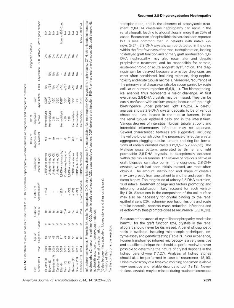
theless, crystals may bemissed during routinemicroscopicTable
5:Literature
review
ofcasesofrecurrent2,8-DHAcrystallinenephropathy—characteristicsatdiagnosis
anddiagnosticmethods
First
Author(ref.)
Year
Ageat
diagnosis
Gender
Orderof
renalTx
History
of
nephrolithiasis2
Suspected
causeofCKD
Delayof
diagnosis
after
Tx(w
eeks)
sCrat
diagnosis
(mmol/L)
Graft
dysfunction
Diagnosticmethods
FTIR
Crystalluria
APRTactivity
APRTgeneanalysis
deJong(5)
1996
56
M1st
þ(40)
CTIN/urate
stones
0.3
Hemodialysis
CGD3
NA
þ0%
NA
Brown(6)
1998
49
F1st
�Undeterm
inedCN
4Hemodialysis
PGNF
þ/GB
NA
NA
NA
Benedetto(7)
2001
48
M1st
�Undeterm
inedCN
19
248
AGD
NA
NA
0%
NA
Cassidy(8)
2004
23
M1st
�Undeterm
inedCN
2361
CGD
þ/NL
NA
0%
NA
Eller(9)
2007
111
M4th
þ(9.5)
2,8-DHA
nephropathy
1Hemodialysis
DGF4
þ/NL
þ0%
hom.c.400þ2
dup
Nasr(10)
2010
42
F2nd
�Oxalate
nephropathy
6486
CGD
þ/GB
NA
0%
NA
Nasr(10)
2010
54
M1st
þ(36)
2,8-DHA
nephropathy
1.3
398
AGD3
NA
NA
0%
NA
Bertram
(11)
2010
56
M3rd
þ(21)
CTIN/urate
stones
0.3
Hemodialysis
DGF
þ/NL
NA
NA
NA
Sharm
a(12)
2012
80
M1st
þ(30)
CTIN/nephrolithiasis
1Hemodialysis
DGF
NA
NA
0%
NA
Kaartinen(13)
2014
63
M2nd
þ(from
childhood)
CTIN/nephrolithiasis
0.3
Hemodialysis
PGNF
NA
þhom.c.188G>A
ref.,referencenumber;F,female;M,male;Tx,transplant;CKD,chronickidneydisease;NA,notavailable;Nx,nephropathy;CN,crystallinenephropathy;CTIN,chronictubulointerstitial
nephropathy;sCr,serum
creatinine;CGD,chronicgraftdysfunction;AGD,acute
graftdysfunction;DGF,delayedgraftfunction;PGNF,primary
graftnonfunction;GB,graftbiopsy;NL,
nephrolithiasis;hom.,homozygousmutation.
1Before
1strenaltransplantation.
2Delaybetw
eenfirstepisodeofkidneystoneanddiagnosis
(years).
3History
ofDGF.
4In
thecontextofacute
rejection.
Recurrent 2,8-Dihydroxyadenine Nephropathy
2629American Journal of Transplantation 2014; 14: 2623–2632

examination (10), and crystalluria may be inconspicuous in
oligo-anuric patients. Enzyme assay measuring APRT
activity in erythrocyte lysates may thus be helpful.
Undetectable enzyme activity confirms the diagnosis (1–
3,23). Importantly, once the diagnosis has been estab-
lished, the siblings should be screened using crystalluria
and APRT enzyme assay. Genetic testing can be consid-
ered, particularly if enzyme assay is not available. Various
germline mutations have been reported with no correlation
between genotype and phenotype (3,28–32). Moreover,
10% of mutations remain undetermined despite sequenc-
ing of the entire coding region and intron-exon junctions of
the APRT gene (3), as in three of our patients.
The management of APRT deficiency is based on allopuri-
nol (23), or the alternative agent febuxostat (33), in order to
effectively reduce the generation of 2,8-DHA. High fluid
intake and avoidance of purine-rich diet can also be advised,
whereas urine alkalinization is not beneficial (23). The initial
dose of allopurinol usually ranges from100 to 300mg/day in
adults. Higher doses, in the range of 300–600mg/day, are
generally required to achieve complete inhibition of 2,8-
DHA formation (10,13). Crystalluria can be used for
monitoring treatment efficiency. The likelihood of complete
regression of crystal deposition and recovery of allograft
function depends on the extent of kidney damage at
treatment initiation. In our series and previous reports,
almost one-third of patients had allograft loss at last follow-
up, andmost of the remaining patients had chronic allograft
dysfunction (13,14,16,19,21,23). Nevertheless, if allopuri-
nol is initiated early enough, graft function may remain
stable or improve (7,11).
2,8-DHA crystalline nephropathy is a rare and underrecog-
nized cause of CKD that can recur in the renal allograft. The
presence of crystals in the renal parenchyma and urine
sediment should not be overlooked. A high index of
suspicion for disease recurrence should be maintained,
Table 6: Literature review of cases of recurrent 2,8-DHA crystalline nephropathy—treatment and outcome
First author (ref.)
Allopurinol therapy
Follow-up
(months)
sCr at diagnosis
(mmol/L)
sCr at last
follow-up (mmol/L) Renal outcomeInitial dose
Maintenance
dose
de Jong (5) – – 6 Hemodialysis Hemodialysis Graft loss
Brown (6) – – 4 Hemodialysis Hemodialysis Graft loss
Benedetto (7) 10mg/kg NA NA 248 177 Chronic graft dysfunction
Cassidy (8) 100mg/day 300mg/day 7 361 262 Chronic graft dysfunction
Eller (9) 150mg�2/week 100mg/day 7 Hemodialysis 240 Chronic graft dysfunction
Nasr (10) 200mg/day 200mg/day 12 486 Hemodialysis Graft loss1
Nasr (10) 300mg/day2 600mg/day 18 398 141 Chronic graft dysfunction
Bertram (11) 150mg/day 300mg/day 9 Hemodialysis NA Chronic graft dysfunction
Sharma (12) NA NA NA Hemodialysis NA NA
Kaartinen (13) 300mg/day 500mg/day 11 Hemodialysis Hemodialysis Graft loss
ref., reference number; NA, not available (because not performed); hom, homozygous mutation; sCr, serum creatinine.1The patient initially improved but later developed acute deterioration of allograft function in the setting of toxic megacolon. Allopurinol was
temporarily discontinued and renal allograft biopsy showed extensive crystal deposition. Despite resumption of allopurinol, the patient
remained dialysis-dependent.2Allopurinol was started before renal transplantation.
Table 7: Recommended tests for the diagnosis of 2,8-DHA crystalline nephropathy and APRT deficiency
Diagnostic tests Advantages Pitfalls
First line investigations
Crystalluria Sensitivity and specificity close to 100% Requires experienced operator
Determines the nature of the crystals (i.e. 2,8-DHA,
oxalate, urate and others)
Not possible in anuric patients
FTIR/kidney biopsy Very high sensitivity and specificity Requires kidney or graft biopsy
Determines the nature of the crystals (i.e. 2,8-DHA,
oxalate, urate and others)
May not characterize very small crystals<12mm
FTIR/kidney stone Very high sensitivity and specificity determines the
nature of the stone
Requires the patient to save any passed stones
for testing
Second line investigations
APRT activity in erythrocyte
lysates
Sensitivity and specificity of 100% Availability
Genetic testing Identification of 80–90% of APRT gene mutations Cost
APRT, adenine phosphoribosyltransferase; 2,8-DHA, 2,8-dihydroxyadenine; FTIR, Fourier transformed infrared microscopy.
Zaidan et al
2630 American Journal of Transplantation 2014; 14: 2623–2632

regardless of the course and delay of allograft dysfunction
and immunological risk. This is particularly important
when the underlying cause of the CKD is unknown.
Accurate diagnosis and prompt pharmacologic inhibition
of xanthine dehydrogenase may allow the stabilization or
even improvement of graft function, reducing the risk of
allograft loss.
Acknowledgments
We thank all the nephrologists and pathologists who have contributed to the
recruitment of patients, including Eric Prinz (Strasbourg, France), Reda
Sharobeem (Olivet, France), Christian Jacquot (Paris, France), Renato
Demontis (Creil, France), Guillaume Bollee (Quebec, Canada), Catterine
Canavese (Novara, Italy), Gabriele Guglielmetti (Novara, Italy), Laure-Helene
Noel (Paris, France), Laurent Doucet (Brest, France),Marie-ChristineMachet
(Tours, France), Remy Kerdraon (Orleans, France), Jerome Olagne
(Strasbourg, France), Dominique Bazin (Paris, France) and Christophe
Sandt (Gif-sur-Yvette, France). RP andVOE are supported by theRare Kidney
Stone Consortium (U54KD083908), a member of the NIH Rare Diseases
Clinical Research Network (RDCRN), supported through a collaboration
between the National Center for advancing Translational Sciences
(NCATS), and the National Institute of Diabetes and Digestive and Kidney
Diseases (NIDDK).
Disclosure
The authors of this manuscript have no conflicts of interest
to disclose as described by the American Journal of
Transplantation.
References
1. Kamatani N, Terai C, Kuroshima S, Nishioka K,Mikanagi K. Genetic
and clinical studies on 19 families with adenine phosphoribosyl-
transferase deficiencies. Hum Genet 1987; 75: 163–168.
2. Edvardsson V, Palsson R, Olafsson I, Hjaltadottir G, Laxdal T.
Clinical features and genotype of adenine phosphoribosyltransfer-
ase deficiency in Iceland. Am J Kidney Dis 2001; 38: 473–480.
3. Bollee G, Dollinger C, Boutaud L, et al. Phenotype and genotype
characterization of adenine phosphoribosyltransferase deficiency.
J Am Soc Nephrol 2010; 21: 679–688.
4. Harambat J, Bollee G, Daudon M, Ceballos-Picot I, Bensman A,
Group AS. Adenine phosphoribosyltransferase deficiency in
children. Pediatr Nephrol 2012; 27: 571–579.
5. de Jong DJ, de Assmann KJ, Abreu RA, et al. 2,8-Dihydroxyade-
nine stone formation in a renal transplant recipient due to adenine
phosphoribosyltransferase deficiency. J Urol 1996; 156: 1754–
1755.
6. Brown HA. Recurrence of 2,8-dihydroxyadenine tubulointerstitial
lesions in a kidney transplant recipient with a primary presentation
of chronic renal failure. Nephrol Dial Transpl 1998; 13: 998–
1000.
7. Benedetto B, Madden R, Kurbanov A, Braden G, Freeman J,
Lipkowitz G. Adenine phosphoribosyltransferase deficiency and
renal allograft dysfunction. Am J Kidney Dis 2001; 37: E37.
8. CassidyMJ,McCulloch T, Fairbanks LD, Simmonds HA. Diagnosis
of adenine phosphoribosyltransferase deficiency as the underlying
cause of renal failure in a renal transplant recipient. Nephrol Dial
Transpl 2004; 19: 736–738.
9. Eller P, Rosenkranz AR, Mark W, Theurl I, Laufer J, Lhotta K. Four
consecutive renal transplantations in a patient with adenine
phosphoribosyltransferase deficiency. Clin Nephrol 2004; 61:
217–221.
10. Nasr S, Sethi S, Cornell L, et al. Crystalline nephropathy due to 2,8-
dihydroxyadeninuria: An under-recognized cause of irreversible
renal failure. Nephrol Dial Transpl 2010; 25: 1909–1915.
11. Bertram A, Broecker V, Lehner F, Schwarz A. Kidney transplanta-
tion in a patient with severe adenine phosphoribosyl transferase
deficiency: Obstacles and pitfalls. Transpl Int 2010; 23: e56–e58.
12. Sharma SG, Moritz MJ, Markowitz GS. 2,8-dihydroxyadeninuria
disease. Kidney Int 2012; 82: 1036.
13. Kaartinen K, Hemmila U, Salmela K, Raisanen-Sokolowski A, Kouri
T, Makela S. Adenine phosphoribosyltransferase deficiency as a
rare cause of renal allograft dysfunction. J Am Soc Nephrol 2014;
25: 671–674.
14. Gagne ER, Deland E, Daudon M, Noel LH, Nawar T. Chronic renal
failure secondary to 2,8-dihydroxyadenine deposition: The first
report of recurrence in a kidney transplant. Am J Kidney Dis 1994;
24: 104–107.
15. Stratta P, Fogazzi G, Canavese C, et al. Decreased kidney function
and crystal deposition in the tubules after kidney transplant. Am J
Kidney Dis 2010; 56: 585–590.
16. Levey AS, Bosch JP, Lewis JB, Greene T, Rogers N, Roth D. A
more accurate method to estimate glomerular filtration rate from
serum creatinine: A new prediction equation. Modification of Diet
in Renal Disease Study Group. Ann Intern Med 1999; 130: 461–
470.
17. Estepa-Maurice L, Hennequin C, Marfisi C, Bader C, Lacour B,
Daudon M. Fourier transform infrared microscopy identification of
crystal deposits in tissues: Clinical importance in various patholo-
gies. Am J Clin Pathol 1996; 105: 576–582.
18. Daudon M, Bader C, Jungers P. Urinary calculi: review of
classification methods and correlations with etiology. Scanning
Microsc 1993; 7: 1081–1104; discussion 1104–1106.
19. DaudonM, Jungers P. Clinical value of crystalluria and quantitative
morphoconstitutional analysis of urinary calculi. Nephron Physiol
2004; 98: p31–36.
20. GreenwoodMC, DillonMJ, Simmonds HA, Barratt TM, Pincott JR,
Metreweli C. Renal failure due to 2,8-dihydroxyadenine urolithia-
sis. Eur J Pediatr 1982; 138: 346–349.
21. Gelb AB, Fye KH, Tischfield JA, et al. Renal insufficiency secondary
to 2,8-dihydroxyadenine urolithiasis. Hum Pathol 1992; 23: 1081–
1085.
22. Arnadottir M, Laxdal T, Hardarson S, Asmundsson P. Acute renal
failure in a middle-aged woman with 2,8-dihydroxyadeninuria.
Nephrol Dial Transpl 1997; 12: 1985–1987.
23. Bollee G, Harambat J, Bensman A, Knebelmann B, Daudon M,
Ceballos-Picot I. Adenine phosphoribosyltransferase deficiency.
Clin J Am Soc Nephrol 2012; 7: 1521–1527.
24. Glicklich D, Gruber HE, Matas AJ, et al. 2,8-dihydroxyadenine
urolithiasis: Report of a case first diagnosed after renal transplant.
Q J Med 1988; 68: 785–793.
25. Herlitz LC, D’Agati VD, Markowitz GS. Crystalline nephropathies.
Arch Pathol Lab Med 2012; 136: 713–720.
26. Verkoelen CF, Verhulst A. Proposed mechanisms in renal tubular
crystal retention. Kidney Int 2007; 72: 13–18.
27. Dessombz A, Bazin D, Dumas P, Sandt C, Sule-Suso J, DaudonM.
Shedding light on the chemical diversity of ectopic calcifications in
kidney tissues: Diagnostic and research aspects. PLoS ONE 2011;
6: e28007.
Recurrent 2,8-Dihydroxyadenine Nephropathy
2631American Journal of Transplantation 2014; 14: 2623–2632

28. Chen J, Sahota A, Laxdal T, et al. Identification of a singlemissense
mutation in the adenine phosphoribosyltransferase (APRT) gene
from five Icelandic patients and a British patient. Am J Hum Genet
1991; 49: 1306–1311.
29. Gathof BS, Sahota A, Gresser U, et al. A splice mutation at the
adenine phosphoribosyltransferase locus detected in a German
family. Adv Exp Med Biol 1991; 309B: 83–86.
30. Hidaka Y, Tarle SA, Fujimori S, Kamatani N, Kelley WN, Palella TD.
Human adenine phosphoribosyltransferase deficiency. Demon-
stration of a single mutant allele common to the Japanese. J Clin
Invest 1988; 81: 945–950.
31. Mimori A, Hidaka Y, Wu VC, et al. A mutant allele common to the
type I adenine phosphoribosyltransferase deficiency in Japanese
subjects. Am J Hum Genet 1991; 48: 103–107.
32. Kamatani N, Hakoda M, Otsuka S, Yoshikawa H, Kashiwazaki S.
Only three mutations account for almost all defective alleles
causing adenine phosphoribosyltransferase deficiency in Japa-
nese patients. J Clin Invest 1992; 90: 130–135.
33. Becker MA, Schumacher HR, Wortmann RL, et al. Febuxostat
comparedwith allopurinol in patients with hyperuricemia and gout.
N Engl J Med 2005; 353: 2450–2461.
Zaidan et al
2632 American Journal of Transplantation 2014; 14: 2623–2632
Copyright © 2022 FDOKUMEN