Short 9q interstitial deletion in a neonate with lethal non-immune hydrops
Upload
khangminh22Category
view
2download
0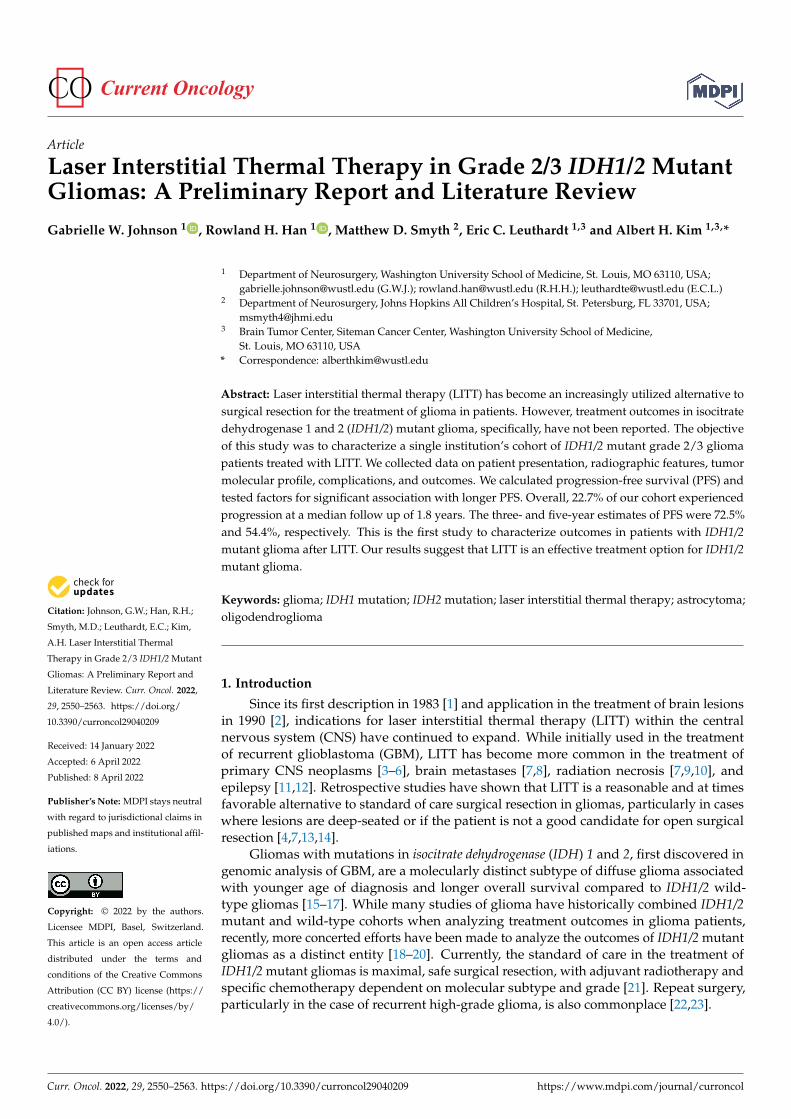
�����������������
Citation: Johnson, G.W.; Han, R.H.;
Smyth, M.D.; Leuthardt, E.C.; Kim,
A.H. Laser Interstitial Thermal
Therapy in Grade 2/3 IDH1/2 Mutant
Gliomas: A Preliminary Report and
Literature Review. Curr. Oncol. 2022,
29, 2550–2563. https://doi.org/
10.3390/curroncol29040209
Received: 14 January 2022
Accepted: 6 April 2022
Published: 8 April 2022
Publisher’s Note: MDPI stays neutral
with regard to jurisdictional claims in
published maps and institutional affil-
iations.
Copyright: © 2022 by the authors.
Licensee MDPI, Basel, Switzerland.
This article is an open access article
distributed under the terms and
conditions of the Creative Commons
Attribution (CC BY) license (https://
creativecommons.org/licenses/by/
4.0/).
Article
Laser Interstitial Thermal Therapy in Grade 2/3 IDH1/2 MutantGliomas: A Preliminary Report and Literature ReviewGabrielle W. Johnson 1 , Rowland H. Han 1 , Matthew D. Smyth 2, Eric C. Leuthardt 1,3 and Albert H. Kim 1,3,*
1 Department of Neurosurgery, Washington University School of Medicine, St. Louis, MO 63110, USA;[email protected] (G.W.J.); [email protected] (R.H.H.); [email protected] (E.C.L.)
2 Department of Neurosurgery, Johns Hopkins All Children’s Hospital, St. Petersburg, FL 33701, USA;[email protected]
3 Brain Tumor Center, Siteman Cancer Center, Washington University School of Medicine,St. Louis, MO 63110, USA
* Correspondence: [email protected]
Abstract: Laser interstitial thermal therapy (LITT) has become an increasingly utilized alternative tosurgical resection for the treatment of glioma in patients. However, treatment outcomes in isocitratedehydrogenase 1 and 2 (IDH1/2) mutant glioma, specifically, have not been reported. The objectiveof this study was to characterize a single institution’s cohort of IDH1/2 mutant grade 2/3 gliomapatients treated with LITT. We collected data on patient presentation, radiographic features, tumormolecular profile, complications, and outcomes. We calculated progression-free survival (PFS) andtested factors for significant association with longer PFS. Overall, 22.7% of our cohort experiencedprogression at a median follow up of 1.8 years. The three- and five-year estimates of PFS were 72.5%and 54.4%, respectively. This is the first study to characterize outcomes in patients with IDH1/2mutant glioma after LITT. Our results suggest that LITT is an effective treatment option for IDH1/2mutant glioma.
Keywords: glioma; IDH1 mutation; IDH2 mutation; laser interstitial thermal therapy; astrocytoma;oligodendroglioma
1. Introduction
Since its first description in 1983 [1] and application in the treatment of brain lesionsin 1990 [2], indications for laser interstitial thermal therapy (LITT) within the centralnervous system (CNS) have continued to expand. While initially used in the treatmentof recurrent glioblastoma (GBM), LITT has become more common in the treatment ofprimary CNS neoplasms [3–6], brain metastases [7,8], radiation necrosis [7,9,10], andepilepsy [11,12]. Retrospective studies have shown that LITT is a reasonable and at timesfavorable alternative to standard of care surgical resection in gliomas, particularly in caseswhere lesions are deep-seated or if the patient is not a good candidate for open surgicalresection [4,7,13,14].
Gliomas with mutations in isocitrate dehydrogenase (IDH) 1 and 2, first discovered ingenomic analysis of GBM, are a molecularly distinct subtype of diffuse glioma associatedwith younger age of diagnosis and longer overall survival compared to IDH1/2 wild-type gliomas [15–17]. While many studies of glioma have historically combined IDH1/2mutant and wild-type cohorts when analyzing treatment outcomes in glioma patients,recently, more concerted efforts have been made to analyze the outcomes of IDH1/2 mutantgliomas as a distinct entity [18–20]. Currently, the standard of care in the treatment ofIDH1/2 mutant gliomas is maximal, safe surgical resection, with adjuvant radiotherapy andspecific chemotherapy dependent on molecular subtype and grade [21]. Repeat surgery,particularly in the case of recurrent high-grade glioma, is also commonplace [22,23].
Curr. Oncol. 2022, 29, 2550–2563. https://doi.org/10.3390/curroncol29040209 https://www.mdpi.com/journal/curroncol
Curr. Oncol. 2022, 29 2551
Over the last decade, LITT has become an increasingly utilized modality in the treat-ment of gliomas. While LITT has been shown to be an effective, well-tolerated alternativeto open surgical resection in both low- [4,24,25] and high-grade [14,26,27] gliomas, LITTtreatment outcomes specific to IDH1/2 mutant grade 2 and 3 gliomas have not been stud-ied. The objective of the present study was to describe the clinical and histopathologicalpresentation, treatment, and outcomes of patients with IDH1/2 mutant grade 2/3 gliomastreated with LITT at a single institution, to determine the progression-free survival of thesepatients, and to compare their clinical outcomes with previously published findings.
2. Materials and Methods
The study was conducted with the approval of the institutional review board (IRB ID#201409046, approval date: 3 February 2022). This study was a single-institution, retrospec-tive case series of patients treated at Washington University, Barnes Jewish Hospital, andSt. Louis Children’s Hospital with LITT for IDH1/2 mutant grade 2 or 3 gliomas from 2014to 2021. Patients who underwent LITT at our institution were identified via combinationof case log databases and departmental billing records. Patients were excluded if theirpathology was not consistent with a glial neoplasm, was determined to be WHO grades Ior IV, was IDH1/2 wild-type, or was of an unknown IDH1/2 mutation status (Figure 1).
Curr. Oncol. 2022, 28, FOR PEER REVIEW 3
Figure 1. Flowchart of patient selection.
Median time to progression was obtained using Kaplan–Meier (KM) product-limit estimates of the survival function and were graphically represented using KM curves. If the survival estimate was above 50% at the end of observation time, a “not reached” indi-cator was used. KM survival tables were used to estimate PFS at 3 and 5 years. KM curves were constructed for the overall cohort, first line versus salvage treatment, prior extent of resection, extent of ablation, and tumor pathology. Log-rank testing was conducted to identify factors independently associated with differences in PFS. Univariate logistic re-gression was used to identify factors independently associated with progression. Factors with p < 0.10 were retained for multivariable analysis (Cox proportional hazard testing and multivariable logistic regression).
Patient consent was not required due to the retrospective nature of the study.
3. Results 3.1. Baseline Characteristics and Presentation
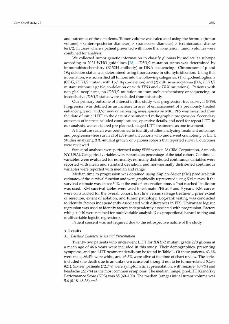
Twenty-two patients who underwent LITT for IDH1/2 mutant grade 2/3 glioma at a mean age of 46.6 years were included in this study. Their demographics, presenting symp-toms, and pre-LITT treatment details can be found in Table 1. Of these patients, 63.6% were male, 86.4% were white, and 95.5% were alive at the time of chart review. The series included one death due to an unknown cause but thought not to be tumor-related (Case #21). Sixteen patients (72.7%) were symptomatic at presentation, with seizure (40.9%) and headache (22.7%) as the most common symptoms. The median (range) pre-LITT Karnofsky Performance Score (KPS) was 85 (60–100). The median (range) initial tumor volume was 5.6 (0.18–48.38) cm3.
Treatment prior to LITT was common, with 72.7% of patients having undergone pre-LITT treatment. Surgical resection was the most common pre-LITT treatment (14 patients, 63.6%), with 57.1% of these patients undergoing gross total resection (GTR) and 42.9% undergoing subtotal resection (STR). Ten patients (45.5%) underwent chemotherapy, with most patients (90%) undergoing chemotherapy with temozolomide. Radiation therapy was also common, with 12 (54.4%) of patients undergoing treatment. Biopsy was less com-mon (four patients, 18.2%), and radiosurgery was the least common pre-LITT treatment (one patient).
Figure 1. Flowchart of patient selection.
Data extracted from the electronic medical record included patient demographics,clinical presentation, radiographic characteristics, treatment prior to LITT, LITT operativedetails, adjuvant treatment, and outcomes after treatment to better classify the presentation
Curr. Oncol. 2022, 29 2552
and outcomes of these patients. Tumor volume was calculated using the formula (tumorvolume) = (antero-posterior diameter) × (transverse diameter) × (craniocaudal diame-ter)/2. In cases where a patient presented with more than one lesion, tumor volumes werecombined for analysis.
We collected tumor genetic information to classify gliomas by molecular subtypeaccording to 2021 WHO guidelines [28]. IDH1/2 mutation status was determined byimmunohistochemistry (R132H antibody) or DNA sequencing. Chromosome 1p and19q deletion status was determined using fluorescence in situ hybridization. Using thisinformation, we reclassified all tumors into the following categories: (1) oligodendroglioma(ODG; IDH1/2 mutant with 1p/19q co-deletion) and (2) diffuse astrocytoma (DA; IDH1/2mutant without 1p/19q co-deletion or with TP53 and ATRX mutations). Patients withnon-glial neoplasms, no IDH1/2 mutation on immunohistochemistry or sequencing, orinconclusive IDH1/2 status were excluded from this study.
Our primary outcome of interest in this study was progression-free survival (PFS).Progression was defined as an increase in area of enhancement of a previously treatedenhancing lesion and/or new or increasing mass lesions on MRI. PFS was measured fromthe date of initial LITT to the date of documented radiographic progression. Secondaryoutcomes of interest included complications, operative details, and need for repeat LITT. Inour analysis, we considered pre-planned, staged LITT treatments as one treatment.
A literature search was performed to identify studies analyzing treatment outcomesand progression-free survival of IDH-mutant cohorts who underwent craniotomy or LITT.Studies analyzing IDH-mutant grade 2 or 3 glioma cohorts that reported survival outcomeswere reviewed.
Statistical analyses were performed using SPSS version 28 (IBM Corporation, Armonk,NY, USA). Categorical variables were reported as percentage of the total cohort. Continuousvariables were evaluated for normality; normally distributed continuous variables werereported with mean and standard deviation, and non-normally distributed continuousvariables were reported with median and range.
Median time to progression was obtained using Kaplan–Meier (KM) product-limitestimates of the survival function and were graphically represented using KM curves. If thesurvival estimate was above 50% at the end of observation time, a “not reached” indicatorwas used. KM survival tables were used to estimate PFS at 3 and 5 years. KM curveswere constructed for the overall cohort, first line versus salvage treatment, prior extentof resection, extent of ablation, and tumor pathology. Log-rank testing was conductedto identify factors independently associated with differences in PFS. Univariate logisticregression was used to identify factors independently associated with progression. Factorswith p < 0.10 were retained for multivariable analysis (Cox proportional hazard testing andmultivariable logistic regression).
Patient consent was not required due to the retrospective nature of the study.
3. Results3.1. Baseline Characteristics and Presentation
Twenty-two patients who underwent LITT for IDH1/2 mutant grade 2/3 glioma ata mean age of 46.6 years were included in this study. Their demographics, presentingsymptoms, and pre-LITT treatment details can be found in Table 1. Of these patients, 63.6%were male, 86.4% were white, and 95.5% were alive at the time of chart review. The seriesincluded one death due to an unknown cause but thought not to be tumor-related (Case#21). Sixteen patients (72.7%) were symptomatic at presentation, with seizure (40.9%) andheadache (22.7%) as the most common symptoms. The median (range) pre-LITT KarnofskyPerformance Score (KPS) was 85 (60–100). The median (range) initial tumor volume was5.6 (0.18–48.38) cm3.
Curr. Oncol. 2022, 29 2553
Table 1. Cohort demographics and initial presentation.
Case Age Sex Presenting Symptoms Pre-LITTKPS
Pre-LITTTreatment Lesion Location
TumorVolume(cm3)
1 43.8 F Sensory changes, headache, syncopalepisodes, fatigue, change in taste/smell 90 None L frontotemporal, insula 21.39
2 44.0 M Seizures 80 None R frontoparietal,cingulate gyrus 2.42
3 65.9 M Seizures 90 None L parietooccipital 0.84
4 35.0 FHeadache, vision problems,neurocognitive deficits,behavioral changes
70 None R parietooccipital 10.35
5 67.3 F Seizures, neurocognitive deficits, tremor 70 Bx L frontotemporal, basalganglia, insula 37.02
6 60.7 M Seizures 80 GTR ×2, RT, C R frontal 10.67
7 49.8 M Seizures, speech difficulties 80 STR, RT, C L occipital 5.95
8 46.9 F None 100 GTR, RT, R frontal 15.75
9 56.9 M Muscle weakness/paralysis, speechdifficulties 70 GTR ×2, RT, Bx L frontoparietal 1.69
10 46.4 F None 100 GTR L frontal 1.08
11 53.3 M None 100 STR, RT, C R temporal, insula 22.28
12 60.0 M Muscle weakness/paralysis, headache 80 STR, C, R frontoparietal 10.01
13 21.8 M Seizures 80 Bx L frontotemporal, insula 48.38
14 47.4 F None 100 STR, RT, R temporal 10.29
15 36.0 M Seizures 80 STR ×2, RT, C, GK L temporal 3.02
16 45.4 M None 100 GTR, RT, C, Bx L frontal 0.38
17 31.3 M Headache 90 None L insula 15.91
18 48.5 M Muscle weakness/paralysis, headache,speech difficulties 60 STR ×2, RT, C L frontoparietal 0.18
19 44.2 F Seizures 100 GTR, RT, C R frontal 1.05
20 41.9 M Earaches 90 None L insula 3.15
21 34.3 M None 100 GTR, RT, C L insula 3.78, 1.46 *
22 44.8 F Seizures 80 STR, RT, C R temporal 5.30
Abbreviations: KPS = Karnosfsky Performance Score, F = female, M = male, L = left, R = right, GTR = gross totalresection, STR = subtotal resection, RT = radiation therapy, C = chemotherapy, GK = gamma knife radiosurgery,Bx = biopsy. * This patient presented with two lesions.
Treatment prior to LITT was common, with 72.7% of patients having undergone pre-LITT treatment. Surgical resection was the most common pre-LITT treatment (14 patients,63.6%), with 57.1% of these patients undergoing gross total resection (GTR) and 42.9%undergoing subtotal resection (STR). Ten patients (45.5%) underwent chemotherapy, withmost patients (90%) undergoing chemotherapy with temozolomide. Radiation therapywas also common, with 12 (54.4%) of patients undergoing treatment. Biopsy was lesscommon (four patients, 18.2%), and radiosurgery was the least common pre-LITT treatment(one patient).
3.2. Tumor Pathology
IDH1/2 mutant gliomas were re-classified according to the 2021 WHO guidelines intooligodendroglioma (ODG, IDH1/2 mutant and 1p/19q co-deleted) and diffuse astrocy-
Curr. Oncol. 2022, 29 2554
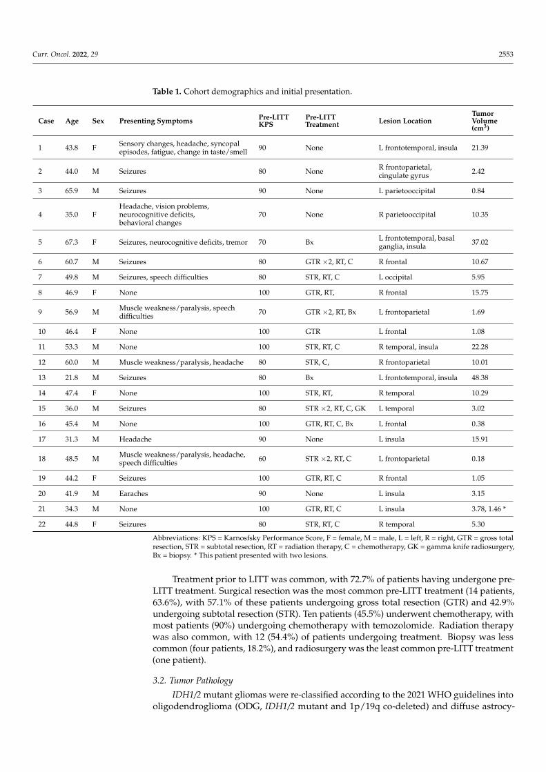
toma (DA, IDH1/2 mutant without 1p/19q co-deletion). Of the 22 patients in the study,12 patients (54.5%) had ODG and 10 (45.5%) had DA. Most tumors (59.1%) were grade 2,while the remaining nine (40.9%) were grade 3. Only one patient (Case #2) had a mutationin IDH2 (R127K); the remaining patients had mutations in IDH1. The most common IDH1mutation was R132H (90.1%), followed by R132C (4.5%). Other mutations in each patient’stumor, along with Ki-67 proliferative index information, can be found in Table 2.
Table 2. Histopathologic findings.
Case Tumor Type WHO Grade Mutations Ki-67 Index Tumor Mutational Burden
1 Diffuse astrocytoma 2 IDH1, TP53, ATRX ≈4 12 Oligodendroglioma 2 IDH2, TERTp, CIC, 1p/19q co-deletion 17 03 Oligodendroglioma 2 IDH1, TERTp, CIC, 1p/19q co-deletion 6 34 Diffuse astrocytoma 2 IDH1, TP53, ATRX 2.5 65 Oligodendroglioma 2 IDH1, 1p/19q co-deletion NT NT6 Diffuse astrocytoma 2 IDH1, TP53, ATRX <10 NT7 Diffuse astrocytoma 2 IDH1 5.7 NT8 Diffuse astrocytoma 2 IDH1, TP53, ATRX 2.3 39 Oligodendroglioma 2 IDH1, 1p/19q co-deletion 8.2 NT10 Oligodendroglioma 2 IDH1, 1p/19q co-deletion 2.1 NT11 Oligodendroglioma 2 IDH1, TERTp, CIC, 1p/19q co-deletion 3.7 012 Oligodendroglioma 2 IDH1, TERTp, CIC, 1p/19q co-deletion NT 4213 Diffuse astrocytoma 2 IDH1 <5 NT14 Diffuse astrocytoma 3 IDH1, TP53, ATRX 1.4 115 Oligodendroglioma 3 IDH1, TERTp, CIC, 1p/19q co-deletion 40 1616 Oligodendroglioma 3 IDH1, TERTp, 1p/19q co-deletion 9.9 1317 Diffuse astrocytoma 3 IDH1, TP53, ATRX NT NT18 Oligodendroglioma 3 IDH1, 1p/19q co-deletion 24 NT19 Oligodendroglioma 3 IDH1, TERTp, CIC, 1p/19q co-deletion 20 NT20 Diffuse astrocytoma 3 IDH1, TP53, ATRX 5–10 421 Diffuse astrocytoma 3 IDH1, TP53, ATRX 31.5 122 Oligodendroglioma 3 IDH1, TERTp, 1p/19q co-deletion 50 3
Abbreviations: NT = not tested.
3.3. LITT Treatment
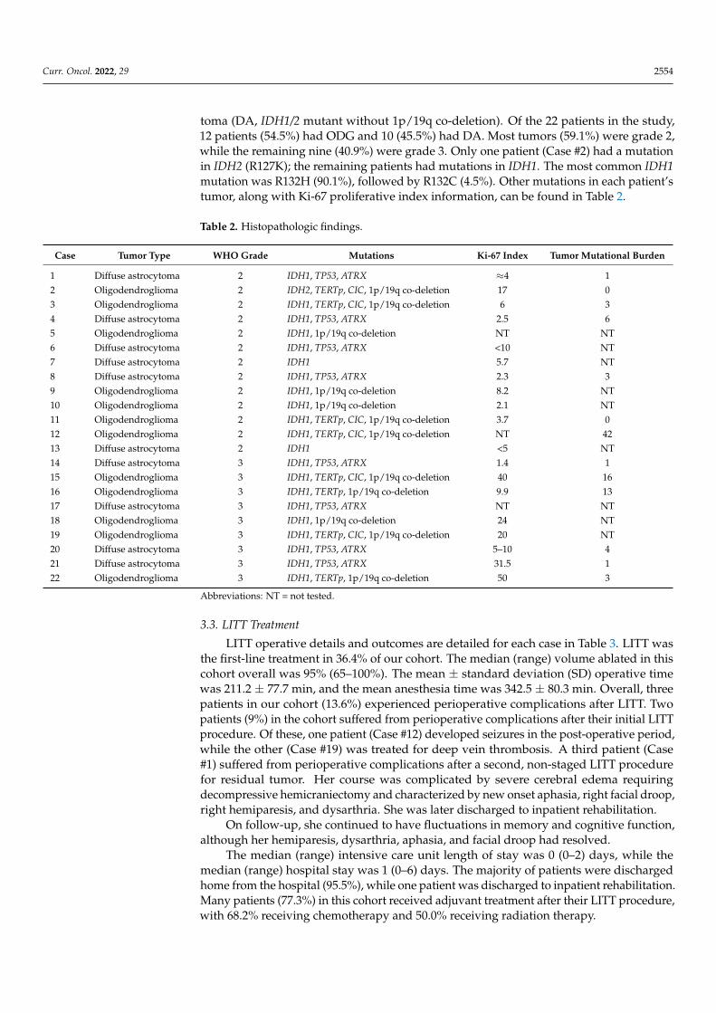
LITT operative details and outcomes are detailed for each case in Table 3. LITT wasthe first-line treatment in 36.4% of our cohort. The median (range) volume ablated in thiscohort overall was 95% (65–100%). The mean ± standard deviation (SD) operative timewas 211.2 ± 77.7 min, and the mean anesthesia time was 342.5 ± 80.3 min. Overall, threepatients in our cohort (13.6%) experienced perioperative complications after LITT. Twopatients (9%) in the cohort suffered from perioperative complications after their initial LITTprocedure. Of these, one patient (Case #12) developed seizures in the post-operative period,while the other (Case #19) was treated for deep vein thrombosis. A third patient (Case#1) suffered from perioperative complications after a second, non-staged LITT procedurefor residual tumor. Her course was complicated by severe cerebral edema requiringdecompressive hemicraniectomy and characterized by new onset aphasia, right facial droop,right hemiparesis, and dysarthria. She was later discharged to inpatient rehabilitation.
On follow-up, she continued to have fluctuations in memory and cognitive function,although her hemiparesis, dysarthria, aphasia, and facial droop had resolved.
The median (range) intensive care unit length of stay was 0 (0–2) days, while themedian (range) hospital stay was 1 (0–6) days. The majority of patients were dischargedhome from the hospital (95.5%), while one patient was discharged to inpatient rehabilitation.Many patients (77.3%) in this cohort received adjuvant treatment after their LITT procedure,with 68.2% receiving chemotherapy and 50.0% receiving radiation therapy.
Curr. Oncol. 2022, 29 2555
Table 3. Operative details and outcomes.
Case First Line vs.Salvage Indication for LITT EOA (%) Perioperative
Complications Adjuvant Tx Repeat LITT Time to Progression (Months) Follow-Up(Months)
1 First line Tumor location 80 Severe edema, new FND RT, C Yes No progression 9.852 First line Tumor location 95 None RT, C No No progression 7.363 First line Unknown 85 None RT, C No No progression 16.524 First line Tumor location 95 None RT, C No No progression 33.805 First line Shorter LOS 65 None RT, C No No progression 73.136 Salvage Refractory to Tx 95 None None No No progression 62.127 Salvage Recurrence NR None STR, RT, C No 7.42 70.148 Salvage Recurrence 95 None RT, C No No progression 19.489 Salvage Recurrence 98 None C No 45.34 88.5010 Salvage Recurrence 100 None None No No progression 23.5511 Salvage Recurrence 80 None C No 18.82 18.8212 Salvage Recurrence 80 Seizure None No No progression 16.2913 First line Aborted craniotomy 70 None None Yes 32.69 85.3214 Salvage Recurrence NR None RT, C No No progression 39.1315 Salvage Recurrence 99 None RT, C No 8.25 15.0116 Salvage Recurrence 80 None C, GK No No progression 11.6617 First line Lower percieved risk 95 None C No No progression 31.5718 Salvage Recurrence 100 None RT No No progression 89.8219 Salvage Recurrence 100 DVT None No No progression 24.7720 First line Favorable safety profile 80 None None No No progression 40.9721 Salvage Recurrence 99 None RT, C No No progression 2.8922 Salvage Recurrence 95 None C x2 No No progression 7.78
Abbreviations: EOA = extent of ablation, FND = focal neurologic deficit, LOS = length of stay, Tx = treatment, DVT = deep vein thrombosis, RT = radiation therapy, C = chemotherapy,GK = gamma knife, STR = subtotal resection, NR = not reported.
Curr. Oncol. 2022, 29 2556
Three patients (13.6%) underwent staged LITT (Cases #5, 11, and 13), and all of themtolerated the staged procedures without complication. One of these patients (Case #13) un-derwent a third staged treatment. Two patients underwent repeat, non-staged LITT due toresidual (Case #1) or recurrent (Case #13) tumor 2 months and 2 years after initial treatment,respectively. One patient (Case #7) underwent subtotal resection due to tumor progressionafter LITT. Repeat pathology in this patient demonstrated malignant transformation of thetumor from grade 2 diffuse astrocytoma to grade 3 anaplastic astrocytoma.
3.4. Progression-Free Survival
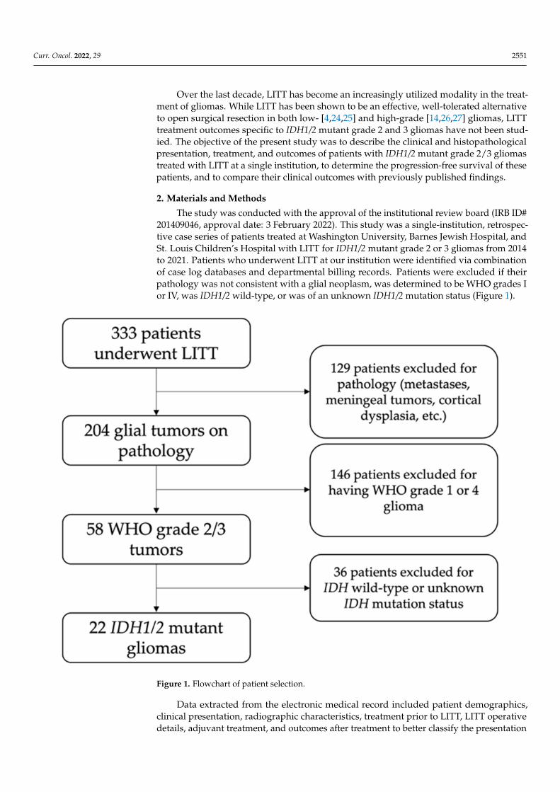
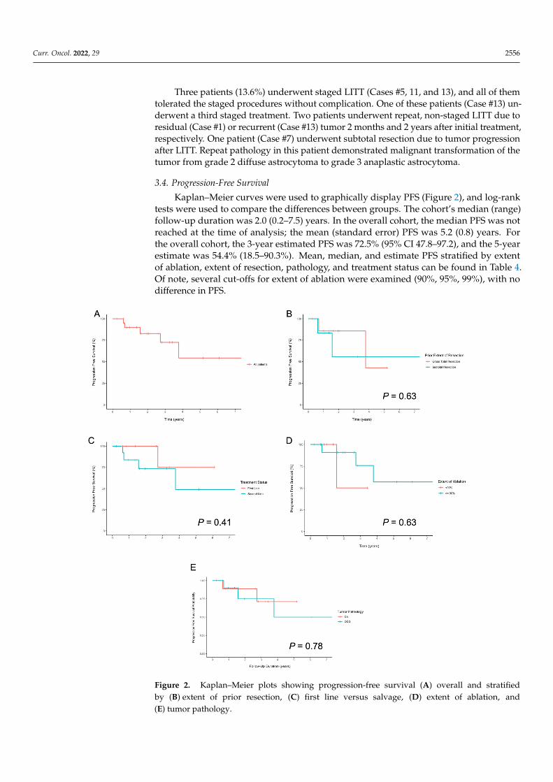
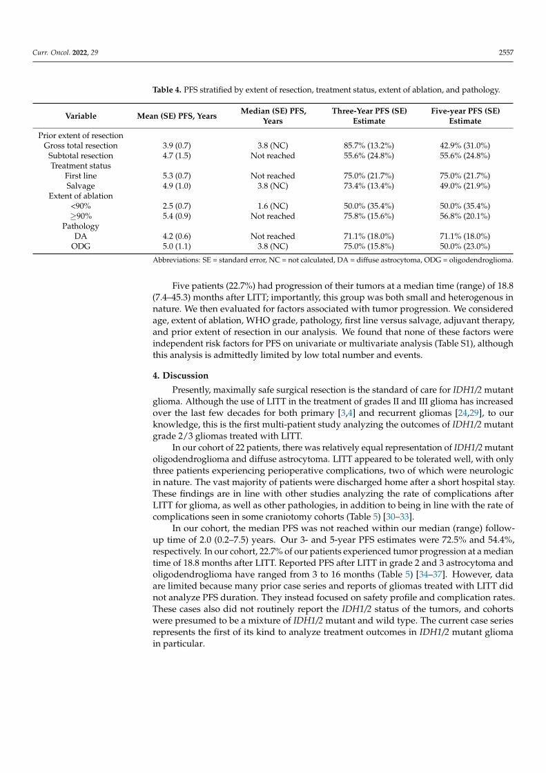
Kaplan–Meier curves were used to graphically display PFS (Figure 2), and log-ranktests were used to compare the differences between groups. The cohort’s median (range)follow-up duration was 2.0 (0.2–7.5) years. In the overall cohort, the median PFS was notreached at the time of analysis; the mean (standard error) PFS was 5.2 (0.8) years. Forthe overall cohort, the 3-year estimated PFS was 72.5% (95% CI 47.8–97.2), and the 5-yearestimate was 54.4% (18.5–90.3%). Mean, median, and estimate PFS stratified by extentof ablation, extent of resection, pathology, and treatment status can be found in Table 4.Of note, several cut-offs for extent of ablation were examined (90%, 95%, 99%), with nodifference in PFS.
Curr. Oncol. 2022, 28, FOR PEER REVIEW 7
independent risk factors for PFS on univariate or multivariate analysis (Table S1), alt-hough this analysis is admittedly limited by low total number and events.
Figure 2. Kaplan–Meier plots showing progression-free survival (A) overall and stratified by (B) extent of prior resection, (C) first line versus salvage, (D) extent of ablation, and (E) tumor pathol-ogy.
Table 4. PFS stratified by extent of resection, treatment status, extent of ablation, and pathology.
Variable Mean (SE) PFS, Years Median (SE) PFS, Years Three-Year PFS (SE) Estimate
Five-year PFS (SE) Estimate
Prior extent of resection Gross total resection 3.9 (0.7) 3.8 (NC) 85.7% (13.2%) 42.9% (31.0%)
Subtotal resection 4.7 (1.5) Not reached 55.6% (24.8%) 55.6% (24.8%) Treatment status
First line 5.3 (0.7) Not reached 75.0% (21.7%) 75.0% (21.7%)
Figure 2. Kaplan–Meier plots showing progression-free survival (A) overall and stratifiedby (B) extent of prior resection, (C) first line versus salvage, (D) extent of ablation, and(E) tumor pathology.
Curr. Oncol. 2022, 29 2557
Table 4. PFS stratified by extent of resection, treatment status, extent of ablation, and pathology.
Variable Mean (SE) PFS, Years Median (SE) PFS,Years
Three-Year PFS (SE)Estimate
Five-year PFS (SE)Estimate
Prior extent of resectionGross total resection 3.9 (0.7) 3.8 (NC) 85.7% (13.2%) 42.9% (31.0%)
Subtotal resection 4.7 (1.5) Not reached 55.6% (24.8%) 55.6% (24.8%)Treatment status
First line 5.3 (0.7) Not reached 75.0% (21.7%) 75.0% (21.7%)Salvage 4.9 (1.0) 3.8 (NC) 73.4% (13.4%) 49.0% (21.9%)
Extent of ablation<90% 2.5 (0.7) 1.6 (NC) 50.0% (35.4%) 50.0% (35.4%)≥90% 5.4 (0.9) Not reached 75.8% (15.6%) 56.8% (20.1%)
PathologyDA 4.2 (0.6) Not reached 71.1% (18.0%) 71.1% (18.0%)
ODG 5.0 (1.1) 3.8 (NC) 75.0% (15.8%) 50.0% (23.0%)
Abbreviations: SE = standard error, NC = not calculated, DA = diffuse astrocytoma, ODG = oligodendroglioma.
Five patients (22.7%) had progression of their tumors at a median time (range) of 18.8(7.4–45.3) months after LITT; importantly, this group was both small and heterogenous innature. We then evaluated for factors associated with tumor progression. We consideredage, extent of ablation, WHO grade, pathology, first line versus salvage, adjuvant therapy,and prior extent of resection in our analysis. We found that none of these factors wereindependent risk factors for PFS on univariate or multivariate analysis (Table S1), althoughthis analysis is admittedly limited by low total number and events.
4. Discussion
Presently, maximally safe surgical resection is the standard of care for IDH1/2 mutantglioma. Although the use of LITT in the treatment of grades II and III glioma has increasedover the last few decades for both primary [3,4] and recurrent gliomas [24,29], to ourknowledge, this is the first multi-patient study analyzing the outcomes of IDH1/2 mutantgrade 2/3 gliomas treated with LITT.
In our cohort of 22 patients, there was relatively equal representation of IDH1/2 mutantoligodendroglioma and diffuse astrocytoma. LITT appeared to be tolerated well, with onlythree patients experiencing perioperative complications, two of which were neurologicin nature. The vast majority of patients were discharged home after a short hospital stay.These findings are in line with other studies analyzing the rate of complications afterLITT for glioma, as well as other pathologies, in addition to being in line with the rate ofcomplications seen in some craniotomy cohorts (Table 5) [30–33].
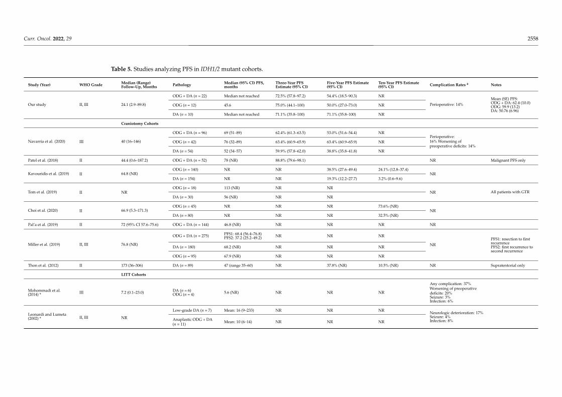
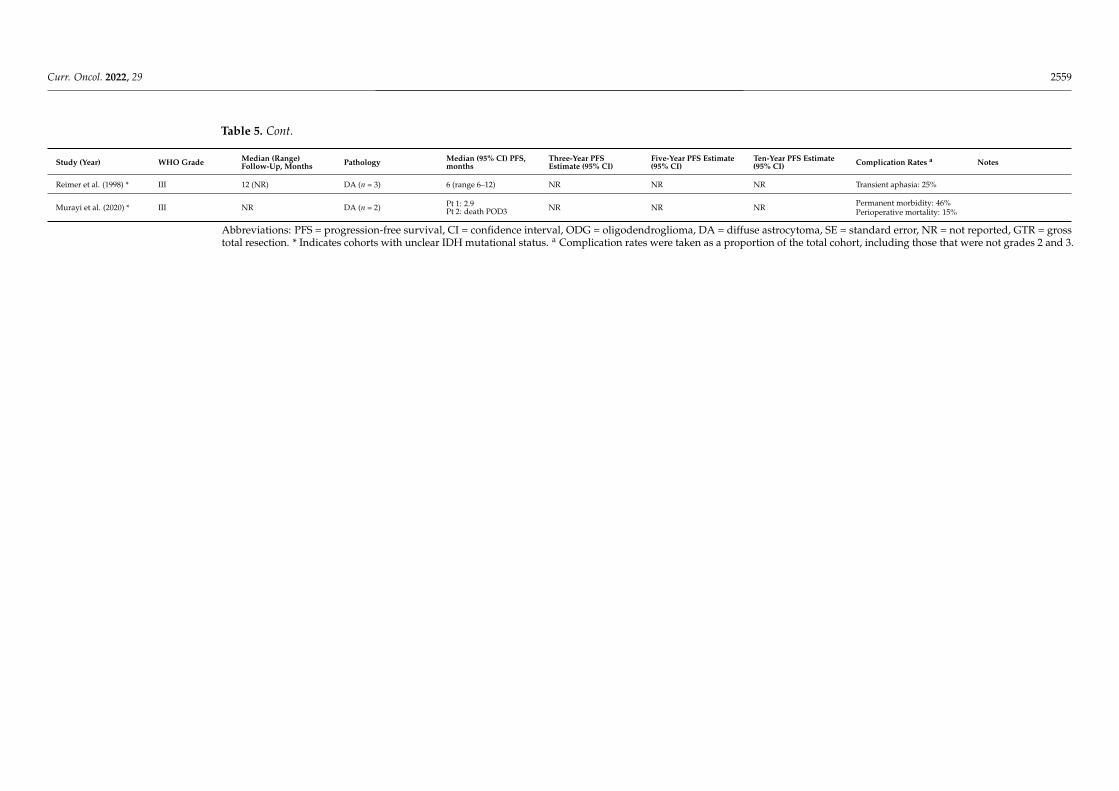
In our cohort, the median PFS was not reached within our median (range) follow-up time of 2.0 (0.2–7.5) years. Our 3- and 5-year PFS estimates were 72.5% and 54.4%,respectively. In our cohort, 22.7% of our patients experienced tumor progression at a mediantime of 18.8 months after LITT. Reported PFS after LITT in grade 2 and 3 astrocytoma andoligodendroglioma have ranged from 3 to 16 months (Table 5) [34–37]. However, dataare limited because many prior case series and reports of gliomas treated with LITT didnot analyze PFS duration. They instead focused on safety profile and complication rates.These cases also did not routinely report the IDH1/2 status of the tumors, and cohortswere presumed to be a mixture of IDH1/2 mutant and wild type. The current case seriesrepresents the first of its kind to analyze treatment outcomes in IDH1/2 mutant gliomain particular.
Curr. Oncol. 2022, 29 2558
Table 5. Studies analyzing PFS in IDH1/2 mutant cohorts.
Study (Year) WHO Grade Median (Range)Follow-Up, Months Pathology Median (95% CI) PFS,
monthsThree-Year PFSEstimate (95% CI)
Five-Year PFS Estimate(95% CI)
Ten-Year PFS Estimate(95% CI) Complication Rates a Notes
Our study II, III 24.1 (2.9–89.8)
ODG + DA (n = 22) Median not reached 72.5% (57.8–97.2) 54.4% (18.5–90.3) NR
Perioperative: 14%Mean (SE) PFS:ODG + DA: 62.4 (10.0)ODG: 59.9 (13.2)DA: 50.76 (6.96)
ODG (n = 12) 45.6 75.0% (44.1–100) 50.0% (27.0–73.0) NR
DA (n = 10) Median not reached 71.1% (35.8–100) 71.1% (35.8–100) NR
Craniotomy Cohorts
Navarria et al. (2020) III 40 (16–146)
ODG + DA (n = 96) 69 (51–89) 62.4% (61.3–63.5) 53.0% (51.6–54.4) NRPerioperative:16% Worsening ofpreoperative deficits: 14%
ODG (n = 42) 76 (32–89) 63.4% (60.9–65.9) 63.4% (60.9–65.9) NR
DA (n = 54) 52 (34–57) 59.9% (57.8–62.0) 38.8% (35.8–41.8) NR
Patel et al. (2018) II 44.4 (0.6–187.2) ODG + DA (n = 52) 78 (NR) 88.8% (79.6–98.1) NR Malignant PFS only
Kavouridis et al. (2019) II 64.8 (NR)ODG (n = 140) NR NR 38.5% (27.6–49.4) 24.1% (12.8–37.4)
NRDA (n = 154) NR NR 19.3% (12.2–27.7) 3.2% (0.6–9.6)
Tom et al. (2019) II NRODG (n = 18) 113 (NR) NR NR
NR All patients with GTRDA (n = 30) 56 (NR) NR NR
Choi et al. (2020) II 66.9 (5.3–171.3)ODG (n = 45) NR NR NR 73.6% (NR)
NRDA (n = 80) NR NR NR 32.5% (NR)
Pal’a et al. (2019) II 72 (95% CI 57.6–75.6) ODG + DA (n = 144) 46.8 (NR) NR NR NR NR
Miller et al. (2019) II, III 76.8 (NR)
ODG + DA (n = 275) PFS1: 68.4 (56.4–76.8)PFS2: 37.2 (25.2–49.2) NR NR NR
NRPFS1: resection to firstrecurrencePFS2: first recurrence tosecond recurrence
DA (n = 180) 68.2 (NR) NR NR NR
ODG (n = 95) 67.9 (NR) NR NR NR
Thon et al. (2012) II 173 (36–306) DA (n = 89) 47 (range 35–60) NR 37.8% (NR) 10.5% (NR) NR Supratentorial only
LITT Cohorts
Mohommadi et al.(2014) * III 7.2 (0.1–23.0) DA (n = 6)
ODG (n = 4) 5.6 (NR) NR NR NR
Any complication: 37%Worsening of preoperativedeficits: 20%Seizure: 3%Infection: 6%
Leonardi and Lumeta(2002) * II, III NR
Low-grade DA (n = 7) Mean: 16 (9–233) NR NR NRNeurologic deterioration: 17%Seizure: 4%Infection: 8%Anaplastic ODG + DA
(n = 11) Mean: 10 (6–14) NR NR NR
Curr. Oncol. 2022, 29 2559
Table 5. Cont.
Study (Year) WHO Grade Median (Range)Follow-Up, Months Pathology Median (95% CI) PFS,
monthsThree-Year PFSEstimate (95% CI)
Five-Year PFS Estimate(95% CI)
Ten-Year PFS Estimate(95% CI) Complication Rates a Notes
Reimer et al. (1998) * III 12 (NR) DA (n = 3) 6 (range 6–12) NR NR NR Transient aphasia: 25%
Murayi et al. (2020) * III NR DA (n = 2) Pt 1: 2.9Pt 2: death POD3 NR NR NR Permanent morbidity: 46%
Perioperative mortality: 15%
Abbreviations: PFS = progression-free survival, CI = confidence interval, ODG = oligodendroglioma, DA = diffuse astrocytoma, SE = standard error, NR = not reported, GTR = grosstotal resection. * Indicates cohorts with unclear IDH mutational status. a Complication rates were taken as a proportion of the total cohort, including those that were not grades 2 and 3.
Curr. Oncol. 2022, 29 2560
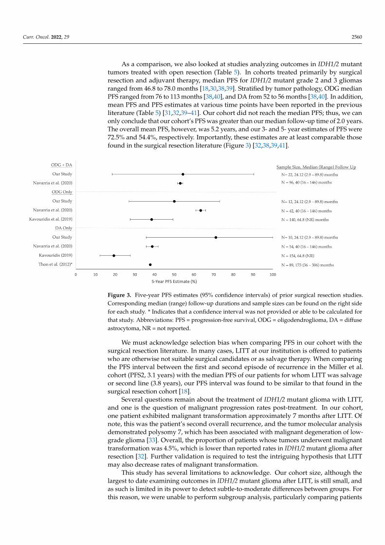
As a comparison, we also looked at studies analyzing outcomes in IDH1/2 mutanttumors treated with open resection (Table 5). In cohorts treated primarily by surgicalresection and adjuvant therapy, median PFS for IDH1/2 mutant grade 2 and 3 gliomasranged from 46.8 to 78.0 months [18,30,38,39]. Stratified by tumor pathology, ODG medianPFS ranged from 76 to 113 months [38,40], and DA from 52 to 56 months [38,40]. In addition,mean PFS and PFS estimates at various time points have been reported in the previousliterature (Table 5) [31,32,39–41]. Our cohort did not reach the median PFS; thus, we canonly conclude that our cohort’s PFS was greater than our median follow-up time of 2.0 years.The overall mean PFS, however, was 5.2 years, and our 3- and 5- year estimates of PFS were72.5% and 54.4%, respectively. Importantly, these estimates are at least comparable thosefound in the surgical resection literature (Figure 3) [32,38,39,41].
Curr. Oncol. 2022, 28, FOR PEER REVIEW 9
Figure 3. Five-year PFS estimates (95% confidence intervals) of prior surgical resection studies. Cor-responding median (range) follow-up durations and sample sizes can be found on the right side for each study. * Indicates that a confidence interval was not provided or able to be calculated for that study. Abbreviations: PFS = progression-free survival, ODG = oligodendroglioma, DA = diffuse as-trocytoma, NR = not reported.
We must acknowledge selection bias when comparing PFS in our cohort with the surgical resection literature. In many cases, LITT at our institution is offered to patients who are otherwise not suitable surgical candidates or as salvage therapy. When compar-ing the PFS interval between the first and second episode of recurrence in the Miller et al. cohort (PFS2, 3.1 years) with the median PFS of our patients for whom LITT was salvage or second line (3.8 years), our PFS interval was found to be similar to that found in the surgical resection cohort [18].
Several questions remain about the treatment of IDH1/2 mutant glioma with LITT, and one is the question of malignant progression rates post-treatment. In our cohort, one patient exhibited malignant transformation approximately 7 months after LITT. Of note, this was the patient’s second overall recurrence, and the tumor molecular analysis demon-strated polysomy 7, which has been associated with malignant degeneration of low-grade glioma [33]. Overall, the proportion of patients whose tumors underwent malignant trans-formation was 4.5%, which is lower than reported rates in IDH1/2 mutant glioma after resection [32]. Further validation is required to test the intriguing hypothesis that LITT may also decrease rates of malignant transformation.
This study has several limitations to acknowledge. Our cohort size, although the larg-est to date examining outcomes in IDH1/2 mutant glioma after LITT, is still small, and as such is limited in its power to detect subtle-to-moderate differences between groups. For this reason, we were unable to perform subgroup analysis, particularly comparing pa-tients treated with first-line versus salvage therapy. Additionally, we were unable to ac-count for heterogeneity in pre-LITT and adjuvant treatments, including those who under-went surgical resection before LITT and those who did not. Our median duration of fol-low-up was shorter than prior literature analyzing outcomes in IDH1/2 mutant gliomas after surgical resection, although our median approached the recommended threshold for IDH1/2 mutant gliomas [16]. Notably, our cohort did not reach the median PFS within our duration of follow-up. While out of the scope of this preliminary, retrospective analysis, a larger, multi-institutional cohort analysis or prospective study is needed to further char-acterize outcomes in IDH1/2 mutant grade 2/3 gliomas treated with LITT and further elu-cidate its effects on PFS compared to both open surgical resection and IDH1/2 wild-type gliomas; this will be the focus of future work.
Figure 3. Five-year PFS estimates (95% confidence intervals) of prior surgical resection studies.Corresponding median (range) follow-up durations and sample sizes can be found on the right sidefor each study. * Indicates that a confidence interval was not provided or able to be calculated forthat study. Abbreviations: PFS = progression-free survival, ODG = oligodendroglioma, DA = diffuseastrocytoma, NR = not reported.
We must acknowledge selection bias when comparing PFS in our cohort with thesurgical resection literature. In many cases, LITT at our institution is offered to patientswho are otherwise not suitable surgical candidates or as salvage therapy. When comparingthe PFS interval between the first and second episode of recurrence in the Miller et al.cohort (PFS2, 3.1 years) with the median PFS of our patients for whom LITT was salvageor second line (3.8 years), our PFS interval was found to be similar to that found in thesurgical resection cohort [18].
Several questions remain about the treatment of IDH1/2 mutant glioma with LITT,and one is the question of malignant progression rates post-treatment. In our cohort,one patient exhibited malignant transformation approximately 7 months after LITT. Ofnote, this was the patient’s second overall recurrence, and the tumor molecular analysisdemonstrated polysomy 7, which has been associated with malignant degeneration of low-grade glioma [33]. Overall, the proportion of patients whose tumors underwent malignanttransformation was 4.5%, which is lower than reported rates in IDH1/2 mutant glioma afterresection [32]. Further validation is required to test the intriguing hypothesis that LITTmay also decrease rates of malignant transformation.
This study has several limitations to acknowledge. Our cohort size, although thelargest to date examining outcomes in IDH1/2 mutant glioma after LITT, is still small, andas such is limited in its power to detect subtle-to-moderate differences between groups. Forthis reason, we were unable to perform subgroup analysis, particularly comparing patients
Curr. Oncol. 2022, 29 2561
treated with first-line versus salvage therapy. Additionally, we were unable to accountfor heterogeneity in pre-LITT and adjuvant treatments, including those who underwentsurgical resection before LITT and those who did not. Our median duration of follow-up was shorter than prior literature analyzing outcomes in IDH1/2 mutant gliomas aftersurgical resection, although our median approached the recommended threshold for IDH1/2mutant gliomas [16]. Notably, our cohort did not reach the median PFS within our durationof follow-up. While out of the scope of this preliminary, retrospective analysis, a larger,multi-institutional cohort analysis or prospective study is needed to further characterizeoutcomes in IDH1/2 mutant grade 2/3 gliomas treated with LITT and further elucidate itseffects on PFS compared to both open surgical resection and IDH1/2 wild-type gliomas;this will be the focus of future work.
5. Conclusions
LITT is an effective alternative to open resection in patients with glioma. In thisstudy, we analyzed the outcomes of patients with IDH1/2 mutant grade 2 and 3 gliomastreated with LITT at a single institution. We found that our cohort had a relatively lowrate of complications that was on par with complication rates seen in craniotomy and LITTcohorts, and malignant progression after LITT was rare. The median time to progressionand three- and five-year PFS estimates in our cohort were on par with those reported in theliterature after surgical resection. While further, multi-institutional studies are needed tobetter characterize treatment outcomes after LITT in patients with IDH1/2 mutant gliomaand to elucidate risk factors for progression, the findings in this study suggest that LITTmay be an effective treatment option for this molecular subtype.
Supplementary Materials: The following are available online at https://www.mdpi.com/article/10.3390/curroncol29040209/s1, Table S1: Univariate time to event and regression analysis.
Author Contributions: Conceptualization, A.H.K. and R.H.H.; methodology, A.H.K., R.H.H. andG.W.J.; formal analysis, G.W.J.; data curation, G.W.J. and R.H.H.; writing—original draft preparation,G.W.J.; writing—review and editing, E.C.L., M.D.S., A.H.K. and R.H.H.; supervision, A.H.K., M.D.S.and E.C.L.; funding acquisition, A.H.K. All authors have read and agreed to the published version ofthe manuscript.
Funding: This work was supported by the Christopher Davidson and Knight Family Fund (to A.H.K.)and the Duesenberg Research Fund (to A.H.K). R.H.H. was supported by a postdoctoral fellowship,PF-21-149-01-CDP, from the American Cancer Society.
Institutional Review Board Statement: The study was conducted in accordance with the Declarationof Helsinki and approved by the Institutional Review Board of Washington University in St. LouisSchool of Medicine (ID# 201409046, approval date: 3 February 2022).
Informed Consent Statement: Patient consent was waived by the institution’s Institutional ReviewBoard due to the retrospective nature of the study.
Data Availability Statement: The dataset used and/or analyzed during the current study are avail-able from the corresponding author on reasonable request.
Conflicts of Interest: A.H.K. is a consultant for Monteris Medical and has received research grantsfrom Monteris Medical for a mouse laser therapy study as well as from Stryker and Collagen Matrixfor clinical outcomes studies about a dural substitute, which have no direct relation to this study.E.C.L.’s disclosures are as follows: Stock ownership: Neurolutions, General Sensing, Osteovantage,Pear Therapeutics, Face to Face Biometrics, Immunovalent, Caeli Vascular, Acera, Sora Neuroscience,Inner Cosmos, Kinetrix, NeuroDev. Consultant: Monteris Medical, E15, Acera, Alcyone, IntellectualVentures, Neurolutions, Osteovantage, Pear Therapeutics, Inc., Sante Ventures, Microbot. SAB: PearTherapeutics, Microbot. Licensing from Intellectual Property: Neurolutions, Osteovantage, CaeliVascular. Licensing/product development agreements or royalties for inventions/IP: Cerovations,Intellectual Ventures. Washington University owns equity in Neurolutions. M.D.S. is a consultant forMonteris Medical. R.H.H. and G.W.J. have nothing to disclose.
Curr. Oncol. 2022, 29 2562
References1. Bown, S.G. Phototherapy of Tumors. World J. Surg. 1983, 7, 700–709. [CrossRef]2. Sugiyama, K.; Sakai, T.; Fujishima, I.; Ryu, H.; Uemura, K.; Yokoyama, T. Stereotactic Interstitial Laser-Hyperthermia Using
Nd-YAG Laser. Stereotact. Funct. Neurosurg. 1990, 54, 501–505. [CrossRef]3. Karampelas, I.; Sloan, A.E. Laser-Induced Interstitial Thermotherapy of Gliomas. Prog. Neurol. Surg. 2018, 32, 14–26. [CrossRef]4. Chen, C.; Lee, I.; Tatsui, C.; Elder, T.; Sloan, A.E. Laser Interstitial Thermotherapy (LITT) for the Treatment of Tumors of the Brain
and Spine: A Brief Review. J. Neuro-Oncol. 2021, 151, 429–442. [CrossRef]5. Rahmathulla, G.; Recinos, P.F.; Kamian, K.; Mohammadi, A.M.; Ahluwalia, M.S.; Barnett, G.H. MRI-Guided Laser Interstitial
Thermal Therapy in Neuro-Oncology: A Review of Its Current Clinical Applications. Oncology 2014, 87, 67–82. [CrossRef]6. Ashraf, O.; Patel, N.V.; Hanft, S.; Danish, S.F. Laser-Induced Thermal Therapy in Neuro-Oncology: A Review. World Neurosurg.
2018, 112, 166–177. [CrossRef]7. Hong, C.S.; Deng, D.; Vera, A.; Chiang, V.L. Laser-Interstitial Thermal Therapy Compared to Craniotomy for Treatment of
Radiation Necrosis or Recurrent Tumor in Brain Metastases Failing Radiosurgery. J. Neuro-Oncol. 2019, 142, 309–317. [CrossRef]8. Carpentier, A.; McNichols, R.J.; Stafford, R.J.; Guichard, J.-P.; Reizine, D.; Delaloge, S.; Vicaut, E.; Payen, D.; Gowda, A.; George, B.
Laser Thermal Therapy: Real-Time MRI-Guided and Computer-Controlled Procedures for Metastatic Brain Tumors. Lasers Surg.Med. 2011, 43, 943–950. [CrossRef]
9. Rao, M.S.; Hargreaves, E.L.; Khan, A.J.; Haffty, B.G.; Danish, S.F. Magnetic Resonance-Guided Laser Ablation Improves LocalControl for Postradiosurgery Recurrence and/or Radiation Necrosis. Neurosurgery 2014, 74, 658–667. [CrossRef]
10. Rahmathulla, G.; Recinos, P.F.; Valerio, J.E.; Chao, S.; Barnett, G.H. Laser Interstitial Thermal Therapy for Focal Cerebral RadiationNecrosis: A Case Report and Literature Review. Stereotact. Funct. Neurosurg. 2012, 90, 192–200. [CrossRef]
11. Miller, B.A.; Salehi, A.; Limbrick, D.D.; Smyth, M.D. Applications of a Robotic Stereotactic Arm for Pediatric Epilepsy andNeurooncology Surgery. J. Neurosurg. Pediatrics 2017, 20, 364–370. [CrossRef]
12. Tovar-Spinoza, Z.; Carter, D.; Ferrone, D.; Eksioglu, Y.; Huckins, S. The Use of MRI-Guided Laser-Induced Thermal Ablation forEpilepsy. Child’s Nerv. Syst. 2013, 29, 2089–2094. [CrossRef]
13. Buckley, R.T.; Wang, A.C.; Miller, J.W.; Novotny, E.J.; Ojemann, J.G. Stereotactic Laser Ablation for Hypothalamic and DeepIntraventricular Lesions. Neurosurg. Focus 2016, 41, E10. [CrossRef]
14. Barnett, G.H.; Voigt, J.D.; Alhuwalia, M.S. A Systematic Review and Meta-Analysis of Studies Examining the Use of Brain LaserInterstitial Thermal Therapy versus Craniotomy for the Treatment of High-Grade Tumors in or near Areas of Eloquence: AnExamination of the Extent of Resection and Major Complication Rates Associated with Each Type of Surgery. Stereotact. Funct.Neurosurg. 2016, 94, 164–173. [CrossRef]
15. Parsons, D.W.; Jones, S.; Zhang, X.; Lin, J.C.-H.; Leary, R.J.; Angenendt, P.; Mankoo, P.; Carter, H.; Siu, I.-M.; Gallia, G.L.; et al. AnIntegrated Genomic Analysis of Human Glioblastoma Multiforme. Science 2008, 321, 1807–1812. [CrossRef]
16. Miller, J.J.; Shih, H.A.; Andronesi, O.C.; Cahill, D.P. Isocitrate Dehydrogenase-Mutant Glioma: Evolving Clinical and TherapeuticImplications. Cancer 2017, 123, 4535–4546. [CrossRef]
17. Yan, H.; Parsons, D.W.; Jin, G.; McLendon, R.; Rasheed, B.A.; Yuan, W.; Kos, I.; Batinic-Haberle, I.; Jones, S.; Riggins, G.J.; et al.IDH1 and IDH2 Mutations in Gliomas. N. Engl. J. Med. 2009, 360, 765–773. [CrossRef]
18. Miller, J.J.; Loebel, F.; Juratli, T.A.; Tummala, S.S.; Williams, E.A.; Batchelor, T.T.; Arrillaga-Romany, I.; Cahill, D.P. AcceleratedProgression of IDH Mutant Glioma after First Recurrence. Neuro-Oncology 2019, 21, 669–677. [CrossRef]
19. Olar, A.; Wani, K.M.; Alfaro-Munoz, K.D.; Heathcock, L.E.; van Thuijl, H.F.; Gilbert, M.R.; Armstrong, T.S.; Sulman, E.P.; Cahill,D.P.; Vera-Bolanos, E.; et al. IDH Mutation Status and Role of WHO Grade and Mitotic Index in Overall Survival in Grade II–IIIDiffuse Gliomas. Acta Neuropathol. 2015, 129, 585–596. [CrossRef]
20. Reuss, D.E.; Mamatjan, Y.; Schrimpf, D.; Capper, D.; Hovestadt, V.; Kratz, A.; Sahm, F.; Koelsche, C.; Korshunov, A.; Olar, A.; et al.IDH Mutant Diffuse and Anaplastic Astrocytomas Have Similar Age at Presentation and Little Difference in Survival: A GradingProblem for WHO. Acta Neuropathol. 2015, 129, 867–873. [CrossRef]
21. Weller, M.; van den Bent, M.; Preusser, M.; le Rhun, E.; Tonn, J.C.; Minniti, G.; Bendszus, M.; Balana, C.; Chinot, O.; Dirven,L.; et al. EANO Guidelines on the Diagnosis and Treatment of Diffuse Gliomas of Adulthood. Nat. Rev. Clin. Oncol. 2021, 18,170–186. [CrossRef]
22. Natsume, K.; Sakakima, H.; Kawamura, K.; Yoshida, A.; Akihiro, S.; Yonezawa, H.; Yoshimoto, K.; Shimodozono, M. FactorsInfluencing the Improvement of Activities of Daily Living during Inpatient Rehabilitation in Newly Diagnosed Patients withGlioblastoma Multiforme. J. Clin. Med. 2022, 11, 417. [CrossRef]
23. Montemurro, N.; Fanelli, G.N.; Scatena, C.; Ortenzi, V.; Pasqualetti, F.; Mazzanti, C.M.; Morganti, R.; Paiar, F.; Naccarato, A.G.;Perrini, P. Surgical Outcome and Molecular Pattern Characterization of Recurrent Glioblastoma Multiforme: A Single-CenterRetrospective Series. Clin. Neurol. Neurosurg. 2021, 207, 106735. [CrossRef]
24. Easwaran, T.; Lion, A.; Vortmeyer, A.; Kingery, K.; Bc, M.D.; Raskin, J. Seizure Freedom from Recurrent Insular Low-GradeGlioma Following Laser Interstitial Thermal Therapy. Child’s Nerv. Syst. 2020, 36, 1055–1059. [CrossRef]
25. Hafez, D.M.; Liekweg, C.; Leuthardt, E.C. Staged Laser Interstitial Thermal Therapy (LITT) Treatments to Left Insular Low-GradeGlioma. Clin. Neurosurg. 2020, 86, E337–E342. [CrossRef]
26. Avecillas-Chasin, J.M.; Atik, A.; Mohammadi, A.M.; Barnett, G.H. Laser Thermal Therapy in the Management of High-GradeGliomas. Int. J. Hyperth. 2020, 37, 44–52. [CrossRef]
Curr. Oncol. 2022, 29 2563
27. Hawasli, A.H.; Kim, A.H.; Dunn, G.P.; Tran, D.D.; Leuthardt, E.C. Stereotactic Laser Ablation of High-Grade Gliomas. Neurosurg.Focus 2014, 37, E1. [CrossRef]
28. Louis, D.N.; Perry, A.; Wesseling, P.; Brat, D.J.; Cree, I.A.; Figarella-Branger, D.; Hawkins, C.; Ng, H.K.; Pfister, S.M.; Reifenberger,G.; et al. The 2021 WHO Classification of Tumors of the Central Nervous System: A Summary. Neuro-Oncology 2021, 23, 1231–1251.[CrossRef]
29. Lee, I.; Kalkanis, S.; Hadjipanayis, C.G. Stereotactic Laser Interstitial Thermal Therapy for Recurrent High-Grade Gliomas.Neurosurgery 2016, 79, S24–S34. [CrossRef]
30. Pal’a, A.; Coburger, J.; Scherer, M.; Ahmeti, H.; Roder, C.; Gessler, F.; Jungk, C.; Scheuerle, A.; Senft, C.; Tatagiba, M.; et al. To Treator Not to Treat? A Retrospective Multicenter Assessment of Survival in Patients with IDH-Mutant Low-Grade Glioma Based onAdjuvant Treatment. J. Neurosurg. 2020, 133, 273–280. [CrossRef]
31. Choi, J.; Kim, S.H.; Ahn, S.S.; Choi, H.J.; Yoon, H.I.; Cho, J.H.; Roh, T.H.; Kang, S.G.; Chang, J.H.; Suh, C.O. Extent of Resection andMolecular Pathologic Subtype Are Potent Prognostic Factors of Adult WHO Grade II Glioma. Sci. Rep. 2020, 10, 2086. [CrossRef]
32. Thon, N.; Eigenbrod, S.; Kreth, S.; Lutz, J.; Tonn, J.C.; Kretzschmar, H.; Peraud, A.; Kreth, F.W. IDH1 Mutations in Grade IIAstrocytomas Are Associated with Unfavorable Progression-Free Survival and Prolonged Postrecurrence Survival. Cancer 2012,118, 452–460. [CrossRef]
33. Idbaih, A.; Carvalho Silva, R.; Crinière, E.; Marie, Y.; Carpentier, C.; Boisselier, B.; Taillibert, S.; Rousseau, A.; Mokhtari, K.; Ducray,F.; et al. Genomic Changes in Progression of Low-Grade Gliomas. J. Neuro-Oncol. 2008, 90, 133–140. [CrossRef]
34. Murayi, R.; Borghei-Razavi, H.; Barnett, G.H.; Mohammadi, A.M. Laser Interstitial Thermal Therapy in the Treatment of ThalamicBrain Tumors: A Case Series. Oper. Neurosurg. 2020, 19, 641–650. [CrossRef]
35. Reimer, P.; Bremer, C.; Horch, C.; Morgenroth, C.; Allkemper, T.; Schuierer, G. MR-Monitored LITT as a Palliative Concept inPatients with High Grade Gliomas: Preliminary Clinical Experience. J. Magn. Reson. Imaging 1998, 8, 240–244. [CrossRef]
36. Mohammadi, A.M.; Hawasli, A.H.; Rodriguez, A.; Schroeder, J.L.; Laxton, A.W.; Elson, P.; Tatter, S.B.; Barnett, G.H.; Leuthardt,E.C. The Role of Laser Interstitial Thermal Therapy in Enhancing Progression-free Survival of Difficult-to-access High-gradeGliomas: A Multicenter Study. Cancer Med. 2014, 3, 971–979. [CrossRef]
37. Leonardi, M.A.; Lumenta, C.B. Stereotactic Guided Laser-Induced Interstitial Thermotherapy (SLITT) in Gliomas with Intra-operative Morphologic Monitoring in an Open MR: Clinical Expierence. MIN Minim. Invasive Neurosurg. 2002, 45, 201–207.[CrossRef]
38. Navarria, P.; Pessina, F.; Clerici, E.; Rossini, Z.; Franceschini, D.; D’Agostino, G.; Franzese, C.; Comito, T.; Loi, M.; Simonelli, M.;et al. Is IDH Status the Only Factor Predicting Prognosis in Newly Diagnosed Anaplastic Glioma Patients? Outcome Evaluationand Prognostic Factor Analysis in a Single-Institution Large Series. J. Neurosurg. 2021, 135, 64–77. [CrossRef]
39. Patel, T.; Bander, E.D.; Venn, R.A.; Powell, T.; Cederquist, G.Y.M.; Schaefer, P.M.; Puchi, L.A.; Akhmerov, A.; Ogilvie, S.; Reiner,A.S.; et al. The Role of Extent of Resection in IDH1 Wild-Type or Mutant Low-Grade Gliomas. Neurosurgery 2018, 82, 808–814.[CrossRef]
40. Tom, M.C.; Varra, V.; Leyrer, C.M.; Park, D.Y.; Chao, S.T.; Yu, J.S.; Suh, J.H.; Reddy, C.A.; Balagamwala, E.H.; Broughman, J.R.; et al.Risk Factors for Progression Among Low-Grade Gliomas After Gross Total Resection and Initial Observation in the MolecularEra. Int. J. Radiat. Oncol. Biol. Phys. 2019, 104, 1099–1105. [CrossRef]
41. Kavouridis, V.K.; Boaro, A.; Dorr, J.; Cho, E.Y.; Iorgulescu, J.B.; Reardon, D.A.; Arnaout, O.; Smith, T.R. Contemporary Assessmentof Extent of Resection in Molecularly Defined Categories of Diffuse Low-Grade Glioma: A Volumetric Analysis. J. Neurosurg.2020, 133, 1291–1301. [CrossRef]
Copyright © 2022 FDOKUMEN