Laser Interstitial Thermal Therapy in Grade 2/3 IDH1/2 Mutant ...
Upload
istituto-bestaCategory
view
1download
0
Pellegatta et al. Acta Neuropathologica Communications (2015) 3:4 DOI 10.1186/s40478-014-0180-0
RESEARCH Open Access
Effective immuno-targeting of the IDH1 mutationR132H in a murine model of intracranial gliomaSerena Pellegatta1, Lorella Valletta1, Cristina Corbetta1, Monica Patanè1, Ileana Zucca1, Federico Riccardi Sirtori2,Maria Grazia Bruzzone1, Gianpaolo Fogliatto2, Antonella Isacchi2, Bianca Pollo1 and Gaetano Finocchiaro1*
Abstract
The R132H mutation of cytosolic isocitrate dehydrogenase (IDH1) is present in the majority of low grade gliomas.Immunotherapy in these tumors has an interesting, still unexploited, therapeutic potential, as they are lessimmunosuppressive than glioblastomas.Using site-directed mutagenesis we introduced the R132H mutation into the murine glioma cell line GL261,creating mIDH1-GL261. Presence of the mutation was confirmed by immunoblotting and production of theoncometabolite 2-hydroxyglutarate (2HG), demonstrated by mass spectrometry (LC-MS/MS) performed on cellsupernatant. In vitro mIDH1-GL261 had different morphology but similar growth rate than parental GL261(p-GL261). After intracranial injection, MRI suggested that the initial growth rate was slower in mIDH1-GL261 thanp-GL261 gliomas but overall survival was similar. mIDH1-GL261 gliomas showed evidence of R132H expression andof intratumoral 2HG production (evaluated by MRS and LC-MS/MS). Immunizations were performed nine days afterintracranial implantation of mIDH1- or p-GL261 cells by three subcutaneous injections of five different peptidesencompassing the IDH1 mutation site, all emulsified with Montanide ISA-51, in association with GM-CSF. Controlmice were injected with four ovalbumin peptides or vehicle. Mice with mIDH1-GL261 but not p-GL261 gliomastreated with mIDH1 peptides survived longer than controls; 25% of them were cured. Immunized mice showedhigher amounts of peripheral CD8+ T cells, higher production of IFN-γ, and evidence of anti-mIDH1 antibodies.Immunizations led to intratumoral up-regulation of IFN-γ, granzyme-b and perforin-1 and down-regulation ofTGF-β2 and IL-10.These results support the translational potential of immunotherapeutic targeting of gliomas carrying IDH1mutations.
Keywords: IDH1-R132H, Immunotherapy, Glioma, 2-hydroxyglutarate
IntroductionThe new era of cancer genomics, due to the revolutionof next generation sequencing, was heralded in 2008 bythe discovery of mutations in the IDH1 gene in glioblast-omas (GBM) [1]. Subsequently it was found that IDH1mutations are present in the large majority of low gradegliomas (LGG) and define secondary GBM more rigorouslythan before [2]. Remarkably, a new function of IDH1 wasfound as a consequence of the R132H mutation that affectsthe active site of the enzyme [3]. IDH1, that normally
* Correspondence: [email protected] of Neuro-Oncology, Unit of Molecular Neuro-Oncology,Fondazione I.R.C.C.S. Istituto Neurologico C. Besta, via Celoria 11, 20133Milano, ItalyFull list of author information is available at the end of the article
© 2015 Pellegatta et al.; licensee BioMed CentCommons Attribution License (http://creativecreproduction in any medium, provided the orDedication waiver (http://creativecommons.orunless otherwise stated.
catalyzes the production of alpha-ketoglutarate (alpha-KG),in its mutated form released 2-hydroxyglutarate (2HG),subsequently defined as an oncometabolite [3]. Mutationsof the cytosolic IDH1, as well as of the mitochondrialisoform IDH2, were subsequently found in other tumorslike cholangiocarcinomas [4], leukemias [5] and others,reviewed in [6]. In gliomas, one main consequence of theR132H mutation is the alteration of the epigenetic pat-terns, as 2HG may act as a competitive inhibitor of histoneand DNA demethylases requiring alpha-KG as a cofactor[7-9]. Initially, increased neo-angiogenesis due to increasedexpression of the pro-angiogenic factor HIF-1α was alsofound as a consequence of IDH1 mutation [10].Due to mutation frequency and importance of the
modulation of glioma biology, an increasing effort has
ral. This is an Open Access article distributed under the terms of the Creativeommons.org/licenses/by/4.0), which permits unrestricted use, distribution, andiginal work is properly credited. The Creative Commons Public Domaing/publicdomain/zero/1.0/) applies to the data made available in this article,
Pellegatta et al. Acta Neuropathologica Communications (2015) 3:4 Page 2 of 12
been placed on pharmacological targeting of mutatedIDH1. New drugs that are acting on the novel function ofIDH1 are under development and are initially tested inpatients [11]. Furthermore, the diagnosis of the mutationin peripheral blood as well as the detection of increasedlevels of 2HG by MRI in the brain are actively pursuedand initial results are available [12,13].As in the case of EGFRvIII, the R132H mutation cre-
ates a novel, cancer-specific epitope, that could be po-tentially targeted by immunotherapy, as recently shownby Schumacher et al. [14]. This could be particularly ef-fective in LGG as these tumors, due to their associationwith longer survival than GBM [15] and with a less im-munosuppressive microenvironment [16], may offer awider window for the development of a therapeuticallymeaningful immune response. To this goal we have de-veloped a glioma model of the R132H mutation that isimmunologically actionable. Our results suggest thatpeptide vaccination may delay and, in a fraction of cases,cure an intracranial glioma that is otherwise lethal inabout one month.
Materials and methodsGeneration of GL261-IDH1-R132H cell lineSite directed mutagenesis was performed on pCMV6kan-neo vector containing the murine wild-type IDH1 cDNA(NM_010497.3) (Origene, Rockville, MD, USA), humanIDH1mutation R132H was introduced (see Additionalfile 1).GL261 adherent cells were seeded in 6-well plates at
2 × 10⁶ cells per well 24 hours prior to the transfection.Cells were transfected with 10 μg of empty or IDH1-R132H– DNA vector using Lipofectamine 2000 Transfec-tion Reagent (LifeTechnologies), according to the manufac-turer’s instructions. Twenty-four hours after transfection,GL261 cells were split and grown in fresh medium contain-ing 0.5 mg/ml of G418 (Lonza) as selective agent.Parental and IDH1-GL261 were grown in DMEM, 1X
Pen Strep,1X L-glu, 20% Fetal Bovine Serum (LifeTechnol-ogies). The cell Proliferation Reagent WST-1 (Roche) wasused to measure proliferation plating 3000 cells/well. Sixreplicates per point were analyzed.
Peptide predictionMice were immunized with two 9-mer (PEP1 - IDH1-R132H126–134: KPIIIGHHA; PEP2 - IDH1-R132H128–136:IIIGHHAYG), two 10-mer (PEP3 - IDH1-R132H131–140:IGHHAYGDQY; PEP4 - IDH1-R132H123–132: GWVKPIIIGH) and a 16-mer (PEP5 - IDH1-R132H126–141: KPIIIGHHAYGDQYRA) peptides spanning the IDH1 mutation.Negative control peptides were: OVA155–62: KVVRFDKL;OVA2107–114: AEERYPIL; OVA3176–183: NAIVFKGL,synthesized by Primm srl (Milano), and OVA4257–264:SIINFEKL (Sigma Aldrich).
In vivo treatmentsFive week-old females C57BL/6N mice (Charles RiverLaboratory, Calco-Lecco Italy) were injected with 50,000pGL261 cells (n = 44) or mIDH1-GL261 (n = 80). A totalof 24 mIDH1-GL261 glioma bearing mice were sacri-ficed after second immunization for immunological andhistological studies (6 mice per group of treatment). Thestereotactic coordinates with respect to the bregmawere: 0.7 mm posterior, 3 mm left lateral, and 3.5 mmdeep, into the nucleus caudatum. Glioma-bearing micewere treated using four different conditions: group I:vehicle only (GM-CSF and Montanide); group II: threeimmunizations spaced one week apart (days 9, 16 and23) with two 9-mer and two 10-mer peptides; group III:three immunization spaced one week apart (days 9, 16and 23) with the 16-mer peptide; group IV: ova peptideimmunizations. Single peptides (15 μg/peptide) wereemulsified with Montanide ISA-51 VG (1:1; SEPPIC) aspreviously described [17,18], and administered by sub-cutaneous injections of the 16-mer peptide (group III)or the four peptides separately (group II and group IV)into different areas of the flank. The immunized mice re-ceived a total of 3 μg of recombinant murine granulocytemacrophage colony-stimulating factor (GM-CSF) (MiltenyiBiotec) spread out over three injections into the same areaof the peptide injections, beginning one day before the firstvaccination.Animal experiments were performed in accordance to
the Italian Principle of Laboratory Animal Care (D. Lgs.26/2014) and European Communities Council Directives(86/609/EEC and 2010/63/UE).
Isolation and characterization of splenocytesSplenocytes from immunized and control mice wereisolated after the second immunization. Before staining,splenocytes were grown in RPMI 1640 (EuroClone) con-taining 10% heat-inactivated fetal bovine serum (FBS)(Life Technologies), 100 U/mL penicillin (EuroClone),100 U/mL streptomycin (EuroClone), 100 μg/mL glu-tamine (EuroClone), 0.1 mM non-essential amino acids(EuroClone), 1 mM sodium pyruvate (EuroClone), 50 μMβ-mercaptoethanol (EuroClone) and 10 U/mL IL-2(Miltenyi Biotec). Cells were stimulated for 4 h with0.25 μM ionomycin and 10 ng/mL phorbol myristateacetate (PMA) and 2 hours with 10 μg/ml brefeldinA. A total of 1.5 × 10⁶ cells were stained in PBS 1X/0.5% bovine serum albumin/2 mM EDTA for 10 minat 4°C with the following antibodies (Miltenyi Biotec):anti-CD3 Vioblue; anti-CD4-APC, anti-CD8-PE-Vio770,anti-CD62L-FITC and anti-CD49d-PE for T cell detection.The cells were then fixed and permealized using theMiltenyi Biotec Inside Stain Kit and intracellularly stainedaccording to the manufacturer’s instructions with IFN-γ-PE or APC (Miltenyi Biotec). The CD8 +CD3+; CD3 +
Pellegatta et al. Acta Neuropathologica Communications (2015) 3:4 Page 3 of 12
CD4+ cells were gated and then analyzed by flow cytome-try for IFN-γ production and for the evaluation of CD62Land CD49d expression. Flow cytometry acquisition wasperformed on a MACSQuant instrument, and the datawere analyzed with the MACSQuantify Software (MiltenyiBiotec).
Cytotoxic and proliferation assaysLymphocytes from immunized and control (vehicle andova-treated) mice were isolated after second immuni-zation. A total of 2 × 10⁶ splenocytes were pre-stimulatedfor 5 days in the presence of 5 × 10⁵ irradiated (3-Gy),naïve splenocytes, acting as antigen presenting cells, and10 μg/mL of each peptide. Pre-stimulated splenocyteswere tested for GL261-specific cytotoxicity using 10:1,20:1 and 40:1 effector:target (E:T) ratios. The cytotoxicMTT assay was performed according to the manu-facturer’s instructions (Millipore). For proliferation assay,2 × 106 splenocytes were primed for 4 days in the presenceof 5 × 10⁵ naïve splenocytes that had received 3 Gy irradi-ation, 10 μg/mL of IDH1 mutated or OVA peptides and10 U/ml of IL-2. After pre-stimulation, 5 × 10⁵ splenocyteswere incubated for 20 h in the presence of single peptides(10 μg/ml) and 10 U/ml IL-2 and tested for their ability toproliferate using MTT Reagent (Millipore). The data areexpressed as the percentage of proliferation, calculatedaccording to the following equation: (OD stimulatedsplenocytes – OD splenocytes without peptide)/ODstimulated splenocytes × 100.
IgG ELISAELISA plates were coated with 0.01 mg/ml IDH1 R132Hpeptides for IgG detection in sera of immunized andcontrol mice. Ovalbumin peptides were used as peptidecoating control. The plate was washed and blocked withPBS 10% FCS for 2 h at room temperature. The platewas washed and mouse sera (1:100) were incubated for2 h at room temperature. After washing the plate, HRP-conjugate secondary antibody IgG-HRP (1:5000) wasincubated for 1 h in the dark at room temperature. Theplate was washed and incubate with tetramethylbenzi-dine (TMB) and the reaction was stopped with H2SO4
1 M. OD at 450 nm was measured.
Real-time PCR (RT-PCR)Total RNA was isolated from freshly harvested GL261gliomas and lymphocytes from immunized and controlmice and used for gene expression analysis. RNA wasextracted with TRIzol reagent (Life Technologies) usingthe RNeasy MINI KIT (Qiagen) and the RNase-FreeDNase Set (Qiagen). cDNA was synthesized from totalRNA using oligo (dT) and M-MLV Reverse Transcript-ase (Life Technologies). Specific primers for target geneswere designed for Fast SYBR Green chemistry (Life
Technologies) and purchased from Primm S.r.l. Therelative mRNA levels were evaluated using a ViiA-7Real-Time PCR System (Life Technologies) and calcu-lated using the ΔΔCt method. The expression levels ofthe target genes were normalized to the expression levelof beta-actin (see oligo sequences in Additional file 1).
Western blotTotal proteins were isolated using tissue lysis with PKbuffer (NaH2PO4 0.2 M; Na2HPO4 0.2 M; NaCl 5 M;EDTA) and protease inhibitor buffer (Triton 100X 2%;SDS 0.25%; Leupeptin 0.90%; Pepstatin A 0.90%; PMSF0.90%), and centrifuged to pellet debris. Protein concen-trations were measured by Micro BCA protein assay kit(Thermo Scientific) at 540 nm. Proteins (20 μg) werediluted in NuPAGE® LDS Sample Buffer 4X (Invitrogen)and NuPAGE Reducing Agent 10X (Invitrogen) and elec-troblotted onto nitrocellulose membranes at 30 Volts for1 h . Membranes with transferred proteins were incubatedwith the primary antibody anti-IDH1-R132H (1:100,Dianova) or anti-vinculin (1:5000, Abcam, Cambridge,UK). The primary antibody incubation was followedby incubation with the secondary antibody anti-mouse(1:10000). A chemiluminescence reaction using the ECL(enhanced chemiluminescence) Plus kit (Amersham, GEHealthcare) was detected using film.
Liquid chromatography (LC-MS/MS) and magnetic resonancespectroscopyTumour lysates (100 μL) were denatured by adding10 μL of thrichloroacetic acid (TCA) 1 M containing2HG-d3 (130 μM) as internal standard. After mixingand centrifugation, the supernatants were analyzed for2-hydroxyglutaric (2HG) determination using an UltraHigh Pressure Liquid Chromatography system (UPLC®,Waters) coupled with a triple quadrupoles mass spec-trometer (TQD, Waters) operating in multiple reactionmonitoring mode (MRM). Protein normalization wasobtained using the microBCA kit (Thermo Scientific).Supernatants were obtained from a sub-confluent cellcultures (3 × 10⁶ cells).MRI/MRS studies were performed on a 7 Tesla BioS-
pec 70/30 USR scanner equipped with a 12 cm innerdiameter actively shielded gradient system reaching amaximum amplitude of 400 mT/m. (Bruker BioSpin,Ettlingen, Germany). T2-weighted images were acquiredin three orthogonal (axial, sagittal, and coronal planes)using a rapid acquisition with relaxation enhancement(RARE) sequence (TR/TE = 3000/13 ms, matrix size =256 × 256, RARE factor = 8, slice thickness = 0.7 mm,FOV = 2 × 2 cm2, in plane resolution = 78 μm, numberof averages(NA) = 1, acquisition time (AT) = 1 min 30 s).1H spectra carried out by two PRESS sequence withdifferent Echo Time (TE) TE =13 ms and 50 ms
Pellegatta et al. Acta Neuropathologica Communications (2015) 3:4 Page 4 of 12
(Point RESolved Spectroscopy, TR = 5000 ms, and ad-justment of the first and second order shims conductedbeforehand by Mapshim macro of Paravision 5.1 software)from a single voxel (~10 μL) located inside a tumor (or,before detecting solid tumor, at the injection site). Thespectra were analyzed with LCModel software (Version6.3) [19] for metabolites absolute quantification. The me-tabolites concentration for each single mouse at differenttime point (before, one week, twenty days and one monthafter the tumor cells injection) are presented as absoluteconcentration ± CRB error [20]. The metabolites withCRB error less than 20% of the estimated metabolite con-centration were considered. Also the relative concentra-tion with respect to Creatine is reported.
Histology and immunohistochemistryFor histology of murine gliomas, brains were carefully re-moved, post-fixed in 4% paraformaldehyde, and embeddedin paraffin. Five μm thick sections were dissected using amicrotome. IHC analysis for IDH1-R132H was performedon paraffin-embedded sections using the anti-IDH1-R132Hantibody (Dianova). The paraffin was removed using xylene,and the sections were rehydrated in graded alcohol.Antigen retrieval was carried out using preheated tar-get retrieval solution (pH 6.0), and the primary anti-bodies were incubated overnight. Single immunostainswere performed using a standard immunoperoxidaseprotocol (Vectastain Elite ABC kit, PK-6100; VectorLaboratories, Inc., Burlingame, CA, USA) followed by adiaminobenzidine chromogen reaction (Peroxidase sub-strate kit, DAB, SK-4100; Vector Lab). All sections werecounterstained with Mayer’s hematoxylin and visualizedusing a LEICA MDLB light microscope.
Statistical analysisStatistical comparisons of the data sets were performedusing a two-tailed Student’s T-test, and the results wereconsidered significant at p < 0.05. Cumulative survivalcurves were constructed using the Kaplan-Meier method(MedCalc 9.3).
ResultsGL261-cells overexpressing IDH1-R132H produce 2HG andare tumorigenic in vivoTo verify the potential effects of IDH1 mutation ontumor progression, we over-expressed IDH1-R132H inGL261 cells creating mIDH1-GL261. Expression of mIDH1mRNA was detectable by RT-PCR in mIDH1-GL26115 days after selection with G418, and highly increased onday 24 after transfection (Figure 1a). Accordingly, expres-sion of the mutant protein was clearly observed from day15 by Western blot with R132H mutation specific anti-bodies (Figure 1b). At the same time point mIDH1-GL261cells appeared smaller that pGL261 and with fibroblast-like
features (Figure 1c). No significant difference in prolifera-tion was visible in vitro, as evaluated at four time points(24, 48, 72 and 96 h, Figure 1d). mIDH1-GL261 released2HG in micromolar amounts in the culture medium, asmeasured by LC-MS/MS (Figure 1e).The overall survival of mice injected intracranially
with mIDH1-GL261 cells appeared initially longer thanin pGL261 controls, but the two curves merged later(Figure 1f ). Observations on tumor size by MRI were co-herent with survival data. Eight days after implantation, inboth groups no signal alteration on T2-weighted (wi) im-ages was evident along the injection track (Figure 1g). Onday 20 the tumor onset was detected as a bright area onthe T2-wi images. pGL261-gliomas were growing morerapidly than mIDH1-gliomas but remained small andcircumscribed (Figure 1h). Thirty days after tumor cell in-jection, the mIDH1-GL261 gliomas were still smaller butbrighter than pGL261 gliomas (Figure 1i). By RT-PCR onexplanted gliomas we found that the expression of IDH1was significantly reduced over the time, from 48.8 ± 1.3 onday 20 to 24. ± 0.4 on day 30 (p < 0.001) (Figure 1l).Histology with H&E staining showed that pGL261-
gliomas were characterized by pleomorphic and mitoti-cally active cells, while mIDH1-gliomas were composedof single small and round cells occasionally organized ascell nodules (Figure 1m). Immunostaining with specificantibodies confirmed the expression of IDH1-R132H ingliomas originating from mIDH1-GL261, but not inpGL261 gliomas (Figure 1n).
mIDH1-GL261 gliomas maintain 2HG productionIn order to evaluate the absolute concentration of 2HG,magnetic resonance spectroscopy (MRS) was performedon mice before and after (on day 8 and 20) pGL261 ormIDH1-GL261 implantation. A preliminary optimizationof 2HG detection was performed by acquiring sequenceswith two different Echo Time values: TE = 13 ms andTE = 50 ms for each examination session (not shown). Amore accurate quantification was obtained using spectrawith the shorter TE, as also previously described [21]. Inpre-injection MRS data, there was no evidence of 2HGin all the examined animals (Figure 2a and b, left panels;quantification in Figure 2c). The improved signal-to noiseratio and the use of quantitative standards allowed toobtain for the first time an absolute quantification of 2HGin mouse brain. On day 8 mIDH1-GL261 gliomas showeda significantly higher concentration of 2HG as comparedto pGL261 gliomas (4–7 mM vs 2 mM, Figure 2c and a, bright panels). Interestingly an accumulation of 2HG higherthan the background was also reported in a subgroup ofhuman wild type IDH gliomas [22]. Decreased concen-tration of 2HG was detected on day 20 (Figure 2c). Nodifference in the absolute concentration of the other me-tabolites investigated was observed (e.g. choline, lactate,
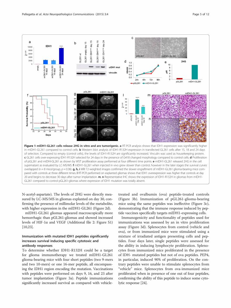
Figure 1 mIDH1-GL261 cells release 2HG in vitro and are tumorigenic. a RT-PCR analysis shows that IDH1 expression was significantly higherin mIDH1-GL261 compared to control cells. b Western blot analysis of IDH1-R132H expression in transfected GL261 cells after 15, 18 and 24 daysof selection. Compared to empty (control cells), the levels of IDH1-R132H are significantly increased. Vinculin was used as housekeeping protein.c GL261 cells over-expressing IDH1-R132H selected for 24 days in the presence of G418 changed morphology compared to control cells. d Proliferationof pGL261 and mIDHI-GL261 as shown by WST proliferation assay performed at four different time points. e mIDH1-GL261 released 2HG in the cellsupernatant as evaluated by LC-MS/MS. f mIDH1-GL261 when injected in vivo grew slower than control, however in the later stages the survival curvesoverlapped (n = 8 mice/group, p = 0.36). g, h, i MRI T2-weighted images confirmed the slower engraftment of mIDH1-GL261 glioma-bearing mice com-pared with controls at three different times. l RT-PCR performed on explanted gliomas shows that IDH1 overexpression was higher that controls at day20 and begins to decrease 30 days after tumor implantation. m, n Representative IHC shows the expression of IDH1-R132H in gliomas from mIDH1-GL261 compared to control pGL261-gliomas where expression of IDH1 mutation was totally absent.
Pellegatta et al. Acta Neuropathologica Communications (2015) 3:4 Page 5 of 12
N-acetyl-aspartate). The levels of 2HG were directly mea-sured by LC-MS/MS in gliomas explanted on day 30, con-firming the presence of millimolar levels of the metabolite,with higher expression in the mIDH1-GL261 (Figure 2d).mIDH1-GL261 gliomas appeared macroscopically more
hemorrhagic than pGL261-gliomas and showed increasedlevels of HIF-1α and VEGF (Additional file 2: Figure S1)[10,23].
Immunization with mutated IDH1 peptides significantlyincreases survival inducing specific cytotoxic andantibody responsesTo determine whether IDH1-R132H could be a targetfor glioma immunotherapy we treated mIDH1-GL261glioma-bearing mice with four short peptides (two 9-mersand two 10-mers) or one 16-mer peptide, all encompass-ing the IDH1 region encoding the mutation. Vaccinationswith peptides were performed on days 9, 16, and 23 aftertumor implantation (Figure 3a). Peptide immunizationssignificantly increased survival as compared with vehicle-
treated and ovalbumin (ova) peptide-treated controls(Figure 3b). Immunization of pGL261-glioma-bearingmice using the same peptides was ineffective (Figure 3c),demonstrating that the immune response induced by pep-tide vaccines specifically targets mIDH1-expressing cells.Immunogenicity and functionality of peptides used for
immunizations was assessed by an in vitro proliferationassay (Figure 3d). Splenocytes from control (vehicle andova), or from immunized mice were stimulated using amixture of irradiated antigen presenting cells and pep-tides. Four days later, single peptides were assessed forthe ability in inducing lymphocyte proliferation. Spleno-cytes from immunized mice proliferated in the presenceof IDH- mutated peptides but not of ova peptides. PEP4,in particular, induced 90% of proliferation. On the con-trary peptides were unable to stimulate splenocytes from“vehicle” mice. Splenocytes from ova-immunized miceproliferated when in presence of one out of four peptides,confirming the ability of this peptide to induce some cyto-lytic response [24].
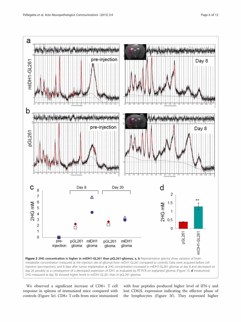
Figure 2 2HG concentration is higher in mIDH1-GL261 than pGL261-gliomas. a, b Representative spectra show variation of brainmetabolite concentration measured at the injection site of gliomas from mIDH1-GL261 compared to controls. Data were acquired before cellinjection (pre-injection), and 8 days after tumor implantation. c 2HG concentration increased in mIDH1-GL261 gliomas at day 8 and decreased onday 20 possibly as a consequence of a decreased expression of IDH1 as evaluated by RT-PCR on explanted gliomas (Figure 1l). d Intratumoral2HG measured at day 30 showed higher levels in mIDH1-GL261 than in pGL261-gliomas.
Pellegatta et al. Acta Neuropathologica Communications (2015) 3:4 Page 6 of 12
We observed a significant increase of CD8+ T cellresponse in spleens of immunized mice compared withcontrols (Figure 3e). CD8+ T cells from mice immunized
with four peptides produced higher level of IFN-γ andlost CD62L expression indicating the effector phase ofthe lymphocytes (Figure 3f ). They expressed higher
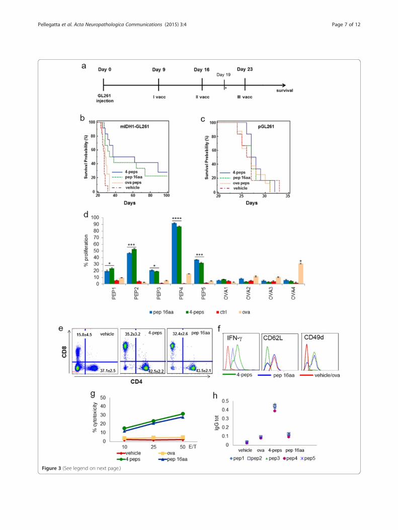
Figure 3 (See legend on next page.)
Pellegatta et al. Acta Neuropathologica Communications (2015) 3:4 Page 7 of 12
(See figure on previous page.)Figure 3 Immunization with mutated IDH1 peptides prolongs survival and promotes specific anti-tumor responses. a Experimentalschema of in vivo treatments. Two groups or mice injected with mIDH1-GL261 or pGL261 were immunized with three subcutaneous vaccinationson days 9, 16 and 23 after tumor implantation with four peptides (two 9-mer and two 10-mer), or with a single peptide (16-mer) emulsified inMontanide (15 μg/single peptide). A total of 3 μg of GM-CSF/mouse were administered during each immunization. Two group of control micewere treated with GM-CSF and Montanide alone or with ova peptides. On day 19 mice were sacrificed for immune monitoring. b, c Kaplan-Meiercurves show that immunized mIDH1-glioma mice (four peptides n = 12; 16-mer peptide n = 12) survived longer than control mice (vehicle, n = 12; ovan = 12). Immunizations of p-GL261 glioma-bearing mice did not modify the survival. d Splenocytes from immunized mice proliferated significantlymore than splenocytes from control (vehicle and ova treated) mice in the presence of IDH1-R132H peptides, particularly of peptide 4 (*p < 0.01;**p < 0.005; ***p < 0.001, ****p < 0.0001), not in presence of ova peptides. e Flow cytometry performed on splenocytes shows that CD8+ but notCD4+ T cell percentage increased significantly in immunized mice compared to controls (n = 6 mice/group). No difference between vehicle and ovapeptide immunizations. Data are reported in dot plots as the mean%± SD. f Flow cytometry histograms show that CD3 + CD8+ T cells produced moreIFN-γ and expressed less CD62L and more CD49d than controls (vehicle and ova histograms were overlapped). g In vitro MTT cytotoxicity assay revealsthat splenocytes from immunized but not from control mice lyse mIDH1-GL261 cells. h Scatter plot shows mIDH1 specific IgG detected in serum frommice immunized with four peptides. Single plots represent the mean of three serum samples/group.
Pellegatta et al. Acta Neuropathologica Communications (2015) 3:4 Page 8 of 12
levels of CD49d, critical for efficient infiltration andhoming into the glioma (Figure 3f ) [25]. CD8+ T cellsfrom mice immunized with the 16-mer peptide showedan intermediate expression of IFN-γ and two subpopula-tions with high or absent CD62L expression, suggestingthe coexistence of both naïve and effector cells (Figure 3f).Part of the splenocytes from immunized mice were pre-stimulated with peptides and tested five days later forcytotoxic ability using an MTT assay. The splenocytesfrom mice treated with IDH1-R132H peptides displayedspecific cytotoxicity against mIDH1-GL261 comparedwith vehicle and ova controls (Figure 3g), but not againstpGL261 (Additional file 2: Figure S2). IDH1-R132H spe-cific IgG were detected in serum of mIDH1-GL261 gliomabearing mice after immunization with the four peptidescompared with controls. No IgG response was found afterimmunization with the single 16-mer peptide (Figure 3h).
Peptide immunizations modulate tumor microenvironmentspecifically affecting mutated IDH1 expressionGliomas from mice treated with vehicle maintainedhighly vascularized features showing large and mediumsize-vessels. In gliomas from immunized mice the numberof blood vessels was reduced (Figure 4a, b) and was simi-lar to pGL261-gliomas (Additional file 2: Figure S1d).HIF-1α and VEGF expression were significantly reducedin gliomas from immunized mice compared with controls(Additional file 2: Figure S3).Vehicle gliomas appeared immune-reactive for IDH1-
R132H, whereas gliomas from immunized mice almostcompletely lost IDH1-R132H expression (Figure 4c), asalso confirmed by RT-PCR (Figure 4d).The immune response induced by peptide immuniza-
tions determined a significant modulation of tumormicroenvironment. IFN-γ, granzyme-b and perforin-1expression levels were 2.6 ± 0.04, 3.2 ± 0.03- and 2.1 ±0.02- fold higher, respectively, in immunized mice than invehicle mice (p < 0.01, p < 0.005, p = 0.01; Figure 4g). In
contrast, TGF-β2 and IL-10 expression levels were 2.4 ±0.22- and 2.3 ± 0.01-fold lower, respectively, in immunizedmice than in control animals (p = 0.01 and p < 0.005;Figure 4h).
DiscussionRecent data have shown that the most frequent IDH1mutation, R132H, can be effectively targeted in a A2.DR1 mice devoid of mouse MHC and transgenic forhuman MHC class I and II [14]. The R132H mutationwas expressed in sarcoma cells obtained by exposure to3-methylcholantrene that were injected subcutaneouslyin the A2.DR1 mice [14]. Our results show for the firsttime in an intracranial glioma model that the R132Hmutation, can be effectively targeted by the immune sys-tem, allowing a significant prolongation of survival andthe cure of 25% of the mice. The mIDH1-GL261 modelwe have used has obvious differences with human gli-omas, as it is established in a malignant glioma while inhumans IDH1 mutations take place early in develop-ment of low grade gliomas. An important similarity,however, is that, as in the human counterpart, mIDH1-GL261 cells produce increased amounts of the oncome-tabolite 2HG in vitro and in vivo. Although at a muchlower level, pGL261 also release detectable amounts of2HG. As mentioned in Results, levels higher than back-ground were found in a subset of wild type IDH gliomas[22]. Interestingly, 2HG production was also found inbreast cancer cells in association to MYC overexpression[26]. This may also be the case of pGL261 cells, as theyshow increased myc expression [27]. The presence ofthe mutation is associated to a decreased growth in vivo,exemplified by MRI scans taken on day 8 and 20 afterintracranial injection of tumor cells (Figure 1g, h) and bythe trend in Kaplan Meier survival curves until day 28(Figure 1f ), even if differences with pGL261 were notstatistically significant. A recent research by Phillips andcolleagues may provide some explanation for this finding,
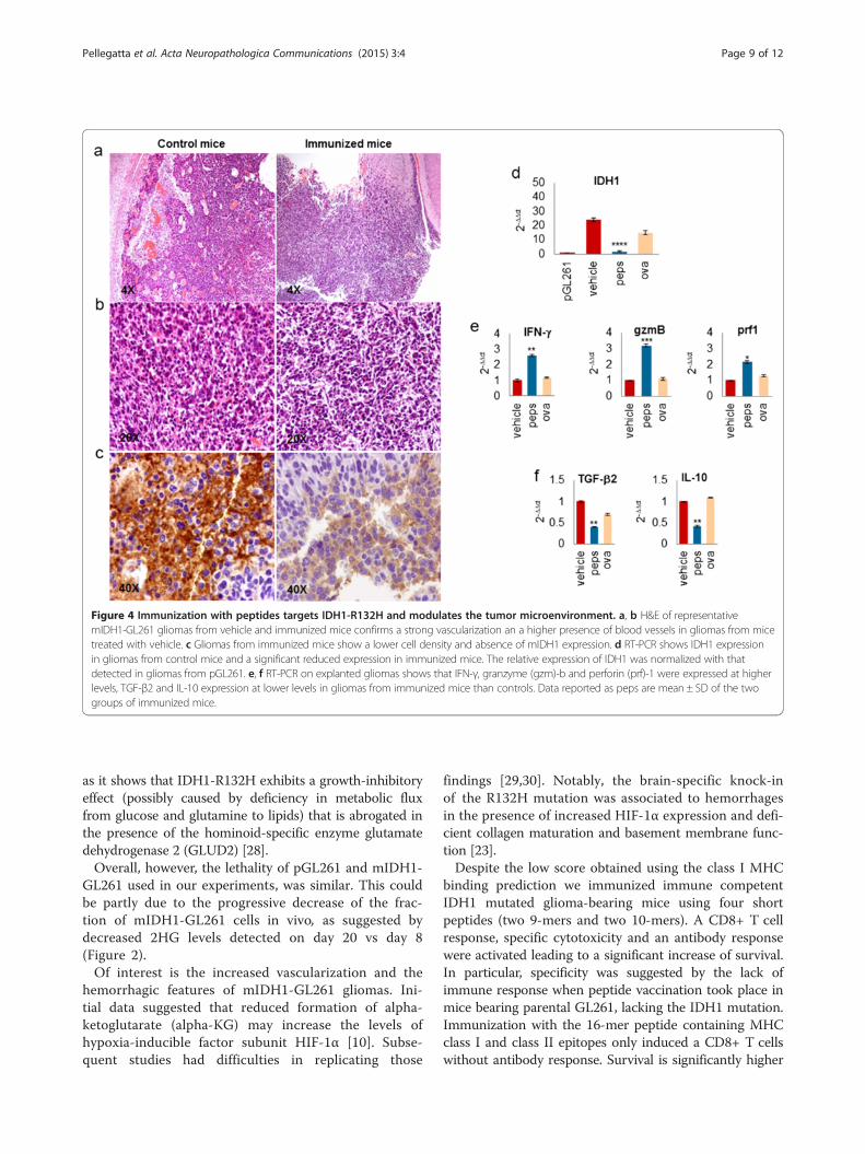
Figure 4 Immunization with peptides targets IDH1-R132H and modulates the tumor microenvironment. a, b H&E of representativemIDH1-GL261 gliomas from vehicle and immunized mice confirms a strong vascularization an a higher presence of blood vessels in gliomas from micetreated with vehicle. c Gliomas from immunized mice show a lower cell density and absence of mIDH1 expression. d RT-PCR shows IDH1 expressionin gliomas from control mice and a significant reduced expression in immunized mice. The relative expression of IDH1 was normalized with thatdetected in gliomas from pGL261. e, f RT-PCR on explanted gliomas shows that IFN-γ, granzyme (gzm)-b and perforin (prf)-1 were expressed at higherlevels, TGF-β2 and IL-10 expression at lower levels in gliomas from immunized mice than controls. Data reported as peps are mean ± SD of the twogroups of immunized mice.
Pellegatta et al. Acta Neuropathologica Communications (2015) 3:4 Page 9 of 12
as it shows that IDH1-R132H exhibits a growth-inhibitoryeffect (possibly caused by deficiency in metabolic fluxfrom glucose and glutamine to lipids) that is abrogated inthe presence of the hominoid-specific enzyme glutamatedehydrogenase 2 (GLUD2) [28].Overall, however, the lethality of pGL261 and mIDH1-
GL261 used in our experiments, was similar. This couldbe partly due to the progressive decrease of the frac-tion of mIDH1-GL261 cells in vivo, as suggested bydecreased 2HG levels detected on day 20 vs day 8(Figure 2).Of interest is the increased vascularization and the
hemorrhagic features of mIDH1-GL261 gliomas. Ini-tial data suggested that reduced formation of alpha-ketoglutarate (alpha-KG) may increase the levels ofhypoxia-inducible factor subunit HIF-1α [10]. Subse-quent studies had difficulties in replicating those
findings [29,30]. Notably, the brain-specific knock-inof the R132H mutation was associated to hemorrhagesin the presence of increased HIF-1α expression and defi-cient collagen maturation and basement membrane func-tion [23].Despite the low score obtained using the class I MHC
binding prediction we immunized immune competentIDH1 mutated glioma-bearing mice using four shortpeptides (two 9-mers and two 10-mers). A CD8+ T cellresponse, specific cytotoxicity and an antibody responsewere activated leading to a significant increase of survival.In particular, specificity was suggested by the lack ofimmune response when peptide vaccination took place inmice bearing parental GL261, lacking the IDH1 mutation.Immunization with the 16-mer peptide containing MHCclass I and class II epitopes only induced a CD8+ T cellswithout antibody response. Survival is significantly higher
Pellegatta et al. Acta Neuropathologica Communications (2015) 3:4 Page 10 of 12
than controls but less than what obtained with short pep-tide immunizations, possibly because of the lack of activa-tion of the antibody response.CTL epitopes alone can be inefficient in inducing a
long-term immune response and a specific memory[31], whereas MHC class I and class II restricted epi-topes in a single longer peptide can improve vaccineefficacy based on the simultaneous activation of CTLand CD4+ T cells [32-34]. This condition was observedafter immunizations with 35 amino-acid long peptidescontaining CTL epitopes showed more efficient thanthose with CTL peptides in inducing effective anti-tumorcytotoxic response [35].A number of reports support the essential role of CD4+
T cells in the anti-tumor responses due to their ability tostimulate dendritic cells and potentiate anti-tumor im-mune response by enhancing antigen presentation [36,37]and to the contribution to the memory response esta-blishment [38]. Importantly CD4+ T cells have also beendescribed to develop cytotoxicity and they are able toeradicate established melanomas [39]. However there isalso evidence that long peptides can modulate the efficacyof immunization by affecting the magnitude of CTLresponse [40,41].In our experiments we used as immune adjuvants
GM-CSF and Montanide ISA-51, a water-in-oil emul-sion enhancing the cytotoxic CD8+ T lymphocyte re-sponse. This was reported in patients [42-44], and wasalso supported by our previous pre-clinical experiments,where immunizations with short peptides induced CD8+T cell positive activation and tumor specific cytotox-icity [17,18].In conclusion, both our data and those of Platten and
coworkers [14] converge in supporting a translationalevolution of immunological targeting of the R132H mu-tation of IDH1 in glioma patients.There is increasing clinical evidence that immune
checkpoint inhibitors like ipilimumab and nivolumabmay prolong survival in patients with melanoma andsmall cell lung cancer [45,46]. Preclinical evidencesupport a role for this therapeutic approach in gli-omas [47] and a clinical trial is now ongoing to testthis hypothesis. It is plausible that a combination ofthis nonspecific form of activation of the immune sys-tem with specific mutation targeting, as is the case forthe R132H mutation, may yield a synergic effect. Thishypothesis deserves further testing in an appropriatemurine model.
Additional files
Additional file 1: Supplementary methods.
Additional file 2: Figure S1. mIDH1-GL261 gliomas maintain 2HGproduction and are strongly hemorrhagic. Figure S2. Splenocytes frommice immunized with IDH1-R132H peptides do not lyse pGL261 cells.Figure S3. Gliomas from immunized mice show a decreased expressionof HIF1-α and VEGF.
Competing interestsThe authors declare that they have no competing interests.
AcknowledgementsThis work was supported by “Il Fondo di Gio”, “Associazione Italiana TumoriCerebrali-AITC” and the Italian Ministry of Health. We thank Silvia Musio forhelpful hints on ELISA assay.
Author details1Department of Neuro-Oncology, Unit of Molecular Neuro-Oncology,Fondazione I.R.C.C.S. Istituto Neurologico C. Besta, via Celoria 11, 20133Milano, Italy. 2Nerviano Medical Sciences, Nerviano S.r.l., Milano, Italy.
Received: 14 December 2014 Accepted: 26 December 2014
References1. Parsons DW, Jones S, Zhang X, Lin JC, Leary RJ, Angenendt P, Mankoo P,
Carter H, Siu IM, Gallia GL, Olivi A, McLendon R, Rasheed BA, Keir S,Nikolskaya T, Nikolsky Y, Busam DA, Tekleab H, Diaz LA Jr, Hartigan J,Smith DR, Strausberg RL, Marie SK, Shinjo SM, Yan H, Riggins GJ, Bigner DD,Karchin R, Papadopoulos N, Parmigiani G et al (2008) An integratedgenomic analysis of human glioblastoma multiforme. Science 321:1807–1812,doi:10.1126/science.1164382
2. Yan H, Parsons DW, Jin G, McLendon R, Rasheed BA, Yuan W, Kos I,Batinic-Haberle I, Jones S, Riggins GJ, Friedman H, Friedman A, Reardon D,Herndon J, Kinzler KW, Velculescu VE, Vogelstein B, Bigner DD (2009) IDH1and IDH2 mutations in gliomas. N Engl J Med 360:765–773, doi:10.1056/NEJMoa0808710
3. Dang L, White DW, Gross S, Bennett BD, Bittinger MA, Driggers EM, FantinVR, Jang HG, Jin S, Keenan MC, Marks KM, Prins RM, Ward PS, Yen KE, LiauLM, Rabinowitz JD, Cantley LC, Thompson CB, Vander Heiden MG, Su SM(2009) Cancer-associated IDH1 mutations produce 2-hydroxyglutarate.Nature 462:739–744, doi:10.1038/nature08617
4. Saha SK, Parachoniak CA, Ghanta KS, Fitamant J, Ross KN, Najem MS,Gurumurthy S, Akbay EA, Sia D, Cornella H, Miltiadous O, Walesky C,Deshpande V, Zhu AX, Hezel AF, Yen KE, Straley KS, Travins J, Popovici-Muller J, Gliser C, Ferrone CR, Apte U, Llovet JM, Wong KK, Ramaswamy S,Bardeesy N (2014) Mutant IDH inhibits HNF-4α to block hepatocytedifferentiation and promote biliary cancer. Nature 513:110–114,doi:10.1038/nature13441
5. Losman J-AJ-A, Looper RE, Koivunen P, Lee S, Schneider RK, McMahon C,Cowley GS, Root DE, Ebert BL, Kaelin WG Jr (2013) (R)-2-Hydroxyglutarate issufficient to promote leukemogenesis and its effects are reversible. Science339:1621–1625, doi:10.1126/science.1231677
6. Krell D, Mulholland P, Frampton AE, Krell J, Stebbing J, Bardella C (2013) IDHmutations in tumorigenesis and their potential role as novel therapeutictargets. Future Oncol 9:1923–1935, doi:10.2217/fon.13.143
7. Turcan S, Rohle D, Goenka A, Walsh LA, Fang F, Yilmaz E, Campos C, FabiusAW, Lu C, Ward PS, Thompson CB, Kaufman A, Guryanova O, Levine R,Heguy A, Viale A, Morris LG, Huse JT, Mellinghoff IK, Chan TA (2012) IDH1mutation is sufficient to establish the glioma hypermethylator phenotype.Nature 483:479–483, doi:10.1038/nature10866
8. Lu C, Ward PS, Kapoor GS, Rohle D, Turcan S, Abdel-Wahab O, Edwards CR,Khanin R, Figueroa ME, Melnick A, Wellen KE, O'Rourke DM, Berger SL,Chan TA, Levine RL, Mellinghoff IK, Thompson CB (2012) IDH mutationimpairs histone demethylation and results in a block to cell differentiation.Nature 483:474–478, doi:10.1038/nature10860
9. Horbinski C (2013) What do we know about IDH1/2 mutations so far, and howdo we use it? Acta Neuropathol 125:621–636, doi:10.1007/s00401-013-1106-9
10. Zhao S, Lin Y, Xu W, Jiang W, Zha Z, Wang P, Yu W, Li Z, Gong L, Peng Y,Ding J, Lei Q, Guan KL, Xiong Y (2009) Glioma-derived mutations in IDH1dominantly inhibit IDH1 catalytic activity and induce HIF-1alpha. Science324:261–265, doi:10.1126/science.1170944
Pellegatta et al. Acta Neuropathologica Communications (2015) 3:4 Page 11 of 12
11. Rohle D, Popovici-Muller J, Palaskas N, Turcan S, Grommes C, Campos C,Tsoi J, Clark O, Oldrini B, Komisopoulou E, Kunii K, Pedraza A, Schalm S,Silverman L, Miller A, Wang F, Yang H, Chen Y, Kernytsky A, Rosenblum MK,Liu W, Biller SA, Su SM, Brennan CW, Chan TA, Graeber TG, Yen KE,Mellinghoff IK (2013) An inhibitor of mutant IDH1 delays growth andpromotes differentiation of glioma cells. Science 340:626–630,doi:10.1126/science.1236062
12. Boisselier B, Pérez-Larraya JG, Rossetto M, Labussière M, Ciccarino P, Marie Y,Delattre JY, Sanson M (2012) Detection of IDH1 mutation in the plasmaof patients with glioma. Neurology 79:1693–1698, doi:10.1212/WNL.0b013e31826e9b0a
13. Kalinina J, Carroll A, Wang L, Yu Q, Mancheno DE, Wu S, Liu F, Ahn J, He M,Mao H, Van Meir EG (2012) Detection of “oncometabolite” 2-hydroxyglutarate by magnetic resonance analysis as a biomarker of IDH1/2mutations in glioma. J Mol Med (Berl). doi:10.1007/s00109-012-0888-x
14. Schumacher T, Bunse L, Pusch S, Sahm F, Wiestler B, Quandt J, Menn O,Osswald M, Oezen I, Ott M, Keil M, Balß J, Rauschenbach K, Grabowska AK,Vogler I, Diekmann J, Trautwein N, Eichmüller SB, Okun J, Stevanović S,Riemer AB, Sahin U, Friese MA, Beckhove P, von Deimling A, Wick W, PlattenM (2014) A vaccine targeting mutant IDH1 induces antitumour immunity.Nature. doi:10.1038/nature13387
15. Bourne TD, Schiff D (2010) Update on molecular findings, management andoutcome in low-grade gliomas. Nat Rev Neurol 6:695–701, doi:10.1038/nrneurol.2010.159
16. Han S, Zhang C, Li Q, Dong J, Liu Y, Huang Y, Jiang T, Wu A (2014)Tumour-infiltrating CD4(+) and CD8(+) lymphocytes as predictors ofclinical outcome in glioma. Br J Cancer 110:2560–2568, doi:10.1038/bjc.2014.162
17. Cantini G, Pisati F, Pessina S, Finocchiaro G, Pellegatta S (2012)Immunotherapy against the radial glia marker GLAST effectively triggersspecific antitumor effectors without autoimmunity. Oncoimmunology1:884–893, doi:10.4161/onci.20637
18. Favaro R, Appolloni I, Pellegatta S, Sanga AB, Pagella P, Gambini E, Pisati F,Ottolenghi S, Foti M, Finocchiaro G, Malatesta P, Nicolis SK (2014) Sox2 is requiredto maintain cancer stem cells in a mouse model of high-grade oligodendroglioma.Cancer Res 74:1833–1844, doi:10.1158/0008-5472.CAN-13-1942
19. Provencher SW (1993) Estimation of metabolite concentrations fromlocalized in vivo proton NMR spectra. Magn Reson Med 30:672–679
20. Gonsalves RA (1976) Cramér-Rao bounds on mensuration errors. Appl Opt15:1270–1275
21. Lazovic J, Soto H, Piccioni D, Lou JR, Li S, Mirsadraei L, Yong W, Prins R,Liau LM, Ellingson BM, Cloughesy TF, Lai A, Pope WB (2012) Detection of2-hydroxyglutaric acid in vivo by proton magnetic resonance spectroscopyin U87 glioma cells overexpressing isocitrate dehydrogenase-1 mutation.Neuro Oncol 14:1465–1472, doi:10.1093/neuonc/nos258
22. Natsumeda M, Igarashi H, Nomura T, Ogura R, Tsukamoto Y, Kobayashi T,Aoki H, Okamoto K, Kakita A, Takahashi H, Nakada T, Fujii Y (2014)Accumulation of 2-hydroxyglutarate in gliomas correlates with survival: astudy by 3.0-tesla magnetic resonance spectroscopy. Acta NeuropatholCommun 2:158, doi:10.1186/s40478-014-0158-y
23. Sasaki M, Knobbe CB, Itsumi M, Elia AJ, Harris IS, Chio II, Cairns RA,McCracken S, Wakeham A, Haight J, Ten AY, Snow B, Ueda T, Inoue S,Yamamoto K, Ko M, Rao A, Yen KE, Su SM, Mak TW (2012) D-2-hydroxyglutarate produced by mutant IDH1 perturbs collagenmaturation and basement membrane function. Genes Dev 26:2038–2049doi:10.1101/gad.198200.112
24. Lipford GB, Hoffman M, Wagner H, Heeg K (1993) Primary in vivo responsesto ovalbumin. Probing the predictive value of the Kb binding motif.J Immunol 150:1212–1222
25. Zhu X, Nishimura F, Sasaki K, Fujita M, Dusak JE, Eguchi J, Fellows-Mayle W,Storkus WJ, Walker PR, Salazar AM, Okada H (2007) Toll like receptor-3 ligandpoly-ICLC promotes the efficacy of peripheral vaccinations with tumorantigen-derived peptide epitopes in murine CNS tumor models. J TranslMed 5:10, doi:10.1186/1479-5876-5-10
26. Terunuma A, Putluri N, Mishra P, Mathé EA, Dorsey TH, Yi M, Wallace TA,Issaq HJ, Zhou M, Killian JK, Stevenson HS, Karoly ED, Chan K, Samanta S,Prieto D, Hsu TY, Kurley SJ, Putluri V, Sonavane R, Edelman DC, Wulff J,Starks AM, Yang Y, Kittles RA, Yfantis HG, Lee DH, Ioffe OB, Schiff R,Stephens RM, Meltzer PS et al (2014) MYC-driven accumulation of2-hydroxyglutarate is associated with breast cancer prognosis. J Clin Invest124:398–412, doi:10.1172/JCI71180
27. Szatmári T, Lumniczky K, Désaknai S, Trajcevski S, Hídvégi EJ, Hamada H,Sáfrány G (2006) Detailed characterization of the mouse glioma 261 tumormodel for experimental glioblastoma therapy. Cancer Sci 97:546–553,doi:10.1111/j.1349-7006.2006.00208.x
28. Chen R, Nishimura MC, Kharbanda S, Peale F, Deng Y, Daemen A, ForrestWF, Kwong M, Hedehus M, Hatzivassiliou G, Friedman LS, Phillips HS (2014)Hominoid-specific enzyme GLUD2 promotes growth of IDH1R132H glioma.Proc Natl Acad Sci 111:14217–14222, doi:10.1073/pnas.1409653111
29. Koivunen P, Lee S, Duncan CG, Lopez G, Lu G, Ramkissoon S, Losman JA,Joensuu P, Bergmann U, Gross S, Travins J, Weiss S, Looper R, Ligon KL,Verhaak RG, Yan H, Kaelin WG Jr (2012) Transformation by the (R)-enantiomer of 2-hydroxyglutarate linked to EGLN activation. Nature483:484–488, doi:10.1038/nature10898
30. Williams SC, Karajannis MA, Chiriboga L, Golfinos JG, von Deimling A,Zagzag D (2011) R132H-mutation of isocitrate dehydrogenase-1 is notsufficient for HIF-1α upregulation in adult glioma. Acta Neuropathol121:279–281, doi:10.1007/s00401-010-0790-y
31. Bijker MS, van den Eeden SJF, Franken KL, Melief CJ, Offringa R, van derBurg SH (2007) CD8+ CTL priming by exact peptide epitopes in incompleteFreund’s adjuvant induces a vanishing CTL response, whereas long peptidesinduce sustained CTL reactivity. J Immunol 179:5033–5040
32. Kissick HT, Sanda MG, Dunn LK, Arredouani MS (2014) Immunization with apeptide containing MHC class I and II epitopes derived from the tumorantigen SIM2 induces an effective CD4 and CD8 T-cell response. PLoS One9:e93231, doi:10.1371/journal.pone.0093231
33. Speetjens FM, Kuppen PJK, Welters MJP, Essahsah F, van den Brink AM V, LantruaMG, Valentijn AR, Oostendorp J, Fathers LM, Nijman HW, Drijfhout JW, van deVelde CJ, Melief CJ, van der Burg SH (2009) Induction of p53-specific immunityby a p53 synthetic long peptide vaccine in patients treated for metastatic colo-rectal cancer. Clin Cancer Res 15:1086–1095, doi:10.1158/1078-0432.CCR-08-2227
34. Krug LM, Dao T, Brown AB, Maslak P, Travis W, Bekele S, Korontsvit T,Zakhaleva V, Wolchok J, Yuan J, Li H, Tyson L, Scheinberg DA (2010) WT1peptide vaccinations induce CD4 and CD8 T cell immune responses inpatients with mesothelioma and non-small cell lung cancer. CancerImmunol Immunother 59:1467–1479, doi:10.1007/s00262-010-0871-8
35. Zwaveling S, Ferreira Mota SC, Nouta J, Johnson M, Lipford GB, Offringa R,van der Burg SH, Melief CJ (2002) Established human papillomavirus type16-expressing tumors are effectively eradicated following vaccination withlong peptides. J Immunol 169:350–358
36. Hung K, Hayashi R, Lafond-Walker A, Lowenstein C, Pardoll D, Levitsky H(1998) The central role of CD4(+) T cells in the antitumor immune response.J Exp Med 188:2357–2368
37. Matsui S, Ahlers JD, Vortmeyer AO, Terabe M, Tsukui T, Carbone DP, LiottaLA, Berzofsky JA (1999) A model for CD8+ CTL tumor immunosurveillanceand regulation of tumor escape by CD4 T cells through an effect on qualityof CTL. J Immunol 163:184–193
38. Pardoll DM, Topalian SL (1998) The role of CD4+ T cell responses inantitumor immunity. Curr Opin Immunol 10:588–594
39. Quezada SA, Simpson TR, Peggs KS, Merghoub T, Vider J, Fan X, Blasberg R,Yagita H, Muranski P, Antony PA, Restifo NP, Allison JP (2010)Tumor-reactive CD4(+) T cells develop cytotoxic activity and eradicate largeestablished melanoma after transfer into lymphopenic hosts. J Exp Med207:637–650, doi:10.1084/jem.20091918
40. Welters MJP, Bijker MS, van den Eeden SJF, Franken KL, Melief CJ, Offringa R,van der Burg SH (2007) Multiple CD4 and CD8 T-cell activation parameterspredict vaccine efficacy in vivo mediated by individual DC-activatingagonists. Vaccine 25:1379–1389, doi:10.1016/j.vaccine.2006.10.049
41. Ossendorp F, Mengedé E, Camps M, Filius R, Melief CJ (1998) Specific T helpercell requirement for optimal induction of cytotoxic T lymphocytes against majorhistocompatibility complex class II negative tumors. J Exp Med 187:693–702
42. Markovic SN, Suman VJ, Ingle JN, Kaur JS, Pitot HC, Loprinzi CL, Rao RD,Creagan ET, Pittelkow MR, Allred JB, Nevala WK, Celis E (2006) Peptidevaccination of patients with metastatic melanoma: improved clinicaloutcome in patients demonstrating effective immunization. Am J ClinOncol 29:352–360, doi:10.1097/01.coc.0000217877.78473.a4
43. Kostic A, Mihailovic D, Veselinovic S, Tasic D, Stefanovic I, Novak V,Stojanovic N, Veselinovic D, Pavlovic S (2008) Tumor size and karyometricvariables in brain astrocytoma. J BU ON 14:473–477
44. Karbach J, Gnjatic S, Bender A, Neumann A, Weidmann E, Yuan J,Ferrara CA, Hoffmann E, Old LJ, Altorki NK, Jäger E (2010) Tumor-reactiveCD8+ T-cell responses after vaccination with NY-ESO-1 peptide, CpG 7909
Pellegatta et al. Acta Neuropathologica Communications (2015) 3:4 Page 12 of 12
and Montanide ISA-51: association with survival. Int J Cancer 126:909–918,doi:10.1002/ijc.24850
45. Pardoll DM (2012) The blockade of immune checkpoints in cancerimmunotherapy. Nat Rev Cancer 12:252–264, doi:10.1038/nrc3239
46. Mullard A (2013) New checkpoint inhibitors ride the immunotherapytsunami. Nat Rev Drug Discov 12:489–492, doi:10.1038/nrd4066
47. Vom Berg J, Vrohlings M, Haller S, Haimovici A, Kulig P, Sledzinska A,Weller M, Becher B (2013) Intratumoral IL-12 combined with CTLA-4blockade elicits T cell-mediated glioma rejection. J Exp Med 210:2803–2811,doi:10.1084/jem.20130678
Submit your next manuscript to BioMed Centraland take full advantage of:
• Convenient online submission
• Thorough peer review
• No space constraints or color figure charges
• Immediate publication on acceptance
• Inclusion in PubMed, CAS, Scopus and Google Scholar
• Research which is freely available for redistribution
Submit your manuscript at www.biomedcentral.com/submit
Copyright © 2022 FDOKUMEN