Novel Noninvasive Methods of Intracranial Pressure ... - DTIC
91
AWARD NUMBER: W81XWH-18-1-0005 TITLE: Novel Noninvasive Methods of Intracranial Pressure and Cerebrovascular Autoregulation Assessment: Seeing the Brain Through the Eyes PRINCIPAL INVESTIGATOR: Mohamad Hakam Tiba, MD, MS CONTRACTING ORGANIZATION: Regents of the University of Michigan Ann Arbor, MI 48109 REPORT DATE: January 2019 TYPE OF REPORT: Annual PREPARED FOR: U.S. Army Medical Research and Materiel Command Fort Detrick, Maryland 21702-5012 DISTRIBUTION STATEMENT: Approved for Public Release; Distribution Unlimited The views, opinions and/or findings contained in this report are those of the author(s) and should not be construed as an official Department of the Army position, policy or decision unless so designated by other documentation.
-
Upload
khangminh22 -
Category
Documents
-
view
0 -
download
0
Transcript of Novel Noninvasive Methods of Intracranial Pressure ... - DTIC

AWARD NUMBER: W81XWH-18-1-0005
TITLE: Novel Noninvasive Methods of Intracranial Pressure and Cerebrovascular Autoregulation Assessment: Seeing the Brain Through the Eyes
PRINCIPAL INVESTIGATOR: Mohamad Hakam Tiba, MD, MS
CONTRACTING ORGANIZATION: Regents of the University of Michigan
Ann Arbor, MI 48109
REPORT DATE: January 2019
TYPE OF REPORT: Annual
PREPARED FOR: U.S. Army Medical Research and Materiel Command Fort Detrick, Maryland 21702-5012
DISTRIBUTION STATEMENT: Approved for Public Release; Distribution Unlimited
The views, opinions and/or findings contained in this report are those of the author(s) and should not be construed as an official Department of the Army position, policy or decision unless so designated by other documentation.

REPORT DOCUMENTATION PAGE Form Approved
OMB No. 0704-0188 Public reporting burden for this collection of information is estimated to average 1 hour per response, including the time for reviewing instructions, searching existing data sources, gathering and maintaining the data needed, and
completing and reviewing this collection of information. Send comments regarding this burden estimate or any other aspect of this collection of information, including suggestions for reducing this burden to Department of Defense,
Washington Headquarters Services, Directorate for Information Operations and Reports (0704-0188), 1215 Jefferson Davis Highway, Suite 1204, Arlington, VA 22202-4302. Respondents should be aware that notwithstanding any
other provision of law, no person shall be subject to any penalty for failing to comply with a collection of information if it does not display a currently valid OMB control number. PLEASE DO NOT RETURN YOUR FORM TO
THE ABOVE ADDRESS.
1. REPORT DATE
January 2019
2. REPORT TYPE
Annual
3. DATES COVERED
1 Jan 2018 - 31 Dec 2018
4. TITLE AND SUBTITLE
Novel Noninvasive Methods of Intracranial Pressure and Cerebrovascular Autoregulation Assessment: Seeing the Brain Through the Eyes
5a. CONTRACT NUMBER
5b. GRANT NUMBER
W81XWH-18-1-0005
5c. PROGRAM ELEMENT NUMBER
6. AUTHOR(S)
Mohamad H. Tiba, MD
Kevin Ward, MD
Amanda Pennington, MS
5d. PROJECT NUMBER
Brendan McCracken, BS
Brandon Cummings, BS
Reza Soroushmehr, PhD
5e. TASK NUMBER
E-Mail: [email protected]
5f. WORK UNIT NUMBER
7. PERFORMING ORGANIZATION NAME(S) AND ADDRESS(ES)
University of Michigan
Michigan Center for Integrative Research in Critical Care
8. PERFORMING ORGANIZATION REPORTNUMBER
2800 Plymouth Road, NCRC Building
10, Room A107
Ann Arbor, Michigan 48109-2800
9. SPONSORING / MONITORING AGENCY NAME(S) AND ADDRESS(ES) 10. SPONSOR/MONITOR’S ACRONYM(S)
U.S. Army Medical Research and Materiel Command
Fort Detrick, Maryland 21702-5012 11. SPONSOR/MONITOR’S REPORT
NUMBER(S)
12. DISTRIBUTION / AVAILABILITY STATEMENT
Approved for Public Release; Distribution Unlimited
13. SUPPLEMENTARY NOTES

14. ABSTRACT
Traumatic brain injury (TBI) is a major public health problem in the U.S. and around the world. It plays a major role in approximately 30%
of injury related civilian deaths in the U.S. and is often referred to as the “silent epidemic” because of associated complications that go
undiagnosed and unnoticed, but might have a lasting effect. Furthermore, the Defense and Veterans Brain Injury Center (DVBIC) has
reported over 34,000 moderate to severe combat-related TBI (CRTBI) since 2000, making it a major source of mortality and morbidity for
the U.S. military between 2000 and 2016. The significance of such numbers and statistics becomes apparent with the military’s increased
focus on Prolonged Field Care (PFC) and prolonged damage control resuscitation (pDCR). PFC is defined by Keenan as the “field medical
care, applied beyond ‘doctrinal planning time-lines’ by a SOCM (Special Operations Combat Medic) or higher, in order to decrease patient
mortality and morbidity, utilize limited resources, and provide sustained care until the patient arrives at an appropriate level of care.”
Approximately 20% of individuals with combat-related severe TBI suffer acute neurological deterioration in the first 72 hours following
injury, the potential time window of PFC. The austere, resource-constrained combat environment and lack of diagnostic capabilities could
lead to delayed recognition of the severity of a TBI or in having rationale treatment end-points, resulting in exacerbated (secondary) brain
damage and increased TBI-related disabilities. This is especially true when TBI-related injuries are combined with other injuries requiring
pDCR.
One of the significant management strategies in the treatment of TBI is aimed at preventing secondary brain damage, which mainly manifests
itself as brain ischemia and inflammation. Monitoring of intracranial pressure (ICP) and cerebral autoregulation (CAR) to optimize cerebral
perfusion pressure (CPP) to a target and maintain cerebral blood flow (CBF) are the primary methods to prevent secondary injury and are the
mainstays of current practice. In a recent study, Juul et al. has concluded that acute neurological deterioration is a powerful predictor of poor
outcomes following TBI. The study showed that 29% of patients with acute neurological decline having an unfavorable outcome and the
most powerful predictor of such neurological deterioration was the patient’s measured ICP. Therefore, it is critical to be able to monitor and
manage ICP as early as possible following TBI. Current guidelines of the Brain Trauma Foundation recommend the use of invasive ICP
monitoring in patients who meet specific criteria, with the aim of achieving significant reduction in mortality in civilian centers. The Joint
Theater Trauma System (JTTS) Clinical Practice Guideline for TBI Management (CPG) has a major focus on early management of ICP for
the management of severe TBI—in particular, the prevention of any secondary neurologic decline as a result of an expanding intracranial
hematoma and the subsequent cerebral herniation and ischemia. However, monitoring of ICP during PFC is problematic and challenging
since invasive monitoring and computed tomography (CT) imaging is unavailable or difficult to implement in these settings. Therefore, the
current CPG relies on clinical neurological deterioration to trigger treatment of raised ICP, by which time irreversible brain injury may have
already occurred. In addition, following ICP in a patient with an already altered mental status or who is intubated and sedated is problematic.
Recently, physicians and healthcare providers began utilizing a more dynamic, patient-oriented optimization of CPP based on CAR.
Autoregulation is considered one of the most important central nervous system auto-protective mechanisms. It is described as the ability of
vessels to modulate their tone in response to changes in CAR is a complex process (critical in preventing secondary brain injury) often
impaired after injury and has been shown to be a significant predictor of outcomes in patients with various acute neurological diseases,
including severe TBI-related and ischemic injuries. Therefore, continuous monitoring of autoregulation may be beneficial as a means to
enable optimization of CPP on a patient-by-patient basis. This represents a more precise and personalized approach to managing the CPP
components (ICP and mean arterial pressure) as there is likely great variation in autoregulation ability among individuals and across injuries.
Assessment methods such as Transcranial-Doppler (TCD), brain tissue oxygenation (ORx), hemoglobin saturation measured by near-infrared
spectroscopy (NIRS), and Laser-Doppler flowmetry of CBF have been used in the past to assess cerebral autoregulation, but with mixed
results. Such methods are problematic in PFC settings for a number of reasons, including the intermittent or invasive nature of the measure,
the need for a high level of operator experience, and the lack of technology available in far-forward echelons of care.
Newer approaches to autoregulation monitoring, such as pressure reactivity monitoring (PRx, a correlation between mean arterial pressure
and ICP), have proven to be independent predictors of outcome. PRx is calculated as the moving Pearson correlation coefficient between
certain count of consecutive 5-10 second averages of mean arterial blood pressure (MAP) and ICP. Since PRx is calculated as a correlation,
its values would range between -1 and 1, with positive values indicating impaired autoregulation (pressure-passive behavior of the arterial
walls) and negative values indicating intact autoregulation (vascular bed with active vasomotor responses). However, PRx is frequently
difficult to interpret due to noise in the signal, and would be difficult to apply in early echelons of care due to the requirement for invasive
ICP and arterial blood pressure monitoring. In addition, because of the complexity of PRx calculation and the requirement for additional
software, PRx monitoring has been limited to research-oriented academic centers. Therefore, there exists an unmet need for non-invasive,
portable diagnostic tools for the early detection impairment of autoregulation and elevated ICP, prior to a potentially catastrophic clinical
decline, in patients or injured warfighters who may require initiation of medical therapy and priority evacuation for neurosurgical intervention.15. SUBJECT TERMS
TBI, Bioimpedance, ICP, Cerebrovascular Autoregulation, Ultrasound, cerebral blood flow, Optic Nerve Sheath, PRX, Non-invasive
16. SECURITY CLASSIFICATION OF: 17. LIMITATIONOF ABSTRACT
18. NUMBEROF PAGES
19a. NAME OF RESPONSIBLE PERSON
USAMRMC a. REPORT
Unclassified
b. ABSTRACT
Unclassified
c. THIS PAGE
Unclassified Unclassified
87 19b. TELEPHONE NUMBER (include area
code)
Standard Form 298 (Rev. 8-98)Prescribed by ANSI Std. Z39.18
Table of Contents
Page
1. Introduction…………………………………………………………. 1
2. Keywords……………………………………………………………. 1
3. Accomplishments………..…………………………………………... 1
4. Impact…………………………...…………………………………… 12
5. Changes/Problems...….……………………………………………… 13
6. Products, Inventions, Patent Applications, and/or Licenses.……. 14
7. Participants & Other Collaborating Organizations…………… 14
8. Special Reporting Requirements………………………………… 18
9. Appendices…………………………………………………………… 18

1
INTRODUCTION: Traumatic brain injury (TBI) is a major public health problem both in the U.S. and around the
world. One of the significant management strategies in the treatment of TBI is aimed at preventing
secondary brain injury, which mainly manifests itself as brain ischemia and inflammation. Monitoring of
intracranial pressure (ICP) and cerebrovascular autoregulation (CAR) to optimize cerebral perfusion
pressure (CPP) and maintain cerebral blood flow (CBF) are the primary methods to prevent secondary
injury and are the mainstays of current practice. Care of moderate to severe combat-related traumatic brain
injury (TBI) continues to pose enormous challenges sometimes compounded by the need to provide
prolonged field care (PFC). TBI in the presence of other injuries requiring prolonged damage control
resuscitation (pDCR) provides additional challenges. The austere, resource-constrained combat
environment and lack of readily available diagnostic capabilities often lead to delayed recognition of the
severity of TBIs, resulting in exacerbated damage and increased TBI-related disabilities. CAR and ICP
monitoring has been used in cases of civilian TBI-related injuries to optimize cerebral perfusion pressure
and blood flow to prevent secondary injury. However, technologies currently available to monitor CAR
and ICP require invasive techniques and a high level of experience, while providing intermittent readings,
making them impractical and unavailable in PFC and pDCR settings. Robust methods of noninvasive
monitoring of CAR and ICP would allow for early application by combat medics, first responders,
emergency departments, surgeons, and critical care staff. The proposed project aims to utilize trans-ocular
brain bioimpedance and optic nerve ultrasound in a novel manner to assess CAR and ICP, utilizing the eye
as a window to the brain.
KEYWORDS: TBI, Bioimpedance, ICP, Cerebrovascular Autoregulation, Ultrasound, cerebral blood flow, Optic Nerve
Sheath, PRX, Non-invasive
ACCOMPLISHMENTS:
What were the major goals of the project?
Major Task 1: Evaluation of ocular bioimpedance in two swine TBI models. Months 0-36
1. A swine model of blunt trauma. Months 3-36
2. A swine TBI model with provocative maneuvers to manipulate cerebral blood flow (elevated blood
pressure, systemic hemorrhage, elevations in ICP and changes in ventilation. Months 3-36
Major Task 2: Evaluation of ocular impedance as an indicator of cerebral autoregulation in
humans who are undergoing both invasive arterial blood pressure and ICP monitoring for
brain injury. Months 0-36
Major Task 3: Collection of ONS ultrasound videos for assessment of ICP in humans who are
undergoing both invasive arterial blood pressure and ICP monitoring for brain injury. Months
0-36
Major Task 4: Development of an ultrasound video analytic system to evaluate ONSD. Months
6-36
What was accomplished under these goals?
1. Major Task 1: Evaluation of ocular bioimpedance in two swine TBI models. Months 0-36
1) Overall target: 5 animals (either model)/quarter
2) Specific objectives:
a. IACUC approval: June 12, 2017
b. ACURO approval: August 11, 2017

2
3) Animals use Data:
a. Species: Sus Scrofa Domestica
b. Total animal number used: 26
c. USDA pain category for all animals used: D
All animals used to date for this study were subjected to Model 1: Provocative maneuvers to
manipulate cerebral blood flow (CBF) systemic blood pressure (MAP) and ICP.
A combination of maneuvers and injuries were performed including hyperventilation, slow infusion of
vasopressors (epinephrine) to increase MAP to ~ 160 mmHg, epidural hematoma by insertion of a Foley
catheter into the epidural region and inflating the balloon, and lastly a slow systematic hemorrhage and
crystalloid resuscitation.
All animals received the same surgical Instrumentation for evaluation of:
Invasive arterial blood pressure (MAP)
Intracranial pressure (ICP)
Cerebral blood flow (CBF)
End tidal CO2 (PetCO2)
Cerebral perfusion pressure (CPP)
Pressure reactivity index (PRx)
Ocular bioimpedance
Laser Doppler flow (LDF)
Transcranial Doppler flow (TAMEAN)
a. The left and right femoral artery are cannulated for removal of blood during controlled hemorrhage,
measurement of arterial blood pressure and blood sampling.
b. One external jugular vein is cannulated for delivery of resuscitation fluids.
c. 2-3mm burr holes are drilled into the skull and used for placement of an LDF probe, pressure
catheter for monitoring ICP and placement of an 8 French Foley catheter to simulate hematoma.
d. Non-invasive bipolar ECG electrodes will be placed and secured on the animal’s upper eyelids to
measure ocular bio-impedance. A Biopac EBI100C electro-bioimpedance amplifier will be
connected to the eye electrodes and used to apply low current (0.1-1 mA) and continuously detect
potential. In addition, an ultrasound probe is placed at the temporal window of the skull for
transcranial doppler assessment of cerebral blood flow, measured as a time averaged mean flow
(TAMEAN) of the middle cerebral artery.
Injury and maneuvers:
a. Hyperventilation: The respiratory rate (RR) is increased by increments of 10 breaths per minute
until PetCO2 is reduced to ~20mmHg. After 10min, RR will be decreased by 10 breaths until back
to baseline.
b. Vasopressor (norepinephrine) administration: Norepinephrine will be administered and titrated
upward to increase MAP to ~ 150-160 mmHg. MAP will be maintained for 2-5 minutes while data
is recorded. Afterward, norepinephrine solution infusion will be stopped and the animal’s MAP
will be allowed to return to near baseline level. The norepinephrine infusion procedure may be
repeated up to three times.
c. Epidural Hematoma: Simulation of an epidural hematoma was created using a 3F Foley catheter
placed under the skull above the parietal cortex of the brain. Catheter’s balloon will be inflated
using a syringe pump and ICP was monitored with a target maximum pressure of 30-40mmHg.
When the target ICP was reached, the pressure will be maintained for a period of 1-5 minutes. After
the monitoring period, balloon will be deflated till ICP reaches baseline level.
d. Systemic Hemorrhage: Approximate 40% of the animal’s estimated blood volume will be removed
to reach a mean blood pressure of 35-40 mmHg. Low pressure will be maintained for up to 60min

3
then animal will be resuscitated with a combination of shed blood and normal saline to return blood
pressure to baseline value.
e. Combination of A-D above will be performed to simulate intracranial pathology and various
concomitant injuries (systemic hemorrhage) and treatments (hyperventilation, transfusion,
vasopressor use).
Measurements of all physiological data and hemodynamics (MAP, ICP, CBF, CPP, as well as ocular
impedance) will be recorded at baseline then at different intervals during maneuvers.
At the end of all maneuvers and monitoring, animals will be euthanized under anesthesia and not allowed
to recover. All non-invasive data was time stamped and matched with invasive data collected by the
monitors described above. This allowed for temporal comparison of invasive and non-invasive data.
4) Significant Results:
a. Data collection on Provocative maneuvers model
b. Data analysis:
Using the data collected during the course of the provocative maneuvers model, we have developed
several novel analytic tools for the assessment of the trans-ocular bioimpedance waveform as it relates to
cerebrovascular autoregulatory parameters such as MAP, ICP, CPP, and CBF. Our initial analysis pipeline
involved the hand-measurement of peak-to-peak respiratory amplitude, however, we have made progress
in two major areas. One, we have been able to fully automate the quantification of the respiratory amplitude
in an effort to move to a continuous real-time analytic. Secondly, we have begun to assess the potential
information available in the cardiac component of the bioimpedance signal in conjunction to the respiratory
component. We have also investigated how the bioimpedance signal interacts with arterial blood pressure
in an effort to create an analytic similar to the pressure-reactivity index (PRx).
In order to automate the quantification of the amplitude of the respiratory component of the trans-
ocular bioimpedance signal, we use a windowed root-mean-square (RMS) envelope. The advantage of
using a time-domain method such as RMS, as compared to spectra in the frequency domain, is that the
RMS envelope is significantly less sensitive to respiratory and heart rate as well as variability in those rates.
The RMS envelope is also more resistant to impulse noise such as movement artifact, as it settles back
down to baseline within 15 seconds. We term this metric dz in the figures below, as it is the peak-to-peak
delta of impedance (often denoted as Z).
In addition to the respiratory component of the bioimpedance signal, there is also a cardiac
frequency that (until the provocative maneuver model) had not been investigated. Using two 4th-order,
zero-phase Butterworth bandpass filters, the respiratory and cardiac components are isolated (Figure 1).
The amplitude of each component is then estimated via windowed RMS envelope, and the ratio of cardiac
to respiratory amplitude is calculated and termed “dz Ratio” in the figures below. In the provocative
maneuvers model, both dz and dz Ratio have been found to track of cerebrovascular autoregulatory
parameters such as MAP, ICP, CPP, and CBF. dz Ratio offers several advantages - namely, being a ratio,
it is somewhat normalized both between animals and with regard to impulse noise. While further analysis
is required to fully investigate the differences between these two analytics, Figures 2 & 3 display the
potential predictive power of these analytics (in both time-domain and regression models).
Lastly, we have attempted create a surrogate of the pressure reactivity index (PRx). PRx is
calculated as the moving Pearson correlation between MAP and ICP, and positive PRx values may be
indicative of impaired CAR function. Similarly, we sought to create a bioimpedance index (termed DZx)
using dz and MAP in a similar calculation. An example is included in Figure 2. We are still investigating
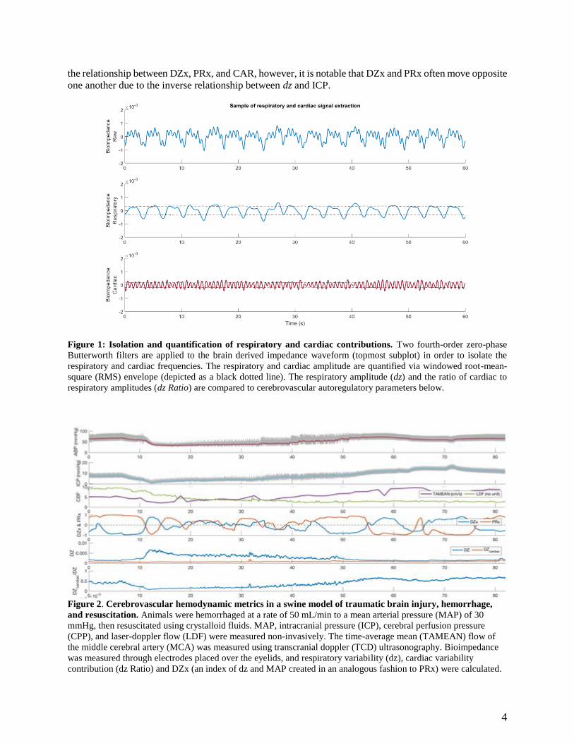
4
the relationship between DZx, PRx, and CAR, however, it is notable that DZx and PRx often move opposite
one another due to the inverse relationship between dz and ICP.
Figure 1: Isolation and quantification of respiratory and cardiac contributions. Two fourth-order zero-phase
Butterworth filters are applied to the brain derived impedance waveform (topmost subplot) in order to isolate the
respiratory and cardiac frequencies. The respiratory and cardiac amplitude are quantified via windowed root-mean-
square (RMS) envelope (depicted as a black dotted line). The respiratory amplitude (dz) and the ratio of cardiac to
respiratory amplitudes (dz Ratio) are compared to cerebrovascular autoregulatory parameters below.
Figure 2. Cerebrovascular hemodynamic metrics in a swine model of traumatic brain injury, hemorrhage,
and resuscitation. Animals were hemorrhaged at a rate of 50 mL/min to a mean arterial pressure (MAP) of 30
mmHg, then resuscitated using crystalloid fluids. MAP, intracranial pressure (ICP), cerebral perfusion pressure
(CPP), and laser-doppler flow (LDF) were measured non-invasively. The time-average mean (TAMEAN) flow of
the middle cerebral artery (MCA) was measured using transcranial doppler (TCD) ultrasonography. Bioimpedance
was measured through electrodes placed over the eyelids, and respiratory variability (dz), cardiac variability
contribution (dz Ratio) and DZx (an index of dz and MAP created in an analogous fashion to PRx) were calculated.

5
Figure 3: Regression models of several cerebrovascular hemodynamic metrics as they relate to dz and dz
Ratio. The hemodynamic and bioimpedance data from the animal displayed in the previous figure are shown here.

6
5) Other achievements:
Established a state of the art large animal model of TBI to be utilized as testing bed for this and
other technologies
Major Task 2: Evaluation of ocular impedance as an indicator of cerebral autoregulation in
humans who are undergoing both invasive arterial blood pressure and ICP monitoring for brain
injury. Months 0-36
1. Specific objectives:
a) IRB approval May 11, 2017
b) HRPO approval September 22, 2017
c) Patient recruitment: 31 patients
Patients who were admitted to the University of Michigan neurosurgery ICU or the trauma ICU
with a ventriculostomy or an ICP monitor and arterial blood pressure monitoring were consented and
enrolled into the study. In cases where the patient was unable to consent, the legally authorized
representative consented on their behalf. A signed copy of the informed consent document was provided.
Patients were admitted to the ICUs most commonly for subarachnoid hemorrhage (15 patients) but also for
brain tumors (4), hematoma (3), trauma (2), intracranial hemorrhage (2), hydrocephalus (1), compression
of the brain stem (1), cortical hemorrhage (1), dermoid cyst (1), and intraventricular hemorrhage (1). 15
females and 17 males were enrolled with an average age of 48.2(16.6).
At a time when the physicians clamped the ventriculostomy as part of routine care, standard
electrode patches (ConMed) were placed over the closed eyes of the patient and anchored at the nasal
bridge, superior orbital rim, and the inferior orbital rim. Bioimpedance data was collected (Biopac Data
Acquisition System) for 20-45 minutes while arterial blood pressures and ICPs were collected
simultaneously. Starting January 4, 2019, the electrodes were changed from the standard electrodes to
proprietary electrodes manufactured by In2Being Inc. and fitted onto a device resembling ocular glass wear
only contacting the patient’s eyelids. This setup has so far been tested on 4 patients with positive feedback
for comfort, skill required for use, as well as signal integrity. This allows us to test on patients with orbital
fractures and other facial traumas.
2. Significant results:
a) Data analysis b) development of analytics for bioimpedance to current standard predictors of autoregulation
As with animals, the trans-ocular bioimpedance signal is largely composed of two frequencies of
interest, relating to the respiratory and cardiac cycles. As expressed in the Q3 Quarterly Technical Progress
Report, we have begun to investigate the cardiac component of the signal in conjunction with the respiratory
oscillations to glean additional information from the signal. Figure 4 depicts a sample of the trans-ocular
bioimpedance signal before and after 30 minutes of ventriculostomy clamping, during which ICP rose
several points. Note the difference in wave morphology - the amplitude of the respiratory signal decreases
slightly, and the higher-frequency cardiac component is significantly lessened. We’ve developed two
different analytic techniques to quantify this change: a spectral analytic tool based on the Peripheral I.V.
Analytic1 and another using the root-mean-square (RMS) envelope described above.
Figure 5 depicts the power spectral density of the same portions of waveform depicted in Figure 4.
Notice that both cardiac and respiratory peaks decrease as the ventriculostomy is clamped and ICP
increases. Figure 6represents a sample of the time-domain analytic, using the root-mean-square envelope
of the band-pass filtered signals as a substitute for the spectral power density estimate. Notably, this analytic
is independent of both respiratory and heart rate as well as respiratory and heart rate variability.

7
We are currently engaged in more analysis of the human data and aim to provide more details in the next
quarterly report.
Figure 4: Sample of transocular bioimpedance waveform before (blue) and 30 minutes after EVD clamping
(red). Note the differences in wave morphology - while the respiratory amplitude decreases slightly, the cardiac
component is significantly lessened.
Figure 5: Power Spectral Density Estimate. Using the same two sections of signal identified above, the differences
in respiratory and cardiac morphology can be quantified. Note that this method is highly dependent on both rate and
rate variability in the two components.

8
Figure 6: Sample of respiratory and cardiac signal extraction. This figure depicts the time-domain analytic using
the root-mean-square (RMS) envelope of the bandpass-filtered signals. Two fourth-order Butterworth filters are
constructed and applied to the impedance waveform to isolate respiratory and cardiac frequencies. The amplitude is
then estimated using a root-mean-square envelope, depicted as a black dotted line. The advantage of this RMS method
is that it performs independently of variations in respiratory or heart rate which may partially confound the spectral
method described above. The respiratory and cardiac RMS envelope are being investigated independently and in ratio
form as an indicator of cerebrovascular autoregulatory status. If successful, this method may allow us to track
cerebrovascular autoregulation without concurrent arterial blood pressure.
References
1. Hocking KM, Sileshi B, Baudenbacher FJ, Boyer RB, Kohorst KL, Brophy CM, Eagle SS.
“Peripheral Venous Waveform Analysis for Detecting Hemorrhage and Iatrogenic Volume
Overload in a Porcine Model.” Shock. 2016 Oct;46(4):447-52.
2. Other achievements
Development of novel electrodes in collaboration with
In2Being, LLC. The electrodes are fitted onto a 3D printed glasses
with only the electrodes contacting patient’s eyelids. This
electrodes/goggles combination prototype has received positive
feedback from patients for comfort and ease of application. This
prototype will be used during patients testing in conjunction with
Biopac system (Figure 7).
Figure 7. Goggles with new
electrodes.

9
Major task 3: Collection of ONS ultrasound videos for assessment of ICP in humans who are
undergoing both invasive arterial blood pressure and ICP monitoring for brain injury. Months 0-
36
1. Specific Objectives:
a. IRB approval May 11, 2017
b. HRPO approval September 22, 2017
c. Patient recruitment: 26 patients
The optic nerve sheath (ONS) is a continuation of the
brain’s dura mater. Characteristics of the ONS, as well as blood
flow to the eye, are known to be affected by ICP and CBF,
potentially allowing the eye to serve as a window into the brain.
Elevated ICP results in swelling of the optic disk (papilledema)
due to the effect of high pressure within the subarachnoid space.
Studies have shown that an increase in ICP results in distension
of the retrobulbar ONS within seconds. Measurement of the
Optic Nerve Sheath Diameter (ONSD) at a standardized 3mm
distance behind the globe could be performed to identify
distension using point-of-care ultrasound devices with specific
transducers with ocular imaging presets (Figure 8).
Patients who were admitted to the University of Michigan
neurosurgery ICU or the trauma ICU with a ventriculostomy or
an ICP monitor and arterial blood pressure monitoring were
consented and enrolled into the study. In cases where the patient
was unable to consent, the legally authorized representative
consented on their behalf. A signed copy of the informed
consent document was provided.
ONUS was performed with the patient’s eye closed, using a linear array transducer placed on the upper
margin of the orbit to obtain a sonographic image of the eye. Imaging was performed on both eyes for each
patient.
Data Collection and Management: The following parameters were monitored for each patient throughout
the experiment.
ONSD using ultrasound (non-invasive); and
Invasive ICP (invasive)
2. Significant results a. Data analysis
In order to find out if there is any correlation between ICP and ONSD we performed several
calculations. In each one we computed the correlation between ICP and different ONSD measures such as
maximum of left and right ONSD, average of left ONSD (L) and right ONSD (R), shown in the following
table.
Max(L, R) Mean(L, R) Abs(L, R)
Pearson (Rho/P-value) 0.1/0.64 0.06/0.75 0.12/0.55
Kendal (Rho/P-value) 0/1 -0.04/0.79 0.13/0.39
Spearman (Rho/P-value) -0.02/0.9 -0.05/0.8 0.19/0.34
Figure 8: Magnified image. Caliper A
demarcates a point 3mm behind the
posterior scleral border and Caliper B
measures the ONSD.

10
This table shows that the most correlation is between the ICP and the absolute difference of right and left
ONSD. However, this correlation is not statistically significant. We are working on recruiting more
patients and optimizing our approach.
Major Task 4: Development of an ultrasound video analytic system to evaluate ONSD. Months
6-36
1. Specific objectives:
a. Development of ultrasound analytic system
b. Compare reading of automated ONSD with manual reading by clinicians
We develop an automated algorithm using image processing techniques to analyze ultrasound(US)
images and calculate the optic nerve sheath diameter(ONSD) in 3 mm posterior to the orbit/globe as shown
in Figure 8. The schematic diagram of the proposed method is shown in Figure 2. In the first stage of the
proposed method, we perform preprocessing in which we crop images using Digital Imaging and
Communications in Medicine (DICOM) attributes that specify the location of the region. We also denoise
images using a technique called image guided filtering [3] which is an edge-preserving method. Suppose
that the filtering input image and guidance image are 𝑃 and 𝐼 respectively. The images are divided to
overlapped windows with radius of 𝑟 and following coefficients are computed in each window:
𝑎𝑘 =𝑐𝑜𝑣𝑘(𝐼, 𝑃)
𝑣𝑎𝑟𝑘(𝐼) + 𝜀
(1)
𝑏𝑘 = 𝑘 − 𝑎𝑘𝐼
where 𝑘 is the window index, 𝐼 and 𝑘 are average of intensities in 𝑘𝑡ℎ window in noisy and guidance
images respectively. Also 𝜀 is called regularization parameter that determines the edge-preserving property
of the filter. The filtered pixel 𝑞𝑖 is the average of 𝑎𝑘𝐼𝑖 + 𝑏𝑘 in all the windows that cover 𝑞𝑖.
After denoising images, we find the region of interest (ROI) by analyzing the image integral. This is done
through calculating the summation of pixel values in each column. Suppose that the denoised image is an
𝑁 × 𝑀 image shown as 𝐼𝑑 . We analyze the following one dimensional signal.
𝑣(𝑖) = ∑ 𝐼𝑑(𝑗, 𝑖)𝑀
𝑗=1 (2)

11
This signal has two main peaks, 𝑀𝑎𝑥1 and 𝑀𝑎𝑥2,
corresponding to the vertical borders of the ROI and
a local minimum between these peaks corresponding
to the dark region inside the sheaths. Suppose that the
minimum of this signal is 𝑔th element of the signal
which corresponds to the column where we can find
the globe.
After finding the globe point and the ROI, we use a
superpixel segmentation technique called simple
linear iterative clustering (SLIC) to segment each
image to superpixels. Suppose that the output of this
method is called 𝐼𝑠 and the row which is 3mm below
the globe is 𝑟3𝑚𝑚th row of 𝐼𝑠. We analyze the peaks
and also derivatives of this row to calculate the
ONSD. We repeat this process for all the images in
the Ultrasound video and then we calculate the
median of all the values (Voting).
2. Significant results
We applied the proposed method on 52 videos of 26 patients (for each patient we have US images of both
eyes) and calculated the average of the error between the proposed method and the ground truth (i.e. point
of care manual measurements). The automated algorithm was able to process images, determine and
measure ONSD with high precision when compared to clinicians’ manual measurement with percentage of
error difference between the two methods at 6.5% safely within the clinically accepted error. We will
continue data collection and refinement of the algorithm to reduce the percentage error even further.
References:
1) Williams, P., 2017. Optic Nerve Sheath Diameter as a Bedside Assessment for Elevated Intracranial
Pressure. Case reports in critical care, 2017.
2) Kimberly HH, Shah S, Marill K, Noble V. Correlation of optic nerve sheath diameter with direct
measurement of intracranial pressure. Academic Emergency Medicine. 2008 Feb 1;15(2):201-4.
3) He K, Sun J, Tang X. Guided image filtering. IEEE transactions on pattern analysis and machine
intelligence. 2013 Jun;35(6):1397-409.
4) Achanta R, Shaji A, Smith K, Lucchi A, Fua P, Süsstrunk S. SLIC superpixels compared to state-
of-the-art superpixel methods. IEEE transactions on pattern analysis and machine intelligence.
2012 Nov;34(11):2274-82.
A. What opportunities for training and professional development has the project provided? Opportunity was provided for Mr. Brandon Cummings, a graduate student in the
Bioinformatics Master Degree Program to present data at the 2018 MHSRS Symposium. Mr.
Cummings works with the PI and Co-I on signal processing and data analysis. Mr. Cummings
presented an abstract in a form of a poster presentation: (Abstract and poster are provided in the
appendices)
Preprocessing (Denoising, ...)
FindingRegion of Interest
(ROI)
Finding Globe
Segmentation (Superpixel)
Analyzing pixels 3mm posterior to
Globe
Optic Nerve Ultrasound Images
Voting
ONSD
Figure 8: Schematic diagram of the proposed method
𝑔 = 𝑎𝑟𝑔𝑚𝑖𝑛 𝑀𝑎𝑥1≤𝑖≤𝑀𝑎𝑥2
𝑣(𝑖) (3)

12
- Brandon Cummings, BS, Brendan McCracken, BS, Chandler Rygalski, BS, Ashwin Belle, PhD,
Kevin Ward, MD, M. Hakam Tiba, MD, MS.: A Signal Processing Approach for the Calculation
of a Bioimpedance Index in the Assessment of Cerebrovascular Autoregulatory Status., Military
Health System Research Symposium (MHSRS), Kissimmee, Florida, 2018.
B. How were the results disseminated to communities of interest? Data was presented to the community of scientific peers at the Military Health System
Research Symposium (MHSRS), Kissimmee, Florida, 2018 in the form of 2 poster presentation.
(Abstracts and posters will be provided in the append 1. M. Hakam Tiba, MD, MS, Krishna Rajajee, MD, Craig Williamson, MD, Ashwin Belle, PhD,
Sardar Ansari, PhD, Brandon Cummings, BS, Brendan McCracken, BS, Amanda Pennington, MS,
Kevin Ward, MD.: Monitoring Traumatic Brain Injury Patients using Transocular Brain Impedance
(TBI)., Military Health System Research Symposium (MHSRS), Kissimmee, Florida, 2018.
2. Brandon Cummings, BS, Brendan McCracken, BS, Chandler Rygalski, BS, Ashwin Belle, PhD,
Kevin Ward, MD, M. Hakam Tiba, MD, MS.: A Signal Processing Approach for the Calculation
of a Bioimpedance Index in the Assessment of Cerebrovascular Autoregulatory Status., Military
Health System Research Symposium (MHSRS), Kissimmee, Florida, 2018.
C. What do you plan to do during the next reporting period to accomplish the goals? The activities in all major task areas will be continued for the duration of the next reporting
period. We plan to continue animal testing using the provocative maneuvers model and begin the
model of blunt trauma TBI at a pace targeting 5 animals (either model)/ quarter. Human subjects’
recruitment and testing will be continued with a target of 15 patients/ quarter. Human subjects’
data collection will be collected using the latest iteration of the Trans-Ocular Bioimpedance
prototype (TOBI), and compared to preliminary data for signal quality device validation. In tandem
with collection, data analysis and signal processing will continue for the duration of the next
reporting period. (Signal processing a validation of bioimpedance signal against autoregulation
parameters such as MAP, ICP, or cerebral blood flow). Further development of ultrasound
technique and algorithm for assessment of ONSD will continue for the duration of the next
reporting period (Patients recruitment and algorithm validation). Data and project progress will
continue to be divulged via presentations at scientific meetings both locally at the University of
Michigan and nationally at the MHSRS and the Shock Society Meeting during the next reporting
period. Lastly, scientific manuscript writing will begin this reporting period with a target of 2-4
publications in major scientific and clinical journals covering all major tasks outlined in the report.
i. Continue testing animals both models 1 & 2
ii. Continue patient recruitment
iii. Continue patients testing using prototype
iv. Further algorithm development for ONSD ultrasound
v. Data analysis and signal processing
vi. Data presentation (national and local
vii. Manuscript writing
D. IMPACT:
a. What was the impact on the development of the principal discipline(s) of the project? Nothing to report at this time as we are still in testing phase. However, we are expecting a high
level of impact by the end of the project on the understanding of cerebrovascular autoregulation and its
relationship to cerebral blood flow with the ability to monitor and track these events using transocular

13
impedance that can be commercialized. We will also continue to assess ICP non-invasively using ONSD
via ultrasound and an automated algorithm.
b. What was the impact on other disciplines?
Nothing to report.
c. What was the impact on technology transfer?
i. Provisional patent application (62/506,971) Ocular Impedance-Based System for Brain Health
Monitoring. Submitted May 16, 2017.
ii. Invention disclosure (D2018-0119) Automated Method to Calculate the Optic Nerve Sheath
Diameter. Filed with the University of Michigan Office of Technology Transfer.
iii. Prototype developed by In2Being, LLC.
iv. Technology now exclusively licensed to New Vital Signs, Inc.
d. What was the impact on society beyond science and technology?
the proposed work is envisioned to lead to development of technologies for noninvasive evaluation of
CAR and ICP. Such technologies are envisioned to be suitable for in-hospital and out-of-hospital setting
in both the civilian and military setting and will allow for:
1) Early application by first responders and military medics for precision management of the severe
TBI patient including providing optimal and personalized cerebral perfusion pressure as opposed
to a range.
2) Rapid point of care diagnostic indicators of severity of TBI allowing for earlier intervention in more
far forward echelons of care.
3) Improved outcomes by earlier detection of injury and prevention of secondary damage.
4) Greater uninterrupted continuum of care as casualties moves from lower to higher levels of care.
5) Reduction in the need for experienced personnel to perform the time consuming procedures
necessary for invasive monitoring as well as elimination of associated complications.
6) Improved resource allocation by providing indications for invasive monitoring as well as earlier
termination of such invasive monitoring (when they are indicated) by transitioning into noninvasive
monitoring.
E. CHANGES/PROBLEMS:
a. Changes in approach and reasons for change
Nothing to report
b. Actual or anticipated problems or delays and actions or plans to resolve them
i. We have encountered a lower than anticipated enrollment at the beginning of the reporting period
due to the low number of Codman ICP monitors placed at Michigan Medicine. In order to continue
to enroll patients we are including patients who have a ventriculostomy which has been clamped as
part of their clinical care as well as a subdural screw or epidural sensor. The addition of
ventriculostomy has enhanced our recruitment to meet our goal.
ii. The Michigan health system has experienced several computer connectivity issues that prevented us
from collecting patients’ waveforms from part of June and July. The health system IT department
resolved the issue and restored connectivity.
c. Changes that had a significant impact on expenditures
Nothing to report

14
d. Significant changes in use or care of human subjects, vertebrate animals, biohazards, and/or
select agents
i. Significant changes in use or care of human subjects: None to report
ii. Significant changes in use or care of vertebrate animals: None to report
iii. Significant changes in use of biohazards and/or select agents: None to report
F. PRODUCTS:
a. Publications, conference papers, and presentations
i. Journal publications. Nothing to report
ii. Books or other non-periodical, one-time publications. Nothing to report
iii. Other publications, conference papers, and presentations.
Two abstracts have been submitted to the 2018 Military Health System Research Symposium
(MHSRS) and have been accepted for poster presentations.
1. MHSRS-18-0614: Hakam Tiba, MD, MS, Krishna Rajajee, MD, Craig Williamson, MD, Ashwin
Belle, PhD, Sardar Ansari, PhD, Brandon Cummings, BS, Brendan McCracken, BS, Amanda
Pennington, MS, Kevin Ward, MD. Monitoring Traumatic Brain Injury Patients using Transocular
Brain Impedance (TBI).
2. MHSRS-18-0734: Brandon Cummings, BS, Brendan McCracken, BS, Chandler Rygalski, BS,
Ashwin Belle, PhD, Kevin Ward, MD, M. Hakam Tiba, MD, MS. A Signal Processing Approach
for the Calculation of a Bioimpedance Index in the Assessment of Cerebrovascular Autoregulatory
Status
b. Website(s) or other Internet site(s) Nothing to report
c. Technologies or techniques Nothing to report
d. Inventions, patent applications, and/or licenses i. Provisional patent application (62/506,971) Submitted May 16, 2017. (Patent application
included as part of the appendices.
ii. Invention disclosure (D2018-0119) Filed June 11, 2018 with the University of Michigan Office of
Technology Transfer.
e. Other Products
Created a one-page description of the methodology for patients and their families. Material is provided
in the appendices.
f. Research material (e.g., Germplasm; cell lines, DNA probes, animal models);
Animal TBI model developed for this project is now being utilized for testing beyond bioimpedance
G. PARTICIPANTS & OTHER COLLABORATING ORGANIZATIONS
a. What individuals have worked on the project? Example:
Name: Mary Smith
Project Role: Graduate Student

15
Researcher Identifier (e.g.
ORCID ID): 1234567
Nearest person month worked: 5
Contribution to Project:
Ms. Smith has performed work in the area of combined error-control and
constrained coding.
Funding Support:
The Ford Foundation (Complete only if the funding support is provided from
other than this award).
b.
Name: Mohamad Hakam Tiba, MD, MS
Project Role: PI
Researcher Identifier (e.g.
ORCID ID):
Nearest person month worked: 3
Contribution to Project: Oversight of data collection and analysis
Funding Support:
Name: Kevin Ward, MD
Project Role: Co-I
Researcher Identifier (e.g.
ORCID ID):
Nearest person month worked: 1
Contribution to Project: Oversight of data collection and analysis
Funding Support:
Name: Venkatakrishna Rajajee, MD
Project Role: Co-I
Researcher Identifier (e.g.
ORCID ID):
Nearest person month worked: 1
Contribution to Project: Perform ultrasounds, medical consultation
Funding Support:
Name: Craig Williamson, MD
Project Role: Co-I
Researcher Identifier (e.g.
ORCID ID):
Nearest person month worked: 1
Contribution to Project: Perform ultrasounds, medical consultation
Funding Support:
Name: Hasan Alam, MD PhD
Project Role: Co-I

16
Researcher Identifier (e.g.
ORCID ID):
Nearest person month worked: 1
Contribution to Project: Oversight of data collection and analysis
Funding Support:
Name: Kayvan Najarian, PhD
Project Role: Co-I
Researcher Identifier (e.g.
ORCID ID):
Nearest person month worked: 1
Contribution to Project: Development of a computer image analysis algorithm
Funding Support:
Name: Reza Soroushmehr, PhD
Project Role: Research Staff
Researcher Identifier (e.g.
ORCID ID):
Nearest person month worked: 5
Contribution to Project: Development of a computer image analysis algorithm
Funding Support:
Name: Amanda Pennington, MS
Project Role: Clinical Project Manager
Researcher Identifier (e.g.
ORCID ID):
Nearest person month worked: 5
Contribution to Project:
Subject screening and enrollment, regulatory and compliance management,
data collection
Funding Support:
Name: Brendan McCracken, BS
Project Role: Laboratory Assistant Director
Researcher Identifier (e.g.
ORCID ID):
Nearest person month worked: 4
Contribution to Project: Oversight and lab management, data collection, data analysis
Funding Support:
Name: Brandon Cummings, BS
Project Role: Research Staff
Researcher Identifier (e.g.
ORCID ID):

17
Nearest person month worked: 1
Contribution to Project: Data collection, signal processing and data analysis
Funding Support:
Name: Carmen Colmenero, BS
Project Role: Research Staff
Researcher Identifier (e.g.
ORCID ID):
Nearest person month worked: 4
Contribution to Project: Animal lab duties, data collection, data analysis
Funding Support:
Name: Danielle Leander, BS
Project Role: Research Staff
Researcher Identifier (e.g.
ORCID ID):
Nearest person month worked: 2
Contribution to Project: Animal lab duties, data collection, data analysis
Funding Support:
Name: Chandler Rygalski, BS
Project Role: Research Staff
Researcher Identifier (e.g.
ORCID ID):
Nearest person month worked: 1
Contribution to Project: Animal lab duties, data collection, data analysis
Funding Support:
Name: Daniel Taylor, MA
Project Role: Data Engineer
Researcher Identifier (e.g.
ORCID ID):
Nearest person month worked: 3
Contribution to Project: Signal processing, data storage and analysis
Funding Support:
Name: Mark Salamango, PhD
Project Role: Data Engineer
Researcher Identifier (e.g.
ORCID ID):
Nearest person month worked: 1
Contribution to Project: Signal processing, data storage and analysis
Funding Support:

18
Name: Justin Massey, BS
Project Role: Research Staff
Researcher Identifier (e.g.
ORCID ID):
Nearest person month worked: 3
Contribution to Project: Screening and consenting patients, data collection
Funding Support:
Name: Erin Bisco, BS
Project Role: Research Staff
Researcher Identifier (e.g.
ORCID ID):
Nearest person month worked: 1
Contribution to Project: Screening and consenting patients, data collection
Funding Support:
c. Has there been a change in the active other support of the PD/PI(s) or senior/key personnel
since the last reporting period? Nothing to report
d. What other organizations were involved as partners? None
e. Other. Nothing to report
H. SPECIAL REPORTING REQUIREMENTS
a. COLLABORATIVE AWARDS:
None
b. QUAD CHARTS: Included with this report before the appendices .
I. APPENDICES:
I. Abstract MHSRS-18-0614 and Poster
II. Abstract MHSRS-18-0734 and Poster
III. Educational Material for
o One-page Protocol Description for Patients and Patients’ families
o Healthcare staff.
IV. Invention Disclosure
V. Patent Application
VI. PI Curriculum Vitae
Novel Noninvasive Methods of Intracranial Pressure and Cerebrovascular Autoregulation
Assessment: Seeing the Brain through the EyesDM160225 Prolonged Field Care Research Award
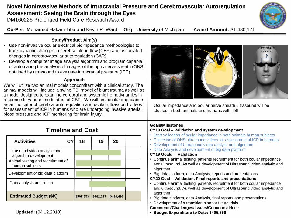
Co-PIs: Mohamad Hakam Tiba and Kevin R. Ward Org: University of Michigan Award Amount: $1,480,171
Study/Product Aim(s)
• Use non-invasive ocular electrical bioimpedance methodologies to
track dynamic changes in cerebral blood flow (CBF) and associated
changes in cerebrovascular autoregulation (CAR).
• Develop a computer image analysis algorithm and program capable
of automating the analysis of images of the optic nerve sheath (ONS)
obtained by ultrasound to evaluate intracranial pressure (ICP).
Approach
We will utilize two animal models concomitant with a clinical study. The animal models will include a swine TBI model of blunt trauma as well as a model designed to examine cerebral and systemic hemodynamics in response to various modulators of CBF. We will test ocular impedance as an indicator of cerebral autoregulation and ocular ultrasound videos for assessment of ICP in humans who are undergoing invasive arterial blood pressure and ICP monitoring for brain injury.
Goals/Milestones
CY18 Goal – Validation and system development
• Start validation of ocular impedance in both animals human subjects
• Collection of ONS ultrasound videos for assessment of ICP in humans
• Development of Ultrasound video analytic and algorithm
• Data Analysis and development of big data platform
CY19 Goals – Validation
• Continue animal testing, patients recruitment for both ocular impedance
and ultrasound. As well as development of Ultrasound video analytic and
algorithm
• Big data platform, data Analysis, reports and presentations
CY20 Goal – Validation, Final reports and presentations
• Continue animal testing, patients recruitment for both ocular impedance
and ultrasound. As well as development of Ultrasound video analytic and
algorithm
• Big data platform, data Analysis, final reports and presentations
• Development of a transition plan for future trials
Comments/Challenges/Issues/Concerns: None
• Budget Expenditure to Date: $495,856Updated: (04.12.2018)
Timeline and Cost
Activities CY 18 19 20
Ultrasound video analytic and
algorithm development
Estimated Budget ($K) $507,353 $482,327 $490,491
Animal testing and recruitment of
human subjects
Data analysis and report
Development of big data platform
Ocular impedance and ocular nerve sheath ultrasound will be
studied in both animals and humans with TBI

II. Abstract MHSRS-18-0614
Monitoring Traumatic Brain Injury Patients using Transocular Brain Impedance (TBI).
M. Hakam Tiba, MD, MS1,2, Krishna Rajajee, MD2,3, Craig Williamson, MD2,3, Ashwin Belle, PhD1,2, Sardar
Ansari, PhD1,2, Brandon Cummings, BS1,2, Brendan McCracken, BS1,2, Amanda Pennington, MS1,2, Kevin
Ward, MD1,2,4
1 Department of Emergency Medicine, University of Michigan, Ann Arbor, MI. 2 Michigan Center of Integrative Research in Critical Care (MCIRCC), University of Michigan, Ann Arbor, MI. 3 Department of Neurosurgery, University of Michigan, Ann Arbor, MI. 4 Department of Biomedical Engineering, University of Michigan, Ann Arbor, MI.
Introduction: Cerebrovascular autoregulation (CAR) is an auto-protective mechanism where intracranial
vessels modulate their tone in order to maintain consistent levels of cerebral blood flow (CBF) in the face of
changing intracranial or systemic pressure. CAR is often impaired after traumatic brain injury (TBI) which may
result in secondary brain injury. CAR has been found to be a significant predictor of outcome after TBI as well
as a beneficial means of optimizing cerebral perfusion pressure (CPP). Monitoring and assessment of CAR is
envisioned to be useful and valuable during the management of TBI patients. However, direct assessment of
CAR is difficult and has proven to be elusive. Current modalities assessing CAR are often invasive and hard to
obtain in a meaningful and timely manner. The pressure-reactivity index (PRx), which describes how intracranial
pressure (ICP) and mean arterial pressure (MAP) vary in relation to each other has been found to be an
acceptable method of CAR assessment. However, the feasibility of PRx measurement is limited as it requires
the use of ICP monitoring and heavy filtering to remove signal noise. In this investigation, we propose a novel
measure to assess CAR non-invasively and in real time using brain bioimpedance measured through a
transocular pathway. Bioimpedance measures the passive electrical properties of tissue and is affected directly
by the volume of blood in the interrogated area, and indirectly by respiration. By harnessing such effects on
bioimpedance, we hypothesize that this methodology will provide a real time and non-invasive assessment of
CAR. Methods: This investigation utilizes a large animal model of TBI as well as monitoring of TBI patients in
the ICU. In the animal model, male Yorkshire swine with a mean(SD) weight of 39.8(1.5) kg were anesthetized,
mechanically ventilated, and instrumented to continuously monitor and record brain bioimpedance, ICP, MAP,
and CBF. Cerebral blood volume and CPP were manipulated with maneuvers such as intravenous
norepinephrine challenge, epidural hematoma or systemic hemorrhage. Brain bioimpedance was also
continuously monitored in human TBI patients without any provocative maneuvers, provided that their ICP and
MAP were already being monitored as part of their clinical management. In both the animal model and human
monitoring, brain bioimpedance was obtained by placing ECG electrodes on the eyelids. PRx was calculated as
the moving Pearson correlation between mean ICP and MAP over a two-minute window. The novel
bioimpedance index (DZx) was calculated as a moving correlation between the respiratory changes in
bioimpedance (dz) and MAP. Results: The diagnostic performance of DZx to predict CAR impairment was
evaluated using the Receiver-Operator Characteristic (ROC) curves and Area Under the Curve (AUC). DZx was
compared to PRx which was used as a reference point and gold standard at a threshold value of zero, with
positive values indicating CAR impairment and negative values indicating active vasogenic vessels and intact
CAR. The mean(SD) area under the ROC curve (AUC) for DZx was 0.82(0.12)% and 0.78 for animals and
patients respectively, indicating a significant predictive ability. Conclusion: In this study, DZx appears to track
changes in PRx with high precision. This indicates that DZx may prove useful as a portable, easily-applied, and
significantly less-invasive alternative to PRx as a diagnostic index for the early assessment and detection of CAR
impairment. Further studies in animals and humans are currently underway to further validate this promising
technology.

University of
Michigan
• The diagnostic ability of DZ and DZx to predict autoregulation impairment was
assessed using:
• Linear Regression
• Receiver-Operator Characteristic (ROC) curves and Area Under the
Curve
• PRx was used as the gold standard at a threshold value of zero
• Mean(SD) AUC: 88.5(4.0)%
• DZx and PRx are typically
inverted about the zero
point
• Negative PRx values indicate
intact autoregulation
• Positive DZx values indicate intact
autoregulation
Traumatic Brain Injury (TBI)
• “Silent epidemic” because of associated
complications
• 2.5 million people sustained a TBI in 2010
• Accounts for 30% of all injury related deaths
Management & Monitoring Strategies
• Monitoring of intracranial pressure (ICP)
• Invasive, increased risk of infection and further damage to the brain
• Optimization of cerebral perfusion pressure (CPP) to a target level
• Preventing secondary brain damage (ischemia, edema)
Cerebrovascular Autoregulation
• Vessels modulate their tone in response to pressure changes
• Prevention of secondary brain injury
• Auto-protective mechanism
• Maintains constant levels of
cerebral blood flow (CBF)
• Impaired in severe head injury or
acute ischemic stroke
• Predictor of poor outcomes in
acute neurological disease
• Opposition to an electrical current flow through tissues
• Passive bioelectricity. Tissues’ response to external electrical excitation.
• Cumulative effect of individual impedances
• Blood has a distinct effect on bioimpedance:
• Good conductor of electricity
• More blood present lower bioimpedance
• RESPIRATION affects bioimpedance indirectly
• Thoracic pressure gradient
• Changes in venous return
Transocular Brain Impedance (TOBI)
• Bipolar impedance measured from noninvasive
electrodes placed over the eyelids
• Brain encounters a significant portion of
the electrical current
• Impedance respiratory
variation reflects changes
in brain blood volume
• Continuous monitoring is beneficial to optimize CPP
• Precision or personalized approach in managing the components of CPP
Measuring autoregulation remains a challenge
Current monitoring modalities include:
• Hemoglobin saturation by near-infrared
spectroscopy (NIRS)
• Laser Doppler flowmetry of CBF
• Transcranial Doppler (TCD)
o Invasive
o Intermittent spot-checks
o Require high levels of expertise
o Unavailable at earlier echelons of care
o Produce mixed results
Pressure reactivity index (PRx)
Moving Pearson correlation between MAP
and ICP
• Requires invasive ICP measurement
• Calculation is complex with noisy signal
• Often unavailable outside of
research-minded academic settings
Monitoring Traumatic Brain Injury Patients using Transocular Brain Impedance (TOBI)M. Hakam Tiba, MD, MS1,2, Brendan M. McCracken , BS1,2, Brandon Cummings, BS1,2,
Krishna Rajajee, MD2,3, Craig Williamson, MD2,3, Ashwin Belle, PhD1,2, Sardar Ansari, PhD1,2,
Amanda Pennington, MS1,2, Kevin Ward, MD1,2,4
1 Department of Emergency Medicine, 2 Michigan Center of Integrative Research in Critical Care (MCIRCC), 3 Department of Neurosurgery,
4 Department of Biomedical Engineering, University of Michigan, Ann Arbor, MI.
INTRODUCTION
• TOBI may be used to track cerebrovascular autoregulation status with high
precision
• Noninvasive techniques to assess CAR may prove to have tremendous
potential in guiding patient triage and management, as well as improving
outcomes
• Utilize Transocular Brain Impedance
to assess cerebrovascular
autoregulation by comparing
respiratory changes in
bioimpedance to MAP, ICP, CPP,
and cerebral blood flow.
• Use TOBI to create new and novel
indices predictive of changes in
cerebrovascular autoregulation.
AIM & Methods
BIOIMPEDANCE
CEREBROVASCULAR AUTOREGULATION
RESULTS
CONCLUSION
Large Animal Model
• Anesthetized animals weighing an average(SD) of 39(0) kg were instrumented
to monitor MAP, ICP, and CBF (using LDF and TCD)
• Bioimpedance was monitored through ECG-electrodes placed over eyelids
• Challenges such as vasopressors (norepinephrine) infusion, creation of
epidural hematoma, and systemic hemorrhage were used to manipulate level
of MAP, ICP and CBF out of optimal zone and temporarily impairing CAR
• PRx: was calculated as moving Pearson correlation of MAP and mean ICP
• DZx (New index): was computed as the moving Pearson correlation of MAP
and DZ (Respiratory variability in the bioimpedance signal)
Clinical testing:
Moderate to severe TBI patients were recruited and bioimpedance was
monitored alongside blood pressure and ICP for a period of 45 – 90min
Cardiac
variationRespiratory
variation
https://www.semantic
scholar.org/paper/Ma
nagement-of-
Intracranial-Pressure.-
Freeman/5e7a88c6be
e65fa8f0be807e88c445
c197e2cfb9
Impaired CAR
MAP ICPPositive Correlation
Intact CAR
MAP ICPNegative Correlation
KRW and MHT have submitted a Patent application (62/506,971) with the Office of Tech Transfer, University of Michigan, Ann Arbor.
The technology has been licensed to New Vital Signs Inc. Ann Arbor, Michigan
This work has been supported by a DoD (PFC) grant award (Award No. W81XWH-18-1-0005, Log No. DM160225).

III. Abstract MHSRS-18-0734:
A Signal Processing Approach for the Calculation of a Bioimpedance Index in the Assessment of Cerebrovascular Autoregulatory Status
Brandon Cummings, BS1,2, Brendan McCracken, BS1,2, Chandler Rygalski, BS1,2, Ashwin Belle, PhD1,2, Kevin
Ward, MD1,2,3 M. Hakam Tiba, MD, MS1,2,
1 Department of Emergency Medicine, University of Michigan, Ann Arbor, MI. 2 Michigan Center of Integrative Research in Critical Care (MCIRCC), University of Michigan, Ann Arbor, MI. 3 Department of Biomedical Engineering, University of Michigan, Ann Arbor, MI.
Background: Cerebrovascular autoregulation (CAR) is an auto-protective mechanism in which the brain maintains consistent levels of cerebral blood flow (CBF) in the face of changing systemic and intracranial pressures by modulating vascular tone. Traumatic brain injuries (TBI) often impair CAR function, which can result in secondary injuries including ischemia, inflammation, and edema. Although CAR is believed to predict outcome and aid in optimization of cerebral perfusion pressure (CPP), direct assessment of CAR has proven elusive. The pressure reactivity index (PRx), which describes the relationship between intracranial pressure (ICP) and mean arterial pressure (MAP), is becoming an acceptable method of CAR measurement. Its values range from negative one to positive one, with negative values indicating intact CAR with active vasogenic vessels and positive values indicating impaired autoregulatory function. However, the feasibility of this measure is limited as it requires the use of continuous and invasive ICP monitoring. We previously described the ability of respiratory-driven changes in trans-ocular impedance (dz) to be a potential indicator of cerebrovascular parameters such as ICP and CBF, and further preliminary work shows that the combination of this metric with continuous MAP recording may provide an alternative to PRx measurement which does not require invasive ICP measurement. However, there exists a previously unmet need for continuous, real-time, algorithmic analysis of this bioimpedance index (DZx). Here, we introduce an algorithm which robustly quantifies respiratory variation and couples this data with continuous MAP to produce an index analogous to PRx. Methods: Data collection: Four anesthetized Yorkshire swine were surgically instrumented to measure ICP, MAP, and CBF. ICP and MAP were manipulated using challenges such as norepinephrine, simulated hematoma, and controlled hemorrhage. Additionally, two human patients with indwelling ICP and arterial blood pressure monitors were recruited from the neurosurgical ICU and passively monitored. In both cases, trans-ocular brain impedance was monitored at 200 Hz from two standard Ag/AgCl ECG electrodes placed over the eyelids for up to one hour. This yielded a combination of over XX hours of data from which to build the algorithm. Algorithm: The trans-ocular bioimpedance was first pre-processed to remove fluctuations in baseline and high-frequency noise. This was accomplished using a fourth-order, zero-phase Butterworth IIR bandpass filter with cutoff frequencies of 0.1 and 2 Hz. The result was a composite signal containing clear respiratory and cardiac component frequencies. The magnitude of the respiratory component (dz) was quantified using a root-mean-square (RMS) envelope with a window size of 30 seconds. This technique was chosen over a more direct peak-to-peak measure as it was found to be more robust when considering the high breath-to-breath variability in respiratory rate and tidal volume observed during spontaneous ventilation. MAP was estimated from a continuous arterial pressure waveform using a windowed moving average with a window size of 30 seconds. The bioimpedance index (DZx) was computed in a fashion analogous to PRx calculation, using a moving Pearson correlation with a window size of five minutes and a step size of one second. Results: The algorithm described above yields a continuous DZx signal with values ranging from negative one to positive one, inclusive. We observed that DZx tracks PRx in an opposite manner, which is consistent with previous observations that respiratory variations in bioimpedance (dz) are negatively correlated with ICP and CBF. Thus, it appears that negative values of DZx are indicate impaired CAR and positive values indicate intact CAR with active vasogenic vessels. Receiver-Operator Characteristic (ROC) curves were used to assess the predictive capability of DZx during several of the maneuvers in animals, with a threshold of PRx = 0. Mean(SD) area under the curve was found to be 0.82(0.12), indicating significant predictive ability. Conclusion: Respiratory variations in bioimpedance (dz) have been previously described as a novel alternative to traditional highly-invasive methods of cerebrovascular autoregulatory (CAR) status. Furthermore, coupling this metric with continuous MAP data results in a bioimpedance index (DZx) which may prove to be a viable alternative to the pressure-reactivity index (PRx) in the assessment of autoregulatory status.

University of
Michigan
• Bioimpedance: the opposition of a tissue to an externally applied electric
current
• Cumulative effect of individual impedances of each tissue component (muscle,
adipose tissue, extracellular fluid, blood, etc.)
• Blood has a distinct effect on bioimpedance:
• Good conductor of electricity
• Changes with cardiac/respiratory cycles
• Respiratory variation causes thoracic pressure
gradient which changes venous return, thus
affecting local blood volume
Transocular Brain Impedance (TOBI)
• Bipolar impedance measured
from noninvasive electrodes
placed over the eyelids
• Brain encounters a significant
portion of the electrical current
sent through the globes
• Signal contains respiratory and
cardiac components
Measuring autoregulation remains a challenge
• Current monitoring modalities include:
• Transcranial Doppler (TCD)
• Near-Infrared Spectroscopy (NIRS)
• Laser Doppler Flowmetry (LDF)
• Many of these methods are:
• Invasive
• Intermittent spot-checks
• Require high levels of expertise
• Unavailable at earlier echelons of care
• Produce mixed results
Pressure reactivity index (PRx)
• Moving Pearson correlation between MAP
and ICP
• Requires invasive ICP measurement
• Calculation produces noisy signal that may
be difficult to interpret
• Often unavailable outside of
research-minded academic settings
Traumatic Brain Injury (TBI)
• 2.5 million people sustained a TBI in 2010
• TBI accounts for 30% of all injury related deaths
Current Management & Monitoring Strategies
• Preventing secondary brain damage (ischemia, edema)
• Monitoring of intracranial pressure (ICP)
• Invasive, increased risk of infection and further damage to the brain
• Optimization of cerebral perfusion pressure (CPP) to a target level
Cerebrovascular Autoregulation (CAR)
• The ability of vessels to modulate their tone in response to
pressure changes
• Auto-protective mechanism which
maintains constant levels of
cerebral blood flow (CBF) to
match metabolic demand
• Critical in the prevention of
secondary brain injury
• Predictor of poor outcomes in
acute neurological disease
• Impaired in severe head injury or acute ischemic stroke
A Signal Processing Approach for the Calculation of a Bioimpedance Index
in the Assessment of Cerebrovascular Autoregulation
Brandon Cummings, BS1,3; Brendan McCracken, BS1,3; Carmen Colmenero, BS1,3;
Amanda Pennington, MS1,3; Kevin Ward, MD1,2,3; M. Hakam Tiba, MD, MS1,3;Departments of: 1Emergency Medicine, 2Biomedical Engineering, 3Michigan Center for Integrative Research in Critical Care
University of Michigan, Ann Arbor, Michigan
INTRODUCTION
• TOBI impedance may be used to track cerebrovascular autoregulation status
with high precision and help optimize CPP
• Noninvasive techniques to assess CAR may prove to have tremendous
potential in guiding patient triage and management, as well as improving
outcomes
To develop a noninvasive index of cerebrovascular
autoregulation using characteristics from the trans-ocular
brain impedance waveform.
AIM
BIOIMPEDANCE
CURRENT MONITORING STRATEGIES
METHODS
RESULTS
CONCLUSIONImpaired CAR
MAP ICPPositive Correlation
Intact CAR
MAP ICPNegative Correlation
Large Animal Model
• Anesthetized swine weighing 40-45 kg were instrumented to monitor ICP,
MAP, CBF, & TCD
• Bioimpedance was monitored noninvasively through ECG electrodes placed
over eyelids
• Maneuvers such as norepinephrine challenges, simulated hematomas, and
controlled arterial hemorrhage were used to manipulate CPP outside of
optimal zone
Bioimpedance Signal Analysis
• Impedance waveform was
pre-processed to remove
baseline fluctuations and high-
frequency noise
• Pre-processing results in
composite signal containing cardiac
and respiratory frequencies
• dz calculated based on respiratory
signal amplitude
• A novel bioimpedance index (DZx) was calculated using MAP and dz in a
fashion analogous to PRx.
KRW and MHT have submitted a Patent application (62/506,971) with the Office of Tech Transfer, University of Michigan, Ann Arbor.
The technology has been licensed to New Vital Signs Inc. Ann Arbor, Michigan
This work has been supported by a DoD (PFC) grant award (Award No. W81XWH-18-1-0005, Log No. DM160225).

ASSESSMENT OF CEREBROVASCULAR AUTOREGULATION AND ICP USING BIOIMPEDANCE AND OCULAR ULTRASOUND
Background: Traumatic brain injury (TBI) is a major public health problem in the U.S. and around the world. The Defense and Veterans Brain Injury Center (DVBIC) has reported over 34,000 moderate to severe combat-related TBI events since 2000, and the CDC states that approximately 30% of injury related civilian deaths in the U.S. One of the significant management strategies in the management of TBI is aimed at preventing secondary brain damage, which mainly manifests itself as a brain ischemia and inflammation. Monitoring of intracranial pressure (ICP) and cerebrovascular autoregulation (CAR) to optimize cerebral perfusion pressure (CPP) to target and maintain cerebral blood flow (CBF) are the primary methods to prevent secondary injury and are the mainstays of current practice. Nonetheless, assessment of these parameters can be difficult, invasive, and are not typically available outside of research-minded neurosurgical ICUs. We propose two novel noninvasive methods to measure ICP and assess CAR using the eye as a window to the brain.
Specific Aim 1: Use non-invasive ocular electrical bioimpedance methodologies to track dynamic changes is cerebral blood flow (CBF) and associated changes in cerebrovascular autoregulation (CAR). Bioimpedance, a passive electrical property of tissues, has previously been shown in animals to track dynamic changes in cerebral blood flow associated with changes in ICP, MAP, CPP, and the pressure-reactivity index (a surrogate measure of CAR). Ultimately, we would like to develop new techniques and technologies that enhance the ability to rapidly and non-invasively assess ICP and CAR in the early stages of care for victims of TBI.
For the purposes of this study, ocular bioimpedance is measured using two noninvasive adhesive ECG electrodes placed over the eyelids for ~ 90 minutes. Our inclusion criteria require that subjects by 18 years old or older, admitted to a Michigan Medicine ICU with acute brain injury (TBI, cerebrovascular accident, subarachnoid hemorrhage, etc.) and have invasive intracranial pressure and arterial blood pressure monitoring in place. Patients who are younger than 18 years old, have a known pregnancy, have pre-existing eye disease (excluding vision issues such as near/far-sightedness, astigmatism, etc.) or traumatic globe injury, or are prisoners will be excluded from the study. Research staff will identify patients who fit these criteria and obtain informed consent. Testing may take up to several hours and may be repeated at a later date up to three times.
Specific Aim 2: Develop a computer image analysis algorithm and software program capable of automating the analysis of images of the optic nerve sheath diameter (ONSD) obtained by ultrasound to evaluate ICP. The optic nerve sheath (ONS) is a continuation of the brain’s dura mater. Characteristics of the ONS, as well as blood flow to the eye, are known to be affected by ICP and CBF, potentially allowing the eye to serve as a window to the brain. Swelling of the optic disk (papilledema) due to the effect of high pressure in the subarachnoid space can take hours or days, making it unsuitable as a guide for the management of acute TBI. However, studies have shown that an increase in ICP results in the distension of the retrobulbular ONS within seconds. Measurement of the ONS diameter (ONSD) at a standardized 3mm distance behind the globe could be performed to identify distension using point-of-care ultrasound devices with specific transducers and imaging presets, however, manual measurement requires a highly trained operator and is subject to inter-operator variability. Preliminary data from our work on automated image analysis has been used to develop a speckle-tracking algorithm to image and quantify changes in ONSD. Once this algorithm has been successfully validated, a software program and user interface will be developed to automate the analysis of ONSD images to evaluate ICP.
For the purpose of this study, a linear array ultrasound transducer will be placed on the upper margin of the orbit (with the eyes closed). B-mode images will be collected and subjected to manual and algorithm measurement. Both of these measures will be evaluated as predictors of ICP. The ultrasound imaging will be performed on both eyes for each patient by Drs. Rajajee or Williamson. The inclusion and exclusion criteria are identical to those listed in Specific Aim 1.
For questions, comments, or concerns, please see the contact information listed below. This study has been approved by IRB-MED as protocol HUM00098976. This study is funded by the Department of Defense (Grant #DM160225) from 01/01/2018 through 12/31/2020.
Contact Information
PI: Dr. Hakam Tiba (734-764-6702) Co-Is: Drs. Krishna Rajajee, Craig Williamson, Kevin Ward Recruitment Contacts: Amanda Pennington (primary, 734-936-5947), Justin Massey, Erin Bisco Research Staff: Brandon Cummings, Chandler Rygalski

Assessment of Cerebrovascular
Autoregulation and ICP using
Bioimpedance and Ocular Ultrasound
26 January 2018

Aims and Hypothesis
Hypothesis: We hypothesize that ocular bioimpedance will
non-invasively and continuously track changes in cerebral
blood volume and predict CAR impairment, and that
ultrasound assessment of ONSD using automated image
analysis will enable a non-invasive estimation of ICP.
Specific Aims:
1. Use non-invasive ocular bioimpedance methodologies
to track dynamic changes in CBF and autoregulation
2. Develop an algorithm to automate optic nerve sheath
diameter measurement obtained by ultrasound to
evaluate ICP

Bioimpedance
• Noninvasive, adhesive ECG
electrodes placed over eyelids
for up to 90 minutes
• Measures passive electrical
properties of the brain through
the optic nerve (bioimpedance)
• Respiratory-driven changes in
bioimpedance are thought to
correspond to changes in ICP
and other neurological
parameters1
• If successful, would result in a
continuous, noninvasive, and
dynamic measure of
cerebrovascular autoregulation 1. Mohamad H. Tiba, Brendan M. McCracken, Sardar Ansari, Ashwin Belle, Brandon
Cummings, Venkatakrishna Rajajee, Hasan Alam, Kevin R. Ward. “Novel Noninvasive
Method of Cerebrovascular Blood Volume Assessment Using Brain Bioimpedance.” Journal
of Neurotrauma, 2017, 34(22):3089-3096.
Figure 1. Illustration of future device prototype

Optic Nerve Ultrasound
• Linear array transducer placed on upper margin of orbit (eyes closed)
• B-mode images will be subjected to novel algorithm to measure optic nerve sheath diameter
• Optic nerve sheath diameter is thought to be a noninvasive method of ICP assessment2
• Imaging will be performed on both eyes for each patient by Drs. Rajajee or Williamson
Figure 2. Optic nerve ultrasound image with
inner and outer diameter of optic nerve
sheath determined by multiple threshold
segmentation algorithm.
2. Rajajee V, Fletcher JJ, Rochlen LR, Jacobs TL. “Comparison of accuracy of optic nerve
ultrasound for the detection of intracranial hypertension in the setting of acutely fluctuating
vs stable intracranial pressure: post-hoc analysis of data from a prospective, blinded single
center study.” Crit Care. 2012 May 11;16(3):R79. doi: 10.1186/cc11336.

Enrollment Criteria
Inclusion
• Age ≥ 18 years
• Admitted to ICU at UMHS with
brain injury
• Have invasive ICP and arterial
blood pressure monitoring
– Invasive ICP can be gathered
from either an ICP monitor or
an EVD which is clamped for ≥
15mins
Exclusion
• Age < 18 years
• Known pregnancy
• Prisoners
• Pre-existing eye disease
– Other than nearsightedness,
farsightedness, age-related
vision loss, or astigmatism
• Traumatic globe or facial injury
Recruitment:
• Research staff will identify patients who fit these criteria and obtain informed
consent.
• Testing may take up to several hours and may be repeated at a later date up
to three times

Personnel
• PI: Hakam Tiba (734-764-6702)
• Co-I: Krishna Rajajee, Craig Williamson,
Kevin Ward
• Recruitment contacts:
– Amanda Pennington (primary): (734 936-5947)
– Justin Massey
– Erin Bisco
• Research Staff: Brandon Cummings,
Chandler Rygalski

Additional Information
• IRB Protocol #: HUM00098976
• Funded by Department of Defense
– Grant #DM160225
– Length of Study: 01/01/2018 to 12/31/2020

Automated method to calculate the optic nerve sheath diameter
The optic nerve is part of the central nervous system surrounded by cerebrospinal fluid (CSF) and
encased in a sheath [1]. The sheath is continuous with the dura mater and diameter of this sheath
changes rapidly with changing CSF pressure [1]. It has been shown that ventriculostomy measurements
of intracranial pressure (ICP) are correlated
with Ultrasound (US) optic nerve sheath
diameter (ONSD) measurements [2].
Therefore, ONSD can be used as a non-
invasive test for elevated ICP [2]. However,
manual ONSD measurement is cumbersome
and prone to human error. An automated
ONSD measurement can help physicians
diagnose TBI patients faster and more
accurately.
Here, we develop an automated algorithm
using image processing techniques to analyze
US images and calculate the ONSD in 3 mm
posterior to the orbit/globe. The schematic
diagram of the proposed method is shown in
figure 1. In the first stage of the proposed
method, we perform preprocessing in which
we denoise images and also normalize pixel
intensities. After that and by analyzing the
image integral we find the globe point. In
order to find the region of interest we also analyze the image integral (summation of pixel values in each
column). After that we use a superpixel segmentation technique called simple linear iterative clustering
(SLIC) [3] to segment images to superpixels. Then, we analyze the row which is 3mm below the globe to
calculate the ONSD. We repeat this process for all the images in the Ultrasound video and then we
calculate the median of all the values (Voting).
References:
1) Williams, P., 2017. Optic Nerve Sheath Diameter as a Bedside Assessment for Elevated
Intracranial Pressure. Case reports in critical care, 2017. 2) Kimberly HH, Shah S, Marill K, Noble V. Correlation of optic nerve sheath diameter with direct
measurement of intracranial pressure. Academic Emergency Medicine. 2008 Feb 1;15(2):201-4. 3) Achanta R, Shaji A, Smith K, Lucchi A, Fua P, Süsstrunk S. SLIC superpixels compared to state-
of-the-art superpixel methods. IEEE transactions on pattern analysis and machine intelligence.
2012 Nov;34(11):2274-82.
Preprocessing (Denoising, ...)
FindingRegion of Interest
(ROI)
Finding Globe
Segmentation (Superpixel)
Analyzing pixels 3mm posterior to
Globe
Optic Nerve Ultrasound Images
Voting
ONSD
Figure 1: Schematic diagram of the proposed method

(12) INTERNATIONAL APPLICATION PUBLISHED UNDER THE PATENT COOPERATION TREATY (PCT)
(19) World Intellectual PropertyOrganization
International Bureau (10) International Publication Number
(43) International Publication Date WO 2018/213456 Al22 November 2018 (22.11.2018) W !P O PCT
(51) International Patent Classification: (US). PATIL, Parag, G.; 4 17 South Ashley Street, Ann A r
A61N 1/36 (2006.01) A61N 1/362 (2006.01) bor, Michigan 48103 (US).A 61B 5/00 (2006 .0 1) A 61N 1/37 (2006 .01)
(74) Agent: STEPHENS, Paul, B.; Marshall, Gerstein & Borun(21) International Application Number: LLP, 233 S. Wacker Drive, 6300 Willis Tower, Chicago, IL
PCT/US20 18/032984 60606-6357 (US).
(22) International Filing Date: (81) Designated States (unless otherwise indicated, for every16 May 2018 (16.05.2018) kind of national protection available): AE, AG, AL, AM,
AO, AT, AU, AZ, BA, BB, BG, BH, BN, BR, BW, BY, BZ,(25) Filing Language: English
CA, CH, CL, CN, CO, CR, CU, CZ, DE, DJ, DK, DM, DO,(26) Publication Language: English DZ, EC, EE, EG, ES, FI, GB, GD, GE, GH, GM, GT, HN,
HR, HU, ID, IL, IN, IR, IS, JO, JP, KE, KG, KH, KN, KP,(30) Priority Data: KR, KW, KZ, LA, LC, LK, LR, LS, LU, LY, MA, MD, ME,
62/506,971 16 May 2017 (16.05.2017) US MG, MK, MN, MW, MX, MY, MZ, NA, NG, NI, NO, NZ,(71) Applicant: THE REGENTS OF THE UNIVERSITY OM, PA, PE, PG, PH, PL, PT, QA, RO, RS, RU, RW, SA,
OF MICHIGAN [US/US]; Office of Technology Tran s SC, SD, SE, SG, SK, SL, SM, ST, SV, SY,TH, TJ, TM, TN,fer, 1600 Huron Parkway, 2nd Floor, Ann Arbor, Michigan TR, TT, TZ, UA, UG, US, UZ, VC, VN, ZA, ZM, ZW.
48109-2590 (US). (84) Designated States (unless otherwise indicated, for every(72) Inventors: WARD, Kevin; 6730 Fleming Creek Dr., Su kind of regional protection available): ARIPO (BW, GH,
perior Twp., Michigan 48198 (US). TIBA, Mohamad GM, KE, LR, LS, MW, MZ, NA, RW, SD, SL, ST, SZ, TZ,Hakam; 3723 Briar Parkway, Ann Arbor, Michigan 48108 UG, ZM, ZW), Eurasian (AM, AZ, BY, KG, KZ, RU, TJ,(US). BELLE, Ashwin; 2755 Windwood Dr., Apt. 215, TM), European (AL, AT, BE, BG, CH, CY, CZ, DE, DK,Ann Arbor, Michigan 48105 (US). ANSARI, Sardar; 1015 EE, ES, FI, FR, GB, GR, HR, HU, IE, IS, IT, LT, LU, LV,Floyd Av., Room 4156, Richmond, Virginia 23284-3083 MC, MK, MT, NL, NO, PL, PT, RO, RS, SE, SI, SK, SM,
(54) Title: OCULAR IMPEDANCE-BASED SYSTEM FOR BRAIN HEALTH MONITORING
FIG. 1
(57) Abstract: Methods and systems monitor and assess brain bioimpedance through the use of an ocular window that assesses dynamicchanges in cerebral blood volume (CBV). That ocular window is implemented through an ocular bioimpedance device that, in a noninvasive manner, measures numerous different brain health indicators using the bioimpedance measurements collected through the
o regions around the eyes. The ocular bioimpedance device may be goggles with localized measurement electrodes that include cathodesand anodes.
o
[Continued on nextpage]

WO 2018/213456 Al llll I I I I 11III II I I II III III I II III II I II
TR), OAPI (BF, BJ, CF, CG, CI, CM, GA, GN, GQ, GW,
KM, ML, MR, NE, SN, TD, TG).
Published:

OCULAR IMPEDANCE-BASED SYSTEM FOR BRAIN HEALTH MONITORING
CROSS-REFERENCE TO RELATED APPLICATION
[0001] This application claims the benefit of U.S. Provisional Application No. 62/506,971, filed
May 16, 2017, entitled "Ocular Impedance Based System for Brain Health Monitoring," which is
hereby incorporated by reference in its entirety.
FIELD OF THE DISCLOSURE
[0002] The present disclosure relates generally to techniques for monitoring and assessing brain
health and, more particularly, to techniques for using an ocular impedance measurement to
monitor and assess brain health.
BACKGROUND
[0003] The background description provided herein is for the purpose of generally presenting the
context of the disclosure. Work of the presently named inventors, to the extent it is described in
this background section, as well as aspects of the description that may not otherwise qualify as
prior art at the time of filing, are neither expressly nor impliedly admitted as prior art against the
present disclosure.
[0004] Traumatic brain injury (TBI) plays a major role in approximately 30% of injury related
deaths in the United States and is often referred t o as the "silent epidemic" due to associated
complications that go undiagnosed and unnoticed, but that may have a lasting effect on health.
Management strategies in the treatment of severe-TBI are usually aimed at preventing secondary
brain injury, which mainly manifests itself as inflammation and brain ischemia. Monitoring of
intracranial pressure (ICP) and optimization of cerebral perfusion pressure (CPP) t o a target level
have been proposed in the past as primary methods t o prevent secondary injury and are the
backbones of current practice. However, recent trials did not demonstrate clear benefits of ICP
monitoring or targeted CPP t o guide management.
[0005] Recent management approaches have attempted to utilize a more dynamic, and
individualized precision optimization of CPP based on cerebrovascular autoregulation (CAR) using
methods such as pressure reactivity index (PRx). Autoregulation is the ability of vessels t o modulate
their tone in response to changes in CPP and in so doing, maintain constant levels of cerebral blood
flow (CBF) t o match cerebral metabolic demand. CAR can be considered one of the most important
central nervous system auto-protective mechanisms against secondary brain injury. It is often
impaired after severe-TBI and has been shown to be a predictor of outcome in patients with
severe-TBI as well as various acute neurological diseases and ischemic injuries such as stroke,
subarachnoid hemorrhage, brain tumors, cardiac arrest, hypertensive crises, and others.
[0006] However, current assessment methods of CAR lack the ability to directly monitor and
track relative changes in cerebral blood volume. In addition, they cannot be utilized in settings
outside the hospital. For example, current techniques using PRx require invasive monitoring.
[0007] There is a need for a technique that can be used t o monitor dynamic changes in cerebral
blood volume (CBV) as a reflection of CAR. There is a need for a portable, non-invasive sensor for
measuring CBV changes in casualties with traumatic head injury and other cerebrovascular
emergencies, suitable for use in varied environments (e.g., in civilian and military prehospital
settings, emergency department trauma centers, intensive care units, etc.). This will allow early
precision monitoring and treatment t o prevent secondary brain damage.
SUMMARY OF THE INVENTION
[0008] The present techniques include methods and systems that monitor and assess brain
bioimpedance through an ocular window as a method of assessing dynamic changes in cerebral
blood volume (CBV). The techniques may be achieved in a non-invasive and continuous manner.
The techniques monitor brain impedance to track changes in CBF, ICP and CPP that are associated
with changes in cerebral blood volume. In this way, the techniques may be additionally used t o
evaluate CAR impairment
[0009] The present techniques provide a non-invasive way t o measure numerous different brain
health indicators using impedance measurements collected through the eye(s) of a subject. An
ocular bioimpedance device is used t o particularly localize measurement electrodes which may
include combinations of cathodes and anodes.
[0010] In an example, an apparatus for evaluating brain health of a subject comprises: one or
more electrodes; one or more processors; a computer-readable memory storing non-transient
instructions that when executed by the one or more processors cause the apparatus to: provide,
using the one or more electrodes, electrical current t o an ocular region of the subject; sense, using
the one or more electrodes, an electrical signal obtained from the ocular region of the subject, and
determine a bioimpedance value of the subject from the electrical signal, wherein the

bioimpedance value represents a bioimpedance for a conduction path that includes at least a
portion of the ocular and brain regions of the subject; and determine a brain health indicator from
the bioimpedance information.
[0011] In another example, a method of evaluating brain health of a subject, the method
comprising: in response to the provision of an electrical signal t o an ocular region of a subject and
detection of the electrical signal over a conduction path that includes at the ocular region and at
least a portion of a brain region, determining an ocular-brain region bioimpedance value of the
subject; determining, from the ocular-brain region bioimpedance value, changes in intracranial
pressure over a sample time period, those changes corresponding to changes in cerebral blood
volume (CBV); determining the effects of arterial pressure of the subject on CBV over the sample
time period; determining the effects of mean intracranial pressure over the sample time period and
mean arterial pressure over the sample time period on CBV; and determining a pressure reactivity
index value from a correlation of the mean intracranial pressure and the mean arterial pressure,
the pressure reactivity index on CBV indicating the brain health of the subject.
[0012] In another example, a method of evaluating brain health of a subject, the method
comprising: receiving mean intracranial pressure data of the subject over a sample time period;
receiving mean arterial pressure data for the subject over the sample time period; receiving a
pressure reactivity index value determined from a correlation of the mean intracranial pressure and
the mean arterial pressure, the pressure reactivity index indicating a brain health of the subject; in
response to the provision of an electrical signal t o an ocular region of the subject and detection of
the electrical signal over a conduction path that includes at the ocular region and at least a portion
of a brain region, determining an ocular-brain region bioimpedance of the subject over the sample
time period; and combining the bioimpedance with the pressure reactivity index and producing a
brain health indicator, the indicator having a positive value indicating a healthy brain state of the
subject and a negative value indicating an unhealthy brain state of the subject.
[0013] In another example, a method of treating a brain condition of a subject, the method
comprising: applying, to an ocular region of the subject, a brain-condition affecting treatment to
the subject, the brain-condition affecting treatment being a transcranial direct current stimulation
(tDCS), a transcranial alternating current stimulation (tACS), a biophotonic stimulation, and/or an
acoustic stimulation.

[0014] In another example, an apparatus for treating a brain condition of a subject, the
apparatus comprising: a housing configured to engage an ocular region of the subject, the housing
having one or more electrodes configured to deliver electrical signals to the ocular region of the
subject; one or more processors; a computer-readable memory storing non-transient instructions
that when executed by the one or more processors cause the apparatus to: supply, using the one or
more electrodes, an electrical signal in the form of a transcranial direct current stimulation (tDCS)
and/or a transcranial alternating current stimulation (tACS) t o the ocular region of the subject t o
treat the brain condition.
BRIEF DESCRIPTION OF THE DRAWINGS
[0015] The figures described below depict various aspects of the system and methods disclosed
herein. It should be understood that each figure depicts an example of aspects of the present
systems and methods.
[0016] FIG. 1 illustrates the placement on a test subject of a Foley catheter balloon (1), an ICP
probe (2), a LDF probe (3) and ocular impedance electrodes ( which may include combinations of
cathodes and anodes) (4).
[0017] FIG. 2 is a plot of impedance waveform showing respiratory as well as cardiac cycle
changes as measured using ocular impedance electrodes, in an example.
[0018] FIG. 3 illustrates scatter plots and correlation coefficients for A: During hyperventilation,
Plot Al) dz vs. MAP, Plot A2) dz vs. ICP, Plot A3) dz vs. CPP, Plot A4) dz vs. CBF Change and Plot A5)
dz vs. PetC02. B: During vasopressors infusion, Plot Bl) dz vs. MAP, Plot B2) dz vs. ICP, Plot B3) dz
vs. CPP and Plot B4) dz vs. CBF Change. C: During epidural hematoma, Plot CI) dz vs. ICP, Plot C2) dz
vs. CPP, Plot C3) dz vs. CBF change and D: During systemic hemorrhage, Plot Dl) dz vs. MAP, Plot
D2) dz vs. ICP, Plot D3) dz vs. CPP and Plot D4) dz vs. CBF Change, where dz is the measured brain
bioimpedance, MAP is the mean arterial pressure, ICP is the intracranial pressure, CPP is the
cerebral perfusion pressure, and CBF is the cerebral blood flow.
[0019] FIG. 4 illustrates plots of an impedance waveform obtained with ocular impedance
electrodes (cathode and anode) placed on various locations, namely plot Al) eyelids and plot A2)
on scalp as well as the power spectral density for plot Bl) ocular impedance and plot B2) scalp
impedance, respectively. The plots use the same scale.

[0020] FIG. 5 illustrates plots of brain-ocular impedance measurements obtained from ocular
impedance electrodes. FIG. 5A) illustrates an example, measured impedance response during
normal breathing. FIG. 5B) illustrates an example, measured impedance response during deep
Breathing. FIG. 5C) illustrates an example, measured impedance response during a Valsalva
maneuver. FIG. D) illustrates an example, measured impedance response while holding ones
breath. Images scales vary change to allow visual inspection of the changes.
[0021] FIG. 6 illustrates changes in ICP, CBF, MAP and impedance during induction of epidural
hematoma (in) then during removal of hematoma (out).
[0022] FIG. 7 illustrates an ocular bioimpedance measurement device, in accordance with an
example.
[0023] FIG. 8 illustrates an ocular bioimpedance measurement device, in accordance with
another example.
[0024] FIG. 9 illustrates an ocular bioimpedance measurement device, in accordance with
another example.
[0025] FIG. 10 illustrates an example ocular bioimpedance assessment system, in accordance
with an example herein.
[0026] FIG. 11 illustrates an ocular bioimpedance measurement device, in accordance with
another example.
[0027] FIG. 12 illustrates an ocular bioimpedance measurement device, in accordance with
another example.
DETAILED DESCRIPTION
[0028] Although the following text sets forth a detailed description of numerous different
embodiments, it should be understood that the legal scope of the invention is defined by the words
of the claims set forth at the end of this patent. The detailed description is t o be construed as
exemplary only and does not describe every possible embodiment, as describing every possible
embodiment would be impractical, if not impossible. One could implement numerous alternate
embodiments, using either current technology or technology developed after the filing date of this
patent, which would still fall within the scope of the claims.

[0029] Bioimpedance is a measure of tissue resistance to an induced current or voltage. When
the current is applied to the body, either as a whole or a portion thereof, bioimpedance will
represent a cumulative effect of the impedances of each of the components through which the
current flows. These components might include muscle tissue, bone, fat, intracellular and
extracellular fluid, and blood. Blood, being a good conductor, has a distinct effect on impedance.
Hence, physiologic or other induced events which modulate blood volume in an area of interest can
be detected with impedance. An example of this is the effect of ventilation and even the cardiac
cycle (FIG. 5). Therefore, the electrical impedance across a segment of tissue increases with
decreased blood volume and decreases with increased blood volume. For example, the Valsalva
maneuver is expected to increase cerebral volume (and hence reduce impedance) by increasing
venous pressure and limiting venous return from the brain. Deep breathing is expected to
demonstrate large swings in impedance as deep inhalation increases venous return from the brain,
whereas breath holding is expected to minimize respiratory induced changes. The plots of FIGS. 5A
- 5D provide examples. FIG. 6 illustrates trends and changes over time for MAP, ICP, CBF and
impedance during the creation then removal of an epidural hematoma, a type of traumatic brain
injury. As the plots of FIG. 6 demonstrate, impedance (dz) increases with decreased blood volume
(i.e., decreased CBF) and vice versa.
[0030] The present techniques provide methods and systems to measure brain bioimpedance
through the eyes, or other portions of the ocular region, using a bipolar arrangement. Example
ocular bioimpedance devices are illustrated in FIGS. 7-9, 11, and 12. Because of the fluid interface
and close proximity of the ocular bioimpedance devices to the brain along with a decrease in
intervening tissues (hair, scalp, muscle, bone) and the direct connection of the ocular nerve to the
brain, the present devices are able to ensure that the brain will encounter a significant portion of
the electrical current sent through the devices. As small current may be applied through the eyes
and the resulting the measured conductivity differences reflect the blood volume between the
electrodes, which will include a large portion of the brain.
[0031] As discussed further herein, we've confirmed the assessment of bioimpedance through
ocular measurements using various experimental maneuvers, such as through increasing ICP using
inflation of the epidural balloon to demonstrate that changes in scalp or facial soft tissue blood flow
are not responsible for the noted changes in impedance measured by the present techniques. In
this way, we demonstrate an entirely new bioimpedance pathway measurement, uncorrelated to
conventional scalp-based measurement techniques and heretofore unrecognized and un-isolated
for measurement and assessment. Further, the present techniques provide unexpected
improvement in measuring bioimpedance and correlating that measurement to indicators of brain
health, such as CBF, ICP and CPP.
[0032] Various experimental maneuvers (hyperventilation, vasopressors infusion, epidural
hematoma and systemic hemorrhage) were used to vary the level of cerebral blood volume
through changes in ICP, CPP and CBF for testing the efficacy of the present techniques. In each case,
the ocular impedance measurement technique was able t o detect changes in cerebral blood
volume associated with the events. These tests demonstrated the ability of the present techniques
to provide an effective mechanism for evaluating CAR and other intracranial events by monitoring
changes in cerebral blood volume through impedance. As such, the techniques herein can be used
t o provide early evaluation of a patient with TBI or other cerebral insult, as a mechanism to
evaluate CAR or the effect of other therapies on changes in cerebral blood volume prior t o
performing an invasive monitoring procedure on the patient or in conjunction with such invasive
monitoring.
[0033] Furthermore, the present techniques may be combined with other monitoring
techniques. For example, approaches such as calculation of the pressure reactivity index (PRx) (a
moving Pearson correlation between mean arterial pressure MAP and ICP) have been shown to
provide an independent predictor of brain health. The ocular measurement, bioimpedance
techniques herein may be used in conjunction with PRx, where using an additional simultaneous
measure such as cerebral impedance may allow improved use of PRx, which is an otherwise high
noise measurement. Although overall tissue impedance changes over time, varies among
individuals, and might be affected by the type and placement of electrodes (cathodes and anodes),
the present techniques may reduce these effects by normalizing the impedance wave to its basal
value negating the need to index to a baseline or normal value. Furthermore a PRx type measure
utilizing MAP and cerebral impedance (using a moving Pearson correlation or other computational
techniques) may be used as a precision measure of CAR.
[0034] The techniques herein can be used with PRx, and CAR more broadly in a number of ways.
In some examples, the bioimpedance measurement techniques herein are used to determine ICP
from which a more accurate PRx value can be determined, and a more accurate assessment of CAR
results. In other examples, PRx may be determined independently, for example through known
techniques, and the P x value may be correlated with bioimpedance for a more accurate
assessment of CAR.
[0035] As an example embodiment, PRx is determined independently and then correlated with
bioimpedance measured using the techniques described herein. For example, the bioimpedance
can be combined with the pressure reactivity index to produce a brain health indicator, where, like
the PRx value itself, that indicator having a negative value indicates a healthy brain state of the
subject (i.e., an intact autoregulation) and the indicator having a positive value indicates an
unhealthy brain state of the subject (i.e., an impaired autoregulation). The combination is a
mathematical combination. For example, where the two values can be correlated over a sample
time period using a moving Pearson correlation. Additionally, a moving Pearson correlation could
be produced using MAP and dz measured by bioimpedance allowing both PRx and the additional
MAP and dz correlation to be compared and tracked together.
[0036] We describe example testing procedures below. In a first example, we measured brain
bioimpedance using an ocular-brain interface in a novel manner to assess real time changes in
cerebral blood volume in response to a number of physiologic challenges. As blood is a good
conductor of electricity, w e hypothesized that changes in brain bioimpedance (dz) would track
changes in cerebral blood volume. Six anesthetized swine were instrumented for invasive
monitoring of ICP, mean arterial blood pressure (MAP), cerebral perfusion pressure (CPP) and
cerebral blood flow (CBF). Bioimpedance was monitored continuously through ECG electrodes
placed over the eyelids. Low current (0.1-1 mA, at 50 kHz) was applied and the electrical potential
sensed through the same electrodes. Interventions such as hyperventilation, vasopressor
administration, creation of an epidural hematoma, and systemic hemorrhage were used t o
manipulate levels of ICP, CPP, and CBF.
[0037] The results of the testing showed that bioimpedance (dz) is highly correlated to changes
in ICP, CPP, and CBF (r = -0.72 t o -0.88, p < 0.0001). The Receiver Operator Curve (ROC) for dz was
plotted at different thresholds of CPP and percent change in CBF. The Area Under the Curve (AUC),
sensitivity and specificity were calculated for each t hreshold dz was shown to have a high
predictive power with areas under the curve between (0.80 - 1.00, p < 0.003) with sensitivity and
specificity varying between (83% - 100%) and (70% - 100%) respectively demonstrating the ability
of dz t o track changes in cerebral blood volume in real time.

[0038] Thus these experiments confirmed brain bioimpedance measured through the ocular
brain interface can be used t o track changes in CPP and CBF with high precision and are valuable
assessing changes in cerebral blood volume and CAR.
[0039] Hyperventilation: The mechanical ventilator was initially set at baseline between 15-18
BPM t o achieve an end tidal C02 (PetC02) at 35-40mm Hg. After baseline line readings, the
respiratory rate (RR) was then increased fourfold in increments of 10 breaths until PetC02 reached
~20mm Hg. PetC02 was maintained at ~20mm Hg for 5-10min. RR was then decreased to baseline
levels.
[0040] Vasopressor (norepinephrine) administration: Norepinephrine ^g/ml) was mixed with
500m Lof 5% dextrose, administered by continuous infusion and titrated to reach an MAP of 160
mm Hg or greater. MAP was maintained at 160 mm Hg or greater for 5 minutes followed by stopping
the infusion and allowing the animal's MAP t o return t o near baseline level. The norepinephrine
infusion was repeated three times.
[0041] Epidural Hematoma: Simulation of an epidural hematoma was created using an 8 F Foley
catheter as described by Metzger and colleagues. The balloon was filled with 6-8m L of normal
saline at a rate of 0.5m L/min. ICP was monitored as the balloon was inflated to reach an ICP of 35-
45mm Hg. The pressure was maintained for up to 5min followed by deflating the balloon at the
same rate to bring ICP back t o baseline level.
[0042] Systemic Hemorrhage: Lastly, animals were hemorrhaged through the femoral artery at a
rate of 50-100mL/min. Hemorrhage continued uninterrupted for 16-20 minutes for a total volume
of 800-1000m L representing 30-40% of the animals' estimated total blood volume.
[0043] In another experiment, three human subjects were consented and had electrodes placed
on their closed eyelids for impedance monitoring using the same current and impedance
monitoring parameters described in the animal experiments. Volunteers were placed in a supine
position and then asked t o perform the following maneuvers: normal breathing, deep breathing,
breath holding, and Valsalva maneuvers.
[0044] We examined and evaluated changes in ocular-brain impedance as well as cerebral and
systemic hemodynamics (CBF, ICP, CPP, MAP, PetC02) throughout baseline and during the various
maneuvers. The raw impedance signal was initially smoothed and filtered using an iterative simple

moving average (three passes through a 20 point moving average). Im pedance changes (dz) were
calculated as dz = (zmax- zmin)/zmax then transformed using the natural logarithm.
[0045] For the two experiments, descriptive statistics used t o assess effectiveness and t o present
means and standard deviations (SD), or median and interquartile ranges (IQR). A number of
statistical analyses were utilized t o compare the performance of dz with the invasive measures of
MAP, ICP, CBF, CPP and PetC02 as follows. Pearson correlation was used t o allow for visual
inspection across a range of values. Receiver-Operator Characteristic (ROC) analysis and Area Under
the Curve (AUC) were constructed t o assess the predictive value of dz across a certain range of CBF
and CPP values. The ROC graph depicts the relationship between true positive and false positive
results; the greater the AUC, the better the predictive value. Significance level was considered at a
= 0.05.
[0046] The results were as follows. For the first experiment, six animals with an average (SD)
weight of 39.3(0.75) kg were tested. FIG. 2 demonstrates a baseline ocular-brain bioimpedance
recording during mechanical ventilation noting both changes induced by respiration as well as
superimposed cardiac cycle changes. Table 1 shows mean and (SD) values for weight MAP, ICP and
CPP at baseline as well as range (minimum and maximum) and direction of changes during the
various maneuvers. Pearson correlation showed high correlation between MAP, ICP, CPP, PetC02,
CBF change and dz (r = 0.6 t o 0.96, p<0.0001) (see, Table 1 and FIG. 3).
[0047] The ROC for dz during maneuvers was plotted at different thresholds of CPP and CBF
changes. As shown in Table 2, AUCs, sensitivity and specificity were calculated for dz at each
maneuver, dz demonstrated a high prediction capability with areas under the curve between (0.81
- 1.00, p < 0.004). The sensitivity and specificity of the impedance method associated with the
above thresholds varied between (0.75 - 1.00) and (0.80 - 1.00) respectively. Table 2 lists various
CPP and percent CBF change thresholds, and corresponding AUCs sensitivities and specificities for
dz during maneuvers.
[0048] In order t o better understand and quantitate the ability of the ocular pathway technique
t o incorporate or capture signal from the brain com pared t o scalp, w e performed a separate
experiment in one additional animal. The effectiveness of ocular current injection was compared t o
injection of the same amount of current through the scalp by placing one pair of electrodes on the
eyelids and another pair close t o the animal's ears with the same distance as the first pair. First, the
resistance between the ocular and scalp electrodes was measured. Injecting current through the

ocular path resulted in a resistance of 0.5 Μ Ω compared t o 3Μ Ω , when current is injected into
through the scalp indicating significantly better conductance through ocular injection of current.
Next, the power spectral density was used t o compare the amplitude of the respiratory component
of dz (see, FIG. 4). The tidal respiration was controlled by the ventilator at rate of 16 breaths per
minute. The power of the respiratory component was 6.5 times larger when the current was
injected through the ocular path compared to the scalp path. The ratio was increased to 46 times
when the animal was hyperventilated ( =56).
[0049] The voltage gradient was then measured inside the brain by creating two burr holes in the
skull, equally distanced between the ocular and scalp electrodes. The voltage gradients exerted by
the current were measured by periodically interrupting the current injection. This was repeated 20
times and averaged using each pair of the electrodes. The results showed that the voltage gradient
inside the brain was 40% higher when the current was injected through the ocular path compared
to the scalp pathway, indicating that a larger portion of the current passes through the brain if
current is injected through the ocular pathway. Finally, the animal was euthanized and the
resistance between the two eyelids was measured absent of the electrical variations caused by
brain activity and changes in the blood flow. This was repeated after a craniotomy was performed
and the brain was removed followed by return of the removed cranium and scalp which was
sutured back in place. The resistance values were RT = lOkQ before and s = 30k after the brain
was removed. Assuming a parallel model for the resistance of the brain and the remaining tissue,
skin and bone between the eyelids, the resistance of the brain was computed as RB= RTRS/(RS-RT) =
300/20 k = 15kQ. As a result, the ratio of current that passes through the brain to total current
can be computed as ΙΒ/ Ιτ = RT/RB = 10/15. Hence, approximately two thirds of the current that is
injected through the ocular pathway passes through the brain. While the current values for the
electrical signal will vary for different subjects, with the improved techniques herein, bioimpedance
can be measured from current values below about 10 mA, including below 5 mA, such as 4 mA and
below or 2 mA and below. The lower bound of the current values will vary but may be 1 mA in
some examples and even lower in other examples.
[0050] For the second experiment, the impedance data collected from the volunteer subjects
demonstrated similar impedance waveforms noted from the animal experiments. Clear respiratory
and cardiac cycle induced changes in the impedance waveform were observed. Deep inspiration
and the Valsalva maneuver produced changes in impedance that would be expected from changes
in cerebral blood volume produced by these respiratory maneuvers (see, FIG. 5).

[0051] FIGS. 7 -9 (as well as FIGS. 11 and 12) illustrate different example ocular bioimpedance
devices and configurations.
[0052] FIG. 7 illustrates an example ocular bioimpedance device 100 in the form of goggles
having a first lens 102 formed of a cap that may transparent, partially transparent or opaque. In
the illustrated example, the first lens 102 further includes one or more electrodes 104, shown in
FIG. 8, on an interior ocular region engagement portion of the lens 102. The engagement portion is
configured such that when the lens 102 is put in place on a subject, the one or more electrodes 104
are in conductive contact with surface skin of the subject. The one or more electrodes 104
provides a conduction path t o the skin for injecting current t o the subject at a point of contact
within and thus through the skin of the ocular region of the subject. That conductive contact may
be a direct contact with the skin (such as the close eyelids), such that the point of contact with the
electrodes 104 is direct, or that point of contact may be through another electrical conductor
positioned between the electrodes 104 and the skin, for example, through a conductive film
positioned on or around the ocular region for dispersing the electrical current more uniformly t o
the patient. The device 100 includes a second lens 106 that may be similar or identical t o the first
lens 102 except that the electrodes in the second lens 106 may be configured for sensing the
injected current from the first lens and thereby being used as a bioimpedance sensor. Electrodes
108 of the second lens 106 may be in direct contact with the skin or in indirect contact, like that of
the electrodes 104. Furthermore, while the electrical current path is described as starting with lens
102 and terminating with lens 106, such orientation may be imposed by the control circuitry
coupled thereto (see, e.g., FIG. 10). The control circuitry could reverse the current flow direction
and the operation would be the same. In some examples, the electrodes 104 and 108 are not
identical, but may differ in electrode pattern and/or positioning. In such examples, the particular
direction of current injection and sensing may be established, at least in part, based on the
differences in those electrodes. The lens 102 and 106 are physically connected by a bridge 110
formed for a non-conducting material t o further provide proper electrical isolation of the
electrodes 104 and 108.
[0053] FIG. 9 illustrates another example configuration of an ocular bioimpedance device 200
similarly formed of a first lens 202 and a second lens 204. For the device 200 each lens includes
both injection electrodes 206 and sensing electrodes 208. The patterning of the electrodes 206 and
208 can vary in pattern and position, as well. In the illustrated example, the injection electrodes
206 are disposed closer t o a centroid of each lens 202 and 204, while the sensing electrodes 208

are positioned distally further from the centroid. The converse orientation may be used instead. In
some examples, the electrodes 206 and 208 may be positioned in an alternating manner around
the engagement surface of the lens. As is the case for the device 100, in some examples, only one
of each electrode type is used on each lens.
[0054] While examples are shown of a device in contact with the skin, in yet other examples,
contact is achieved between the corneal tissue and the device in a contact lens type manner.
[0055] The ocular bioimpedance techniques herein may be implemented in devices offering a
combination of features. For example, lens-based devices for measuring bioimpedance may be
combined with lens based devices and also include light transmitters in the lens cap, transmitters
that are able to provide light therapy to a patient, such as goggles that provide white light therapy
through light emitting diodes (LEDs), high color temperature light therapy (500 lux, 1000 lux, 1500
lux, or higher), blue light therapy devices (e.g., emitting at wavelengths at or between 450 nm and
500 nm), various near infrared and infrared wavelengths (730-770 nm, 850-890 nm, 880-920 nm,
950-970 nm) for cerebral blood oxygenation monitoring, mitochondrial repair, and others. Some
such devices are used to treat Seasonal Affective Disorder (SAD), as well as migraines and other
brain related conditions. In yet other examples, the ocular bioimpedance techniques herein may
be used with an acoustic energy application device, such as that described in U.S. Patent No.
8,172,769, entitled "Method and apparatus for monitoring intra ocular and intra cranial pressure,"
the entire specification of which is hereby incorporated by reference.
[0056] Example devices are illustrated in FIGS. 11 and 12. FIG. 11 illustrates an ocular treatment
device 400 having a lens 402 and a lens 406, each embedded with a photonic treatment LED array
410 and 412, respectively. The LED arrays 410 and 412 may be positioned over centralized portion
of the cap forming the lens 402 and 406. These caps, as with the other example lens herein, may
be opaque blocking external light from impinging upon the subject. Instead, for these examples,
only photons from the arrays 410 and 412 would imping upon the subject. Of course, in other
examples, the lens described herein may be transparent or sem i-transparent (i.e., translucent). The
arrays 410 and 412 generate photonic stimulation thereby providing therapy to the subject and/or
diagnostic information to the subject, via this integrated goggle configuration. The device 400 may
be controlled by a controller, an example of which is described in FIG. 10, that controls both
photonic stimulation and electrical signals. The photonic stimulation may be provided during
supply of an electrical signal applied and sensed through electrodes (not shown) that may be

positioned at the lens rim for ocular region contact, as shown in the examples of Figs. 8 and 9. The
electrodes (not shown) would provide electrical signals for sensing bioimpedance and/or for
treating the subject. In some examples, the photonic stimulation signals do not overlap with the
supply of the electrical signal.
[0057] FIG. 12 illustrates a device 500 having lens 502 and 504 and integrated with an acoustic
stimulation stage formed by two speakers 506 that are configured to provide acoustic stimulation
for therapeutic or diagnostic purposes to the subject. While not limited to these examples, the
speakers may be ear-plug styled headphones, over the ear headphones, miniature speakers
attached t o side of the subject or near the ocular region, including near or at a subject's temple,
etc. That is, the speakers 506 may be configured t o provide the acoustic stimulation at the ocular
region and/or at a region on the subject other than the ocular region. Electrodes (not shown) would
provide electrical signals for sensing bioimpedance and/or for treating the subject. A controller, like
that of Fig. 10 would be used t o control both electrical signals and acoustic stimulation signals.
[0058] FIG. 10 is an example block diagram 300 illustrating the various components used in
implementing an example embodiment of the ocular bioimpedance measuring techniques herein.
An analysis apparatus 302 is coupled t o a patient 320 (e.g., a human or animal) via an ocular
bioimpedance device 316 in accordance with executing the functions of the disclosed
embodiments, and more specifically by current injecting electrodes 350 electrically coupled to the
ocular region of the patient 320 and sensing electrodes 352 also electrically coupled to the ocular
region of the patient 320. The analysis apparatus 302 may have a controller 304 operatively
connected to the database 314 via a link 322 connected t o an input/output (I/O) circuit 312. It
should be noted that, while not shown, additional databases may be linked to the controller 304 in
a known manner. The controller 304 includes a program memory 306, the processor 308 (may be
called a microcontroller or a microprocessor), a random-access memory (RAM) 310, and the
input/output (I/O) circuit 312, all of which are interconnected via an address/data bus 320. It
should be appreciated that although only one microprocessor 308 is shown, the controller 304 may
include multiple microprocessors 308. Similarly, the memory of the controller 304 may include
multiple RAMs 310 and multiple program memories 306. Although the I/O circuit 312 is shown as a
single block, it should be appreciated that the I/O circuit 312 may include a number of different
types of I/O circuits. The RAM(s) 310 and the program memories 306 may be implemented as
semiconductor memories, magnetically readable memories, and/or optically readable memories,
for example. A link 324 may operatively connect the controller 304 to the ocular bioimpedance

device 316 through the I/O circuit 312. The ocular bioimpedance device 316 is operatively
connected to the patient 320 via electrodes 350 and 352.
[0059] The program memory 306 and/or the RAM 310 may store various applications (i.e.,
machine readable instructions) for execution by the microprocessor 308. For example, an
operating system 330 may generally control the operation of the testing apparatus 302 and provide
a user interface to the testing apparatus 302 to im plement the processes described herein. The
program memory 306 and/or the RAM 310 may also store a variety of subroutines 332 for
accessing specific functions of the testing apparatus 302. By way of example, and without
limitation, the subroutines 332 may include, among other things: a subroutine for providing
electrical current to the ocular region, a subroutine for taking bioimpedance measurements with
the ocular bioimpedance device 316, a subroutine for determining a brain health indicator such as
MAP, ICP, CBF, CPP, and ocular-brain impedance, and other subroutines, for example,
implementing software keyboard functionality, interfacing with other hardware in the analysis
apparatus 302, etc. For example, the processes described hereinabove may be stored on the
program memory 306 for execution by the processor 308. The program memory 306 and/or the
RAM 310 may further store data related to the configuration and/or operation of the analysis
apparatus 302, and/or related to the operation of one or more subroutines 252. For example, the
data may be data gathered by the ocular bioimpedance device 316, data determined and/or
calculated by the processor 308, etc. In addition to the controller 304, the analysis apparatus 302
may include other hardware resources. The analysis apparatus 302 may also include various types
of input/output hardware such as a visual display 326 and input device(s) 328 (e.g., keypad,
keyboard, etc.). In an embodiment, the display 326 is touch-sensitive, and may cooperate with a
software keyboard routine as one of the software routines 332 to accept user input. It may be
advantageous for the analysis apparatus to communicate with a broader medical treatment
network (not shown) through any of a number of known networking devices and techniques (e.g.,
through a commuter network such as a hospital or clinic intranet, the Internet, etc.). For example,
the analysis apparatus may be connected to a medical records database, hospital management
processing system, health care professional terminals (e.g., doctor stations, nurse stations), patient
monitoring systems, automated drug delivery systems such as smart pumps, smart infusion
systems, automated drug delivery systems, etc. Accordingly, the disclosed embodiments may be
used as part of an automated closed loop system or as part of a decision assist system. By way of

example, a network interface 334 is coupled to the I/O interface 312 for connecting the analysis
apparatus 302 to a network 336, through a wired or wireless connection.
[0060] In this way, the system 300 may be configured to determine the bioimpedance of the
patient and then further assess brain health, by determining, for example, whether the
bioimpedance changes over time, changes in response t o treatment, or changes based on some
other conditions. The system 300 is configured to determine brain health indicators such as MAP,
ICP, CBF, CPP, and/or ocular-brain impedance and measure the same over time. As discussed
further, changes in brain impedance can be used t o titrate specific therapies such as MAP,
ventilation parameters, ICP (through removal of cerebral spinal fluid), blood and fluid transfusions
in order t o optimize CPP and preserve CAR t o improve cerebral outcomes. For example decreases
in brain impedance in response to a rising MAP (indicating abnormal CAR) may prompt health care
providers to reduce MAP. Another example may include an increase in impedance with no change
in MAP or current care may indicate a rise in ICP thus prompting therapies to reduce ICP.
[0061] Thus, in further example embodiments, the bioimpedance determination techniques
herein are com bined with treatment techniques t o improve the efficacy of such treatments.
[0062] For example, transcranial direct current stimulation (tDCS) has been proposed as a
neuromodulation technique in the treatment of psychiatric illnesses, such as depression or
schizophrenia, as well as in providing cognitive enhancement, such as memory enhancement,
executive function enhancement, attention enhancement, and fluency enhancement. The
techniques can include applying direct current stimulation to the brain through the use of
electrodes externally placed on the skin at various locations on the scalp. However, the amount of
current that actually penetrates the scalp and flows into the brain is believed to be very small. By
including, through the techniques herein, electrodes in the ocular region it is now possible to
deliver higher levels of current to the brain as outlined in the previously described experiments
where ocular versus scalp pathways were compared in their ability to penetrate into the brain. This
ocular pathway for delivery of direct current may be coupled with the simultaneous or intermittent
measuring of brain bioimpedance in accordance with the present techniques as a means to help
monitor therapy, as desired. This bioimpedance-based feedback can then be used t o further guide
the treatment, either manually or through completely- or partially-automated computer processing
of the treatment signal. For example, in the context of determining an enhanced PRx using the
present techniques, the transcranial direct current stimulation controller that controls the electrical

stimulation signals sent to the brain can be configured t o automatically re-adjust the electrical
signals (i.e., current value, frequency, waveform, voltage, etc.) in response to changes in the
enhanced P x, e.g., from the PRx changing from a negative value to a positive value.
[0063] In some example embodiments, traditional tDCS using the scalp as the site of current
injection may be directed by using the ocular-brain bioimpedance signal t o optimize the location of
the tDCS electrodes. For example, tDCS electrodes may be positioned on a subject and a treatment
is commenced. The bioimpedance is measured; and the electrodes are placed at another location,
from which the bioimpedance is re-measured. By assessing the bioimpedance at each location, or a
brain health indicator determined from the bioimpedance at each location, a treatment
professional can determine which tDCS electrode location is better for treating the subject, for
example, which location results in the better brain health indicator value.
[0064] These ocular-brain region bioimpedance enhanced treatment techniques are not limited
to tDCS. The techniques can be used in a similar manner with transcranial alternating current
stimulation (tACS) t o control stimulation signal characteristics, the location of the tACS stimulation
electrodes, etc. tACS is used similar t o tDCS for numerous neuro- and neuro-psychiatric conditions
ranging from stroke to depression. Thus the ocular-brain pathway techniques herein may be used
t o both deliver tACS and/or tDCS as well as monitor brain bioimpedance in addition to the ocular-
brain pathway of bioimpedance being used t o optimize scalp electrode placement for tDCS and
tACS.
[0065] In yet other examples, these ocular-brain bioimpedance enhanced treatments may
include biophotonic-based treatments and acoustic-based treatments. Biophotonic treatments
include proton photonic stimulation to a subject and monitoring the effects thereof. These
biophotonic treatments include what is commonly referred to as red light therapy, blue light
therapy, infrared therapy, where stimulation photons are provided through the vision system of a
subject. The bioimpedance techniques described herein may be used t o monitor the effectiveness
of biophotonic therapy by measuring, for example, a brain health indicator during treatment and
assessing the effectiveness of that treatment in response.
[0066] Acoustic-based treatments may be analyzed in a similar manner. In some examples,
acoustic energy is applied to the head of a subject t o detect increases in intracranial pressure.
Acoustic eye patches, for example, are applied to a patient's eye or eyelid, and an ultrasonic sweep
generator applies an acoustic signal across the patient's skull, the signal being swept across a

predetermined range. The eye patches have piezoelectric film sensors for measuring the acoustic
signal. In one embodiment the predetermined range is in the ultrasonic band and an analyzer
determines from the output of the sensors a resonant frequency and a damping of acoustic
amplitude at said resonant frequency, there being a correlation between said damping and intra
cranial pressure. In another embodiment the predetermined range includes a range less than 20
kHz and the analyzer determines retinal artery pulsations, with pressure being applied to the eye
until the pulsations disappear, such pressure being a measure of intra cranial pressure. These
acoustic eye patches are configured with bioimpedance electrodes that measure the ocular-brain
region bioimpedance of the subject during application of the acoustic signal. The effectiveness of
the acoustic signals may then be assessed based on the changes in the bioimpedance values or
brain health indicator(s) derived therefrom. And, as is the case with the other treatment examples
herein (tDCS, tACS, biophotonics, etc.), the treatment signals may be adjusted to improve brain
health based on the measured bioimpedance response.
[0067] Throughout this specification, plural instances may implement components, operations,
or structures described as a single instance. Although individual operations of one or more methods
are illustrated and described as separate operations, one or more of the individual operations may
be performed concurrently, and nothing requires that the operations be performed in the order
illustrated. Structures and functionality presented as separate components in example
configurations may be implemented as a combined structure or component. Similarly, structures
and functionality presented as a single component may be implemented as separate components.
These and other variations, modifications, additions, and improvements fall within the scope of the
subject matter herein.
[0068] Additionally, certain embodiments are described herein as including logic or a number of
routines, subroutines, applications, or instructions. These may constitute either software (e.g., code
embodied on a machine-readable medium or in a transmission signal) or hardware. In hardware,
the routines, etc., are tangible units capable of performing certain operations and may be
configured or arranged in a certain manner. In example embodiments, one or more computer
systems (e.g., a standalone, client or server computer system) or one or more hardware modules of
a computer system (e.g., a processor or a group of processors) may be configured by software (e.g.,
an application or application portion) as a hardware module that operates to perform certain
operations as described herein.

[0069] In various embodiments, a hardware module may be implemented mechanically or
electronically. For example, a hardware module may comprise dedicated circuitry or logic that is
permanently configured (e.g., as a special-purpose processor, such as a field programmable gate
array (FPGA) or an application-specific integrated circuit (ASIC)) to perform certain operations. A
hardware module may also comprise programmable logic or circuitry (e.g., as encompassed within
a general-purpose processor or other programmable processor) that is temporarily configured by
software t o perform certain operations. It will be appreciated that the decision to implement a
hardware module mechanically, in dedicated and permanently configured circuitry, or in
temporarily configured circuitry (e.g., configured by software) may be driven by cost and time
considerations.
[0070] Accordingly, the term "hardware module" should be understood to encompass a tangible
entity, be that an entity that is physically constructed, permanently configured (e.g., hardwired), or
temporarily configured (e.g., programmed) to operate in a certain manner or to perform certain
operations described herein. Considering embodiments in which hardware modules are
temporarily configured (e.g., programmed), each of the hardware modules need not be configured
or instantiated at any one instance in time. For example, where the hardware modules comprise a
general-purpose processor configured using software, the general-purpose processor may be
configured as respective different hardware modules at different times. Software may accordingly
configure a processor, for example, to constitute a particular hardware module at one instance of
time and to constitute a different hardware module at a different instance of time.
[0071] Hardware modules can provide information to, and receive information from, other
hardware modules. Accordingly, the described hardware modules may be regarded as being
comm unicatively coupled. Where multiple of such hardware modules exist contemporaneously,
comm unications may be achieved through signal transmission (e.g., over appropriate circuits and
buses) that connects the hardware modules. In embodiments in which multiple hardware modules
are configured or instantiated at different times, communications between such hardware modules
may be achieved, for example, through the storage and retrieval of information in memory
structures t o which the multiple hardware modules have access. For example, one hardware
module may perform an operation and store the output of that operation in a memory device to
which it is communicatively coupled. A further hardware module may then, at a later time, access
the memory device to retrieve and process the stored output. Hardware modules may also initiate
comm unications with input or output devices, and can operate on a resource (e.g., a collection of
information).
[0072] The various operations of the example methods described herein may be performed, at
least partially, by one or more processors that are temporarily configured (e.g., by software) or that
are permanently configured to perform the relevant operations. Whether temporarily or
permanently configured, such processors may constitute processor-implemented modules that
operate to perform one or more operations or functions. The modules referred to herein may, in
some example embodiments, comprise processor-implemented modules.
[0073] Similarly, the methods or routines described herein may be at least partially processor-
implemented. For example, at least some of the operations of a method may be performed by one
or more processors or by processor-implemented hardware modules. The performance of certain
of the operations may be distributed among the one or more processors, not only residing within a
single machine (having different processing abilities), but also deployed across a number of
machines. In some example embodiments, the processors may be located in a single location (e.g.,
deployed in the field, in an office environment, or as part of a server farm), while in other
embodiments the processors may be distributed across a number of locations.
[0074] Unless specifically stated otherwise, discussions herein using words such as "processing,"
"computing," "calculating," "determining," "presenting," "displaying," or the like may refer to
actions or processes on a GPU thread that manipulates or transforms data represented as physical
(e.g., electronic, magnetic, or optical) quantities within one or more memories (e.g., volatile
memory, non-volatile memory, or a combination thereof), registers, or other machine components
that receive, store, transmit, or display information.
[0075] As used herein any reference to "one embodiment" or "an embodiment" means that a
particular element, feature, structure, or characteristic described in connection with the
embodiment is included in at least one embodiment. The appearances of the phrase "in one
embodiment" in various places in the specification are not necessarily all referring to the same
embodiment.
[0076] Some embodiments may be described using the expression "coupled" and "connected"
along with their derivatives. For example, some embodiments may be described using the term
"coupled" t o indicate that two or more elements are in direct physical or electrical contact. The
term "coupled," however, may also mean that two or more elements are not in direct contact with

each other, but yet still co-operate or interact with each other. The embodiments are not limited in
this context.
[0077] As used herein, the terms "comprises," "comprising," "includes," "including," "has,"
"having" or any other variation thereof, are intended to cover a non-exclusive inclusion. For
example, a process, method, article, or apparatus that comprises a list of elements is not
necessarily limited to only those elements but may include other elements not expressly listed or
inherent to such process, method, article, or apparatus. Further, unless expressly stated to the
contrary, "or" refers to an inclusive or and not to an exclusive or. For example, a condition A or B is
satisfied by any one of the following: A is true (or present) and B is false (or not present), A is false
(or not present) and B is true (or present), and both A and B are true (or present).
[0078] In addition, use of the "a" or "an" are employed to describe elements and components of
the embodiments herein. This is done merely for convenience and to give a general sense of the
description. This description, and the claims that follow, should be read to include one or at least
one and the singular also includes the plural unless it is obvious that it is meant otherwise.
[0079] This detailed description is to be construed as an example only and does not describe
every possible embodiment, as describing every possible embodiment would be impractical, if not
impossible. One could implement numerous alternate embodiments, using either current
technology or technology developed after the filing date of this application.

WHAT IS CLAIMED:
1. A apparatus for evaluating brain health of a subject, the apparatus comprising:
one or more electrodes;
one or more processors;
a computer-readable memory storing non-transient instructions that when executed
by the one or more processors cause the apparatus to:
provide, using the one or more electrodes, electrical current to an ocular region of the
subject;
sense, using the one or more electrodes, an electrical signal obtained from the ocular
region of the subject, and determine a bioimpedance value of the subject from the electrical
signal, wherein the bioimpedance value represents a bioimpedance for a conduction path that
includes at least a portion of the ocular and brain regions of the subject; and
determine a brain health indicator from the bioimpedance information.
2. The apparatus of claim 1, wherein the brain health indicator indicates at least one
of changes (i) in cerebral blood volume (CBV), (ii) cerebral autoregulation (CAR), (iii)
intracranial pressure (ICP), (iv) cerebral perfusion pressure (CPP), (v) a perfusion reactivity
index (PRx), (vi) cerebral blood flow (CBF), (vii) blood pressure, and (viii) ventilation.
3. The apparatus of claim 1, wherein the computer-readable memory storing non-
transient instructions that when executed by the one or more processors cause the apparatus
t o determine respiration rate, respiration quality, and/or heart rate of the subject.
4. The apparatus of claim 1, further comprising a goggle having a first lens and a
second lens, wherein the one or more electrodes are positioned on interior surfaces of the
first and second lens, respectively, to provide electrical conduction path from the ocular
region of the subject to the brain.

5. The apparatus of claim 4, wherein the first lens comprises current injecting
electrodes and the second lens comprises current sensing electrodes.
6. The apparatus of claim 4, wherein the first lens comprises current injecting
electrodes and current sensing electrodes, and wherein the second lens comprises current
injecting electrodes and current sensing electrodes.
7. A method of evaluating brain health of a subject, the method comprising:
in response to the provision of an electrical signal to an ocular region of a subject and
detection of the electrical signal over a conduction path that includes at the ocular region and
at least a portion of a brain region, determining an ocular-brain region bioimpedance value of
the subject;
determining, from the ocular-brain region bioimpedance value, changes in intracranial
pressure over a sample time period, those changes corresponding to changes in cerebral blood
volume (CBV);
determining the effects of arterial pressure of the subject on CBV over the sample
time period;
determining the effects of mean intracranial pressure over the sample time period and
mean arterial pressure over the sample time period on CBV; and
determining a pressure reactivity index value from a correlation of the mean
intracranial pressure and the mean arterial pressure, the pressure reactivity index on CBV
indicating a brain health of the subject.
8. A method of evaluating brain health of a subject, the method comprising:
receiving mean intracranial pressure data of the subject over a sample time period;
receiving mean arterial pressure data for the subject over the sample time period;

receiving a pressure reactivity index value determined from a correlation of the mean
intracranial pressure and the mean arterial pressure, the pressure reactivity index indicating a
brain health of the subject;
in response to the provision of an electrical signal to an ocular region of the subject
and detection of the electrical signal over a conduction path that includes at the ocular region
and at least a portion of a brain region, determining an ocular-brain region bioimpedance of
the subject over the sample time period; and
combining the bioimpedance with the pressure reactivity index and producing a brain
health indicator, the indicator having a positive value indicating a healthy brain state of the
subject and a negative value indicating an unhealthy brain state of the subject.
9. The method of claim 8, wherein combining the bioimpedance with the pressure
reactivity index comprises performing a moving Pearson correlation between pressure
reactivity index and the bioimpedance and/or a moving Pearson correlation between mean
arterial pressure and bioimpedance.
10. A method of treating a brain condition of a subject, the method comprising:
applying, t o an ocular region of the subject, a brain-condition affecting treatment t o
the subject, the brain-condition affecting treatment being a transcranial direct current
stimulation (tDCS), a transcranial alternating current stimulation (tACS), a biophotonic
stimulation, and/or an acoustic stimulation.
11. The method of treatment of claim 10, further comprising:
measuring an ocular-brain region bioimpedance of the subject over a treatment time
of the brain-condition affecting treatment; and
assessing an efficacy of the brain-condition affecting treatment based on changes in
the ocular-brain region bioimpedance over the treatment time.

12. The method of treatment of claim 11, wherein measuring the ocular-brain region
bioimpedance of the subject over the treatment time of the brain-condition affecting
treatment comprises:
providing an electrical signal t o an ocular region of the subject through one or more
electrodes placed on an outer surface of the subject at the ocular region of the subject;
detecting the electrical signal over a conduction path of the subject, that conduction
path including the ocular region and at least a portion of a brain region; and
determining the ocular-brain region bioimpedance from the detected electrical signal.
13. The method of claim 12, wherein assessing the efficacy of the brain-condition
affecting treatment based on changes in the ocular-brain region bioimpedance over the
treatment time comprises:
determining a brain health indicator from the bioim pedance information, wherein the
brain health indicator indicates at least one of changes (i) in cerebral blood volume (CBV), (ii)
cerebral autoregulation (CAR), (iii) intracranial pressure (ICP), (iv) cerebral perfusion pressure
(CPP), (v) a perfusion reactivity index (PRx), (vi) cerebral blood flow (CBF), (vii) blood pressure,
and (viii) ventilation; and
assessing the brain health indicator t o determine if the brain health indicator has an
acceptable brain health indicator value or an unacceptable brain health indicator value.
14. An apparatus for treating a brain condition of a subject, the apparatus comprising:
a housing configured to engage an ocular region of the subject, the housing having one
or more electrodes configured to deliver electrical signals t o the ocular region of the subject;
one or more processors;
a computer-readable memory storing non-transient instructions that when executed
by the one or more processors cause the apparatus to:

supply, using the one or more electrodes, an electrical signal in the form of a
transcranial direct current stimulation (tDCS) and/or a transcranial alternating current
stimulation (tACS) to the ocular region of the subject to treat the brain condition.
15. The apparatus of claim 14, wherein the housing is a goggle having a first lens and a
second lens, wherein the one or more electrodes are positioned on interior surfaces of the
first and second lens, respectively, to provide electrical conduction path from the ocular
region of the subject to the brain.
16. The apparatus of claim 15, further comprising a photonic stimulation to provide
therapy and/or diagnostic information integrated with the goggle and configured to provide
photonic stimulation for therapy and/or diagnostic information to the brain through the ocular
region.
17. The apparatus of claim 16, wherein the photonic stimulation is provided during
supply of the electrical signal.
18. The apparatus of claim 16, wherein the photonic stimulation does not overlap with
the supply of the electrical signal.
19. The apparatus of claim 15, further comprising an acoustic stimulation stage
configured to provide acoustic stimulation for therapeutic or diagnostic purposes to the
subject.
20. The apparatus of claim 19, wherein the acoustic stimulation stage is configured t o
provide the acoustic stimulation at the ocular region.

21. The apparatus of claim 19, wherein the acoustic stimulation stage is configu
rovide the acoustic stimulation at a region on the subject other than the ocular region.


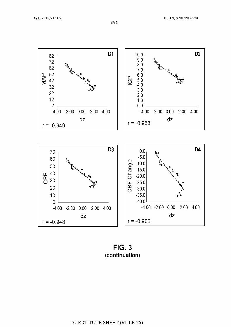


INTERNATIONAL SEARCH REPORT International application No.
PCT/US20 18/032984
A . CLASSIFICATION O F SUBJECT MATTER
IPC(8) - A61 N 1/36; A61 B 5/00; A61 N 1/362; A61 N 1/37 (2018.01 )
CPC - A61 N 1/36; A61 B 5/00; A61 B 5/0042; A61 1/37 (201 8.05)
According to International Patent Classification (IPC) or to both national classification and IPC
B . FIELDS SEARCHED
Minimum documentation searched (classification system followed by classification symbols)
See Search History document
Documentation searched other than minimum documentation to the extent that such documents are included in the fields searched
USPC - 607/45; 600/544; 607/2 (keyword delimited)
Electronic data base consulted during the international search (name of data base and, where practicable, search terms used)
See Search History document
C . DOCUMENTS CONSIDERED T O B E RELEVANT
Category* Citation of document, with indication, where appropriate, o f the relevant passages Relevant to claim No.
US 2008/0200787 A 1 (SHAPIRA et al) 2 1 August 2008 (21 .08.2008) entire document I -3
4-9, 1-21
US 2010/0189698 A 1 (WILLIS) 29 July 2010 (29.07.2010) entire document 10
I I -13, 16-18
US 2004/0176820 A 1 (PAUL) 09 September 2004 (09.09.2004) entire document 4-6, l b- 1
US 2013/0190632 A 1 (BARUCH et al) 25 July 2013 (25.07.2013) entire document 7
US 2014/0371545 A 1 (BEN-ARI et al) 18 December 2014 (18.12.2014) entire document 8 , 9
US 2010/0030054 A 1 (BARUCH et al) 04 February 2010 (04.02.2010) entire document 9
US 201 1/0245734 A 1 (WAGNER et al) 06 October 201 1 (06.10.201 1) entire document 14-21
Further documents are listed in the continuation o f Box C . | | Sec patent family annex.
Special categories of cited documents: "T" later document published after the international filing date or priorityA" document defining the general state of the art which is not considered date and not in conflict with the application but cited to understand
to be of particular relevance the principle or theory underlying tne invention
E" earlier application or patent but published on or after the international "X" document of particular relevance, the claimed invention cannot befiling date considered novel or cannot be considered to involve an inventive
L" document which may throw doubts on priority claim(s) or which is step when the document is taken alonecited to establish the publication date of another citation or otherspecial reason (as specified) "Y" document of particular relevance; the claimed invention cannot be
considered to involve an inventive step when the document isO" document referring to an oral disclosure, use, exhibition or other combined with one or more other such documents, such combination
means being obvious to a person skilled in the art
P" document published prior to the international filing date but later than "&" document member of the same patent familythe priority date claimed
Date of the actual completion of the international search Date of mailing of the international search report
20 July 20180 3 AUG 2018
Name and mailing address of the ISA/US Authorized officer
Mail Stop PCT, Attn: ISA/US, Commissioner for Patents Blaine R. CopenheaverP.O. Box 1450, Alexandria, VA 2231 3-1450
Facsimile No. 571-273-8300
Form PCT/ISA/210 (second sheet) (January 201 )

Mohamad Hakam Tiba Dec 04, 2018 1 of 12
Mohamad Hakam TibaResearch Assistant ProfessorNorth Campus Reseach Complex
2800 Plymouth Road, 026-323N, Ann Arbor, MI 48109Phone: (734) 764-6702
Fax: (734) 763-9389Email: [email protected]
Education and TrainingEducation
09/1985-09/1991 MD, Medicine, Damascus University, Damascus, Syrian Arab Republic10/2013-04/2015 MS, Clinical Research Design and Statistical Analysis, University Of Michigan, Ann Arbor,
MI
PostDoctoral Training10/1991-08/1992 Internship, Intern in General Medicine, Ministry of Health Hospitals and Alshifa Hospital,
Damascus, Syrian Arab Republic
Academic, Administrative, Clinical, Research and Military AppointmentsAcademic Appointments
07/2012-08/2016 Research Investigator in Emergency Medicine - Adult, University of Michigan - Ann Arbor, Ann Arbor, Michigan
09/2016-present Research Assistant Professor in Emergency Medicine - Adult, University of Michigan - Ann Arbor, Ann Arbor, Michigan
Administrative Appointments06/1994-09/1994 Observership and research assistant, Receiving Hospital, Wayne State University, Detroit,
MI01/1995-05/1995 Observership, Good Samaritan Hospital, Dayton, OH02/1996-04/1998 Volunteer Research Assistant, Tufts University, Boston, MA06/1998-05/2000 Senior Quality Control and Production Chemist, Owl Separation System, Portsmouth, New
Hampshire06/2000-05/2010 Life/Phys Science Research Associate, Virginia Commonwealth University, Richmond,
Virginia05/2010-06/2012 Laboratory and Research Manager, Virginia Commonwealth University, Richmond, Virginia
Research Interests
Development of large animals models to study critical care illness and injuryDevelopment of non-invasive technologies to evaluate intravenous volume status in critically ill patients using bioimpedanceDevelopment of non-invasive technologies to evaluate cerebrovascular autoregulation using ocular bioimpedanceDevelopment of non-invasive spectroscopic technologies to monitor tissue oxygenation using Resonance Raman Spectroscopy (RRS)Development of resuscitation Fluids, hemoglobin and non-hemoglobin oxygen carriersDevelopment of closed-loop feedback CPR devicesDevelopment of hemorrhage control materials and devices

Mohamad Hakam Tiba Dec 04, 2018 2 of 12
GrantsCurrent Grants
MICHRSwine-Sepsis and ARDSCo-I with Effort (Principal Investigator: Robert Dickson and Kathleen Stringer)09/2018-08/2019. $100,000 ($100,000)
The Massey Family FoundationIntracranial Pressure Monitor Enhancement for Cerebral Hemodynamic MonitoringCo-I with Effort (Principal Investigator: Kenn Oldham)07/2018-06/2019. $104,433 ($104,433)
The Massey Family FoundationReal-Time, Non-Invasive Brain MetabolismCo-I with Effort (Principal Investigator: Oliver D. Kripfgans)07/2018-06/2019. $143,666 ($143,666)
Dept. of the Army -- USAMRAA- 16-DM160299: Gastroesophageal Resuscitative Occlusion of the Aorta (GROA)PAF08148Co-I with Effort (Principal Investigator: Ward, Kevin Ralph)02/2018-01/2021. $2,995,616
DM160294: Development and Testing of New Noninvasive Monitoring Tools for Prolonged Field Care Goal-Directed Dept. of the Army -- USAMRAATherapy
Co-I with Effort (Principal Investigator: Kevin Ward)01/2018-12/2020. $2,999,754 ($969,599)
DM160225: Novel Noninvasive Methods of Intracranial Pressure and Cerebrovascular Autoregulation Assessment: Dept. of the Army -- USAMRAA- 17-PAF00214Seeing the Brain through the Eyes
Tiba, Mohamad Hakam, PI01/2018-12/2020. $1,481,538
NIH-DHHS-5 R01 HL133129-04: ECPR After Prolonged Cardiac Arrest: Targeting Mechanisms of the No-ReflowUS- 16-PAF07624Co-I with Effort (Principal Investigator: Neumar, Robert;Bartlett, Robert Hawes)07/2017-06/2022. $3,894,128
Submitted Grants
SubK-NIH-DHHS-US through a Resonance Raman Spectroscopy for Tissue Oxygenation Monitoring in Sepsisconsortium with Pendar Medical, LLC- 19-PAF02234Co-I with Effort (Principal Investigator: Ward, Kevin Ralph)01/2019-12/2019. $349,726
SubK-NIH-DHHS-US through a consortium Dynamic Respiratory Impedance Volume Evaluation (DRIVE) for Sepsiswith New Vital Signs, Inc.- 19-PAF02230Tiba, Mohamad Hakam;Ward, Kevin Ralph, PI01/2019-12/2019. $350,170
Zoll Foundation- 19-PAF01707ART-123 (recombinant human thrombomodulin alpha) in porcine cardiac arrestCo-I without Effort (Principal Investigator: Greineder, Colin F)12/2018-11/2019. $47,773
Past Grants
The Massey Novel Noninvasive Method of Cerebrovascular Blood Volume Assessment Using Brain BioimpedanceFamily foundationTiba, MH and Ward, KR, Co-PI07/2017-09/2018. $130,565 ($130,565)
The Massey Family FoundationSystolic Target Assessment Tool (STAT) for TBI ManagementCo-I (Principal Investigator: Hackenson)07/2017-09/2018. $73,975 ($73,975)

Mohamad Hakam Tiba Dec 04, 2018 3 of 12
Redox POC Platform for Evaluation and Treatment of Sepsis, Septic Shock, and Multiple Organ DysfunctionMichigan Translational Research and Commercialization ProgramCo-I with Effort (Principal Investigator: Rodney C. Daniels)02/2016-01/2017. $155,435 ($155,435)
15-PAF06146: Comparison of Respiratory Induced Limb Bioimpedance Changes With Inferior Vena Cava Diameter Renal Research Institute, LLCChanges to Assess Intravascular Volume Status in Patients Undergoing Hemodialysis
Co-I with Effort (Principal Investigator: Michael Heung)06/2015-12/2016. $42,830 ($42,830)
G016123: Novel Noninvasive Methods of Intracranial Pressure Assessment: Seeing the Brain Through the EyesMassey Family FoundationCo-I with Effort (Principal Investigator: Krishna Rajajee and Kevin Ward)02/2015-02/2016. $106,262 ($106,262)
Comparison of Respiratory Induced Limb Bioimpedance Changes with Inferior Vena Cava Diameter Changes to Baxter Healthcare Corporation- 15-PAF03360Assess Intravascular Volume Status
Tiba, Mohamad Hakam;Ward, Kevin Ralph, PI01/2015-06/2015. $150,476
Michigan Translation and N017668: Dynamic Respiratory Impedance Volume Evaluation (DRIVE)Commercialization (MTRAC) for Life Sciences ProgramMohamad Hakam Tiba, PI02/2014-01/2015. $92,958 ($92,958)
St. Michaels Hospital14-PAF05256: Multi-laboratory Study of Epinephrine in Experimental Cardiac ArrestRobert Neumar Mohamad Hakam Tiba, Co-PI11/2013-12/2015. $10,000
Honors and AwardsRegional
2010 Mentorship Award, Goldwater Scholarship committee. For mentorship of Elizabeth Proffitt, a Goldwater Scholar
2016 Outstanding Poster Award. Massey Regional TBI Conference
Institutional2014 Spoor Memorial Scholarship Award2015 First place in the poster competition award. The Eighth Annual Symposium, A. Alfred
Taubman Medical Research Institute.
Memberships in Professional Societies
2012-present Member, The Shock Society2013-present Member, American Association for the Advancement of Science2013-present Member, Society for Academic Emergency Medicine2013-present Member, Society of Critical Care Medicine2014-present Member, American Heart Association
Editorial Positions, Boards, and Peer-Review Service
Study SectionsNational
2015-present Peer review panel of the 2015 Combat Casualty Care Research Program (CCCRP) for the Department of Defense U.S. Army Medical Research and Materiel Command (MRMC).
2018-present Autonomous and Unmanned Medical Capability (AUMC-1) peer review panel of the 2017

Mohamad Hakam Tiba Dec 04, 2018 4 of 12
2018-present Autonomous and Unmanned Medical Capability (AUMC-1) peer review panel of the 2017 Defense Medical Research and Development Program (DMRDP) Joint Program Committee-1 (JPC-1) for the Department of Defense Congressionally Directed Medical Research Programs (CDMRP).
Journal Reviewer2016-present American Journal of Emergency Medicine (Ad Hoc)
TeachingClinical Fellow
10/2018-10/2020 Yub Raj Sedhai, MD, Spectrum system, Grand Rapids, MI
Graduate Student06/2013-12/2015 Barry Belmont, MS, Biomedical Engineering, University of Michigan10/2014-04/2015 Brandon Cummings, MS, The Undergraduate Research Opportunity Program (UROP).
University of Michigan
Medical Student06/2015-08/2015 Spencer Thompson, BS, Summer Biomedical Research Proposal. University of Michigan06/2015-08/2015 Shawn Kache, BS, Summer Biomedical Research Proposal. University of Michigan08/2015-11/2015 Isaac Ezra Perry, BS, Edward Via College of Osteopathic Medicine06/2017-08/2017 Alexander Khouri, BS, Summer Biomedical Research Proposal, University of Michigan.
Undergraduate Student01/2006-01/2010 Elizabeth K. Proffitt, Virginia Commonwealth University10/2013-05/2014 Eman Hejab, The Undergraduate Research Opportunity Program (UROP). University of
Michigan10/2015-04/2016 Justine Garfinkel, The Undergraduate Research Opportunity Program (UROP). University
of Michigan10/2015-04/2016 Mambwe C. Lupiya, The Undergraduate Research Opportunity Program (UROP).
University of Michigan05/2016-08/2016 John Soukar, University of Michigan07/2016-05/2017 Stephen Dowker, University of Michigan10/2016-04/2017 Stephanie Francalancia, The Undergraduate Research Opportunity Program (UROP).
University of Michigan10/2017-04/2018 Alysha Loraff, The Undergraduate Research Opportunity Program (UROP). University of
Michigan10/2017-04/2018 Tessa Magsoudi, The Undergraduate Research Opportunity Program (UROP). University
of Michigan10/2017-04/2018 Varisha Essani, The Undergraduate Research Opportunity Program (UROP). University of
Michigan10/2017-04/2018 Benjamin Koehler, The Undergraduate Research Opportunity Program (UROP).
University of Michigan
Visiting Scholars09/2015-08/2016 Jae Hyuk Lee, MD, Seoul National University Bundang Hospital, South Korea
Teaching ActivityInstitutional
08/2013 Surgery concentration. Surgical techniques training session for University Laboratory Animal Medicine (ULAM) veterinary residents
08/2014 Surgery concentration. Surgical techniques training session for University Laboratory Animal Medicine (ULAM) veterinary residents
08/2016 Surgery concentration. Surgical techniques training session for University Laboratory

Mohamad Hakam Tiba Dec 04, 2018 5 of 12
08/2016 Surgery concentration. Surgical techniques training session for University Laboratory Animal Medicine (ULAM) veterinary residents
08/2018-09/2018 Surgery concentration. Surgical techniques training session for University Laboratory Animal Medicine (ULAM) veterinary residents
Committee and Administrative Services
Committee ServicesNational
2014-2015 Task force to evaluate animal research publications' compliance with ARRIVE reporting standards. Shock Society, Member
Institutional2013-present Institutional Animal Care and Use Committee (IACUC), Member2014-2016 Large Animals Working Group, Member2015-2016 IACUC Subcommittee for eRAM Online Protocol Changes and Updates, Member2015 IACUC Subcommittee for Standard Operating Procedures (SOP) Approval, Member2016-2017 IACUC Task Force for Review of PI Managed Space Request Application, Member2017-present Animal Care and Use Faculty Advisory Committee. ACU-FAC, Chair2018-present Strategic Advisory Committee to the UM Animal Care and Use Program (ACUP)., Member
Administrative ServicesVolunteer
2015-present Judge, The Undergraduate Research Opportunity Program (UROP), University of Michigan, Ann Arbor, The Undergraduate Research Opportunity Program's Annual Research Spring Symposium
Visiting Professorships and Extramural Invited PresentationsExtramural Invited Presentations
1. Mohamad H. Tiba, H. Mowafi, G. Gawor, R. W. Barbee, R. Ivatury, K. R. Ward. Preliminary Studies of a New Impedance Based Measure to Noninvasively Determine Central Venous Pressure, Advanced Technologies Applications in Combat Casualty Care, August 2003, St. Pete Beach, Florida. (Poster Presentation)
2. Mohamad H. Tiba, T. A. Timmons, R. W. Barbee, R. R. Ivatury, B. D. Spiess, P. S. Reynolds, and K. R. Ward. Bispectral Index Monitoring of Intubated Trauma Patients Indicates Inadequate Sedation, Advanced Technologies Applications in Combat Casualty Care, August 2004, St. Pete Beach, Florida. (Poster Presentation)
3. Mohamad H. Tiba, Kate Proffitt; R. Wayne Barbee, Gerard Draucker, Rao R. Ivatury, Kevin R. Ward. Noninvasive Bioimpedance Base for Central Venous Pressure Measurement, Advanced Technologies Applications in Combat Casualty Care, August 2005, Pete Beach Florida. (Poster Presentation)
4. Mohamad H. Tiba, G. T Draucker, R. W. Barbee, J. Terner, I. P. Torres Filho, K. R. Ward. Performance of Tissue Hemoglobin Oxygen Saturation as Measured by Resonance Raman Spectroscopy and Near Infrared Spectroscopy Compared to Mixed Venous Hemoglobin Oxygen Saturation and Systemic Lactate Levels During Hemorrhage, 36th Annual Conference on Shock, June 2013, San Diego, California. (Poster Presentation)
5. Mohamad H. Tiba, G. T. Draucker, B. M. McCracken, K. R. Ward. Controlling Pelvic Hemorrhage using a Novel Pressure Garment, 37th Annual Conference on Shock, June 2014, Charlotte, North Carolina. (Poster Presentation)
6. Mohamad H. Tiba, G. T. Draucker, B. M. McCracken, H. B. Alam, J. L. Eliason, K. R. Ward. Testing of a novel Pelvic Hemostasis Belt to Control Lethal Pelvic Arterial Hemorrhage, American Heart Association. Resuscitation Science Symposium, November 2014, Chicago, Illinois. (Poster Presentation)
7. Mohamad H. Tiba, Barry Belmont, Nik Theyyunni, Robert Huang, David F. Barton, Amanda J. Pennington, Gerard T. Draucker, Albert J. Shih, Kevin R. Ward. Coparison of Respiratory Induced Inferior Vena Cava Diameter Changes With Limb Bioimpedance Changes to Assess Intravascular Volume Status, 38th Annual Conference on Shock, June 2015, Denver Colorado. (Poster Presentation)

Mohamad Hakam Tiba Dec 04, 2018 6 of 12
Status, 38th Annual Conference on Shock, June 2015, Denver Colorado. (Poster Presentation)8. Evaluation of Intravascular Volume Status Using Dynamic Respiratory Induced Bioimpedance of the
Limb, Society for Academic emergency Medicine, May 2016, New Orleans, LA. (Oral Presentation)9. Mohamad H. Tiba, Barry Belmont, Michael Heung, Nik Theyyunni, Robert Huang, Christopher D. Fung,
Amanda J. Pennington, Kevin R. Ward: Comparison of Respiratory Induced Inferior Vena Cava Diameter Changes with Limb Bioimpedance Changes In dialysis and mechanically ventilated patients, Thirty-Ninth Annual Conference on Shock, June 2016, Austin, Texas. (Poster Presaentation)
10. Mohamad H. Tiba, Amanda Pennington, Kyle Gunnerson, Kevin R. Ward. Monitoring of Tissue Microvasculature Oxygenation using Resonance Raman Spectroscopy, Thirty-Ninth Annual Conference on Shock, June 2016, austin, Texas. (Poster Presentation)
11. Mohamad H. Tiba, Brendan McCracken, Gerard Draucker, Hasan Alam, Jonathan Eliason, Kevin Ward: Use of a Novel Pelvic Hemostasis Belt to Control Lethal Arterial Hemorrhage, The 2016 Military Health System Research Symposium (MHSRS), August 2016, Orlando, Florida. (Poster Presntation)
12. Mohamad. Tiba, Michael Heung, Nik Theyyunni, Barry Belmont, Robert Huang, Ross Kessler, Christopher Fung, Amanda Pennington, Brandon Cummings, Kevin Ward: Development of a Novel Non-Invasive Technologies to Evaluate Intravascular Volume Status Using Bioimpedance, The 2016 Military Health System Research Symposium (MHSRS), August 2016, Orlando, Florida. (Poster Presentation)
13. Mohamad H. Tiba, Robert Barbee, James Terner, Ivo. Torres Filho, Kevin Ward: Measurement of tissue hemoglobin oxygen saturation using Resonance Raman Spectroscopy in a large animal model of hemorrhage, The 2016 Military Health System Research Symposium (MHSRS), August 2016, Orlando, Florida. (Poster Presentation)
14. Mohamad Hakam Tiba; Brendan McCracken; Sardar Ansari; Venkatakrishna Rajajee; Kevin Ward. Novel Noninvasive Method of Cerebrovascular Autoregulation Assessment: Seeing the Brain through the Eyes., Fortieth Annual Conference on Shock., June 2017, Fort Lauderdale, Florida. (Poster Presentation)
15. Novel Noninvasive Method of Cerebrovascular Autoregulation Assessment, Military Health System Research Symposium (MHSRS), August 2017, Kissimmee, Florida (Oral Presentation)
16. A Novel Large Animal Model of Sepsis, Forty-First Annual Conference on Shock., June 2018, Scottsdale, Arizona. (Poster Presentation)
17. Monitoring Traumatic Brain Injury Patients using Transocular Brain Impedance (TBI)., Military Health System Research Symposium (MHSRS), August 2018, Orlando, Florida. (Poster Presentation)
18. Use of Resuscitative Balloon Occlusion of the Aorta in a Swine Model of Prolonged Cardiac Arrest., American Heart Association. Resuscitation Symposium. (Poster Presentation), November 2018, Chicago, IL
Other1. DRIVE Performance and update, Baxter Healthcare, March 2015, Chicago, Illinois2. Mohamad H. Tiba, Barry Belmont, Spencer Thompson, Michael Heung, Nik Theyyunni, Robert Huang,
Christofer Fung, Amanda Pennington, Gerard Draucker, Kevin R. Ward. Development of a Novel Non-Invasive Technologies to Evaluate Intravascular Volume Status Using Bioimpedance, Eighth Annual Symposium, A. Alfred Taubman Medical Research institute. University of Michigan., October 2015, Ann Arbor, Michigan. (Poster Presentation)
3. Mohamad H. Tiba, Shawn Kache, Brandon Cummings, Amanda Pennington, Kyle Gunnerson, Kevin R. Ward. Monitoring of Tissue Microvasculature Oxygenation using Resonance Raman Spectroscopy, Eighth Annual Symposium, A. Alfred Taubman Medical Research institute. University of Michigan., October 2015, Ann Arbor, Michigan. (Poster Presentation)
4. Mohamad H. Tiba. Sample Size and Power Analysis: What Do We Need to Know, Institutional Animal Care and Use committee (IACUC). A presentation as part of the training protocol for IACUC members, February 2016, Ann Arbor, MI
5. Mohamad H. Tiba, MD, MS; Nik Theyyunni, MD; Barry Belmont, PhD; Michael Heung, MD, MS; Robert Huang, MD; Christopher Fung, MD; Amanda Pennington, MS; Brandon Cummings; Gerard Draucker, EMT; Kevin R. Ward, MD. Comparison of Respiratory Induced Inferior Vena Cava Diameter Changes with Limb Impedance Changes in Hemodialysis Patients., First Annual William G. Barsan Emergency Medicine Research Forum. (Poster Presentation), April 2016, Ann Arbor, Michigan
6. Mohamad H. Tiba, MD; Amanda Pennington, MS; Gerard Draucker, EMT; Brandon Cummings; Kyle Gunnerson, MD; Kevin R. Ward, MD. Monitoring of Tissue Microvasculature Oxygenation using Resonance Raman Spectroscopy, First Annual William G. Barsan Emergency Medicine Research Forum. (Poster Presentation), April 2016, Ann Arbor, Michigan
7. Mohamad H. Tiba, MD; Gerard T. Draucker, EMT-P; Brendan M. McCracken, BS; Hasan B. Alam, MD;

Mohamad Hakam Tiba Dec 04, 2018 7 of 12
7. Mohamad H. Tiba, MD; Gerard T. Draucker, EMT-P; Brendan M. McCracken, BS; Hasan B. Alam, MD; Jonathan L. Eliason, MD; Kevin R. Ward, MD. Controlling Pelvic Hemorrhage Using a Novel Pressure Garment, First Annual William G. Barsan Emergency Medicine Research Forum. (Poster Presentation), April 2016, Ann ARbor, Michigan
8. Novel Noninvasive Method of Cerebrovascular Autoregulation Assessment: Seeing the Brain through the Eyes, University of Michigan / Massey Foundation, October 2016, Ann Arbor, MI. (Oral Presentation)
9. Mohamad H. Tiba, MD, MS; Brendan McCracken, BS; Sardar Ansari, PhD; Ashwin Belle, PhD; Kevin Ward, MD. Novel Noninvasive Method of Cerebrovascular Autoregulation Assessment: Seeing the Brain through the Eyes, Second Annual William G. Barsan Emergency Medicine Research Forum. (Poster Presentation), April 2017, Ann Arbor, Michigan
10. Noninvasive Assessment of Cerebrovascular Blood Volume and Autoregulation Using Bioimpedance, Massey Foundation Second TBI Summit. (Oral Presentation), October 2017, Ann ARbor, Michigan
11. Monitoring Traumatic Brain Injury Patients using Transocular Brain Impedance (TBI). Grand Challenge Experience., The Massey Foundation TBI Grand Challenge, March 2018, Ann Arbor, Michigan
12. Trans-Ocular Brain Impedance: Novel Non-invasive Monitoring of the TBI Patient, 2018 Massey TBI Regional Conference. Oral Presentation), October 2018, Ann Arbor, MI
Seminars1. Tissue Hemoglobin Oxygen Saturation in the critically ill, as Measured by Resonance Raman
Spectroscopy, University Laboratory Animals Medicine (ULAM) Fall Seminar Series, November 2013, Ann Arbor, Michigan
2. Emergency Department Tour and Description of Two Non-Invasive Monitoring Modalities, University of Michigan. The Undergraduate Research Opportunity Program (UROP) Seminar Series, December 2015, Ann Arbor, Michigan
3. Bolus Versus Continuous Epinephrine Infusion In a Large animal model of Cardiac Arrest: A Multicenter Trial, MCIRCC/OSCAR CONFERENCE, March 2016, Ann Arbor, MI
4. Large Animal Model of Sepsis, University Laboratory Animals Medicine (ULAM) Fall Seminar Series, November 2017, Ann Arbor, Michigan
PatentsApplication in Process
EVALUATING CARDIOVASCULAR HEALTH USING INTRAVASCULAR VOLUME, 20150031966, Co-inventor, Submitted on 07/2014Ocular Impedance-Based System for Brain Health Monitoring, Co-inventor, Submitted on 05/2017
Bibliography
Peer-Reviewed Journals and Publications
1. Torres LN, Torres Filho IP, Barbee RW, Tiba MH, Ward KR, Pittman RN: Systemic responses to prolonged hemorrhagic hypotension Am. J. Physiol. Heart Circ. Physiol. 286(5 55-5): H1811-H1820, 2004. PM14726303
2. Smith L, Tiba MH, Goldberg ME, Barbee RW: Chronic implantation of transit-time flow probes on the ascending aorta of rodents Lab. Anim. 38(4): 362-370, 2004. PM15479550
3. Torres LN, Torres Filho IP, Barbee RW, Tiba MH, Ward KR, Pittman RN: Continuous peripheral resistance measurement during hemorrhagic hypotension Am. J. Physiol. Heart Circ. Physiol. 287(5 56-5): H2341-H2345, 2004. PM15256369
4. Ward KR, Torres Filho I, Barbee RW, Torres L, Tiba MH, Reynolds PS, Pittman RN, Ivatury RR, Terner J: Resonance Raman spectroscopy: A new technology for tissue oxygenation monitoring Crit. Care Med. 34(3): 792-799, 2006. PM16521273
5. Ward KR, Tiba MH, Barbee RW, Ivatury RR, Arrowood JA, Spiess BD, Hummel R: A new noninvasive method to determine central venous pressure Resuscitation 70(2): 238-246, 2006. PM16820258
6. Ward KR, Barbee RW, Reynolds PS, Filho IP, Tiba MH, Torres L, Pittman RN, Terner J: Oxygenation monitoring of tissue vasculature by resonance Raman spectroscopy Anal. Chem. 79(4): 1514-1518, 2007. PM17297949
7. Evans MC, Diegelmann RF, Barbee RW, Tiba MH, Edwards E, Sreedhar S, Ward KR: Protein synthesis

Mohamad Hakam Tiba Dec 04, 2018 8 of 12
7. Evans MC, Diegelmann RF, Barbee RW, Tiba MH, Edwards E, Sreedhar S, Ward KR: Protein synthesis inhibition as a potential strategy for metabolic down-regulation Resuscitation 73(2): 296-303, 2007. PM17250947
8. Ward KR, Tiba MH, Holbert WH, Blocher CR, Draucker GT, Proffitt EK, Bowlin GL, Ivatury RR, Diegelmann RF: Comparison of a new hemostatic agent to current combat hemostatic agents in a swine model of lethal extremity arterial hemorrhage J Trauma 63(2): 276-283, 2007. PM17693824
9. Ward KR, Tiba MH, Draucker GT, Proffitt EK, Barbee RW, Gunnerson KJ, Reynolds PS, Spiess BD: A novel noninvasive impedance-based technique for central venous pressure measurement Shock 33(3): 269-273, 2010. PM19487978
10. Ward KR, Tiba MH, Ryan KL, Filho IP, Rickards CA, Witten T, Soller BR, Ludwig DA, Convertino VA: Oxygen transport characterization of a human model of progressive hemorrhage Resuscitation 81(8): 987-993, 2010. PM20418009
11. Leong B, Reynolds PS, Tiba MH, Holbert WH, Draucker GT, Medina JA, Barbee RW, White NJ, Ward KR: Effects of a combination hemoglobin based oxygen carrier-hypertonic saline solution on oxygen transport in the treatment of traumatic shock Resuscitation 82(7): 937-943, 2011. PM21497981
12. Tiba MH, Draucker GT, Barbee RW, Terner J, Filho IT, Romfh P, Vakhshoori D, Ward KR: Tissue oxygenation monitoring using resonance Raman spectroscopy during hemorrhage J Trauma Acute Care Surg 76(2): 402-408, 2014. PM24378619
13. Tiba MH, Draucker GT, McCracken BM, Alam HB, Eliason JL, Ward KR: Use of pelvic hemostasis belt to control lethal pelvic arterial hemorrhage in a swine model. The journal of trauma and acute care surgery 78(3): 524-9, 2015. PM25710422
14. Tiba MH, Belmont B, Theyyunni N, Heung M, Huang RD, Fung, CM, Pennington AJ, Cummings BC, Draucker GT, Shih AJ, Ward KR: Dynamic Limb Bioimpedance and Inferior Vena Cava Ultrasound in Patients Undergoing Hemodialysis ASAIO J 62(4): 463-9, 2016. PM26919184
15. Tiba MH, McCracken BM, Ansari S, Belle A, Cummings BC, Rajajee V, Patil PG, Alam HB, Ward KR: Novel Noninvasive Method of Cerebrovascular Blood Volume Assessment Using Brain Bioimpedance. J Neurotrauma 34(22): 3089-3096, 2017. PM28657491
16. Daniels RC, Jun H, Tiba H, McCracken B, Herrera-Fierro P, Collinson M, Ward KR: Whole Blood Redox Potential Correlates With Progressive Accumulation of Oxygen Debt and Acts as A Marker of Resuscitation in A Swine Hemorrhagic Shock Model. Shock 49(3): 345-351, 2018. PM28658006/PMC5745311
17. Belmont B, Kessler R, Theyyunni N, Fung C, Huang R, Cover M, Ward KR, Shih AJ, Tiba M: Continuous Inferior Vena Cava Diameter Tracking through an Iterative Kanade-Lucas-Tomasi-Based Algorithm. Ultrasound Med Biol 44(12): 2793-2801, 2018. PM30213669
18. Coute RA, Shields TA, Cranford JA, Ansari S, Abir M, Tiba MH, Dunne R, O'Neil B, Swor R, Neumar RW, SaveMiHeart Consortium and the CARES Surveillance Group.: Intrastate Variation in Treatment and Outcomes of Out-of-Hospital Cardiac Arrest. Prehosp Emerg Care 22(6): 743-752, 2018. PM29624088
19. Sando IC, Plott JS, McCracken BM, Tiba MH, Ward KR, Kozlow JH, Cederna PS, Momoh AO: Simplifying Arterial Coupling in Microsurgery-A Preclinical Assessment of an Everter Device to Aid with Arterial Anastomosis. J Reconstr Microsurg: 2018. PM29452442
Abstracts
1. Ward KR, Barbee RW, Tiba, MH, Arrowood J, Ivatury RR, Lyders E, Spiess BP.: A Noninvasive Method of Determining Central Venous Pressure, 25 Annual Conference on Shock, Big Sky, Montana, Shock, th
17, Supplement 1, 15, 2002.2. Carr M, Kenawy E, Layman G, Wnek G, Ward KR, Barbee W, Tiba MH.: Development of The
BioHemostat- A Treatment Modality for High Pressure Bleeding Based on Super Absorbent Polymers, 25 Annual Conference on Shock, Big Sky, Shock, 17, Supplement 1, 54, 2002.th
3. Barbee RW, Ward KR, Terner J, Torres I, Tiba MH, Torres L, Ivatury R, Spiess B, Pittman R.: Noninvasive Tissue Oxygenation Monitoring Using Resonance Raman Spectroscopy (RRS), 25 Annual th
Conference on Shock, Big Sky, Montana, Shock, 17, Supplement 1, 58, 2002.4. Barbee RW, Ward KR, Terner J, Torres IP, Torres L, Pittman R, Ivatury R, Tiba MH.: Preliminary Studies
Using Near Ultraviolet Excitation Fluorescence Spectroscopy to Monitor Tissue Dysoxia During Hemorrhage, 26 Annual Conference on Shock, Phoenix, Arizona, Shock, 19, Supplement 1, 26, 2003.th
5. Ward K, Tiba M. H, Barbee W, Timmons T, Spiess B, Ivatury R.: Bispectral Index Monitoring of Intubated

Mohamad Hakam Tiba Dec 04, 2018 9 of 12
5. Ward K, Tiba M. H, Barbee W, Timmons T, Spiess B, Ivatury R.: Bispectral Index Monitoring of Intubated Paralyzed Trauma Patient Indicates Inadequate Sedation., Critical Care Medicine, 33, Supplement, A45, 2005.
6. Ward K, Tiba M. H, Proffitt K, Draucker J, Ivatury R, Barbee R, Spiess B, Arrowood J.: Noninvasive Central Venous Pressure Monitoring, Critical Care Medicine, 33, Supplement, A54, 2005.
7. Ward K, Tiba MH, Draucker G, Reynolds P, Torres R, Barbee RW, Ivatury RR.: Performance of Noninvasive Tissue Oxygenation Indicators in Detecting Shock Due to Hemorrhage, Critical Care Medicine, 33, Supplement, A23, 2005.
8. Ward K, Torres I, Barbee W, Torres L, Tiba M. H, Reynolds P, Pittman R, Ivatury R, Terner J.: Resonance Raman Spectroscopy for Noninvasive Tissue Oxygenation Monitoring, Critical Care Medicine, 33, Supplement, A35, 2005.
9. Ward K, Tiba M. H, Holbert W, Blocher C, Draucker G, Proffitt E, Bowlin G, Ivatury R, Diegelmann R.: Comparison of New Hemostatic Agent to Current Combat Hemostatic Agents in a Swine Model of Lethal Extremity Arterial Hemorrhage, Society for Academic Emergency Medicine (SAEM) Annual Meeting, Chicago, Illinois, Academic Emergency Medicine, 14, Supplement, pS61, 2007.
10. Ward K, Tiba M. H, Draucker G, Proffitt K, Gawor G, Barbee R.: Noninvasive Measurement of Central Venous Pressure, Society for Academic Emergency Medicine (SAEM) Annual Meeting, Chicago, Illinois, Academic Emergency Medicine, 14, Supplement, pS182, 2007.
11. Mohamad H. Tiba, Kathy Ryan, Ivo Torres Filho, Carloine Rickards, Tarynn M Witten, Babs Soller, Victor Convertino, Kevin R. Ward: , Oxygen Transport Characterization of a Human Model of Hemorrhage American Heart Association Resuscitation Science Symposium, New Orleans, Louisiana, Circulation, 118, S_1447, 2008.
12. Ward K, Tiba M. H, Draucker G, Barbee R, Proffitt E, Gunnerson K: Noninvasive Central Venous Pressure Measurements in Mechanically Ventilated Patients, Society for Academic Emergency Medicine (SAEM) Annual Meeting, Washington, DC, Academic Emergency Medicine, 15, Supplement 1, S32, 2008.
13. Ward K, Tiba M. H, Medina J, Holbert W, Draucker J, Reynolds P, Blocher C, Barbee R.: Oxygen Debt and its Relationship to Lactate, Hemoglobin Levels, and Hemorrhage Volume in Hemorrhagic Shock and Resuscitation, Society for Academic Emergency Medicine (SAEM) Annual Meeting, Washington, DC, Academic Emergency Medicine, 15, Supplement 1, S32, 2008.
14. Kevin R. Ward, Mohamad H. Tiba, Gerald T. Draucker, Kyle Gunnerson, Robert W. Barbee, Penny S. Reynolds, Bruce Spiess, Rao R. Ivatury: Noninvasive Measurement of Central Venous Pressure, American Heart Association Resuscitation Science Symposium, Orlando, Florida, Circulation, 120, S1460, 2009.
15. Benjamin Leong, Nathan White, Mohamad H. Tiba, William Holbert, Gerard T. Draucker, Juliana Medina, Mary A Peberdy, Joseph P Ornato, Kevin R. Ward: Oxygen Transport in Post Cardiac Arrest Syndrome with Goal Directed Hemodynamic Optimization, American Heart Association Resuscitation Science Symposium, Orlando, Florida, Circulation, 120, S1469, 2009.
16. Benjamin Leong, Mohamad H. Tiba, Gerard T. Draucker, Juliana Medina, William Holbert, Robert W. Barbee, Penny S. Reynolds, Kevin R. Ward: The Importance of Measuring Oxygen Debt in Hemorrhagic Shock., American Heart Association Resuscitation Science Symposium, Orlando, Florida, Circulation, 120, S1465-S1466, 2009.
17. S. Demir, N. Mirshahi, MH. Tiba, G. Draucker, K. Ward, R. Hobson, and K. Najarian: Image Processing and Machine Learning for Diagnostic Analysis of Microcirculation, ICME International Conference on Complex Medical Engineering, CME, 1-5, 9-11, 2009.
18. Sardar Ansari, Kavan Najarian, Kevin R. Ward, Mohamad H. Tiba: Extraction of Respiratory Rate from Impedance Signal Measured on Arm: A Portable Respiratory Rate Measurement Device, IEEE Conference on Bioinformatics and Biomedicine, Washington, DC, IEEE BIBM, 197-202, 2009.
19. Leong B, Tiba M. H, Holbert W, Draucker G, Medina J, Barbee R, Reynolds P, Ward K.: Low Volume Resuscitation and Repayment of Oxygen Debt from Traumatic Shock, Society for Academic EmergencyMedicine (SAEM) Annual Meeting, New Orleans, Louisiana, Academic Emergency Medicine, 16, Suppl 1, S9, 2009.
20. Leong B, Tiba M. H, Holbert W, Draucker G, Medina J, Barbee R, Reynolds P, Ward K.: Low Volume Resuscitation and Repayment of Oxygen Debt from Traumatic Shock, Society for Academic EmergencyMedicine (SAEM) Annual Meeting, New Orleans, Louisiana, Academic Emergency Medicine, 16, Suppl 1, S9, 2009.
21. Robert W. Barbee, Penny S. Reynolds, Nathan White, Mohamad H. Tiba, Kevin R. Ward: Oxygen Debt

Mohamad Hakam Tiba Dec 04, 2018 10 of 12
21. Robert W. Barbee, Penny S. Reynolds, Nathan White, Mohamad H. Tiba, Kevin R. Ward: Oxygen Debt Repayment Predicts Survival in a Swine Model of Trauma Shock, 33 Annual Conference on Shock, rd
Portland, Oregon, Shock, Supplement 1, 2010.22. Penny S. Reynolds, Robert W. Barbee, Nathan White, Mohamad H. Tiba, Kevin R. Ward: Sequential
Lactate Predicts 3-Hr Survival is a Swine Model of Traumatic Shock, 33 Annual Conference on Shock, rd
Portland, Oregon, Shock, Supplement 1, 2010.23. Penny S. Reynolds, Robert W. Barbee, Nathan J. White, Mohammed H. Tiba, Kevin R. Ward:
, Physiological Response Space: Mapping Resuscitation Response in a Swine Model of Traumatic Shock American Heart Association. Resuscitation Science Symposium, Los Angeles, California, Circulation, 122, Suppl 3, 2010.
24. Musana AK, Ward KR, Draucker GT, Tiba MH, Stravitz RT, Bajaj JS, Sanyal AJ: Hyperbilirubinemia Drives Microcirculatory Dysfunction, Tissue Hypoxia and Development of SIRS in Cirrhotic Subjects, The 61st Annual Meeting of the American Association for the Study of Liver Disease., Boston, MA, Hepatology, 890A, 2010.
25. Penny S. Reynolds, Robert W. Barbee, Mohamad H. Tiba, Kevin R. Ward: Visualization of Lactate-Perfusion Relationships During Hemorrhage and Resuscitation Using Artificial Neural Networks, 34 th
Annual Conference on Shock, Norfolk, Virginia, Shock, Supplement 1, 2011.26. Bhogal A.K. Musana, K.R. Ward, M.H. Tiba, G.T. Draucker, V. Mishra, A. Sanyal: Microcirculatory
Dysfunction and Tissue Hypoxia Drive Mortality in Patients with Cirrhosis, The 63 Annual Meeting of the rd
American Association for the Study of Liver Disease., Boston, Massachusetts, Hepatology, 931A, 2012.27. Kevin R. Ward, Mohamad H. Tiba, Gerard T. Draucker: Comparison of Tissue Hemoglobin Oxygen
Desaturation Using Resonance Raman Spectroscopy Versus Near Infrared Spectroscopy, 36 Annual th
Conference on Shock, San Diego, California, Shock, 39, Supplement 2, 64, 2013.28. Mohamad H. Tiba, GGerard T. Draucker, Robert W. Barbee, James Terner, Ivo P. Torres Filho, Kevin R.
Ward: Performance of Tissue Hemoglobin Oxygen Saturation as Measured by Resonance Raman Spectroscopy and Near Infrared Spectroscopy Compared to Mixed Venous Hemoglobin Oxygen Saturation and Systemic Lactate Levels During Hemorrhage, 36 Annual conference on Shock, San th
Diego, California, Shock, 39, Supplement 2, 56, 2013.29. Mohamad H. Tiba, Gerard T. Draucker, Brendan M. McCracken, Kevin R. Ward.: Controlling Pelvic
Hemorrhage Using a Novel Pressure Garment., 37 Annual Conference on Shock, Charlotte, North th
Carolina, Shock, 2014.30. Mohamad H. Tiba, Gerard T Draucker,Brendan M. McCracken, Hasan B. Alam, Jonathan L. Eliason,
Kevin R. Ward: Testing of a Novel Pelvic Hemostasis Belt to Control Lethal Pelvic Arterial Hemorrhage, American Heart Association. Resuscitation Science Symposium, Chicago, Illinois, Circulation, 130, A177, 2014.
31. Marwan R. Al-hajeili, Faris Elkhider, Mohamad H. Tiba: Cost-Effectiveness for Extended RAS/RAF Testing in Metastatic Colorectal Cancer, 2015 ASCO Annual Meeting, Chicago, Illinois, Journal of Clinical Oncology, 33, 5s, 3572, 2015.
32. Mohamad H Tiba, Barry Belmont, Nik Theyyunni, Robert Huang, David F. Barton, Amanda J. Pennington, Gerard T. Draucker, Albert J. Shih, Kevin R. Ward: Comparison of Respiratory Induced Inferior Vena CavaDiameter Changes with Limb Bioimpedance Changes to Assess Intravascular Volume Status, 38th Annual Conference on Shock, Denver, Colorado, Shock, 43, Supplement 1, 116, 2015.
33. Sardar Ansari, Daniel Slavin, Mohamad H. Tiba, Harm Derksen, Kenn Oldham, Kevin Ward and Kayvan Najarian: , American A Novel Portable Polyvinylidene Fluoride Based Sensor for Detection of HemorrhageHeart Association. Resuscitation Science Symposium, Orlando, Florida, Circulation, 132, Suppl 3, 2015.
34. Steve Lin, Li Ka Shing, Matthew L Sundermann, Paul Dorian, Sarah Fink, Henry Halperin, Alex Kiss, Allison C Koller, Brendan M McCracken, Laurie J Morrison, Robert W Neumar, James T Niemann, Andrew Ramadeen, David D Salcido, Mohamad H Tiba, Scott T Youngquist, Menekam Zviman, James J Menegazzi.: Epinephrine In Cardiac Arrest: A Randomized, Multicenter, Doubleblinded, PlaceboControlled Experimental Trial, American Heart Association. Resuscitation Science Symposium, Orlando, Florida, Circulation, 132, Suppl 3, 2015.
35. Ryan A Coute, Theresa A Shields, James A Cranford, Sardar Ansari, M. Hakam Tiba and Robert W Neumar: Intrastate Variation in Treatment and Outcome Measures for Out-of-Hospital Cardiac Arrest, American Heart Association. Resuscitation Science Symposium, New Orleans, LA, Circulation, 134, Suppl 1, 2016.
36. Ryan A Coute, Nicole L Werner, Alvaro Rojas-Pena, Stephanie Rakestraw, Fares Alghanem, M. Hakam

Mohamad Hakam Tiba Dec 04, 2018 11 of 12
36. Ryan A Coute, Nicole L Werner, Alvaro Rojas-Pena, Stephanie Rakestraw, Fares Alghanem, M. Hakam Tiba, Robert H Bartlett and Robert W Neumar: Intravascular Coagulation During Prolonged Cardiac Arrest, American Heart Association. Resuscitation Science Symposium, New Orleans, Circulation, 134, Suppl 1, 2016.
37. Mohamad Hakam Tiba, Barry Belmont, Michael Heung, Nik Theyyunni, Robert D. Huang, Christopher M. Fung, and Kevin R. Ward: Evaluation of Intravascular Volume Status Using Dynamic Respiratory Induced Bioimpedance of the Limb, Society of Academic Emergency Medicine Annual Meeting, New Orleans, LA, Academic Emergency Medicine, Volume 23, Supplement S1, S18-S19, 2016.
38. Christopher Fung, Robert Huang, Mohamad H. Tiba, Barry Belmont, Amanda J. Pennington, Brandon C. Cummings, Gerard T. Draucker, Kevin R. Ward, and Nik Theyyunni: Measurement of Carotid Artery Flow Time Via Point-of-Care Ultrasound in Hemodialysis Patients, Society of Academic Emergency Medicine Annual Meeting, New Orleans, LA, Academic Emergency Medicine, 23, S1, A170, 2016.
39. Mohamad H. Tiba, Barry Belmont, Michael Heung, Nik Theyyunni, Robert Huang, Christopher D. Fung, Amanda J. Pennington, Kevin R. Ward: Comparison of Respiratory Induced Inferior Vena Cava Diameter Changes with Limb Bioimpedance Changes In dialysis and mechanically ventilated patients, Thirty-Ninth Annual Conference on Shock, Austin, Texas, Shock, 45, Supplement 1, 125-126, 2016.
40. Rodney C. Daniels, Hyesun Jun, Mohamad H. Tiba, Pilar Herrera-Fierro, Kevin Ward MD: Evaluating Whole Blood vs. Plasma Redox Measures in Healthy Humans and in Swine Hemorrhagic Shock Model, Thirty-Ninth Annual Conference on Shock, Austin, Texas, Shock, 45, Supplement 1, 91, 2016.
41. Mohamad H. Tiba, Amanda Pennington, Kyle Gunnerson, Kevin R. Ward: Monitoring of Tissue Microvasculature Oxygenation using Resonance Raman Spectroscopy, Thirty-Ninth Annual Conference on Shock, Austin, Texas, Shock, 45, Supplement 1, 118, 2016.
42. Sardar Ansari, Mohamad Hakam Tiba, Kenn Oldham, Kevin R Ward and Kayvan Najarian: Noninvasive Peripheral Vascular Resistance Measured by a Polyvinylidene Fluoride Sensor Identifies Patterns of Oxygen Debt Repayment During Resuscitation After Hemorrhage, american Heart Assocition. Resuscitation Science Symposium, New Orleans, LA, Circulation, 134, Suppl 1, 2016.
43. Sando IC, Plott JS, McCracken BM, Tiba MH, Ward KR, Kozlow JH, Cederna PS, Momoh AO.: A Preclinical Assessment of an Everter Device to Aid with Arterial Anastomosis, American Society for Reconstructive Microsurgery Annual Meeting, Waikoloa, HI., 2017.
44. Amanda Pennington; Mohamad H. Tiba; Kevin R. Ward.: Novel Monitoring of Tissue Microvasculature Oxygenation Using Resonance Raman Spectroscopy, Forthieth Annual Conference on Shock, Fort Lauderdale, Florida, Shock, 47, Supplement 1, 115, 2017.
45. Mohamad Hakam Tiba; Brendan McCracken; Sardar Ansari; Venkatakrishna Rajajee; Kevin Ward.: Novel Noninvasive Method of Cerebrovascular Autoregulation Assessment: Seeing the Brain through the Eyes, Forthieth Annual Conference on Shock, Fort Lauderdale, Florida, Shock, 47, Supplement 1, 62, 2017.
46. Brendan M McCracken, Mohamad H Tiba, Brandon C Cummings, Carmen I Colmenero, Alvaro Rojas-Pena, Pavel Hala, Matias Caceres, Cindy H Hsu, Aaron Prater, Jensyn J VanZalen, Kevin R Ward, Robert H Bartlett, and Robert W Neumar: Examination of the Effects of Extracorporeal Cardiopulmonary Resuscitation on Sublingual Microcirculation in a Swine Model of Cardiac Arrest., American Heart Association. Resuscitation Symposium (ReSS), Chicago, IL, Circulation, 138, Suppl_2, 2018.
47. Pavel Hala, Matias Caceres, Aaron Prater, Josh Jung, Jensyn Van Zalen, Brendan M McCracken, Mohamad H Tiba, Cindy H Hsu, Stephen Harvey, Alyssa Enciso, Jake Pitcher, Brandon C Cummings, Robert H Bartlett, Alvaro Rojas-Pena, and Robert W Neumar: Goal-Directed CPR is Less Effective with an Increased Interval from Cardiac Arrest Onset to Initiation of Chest Compressions., American Heart Association. Resuscitation Symposium (ReSS), Circulation, 138, Suppl_2, 2018.
48. Pavel Hala, Matias Caceres, Aaron Prater, Jensyn Van Zalen, Joshua Jung, Brendan M McCracken, Mohamad H Tiba, Cindy H Hsu, Jake Pitcher, Stephen Harvey, Katia Shpilband, Brandon C Cummings, Robert H Bartlett, Alvaro Rojas-Pena, and Robert W Neumar: Impact of No-Flow Time on the Coagulopathy of Prolonged Cardiac Arrest, American Heart Association. Resuscitation Symposium (ReSS), Chicago, IL, Circulation, 138, Suppl_2, 2018.
49. Mohamad H Tiba, Brendan M McCracken, Brandon C Cummings, Carmen I Colmenero, Chandler J Rygalski, Cindy H Hsu, Thomas H Sanderson, Brahmajee K Nallamothu, Robert W Neumar, and Kevin R Ward: Use of Resuscitative Balloon Occlusion of the Aorta in a Swine Model of Prolonged Cardiac Arrest., American Heart Association. Resuscitation Symposium (ReSS), Chicago, IL, Circulation, 138, Suppl_2, 2018.
50. Brandon Cummings, BS, Brendan McCracken, BS, Chandler Rygalski, BS, Ashwin Belle, PhD, Kevin

Mohamad Hakam Tiba Dec 04, 2018 12 of 12
50. Brandon Cummings, BS, Brendan McCracken, BS, Chandler Rygalski, BS, Ashwin Belle, PhD, Kevin Ward, MD, M. Hakam Tiba, MD, MS.: A Signal Processing Approach for the Calculation of a Bioimpedance Index in the Assessment of Cerebrovascular Autoregulatory Status., Military Health System Research Symposium (MHSRS), Kissimmee, Florida, 2018.
51. M. Hakam Tiba, MD, MS, Krishna Rajajee, MD, Craig Williamson, MD, Ashwin Belle, PhD, Sardar Ansari, PhD, Brandon Cummings, BS, Brendan McCracken, BS, Amanda Pennington, MS, Kevin Ward, MD.: Monitoring Traumatic Brain Injury Patients using Transocular Brain Impedance (TBI)., Military Health System Research Symposium (MHSRS), Kissimmee, Florida, 2018.
52. Brandon Cummings; Hakam Tiba; Ashwin Belle; Brendan McCracken; Sardar Ansari; Krishna Rajajee; Kevin Ward: A Novel Method of Cerebrovascular Autoregulation Assessment Using Bioimpedance, SHOCK, Scottsdale Arizona, SHOCK, 49, Supplement 1, 73, 2018.
53. Jae Hyuk Lee; M. Hakam Tiba; Brendan McCracken; Brian Carlson; Kevin Ward: Basal oxygen delivery-consumption status and its impact on injury response in experimental traumatic hemorrhagic shock, SHOCK, Scottsdale, Arizona, SHOCK, 49, Supplement 1, 87, 2018.
54. Hakam Tiba; Brendan McCracken; Brandon Cummings; Kathleen Stringer; Robert Dickson; Jean Nemzek; Rodney Daniels; Alvaro Rojas-Pena; Scott VanEpps; Christopher Fung; Timothy Cornell; Kevin Ward: Novel Porcine Large Animal Model of Sepsis: A Pilot Study, SHOCK, Scottsdale Arizona, SHOCK, 49, Supplement 1, 68-69, 2018.
55. Amanda Pennington; Hakam Tiba; Brandon Cummings; Kyle Gunnerson; Kevin Ward: Novel monitoring of tissue microvasculature oxygenation using resonance Raman spectroscopy, SHOCK, Scottsdale, Arizona, SHOCK, 49, Supplement 1, 53, 2018.
56. Brendan McCracken; Hakam Tiba; Brandon Cummings; Cindy Hsu; Alvaro Rojas-Pena; Pavel Hala; Matias Caceres; Kevin Ward; Robert Neumar: Reverse Translation of Goal Directed Cardiopulmonary Resuscitation in a Swine Model of Cardiac Arrest, SHOCK, Scottsdale, Arizona, SHOCK, 49, Supplement 1, 52-53, 2018.
57. Rodney C. Daniels; Yan Rou Yap; Hakam Tiba; Brendan McCracken; Brandon Cummings; Kevin R. Ward; Kathleen A. Stringer: WHOLE BLOOD REDOX POTENTIAL CORRELATES WITH CHANGES IN METABOLITE CONCENTRATIONS ATTRIBUTABLE TO PATHWAYS INVOLVED IN OXIDATIVE STRESS IN A SWINE MODEL OF HEMORRHAGIC SHOCK, SHOCK, Scottsdale, Arizona, SHOCK, 49, Supplement 1, 53, 2018.
58. Cover M, Tiba MH, Cummings B, Pennington A, Huang R, Kessler R, Theyyunni N: Comparison of Algorithm-Assisted to Manually Obtained Left Ventricular Outflow Tract Velocity Time Integral, Society of Academic Emergency Medicine Annual Meeting, Indianapolis, Indiana, Academic Emergency Medicine, 25, S1, S59-S60, 2018.