Nonlocal intracranial cavity extraction
12
Research Article Nonlocal Intracranial Cavity Extraction José V. Manjón, 1 Simon F. Eskildsen, 2 Pierrick Coupé, 3 José E. Romero, 1 D. Louis Collins, 4 and Montserrat Robles 1 1 Instituto de Aplicaciones de las Tecnolog´ ıas de la Informaci´ on y de las Comunicaciones Avanzadas (ITACA), Universitat Polit` ecnica de Val` encia, Camino de Vera s/n, 46022 Valencia, Spain 2 Center of Functionally Integrative Neuroscience, Department of Clinical Medicine, Aarhus University, Nørrebrogade 44, 8000 Aarhus, Denmark 3 Laboratoire Bordelais de Recherche en Informatique, Unit´ e Mixte de Recherche CNRS (UMR 5800), PICTURA Research Group, 351 Cours de la Lib´ eration, 33405 Talence cedex, France 4 McConnell Brain Imaging Centre, Montreal Neurological Institute, McGill University, 3801 University Street, Montreal, QC, Canada Correspondence should be addressed to Jos´ e V. Manj´ on; jmanjon@fis.upv.es Received 27 June 2014; Accepted 28 August 2014; Published 28 September 2014 Academic Editor: Guowei Wei Copyright © 2014 Jos´ e V. Manj´ on et al. is is an open access article distributed under the Creative Commons Attribution License, which permits unrestricted use, distribution, and reproduction in any medium, provided the original work is properly cited. Automatic and accurate methods to estimate normalized regional brain volumes from MRI data are valuable tools which may help to obtain an objective diagnosis and followup of many neurological diseases. To estimate such regional brain volumes, the intracranial cavity volume (ICV) is oſten used for normalization. However, the high variability of brain shape and size due to normal intersubject variability, normal changes occurring over the lifespan, and abnormal changes due to disease makes the ICV estimation problem challenging. In this paper, we present a new approach to perform ICV extraction based on the use of a library of prelabeled brain images to capture the large variability of brain shapes. To this end, an improved nonlocal label fusion scheme based on BEaST technique is proposed to increase the accuracy of the ICV estimation. e proposed method is compared with recent state-of-the-art methods and the results demonstrate an improved performance both in terms of accuracy and reproducibility while maintaining a reduced computational burden. 1. Introduction Automated brain image analysis has a huge potential to objec- tively help in the diagnosis and followup of many neurological diseases. To perform such analysis tasks, one of the first image processing operations is the delimitation of the area of interest. For brain image analysis, this operation has received many different names such as brain extraction, skull strip- ping, or intracranial cavity masking. In each case, the aim is to isolate the brain or intracranial tissues (depending on area definition) from the raw image. e accurate estimation of the intracranial volume plays crucial role to obtain robust and reliable normalized measurements of brain structures [1]. e importance of this operation is reflected by the large number of methods proposed over the past decade [2–13]. Many of these methods are based on the modeling of brain intensities (normally using T1 weighted images due to their excellent contrast for brain tissues) combined with a set of morphological operations [3, 5, 12] or atlas priors [9]. e most widely used automated methods correspond to those that are publically available. For example, the BET (brain extraction tool) soſtware from the FSL image process- ing library [2] is one of the most used techniques probably due to its accuracy, ease of use, and low computational load. Other techniques like 3dIntracranial [6], hybrid watershed algorithm (HWA) [5], or brain surface extractor (BSE) [13] have been also widely used. Intracranial cavity extraction can also be obtained indi- rectly as part of a full modeling of brain intensities using a parametric model such as that done in Statistical Paramet- ric Mapping (SPM) [14] or VBM8 (http:/dbm.neuro.uni- jena.de/vbm/) soſtware packages. Over the last decade, methods have been proposed to automatically measure the intracranial cavity volume (ICV) Hindawi Publishing Corporation International Journal of Biomedical Imaging Volume 2014, Article ID 820205, 11 pages http://dx.doi.org/10.1155/2014/820205
Transcript of Nonlocal intracranial cavity extraction

Research ArticleNonlocal Intracranial Cavity Extraction
Joseacute V Manjoacuten1 Simon F Eskildsen2 Pierrick Coupeacute3
Joseacute E Romero1 D Louis Collins4 and Montserrat Robles1
1 Instituto de Aplicaciones de las Tecnologıas de la Informacion y de las Comunicaciones Avanzadas (ITACA)Universitat Politecnica de Valencia Camino de Vera sn 46022 Valencia Spain
2 Center of Functionally Integrative Neuroscience Department of Clinical Medicine Aarhus University Noslashrrebrogade 448000 Aarhus Denmark
3 Laboratoire Bordelais de Recherche en Informatique Unite Mixte de Recherche CNRS (UMR 5800) PICTURA Research Group351 Cours de la Liberation 33405 Talence cedex France
4McConnell Brain Imaging Centre Montreal Neurological Institute McGill University 3801 University Street Montreal QC Canada
Correspondence should be addressed to Jose V Manjon jmanjonfisupves
Received 27 June 2014 Accepted 28 August 2014 Published 28 September 2014
Academic Editor Guowei Wei
Copyright copy 2014 Jose V Manjon et alThis is an open access article distributed under the Creative Commons Attribution Licensewhich permits unrestricted use distribution and reproduction in any medium provided the original work is properly cited
Automatic and accurate methods to estimate normalized regional brain volumes from MRI data are valuable tools which mayhelp to obtain an objective diagnosis and followup of many neurological diseases To estimate such regional brain volumes theintracranial cavity volume (ICV) is often used for normalization However the high variability of brain shape and size due tonormal intersubject variability normal changes occurring over the lifespan and abnormal changes due to disease makes the ICVestimation problem challenging In this paper we present a new approach to perform ICV extraction based on the use of a library ofprelabeled brain images to capture the large variability of brain shapes To this end an improved nonlocal label fusion scheme basedon BEaST technique is proposed to increase the accuracy of the ICV estimation The proposed method is compared with recentstate-of-the-artmethods and the results demonstrate an improved performance both in terms of accuracy and reproducibility whilemaintaining a reduced computational burden
1 Introduction
Automated brain image analysis has a huge potential to objec-tively help in the diagnosis and followupofmanyneurologicaldiseases To perform such analysis tasks one of the first imageprocessing operations is the delimitation of the area ofinterest For brain image analysis this operation has receivedmany different names such as brain extraction skull strip-ping or intracranial cavitymasking In each case the aim is toisolate the brain or intracranial tissues (depending on areadefinition) from the raw imageThe accurate estimation of theintracranial volume plays crucial role to obtain robust andreliable normalized measurements of brain structures [1]
The importance of this operation is reflected by the largenumber of methods proposed over the past decade [2ndash13]Many of these methods are based on the modeling of brainintensities (normally using T1 weighted images due to their
excellent contrast for brain tissues) combined with a set ofmorphological operations [3 5 12] or atlas priors [9]
The most widely used automated methods correspondto those that are publically available For example the BET(brain extraction tool) software from the FSL image process-ing library [2] is one of the most used techniques probablydue to its accuracy ease of use and low computational loadOther techniques like 3dIntracranial [6] hybrid watershedalgorithm (HWA) [5] or brain surface extractor (BSE) [13]have been also widely used
Intracranial cavity extraction can also be obtained indi-rectly as part of a full modeling of brain intensities using aparametric model such as that done in Statistical Paramet-ric Mapping (SPM) [14] or VBM8 (httpdbmneurouni-jenadevbm) software packages
Over the last decade methods have been proposed toautomatically measure the intracranial cavity volume (ICV)
Hindawi Publishing CorporationInternational Journal of Biomedical ImagingVolume 2014 Article ID 820205 11 pageshttpdxdoiorg1011552014820205
2 International Journal of Biomedical Imaging
by using nonlinear registration atlas-based approaches [1516]
More recent works of special interest for the brainextraction problem are methods like MAPS [10] and BEaST[11] Bothmethods rely on the application of amultiatlas labelfusion strategy MAPS uses multiple nonlinear registrationsfollowed by a voxel-wise label fusion while BEaST uses asingle linear registration in combinationwith nonlocal patch-based label fusion Both techniques scored well on the LONIsegmentation validation engine (SVE) [17] comparison forbrain extraction (see httpsvebmapuclaeduarchive)although MAPS has a much larger computational loadcompared to BEaST
In this paper we present an extension of the BEaSTmethodology where we aim to improve the accuracy whilereducing the computational load The main contributions ofthe proposed method are threefold first the use of a newpipeline for the multiatlas library construction for improvednormalization between template library subjects second theuse of a new bilateral patch similarity measure to betterestimate pattern similarities and finally a blockwise labelingapproach that enables significant savings in computationalcost and imposing at the same time a regularization con-straint that increases the methodrsquos accuracy
2 Materials and Methods
Since the method proposed in this paper is based on the useof a library of prelabeled cases to perform the segmentationprocess we will first describe the template library construc-tion and then present the proposed method
21 Template Library Construction
211 Library Dataset Description A library of manuallylabeled templates was constructed using subjects from differ-ent publically available datasets To include as large age rangeas possible different datasets nearly covering the entirehuman lifespan were considered MRI data from the follow-ing databases were used
(i) Normal Adults Dataset Thirty normal subjects (age range24ndash75 years) were randomly selected from the open accessIXI dataset (httpwwwbrain-developmentorg) Thisdataset contains images from nearly 600 healthy subjectsfrom several hospitals in London (UK) Both 15 T (7 cases)and 3 T (23 cases) images were included in this dataset 3 Timages were acquired on a Philips Intera 3 T scanner (TR =96ms TE = 46ms flip angle = 8∘ slice thickness = 12mmvolume size = 256 times 256 times 150 voxel dimensions = 094 times094 times 12mm3) 15 T images were acquired on a PhilipsGyroscan 15 T scanner (TR = 98ms TE = 46ms flip angle =8∘ slice thickness = 12mm volume size = 256 times 256 times 150voxel dimensions = 094 times 094 times 12mm3)
(ii) Alzheimerrsquos Disease (AD) Dataset Nine patients withAlzheimerrsquos disease (age range = 75ndash80 years MMSE = 237 plusmn35 CDR = 11 plusmn 04) scanned using a 15 T General Elec-tric Signa HDx MRI scanner (General Electric Milwaukee
WI) were randomly selected This dataset consisted of highresolution T1-weighted sagittal 3D MP-RAGE images (TR =86ms TE = 38ms TI = 1000ms flip angle = 8∘ slice thick-ness = 12mm matrix size = 256 times 256 voxel dimensions =0938 times 0938 times 12mm3) These images were downloadedfrom the brain segmentation testing protocol [18] website(httpssitesgooglecomsitebrainseg) while they belongoriginally to the open access OASIS dataset (httpwwwoasis-brainsorg)
(iii) Pediatric Dataset Ten infant datasets were also down-loaded from the brain segmentation testing protocol [18]website (httpssitesgooglecomsitebrainseg) These datawere originally collected by Gousias et al [19] and are alsoavailable at httpwwwbrain-developmentorg (this datasetis property of the Imperial College of Science Technology ampMedicine and has been used after accepting the licenseagreement) The selected 10 cases are from the full sample of32 two-year-old infants born prematurely (age = 248 plusmn 24months) Sagittal T1 weighted volumes were acquired fromeach subject (10 T Phillips HPQ scanner TR = 23ms TE =6ms slice thickness = 16mm matrix size = 256 times 256 voxeldimensions = 104 times 104 times 16mm3 resliced to isotropic104mm3)
Downloaded images from the different websites consistedof raw images with no preprocessing and no intracranialcavity masks were supplied with these data To generate thetemplate library all 49 selected T1-weighted images werepreprocessed as follows
212 Denoising and Inhomogeneity Correction All images inthe database were denoised using the spatially adaptive non-local means (SANLM) filter [20] to enhance the image qual-ity The SANLM filter can deal with spatially varying noiselevels across the image without the need of explicitly estimat-ing the local noise level which makes it ideal to process datawith either stationary or spatially varying noise (as in the caseof parallel imaging) in a fully automatic manner To furtherimprove the image quality an inhomogeneity correction stepwas applied using the N4 method [21] The N4 method isan incremental improvement of the N3 method [22] that hasbeen implemented in the ITK toolbox [23] and has proven tobe more efficient and robust
213 MNI Space Registration In order to perform the seg-mentation process templates and the subject to be segmentedhave to be placed in the same stereotactic space Therefore aspatial normalization based on a linear registration to theMontreal Neurological Institute (MNI 152) space was per-formed using ANTS software [24] The resulting images intheMNI space have a size of 181 times 217 times 181 voxels with 1mm3voxel resolution
214 Intensity Normalization As the proposed method isbased on the estimation of image similarities using intensity-derivedmeasures every image in the librarymust be intensitynormalized in order to make the intensity distributionscomparable among them We use a tissue-derived approach
International Journal of Biomedical Imaging 3
to force mean intensities of white matter (WM) grey matter(GM) and cerebrospinal fluid (CSF) to be as similar aspossible across subjects of the library in a similar manneras done by Lotjonen et al [25] For this purpose meanvalues of CSF GM and WM tissues were estimated usingthe trimmed mean segmentation (TMS) method [26] whichrobustly estimates the mean values of the different tissues byexcluding partial volume voxels from the estimation jointlywith the use of an unbiased robust mean estimator Suchestimation was performed using only voxels within the stan-dard brain mask area of MNI 152 template to minimize theinclusion of external tissues Finally a piecewise linear inten-sity mapping [25 27] was applied ensuring that WM hadan average intensity of 250 GM of 150 and CSF of 50 (seeFigure 1)
215Manual Labeling As commented previously there is nostandard definition of what should be included in brain orintracranial masks (it all depends on what you are lookingfor) In BEaST the mask definition included the followingtissues
(i) all cerebral and cerebellar white matters(ii) all cerebral and cerebellar gray matters(iii) CSF in ventricles (lateral 3rd and 4th) and the
cerebellar cistern(iv) CSF in deep sulci and along the surface of the brain
and brain stem(v) the brainstem (pons medulla)(vi) internal brain blood vessels
In the present work we extended that definition by includingall external CSF (thus covering total CSF of IC) and thereforeselecting most of the intracranial cavity volume We have notincluded other intracranial tissues in our mask definitionsuch as dura exterior blood vessels or veins because they arenormally of no interest for brain analysis This mask defini-tion has been traditionally used to estimate the total intracra-nial volume (TIV) in many methods such RBM [28] SPM8or VBM8methods to normalize brain tissue volumes [29 30]as it is expected to be nearly constant in each subject duringthe adult lifespan
To generate the template masks we followed a similarapproach as described in BEaST paper since full manuallabelingwas too time consuming and error prone as discussedin Eskildsen et al [11] All template images in the library wereautomatically segmented using BEaST software to have aninitial mask Conditional mask dilation (only over CSFvoxels) was applied to include external CSF not alreadyincluded in the BEaST mask Finally all the images weremanually corrected by an expert on brain anatomy using theITK-SNAP software [31] to remove segmentation errors InFigure 2 we show an example of our mask definition com-pared to BEaST definition for a patient with Alzheimerrsquosdisease
To further increase the number of available priors on thelibrary all the cases were flipped along the midsagittal planeusing the symmetric properties of the human brain yielding
Nor
mal
ized
inte
nsity
spac
e
250
150
50
Native intensity spaceCSF GM WM
Figure 1 Proposed intensity normalization via a piecewise lin-ear mapping CSF GM and WM mean values are automaticallyestimated using TMS method and mapped to their correspondingnormalized values (50 150 and 250)
a total number of 98 labeled templates (original and flipped)as done in BEaST paper [11]
Compared to BEaST template library creation the maindifferences are the use of a denoisingmethod to improve dataquality the use of a different registration method (ANTSinstead of ANIMAL) and the application of different inten-sity normalization method The scheme of the template lib-rary construction pipeline is summarized in Figure 3
22 Proposed Method While the BEaST technique wasdesigned to improve downstream analysis such as the assess-ment of cortical thickness our proposed method has exten-ded the mask definition to include extracerebral spinal fluidas it can be interesting to obtain normalized brain andtissue specific volumes in many neurological diseases such asAlzheimer or Parkinson We will refer our proposed methodas NICE (nonlocal intracranial cavity extraction) Since themethod proposed in this paper is an evolution of the BEaSTbrainmaskingmethod [11] we refer the reader to the originalpaper for the detailed method overview Here we summarizethe NICE method and present the main improvementsintroduced to increase the method performance
221 Preprocessing To segment a new case it must be firstpreprocessed using the proposed normalization pipeline (seeSection 21 and Figure 2) so that the new case is spatiallyaligned with the template library and to ensure that it has thesame intensity characteristics
222 Improved Nonlocal Means Label Fusion In the classicalnonlocal means label fusion technique proposed by Coupeet al [32] for each voxel 119909
119894from the new image to be seg-
mented the method estimates the final label by performing
4 International Journal of Biomedical Imaging
(a) (b) (c)
Figure 2 Example of mask differences between our mask definition (b) and BEaST mask (c) for an Alzheimerrsquos case (a) As can be noticedall external CSF is included in NICE mask while this is not case at the corresponding BEaST mask (example case from Oasis dataset)
Original data Filtered data MNI registered I normalization Manual labelling
Figure 3 Template library construction pipeline
aweighted label fusion V(119909119894) of all surrounding samples inside
the search area 119881119894from119873 subjects selected from the library
V (119909119894) =
sum119873
119904=1
sum119895isin119881119894
119908(119909119894 119909119904119895) 119897119904119895
sum119873
119904=1
sum119895isin119881119894
119908(119909119894 119909119904119895)
(1)
where 119897119904119895
is the label from the voxel 119909119904119895at the position 119895
in the template library subject 119904 and 119908(119909119894 119909119904119895) is the weight
calculated by patch comparison which is computed depend-ing on the similarity of the surrounding patch for 119909
119894and for
119909119904119895
This weight is estimated as follows
119908(119909119894 119909119904119895) =
expminus119875(119909119894)minus119875(119909119904119895)2
2ℎ
2
if ss lt th0 otherwise
(2)
where 119875(119909119894) is the patch around the voxel 119909
119894 119875(119909119904119895) is the
patch around the voxel 119909119895in the templates and sdot
2is
the normalized L2-norm (normalized by the number ofelements) calculated by the distance between each pair ofvoxels from both patches 119875(119909
119894) and 119875(119909
119904119895) andmodulated by
ℎ parameter If ss (structural similarity index [33] betweenpatches) is less than a threshold th then 119908 is not computedthus avoiding unneeded computations
The structural similarity index ss is calculated as follows
ss =2120583119894120583119904119895
1205832
119894
+ 1205832
119904119895
sdot
2120590119894120590119904119895
1205902
119894
+ 1205902
119904119895
(3)
where 120583 and 120590 are the mean and standard deviation of thepatches surrounding 119909
119894and 119909
119904119895at location 119895 of the template
119904
Finally the final label 119871(119909119894) is computed as
119871 (119909119894) =
1 V (119909119894) ge 05
0 V (119909119894) lt 05
(4)
In this paper we introduce twomodifications to this strategyFirst wemake use of the fact that all the images are registeredto a common space and therefore a locality principle can beused assuming that samples that are spatially closer are likelyto be more similar in their labels However this localityprinciple is limited by residual anatomical variability andregistration errors in the template library spaceTherefore weredefined the similarity weight to take into account not onlyintensity similarity but also spatial patch proximity
119908(119909119894 119909119904119895) =
expminus[(119909119894minus1199091198952120590119889)+(119875(119909119894)minus119875(119909119904119895)
2
2ℎ)]
if ss lt th0 otherwise
(5)
where 119909119894and 119909
119895are the coordinates of patch centers and 120590
119889
is normalization constant We set 120590119889= 8mm experimentally
which curiously coincides with the typical Gaussian blurringkernel size normally used on voxel based morphometry(VBM) to deal with registration error and subject anatomicalvariabilityThis approach shares some similarities to the bilat-eral filter proposed by Tomasi and Manduchi [34] for imagedenoising We experimentally set the threshold th to 097instead of 095 as used in BEaST (this difference can beexplained due to the use of filtered data and a different
International Journal of Biomedical Imaging 5
intensity normalization method) Also a comment about ℎparameter of (5) is required since it plays a major role in theweight computation process In [32] this value was set to
ℎ (119909119894) = 120582 argmin
119909119904119897
10038171003817100381710038171003817119875 (119909119894) minus 119875(119909
119895119904)
100381710038171003817100381710038172+ 120576 (6)
where 120576 is a small constant to ensure numerical stability In[32] 120582was set to 1 but we found experimentally that a value of01 produced better results in the proposed method possiblydue to the improved intensity normalization
The second modification concerns the voting schemeClassical nonlocal label fusion works in a voxelwise mannerwhich sometimes results in a lack of regularization on thefinal labels Given that we wish to segment a continuous ana-tomical structure some level of regularization can be usedas a constraint achieved by a blockwise vote scheme similarto the one used by Rousseau et al [35] for label fusion andderived from MRI denoising [36] This bilateral blockwisevote is computed as follows
V (119861 (119909119894)) =
sum119873
119904=1
sum119895isin119881119894
119908(119909119894 119909119904119895) 119897 (119861 (119910
119904119895))
sum119872
119896=0
sum119873
119904=1
sum119895isin119881119894
119908(119909119894 119909119904119895)
(7)
where 119861(119909119894) is a 3D region which is labeled at the same time
Finally the vote for the voxel 119909119894is obtained in an overcom-
plete manner by averaging over all blocks containing 119909119894and
the label 119871(119909119894) is decided as in (4)
With overlapping blocks it is worth noting that thedistance between adjacent block centers can be increased tobe equal or higher than 2 voxels Therefore we can obtainimportant accelerating factors compared to the voxelwiseversion of the algorithm (eg for a distance equal to 2 voxelsin all three directions a speedup factor of 23 = 8 canbe obtained) The described approach is used within themultiresolution framework as described in the BEaST paper[11]
3 Experiments
31 Experimental Datasets To validate the proposedmethoddifferent datasets were used These datasets can be classifiedtwo categories (a) those that were used to measure theaccuracy of the different methods compared and (b) thoseused to measure their reproducibility
311 Accuracy Datasets
LOO Dataset To measure the accuracy of the proposedmethod we used the template library dataset by using a leave-one-out (LOO) cross validation The characteristics of thisdataset have already been described in Section 21 Each ofthe 49 (nonflipped) library images was processed with theremaining images as priors (after removing the current caseand its flipped version) The resulting segmentation wascompared to the corresponding manual labels in the library
Independent Validation Dataset To avoid any factor associ-ated to our ICmask definition that could bias the comparison
of the compared methods we decided to use an independentdataset with its correspondingmanual segmentationsThere-fore we performed a validation using an independent datasetavailable in the online SegmentationValidation Engine (SVE)[17]The SVE IC segmentation followed rules similar to thoseused here This dataset consists of 40 T1w MRI scans andits associated manual labels (20 males and 20 females agerange 19ndash40)This high-resolution 3D Spoiled Gradient Echo(SPGR)MRI volumewas acquired on aGE 15 T system as 124contiguous 15mm coronal brain slices (TR range 100msndash125ms TE range 422msndash45ms FOV 220mm or 200mmflip angle 20∘) with in-plane voxel resolution of 086mm (38subjects) or 078mm (2 subjects)
312 Reproducibility Dataset Although the accuracy of amethod is very important another important feature is itsreproducibility Indeed the capability to detect changesinduced by the pathology in a consistent manner is a key asp-ect To measure the reproducibility of the different comparedmethods we used the reproducibility dataset of the brainsegmentation testing protocol website (httpssitesgooglecomsitebrainseg) This dataset consists of a test-retestset of 20 subjects scanned twice in the same scanner andsequence (SSS) and another set of 36 subjects scanned twiceon different scanner and different magnetic field strength(DSDF) (15 and 3 Tesla)
SSS Dataset To measure the reproducibility of the differentmethods compared on the same subjects and using the sameMRI scanner we used a subset of the OASIS (wwwoasis-brainsorg) dataset consisting in 20 subjects (age = 234 plusmn 40years 8 females) who were scanned using the same pulsesequence two times (15 T Siemens Vision scanner TR =97ms TE= 4ms TI = 20ms flip angle = 10∘ slice thickness =125mm matrix size = 256 times 256 voxel dimensions = 1 times 1 times125mm3 resliced to 1 times 1 times 1mm3 averages = 1) [37]
DSDF Dataset To determine the consistency of the segmen-tations when different MRI scanners and different magneticfield strength were used 36 adult subjects were scanned usingtwo MRI scanners (15 T and 30 T General Electric SignaHDx scanner) mean interscan interval between 15 T and 3 Tscanner = 67 plusmn 42 days) [18]
32 Method Parameter Settings To study the impact of themethod parameters an exhaustive search of the optimumvalues was performed using the LOOdataset using the librarysegmentations as gold standard references Each one of the49 subjects in the library was processed using the remainingcases of the library as priors and the resulting segmentationwas compared to the manual labeling To measure segmen-tation accuracy the Dice coefficient [38] was used Methodparameters such as patch size and search area were set as inBEaST method while an exhaustive search for the optimalnumber of templates 119873 used for the segmentation processwas carried out (see Figure 4)This search demonstrated thatthe segmentation accuracy stabilizes around 119873 = [20ndash30]range which is in good agreement with previous results from
6 International Journal of Biomedical Imaging
Table 1 Average DICE coefficient for the different methods compared on the different used datasets The best results from each column arein bold
Method Data All (119873 = 49) Adults (119873 = 30) AD (119873 = 9) Infants (119873 = 10)
NICEDICE 09911 plusmn 00020 09921 plusmn 00015 09892 plusmn 00016 09899 plusmn 00019SEN 09907 plusmn 00036 09916 plusmn 00035 09887 plusmn 00029 09898 plusmn 00038SPE 09971 plusmn 00012 09975 plusmn 00010 09964 plusmn 00015 09965 plusmn 00009
BEASTDICE 09880 plusmn 00032 09891 plusmn 00030 09857 plusmn 00018 09866 plusmn 00034SEN 09889 plusmn 00062 09902 plusmn 00060 09830 plusmn 00049 09900 plusmn 00050SPE 09955 plusmn 00019 09958 plusmn 00017 09960 plusmn 00016 09940 plusmn 00019
VBM8DICE 09762 plusmn 00052 09788 plusmn 00026 09690 plusmn 00064 09750 plusmn 00033SEN 09740 plusmn 00121 09796 plusmn 00051 09587 plusmn 00132 09710 plusmn 00138SPE 09926 plusmn 00027 09924 plusmn 00019 09931 plusmn 00033 09926 plusmn 00041
Table 2 NICE compared to the other two methods (119875 values) Significant differences (119875 lt 005) are in bold
Method Data All (119873 = 49) Adults (119873 = 30) AD (119873 = 9) Infants (119873 = 10)
BEASTDICE 630 times 10minus8 550 times 10minus6 516 times 10minus4 0014SEN 0074 0283 0012 0913SPE 254 times 10minus6 297 times 10minus5 0498 0002
VBM8DICE 126 times 10minus33 397 times 10minus32 335 times 10minus7 329 times 10minus10
SEN 499 times 10minus15 260 times 10minus15 161 times 10minus5 618 times 10minus4
SPE 206 times 10minus18 273 times 10minus19 0010 0009
0 5 10 15 20 25 30 35 400976
0978
098
0982
0984
0986
0988
099
0992
0994
0996
Number of templates
DIC
E
Figure 4 Evolution of segmentation accuracy in function of thenumber of training subject templates used in the segmentationprocess
BEaST We decided to use 119873 = 30 as default value given thereduced computational cost of the proposed method
Another parameter of our proposed block-basedapproach is the spacing between adjacent blocks whichjointly with patch size defines the degree of overlap betweenblocksWeobserved experimentally that the optimal value forthat parameter was 2 voxels in all 3 dimensions since theresulting accuracy was virtually the same from full overlap (1voxel spacing) while computation time was greatly reducedThis is in good agreement with previous results on blockwise
MRI denoising [36] Higher block spacing resulted in worsesegmentation results
33 Compared Methods The proposed method was com-pared with BEaST and VBM8 methods Both BEaST andVBM8 methods were selected because of their public acces-sibility and because they are among the highest rankingmethods on the online Segmentation Validation Enginewebsite [17] (httpsvebmapuclaeduarchivel)
To ensure a fair comparison all three methods we usedthe same preprocessing pipeline with the exception of theintensity normalization step (ie using ANTS registration toensure the same image space and the same homogenizationand filtering to ensure the same image quality) In this wayonly the labeling process was evaluated eliminating othersources of variability
BothNICE and BEaSTwere runwith the same number ofpreselected templates (119873 = 30) to ensure a fair comparisonWe used release 435 of VBM8 which was the latest version atthe time of writing To compare the segmentation results ofthe different methods several quantitativemetrics were usedDICE coefficient [38] sensitivity and specificity
4 Results
41 Accuracy Results InTable 1 the averageDICE coefficientsensitivity and specificity for all 49 cases of LOO datasetusing the different methods compared are provided Resultsfor all the cases together and separated by dataset subtype areprovided (Alzheimerrsquos disease (AD) normal infants (infant)and normal adult subjects (adult)) As can be noticed NICEmethod obtained the best results in all the situations Table 2
International Journal of Biomedical Imaging 7
1650 1700 1750 1800 1850 1900 19501600
1700
1800
1900
2000
Real volume (mL)
NIC
E es
timat
edvo
lum
e (m
L)NICE
1650 1700 1750 1800 1850 1900 19501600
1700
1800
1900
2000
Real volume (mL)
BEA
ST es
timat
edvo
lum
e (m
L)
BEAST
1650 1700 1750 1800 1850 1900 19501600
1700
1800
1900
2000
Real volume (mL)
VBM
8 es
timat
edvo
lum
e (m
L)
VBM8
R = 097609 R = 092313 R = 077793
(a)
1650 1700 1750 1800 1850 1900 19501600
1700
1800
1900
2000
Real volume (mL)
NIC
E es
timat
edvo
lum
e (m
L)
1650 1700 1750 1800 1850 1900 19501600
1700
1800
1900
2000
Real volume (mL)
BEA
ST es
timat
edvo
lum
e (m
L)
1650 1700 1750 1800 1850 1900 19501600
1700
1800
1900
2000
Real volume (mL)
VBM
8 es
timat
edvo
lum
e (m
L)
R = 097994 R = 094038 R = 093794
(b)
1750 1800 1850 1900 19501750
1800
1850
1900
1950
Real volume (mL)
NIC
E es
timat
edvo
lum
e (m
L)
1750 1800 1850 1900 19501750
1800
1850
1900
1950
Real volume (mL)
BEA
ST es
timat
edvo
lum
e (m
L)
1750 1800 1850 1900 1950170017501800185019001950
Real volume (mL)
VBM
8 es
timat
edvo
lum
e (m
L)
R = 097834 R = 097953 R = 075736
(c)
1800 1820 1840 1860 1880 1900 19201800
1850
1900
1950
Real volume (mL)
NIC
E es
timat
edvo
lum
e (m
L)
1800 1820 1840 1860 1880 1900 19201800
1850
1900
1950
Real volume (mL)
BEA
ST es
timat
edvo
lum
e (m
L)
1800 1820 1840 1860 1880 1900 19201750
1800
1850
1900
1950
Real volume (mL)
VBM
8 es
timat
edvo
lum
e (m
L)
R = 095468 R = 089568 R = 036773
(d)
Figure 5 Comparison of intracranial cavity volume estimation results Automatic versus manual volume correlation for all the comparedmethods and datasets used The first row shows results for the whole library (119873 = 49) the second only for normal adults (119873 = 30) the thirdonly for AD subjects (119873 = 9) and the fourth only for infant cases (119873 = 10) Red line represents ideal mapping between estimated and realvolumes to highlight eventual over or under volume estimations (it does not represent the fitting line)
shows the statistical significance of these differences (paired119905-test)
Intracranial cavity volume is normally used to normalizebrain tissue volumes to provide a tissuemeasure independentof head size Therefore the ability of the compared methodsto provide an accurate ICV estimation has to be assessed To
this end volume estimations using the different comparedmethods were obtained and compared to gold standardmanual volumes Figure 5 shows the automatic versusmanualvolume correlation for all the compared methods and datasetused As can be noticed theNICEmethod had highest overallcorrelation (0976) while BEaST and VBM8 had 0923 and
8 International Journal of Biomedical Imaging
AD Infant Adult
(a)
AD Infant Adult
(b)
AD Infant Adult
(c)
Figure 6 Example segmentation results usingNICE (a) BEaST (b) andVBM8 (c)methods on the three different population samples Sagittalslices and 3D renderings of the segmentations are shown Red voxels correspond to correct voxels in the segmentation compared to the goldstandard Blue voxels are false positives and green voxels are false negatives (AD case belongs to Oasis dataset and the adult and infant casesbelong to the IXI dataset)
0778 respectively In Figure 6 a visual comparison of thesegmentation results of three examples belonging to the threedifferent subject populations can be performed
To perform an independent validation of the comparedmethods the SVE dataset was used The SVE web serviceallows the comparison of results with hand-corrected brainmasks As done in BEaST and MAPS papers we used thebrain masks provided by the SVE website which included allthe internal ventricular CSF and some external sulcal CSFAlthough this mask definition slightly differs from our maskdefinition (not all CSF was included) this does not representa problem for the methodrsquos comparison since all the methodsshared the same references
Validation of NICE using the SVE test dataset resultedin a mean DICE of 09819 plusmn 00024 (see httpsvebmapuclaeduarchivel) At the time of writing this result was the best(119875 lt 001) of all themethods published on thewebsite BEaSThad a DSC of 09781 plusmn 00047 and VBM8 obtained a DSC of09760 plusmn 00025 Sensitivity and specificity results are alsoincluded in Table 3 A visual representation of false positiveand false negative as supplied by the website is presented atFigure 7
42 Reproducibility Results In Table 4 the average ICV dif-ferences for the differentmethods anddatasets is providedAs
Table 3 Segmentation results of SVE dataset using different qualitymeasures NICEwas compared to the other twomethods (119875 values)Best results are in bold (note that in this dataset BEaST methodobtained a significant higher sensitivity than NICE at the expenseof a lower specificity)
Method DICE Sensitivity SpecificityNICE 09819 plusmn 00024 09857 plusmn 00044 09960 plusmn 00015
BEAST 09781 plusmn 00047 09887 plusmn 00035 09940 plusmn 00025(119875 = 982 times 10minus7) (119875 = 00001) (119875 = 211 times 10minus5)
VBM8 09760 plusmn 00025 09840 plusmn 00046 09942 plusmn 00014(119875 = 256 times 10minus15) (119875 = 006) (119875 = 431 times 10minus8)
can be noticedNICEmethod obtained themost reproducibleresults in all situations For the SSS dataset experiment (test-retest) NICE significantly improved BEaST method whilethese differences were not significant for VBM8 method Forthe DSDF dataset experiment volume differences werehigher than in the previous experiment In this case NICEwas found to yield significantly improved estimates (119875 lt
005) compared to the two other methodsFinally execution times of the different methods were
compared NICE method took around 4 minutes (NICE wasimplemented as amultithreadedMEXC file) BEaSTmethod
International Journal of Biomedical Imaging 9
Average false positive Average false positive Average false positive1645
0
22225
0
11225
0
Average false negative Average false negative Average false negative6275
0
475
0
9925
0
NICE BEAST VBM8
Figure 7 False positive and false negative maps for NICE BEaST and VBM8 on SVE dataset VBM8 tended to produce a systematicoversegmentation compared to the used manual gold standard The errors obtained by NICE and BEaST were more uniformly distributedindicating nonsystematic segmentation errors Note that in the images provided by the SVE website the vertical scale measuring error is notthe same over all images
Table 4 Percent mean IC volume differences for the SSS datasetNICE was compared to the other two methods (119875 values) Bestresults are in bold
Method SSS dataset DSDF datasetNICE 14046 plusmn 02447 31856 plusmn 10280
BEAST 21463 plusmn 06622 54696 plusmn 19097(119875 = 321 times 10minus4) (119875 = 155 times 10minus10)
VBM8 14268 plusmn 03843 37741 plusmn 11627(119875 = 08073) (119875 = 00242)
took around 25 minutes (we have to note that no multi-threading optimizations were used here) and VBM8 tookaround 8minutes on average (in this time it was also includedthe different tissue segmentations) All the experiments wereperformed using MATLAB 2009b 64 bits (Mathworks Inc)on a desktop PC with an Intel Core i7 with 16GB RAMrunning windows 7
However it is worth noting that if we reduce the numberof selected templates to 10 cases we can reduce the processingtime to less than 1 minute with only a small reduction of thesegmentation accuracy (09911 to 09901 in the LOO accuracyexperiment)
5 Discussion
We have presented a new method for intracranial cavityextraction that outperforms related state-of-the-art methodsand a previously proposed method (BEaST) by our group
both in terms of accuracy and reduced computational loadIn addition we demonstrated that the new proposed methodis more robust in terms of measurement reproducibility
This last point is of special interest since in many caseswe are not only interested in the specific brain volume atone time point but in its evolution in a longitudinal studyNICE method was demonstrated to be significantly morereproducible and accurate than BEaST method In additionVBM8 was found to be almost as reproducible as NICE butat the expense of introducing larger systematic errors on thesegmentations The high level of reproducibility of VBM8may be explained by the fact that it uses a single templateand thus a more deterministic pipeline is applied Also thefact that it operates at 15mm3 resolution introduces ablurring effect which increases the method reproducibility atthe expense of the accuracy The increased reproducibilityaccuracy of our proposed method may have a significantimpact on the brain image analysis methods by increasingtheir sensitivity to detect subtle changes produced by the dis-ease While the advantage of NICE in segmentation accuracyof 09911 versus 09762 for VBM8 may appear small whencompared over the three datasets evaluated it is statisticallysignificant and corresponds to more than a 2-fold reductionin error from 238 to 089 In a large volume such as theintracranial cavity (1500 cc) this reduction in error canrepresent a volume of approximately 20 cc a nonnegligibleamount The improvement over BEaST is smaller (35) butstill statistically significant When evaluated on the SVEdataset the NICE yields a Dice overall of 09819 while BEaSTand VBM8 yield 09781 and 09760 corresponding to 20and 32 less error on average respectively
10 International Journal of Biomedical Imaging
The improved results of NICE over BEaST can be under-stood thanks to improvements on two parts of the proposedmethod First improvements on template library construc-tion such as the improved intensity normalization yieldmore coherent and better defined priors This fact positivelyimpacts the intensity driven image similarities of the labelfusion part One limitation of the first part of our validationis in the use of manually corrected masks that may inducea favourable bias toward BEaST and NICE However afterthe conditional dilation and manual correction steps almostall edge voxels were modified thus minimizing any biasSecond the blockwise and new bilateral label fusion schemeresults in more regular and accurate segmentations Theadvantages of using a 3D blockwise approach in comparisonto the previously used voxelwise are twofold first the factthat we label together the whole block imposes an intrinsicregularization which forces connected voxels to have similarlabels and second if a space between block centers is useda significant speed-up factor can be obtained in compari-son with the voxelwise version Finally the new similaritymeasure using spatial distance weighting takes into accounta locality principle that favors the contribution of closerpatches by assuming that after linear registration similarstructures are close in a similar manner as done for the well-known bilateral filter for image denoising [34]
It is also worth noting that the segmentation accuracydepends on the preexistence of similar local patterns withinthe library In our method we do not need to have totallysimilar templates to the case to be segmented within thelibrary since it is able to find locally similar patterns fromdifferent templates in the library However it is also true thatif some specific pattern is not present in the library it willnot be correctly identified and therefore the resulting seg-mentation will be incorrect This risk is normally reducedwhen using nonlinear registrations at the expense of a muchhigher computational load and the introduction of interpola-tion artifacts in both images and associated labels Howeverthis issue can be solved more efficiently (mainly in terms ofcomputational cost) by increasing library size with uncom-mon cases and their associated corrected manual labelsmaking it unnecessary to perform costly nonlinear registra-tions (but making necessary the manual label correction ofnew library cases) We experimentally found that increasingthe size of the library just using the symmetric versions ofthe original library improved the segmentation results aspreviously reported in the BEAST paper Finally it is alsopossible to construct disease specific libraries (as done fortemplates in SPM) maximizing the likelihood to find suitablematches for the segmentation process or to improve templatespreselection by adding extra information such as age or sexwhich could help to find optimal matches (especially usefulwhen the library size will grow)
6 Conclusion
We have presented an improvedmethod to perform intracra-nial cavity extraction that has been shown to be fast robustand accurate The improvements proposed have been shown
to increase segmentation quality and reduce the computa-tional load at the same time (the proposed method is able towork in a reasonable time of approximately 4 minutes) Weplan to make the NICE pipeline publicly accessible througha web interface in the near future so everybody can benefitfrom its use independently of their location and local com-putational resources Finally the usefulness of the proposedapproach to provide accurate ICV based normalization braintissue measurements has to be addressed on future clinicalstudies
Conflict of Interests
The authors declare that there is no conflict of interestsregarding the publication of this paper
Acknowledgments
The authors want to thank the IXI (EPSRC GRS2153302)and OASIS (P50 AG05681 P01 AG03991 R01 AG021910P50 MH071616 U24 RR021382 and R01 MH56584) datasetpromoters for making available this valuable resource to thescientific community which surely will boost the research inbrain imaging This work has been supported by the SpanishGrant TIN2011-26727 from Ministerio de Ciencia e Inno-vacion This work was funded in part by operating grantsfrom the Canadian Institutes ofHealth Research les Fonds dela recherche sante du Quebec MINDLab UNIK initiative atAarhus University funded by the DanishMinistry of ScienceTechnology and InnovationGrant agreement no 09-065250
References
[1] R Nordenskjold F Malmberg E-M Larsson et al ldquoIntracra-nial volume estimated with commonly used methods couldintroduce bias in studies including brain volume measure-mentsrdquo NeuroImage vol 83 pp 355ndash360 2013
[2] S M Smith ldquoFast robust automated brain extractionrdquo HumanBrain Mapping vol 17 no 3 pp 143ndash155 2002
[3] L Lemieux G Hagemann K Krakow and F G WoermannldquoFast accurate and reproducible automatic segmentation of thebrain in T1-weighted volumeMRI datardquoMagnetic Resonance inMedicine vol 42 no 1 pp 127ndash135 1999
[4] F Segonne A M Dale E Busa et al ldquoA hybrid approach to theskull stripping problem in MRIrdquo NeuroImage vol 22 no 3 pp1060ndash1075 2004
[5] DW Shattuck S R Sandor-Leahy K A Schaper D A Rotten-berg and R M Leahy ldquoMagnetic resonance image tissue clas-sification using a partial volume modelrdquo NeuroImage vol 13no 5 pp 856ndash876 2001
[6] B D Ward ldquo3dIntracranial Automatic segmentation of intra-cranial regionrdquo 1999
[7] A H Zhuang D J Valentino and A W Toga ldquoSkull-strippingmagnetic resonance brain images using amodel-based level setrdquoNeuroImage vol 32 no 1 pp 79ndash92 2006
[8] S A Sadananthan W Zheng M W L Chee and V Zagorod-nov ldquoSkull stripping using graph cutsrdquo NeuroImage vol 49 no1 pp 225ndash239 2010
[9] J Ashburner andK J Friston ldquoVoxel-basedmorphometrymdashthemethodsrdquo NeuroImage vol 11 no 6 pp 805ndash821 2000
International Journal of Biomedical Imaging 11
[10] K K Leung J Barnes M Modat et al ldquoBrain MAPS an auto-mated accurate and robust brain extraction technique using atemplate libraryrdquo Neuroimage vol 55 no 3 pp 1091ndash1108 2011
[11] S F Eskildsen P Coupe V Fonov et al ldquoBEaST brain extrac-tion based on nonlocal segmentation techniquerdquo NeuroImagevol 59 no 3 pp 2362ndash2373 2012
[12] J G Park and C Lee ldquoSkull stripping based on region growingfor magnetic resonance brain imagesrdquo NeuroImage vol 47 no4 pp 1394ndash1407 2009
[13] S Sandor and R Leahy ldquoSurface-based labeling of corticalanatomy using a deformable atlasrdquo IEEE Transactions on Medi-cal Imaging vol 16 no 1 pp 41ndash54 1997
[14] J Ashburner and K J Friston ldquoUnified segmentationrdquo Neu-roImage vol 26 no 3 pp 839ndash851 2005
[15] R L Buckner D Head J Parker et al ldquoA unified approachfor morphometric and functional data analysis in youngold and demented adults using automated atlas-based headsize normalization reliability and validation against manualmeasurement of total intracranial volumerdquoNeuroImage vol 23no 2 pp 724ndash738 2004
[16] I Driscoll C Davatzikos Y An et al ldquoLongitudinal pattern ofregional brain volume change differentiates normal aging fromMCIrdquo Neurology vol 72 no 22 pp 1906ndash1913 2009
[17] DW Shattuck G PrasadMMirza K L Narr and AW TogaldquoOnline resource for validation of brain segmentation meth-odsrdquo NeuroImage vol 45 no 2 pp 431ndash439 2009
[18] M J Kempton T S A Underwood S Brunton et al ldquoA com-prehensive testing protocol for MRI neuroanatomical segmen-tation techniques evaluation of a novel lateral ventricle seg-mentation methodrdquo NeuroImage vol 58 no 4 pp 1051ndash10592011
[19] I S Gousias D Rueckert R A Heckemann et al ldquoAutomaticsegmentation of brain MRIs of 2-year-olds into 83 regions ofinterestrdquo NeuroImage vol 40 no 2 pp 672ndash684 2008
[20] J V Manjon P Coupe L Martı-Bonmatı D L Collinsand M Robles ldquoAdaptive non-local means denoising of MRimages with spatially varying noise levelsrdquo Journal of MagneticResonance Imaging vol 31 no 1 pp 192ndash203 2010
[21] N J Tustison B B Avants P A Cook et al ldquoN4ITK improvedN3 bias correctionrdquo IEEE Transactions on Medical Imaging vol29 no 6 pp 1310ndash1320 2010
[22] J G Sied A P Zijdenbos and A C Evans ldquoA nonparametricmethod for automatic correction of intensity nonuniformity inMRI datardquo IEEE Transactions on Medical Imaging vol 17 no 1pp 87ndash97 1998
[23] L IbanezW Schroeder L Ng J Cates T I S Consortium andR HammingThe ITK Software Guide Kitware 2003
[24] B B Avants C L Epstein M Grossman and J C Gee ldquoSym-metric diffeomorphic image registration with cross-correlationevaluating automated labeling of elderly and neurodegenerativebrainrdquoMedical Image Analysis vol 12 no 1 pp 26ndash41 2008
[25] J M Lotjonen R Wolz J R Koikkalainen et al ldquoFast androbust multi-atlas segmentation of brain magnetic resonanceimagesrdquo NeuroImage vol 49 no 3 pp 2352ndash2365 2010
[26] J VManjon J Tohka G Garcıa-Martı et al ldquoRobustMRI braintissue parameter estimation by multistage outlier rejectionrdquoMagnetic Resonance in Medicine vol 59 no 4 pp 866ndash8732008
[27] L G Nyul and J K Udupa ldquoStandardizing theMR image inten-sity scales making MR intensities have tissue specific mean-ingrdquo in Proceedings of the SPIE 3976 Medical Imaging 2000Image Display and Visualization vol 1 pp 496ndash504
[28] S Keihaninejad R A Heckemann G Fagiolo M R Symms JV Hajnal and A Hammers ldquoA robust method to estimate theintracranial volume across MRI field strengths (15T and 3T)rdquoNeuroImage vol 50 no 4 pp 1427ndash1437 2010
[29] J E Peelle R Cusack and R N A Henson ldquoAdjusting forglobal effects in voxel-based morphometry gray matter declinein normal agingrdquoNeuroImage vol 60 no 2 pp 1503ndash1516 2012
[30] G Pengas J M S Pereira G B Williams and P J NestorldquoComparative reliability of total intracranial volume estimationmethods and the influence of atrophy in a longitudinal semanticdementia cohortrdquo Journal of Neuroimaging vol 19 no 1 pp 37ndash46 2009
[31] P A Yushkevich J Piven H C Hazlett et al ldquoUser-guided 3Dactive contour segmentation of anatomical structures signifi-cantly improved efficiency and reliabilityrdquo NeuroImage vol 31no 3 pp 1116ndash1128 2006
[32] P Coupe J V Manjon V Fonov J Pruessner M Robles andD L Collins ldquoPatch-based segmentation using expert priorsapplication to hippocampus and ventricle segmentationrdquo Neu-roImage vol 54 no 2 pp 940ndash954 2011
[33] ZWang A C Bovik H R Sheikh and E P Simoncelli ldquoImagequality assessment from error visibility to structural similarityrdquoIEEE Transactions on Image Processing vol 13 no 4 pp 600ndash612 2004
[34] C Tomasi and R Manduchi ldquoBilateral filtering for gray andcolor imagesrdquo in Proceedings of the IEEE International Confer-ence on Computer Vision pp 839ndash846 Bombay India January1998
[35] F Rousseau P A Habas and C Studholme ldquoA supervisedpatch-based approach for human brain labelingrdquo IEEE Trans-actions on Medical Imaging vol 30 no 10 pp 1852ndash1862 2011
[36] P Coupe P Yger S Prima P Hellier C Kervrann and CBarillot ldquoAn optimized blockwise nonlocal means denoisingfilter for 3-Dmagnetic resonance imagesrdquo IEEE Transactions onMedical Imaging vol 27 no 4 pp 425ndash441 2008
[37] D S Marcus T H Wang J Parker J G Csernansky J C Mor-ris and R L Buckner ldquoOpen Access Series of Imaging Studies(OASIS) cross-sectional MRI data in young middle agednondemented and demented older adultsrdquo Journal of CognitiveNeuroscience vol 19 no 9 pp 1498ndash1507 2007
[38] A P Zijdenbos BMDawant R AMargolin andAC PalmerldquoMorphometric analysis of white matter lesions in MR imagesmethod and validationrdquo IEEE Transactions onMedical Imagingvol 13 no 4 pp 716ndash724 1994
Submit your manuscripts athttpwwwhindawicom
VLSI Design
Hindawi Publishing Corporationhttpwwwhindawicom Volume 2014
International Journal of
RotatingMachinery
Hindawi Publishing Corporationhttpwwwhindawicom Volume 2014
Hindawi Publishing Corporation httpwwwhindawicom
Journal ofEngineeringVolume 2014
Hindawi Publishing Corporationhttpwwwhindawicom Volume 2014
Shock and Vibration
Hindawi Publishing Corporationhttpwwwhindawicom Volume 2014
Mechanical Engineering
Advances in
Hindawi Publishing Corporationhttpwwwhindawicom Volume 2014
Civil EngineeringAdvances in
Acoustics and VibrationAdvances in
Hindawi Publishing Corporationhttpwwwhindawicom Volume 2014
Hindawi Publishing Corporationhttpwwwhindawicom Volume 2014
Electrical and Computer Engineering
Journal of
Hindawi Publishing Corporationhttpwwwhindawicom Volume 2014
Distributed Sensor Networks
International Journal of
The Scientific World JournalHindawi Publishing Corporation httpwwwhindawicom Volume 2014
SensorsJournal of
Hindawi Publishing Corporationhttpwwwhindawicom Volume 2014
Modelling amp Simulation in EngineeringHindawi Publishing Corporation httpwwwhindawicom Volume 2014
Hindawi Publishing Corporationhttpwwwhindawicom Volume 2014
Active and Passive Electronic Components
Hindawi Publishing Corporationhttpwwwhindawicom Volume 2014
Chemical EngineeringInternational Journal of
Control Scienceand Engineering
Journal of
Hindawi Publishing Corporationhttpwwwhindawicom Volume 2014
Antennas andPropagation
International Journal of
Hindawi Publishing Corporationhttpwwwhindawicom Volume 2014
Hindawi Publishing Corporationhttpwwwhindawicom Volume 2014
Navigation and Observation
International Journal of
Advances inOptoElectronics
Hindawi Publishing Corporation httpwwwhindawicom
Volume 2014
RoboticsJournal of
Hindawi Publishing Corporationhttpwwwhindawicom Volume 2014
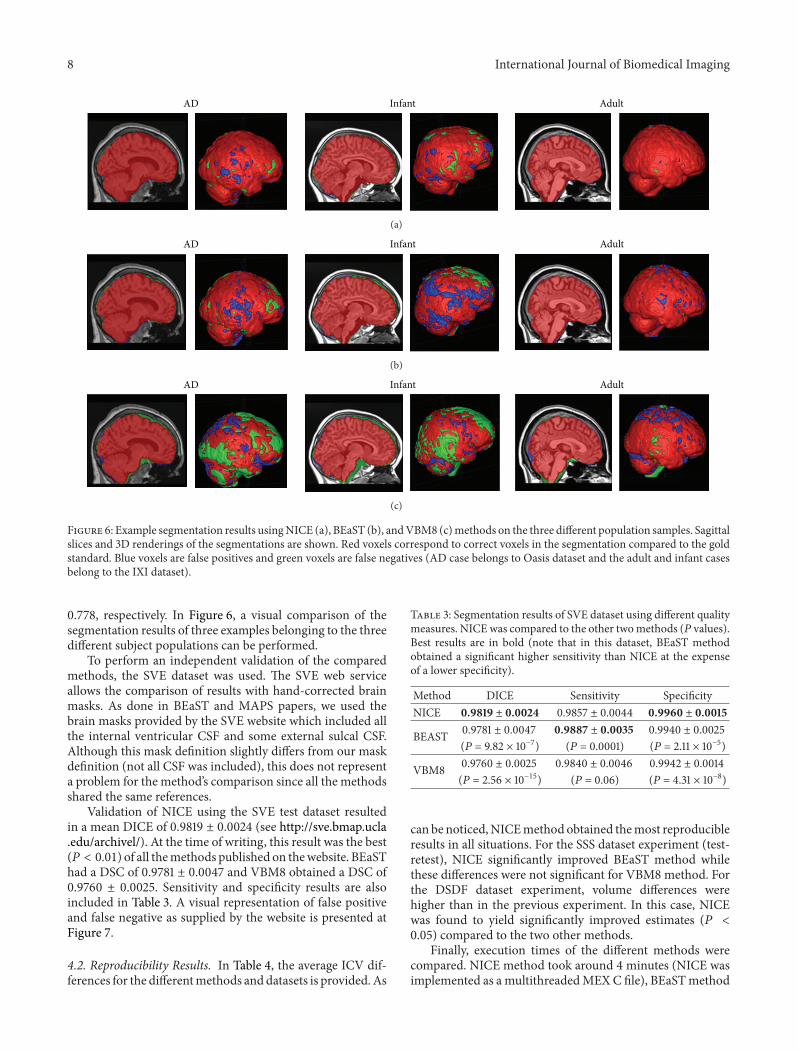