Oral cavity
38
Oral cavity & Oral mucosa Arvin Shahbazi D.M.D, Ph.D fellow Department of Anatomy, Histology and Embryology Semmelweis University [email protected]
-
Upload
khangminh22 -
Category
Documents
-
view
0 -
download
0
Transcript of Oral cavity

Oral cavity & Oral mucosa
Arvin Shahbazi D.M.D, Ph.D fellow
Department of Anatomy, Histology and Embryology Semmelweis University

Extends from the lips & cheeks externally to the pillars of the fauces internally, where it continues into the oropharynx.
Oral cavity
The oral cavity is divided
1) Oral cavity vestibule 2) Oral cavity proper
Arvin Shahbazi D.M.D, Ph.D fellow

Borders of the oral cavity: The palate (hard & soft palate) forms the roof of the mouth and separates the oral & nasal cavities.The floor of the oral cavity consists of mucous membrane covering the mylohyoid muscle and is occupied mainly by the tongue.The lateral walls of the oral cavity are defined by the cheeks & retromolar regions.
Oral cavity
Arvin Shahbazi D.M.D, Ph.D fellow

Functions of the oral cavity: Primary functions —> ingestion of food with mastication & swallowing. Secondary functions —> speech & ventilation (breathing).


It is a slit-like space between the lips / cheeks,and the teeth & alveolus.The oral cavity vestibule is separated from the oral cavity proper by the alveolar bone & teeth.The mucosa covering the alveolus is reflected on to the lips and cheeks, forming a sulcus called as vestibular fornix.The upper and lower labial frena or frenula are such folds in the midline. All folds contain loose connective tissue (no muscle attachments can be found).
Oral vestibule
Arvin Shahbazi D.M.D, Ph.D fellow

When the teeth occlude, the vestibule is a closed space that communicates with the oral cavity proper only behind the last molars (the retromolar regions). This provides a pathway for the administration of nutrients in a patient whose jaws have been wired together following a fracture.
Oral vestibule
Retromolar area
Arvin Shahbazi D.M.D, Ph.D fellow
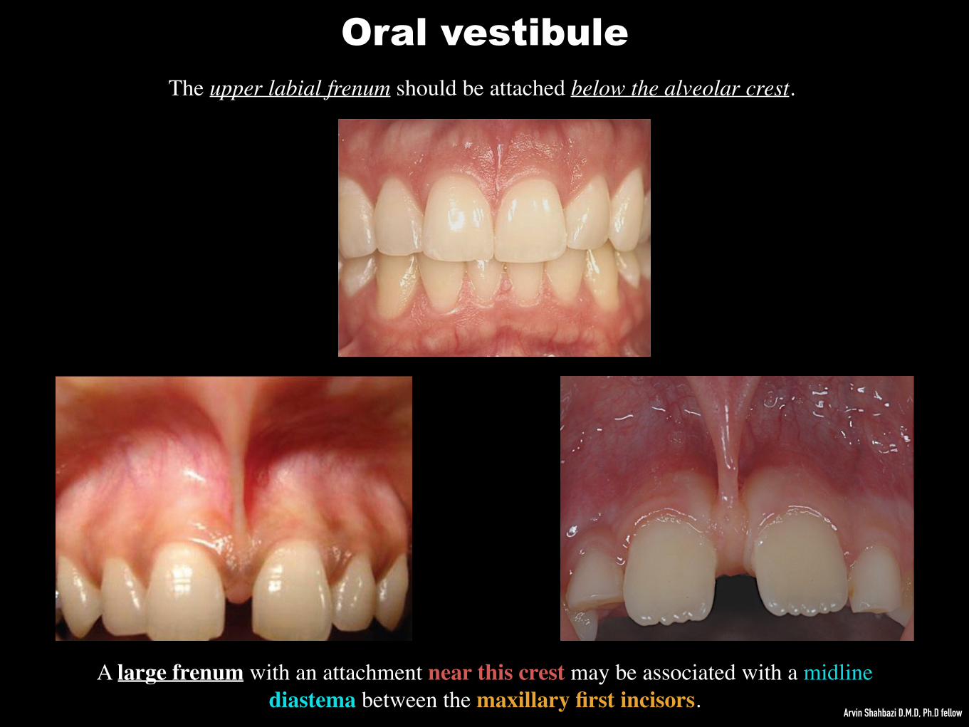
The upper labial frenum should be attached below the alveolar crest.
A large frenum with an attachment near this crest may be associated with a midline diastema between the maxillary first incisors.
Oral vestibule
Arvin Shahbazi D.M.D, Ph.D fellow

Oral vestibule
Prominent frena may also influence the stability of dentures !!
Arvin Shahbazi D.M.D, Ph.D fellow

Oral vestibule Lips Lips are composed of a muscular skeleton (orbicularis oris muscle) & connective tissue, Lips are covered externally by skin and internally by mucous membrane.
The red portion of the lip (the vermilion) is a feature characteristic of humans, it is formed due to: 1) The dilation of the small vessels in the connective tissue
2) Thickness of the epithelium 3) Degree of keratinization and amount of melanin pigments in the epithelium.
The sharp junction of the vermilion & the skin is termed the vermilion border.
The corners of the lips (labial commissures) are usually located adjacent to the maxillary canine & mandibular 1st premolar teeth.
In the upper lip the vermilion protrudes in the midline to form the tubercle. In the midline of the upper lip runs the philtrum.The lower lip shows a slight depression in the midline corresponding to the tubercle.
Laterally, the upper lip is separated from the cheeks by nasolabial grooves. Similar grooves appear with age at the corners of the mouth. The labiomental groove separates the lower lip from the chin.
Arvin Shahbazi D.M.D, Ph.D fellow

Ectopic sebaceous glands without any associated hair
follicles may be evident in the mucosa and are called
Fordyce spots.
They are small, yellowish-white spots, occurring singly
or in clusters on the margin of the lips or the mucosa
of the cheeks.
They can be seen in the majority of patients and are said
to increase with age.
Oral vestibuleCheeks
Extend intra-orally: from the labial commissures anteriorly to the ridge of mucosa on the ascending ramus of the mandible posteriorly. They are bounded superiorly & inferiorly by the upper & lower vestibular fornices. The mucosa is non-keratinized & tightly attaches to the buccinator muscle, The cheek is stretched when the mouth is opened and wrinkled when closed.
Arvin Shahbazi D.M.D, Ph.D fellow
Linea alba
Fordyce spots

In the retromolar region:
Anterior to the pillars of the fauces —> a fold of mucosa
containing the pterygomandibular raphe extends from the
upper to the lower alveolus.
Oral vestibule
Retromolar area
There is a groove located between:
pterygomandibular raphe & the ramus of the mandible is
an important landmark for insertion of a needle for local
anaesthesia of the lingual & inferior alveolar nerves.
Cheeks
Arvin Shahbazi D.M.D, Ph.D fellow

The parotid duct (stensen duct) drains into the cheek opposite
the maxillary 2nd molar tooth and its opening may be covered
by a small fold of mucosa called as parotid papilla.
Oral vestibuleCheeks

Maxilla (upper jaw)
Anterior surface (malar surface)
Forms the skeleton of the anterior part of the cheek
A = frontal process B = zygomatic process C = alveolar process D = anterior nasal spineE = canine fossaF = jugal crest (zygomatico-alveolar crest) The infra-orbital foramen is arrowed.
A
B
CCC
DE
F
Arvin Shahbazi D.M.D, Ph.D fellow

Anterolateral sueface (malar surface)
A = frontal process B = zygomatic process C = alveolar process D = anterior nasal spineE = canine fossaF = orbital plateG = jugal crest (zygomatico-alveolar crest) The infra-orbital foramen is arrowed.
A
B
C C C
DE
F
G
Arvin Shahbazi D.M.D, Ph.D fellow
Maxilla (upper jaw)

A= Alveolar socket (dental alveolus)B= Interdental septum C= Interradicular septum D= Retromolar fossa (triangular depression in the mandible behind to the 3rd molar tooth)E= Retromolar foramen (connects the retromolar canal to the alveolus of 3rd molar)
AB
C
A
C
D
E
D
A
A
AA
B
B
B
D
C
Mandible (lower jaw)
Arvin Shahbazi D.M.D, Ph.D fellow

Palate
Forms the roof of the mouth & separates the oral and nasal cavities. It is divided into the immovable hard palate anteriorly and the movable soft palate posteriorly.The skeleton of the hard palate is bony while that of the soft palate is fibrous.
The hard palate is covered by a masticatory, keratinized mucosa that is attaching tightly to the bone & also contains some taste buds. Immediately behind the maxillary central incisors we can find the —> incisive papilla (located at the incisive fossa). In the incisive fossa —> opening of the incisive canal can be found which carries nasopalatine nerve.
Oral cavity proper
A = incisive papilla
B = palatine raphe
C = palatine rugae
D = alveolus
On the midline from the incisive papilla if we move posteriorly, we can find a ridge termed as palatine raphe (here the oral mucosa is attached directly to bone without the presence of a submucous layer of tissue) Palatine rugae are elevated ridges in the anterior part of the hard palate that radiate somewhat transversely from the incisive papilla & the anterior part of the palatine raphe. Their pattern is unique to the individual and, like fingerprints, can be used for forensic purposes to help identify individuals.
Arvin Shahbazi D.M.D, Ph.D fellow

Bony skeleton of the palateIt is mainly formed by: 1) Premaxilla 2) Palatine process of the maxilla 3) Horizontal plate of the palatine bone
A = palatine processes of maxillaeB = horizontal plates of the palatine bones C = median palatine sutureD = incisive fossa E = transverse palatine sutureF = greater palatine foramina G = posterior nasal spine
A A
A
B B
C
D
E
FG
The maxillary palatine processes arise as horizontal plates at the junction of the bodies and alveolar processes of the maxillae.
Arvin Shahbazi D.M.D, Ph.D fellow

Hard palate
When the teeth are removed!A = buccal alveolar plate
B = palatal alveolar plateC = interdental bony septa (separates the
alveolar socket) D = inter-radicular septum between the buccal
roots of 1st permanent molar.E = Alveolar socket or dental alveolus (holds
the root of the teeth)
BC
D
A
Arvin Shahbazi D.M.D, Ph.D fellow
E
C
CC C

At a location sometimes referred to as the ‘vibrating line’ because the soft palate can be seen to move here on asking a patient to say ‘ah’. There are 2 small pits called as fovea palatini, may be seen on either side of the midline; they represent the orifices of ducts from some of the minor mucous glands of the palate. The fovea palatini can also be seen on impressions of the palate and a postdam may usually be safely placed a couple of millimetres behind the pits. Knowledge of the anatomy of the palate has clinical relevance when siting the posterior border (postdam) of an upper denture.
Palate
Arvin Shahbazi D.M.D, Ph.D fellow
The denture needs to sit on the anterior border of the soft palate:

Palate
Oral disease

1) Levator veli palatini: Origin: bellow the apex of the petrous part of the temporal bone (anterior to the carotid canal) and from cartilage of the auditory tube. It is the elevator muscle of the soft palate. During swallowing, it elevates the soft palate to help to prevent from entry of food into the nasopharynx. Innervation: Vagus nerve
2) Tensor veli palatini: Origin: Scaphoid fossa at the base of the medial pterygoid plate, from the spine of the sphenoid and from the cartilage of the auditory tube Insertion: Palatine aponeurosis Innervation: Mandibular nerve Function: Elevates the soft palate and opens the Auditory tube
4) Palatopharyngeus:Origin: Palatine aponeurosis Insert: Upper part of thyroid cartilage Innervation: Vagus nerve Function: Pulls pharynx & larynx upward
5) Uvula:Origin: Psterior nasal spine It is the projection of posterior part of the soft palate.
3) Palatoglossus:Origin: Palatine aponeurosis of the soft palate Insertion: Side of the tongue Innervation: Vagus nerve Function: Elevates posterior tongue, closes the oropharyngeal isthmus
The arterial supply to the muscles of the soft palate is derived from: 1) Facial artery (ascending palatine branch) 2) Ascending pharyngeal artery 3) Maxillary artery (palatine branches)
Soft palateThe border between the soft palate & the hard palate is
readily palpable and can be recognized by a change in
colour, the soft palate having a yellowish appearance.
Supported by the fibrous palatine aponeurosis,
Arvin Shahbazi D.M.D, Ph.D fellow


A small, horseshoe-shaped region above the mylohyoid muscle & bellow the movable part of the tongue. It is a region located between the medial surface of the mandible, the inferior surface of the tongue and the mylohyoid muscles.Covered by non-keratinized mucosa. In the midline, near the base of the tongue, a fold of tissue called the lingual frenum extends on to the inferior surface of the tongue. The sublingual papilla where submandibular salivary ducts open into the mouth, is a large centrally positioned projection at the base of the tongue. On both side of sublingual papilla are the sublingual folds & bellow the submandibular ducts and sublingual salivary glands can be found.
Floor of the mouth
Arvin Shahbazi D.M.D, Ph.D fellow

Floor of the mouth

Floor of the mouth

MYLOHYOID
The mylohyoid muscle arises from the mylohyoid line on the inner surface of the body of the mandible. Its fibers moves down- wards, forwards and inwards. The anterior fibres of the mylohyoid muscle interdigitate with the corresponding fibres on the opposite side to form a median raphe. This raphe is attached above to the chin and below to the hyoid bone. The posterior fibres are inserted on to the anterior surface of the body of the hyoid bone. Function: The muscle raises the floor of the mouth during the early stages of swallowing. Helps to depress the mandible when the hyoid bone is fixed. Innervation: Mylohyoid branch of the inferior alveolar branch of the mandibular division of the trigeminal nerve. Blood supply: lingual artery (sublingual branch), maxillary artery (mylohyoid branch of the inferior alveolar artery) and the facial artery (submental branch). The 2 mylohyoid muscles form a muscular diaphragm for the floor of the mouth. Above this diaphragm are found the genioglossus and geniohyoid muscles medially and the hyoglossus muscles laterally. Below the diaphragm lie the digastric and stylohyoid muscles.
Floor of the mouth
Arvin Shahbazi D.M.D, Ph.D fellow

GENIOHYOID
Originates: Inferior genial spine (inferior mental spine)
It passes backwards and slightly downwards
Insertion: on to the anterior surface of the body of the hyoid bone.
Function: Elevates the hyoid bone and is a weak depressor of the mandible.
Innervation: Deep cervical ansa (cervical plexus)
Blood supply: lingual artery (sublingual branch).
Floor of the mouth
Arvin Shahbazi D.M.D, Ph.D fellow

The palatine tonsil contains deep crypts
The oropharyngeal isthmus is where the oral cavity and the oropharynx meet.
Isthmus of fauces
Borders: Sup: Soft palate (uvula)Inf: Root of the tongueLat: Palato-glossal & Palato-pharyngeal folds
Arvin Shahbazi D.M.D, Ph.D fellow
These folds cover the palatoglossus & palatopharyngeus muscles,
Between these folds the tonsillar fossa is located which houses the palatine tonsil.
The palatine tonsil is a collection of lymphoid material of variable size that is likely to atrophy in the adult.

Oral mucosaTerm of the mucus membrane is used to describe the moist lining of GI tract, nasal passages and other body cavities that communicates with the exterior. In the oral cavity, this lining is called as oral mucosa: Stratified squamous epithelium (oral epithelium) + connective tissue (lamina propria)The oral mucosa is located between the lips (continues with skin - bounded by vermilion border) & pharynx (continues with the rest of the gut).
Arvin Shahbazi D.M.D, Ph.D fellow

1) Protection: Surface lining (stratified squamous epithelium) separates & protects deeper tissues and organs. Biting and chewing expose the oral soft tissues to mechanical forces (compression, stretching) and surface abrasions so the oral mucosa shows number of adaptions of the epithelium and the connective tissue to these mechanical forces. The epithelium acts as a major barrier to the microorganisms and their toxic products which might cause infection.
2) Sensation:Receptors for the temperature + pain (protopathic), touch, pressure. In the tongue also taste buds (taste receptors) are present. Note: Reflexes like swallowing, salivating and gagging are initiated by the receptors of the oral cavity.
3) Secretion: The major secretion associated with the oral mucosa is saliva, produced by the salivary glands. Sebaceous glands commonly are present in the oral mucosa, but their secretion are insignificant.
4) Thermal regulation: In some animals (such as dog), the mucosa plays a major role in the regulation of the body temperature. The human oral mucosa plays no practical role in regulation of body temperature and no obvious specializations of blood vessels exist for controlling heat transfer (such as arteriovenous shunts).
Functions of oral mucosa
Arvin Shahbazi D.M.D, Ph.D fellow

1) Masticatory mucosa (forms 25% of the total oral mucosa): Stratified squamous keratinized epithelium, found on:a) Dorsum of the tongueb) Hard palate c) Attached gingiva .
2) Lining mucosa (forms 60% of the total oral mucosa):
Stratified squamous nonkeratinized epithelium, found on:
a) Buccal mucosa (inner lining of the cheeks)b) Floor of the oral cavityc) Labial mucosa (inner lining of the lips)d) Alveolar mucosa (lining between the buccal and labial mucosae)e) Soft palatef) Underside the tongue
3) Specialized mucosa (forms 15% of the total oral mucosa):Taste buds, tactile receptors
Types of the oral mucosa according to their primary functions
Arvin Shahbazi D.M.D, Ph.D fellow

1) Stratified squamous keratinized:
Present in the epithelial surface of the masticatory mucosa & in some regions of the specialized mucosa. It is inflexible, tough, resistant to abrasion and tightly bounded to the lamina propria.
Layers: Startum basale (stratum germinativum): cuboidal/columnar cells sitting on basal lamina Startum spinosum (prickle cell layer): several large elliptical spherical cell layers Called spinosum because the cells shrink during histological preparation and the cells remain in contact by intercellular bridges or desmosomes.Note: Stratum basale + Stratum spinosum —> forms 2/3 of the entire epithelium. Stratum granulosum: contains small granules of keratohyalin which can be stained intensly by the acid dyes (such as hematoxylin).Stratum spinosum: transparent, tiny pink squamous layer which contains eleidin protein and it can be stained by eosin staining.Stratum corneum (cornified layer): it is the keratinized layer. Note: The pattern of maturation of these cell layers is termed orthokeratinization.Note: The masticatory mucosa (parts of the hard palate and gingiva) presents parakeratinization (means the surface layer stains for keratin but pyknotic nuclei are retained).
Types of epithelial linings in the oral cavity
Arvin Shahbazi D.M.D, Ph.D fellow


2) Stratified squamous non keratinized:
Present in the lining mucosa of the oral cavity. In some regions (such as labial and buccal mucosa) the epithelium is thicker then keratenized epithelium and shows different pattern of connective tissue.
Layers:
Startum basale (stratum germinativum): cuboidal/columnar cells sitting on basal lamina (slightly larger then the keratinized epithelium) Startum spinosum: several large elliptical spherical cell layers Note: Here the intercellular bridges are less conspicuous.
Note: The outer layer divides in two zones: 1) Stratum intermedium 2) Stratum superficiale
Types of epithelial linings in the oral cavity
Arvin Shahbazi D.M.D, Ph.D fellow

Major Features of Maturation in Keratinized & Nonkeratinized Epithelium
Characteristics of Nonkeratinocytes in Oral Epithelium
LEVEL IN EPITHELIUM

May 11, 2015 ED II/4
08/05/2017ED II/4
May 12, 2016 EM II/16
2017/10/01EDI/4
2018/09/29EDI/5