Toxic Indoor Air Is a Potential Risk of Causing Immuno ... - MDPI
18
Citation: Vaali, K.; Tuomela, M.; Mannerström, M.; Heinonen, T.; Tuuminen, T. Toxic Indoor Air Is a Potential Risk of Causing Immuno Suppression and Morbidity—A Pilot Study. J. Fungi 2022, 8, 104. https:// doi.org/10.3390/jof8020104 Academic Editor: Laurent Dufossé Received: 22 November 2021 Accepted: 4 January 2022 Published: 21 January 2022 Publisher’s Note: MDPI stays neutral with regard to jurisdictional claims in published maps and institutional affil- iations. Copyright: © 2022 by the authors. Licensee MDPI, Basel, Switzerland. This article is an open access article distributed under the terms and conditions of the Creative Commons Attribution (CC BY) license (https:// creativecommons.org/licenses/by/ 4.0/). Fungi Journal of Article Toxic Indoor Air Is a Potential Risk of Causing Immuno Suppression and Morbidity—A Pilot Study Kirsi Vaali 1, *, Marja Tuomela 2,3 , Marika Mannerström 4 , Tuula Heinonen 4 and Tamara Tuuminen 5 1 SelexLab Oy, Kalevankatu 17 A, 00100 Helsinki, Finland 2 Co-op Bionautit, Viikinkaari 9, 00790 Helsinki, Finland; marja.tuomela@helsinki.fi 3 Department of Microbiology, University of Helsinki, 00014 Helsinki, Finland 4 The Finnish Centre for Alternative Methods, Faculty of Medicine and Health Technology, Tampere University, Arvo Ylpön katu 34, 33014 Tampere, Finland; marika.mannerstrom@tuni.fi (M.M.); tuula.heinonen@tuni.fi (T.H.) 5 Medical Center Kruunuhaka Oy, Kaisaniemenkatu 8B a, 00100 Helsinki, Finland; [email protected] * Correspondence: [email protected]; Tel.: +358-50-550-1131 Abstract: We aimed to establish an etiology-based connection between the symptoms experienced by the occupants of a workplace and the presence in the building of toxic dampness microbiota. The occupants (5/6) underwent a medical examination and urine samples (2/6) were analyzed by LC-MS/MS for mycotoxins at two time-points. The magnitude of inhaled water was estimated. Building-derived bacteria and fungi were identified and assessed for toxicity. Separate cytotoxicity tests using human THP-1 macrophages were performed from the office’s indoor air water condensates. Office-derived indoor water samples (n = 4/4) were toxic to human THP-1 macrophages. Penicillium, Acremonium sensu lato, Aspergillus ochraceus group and Aspergillus section Aspergillus grew from the building material samples. These colonies were toxic in boar sperm tests (n = 11/32); four were toxic to BHK-21 cells. Mycophenolic acid, which is a potential immunosuppressant, was detected in the initial and follow-up urine samples of (2/2) office workers who did not take immunosuppressive drugs. Their urinary mycotoxin profiles differed from household and unrelated controls. Our study suggests that the presence of mycotoxins in indoor air is linked to the morbidity of the occupants. The cytotoxicity test of the indoor air condensate is a promising tool for risk assessment in moisture- damaged buildings. Keywords: indoor air water; dampness and mold hypersensitivity syndrome; mycotoxins; clinical toxicology; sick building syndrome; urine 1. Introduction There is convincing scientific data that the exposure to dampness microbiota and the decay products of construction materials may cause the development of polymorbidities and systemic inflammation [1,2], now collectively called dampness and mold hypersen- sitivity syndrome (DMHS) [3], although this designation has not yet received official recognition. The causality between the exposure to dampness microbiota (the source) and the clinical outcome (the effect) is still being questioned. Polymorbidity in the occupants of moisture-damaged buildings with indoor air toxicity has been extensively studied in Finnish schoolchildren and the occupants of a moisture-damaged hospital and a police station [1,4–8]. These studies have demonstrated that the prolonged exposure to dampness microbiota and the decay products of a building’s construction materials may cause a plethora of non-respiratory symptoms such as profound fatigue, neurological, gastrointesti- nal, and muscular-skeletal problems in addition to the already acknowledged respiratory symptoms. Because no single biomarker to diagnose DMHS has yet been devised, the diagnosis remains clinical and is based on interview. The patients often recall some form of water leakage in their homes or workplaces and a gradual worsening of their symptoms. J. Fungi 2022, 8, 104. https://doi.org/10.3390/jof8020104 https://www.mdpi.com/journal/jof
-
Upload
khangminh22 -
Category
Documents
-
view
0 -
download
0
Transcript of Toxic Indoor Air Is a Potential Risk of Causing Immuno ... - MDPI

�����������������
Citation: Vaali, K.; Tuomela, M.;
Mannerström, M.; Heinonen, T.;
Tuuminen, T. Toxic Indoor Air Is a
Potential Risk of Causing Immuno
Suppression and Morbidity—A Pilot
Study. J. Fungi 2022, 8, 104. https://
doi.org/10.3390/jof8020104
Academic Editor: Laurent Dufossé
Received: 22 November 2021
Accepted: 4 January 2022
Published: 21 January 2022
Publisher’s Note: MDPI stays neutral
with regard to jurisdictional claims in
published maps and institutional affil-
iations.
Copyright: © 2022 by the authors.
Licensee MDPI, Basel, Switzerland.
This article is an open access article
distributed under the terms and
conditions of the Creative Commons
Attribution (CC BY) license (https://
creativecommons.org/licenses/by/
4.0/).
FungiJournal of
Article
Toxic Indoor Air Is a Potential Risk of Causing ImmunoSuppression and Morbidity—A Pilot StudyKirsi Vaali 1,*, Marja Tuomela 2,3, Marika Mannerström 4, Tuula Heinonen 4 and Tamara Tuuminen 5
1 SelexLab Oy, Kalevankatu 17 A, 00100 Helsinki, Finland2 Co-op Bionautit, Viikinkaari 9, 00790 Helsinki, Finland; [email protected] Department of Microbiology, University of Helsinki, 00014 Helsinki, Finland4 The Finnish Centre for Alternative Methods, Faculty of Medicine and Health Technology, Tampere University,
Arvo Ylpön katu 34, 33014 Tampere, Finland; [email protected] (M.M.);[email protected] (T.H.)
5 Medical Center Kruunuhaka Oy, Kaisaniemenkatu 8B a, 00100 Helsinki, Finland; [email protected]* Correspondence: [email protected]; Tel.: +358-50-550-1131
Abstract: We aimed to establish an etiology-based connection between the symptoms experiencedby the occupants of a workplace and the presence in the building of toxic dampness microbiota.The occupants (5/6) underwent a medical examination and urine samples (2/6) were analyzed byLC-MS/MS for mycotoxins at two time-points. The magnitude of inhaled water was estimated.Building-derived bacteria and fungi were identified and assessed for toxicity. Separate cytotoxicitytests using human THP-1 macrophages were performed from the office’s indoor air water condensates.Office-derived indoor water samples (n = 4/4) were toxic to human THP-1 macrophages. Penicillium,Acremonium sensu lato, Aspergillus ochraceus group and Aspergillus section Aspergillus grew from thebuilding material samples. These colonies were toxic in boar sperm tests (n = 11/32); four were toxicto BHK-21 cells. Mycophenolic acid, which is a potential immunosuppressant, was detected in theinitial and follow-up urine samples of (2/2) office workers who did not take immunosuppressivedrugs. Their urinary mycotoxin profiles differed from household and unrelated controls. Our studysuggests that the presence of mycotoxins in indoor air is linked to the morbidity of the occupants.The cytotoxicity test of the indoor air condensate is a promising tool for risk assessment in moisture-damaged buildings.
Keywords: indoor air water; dampness and mold hypersensitivity syndrome; mycotoxins; clinicaltoxicology; sick building syndrome; urine
1. Introduction
There is convincing scientific data that the exposure to dampness microbiota and thedecay products of construction materials may cause the development of polymorbiditiesand systemic inflammation [1,2], now collectively called dampness and mold hypersen-sitivity syndrome (DMHS) [3], although this designation has not yet received officialrecognition. The causality between the exposure to dampness microbiota (the source) andthe clinical outcome (the effect) is still being questioned. Polymorbidity in the occupantsof moisture-damaged buildings with indoor air toxicity has been extensively studied inFinnish schoolchildren and the occupants of a moisture-damaged hospital and a policestation [1,4–8]. These studies have demonstrated that the prolonged exposure to dampnessmicrobiota and the decay products of a building’s construction materials may cause aplethora of non-respiratory symptoms such as profound fatigue, neurological, gastrointesti-nal, and muscular-skeletal problems in addition to the already acknowledged respiratorysymptoms. Because no single biomarker to diagnose DMHS has yet been devised, thediagnosis remains clinical and is based on interview. The patients often recall some form ofwater leakage in their homes or workplaces and a gradual worsening of their symptoms.
J. Fungi 2022, 8, 104. https://doi.org/10.3390/jof8020104 https://www.mdpi.com/journal/jof

J. Fungi 2022, 8, 104 2 of 18
Initially, the symptoms are reversible and related to the so-called sick building syndrome,(SBS) [9]. However, if there is continuous or cumulative exposure (e.g., starting alreadyin the kindergarten, and then in school, high school and then workplaces) the symptomsmay become aggravated and progress into irreversible multi-organ DMHS [3]. Importantly,DMHS is associated with an increased rate of respiratory infections [3] probably indicat-ing decreased immunologic tolerance to environmental viruses and bacteria. Moreover,deprivation of the immune system has been proposed by us when reviewing the long-termmorbidity among the occupants of one of the moisture-damaged school [10]. A higherprevalence of different oncological diseases, unexplained succeptibility to sepsis and ahigher than normal rate of autoimmune diseases have been reported [10].
The investigations of the indoor air quality do not always recognize existing problemswithin buildings; for example, the impact of air toxicity has been overlooked. Most ofthe toxic metabolites produced by fungi or bacteria recovered from moisture-damagedbuildings have a molecular weight of 300–2000 g/mol, i.e., they are non-volatile. However,mycotoxins may be present in indoor air either attached to fungal spores or to fragmentsof these microorganisms, e.g., broken or fractured conidia and hyphae. These particlesmay remain suspended in the air for a long time [11]. Recently, it was reported [12–14]that some species of dampness microbiota produce vesicles, or the so-called “guttationdroplets”, or exudates on a culture dish. These droplets may contain substances whichare toxic to eukaryotic cell types at dilutions of 1/100–20,000 [12,15,16]. The most efficientsolvents for mycotoxins are relatively polar solvents, such as methanol or ethanol, only a fewmycotoxins are water soluble [17]. The relative humidity (RH %) of the air becomes elevatedwhen the environment is crowded with occupants (e.g., schools during the working hours)which allows these droplets to move around aerodynamically and thus the toxins canspread throughout the environment [18]. For example, Penicillium expansum recovered fromgypsum boards produced these kinds of toxic droplets, i.e., exudates that were released intothe air and were >100-times more toxic in the cell culture assays than indoor air isolates ofAspergillus, Chaetomium, Stachybotrys and Paecilomyces [13]. These droplets may represent aneven greater health hazard than the spores and nanoparticles of a microbial mass growingin the environment.
To address the issue of air toxicity, we have devised a novel method of collectionof indoor air water vapour by adopting a newly patented technique with the so-calledE-collector: (US Patent 10,502,722 B2; www.sisailmatutkimuspalvelut.fi, accessed on 21November 2021) [18,19]. Briefly, the air condensate is collected on a cooled surface of asteel plate, the condensate then can be tested for its cytotoxicity against living cells [19].Boar sperm cells [20,21] or several eukaryotic cell lines, such as porcine kidney cell line(PK-15) [13,22,23] or murine neuroblastoma cells (MNA) [13,23,24], have been used intoxicity studies instead of experiments in production animals. In this publication, wehave successfully used boar spermatozoids and baby hamster kidney (BHK) cells for thesame purpose.
Here, we describe a plausible pathway of how the mycotoxins present in the indoorair water samples gain access to the exposed individuals. We demonstrate the usefulnessof urinalysis for the detection of mycotoxins as biomarkers of DMHS. A novel method [19]for the sampling and testing of indoor air toxicity is presented and shortly discussed.
2. Materials and Methods2.1. The Patients
This working community comprised six members, and their work premises whichwere located partly below ground level in an office building (Figure 1). Within approx-imately three months after the move into this office, every member of this workplacecommunity started to suffer from symptoms compatible with their exposure to indoor airdampness microbiota. Finally, the symptoms became unbearable. The patients wanted toremain anonymous and therefore their gender and age are not reported.

J. Fungi 2022, 8, 104 3 of 18
J. Fungi 2022, 8, x FOR PEER REVIEW 3 of 18
to indoor air dampness microbiota. Finally, the symptoms became unbearable. The patients wanted to remain anonymous and therefore their gender and age are not reported.
Five (Patients 1-5) out of six occupants of the problematic building were examined by one of us (TT) and one (Patient 6) was interviewed over the telephone. All were healthy before the move into the problematic office. Their life histories and the symptoms were recorded during their visits and then retrieved with the patients’ written consent.
For comparison of urinalysis profiles, we investigated two unrelated patients (Patient X and Y) who volunteered to have their urine samples tested because their symptoms were compatible with an exposure to dampness microbiota. They were unrelated to each other, they lived and worked in different cities than the working community in question. Neither microbiological nor toxicological investigations in their homes or workplaces had been performed at the time of our investigation. Patient X moved with all his/her belongings to a new home but continued to experience a general feeling of sickness, fatigue, and mucosal irritation.
Figure 1. The study design.
2.2. Detection of Mycotoxins in the Patients’ Urine Samples The detection of mycotoxins from the patients’ urine was performed in the Great
Plains Laboratory Inc. (Lenexa, KS, USA) using a liquid chromatography tandem mass spectrometry (LC-MS/MS) technique. The urine specimens from Patients 1 and 2 of the working community and a sample from a household control (HHC) of Patient 1, and from Patients X and Y were analyzed. Follow-up samples from Patients 1 and 2 were taken 2.5 months later after the working community had moved to a clean building.
The detection of mycotoxins was performed according to the standard operational procedure of the laboratory. The presence of the mycotoxin mycophenolic acid was confirmed after extraction of urine and LC/MS/MS testing of the extract with a deuterated mycophenolic acid internal standard [25], indicating that a peak with all the characteristic ions of authentic mycophenolic acid [26,27] and the identical retention time of mycophenolic acid was present in many patients [28]. In addition, patients with high mycophenolic acid in urine has been tested as positive for mycophenolic acid by a commercial specific immunoassay (Cedia) from ThermoScientific for mycophenolic acid in urine. Mycophenolic acid can be produced by multiple species of Penicillium and other molds [29–31].
Figure 1. The study design.
Five (Patients 1–5) out of six occupants of the problematic building were examined byone of us (TT) and one (Patient 6) was interviewed over the telephone. All were healthybefore the move into the problematic office. Their life histories and the symptoms wererecorded during their visits and then retrieved with the patients’ written consent.
For comparison of urinalysis profiles, we investigated two unrelated patients (PatientX and Y) who volunteered to have their urine samples tested because their symptoms werecompatible with an exposure to dampness microbiota. They were unrelated to each other,they lived and worked in different cities than the working community in question. Neithermicrobiological nor toxicological investigations in their homes or workplaces had beenperformed at the time of our investigation. Patient X moved with all his/her belongingsto a new home but continued to experience a general feeling of sickness, fatigue, andmucosal irritation.
2.2. Detection of Mycotoxins in the Patients’ Urine Samples
The detection of mycotoxins from the patients’ urine was performed in the GreatPlains Laboratory Inc. (Lenexa, KS, USA) using a liquid chromatography tandem massspectrometry (LC-MS/MS) technique. The urine specimens from Patients 1 and 2 of theworking community and a sample from a household control (HHC) of Patient 1, and fromPatients X and Y were analyzed. Follow-up samples from Patients 1 and 2 were taken2.5 months later after the working community had moved to a clean building.
The detection of mycotoxins was performed according to the standard operationalprocedure of the laboratory. The presence of the mycotoxin mycophenolic acid was con-firmed after extraction of urine and LC/MS/MS testing of the extract with a deuteratedmycophenolic acid internal standard [25], indicating that a peak with all the characteristicions of authentic mycophenolic acid [26,27] and the identical retention time of mycopheno-lic acid was present in many patients [28]. In addition, patients with high mycophenolicacid in urine has been tested as positive for mycophenolic acid by a commercial specificimmunoassay (Cedia) from ThermoScientific for mycophenolic acid in urine. Mycophenolicacid can be produced by multiple species of Penicillium and other molds [29–31].
2.3. The Office Building
The working community had their office located on the ground floor of a buildingwith a cellar. It was built from concrete elements and had a mechanical ventilation system.One of the walls in the office faced a non-heated hollow area which was below the soil

J. Fungi 2022, 8, 104 4 of 18
surface and was lined on the bottom with a layer of gravel and on the top with a closingplate mostly without waterproofing. Thus, water from rain and melting snow had leakedinside the hollow area enabling microbial growth. There were several air leaks in the wall ofthe office adjacent to the hollow area. Because of the negative pressure difference (−12 Paon average, momentarily even −40 Pa) in the office compared with the hollow area, theair from this area was able to penetrate the office. The hollow area remained moist allyear round.
The office was also examined by sniffer dogs which are trained in mold detection. Thedogs marked six locations, with five of these being beside the wall facing the hollow area.In some locations, the characteristic odors of microbes were noticeable also to the occupants.
2.4. Sampling of the Indoor Air Condensate Water
The device and the technique to collect indoor air water samples have been describedelsewhere [19]. Briefly, the principle of the collection is based on the phenomenon of thecondensation of water molecules on the top of cold surfaces of metal plates. After meltingof the frozen water to room temperature, the condensate is collected from the tray of thedevice into Eppendorf tubes that are transferred to the laboratory where the condensate isanalyzed in a cell culture assay. This collection technique enables the harvesting of airbornetoxic substances in indoor air vapor that may contain mycotoxins [13] as well as variouslarge molecules [32].
The condensing water samples from the six locations marked by the sniffer dogs werecollected with the E-collector from the office building. Samples were also collected fromthe home of Patient 1.
2.5. Indoor Air Toxicity Studies
The toxicity of the indoor air water condensates collected from different locations ofthe office and from the home of Patient 1 was studied using the human acute monocyticleukemia cell line (THP-1) using water-soluble tetrazolium salts (WST-1) in a cytotoxicityassay [19,33]. The WST-1 assay is an indicator of mitochondrial activity and commonlyused to assess cell viability; decreased mitochondrial activity reflects a loss of cell via-bility, i.e., cell death. Increased mitochondrial activity reflects an increased cell number,i.e., proliferation, or increased cellular respiration induced by mitochondrial uncouplingreactions [34,35]. The WST-1 Cell Proliferation Reagent was obtained from Roche (Basel,Switzerland). Human THP-1 monocytes (Cat. No. TIB-202) were from ATCC (LGCPromochem AB, Boras, Sweden), and were verified to be Mycoplasma-free (MycoAlert™kit, Lonza Basel, Switzerland) prior to use. THP-1 monocytes were maintained in RPMI1640 Medium supplemented with 10% fetal bovine serum (FBS), and when differentiatedto macrophages, challenged with 25 nM phorbol 12-myristate 13-acetate (PMA) (SigmaAldrich, Steinheim, Germany) for 48 h followed by a 24 h recovery period.
The cells were seeded into 96-well plates at a density of 105 cells/well and exposed for24 h to the indoor air condensates at 10% and 25% concentrations (in RPMI supplementedwith 5% FBS). Cells exposed to an equal volume of distilled water (10% or 25%) served asnegative controls, and to nickel II sulphate hexahydrate (2.0 and 20.0 µg/mL) as a positivecontrol. All samples and controls were tested in six replicates. In the assessment of cellviability, 10 µL/well WST-1 reagent was added to the cells for 2 hours, and subsequentlyabsorbance was read at 450 nm. The absorbance is directly proportional to the mitochon-drial activity (cell viability). The absorbances were normalized, i.e., the untreated controlwas set as 100%, and all other data were calculated relative to the control absorbance aseither % decrease in cell viability (negative values) or % increase in proliferation (positivevalues). The statistical significance of the changes compared to the untreated control wastested with Student’s t-test.
The indoor air condensates from the office were tested using both THP-1 monocytesand THP-1 macrophages at a 10% sample concentration. The condensates from the home
J. Fungi 2022, 8, 104 5 of 18
of Patient 1 were tested at two concentrations, i.e., 10% and 25%, but using only THP-1 macrophages.
2.6. Detection of Mycotoxins from the Condensed Indoor Air Sample
A left-over aliquot of 0.5 mL of the condensed indoor air water sample in the Eppen-dorf tube was sent at ambient temperature for LC-MS/MS analysis to the Great Plainslaboratory. This sample had not been stored protected from light.
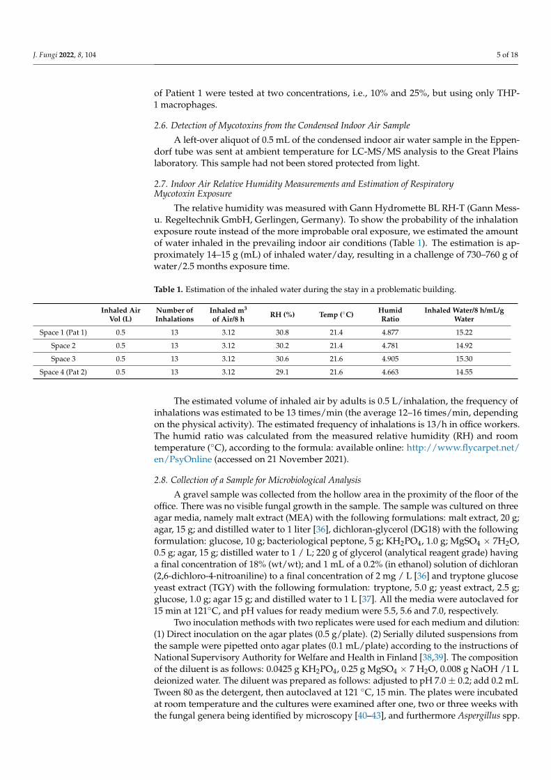
2.7. Indoor Air Relative Humidity Measurements and Estimation of RespiratoryMycotoxin Exposure
The relative humidity was measured with Gann Hydromette BL RH-T (Gann Mess-u. Regeltechnik GmbH, Gerlingen, Germany). To show the probability of the inhalationexposure route instead of the more improbable oral exposure, we estimated the amountof water inhaled in the prevailing indoor air conditions (Table 1). The estimation is ap-proximately 14–15 g (mL) of inhaled water/day, resulting in a challenge of 730–760 g ofwater/2.5 months exposure time.
Table 1. Estimation of the inhaled water during the stay in a problematic building.
Inhaled AirVol (L)
Number ofInhalations
Inhaled m3
of Air/8 h RH (%) Temp (◦C) HumidRatio
Inhaled Water/8 h/mL/gWater
Space 1 (Pat 1) 0.5 13 3.12 30.8 21.4 4.877 15.22
Space 2 0.5 13 3.12 30.2 21.4 4.781 14.92
Space 3 0.5 13 3.12 30.6 21.6 4.905 15.30
Space 4 (Pat 2) 0.5 13 3.12 29.1 21.6 4.663 14.55
The estimated volume of inhaled air by adults is 0.5 L/inhalation, the frequency ofinhalations was estimated to be 13 times/min (the average 12–16 times/min, dependingon the physical activity). The estimated frequency of inhalations is 13/h in office workers.The humid ratio was calculated from the measured relative humidity (RH) and roomtemperature (◦C), according to the formula: available online: http://www.flycarpet.net/en/PsyOnline (accessed on 21 November 2021).
2.8. Collection of a Sample for Microbiological Analysis
A gravel sample was collected from the hollow area in the proximity of the floor of theoffice. There was no visible fungal growth in the sample. The sample was cultured on threeagar media, namely malt extract (MEA) with the following formulations: malt extract, 20 g;agar, 15 g; and distilled water to 1 liter [36], dichloran-glycerol (DG18) with the followingformulation: glucose, 10 g; bacteriological peptone, 5 g; KH2PO4, 1.0 g; MgSO4 × 7H2O,0.5 g; agar, 15 g; distilled water to 1 / L; 220 g of glycerol (analytical reagent grade) havinga final concentration of 18% (wt/wt); and 1 mL of a 0.2% (in ethanol) solution of dichloran(2,6-dichloro-4-nitroaniline) to a final concentration of 2 mg / L [36] and tryptone glucoseyeast extract (TGY) with the following formulation: tryptone, 5.0 g; yeast extract, 2.5 g;glucose, 1.0 g; agar 15 g; and distilled water to 1 L [37]. All the media were autoclaved for15 min at 121◦C, and pH values for ready medium were 5.5, 5.6 and 7.0, respectively.
Two inoculation methods with two replicates were used for each medium and dilution:(1) Direct inoculation on the agar plates (0.5 g/plate). (2) Serially diluted suspensions fromthe sample were pipetted onto agar plates (0.1 mL/plate) according to the instructions ofNational Supervisory Authority for Welfare and Health in Finland [38,39]. The compositionof the diluent is as follows: 0.0425 g KH2PO4, 0.25 g MgSO4 × 7 H2O, 0.008 g NaOH /1 Ldeionized water. The diluent was prepared as follows: adjusted to pH 7.0 ± 0.2; add 0.2 mLTween 80 as the detergent, then autoclaved at 121 ◦C, 15 min. The plates were incubatedat room temperature and the cultures were examined after one, two or three weeks withthe fungal genera being identified by microscopy [40–43], and furthermore Aspergillus spp.
J. Fungi 2022, 8, 104 6 of 18
was identified at a species or section level [44]. The fungal identification was based on theinspection of the morphology as recommended by the Finnish authorized bodies, althoughthe current nomenclature has been recently changed in accordance with the advancesin molecular methods [38]. The bacterial colonies were counted and Streptomycetes wereidentified [38].
2.9. Detection of Toxicity from the Microbial Growth
The colonies of all the fungal and bacterial species (n = 32) that grew on the agarplates were tested in the boar sperm test according to Andersson et al. [20] and Castagnoliet al. [21], and in a mammalian cell test according to Rasimus et al. [45]. A colony or apart of it (20–30 mg) was extracted into 96 % ethanol (0.2 mL). In the toxicity tests, boarspermatozoa that are used in artificial insemination (Figen Oy, Seinäjoki, Finland) and babyhamster kidney cells (BHK-21 [C13] ATCC® CCL10™) were exposed to the ethanol extracts(n = 32). After incubation with the ethanol extracts, the inhibition of BHK-21 cell growthwas assessed using the resazurin reduction assay [46], while toxicity to boar spermatozoawas assessed as a lack of motility observed with the CellSens computer program [21] andvisually with a microscope [20]. Pure ethanol was a negative control in both tests. The BHKcells were maintained in Dulbecco’s modified Eagle’s medium (DMEM) supplementedwith 10% fetal bovine serum (FBS), 2 mM L-glutamine, 100 U/mL penicillin and 100 µg/mLstreptomycin.
2.10. Ethical Considerations
All participants of this study provided their written informed consent to use their records.
3. Results3.1. Clinical Picture in Persons Exposed to Toxic Indoor Air
The symptoms reported by Patients 1–5 are summarized in Table 2. Patient 6 startedto experience a very severe headache that did not become better during the weekends. Thisperson was given a diagnosis of tension neck, and the symptoms were not interpreted asbeing associated with an exposure to poor indoor air quality. The patient was often onsick leave and changed the workplace before this investigation started. The work abilityreturned after changing the employer, and the headache did not reappear.
Table 2. The symptoms experienced by the working community. The symptoms often reported bypatients with chronic mold illness are marked with *.
Patient’s History Symptoms Findings during the Visit
No 1 No previous exposure todampness microbiota * Irritation of eyes * Symptoms became worse when
entering the problematic building
No change in the diet * Very severe headache * Symptoms did not relieveduring weekends
*Recurrent sinusitis Status uneventful
* Multiple courses of antibiotics
The lesions were indurated,watery and had been scratchedexcept for dermatitis on hands,legs, stomach, knees and back
* Thermoregulation problems* Itching of the skin* Exanthema* Sore throat* Gastrointestinal reflux for months* “Brain fog”* A flu-like feeling
J. Fungi 2022, 8, 104 7 of 18
Table 2. Cont.
Patient’s History Symptoms Findings during the Visit
No 2 Lived in a town house withhis/her family * Dyspnoea
* Symptoms had started to ease in2 weeks if the person was absencefrom work
All the other family memberswere asymptomatic * Migraine
* Large psoriatic lesions especiallyon the elbows, back, scalp andchest, lips were cracked open
* Phlegm * The blood pressure was elevated* Muscle pain* Fatigue* Concentration problems* Prolonged cough* Sleeping problems
No 3Previously suffered fromgastrointestinal problemsand allergy
* Sore throat *Redness of eye conjunctiva,unrelated to season
* Blisters on the oral mucosa Exanthema in the lowerneck region
* A flu-like feeling* Blurred vision* Severe fatigue* Dyspnea* Joint painIrregular peristaltic action andmenorrhagia
No 4 Gastrointestinal dysbiosis,leaky gut * Thermoregulation problems Otherwise, the status uneventful
* Two flu-like episodesAxillary temperature 36.8 ◦C,exanthema in the lowerneck region
* Increased sputum production
No 5Previously healthy, the individualhad not been exposed todampness and mold
* Two courses of antibiotics for Status uneventful
tonsillitis* Sore throat* Evenings: eyes dry and voice hoarse* Occasional instances of fatigue* Memory problems
No 6 Did not attend the doctor’s office Was interviewed by telephone
3.2. Detection of Mycotoxins from the Urine Samples
The mycotoxin profiles of Patients 1 and 2 and HHC, Patients X and Y, the controls,are shown in Table 3. It is noteworthy that the HHC for Patient 1 showed evidence of theexcretion of ochratoxin A in the urine but the profile was different from that of Patient 1and there was no mycophenolic acid detectable in his/her urine. The LC-MS/MS plots ofmycophenolic acid are presented in Figure 2A,B. The mycotoxin profiles of Patients X andY did not contain mycophenolic acid, and were different from those of Patients 1 and 2who represented the work community exposure cohort.

J. Fungi 2022, 8, 104 8 of 18
Table 3. Detection of mycotoxins in urine by LC-MS/MS.
MycotoxinsReference Valuesin ng/g Creatinine
Patient 1First Test
Patient 1Follow-Up Test
HHC to Patient 1(Time-Point as forPat 1 Follow-Up)
Patient 2First Test
Patient 2Follow-Up Test Patient X Patient Y
Aflatoxin M1(3.5–20) 0 0 0 0 0 0 0
Ochratoxin A(4–20) 11.23 9.82 * 63.52 7.01 10.96 8.58 18.36
Gliotoxin(200–2000) 0 0 0 0 0 0 * 910.98
Sterigmatocystin(0.2–1.75) 1.14 0 0 0.74 0 0 0
Mycophenolic acid(5–50) * 284.64 30.45 0 * 50 * 130.98 17.79 7.11
Roridin E(1–6) 0 0 0 0 0 0 0
Verrucarin A(1–10) 0 0 0 0 0 0 0
Enniantin B(0.07–1) <0.07 0 0 0 0 0.43 0
Zearalenone(0.5–10) 0 0 0 4.36 * 15.54 5.55 0
Chaetoglobosin A(20–80) 0 0 0 0 0 0 0
Citrinin(10–50) <10 <10 14.9 17.47 21.56 0 0
The * indicates excessively high values.
3.3. Evaluation of the Office before Our Investigations
In addition to the notable negative pressure difference and air leaks (from −12 to−40 Pa), the assessment of the office revealed insufficient ventilation and a water damagedarea in the ceiling covered with gypsum board. The cultured ceiling material contained107 colony forming units/gram (cfu/g) (on MEA plates) or 7 × 10⁶ cfu/g of fungi (onDG18 plates), and 3 × 10⁷ cfu/g of bacteria (on TGY plates). The identified fungi wereAureobasidium sp. (88–97%), and yeasts. It should be emphasized that these counts exceedthe cut-off values, which are 104 for fungi and 105 for bacteria [38].
The sniffer dogs marked six locations, five of which were beside the wall facing thehollow area from where a sample was taken for microbiological analysis.
3.4. Detection of Mycotoxins from the Water Condensate
After the toxicity studies, a left-over sample from the condensed air was analyzedby the LC-MS/MS. No other peaks of mycotoxins were detected except for mycophenolicacid that was however below the limit of accurate detection. The absence of mycophenolicacid could not be ascertained beyond doubt from the indoor air sample because thissample had not been protected from light and this might have led to the degradation ofmycophenolic acid.
3.5. Toxicity Studies from the Water Condensates
Toxicity studies from the office’s indoor air are shown in Table 4, those from thehome of Patient 1 and the HHC are presented in Table 5. All indoor air condensates(tested at a 10% concentration in the final cell culture test system) collected from the officecaused adverse effects on cells: Samples from 2 locations out of 4 induced THP-1 monocyteproliferation, whereas samples from all 4 locations were toxic to the THP-1 macrophages.None of the indoor air water condensates collected from the home of Patient 1 and HHCwere toxic to THP-1 macrophages tested at the same i.e., 10% concentration. However, after

J. Fungi 2022, 8, 104 9 of 18
increasing the indoor water condensate amount from 10% to 25% in the test system, toxicitywas observed in the home of Patient 1 and the HHC. The samples taken from the bedroomlocated on the 1st floor, and that from the living room located on a 2nd floor induced aslight proliferation of THP-1 macrophages. Samples collected from the home were nottested on THP-1 monocytes, Table 5.
J. Fungi 2022, 8, x FOR PEER REVIEW 9 of 18
Chaetoglobosin A (20–80)
0 0 0 0 0 0 0
Citrinin (10–50)
<10 <10 14.9 17.47 21.56 0 0
The * indicates excessively high values.
(A)
Figure 2. Cont.

J. Fungi 2022, 8, 104 10 of 18J. Fungi 2022, 8, x FOR PEER REVIEW 10 of 18
(B)
Figure 2. (A) LC-MS/MS plot of mycophenolic acid in the urine sample of Patient 1 above and the internal standard below. The graph shows a tracing in blue which is the quantifier ion (mass = 207.1) and a tracing in red which is the qualifier ion at mass = 159.1. The peak below with only a single blue tracing is the tracing of the deuterium-labeled mycophenolic internal standard (mass = 210.1). The mycophenolic acid internal standard has a virtually identical retention time to mycophenolic acid in the patient, but the quantifier ion is mass = 210.1 because of three deuterium atoms in the internal standard; (B) LC-MS/MS plot of mycophenolic acid in the urine sample of Patient 2. The upper graph of the patient shows a tracing in blue which is the quantifier ion (mass = 207.1) and a tracing in red which is the qualifier ion at mass = 159.1. The peak below with only a single blue tracing is the tracing of the deuterium-labeled mycophenolic internal standard (mass = 210.1).
Figure 2. (A) LC-MS/MS plot of mycophenolic acid in the urine sample of Patient 1 above and theinternal standard below. The graph shows a tracing in blue which is the quantifier ion (mass = 207.1)and a tracing in red which is the qualifier ion at mass = 159.1. The peak below with only a singleblue tracing is the tracing of the deuterium-labeled mycophenolic internal standard (mass = 210.1).The mycophenolic acid internal standard has a virtually identical retention time to mycophenolicacid in the patient, but the quantifier ion is mass = 210.1 because of three deuterium atoms in theinternal standard; (B) LC-MS/MS plot of mycophenolic acid in the urine sample of Patient 2. Theupper graph of the patient shows a tracing in blue which is the quantifier ion (mass = 207.1) anda tracing in red which is the qualifier ion at mass = 159.1. The peak below with only a single bluetracing is the tracing of the deuterium-labeled mycophenolic internal standard (mass = 210.1).

J. Fungi 2022, 8, 104 11 of 18
Table 4. Testing of indoor air condensates collected from the office.
Sample RH % T ◦CChange in Cell Viability %, Mean ± stdev, p
THP-1 Monocytes THP-1 Macrophages
Working room 1 30.8 21.4 22.26 ± 20.81 * −9.54 ± 1.25 ***
Sample 2 30.2 21.4 18.12 ± 16.6 −2.87 ± 2.06 *
Sample 3 30.6 21.6 7.66 ± 10.70 −6.26 ± 2.40 ***
Sample 4 29.1 21.7 28.40 ± 12.59 *** −6.62 ± 1.26 ***Patient 1 worked in room 1, sample 4 was taken close from working area of Patient 2. The toxicity of thecondensates was tested at a 10% concentration on THP-1 monocytes and THP-1 macrophages. The results arenormalized against control and expressed as % change in cell viability, mean ± stdev, as compared to the control.Negative values refer to a decrease in cellular viability, positive values refer to an increase in mitochondrial activityor proliferation. Both are considered as adverse effects. Each was tested in six parallel samples. The statisticallysignificant changes in viability are bolded and indicated as * p < 0.05; and *** p < 0.001.
Table 5. Testing of indoor air condensates collected from the home of Patient 1 and the HHC. Toxicityof condensates on THP-1 macrophage when tested at 10% and 25% concentrations. The methods andexplanations are as in Table 4.
Sample RH % T ◦C
Change in THP-1 Macrophage Viability %,Mean ± Stdev, p (Significance)
10% Condensate 25% Condensate
Dormitory 1st floor(Patient 1 and HHC) 35.6 22.4 −2.13 ± 4.55 −13.35 ± 5.84 ***
Bedroom (son)2nd floor 37.8 22.2 1.78 ± 3.02 1.56 ± 3.72
Bedroom (daughter)2nd floor 38.7 22.0 6.33 ± 10.30 1.07 ± 8.56
Living room 2ndfloor (HHC worked
in this space)39.6 22.0 2.83 ± 6.17 4.50 ± 4.65 *
The statistically significant changes in viability are bolded and indicated as * p < 0.05; and *** p < 0.001.
3.6. Microbiological Analysis and the Toxicity of the Cultured Microbial Colonies
The microbial growth from the office gravel sample was abundant on all the plates:the total fungal colonies were 13,000 cfu/g on MEA and 3900 cfu/g on DG18, and the totalbacterial colony count was 42,000 cfu/g on TGY. Penicillium and Acremonium sensu latoconstituted 97% of the fungal colonies on MEA plates, and on DG18 plates 92% of colonieswere only Acremonium sensu lato. The other fungi, namely Aspergillus section Aspergillus(formerly Eurotium), Aspergillus ochraceus group, Cladosporium and Trichoderma grew only asa few colonies. No Streptomyces were detected.
Altogether, 11 microbial colonies (10 fungi and one bacterium) out of 32 colonies testedwere toxic to the cultured cells (Table 6). All 11 colonies were toxic in the boar sperm test,and four of them were also toxic to the BHK-21 cells. The species that were toxic in bothtests belonged to the fungal genera Acremonium sensu lato, which grew on MEA plates, andAspergillus section Aspergillus, which grew on DG18 plates. The fungi which were toxic inthe boar sperm test were only from the Aspergillus ochraceus group, which grew on DG18plates, as well as from several Penicillium species, which grew on DG18 or MEA plates.

J. Fungi 2022, 8, 104 12 of 18
Table 6. Toxic colonies: A total of 32 colonies were tested for toxicity to cultured cells.
Sample Fungal Genusor Group Culture Plate Toxicity in
Sperm TestToxicity inBHK-Test
Sample 2 Penicillium MEA Toxic No
Sample 4 Penicillium MEA Toxic No
Sample 9 Acremoniumsensu lato MEA Toxic Toxic
Sample 16 Acremoniumsensu lato MEA Toxic Toxic
Sample 18 Penicillium DG18 Toxic No
Sample 19 Penicillium DG18 Toxic No
Sample 21 Aspergillusochraceus-group DG18 Toxic No
Sample 27 Aspergillusochraceus-group DG18 Toxic No
Sample 28
Aspergillussection
Aspergillus(Eurotium)
DG18, directcultivation Toxic Toxic
Sample 29
Aspergillussection
Aspergillus(Eurotium)
DG18, directcultivation Toxic Toxic
Sample 32 Bacterium TGY Toxic No
4. Discussion
This pilot study revealed the presence of mycotoxins in the urine of the occupantswho had been exposed to toxic indoor air, and emphasizes that a urinalysis for mycotoxins,when combined with a careful patient history and medical check-up, is a valuable toolin the diagnosis of DMHS. Until today, exclusively gaseous and particle exposure oftoxic indoor air has been investigated, but the condensed water component of the airhas been largely overlooked [7,19]. Therefore, our condensed water approach followedby cytotoxicity assays provides a rationale of conducting a risk assessment producing anumerical outcome. Recently, a call for such health risk-based indoor air methods has beenpublished [47].
We detected a high concentration of mycophenolic acid in the first and follow-up urinesamples of the two occupants of the problematic office. Moreover, traces of mycophenolicacid in the condensed water sample were also evident. It is assumed that the content ofmycophenolic acid would have been higher if the sample tubes been protected duringtheir sampling, storage, transportation and analysis; unfortunately, this was not the case.The fungal species recovered from the office are a potential source of mycophenolic acidproduction. Although the amount of toxin might be small in terms of absolute values, theexposure is, however, significant because large volumes of indoor air are inhaled each day.This exposure is dependent on the relative humidity (RH %) of the air, i.e., when the RH %and temperature increases, the quantity of inhaled mycotoxins will increase. Toxins canalso be inhaled along with fungal particles.
All occupants of this working community experienced symptoms that were com-patible with the advanced SBS, or DMHS (Table 2). The mycotoxin profiles of the twooccupants exposed to the same dampness microbiota were similar but were different fromthe profiles of two unrelated patients (Patients X and Y, Table 3). Patients X and Y alsosuspected that they had been exposed to dampness microbiota. They had also complainedof symptoms, but the composition of dampness microbiota at the species level in their

J. Fungi 2022, 8, 104 13 of 18
homes or workplaces must have been different. Therefore, these patients were taken asputative disease-controls for Patients 1 and 2.
Mycotoxins are secondary metabolites, and their production is highly species spe-cific. Although mycotoxin production is common among indoor and outdoor fungi, somespecies are not thought to be mycotoxin producers, for example Cladosporium spp. [48].Mycophenolic acid can be produced by the genera Penicillium [27] and Aspergillus sectionAspergillus (formerly Eurotium) [49]. It was observed that several Penicillium species grewabundantly from the gravel sample taken from the hollow area in the proximity to theoffice; this was a location from which there was an air flow into the office. Four of the testedPenicillium species were found to be toxic in the boar sperm test as was the Aspergillussection Aspergillus which also grew from the gravel samples; this mold was not only toxicin the boar sperm test but also in the BHK-21 test. Half of the colonies of Acremonium sensulato, which was a fungal group thriving in the gravel sample, was also shown to be toxicin both tests. There are only a few reports on the mycotoxins produced by Acremoniumspecies. For example, it has been demonstrated that Acremonium exuviarum isolated fromthe building material produce a toxin called acrebol [24].
Previously, fungal identification was very difficult but now DNA-aided identificationhas proved to be beneficial. Here, we used conventional identification techniques estab-lished for the standard operational procedure of the accredited laboratory. It has beenreported that a few fungal species may produce mycophenolic acid [50] and the speciesidentified in this study belonged to the potential producers of mycophenolic acid. Impor-tantly, we collected indoor air water condensate samples from other moisture-damagedoffices, and six out of ten were also positive for mycophenolic acid (data not published).These samples were also toxic in the THP-1 tests. These findings suggest that mycophenolicacid is a common contaminant in Finnish moisture-damaged buildings.
The route of entry of mycotoxins into the body has been a topic of debate. Forexample, the urine of 3000 Swedish adolescents was recently analyzed with the presenceof 35 different mycotoxins being identified [51]. It was speculated that children had beenexposed by oral ingestion of contaminated foodstuffs. Instead, the exposure via indoor airwas not considered although it is recognized that there are moisture-damaged buildings inSweden and therefore the possibility of inhalation exposure should not have been excluded.
The oral intake of mycophenolate mofetil by Patients 1 and 2 was ruled out becausethey were not receiving any immunosuppressive medication. Taking into account thegrowth of potential mycophenolic acid producers in the office building, it does seem thatthe exposure occurred via inhalation of the office’s air supply. Mycophenolate mofetil is afirst-line immunosuppressive drug used in transplantation immunology [52]. It is used toinhibit the proliferation of B and T lymphocytes and to prevent graft rejection and is alsoadministered in the therapy of lupus nephritis [53]. If our pilot results are corroborated in alarger trial, we may be able to provide mechanisms by which some individuals exposedto dampness microbiota often experience recurrent infections [3]. These infections pointto a dysfunction of the individual’s immune system. This can be either local through theinhibition of innate immunity of the cilia cells lining the mucosal epithelium, or systemicinvolving acquired immunity. Prolonged exposure to mycophenolic acid may furtherresult in a dysregulation of immune checkpoints leading to uncontrolled cell proliferatione.g., cancer. We have already reported an epidemiological observation that a prolongedexposure to dampness microbiota is associated with higher oncological morbidity [10].
Another interesting finding emerging from this study was the excretion of citrinin byPatient 2. This toxin can be produced by Acremonium and Penicillium species [54]. Bothgenera grew abundantly from the gravel sample. Fungi of the A. ochraceus group are knownto produce ochratoxin A (OTA) and this potent mycotoxin was detected in minor amountsin the urine of Patients 1 and 2. This fungus was present only as a few colonies on theagar plates.
OTA was found also in the urine of the HHC to Patient 1. This finding is consistentwith the toxicological studies from the bedroom of both Patient 1 and the HHC (Table 5),

J. Fungi 2022, 8, 104 14 of 18
however, no microbiological studies were undertaken in their home. The HHC workedfrom home in their apartment, whereas Patient 1 worked in the office building, in the testedroom 1.
Ochratoxin and citrinin have been identified in raisins, coffee, cereals, wine andbeer; enniatin and zearalenone have also been commonly detected in cereal product. Thesymptoms experienced by our occupants related to the workplace and their diets wereunchanged. Therefore, the inhalation route of entry remains as a possible option. In ourview, even minute amounts of mycotoxins inhaled from indoor air may be a potentialhealth hazard, since the levels of inhaled mycotoxins are dependent on relative humidity(RH %) and temperature.
We performed the urinalysis twice in Patients 1 and 2 and detected changes in thelevels of mycophenolic acid; in Patient 1, the concentration decreased, whereas in Patient2, it increased after 2.5 months. Many mycotoxins are better extracted and dissolved inrelatively non-polar solvents thus leading to the assumption that they are soluble in fattytissues (reviewed in [2]). Therefore, their excretion kinetics would be expected to dependon an individual’s mass of body fat as well as their genetic background [55]. Unfortunately,we did not examine the home of Patient 2. We emphasize that it is important to compare themycotoxin profiles of the occupants who have had similar exposures, since it is very likelythat in persons exposed to the same ecological system, their mycotoxin profile will displaysimilarities. As an example, Patient Y (unrelated to the working community) excreted highlevels of gliotoxin, whereas Patients 1 and 2 excreted mycophenolic acid, and the levelsremained high in the follow-up.
The strength of this study is the comprehensive and holistic multidisciplinary ap-proach to assessing not only the patients but also the environments where they lived andworked. The environment was studied not only by accepted conventional microbiolog-ical techniques but also by a novel indoor air condensation technique supported by thefunctional cytotoxicity tests.
The importance of this communication is the idea of adopting a holistic approach tohelp the occupants to identify health risks of indoor air e.g., due to the presence of molds.The cumulative or prolonged exposure to toxic indoor air will inevitably be detrimentalwhen the reversible SBS will be transformed into an irreversible DMHS with the so-calledloss of tolerance to many unrelated compounds. This may lead to the development ofmultiple chemical sensitivity (MCS), chronic fatigue syndrome (CFS), autoimmune diseases,debilitating neurological functions, and disturbances in peripheral nervous system, tomention only a few hazardous outcomes.
The limitations of this study are as follows: Firstly, we have investigated a smallworking community and therefore this study is a pilot. Secondly, we were unable toexplicitly demonstrate that the condensate of the workplace indoor air contained the samemycotoxins as the urine from Patients 1 and 2, but the findings were suggestive that thiswas the case. In the future, it would be valuable to apply our holistic approach as presentedin Figure 3 as a roadmap to explain the morbidity caused by toxic indoor air. This approachmight prove beneficial to encourage collaboration and cooperation between the treatingphysicians, environmental scientists, public health care providers and insurance companies.

J. Fungi 2022, 8, 104 15 of 18
J. Fungi 2022, 8, x FOR PEER REVIEW 15 of 18
on an individual’s mass of body fat as well as their genetic background [55]. Unfortunately, we did not examine the home of Patient 2. We emphasize that it is important to compare the mycotoxin profiles of the occupants who have had similar exposures, since it is very likely that in persons exposed to the same ecological system, their mycotoxin profile will display similarities. As an example, Patient Y (unrelated to the working community) excreted high levels of gliotoxin, whereas Patients 1 and 2 excreted mycophenolic acid, and the levels remained high in the follow-up.
The strength of this study is the comprehensive and holistic multidisciplinary approach to assessing not only the patients but also the environments where they lived and worked. The environment was studied not only by accepted conventional microbiological techniques but also by a novel indoor air condensation technique supported by the functional cytotoxicity tests.
The importance of this communication is the idea of adopting a holistic approach to help the occupants to identify health risks of indoor air e.g., due to the presence of molds. The cumulative or prolonged exposure to toxic indoor air will inevitably be detrimental when the reversible SBS will be transformed into an irreversible DMHS with the so-called loss of tolerance to many unrelated compounds. This may lead to the development of multiple chemical sensitivity (MCS), chronic fatigue syndrome (CFS), autoimmune diseases, debilitating neurological functions, and disturbances in peripheral nervous system, to mention only a few hazardous outcomes.
The limitations of this study are as follows: Firstly, we have investigated a small working community and therefore this study is a pilot. Secondly, we were unable to explicitly demonstrate that the condensate of the workplace indoor air contained the same mycotoxins as the urine from Patients 1 and 2, but the findings were suggestive that this was the case. In the future, it would be valuable to apply our holistic approach as presented in Figure 3 as a roadmap to explain the morbidity caused by toxic indoor air. This approach might prove beneficial to encourage collaboration and cooperation between the treating physicians, environmental scientists, public health care providers and insurance companies.
Figure 3. The roadmap to prove causality between the symptoms experienced by the occupant and the exposure to toxic indoor air. Figure 3. The roadmap to prove causality between the symptoms experienced by the occupant and
the exposure to toxic indoor air.
5. Conclusions
We utilized a multidisciplinary, etiology-orientated approach for studying the mor-bidity associated with the toxic indoor air. We used (1) a technique for the collection ofcondensed indoor air water samples, (2) functional cytotoxicity tests to assess the toxicityof the indoor air condensate, (3) clinical evaluation of a working community exposed topoor indoor air, and (4) urinalysis for mycotoxin detection. We detected mycophenolicacid in the urine in both of the two studied occupants of the same moisture-damagedbuilding. This potentially immunosuppressive mycotoxin can cause immune dysregulationthat in the long run may be related to increased oncologic morbidity and susceptibility toinfections.
Our recommendations are: 1. Examine the patients keeping in mind the possibilityof a systemic and a multi-organ disease with high inter-individual variability; 2. Performappropriate laboratory tests for toxicosis, depending on the clinical symptoms; 3. Undertakea urinalysis for mycotoxins; 4. Study the patient´s environment using microbiological andtoxicological methods, especially techniques capable of detecting particulate matter, volatileorganic compounds (VOC) of gases and indoor condensed water; 5. Recommend thatpatients should avoid the inhalation of toxic indoor air and provide prompt rehabilitation.If possible, one should attempt to identify the source of the health risk and in this respect, itis recommended that the toxicity of the indoor air should be investigated and a urinalysisalso performed on their household contacts (HHC).
Larger studies are needed to test the validity of our holistic approach.
Author Contributions: Conceptualization, K.V. and T.T.; methodology, M.T. and M.M.; software,M.M. and M.T.; validation, M.M. and T.H.; investigation, M.T. and M.M.; resources, T.H.; writing—original draft preparation T.T.; writing—review and editing, K.V., M.T., and M.M; visualization, K.V.and T.T; supervision, T.T.; project administration, T.H.; funding acquisition, T.H. All authors haveread and agreed to the published version of the manuscript.
Funding: Sisäilmatutkimuspalvelut Elisa Aattela OY funded the assessment of toxicity of the condensedindoor air samples. M.M. and T.H. were funded by FICAM Institute, University of Tampere, Finland.

J. Fungi 2022, 8, 104 16 of 18
Institutional Review Board Statement: These were waived in this study due to the fact that all ofthe cases provided written permission and that no invasive procedures were involved that couldhave harmed the participants.
Informed Consent Statement: Informed consent was obtained from all subjects involved in thestudy and no invasive methods were in use.
Data Availability Statement: Not applicable.
Acknowledgments: We thank Great Plains Laboratory for discussion and advice, all the patientswho participated in this study and emeritus lecturer Ewen MacDonald for his help with the paper’sstyle and grammar.
Conflicts of Interest: KV is owner of the SelexLab laboratory providing assays to the Great PlainsLaboratory. The other authors’ declarations of interest: none. The funders had no role in the designof the study; in the collection, analyses, or interpretation of data; in the writing of the manuscript, orin the decision to publish the results.
References1. Salin, J.T.; Salkinoja-Salonen, M.; Salin, P.J.; Nelo, K.; Holma, T.; Ohtonen, P.; Syrjala, H. Building-related symptoms are linked to
the in vitro toxicity of indoor dust and airborne microbial propagules in schools: A cross-sectional study. Environ. Res. 2017, 154,234–239. [CrossRef] [PubMed]
2. Tuuminen, T.; Valtonen, V.; Vaali, K. Dampness and Mold Hypersensitivity Syndrome as an Umbrella for Many Chronic Diseases—TheClinician’s Point of View, 2nd ed.; Science Direct: Reference Module in Earth Systems and Environmental Sciences; Elsevier:Amsterdam, Netherlands, 2019.
3. Valtonen, V. Clinical Diagnosis of the Dampness and Mold Hypersensitivity Syndrome: Review of the Literature and SuggestedDiagnostic Criteria. Front. Immunol. 2017, 8, 951. [CrossRef] [PubMed]
4. Hyvonen, S.; Lohi, J.; Tuuminen, T. Moist and Mold Exposure is Associated with High Prevalence of Neurological Symptoms andMCS in a Finnish Hospital Workers Cohort. Saf. Health Work 2020, 11, 173–177. [CrossRef] [PubMed]
5. Hyvönen, S.; Poussa, T.; Lohi, J.; Tuuminen, T. High prevalence of neurological sequelae and multiple chemical sensitivity amongoccupants. Arch. Environ. Occup. Health 2020, 76, 145–151. [CrossRef]
6. Vornanen-Winqvist, C.; Jarvi, K.; Andersson, M.A.; Duchaine, C.; Letourneau, V.; Kedves, O.; Kredics, L.; Mikkola, R.; Kurnitski,J.; Salonen, H. Exposure to indoor air contaminants in school buildings with and without reported indoor air quality problems.Environ. Int. 2020, 141, 105781. [CrossRef]
7. Hyvönen, S.M.; Lohi, J.J.; Räsänen, L.A.; Heinonen, T.; Mannerström, M.; Vaali, K.K.; Tuuminen, T. Association of toxic indoor airwith multi-organ symptoms in pupils attending a moisture-damaged school in Finland. Am. J. Clin. Exp. Immunol. 2020; in press.
8. Salin, J.; Ohtonen, P.; Syrjala, H. Teachers’ work-related non-literature-known building-related symptoms are also connected toindoor toxicity: A cross-sectional study. Indoor Air 2021. Available online: https://onlinelibrary.wiley.com/doi/full/10.1111/ina.12822 (accessed on 21 November 2021). [CrossRef]
9. Tuuminen, T. The Roles of Autoimmunity and Biotoxicosis in Sick Building Syndrome as a "Starting Point" for IrreversibleDampness and Mold Hypersensitivity Syndrome. Antibodies 2020, 9, 26. [CrossRef]
10. Tuuminen, T.; Rinne, K.S. Severe Sequelae to Mold-Related Illness as Demonstrated in Two Finnish Cohorts. Front Immunol. 2017,8, 382. [CrossRef]
11. Lu, R.; Pørneki, A.D.; Lindgreen, J.N.; Li, Y.; Madsen, A.M. Species of Fungi and Pollen in the PM1 and the Inhalable Fraction ofIndoor Air in Homes. Atmosphere 2021, 12, 404. [CrossRef]
12. Castagnoli, E.; Marik, T.; Mikkola, R.; Kredics, L.; Andersson, M.A.; Salonen, H.; Kurnitski, J. Indoor Trichoderma strains emittingpeptaibols in guttation droplets. J. Appl. Microbiol. 2018, 125, 1408–1422. [CrossRef] [PubMed]
13. Salo, M.J.; Marik, T.; Mikkola, R.; Andersson, M.A.; Kredics, L.; Salonen, H.; Kurnitski, J. Penicillium expansum strain isolatedfrom indoor building material was able to grow on gypsum board and emitted guttation droplets containing chaetoglobosins andcommunesins A, B and D. J. Appl. Microbiol. 2019, 127, 1135–1147. [CrossRef] [PubMed]
14. Andersson, M.A.; Salo, J.; Kedves, O.; Kredics, L.; Druzhinina, I.; Kurnitski, J.; Salonen, H. Bioreactivity, Guttation and AgentsInfluencing Surface Tension of Water Emitted by Actively Growing Indoor Mould Isolates. Microorganisms 2020, 8, 1940. [CrossRef]
15. Gareis, M.; Gareis, E.M. Guttation droplets of Penicillium nordicum and Penicillium verrucosum contain high concentrations ofthe mycotoxins ochratoxin A and B. Mycopathologia 2007, 163, 207–214. [CrossRef] [PubMed]
16. Gareis, M.; Gottschalk, C. Stachybotrys spp. and the guttation phenomenon. Mycotoxin Res. 2014, 30, 151–159. [CrossRef][PubMed]
17. Rahmani, A.; Jinap, S.; Soleimany, F. Qualitative and Quantitative Analysis of Mycotoxins. Compr. Rev. Food Sci. Food Saf. 2009, 8,202–251. [CrossRef] [PubMed]
18. Salo, J. Development of Analytical Methods for Assaying Metabolites of Molds in Buildings; Aalto University: Espoo, Finland, 2014.19. Mannerström, M.; Toimela, T.; Ahoniemi, J.; Makiou, A.S.; Heinonen, T. Cytotoxicity of Water Samples Condensed from Indoor
Air: An Indicator of Poor Indoor Air Quality. Appl. Vitr. Toxicol. 2020, 6, 120–130. [CrossRef]

J. Fungi 2022, 8, 104 17 of 18
20. Andersson, M.A.; Mikkola, R.; Rasimus, S.; Hoornstra, D.; Salin, P.; Rahkila, R.; Heikkinen, M.; Mattila, S.; Peltola, J.; Kalso, S.; et al.Boar spermatozoa as a biosensor for detecting toxic substances in indoor dust and aerosols. Toxicol. Vitr. 2010, 24, 2041–2052.[CrossRef]
21. Castagnoli, E.; Salo, J.; Toivonen, M.S.; Marik, T.; Mikkola, R.; Kredics, L.; Vicente-Carrillo, A.; Nagy, S.; Andersson, M.T.;Andersson, M.A.; et al. An Evaluation of Boar Spermatozoa as a Biosensor for the Detection of Sublethal and Lethal Toxicity.Toxins 2018, 10, 463. [CrossRef]
22. Mikkola, R.; Andersson, M.A.; Hautaniemi, M.; Salkinoja-Salonen, M.S. Toxic indole alkaloids avrainvillamide and stephacidin Bproduced by a biocide tolerant indoor mold Aspergillus westerdijkiae. Toxicon 2015, 99, 58–67. [CrossRef]
23. Salo, J.M.; Kedves, O.; Mikkola, R.; Kredics, L.; Andersson, M.A.; Kurnitski, J.; Salonen, H. Detection of Chaetomium globosum,Ch. cochliodes and Ch. rectangulare during the Diversity Tracking of Mycotoxin-Producing Chaetomium-Like Isolates Obtainedin Buildings in Finland. Toxins 2020, 12, 443. [CrossRef] [PubMed]
24. Andersson, M.A.; Mikkola, R.; Raulio, M.; Kredics, L.; Maijala, P.; Salkinoja-Salonen, M.S. Acrebol, a novel toxic peptaibolproduced by an Acremonium exuviarum indoor isolate. J. Appl. Microbiol. 2009, 106, 909–923. [CrossRef] [PubMed]
25. Muth, W.L.; Nash, C.H., 3rd. Biosynthesis of mycophenolic acid: Purification and characterization of S-adenosyl-L-methionine:Demethylmycophenolic acid O-methyltransferase. Antimicrob. Agents Chemother. 1975, 8, 321–327. [CrossRef] [PubMed]
26. Puel, O.; Tadrist, S.; Galtier, P.; Oswald, I.P.; Delaforge, M. Byssochlamys nivea as a source of mycophenolic acid. Appl. Environ.Microbiol. 2005, 71, 550–553. [CrossRef]
27. Vinokurova, N.G.; Ivanushkina, N.E.; Kochkina, G.A.; Rinbasarov, M.U.; Ozerskaia, S.M. Production of mycophenolic acid byfungi of the genus Penicillium link. Prikl. Biokhim. Mikrobiol. 2005, 41, 95–98. [CrossRef]
28. Bentley, R. Mycophenolic Acid: A one hundred year odyssey from antibiotic to immunosuppressant. Chem. Rev. 2000, 100,3801–3826. [CrossRef]
29. Regueira, T.B.; Kildegaard, K.R.; Hansen, B.G.; Mortensen, U.H.; Hertweck, C.; Nielsen, J. Molecular basis for mycophenolic acidbiosynthesis in Penicillium brevicompactum. Appl. Environ. Microbiol. 2011, 77, 3035–3043. [CrossRef]
30. Ismaiel, A.A.; Ahmed, A.S.; El-Sayed el, S.R. Optimization of submerged fermentation conditions for immunosuppressantmycophenolic acid production by Penicillium roqueforti isolated from blue-molded cheeses: Enhanced production by ultravioletand gamma irradiation. World J. Microbiol. Biotechnol. 2014, 30, 2625–2638. [CrossRef]
31. Lafont, P.; Debeaupuis, J.P.; Gaillardin, M.; Payen, J. Production of mycophenolic acid by Penicillium roqueforti strains. Appl.Environ. Microbiol. 1979, 37, 365–368. [CrossRef]
32. van Oss, C.J.; Giese, R.F.; Docoslis, A. Hyperhydrophobicity of the Water-Air Interface. J. Dispers. Sci. Technol. 2005, 26, 585–590.[CrossRef]
33. Salonen, H.; Heinonen, T.; Mannerström, M.; Jackson, M.; Andesson, M.; Mikkola, R.; Kurnitski, J.; Khurshid, S.; Novoselac, A.;Corsi, R. Assessing indoor air toxicity with condensate collected from air using the mitochondrial activity of human BJ fibroblastsand THP-1 monocytes. In Proceedings of the Conference of International Society of Indoor Air Quality and Climate, Philadelphia,PA, USA, 22–27 July 2018.
34. Mosmann, T. Rapid colorimetric assay for cellular growth and survival: Application to proliferation and cytotoxicity assays. J.Immunol. Methods 1983, 65, 55–63. [CrossRef]
35. Ost, M.; Keipert, S.; Klaus, S. Targeted mitochondrial uncoupling beyond UCP1—The fine line between death and metabolichealth. Biochimie 2017, 134, 77–85. [CrossRef] [PubMed]
36. ISO Standard. Indoor Air—Part 21: Detection and Enumeration of Moulds. Sampling from Materials; ISO 16000-21; ISO: Geneva,Switzerland, 2013.
37. American Public Health Association; American Water Works Association. Standard Methods for the Examination of Water andWastewater; APHA: Washington, DC, USA, 1980; Volume 15.
38. Valvira, Asumisterveysasetuksen soveltamisohje, Osa IV. In Asumisterveysasetus §20; Updated 2020; National Supervisory Au-thority for Welfare and Health, Valvira: Helsinki, Finland, 2016; Volume Ohje 8/2016, Available online: https://www.valvira.fi/documents/14444/261239/Asumisterveysasetuksen+soveltamisohje+osa+IV.pdf/cdfaaa39-d2e5-4bd6-b9e9-6d9c0f60bff6 (ac-cessed on 21 November 2021).
39. Pessi, A.-M.; Jalkanen, K. Laboratorio-Opas, 1st ed.; Suomen Ympäristö-ja Terveysalan Kustannus Oy: Pori, Finland, 2018.40. Gravesen, S.; Frisvad, J.C.; Samson, R.A. Microfungi; Special-Trykkeriet Viborg a/s: Copenhagen, Denmark, 1994.41. Institut Scientifique de Sante Publique & BCCM. Moulds in the Indoor Environment and Outdoor; Institut Scientifique de Sante
Publique & BCCM: Bruxelles, Belgium, 2017.42. Bequin, H. Moisiss ures de L’Environnement Intérieur et Extérieur; Section of Mycology & Aerobiology, Ed.; Institut Scientifique de
Sante Publique: Bruxelles, Belgium, 2004.43. Larone, D.H. Medically Important Fungi. A Guide to Identification, 2nd ed.; American Society for Microbiology: Washington, DC,
USA, 1993.44. Klich, M.A. Identification of Common Aspergillus Species; CAB Direct: Wageningen, The Netherlands, 2002.45. Rasimus, S.; Mikkola, R.; Andersson, M.A.; Teplova, V.V.; Venediktova, N.; Ek-Kommonen, C.; Salkinoja-Salonen, M. Psychrotol-
erant Paenibacillus tundrae isolates from barley grains produce new cereulide-like depsipeptides (paenilide and homopaenilide)that are highly toxic to mammalian cells. Appl. Environ. Microbiol. 2012, 78, 3732–3743. [CrossRef] [PubMed]

J. Fungi 2022, 8, 104 18 of 18
46. Bencsik, O.; Papp, T.; Berta, M.; Zana, A.; Forgo, P.; Dombi, G.; Andersson, M.A.; Salkinoja-Salonen, M.; Vagvolgyi, C.; Szekeres,A. Ophiobolin A from Bipolaris oryzae perturbs motility and membrane integrities of porcine sperm and induces cell death onmammalian somatic cell lines. Toxins 2014, 6, 2857–2871. [CrossRef]
47. Morawska, L.; Allen, J.; Bahnfleth, W.; Bluyssen, P.M.; Boerstra, A.; Buonanno, G.; Cao, J.; Dancer, S.J.; Floto, A.;Franchimon, F.; et al. A paradigm shift to combat indoor respiratory infection. Science 2021, 372, 689–691. [CrossRef]
48. Nielsen, K.F.; Frisvad, J.C. Mycotoxins on Building Materials; Wageningen Academic Publishers: Wageningen, The Netherlands, 2011.49. Seguin, V.; Gente, S.; Heutte, N.; Verite, P.; Kientz-Bouchart, V.; Sage, L.; Goux, D.; Garon, D. First report of mycophenolic
acid production by Eurotium repens isolated from agricultural and indoor environments. World Mycotoxin J. 2014, 7, 321–328.[CrossRef]
50. Samson, R.A.; Houbraken, J.; Thrane, U.; Frisvad, J.C.; Andersen, B. Food and Indoor Fungi; Westerdijk Fungal BiodiversityInstitute: Utrecht, The Netherlands, 2019; Volume 2, p. 481.
51. Warensjo Lemming, E.; Montano Montes, A.; Schmidt, J.; Cramer, B.; Humpf, H.U.; Moraeus, L.; Olsen, M. Mycotoxins in bloodand urine of Swedish adolescents-possible associations to food intake and other background characteristics. Mycotoxin Res. 2020,36, 193–206. [CrossRef]
52. van Gelder, T.; Hesselink, D.A. Mycophenolate revisited. Transpl. Int. 2015, 28, 508–515. [CrossRef]53. Mok, C.C. Mycophenolate mofetil for lupus nephritis: An update. Expert Rev. Clin. Immunol. 2015, 11, 1353–1364. [CrossRef]54. Johannessen, L.N.; Nilsen, A.M.; Lovik, M. The mycotoxins citrinin and gliotoxin differentially affect production of the pro-
inflammatory cytokines tumour necrosis factor-alpha and interleukin-6, and the anti-inflammatory cytokine interleukin-10. Clin.Exp. Allergy 2005, 35, 782–789. [CrossRef]
55. Genuis, S.J.; Kyrillos, E. The chemical disruption of human metabolism. Toxicol. Mech. Methods 2017, 27, 477–500. [CrossRef][PubMed]