Stevens–Johnson Syndrome and Toxic Epidermal Necrolysis
15
medicina Review Stevens–Johnson Syndrome and Toxic Epidermal Necrolysis: A Review of Diagnosis and Management Robert Frantz 1 , Simo Huang 2 , Abhirup Are 1 and Kiran Motaparthi 1,3, * Citation: Frantz, R.; Huang, S.; Are, A.; Motaparthi, K. Stevens–Johnson Syndrome and Toxic Epidermal Necrolysis: A Review of Diagnosis and Management. Medicina 2021, 57, 895. https://doi.org/10.3390/ medicina57090895 Academic Editor: Mauro Salvatore Alessandro Alaibac Received: 19 July 2021 Accepted: 24 August 2021 Published: 28 August 2021 Publisher’s Note: MDPI stays neutral with regard to jurisdictional claims in published maps and institutional affil- iations. Copyright: © 2021 by the authors. Licensee MDPI, Basel, Switzerland. This article is an open access article distributed under the terms and conditions of the Creative Commons Attribution (CC BY) license (https:// creativecommons.org/licenses/by/ 4.0/). 1 College of Medicine, University of Florida, Gainesville, FL 32606, USA; rfrantz@ufl.edu (R.F.); [email protected] (A.A.) 2 Department of Dermatology, Lewis Katz School of Medicine, Temple University, Philadelphia, PA 19140, USA; [email protected] 3 Department of Dermatology, College of Medicine, University of Florida, Gainesville, FL 32606, USA * Correspondence: [email protected]fl.edu Abstract: Stevens–Johnson Syndrome (SJS) and Toxic Epidermal Necrolysis (TEN) are rare diseases that are characterized by widespread epidermal necrosis and sloughing of skin. They are associated with significant morbidity and mortality, and early diagnosis and treatment is critical in achieving favorable outcomes for patients. In this scoping review, Excerpta Medica dataBASE and PubMed were searched for publications that addressed recent advances in the diagnosis and management of the disease. Multiple proteins (galectin 7 and RIP3) were identified that are promising potential biomarkers for SJS/TEN, although both are still in early phases of research. Regarding treatment, cyclosporine is the most effective therapy for the treatment of SJS, and a combination of intravenous immunoglobulin (IVIg) and corticosteroids is most effective for SJS/TEN overlap and TEN. Due to the rare nature of the disease, there is a lack of prospective, randomized controlled trials and conducting these in the future would provide valuable insights into the management of this disease. Keywords: Stevens–Johnson Syndrome; Toxic Epidermal Necrolysis; cutaneous adverse drug reactions 1. Introduction Stevens–Johnson Syndrome (SJS) and Toxic Epidermal Necrolysis (TEN) are derma- tologic emergencies characterized by widespread epidermal necrolysis and sloughing. They are considered to have the same pathophysiology and are classified based on body surface area (BSA) involved (Table 1)[1]. These are rare diseases and reported incidence rates vary by location. Frey et al. [2] reported an incidence of 5.76 cases of SJS/TEN per million persons per year in the UK from 1995–2013. Hsu et al. [3] reported 9.2, 1.6, and 1.9 cases per million adults per year in the US from 2009–2012 for SJS, SJS/TEN, and TEN, respectively. Yang et al. [4] reported incidence rates in Korea from 2009–2013 as 3.96–5.03 and 0.94–1.45 per million persons per year for SJS and TEN, respectively. Regarding the pediatric population, Hsu et al. [5] reported an incidence rate of 5.3 and 0.4 cases per million children for SJS and TEN, respectively. Additionally, females are more commonly affected than males at a ratio of approximately 1.5:1 [6–11]. The mortality rates are 4.8–9% for SJS, 19.4–29% for SJS/TEN, and 14.8–48% for TEN [3,6]. Table 1. Diagnosis of Stevens-Johnson Syndrome (SJS) and Toxic Epidermal Necrolysis (TEN) based on body surface area (BSA) (%) involvement. Diagnosis Based on BSA (%) SJS <10% SJS/TEN Overlap 10–30% TEN >30% Medicina 2021, 57, 895. https://doi.org/10.3390/medicina57090895 https://www.mdpi.com/journal/medicina
-
Upload
khangminh22 -
Category
Documents
-
view
1 -
download
0
Transcript of Stevens–Johnson Syndrome and Toxic Epidermal Necrolysis

medicina
Review
Stevens–Johnson Syndrome and Toxic Epidermal Necrolysis:A Review of Diagnosis and Management
Robert Frantz 1, Simo Huang 2, Abhirup Are 1 and Kiran Motaparthi 1,3,*
�����������������
Citation: Frantz, R.; Huang, S.; Are,
A.; Motaparthi, K. Stevens–Johnson
Syndrome and Toxic Epidermal
Necrolysis: A Review of Diagnosis
and Management. Medicina 2021, 57,
895. https://doi.org/10.3390/
medicina57090895
Academic Editor: Mauro Salvatore
Alessandro Alaibac
Received: 19 July 2021
Accepted: 24 August 2021
Published: 28 August 2021
Publisher’s Note: MDPI stays neutral
with regard to jurisdictional claims in
published maps and institutional affil-
iations.
Copyright: © 2021 by the authors.
Licensee MDPI, Basel, Switzerland.
This article is an open access article
distributed under the terms and
conditions of the Creative Commons
Attribution (CC BY) license (https://
creativecommons.org/licenses/by/
4.0/).
1 College of Medicine, University of Florida, Gainesville, FL 32606, USA; [email protected] (R.F.);[email protected] (A.A.)
2 Department of Dermatology, Lewis Katz School of Medicine, Temple University,Philadelphia, PA 19140, USA; [email protected]
3 Department of Dermatology, College of Medicine, University of Florida, Gainesville, FL 32606, USA* Correspondence: [email protected]
Abstract: Stevens–Johnson Syndrome (SJS) and Toxic Epidermal Necrolysis (TEN) are rare diseasesthat are characterized by widespread epidermal necrosis and sloughing of skin. They are associatedwith significant morbidity and mortality, and early diagnosis and treatment is critical in achievingfavorable outcomes for patients. In this scoping review, Excerpta Medica dataBASE and PubMedwere searched for publications that addressed recent advances in the diagnosis and managementof the disease. Multiple proteins (galectin 7 and RIP3) were identified that are promising potentialbiomarkers for SJS/TEN, although both are still in early phases of research. Regarding treatment,cyclosporine is the most effective therapy for the treatment of SJS, and a combination of intravenousimmunoglobulin (IVIg) and corticosteroids is most effective for SJS/TEN overlap and TEN. Dueto the rare nature of the disease, there is a lack of prospective, randomized controlled trials andconducting these in the future would provide valuable insights into the management of this disease.
Keywords: Stevens–Johnson Syndrome; Toxic Epidermal Necrolysis; cutaneous adverse drugreactions
1. Introduction
Stevens–Johnson Syndrome (SJS) and Toxic Epidermal Necrolysis (TEN) are derma-tologic emergencies characterized by widespread epidermal necrolysis and sloughing.They are considered to have the same pathophysiology and are classified based on bodysurface area (BSA) involved (Table 1) [1]. These are rare diseases and reported incidencerates vary by location. Frey et al. [2] reported an incidence of 5.76 cases of SJS/TEN permillion persons per year in the UK from 1995–2013. Hsu et al. [3] reported 9.2, 1.6, and1.9 cases per million adults per year in the US from 2009–2012 for SJS, SJS/TEN, and TEN,respectively. Yang et al. [4] reported incidence rates in Korea from 2009–2013 as 3.96–5.03and 0.94–1.45 per million persons per year for SJS and TEN, respectively. Regarding thepediatric population, Hsu et al. [5] reported an incidence rate of 5.3 and 0.4 cases permillion children for SJS and TEN, respectively. Additionally, females are more commonlyaffected than males at a ratio of approximately 1.5:1 [6–11]. The mortality rates are 4.8–9%for SJS, 19.4–29% for SJS/TEN, and 14.8–48% for TEN [3,6].
Table 1. Diagnosis of Stevens-Johnson Syndrome (SJS) and Toxic Epidermal Necrolysis (TEN) basedon body surface area (BSA) (%) involvement.
Diagnosis Based on BSA (%)
SJS <10%SJS/TEN Overlap 10–30%
TEN >30%
Medicina 2021, 57, 895. https://doi.org/10.3390/medicina57090895 https://www.mdpi.com/journal/medicina
Medicina 2021, 57, 895 2 of 15
2. Clinical Features
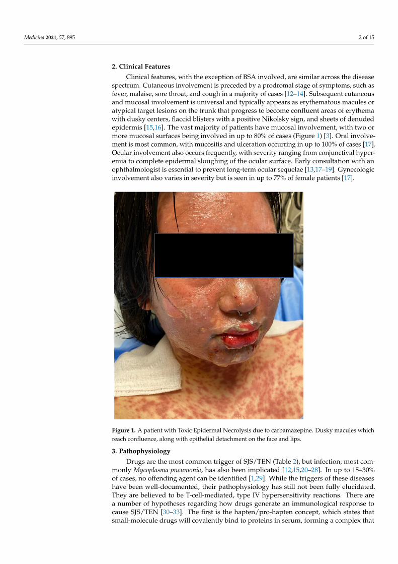
Clinical features, with the exception of BSA involved, are similar across the diseasespectrum. Cutaneous involvement is preceded by a prodromal stage of symptoms, such asfever, malaise, sore throat, and cough in a majority of cases [12–14]. Subsequent cutaneousand mucosal involvement is universal and typically appears as erythematous macules oratypical target lesions on the trunk that progress to become confluent areas of erythemawith dusky centers, flaccid blisters with a positive Nikolsky sign, and sheets of denudedepidermis [15,16]. The vast majority of patients have mucosal involvement, with two ormore mucosal surfaces being involved in up to 80% of cases (Figure 1) [3]. Oral involve-ment is most common, with mucositis and ulceration occurring in up to 100% of cases [17].Ocular involvement also occurs frequently, with severity ranging from conjunctival hyper-emia to complete epidermal sloughing of the ocular surface. Early consultation with anophthalmologist is essential to prevent long-term ocular sequelae [13,17–19]. Gynecologicinvolvement also varies in severity but is seen in up to 77% of female patients [17].
Medicina 2021, 57, 895 2 of 15
Table 1. Diagnosis of Stevens-Johnson Syndrome (SJS) and Toxic Epidermal Necrolysis (TEN) based on body surface area (BSA) (%) involvement.
Diagnosis Based on BSA (%) SJS <10%
SJS/TEN Overlap 10–30% TEN >30%
2. Clinical Features Clinical features, with the exception of BSA involved, are similar across the disease
spectrum. Cutaneous involvement is preceded by a prodromal stage of symptoms, such as fever, malaise, sore throat, and cough in a majority of cases [12–14]. Subsequent cuta-neous and mucosal involvement is universal and typically appears as erythematous mac-ules or atypical target lesions on the trunk that progress to become confluent areas of er-ythema with dusky centers, flaccid blisters with a positive Nikolsky sign, and sheets of denuded epidermis [15,16]. The vast majority of patients have mucosal involvement, with two or more mucosal surfaces being involved in up to 80% of cases (Figure 1) [3]. Oral involvement is most common, with mucositis and ulceration occurring in up to 100% of cases [17]. Ocular involvement also occurs frequently, with severity ranging from con-junctival hyperemia to complete epidermal sloughing of the ocular surface. Early consul-tation with an ophthalmologist is essential to prevent long-term ocular sequelae [13,17–19]. Gynecologic involvement also varies in severity but is seen in up to 77% of female patients [17].
Figure 1. A patient with toxic epidermal necrolysis due to carbamazepine. Dusky macules which reach confluence, along with epithelial detachment on the face and lips.
Figure 1. A patient with Toxic Epidermal Necrolysis due to carbamazepine. Dusky macules whichreach confluence, along with epithelial detachment on the face and lips.
3. Pathophysiology
Drugs are the most common trigger of SJS/TEN (Table 2), but infection, most com-monly Mycoplasma pneumonia, has also been implicated [12,15,20–28]. In up to 15–30%of cases, no offending agent can be identified [1,29]. While the triggers of these diseaseshave been well-documented, their pathophysiology has still not been fully elucidated.They are believed to be T-cell-mediated, type IV hypersensitivity reactions. There area number of hypotheses regarding how drugs generate an immunological response tocause SJS/TEN [30–33]. The first is the hapten/pro-hapten concept, which states thatsmall-molecule drugs will covalently bind to proteins in serum, forming a complex that
Medicina 2021, 57, 895 3 of 15
is recognized by certain HLA molecules and presented to T-cells to generate an immuneresponse. The next hypothesis, called the pharmacological interaction (p-i) concept, statesthat chemically inert drugs, which cannot undergo covalent binding with serum proteins,bind HLA molecules directly leading to T cell activation. The final hypothesis is the alteredpeptide concept, which states that drugs bind inside HLA binding pockets in a way thatalters presentation of self-proteins to T cells, such that they are no longer recognized asself, leading to an immune response [30–33]. Despite uncertainty regarding the exactmechanism, the end result is activation of T-cells in response to a drug or infection anddownstream epidermal necrosis.
Table 2. Common drugs implicated in the pathogenesis of SJS/TEN.
Common Drug Triggers of SJS/TEN
Anti-epileptics Antibiotics# Lamotrigine # TMP-SMX# Phenytoin # Aminopenicillins# Carbamazepine # Tetracyclines# Valproic Acid # Cephalosporins# Phenobarbital Immune Checkpoint InhibitorsNSAIDs # NivolumabAllopurinol # PembrolizumabNevirapine
Early hypotheses postulated that keratinocyte death was mediated by soluble Fasligand (sFasL) interactions with the Fas receptor on the surface of keratinocytes [34].Subsequent studies identified granulysin as a more important mediator of apoptosis.Chung et al. [35] analyzed the blister fluid of SJS/TEN patients and found that granulysinlevels were 2–4 times higher than perforin, granzyme B, and sFasL. Additionally, reducinggranulysin levels reduced cytotoxicity and injection of granulysin into the skin of miceinduced an SJS/TEN-like reaction [35]. Further studies confirmed the role of granulysin asa major mediator of the disease and showed that the levels of granulysin in blister fluidcorrelated with the severity of the disease [36–38]. While granulysin seems to be the maindriver of epidermal necrosis, it does not act alone. Su et al. [39] examined the serum levelsof 28 different cytokines and chemokines and found a number that were upregulated inpatients with SJS/TEN, of which granulysin and IL-15 correlated with the severity of thedisease. Additionally, the role of necroptosis, or programmed necrosis, has been examinedand was found to contribute to keratinocyte death, which could have important diagnosticimplications [30,40–42].
4. Differential Diagnosis
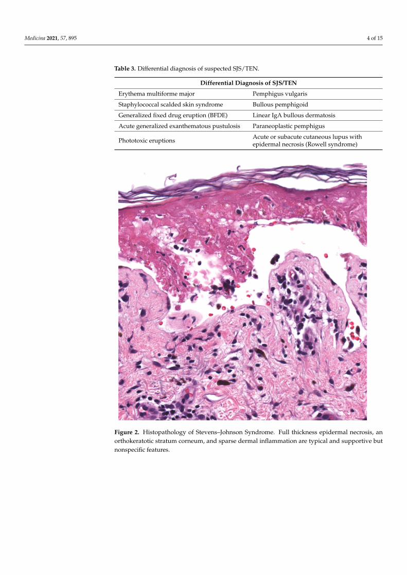
Prior to diagnosis of SJS/TEN, a broad differential diagnosis may be considered(Table 3). This includes other desquamating and vesiculobullous dermatoses, such aspemphigus vulgaris, linear IgA bullous dermatosis, staphylococcal scalded skin syndrome(SSSS), and erythema multiforme major (EMM). Importantly, EMM and SJS/TEN were his-torically classified as existing on the same disease spectrum, as the clinical and histopatho-logic presentation (Figure 2) of these diseases can be similar, but were later determined tobe distinct diseases [6,13–16]. Thus, the diagnosis must be made on clinical parameters.Key features of these disorders are outlined in Table 4 [1,6,13–16,43–45].
Medicina 2021, 57, 895 4 of 15
Table 3. Differential diagnosis of suspected SJS/TEN.
Differential Diagnosis of SJS/TEN
Erythema multiforme major Pemphigus vulgaris
Staphylococcal scalded skin syndrome Bullous pemphigoid
Generalized fixed drug eruption (BFDE) Linear IgA bullous dermatosis
Acute generalized exanthematous pustulosis Paraneoplastic pemphigus
Phototoxic eruptions Acute or subacute cutaneous lupus withepidermal necrosis (Rowell syndrome)
Medicina 2021, 57, 895 5 of 15
Figure 2. Histopathology of Stevens–Johnson syndrome. Full thickness epidermal necrosis, an or-thokeratotic stratum corneum, and sparse dermal inflammation are typical and supportive but non-specific features.
5. Management The management of SJS/TEN is multifaceted and begins with identification and ces-
sation of the causative agent [46]. A thorough history is important to identify the causative agent [47], as symptoms typically present within 8 weeks of beginning therapy, with most cases appearing between 4 days and 4 weeks of starting a drug [12]. If history is not suffi-cient to ascertain the causative drug, a number of causality assessment tools (CATs) can be useful. The Algorithm for Drug Causality for Epidermal Necrolysis (ALDEN) [29] and the Liverpool Adverse Drug Reaction CAT [48] are algorithms that have proven to be effective identifiers of causative drugs. The lymphocyte transformation test (LTT) is an in vitro test that can detect sensitization of T cells to antigens and can be helpful in identifi-cation of causative drugs in SJS/TEN, although at this time it is largely used for research purposes [45].
Prognostication is also an important step in the management of SJS/TEN, as it can guide management and placement in an intensive care or burn unit [49]. The severity-of-illness score for Toxic Epidermal Necrolysis (SCORTEN) scale is the most widely used tool for determining prognosis in patients with SJS/TEN. This has been verified as an ef-fective tool in a number of studies [50,51]. Other studies, however, have shown that SCORTEN may overestimate actual mortality rates [52,53]. However, this discordance may potentially be attributed to improvements in supportive care since the development of SCORTEN in 1979 [51]. Noe et al. [54] developed an alternative prognostic algorithm
Figure 2. Histopathology of Stevens–Johnson Syndrome. Full thickness epidermal necrosis, anorthokeratotic stratum corneum, and sparse dermal inflammation are typical and supportive butnonspecific features.
Medicina 2021, 57, 895 5 of 15
Table 4. Distinguishing characteristics of SJS/TEN and EM.
SJS/TEN vs. EM
SJS/TEN EM
Characteristic Lesions
Atypical target lesions: macules with centralclearing and 2 poorly demarcated components Typical target lesions: papules with a dark center
and 3 well-demarcated, concentric componentsLarge sheets of painful desquamation inlater lesions
Distribution Typically begins on the face and trunk withcentrifugal spread Face and acral skin, rare involvement of trunk
Triggers Drugs (see Table 2) Infection (most commonly HSV andM. pneumonia)
Mucosal Involvement Very common—most cases have involvement of≥2 mucosal surfaces
Rare—typically only one mucosal surfaceinvolved if present
Recurrence Rarely seen with removal and avoidance ofcausative drug Frequently seen
Histopathology(Figure 2)
Early StageBasal layer liquefaction with vacuolar interface changes, scattered necrotic keratinocytes, andinterface lymphocytes
Late Stage *Subepidermal split with full-thickness epidermal necrosis
* Biopsy in the late stages of SJS/TEN may show comparatively little inflammation compared to EM.
5. Management
The management of SJS/TEN is multifaceted and begins with identification andcessation of the causative agent [46]. A thorough history is important to identify thecausative agent [47], as symptoms typically present within 8 weeks of beginning therapy,with most cases appearing between 4 days and 4 weeks of starting a drug [12]. If history isnot sufficient to ascertain the causative drug, a number of causality assessment tools (CATs)can be useful. The Algorithm for Drug Causality for Epidermal Necrolysis (ALDEN) [29]and the Liverpool Adverse Drug Reaction CAT [48] are algorithms that have proven tobe effective identifiers of causative drugs. The lymphocyte transformation test (LTT) isan in vitro test that can detect sensitization of T cells to antigens and can be helpful inidentification of causative drugs in SJS/TEN, although at this time it is largely used forresearch purposes [45].
Prognostication is also an important step in the management of SJS/TEN, as it canguide management and placement in an intensive care or burn unit [49]. The severity-of-illness score for Toxic Epidermal Necrolysis (SCORTEN) scale is the most widely usedtool for determining prognosis in patients with SJS/TEN. This has been verified as aneffective tool in a number of studies [50,51]. Other studies, however, have shown thatSCORTEN may overestimate actual mortality rates [52,53]. However, this discordance maypotentially be attributed to improvements in supportive care since the development ofSCORTEN in 1979 [51]. Noe et al. [54] developed an alternative prognostic algorithm calledABCD-10. This scoring system uses prior dialysis as a proxy for severe renal dysfunction,distinguishing it from SCORTEN. Both scoring systems seem to be reliable predictors ofmortality, but one study [55] showed that SCORTEN was more accurate. The SCORTENand ABCD-10 scoring systems and predicted mortality are outlined in Tables 5 and 6.One important note for both scoring systems is how to determine the BSA involved, asan accurate estimate is critical for classification and prognostication. Creamer et al. [56]described that BSA involved includes both epidermis that is detachable (positive Nikolskysign) and epidermis that is already detached. Areas of erythema without evidence ofdetachment or impending detachment are not included.
Medicina 2021, 57, 895 6 of 15
Table 5. SCORTEN and ABCD-10 Scoring Systems.
SCORTEN ABCD-10Parameter Weight Parameter Weight
Age ≥ 40 years 1 Age ≥ 50 years 1Malignancy—Yes 1 Serum Bicarbonate < 20 mmol/L 1BSA detached > 10% 1 Active Cancer—Yes 2Serum bicarbonate < 20 mmol/L 1 Dialysis prior to admission—Yes 3Serum urea nitrogen > 28 mg/dL 1
BSA Involvement > 10% 1Serum glucose > 252 mg/dL 1Tachycardia ≥ 120 bpm 1Maximum score possible 7 8
Table 6. Estimated mortality in patients with SJS/TEN.
Estimated Mortality in Patients with SJS/TEN
SCORTEN Score Estimated Mortality(%) ABCD-10 Score Estimated Mortality
(%)0–1 3.2 0 2.3
2 12.1 1 5.43 35.3 2 12.34 58.3 3 25.5
>5 >904 45.75 67.4
>6 83.6
Removal of the offending agent and supportive care are the mainstays in treatment ofSJS/TEN [57]. Adjunctive therapies, such as corticosteroids and intravenous immunoglob-ulin (IVIg), are often utilized, although there is still no consensus on the most effectiveadjunctive therapy. The goal of this article is to review the most recent updates in bothdiagnosis and management of SJS/TEN in order to educate dermatologists and otherphysicians who are managing the acute care of patients with SJS/TEN.
6. Materials and Methods
A database search of PubMed and Embase was performed, initially focusing on reviewarticles in the past 5 years, from March 2017 through March 2021, with keywords “Stevens–Johnson Syndrome”, “Toxic Epidermal Necrolysis”, “therapy”, “diagnosis”, “manage-ment”, and synonyms of all these key words. The reference section of each of the reviewarticles was also reviewed to find other articles that contained pertinent information.
7. Clinical Updates7.1. Updates on Diagnosis7.1.1. Potential Biomarkers
Rapid diagnosis of SJS/TEN is critical in order to discontinue the offending agent,begin supportive and adjunctive therapies, and improve outcomes. However, the clinicalpresentation can be similar to a number of other blistering disorders, and diagnosis is notalways straightforward. Given that the diagnosis of SJS/TEN is time sensitive, frozensections can be utilized for more rapid decision making. SJS/TEN can be distinguishedfrom SSSS by the level of epidermal detachment, which is subcorneal in SSSS and at thedermo-epidermal junction in SJS/TEN. Widespread keratinocytic necrosis is characteristicof SJS/TEN on histopathology [43,58]. The distinction between SJS/TEN and EMM isdifficult to make because their histopathology can be identical [13,16]. In the early stage ofboth diseases, a vacuolar or lichenoid interface with scattered necrotic keratinocytes can beseen. As both diseases progress, a subepidermal split with increased epidermal necrosisis observed. In these cases, a heavier lymphocytic infiltrate favors EM while increasedeosinophils and confluent epidermal necrosis favors SJS/TEN. However, these are notreliable distinguishing features and clinicopathologic correlation is required.

Medicina 2021, 57, 895 7 of 15
There are a number of studies that have investigated potential diagnostic markers(Table 7) of the disease, with early studies focusing on the role of granulysin. Abe et al. [37]analyzed the serum of 5 patients with SJS/TEN and found elevated levels of granulysinin 4 out of 5 patients, even before cutaneous detachment and mucosal involvement. Serafrom thirty-one control patients were also analyzed and showed no elevations in serumgranulysin levels. Chen et al. [36] found that granulysin levels in blister fluid were markedlyelevated and correlated with disease severity in SJS/TEN. However, these findings wereconsistent across all cytotoxic T-lymphocyte (CTL)-mediated bullous blistering disorders,such as EMM and bullous fixed drug eruption (BFDE). Elevated serum granulysin levelswere also observed in patients with drug reaction with eosinophilia and systemic symptoms(DRESS) [38]. Therefore, while granulysin is elevated in both serum and blister fluid, it isnot a specific finding for SJS/TEN and has limited utility in early diagnosis.
Table 7. Potential Biomarkers for the Diagnosis of SJS/TEN
Common Drug Triggers of SJS/TENNon-Specific for SJS/TEN Specific for SJS/TEN# Granulysin# CCL-27
# Galectin-7# RIP3
CCL-27 is another nonspecific cytokine that is likely involved in the pathogenesis ofSJS/TEN and aids in the trafficking of T cells to the skin at sites of inflammation [30]. Tapiaet al. [59] reported that CCL-27 levels were elevated in skin from patients with SJS/TENduring the acute phase. Wang et al. [60] then analyzed the levels of CCL-27 in sera from27 patients with SJS/TEN and found elevations during the acute phase compared with39 healthy controls. This implicates CCL-27 in the pathogenesis of SJS/TEN, but elevatedCCL27 levels were also identified in non-bullous drug-induced exanthems. Therefore, theuse of CCL-27 in diagnosis is limited in the same manner as granulysin, due to the lack ofspecificity.
There are a number of other potential biomarkers under investigation that may demon-strate specificity for SJS/TEN. In one study [61], peripheral blood mononuclear cells(PBMCs) from patients who had recovered from SJS/TEN were cultured and re-exposed tothe causative drug. The supernatant of the culture fluid was analyzed using proteomicsto identify potential biomarkers. This protocol was also used to evaluate the moleculessecreted by PBMCs in non-severe cutaneous adverse drug reactions (cADRs). When com-paring the two groups, Hama et al. [61] discovered one protein, galectin-7, exhibited higherlevels in sera of SJS/TEN patients than in sera of non-severe cADRs (p = 0.005). Serumgalectin-7 also correlated with disease severity with significantly higher levels during theacute phase and decreased levels in the late phase of the disease (>7 days). Galectin-7 couldbe a potential mediator of SJS/TEN and a helpful biomarker for diagnosis.
The role of necroptosis, or programmed necrosis, in SJS/TEN has been the focusof multiple studies. Necroptosis differs from apoptosis in that cell death is the result ofexternal triggers that alter membrane permeability and result in cell lysis without theinvolvement of caspases. Recent studies have identified receptor-interacting kinase-3(RIP3) as an important mediator [41,62]. Hasegawa et al. [41] confirmed that necroptotickeratinocytes release RIP3 into the sera of patients, and its levels correlated with the degreeof necroptosis and severity of disease. Notably, the investigators also measured RIP3 levelsin the sera of patients with EMM and found significantly higher levels in patients withSJS/TEN than EMM (p < 0.001). The use of serum RIP3 as a biomarker for the diagnosis ofSJS/TEN could help distinguish between SJS/TEN and EMM.
7.1.2. Diagnostic Subclassification in Pediatric Patients
The diagnostic classification for pediatric patients has been recently updated. Canavanet al. [63] performed a systematic review of 202 patients with an SJS/TEN-like reaction toMycoplasma pneumoniae infection. They noted that these patients had impressive mucosal
Medicina 2021, 57, 895 8 of 15
involvement, but the cutaneous involvement was less significant and the prognosis morefavorable compared with SJS/TEN. Canavan et al. named this dermatosis Mycoplasmapneumoniae-induced rash and mucositis (MIRM) and classified it as distinct from SJS/TENand EM. Subsequently, multiple other studies implicated other infections as causes ofMIRM-like reactions including adenovirus [64], influenza B [65], and Chlamydia pneumo-niae [66].
In light of these findings, Ramien et al. [67] proposed a new classification for blis-tering disorders in pediatric patients. In this new system, SJS, SJS/TEN, and TEN arecondensed into a single disorder called drug-induced epidermal necrolysis (DEN). Theinfection-related cases with severe mucosal involvement and relatively sparse cutaneousinvolvement were considered a distinct clinical identity and termed reactive infectiousmucocutaneous eruption (RIME). Erythema multiforme (EM) was classified as a distinctdisease from DEN and RIME [68]. This new classification is worthwhile because thetreatment of DEN and RIME differ. In DEN, identification and cessation of the causativedrug with supportive care and possible immunosuppressive therapy are the pillars oftreatment. RIME requires identification and treatment of the underlying infection, sup-portive care, and potential antimicrobial and immunosuppressive therapies. In fact, theuse of antibiotics to treat community acquired pneumonia in patients with RIME has beenemphasized [69,70]. One study examined the role of etanercept treatment in RIME andshowed that this therapy led to improvement in physical findings within 2 days of drugadministration [71]. However, this study is limited by its small sample size (n = 6) andtreatment with antibiotics in 5/6 patients prior to administration of etanercept, whichcould also have contributed to the observed improvement. Further studies are required toclarify proper treatment strategies for both DEN and RIME.
7.2. Updates on Management7.2.1. Non-Pharmacologic Treatment
Supportive care is the mainstay of treatment for patients with SJS/TEN and includescessation of the causative drug, fluid and electrolyte management, infection control, andwound care. Of these components, identification and cessation of the causative drug ismost important [46,49,72], but optimization of each measure is necessary to achieve thebest outcomes.
Fluid, electrolyte, and nutrition management is important in SJS/TEN patients andmirrors the requirements of burn patients due to insensible losses through wounds, al-though fluid requirements are about 30% less in SJS/TEN patients than in burn patientswith similar degrees of cutaneous involvement [73,74]. The environment should be keptwarm (30–32 ◦C) [49] due to loss of thermoregulatory function of skin, and fluid replace-ment should be driven by urine output, with a goal of 0.5–1 mL/kg/h [73]. Enteral feedingshould be initiated as early as possible and through nasogastric tube feeds if necessary [49].
Prophylactic antibiotics do not improve outcomes [75], but proper wound care andsterile handling are important in preventing infection. The role of surgical debridement hasbeen controversial, and the decision to pursue this treatment option largely depends onwhere the care is being delivered. McCullough et al. [57] described a series of 40 SJS/TENpatients who were treated with their treatment algorithm, which included aggressivesupportive care, surgical wound debridement with subsequent coverage with antimicrobialdressings, steroid cessation (if the patient was receiving steroids upon transfer), and IVIg.The authors of this study reported a 10% mortality rate, which was lower than the 16.7%mortality rate predicted by SCORTEN. While this result reflects an effective combinationof treatments, it is difficult to identify surgical debridement as the cause of that efficacy.Dorafshar et al. [76] analyzed the efficacy of “anti-shear” therapy, in which blister fluidis aspirated and denuded epidermis is left in place to act as a biological skin graft. Theauthors described 48 patients at their care center who received this treatment and presenteda mortality reduction of 11 percent compared to the expected mortality predicted bySCORTEN. Anti-shear therapy is an alternative to surgical debridement and could reduce

Medicina 2021, 57, 895 9 of 15
hospital costs as well as pain. However, there is a lack of high-quality evidence to guidedecision-making regarding surgical debridement [77], and further studies are needed tofully understand the role of this therapy.
7.2.2. Pharmacologic Treatment
Due to the rarity of the disease, few prospective studies have analyzed the efficacy ofspecific adjunctive therapies for SJS/TEN. As a result, there is no established standard ofcare pertaining to pharmacologic treatment. Due to the immunologic nature of the disease,it is believed that immunosuppressive therapies will aid in treatment, and many casereports have reported positive results with varying treatment regimens involving differentcombinations of corticosteroids, IVIg, cyclosporine, and TNF-alpha inhibitors [21,78–81].However, it is difficult to determine if disease remission was due to specific treatment orsimply the natural history of the disease. Several systematic reviews and meta-analyseshave attempted to overcome these methodologic limitations and clarify the role of pharma-cologic therapies in the treatment of SJS/TEN.
The role of corticosteroids as monotherapy is still debated [82]. Recently, Zimmermannet al. [83] performed a meta-analysis of 11 studies to compare the use of corticosteroids ver-sus supportive therapy and found a positive, although statistically insignificant (OR, 0.54;95% CI, 0.29–1.01), treatment effect. Other studies have shown no improvement in mortalitywith the use of corticosteroids alone [10,30]. The role of IVIg has also been controversial,and there appears to be no mortality benefit associated with monotherapy [56,58,84,85].
Despite uncertainty in the results of these therapies, there are a number of othertreatment options that show promise. Cyclosporine has shown positive results in a numberof studies to this point [83,86–89]. Ng et al. [88] performed a meta-analysis of 10 studiesand reported on the standardized mortality ratio (SMR) of cyclosporine compared withsupportive care. The SMR takes into account baseline severity of the disease, allowing for amore accurate depiction of mortality improvement as compared to mortality ratios (MR). Inthis study, the authors reported an SMR of 0.320 (95% CI, 0.119–0.522, p = 0.002), indicatinga survival benefit in patients treated with cyclosporine. Chen et al. [89] performed a meta-analysis of 7 studies and reported similarly positive results with an SMR of 0.42 (95% CI,0.19–0.95) when cyclosporine was administered.
In another study, Tsai et al. [90] analyzed treatment outcomes of a number of therapiesand performed a meta-analysis of 67 studies involving 2079 patients. The authors onlyexamined the mortality outcomes of patients with SJS/TEN overlap and TEN, choosingto exclude SJS outcomes because mortality is typically lower. The only therapy thatshowed statistically significant improvements in outcomes was the combination of IVIgand corticosteroids, with an SMR of 0.53 (95% CI, 0.31–0.93). Historically, IVIg has beenused as a monotherapy [91], but it only appears to be effective when combined withcorticosteroids. These authors also reported promising results for cyclosporine (with orwithout IVIg), IVIg and plasmapheresis, and etanercept, although they emphasized theneed for further studies.
Han et al. [92] performed a prospective observational study of 28 patients withSJS/TEN overlap or TEN, 13 of whom received plasmapheresis and 15 of whom didnot. Of the 13 that received plasmapheresis, 7 were also treated with concomitant corticos-teroids or IVIg. Using a severity of illness score that evaluated mucosal lesions, cutaneouslesions, and overall general condition (scores 0–39), it was shown that patients who re-ceived plasmapheresis had a lower severity of illness scores later in the disease course(days 7, 10, and 20).
TNF-alpha inhibitors are also of interest due to their immunosuppressive effects.Zhang et al. [93] reviewed 21 case reports, 4 case series, and 2 randomized controlled trials(RCTs) that analyzed the use of TNF-alpha inhibitors and reported positive outcomes in86.8% of patients. One of these RCTs [94] included 91 patients and showed improvement inmortality. The observed mortality rate of 8.3% was lower than that predicted by SCORTEN

Medicina 2021, 57, 895 10 of 15
(17.7%) and lower than the mortality associated with corticosteroid treatment (16.3%),although these results were not statistically significant.
Given the lack of consensus on the most effective pharmacological treatment forSJS/TEN, practical issues such as cost must also be taken into account when determiningtreatment course. Tables 8 and 9 outline practical considerations for these drugs, includingdosing regimens and costs.
Table 8. Typical dosing regimens to treat SJS/TEN for selected drugs
Dosing Regimen for SJS/TEN of Selected Drugs
IVIg 3 g/kg, divided over 3 days [90]
TNF-alpha inhibitors - Infliximab: 5 mg/kg as a single dose [92]- Etanercept: Single 50 mg dose [92]
Cyclosporine 2.5–5 mg/kg/day for 7–10 days, followed by gradual taper [87,88]
Corticosteroids Prednisone 0.5–1 mg/kg/day or pulse methylprednisolone1 mg/kg/d for 3 days [81]
Table 9. Relative cost of selected drugs at a single academic center
Relative Cost of Selected Drugs **
IVIG $1932 for a treatment course *
Etanercept $1386 for a single 50 mg subcutaneous dose
Infliximab $4900 *
Cyclosporine ~$336 for a 3-week course/taper at $16 per day
Prednisone <$20 for 2–3-week taper at $1 per day* Assuming a 70 kg individual. ** Cost and access may vary by medical center.
8. Discussion
SJS/TEN is a dermatologic emergency that causes significant morbidity and mortal-ity. Early in the disease course, there is a broad differential diagnosis that needs to beconsidered, and prompt diagnosis is critical in achieving optimal outcomes. For most ofthe potential differential diagnoses, clinical morphology and histopathology can readilydistinguish them from SJS/TEN. However, EMM is a disease that has identical histopatho-logical features to SJS/TEN, and confident distinction between these two disorders requiresexperience and more careful correlation. SJS/TEN has significantly higher mortality andmorbidity rates than those of EMM, often necessitating surgical or medical therapy beyondsupportive care alone. A number of serological tests show promise in expanding the abilityto diagnose SJS/TEN. Granulysin and CCL-27 serum markers are elevated in patients withSJS/TEN and can be helpful markers to monitor disease severity. However, these markersare not specific for SJS/TEN and are elevated in other disorders, limiting their specificity.Both galectin-7 and RIP3 play a pathogenic role and are elevated to a greater degree inthe sera of patients with SJS/TEN compared to other cADRs. Further research is requiredbefore these markers can be reliably used for diagnosis.
Once diagnosed, the management of SJS/TEN focuses primarily on supportive careand wound management with the addition of adjunctive medications. The role of surgicaldebridement has been debated, and evidence has shown that both surgical debridementand anti-shear therapy improve patient outcomes. Wound management and infectionprevention improve outcomes, as both therapies are effective. Corticosteroids, IVIg, cy-closporine, TNF-alpha inhibitors, and plasmapheresis are therapeutic options. Variableresults have been described, and there is still no consensus on the treatment of choice. FewRCTs have been conducted, and most of the research published has been in the form ofcase reports, case studies, and systematic reviews and meta-analyses. The most efficacioustreatments appear to be cyclosporine and a combination of corticosteroids with IVIg. Both

Medicina 2021, 57, 895 11 of 15
have shown statistically significant improvements to patient mortality. It is also importantto consider cost effectiveness when selecting therapies. Of the drugs described in thetreatment of SJS/TEN, the most expensive is infliximab, followed by IVIg and etaner-cept. The least expensive options are cyclosporine and corticosteroids. Ultimately, furtherprospective studies are required to solidify treatment guidelines.
Author Contributions: Conceptualization, K.M., R.F. and S.H.; writing-original draft preparation,R.F. and S.H.; writing-review and editing, K.M. and A.A.; supervision, K.M. All authors have readand agreed to the final version of the published manuscript.
Funding: This research received no external funding.
Institutional Review Board Statement: Not applicable.
Informed Consent Statement: Not applicable.
Conflicts of Interest: The authors declare no conflict of interest.
References1. Duong, T.A.; Valeyrie-Allanore, L.; Wolkenstein, P.; Chosidow, O. Severe cutaneous adverse reactions to drugs. Lancet 2017, 390,
1996–2011. [CrossRef]2. Frey, N.; Jossi, J.; Bodmer, M.; Bircher, A.; Jick, S.; Meier, C.R.; Spoendlin, J. The Epidemiology of Stevens-Johnson Syndrome and
Toxic Epidermal Necrolysis in the UK. J. Investig. Dermatol. 2017, 137, 1240–1247. [CrossRef]3. Hsu, D.; Brieva, J.; Silverberg, N.B.; Silverberg, J. Morbidity and Mortality of Stevens-Johnson Syndrome and Toxic Epidermal
Necrolysis in United States Adults. J. Investig. Dermatol. 2016, 136, 1387–1397. [CrossRef]4. Yang, M.-S.; Lee, J.Y.; Kim, J.; Kim, G.-W.; Kim, B.-K.; Kim, J.-Y.; Park, H.-W.; Cho, S.-H.; Min, K.-U.; Kang, H.-R. Incidence of
Stevens-Johnson Syndrome and Toxic Epidermal Necrolysis: A Nationwide Population-Based Study Using National HealthInsurance Database in Korea. PLoS ONE 2016, 11, e0165933. [CrossRef]
5. Hsu, D.Y.; Brieva, J.; Silverberg, N.B.; Paller, A.S.; Silverberg, J.I. Pediatric Stevens-Johnson syndrome and toxic epidermalnecroly-sis in the United States. J. Am. Acad. Dermatol. 2017, 76, 811–817.e4. [CrossRef] [PubMed]
6. Paulmann, M.; Mockenhaupt, M. Severe skin reactions: Clinical picture, epidemiology, etiology, pathogenesis, and treatment.Allergo J. Int. 2019, 28, 311–326. [CrossRef]
7. Lim, V.M.; Do, A.; Berger, T.G.; Nguyen, A.H.; Deweese, J.; Malone, J.D.; Jordan, K.; Hom, F.; Tuffanelli, L.; Fillari, P.; et al. Adecade of burn unit experience with Stevens-Johnson Syndrome/Toxic Epidermal Necrolysis: Clinical pathological diagnosisand risk factor awareness. Burns 2016, 42, 836–843. [CrossRef] [PubMed]
8. Lissia, M.; Mulas, P.; Bulla, A.; Rubino, C. Toxic epidermal necrolysis (Lyell’s disease). Burns 2010, 36, 152–163. [CrossRef][PubMed]
9. Richer, V.; Bouffard, D.; Belisle, A.; Duranceau, L.; Perreault, I.; Provost, N. Acute blistering diseases on the burn ward: BeyondStevens–Johnson Syndrome and Toxic Epidermal Necrolysis. Burns 2013, 39, 1290–1296. [CrossRef]
10. Sekula, P.; Dunant, A.; Mockenhaupt, M.; Naldi, L.; Bavinck, J.N.B.; Halevy, S.; Kardaun, S.; Sidoroff, A.; Liss, Y.; Schumacher, M.;et al. Comprehensive Survival Analysis of a Cohort of Patients with Stevens–Johnson Syndrome and Toxic Epidermal Necrolysis.J. Investig. Dermatol. 2013, 133, 1197–1204. [CrossRef] [PubMed]
11. Paggiaro, A.O.; Silva, E.; Filho, M.L.; de Carvalho, V.F.; Isaac, C.; Gemperli, R. The Role of Biological Skin Substitutes inSte-vens-Johnson Syndrome: Systematic Review. Plast. Surg. Nurs. Off. J. Am. Soc. Plast. Reconstr. Surg. Nurses 2018, 38, 121–127.
12. Charlton, O.A.; Harris, V.; Phan, K.; Mewton, E.; Jackson, C.; Cooper, A. Toxic Epidermal Necrolysis and Steven-JohnsonSyn-drome: A Comprehensive Review. Adv. Wound Care 2020, 9, 426–439. [CrossRef]
13. Guvenir, H.; Arikoglu, T.; Vezir, E.; Misirlioglu, E.D. Clinical Phenotypes of Severe Cutaneous Drug Hypersensitivity Reactions.Curr. Pharm. Des. 2019, 25, 3840–3854. [CrossRef] [PubMed]
14. Paulmann, M.; Mockenhaupt, M. Fever in Stevens–Johnson Syndrome and Toxic Epidermal Necrolysis in Pediatric Cases. Pediatr.Infect. Dis. J. 2017, 36, 513–515. [CrossRef] [PubMed]
15. Alerhand, S.; Cassella, C.; Koyfman, A. Stevens-Johnson Syndrome and Toxic Epidermal Necrolysis in the Pediatric Popula-tion:A Review. Pediatric Emerg. Care 2016, 32, 472–478. [CrossRef] [PubMed]
16. Grünwald, P.; Mockenhaupt, M.; Panzer, R.; Emmert, S. Erythema multiforme, Stevens-Johnson syndrome/Toxic EpidermalNecrolysis—Diagnosis and treatment. JDDG J. Ger. Soc. Dermatol. 2020, 18, 547–553. [CrossRef]
17. Shanbhag, S.; Chodosh, J.; Fathy, C.; Goverman, J.; Mitchell, C.; Saeed, H.N. Multidisciplinary care in Stevens-Johnson syndrome.Ther. Adv. Chronic Dis. 2020, 11. [CrossRef] [PubMed]
18. Basu, S.; Shanbhag, S.S.; Gokani, A.; Kedar, R.; Bahuguna, C.; Sangwan, V.S. Chronic Ocular Sequelae of Stevens-JohnsonSyndrome in Children: Long-term Impact of Appropriate Therapy on Natural History of Disease. Am. J. Ophthalmol. 2018, 189,17–28. [CrossRef]
19. Gregory, D.G. New Grading System and Treatment Guidelines for the Acute Ocular Manifestations of Stevens-Johnson Syn-drome.Ophthalmology 2016, 123, 1653–1658. [CrossRef]

Medicina 2021, 57, 895 12 of 15
20. Aghdam, M.K.; Ahmadiafshar, A.; Eftekhari, K. Toxic epidermal necrolysis syndrome following single-dose diclofenac suppos-itory, a case report. J. Compr. Pediatrics 2020, 11, e100496.
21. Alajmi, A.; Jfri, A.; Gomolin, A.; Jafarian, F. A pediatric case of Stevens-Johnson syndrome/Toxic Epidermal Necrolysis withrap-id response to intravenous cyclosporine. JAAD Case Rep. 2020, 6, 555–557. [CrossRef] [PubMed]
22. Maloney, N.J.; Ravi, V.; Cheng, K.; Bach, D.Q.; Worswick, S. Stevens-Johnson syndrome and Toxic Epidermal Necrolysis-likereac-tions to checkpoint inhibitors: A systematic review. Int. J. Dermatol. 2020, 59, e183–e188. [CrossRef]
23. Simonsen, A.B.; Kaae, J.; Ellebaek, E.; Svane, I.M.; Zachariae, C. Cutaneous adverse reactions to anti–PD-1 treatment—Asystematic review. J. Am. Acad. Dermatol. 2020, 83, 1415–1424. [CrossRef]
24. Madabhavi, I.; Revannasiddaiah, S.; Patel, A.; Anand, A. Toxic epidermal necrolysis with the use of tamoxifen. BMJ Case Rep.2015, 2015, bcr2014209102. [CrossRef] [PubMed]
25. Mani, R.; Monteleone, C.; Schalock, P.C.; Truong, T.; Zhang, X.B.; Wagner, M.L. Rashes and other hypersensitivity reactionsassoci-ated with antiepileptic drugs: A review of current literature. Seizure 2019, 71, 270–278. [CrossRef] [PubMed]
26. Rashid, M.; Kashyap, A.; Undela, K. Valproic acid and Stevens-Johnson syndrome: A systematic review of descriptive studies.Int. J. Dermatol. 2019, 58, 1014–1022. [CrossRef] [PubMed]
27. Gupta, S.S.; Sabharwal, N.; Patti, R.; Kupfer, Y. Allopurinol-Induced Stevens-Johnson Syndrome. Am. J. Med. Sci. 2019, 357,348–351. [CrossRef] [PubMed]
28. Sibbald, C.; Putterman, E.; Micheletti, R.; Treat, J.; Castelo-Soccio, L. Retrospective review of drug-induced Stevens-Johnsonsyn-drome and Toxic Epidermal Necrolysis cases at a pediatric tertiary care institution. Pediatric Dermatol. 2020, 37, 461–466.[CrossRef]
29. Sassolas, B.; Haddad, C.; Mockenhaupt, M.; Dunant, A.; Liss, Y.; Bork, K.; Haustein, U.F.; Vieluf, D.; Roujeau, J.C.; Le Louet, H.;et al. ALDEN, an algorithm for assessment of drug causal-ity in stevens-johnson syndrome and Toxic Epidermal Necrolysis:Comparison with case-control analysis. Clin. Pharmacol. Ther. 2010, 88, 60–68. [CrossRef] [PubMed]
30. Hasegawa, A.; Abe, R. Recent advances in managing and understanding Stevens-Johnson syndrome and Toxic EpidermalNecrolysis. F1000Research 2020, 9, 612. [CrossRef] [PubMed]
31. Abe, R. Immunological response in Stevens-Johnson syndrome and Toxic Epidermal Necrolysis. J. Dermatol. 2015, 42, 42–48.[CrossRef]
32. Adam, J.; Pichler, W.J.; Yerly, D. Delayed drug hypersensitivity: Models of T-cell stimulation. Br. J. Clin. Pharmacol. 2010, 71,701–707. [CrossRef] [PubMed]
33. Pichler, W.J. Modes of presentation of chemical neoantigens to the immune system. Toxicology 2002, 181–182, 49–54. [CrossRef]34. Abe, R.; Shimizu, T.; Shibaki, A.; Nakamura, H.; Watanabe, H.; Shimizu, H. Toxic epidermal necrolysis and Stevens-Johnson
syn-drome are induced by soluble fas ligand. Am. J. Pathol. 2003, 162, 1515–1520. [CrossRef]35. Chung, W.-H.; Hung, S.-I.; Yang, J.-Y.; Su, S.-C.; Huang, S.-P.; Wei, C.-Y.; Chin, S.-W.; Chiou, C.-C.; Chu, S.-C.; Ho, H.-C.; et al.
Granulysin is a key mediator for disseminated keratinocyte death in Stevens-Johnson syndrome and Toxic Epidermal Necrolysis.Nat. Med. 2008, 14, 1343–1350. [CrossRef] [PubMed]
36. Chen, C.B.; Kuo, K.L.; Wang, C.W.; Lu, C.W.; Chung-Yee, H.R.; Lu, K.L.; Chang, W.C.; Chen, W.T.; Yun, F.; Teng, Y.C.; et al.Detecting Lesional Granulysin Levels for Rapid Di-agnosis of Cytotoxic T lymphocyte–Mediated Bullous Skin Disorders. J.Allergy Clin. Immunol. Pract. 2020, 9, 1327–1337. [CrossRef] [PubMed]
37. Abe, R.; Yoshioka, N.; Murata, J.; Fujita, Y.; Shimizu, H. Granulysin as a marker for early diagnosis of the Stevens-Johnsonsyn-drome. Ann. Intern. Med. 2009, 151, 514–515. [CrossRef] [PubMed]
38. Saito, N.; Abe, R.; Yoshioka, N.; Murata, J.; Fujita, Y.; Shimizu, H. Prolonged elevation of serum granulysin in drug-inducedhy-persensitivity syndrome. Br. J. Dermatol. 2012, 167, 452–453. [CrossRef] [PubMed]
39. Su, S.-C.; Mockenhaupt, M.; Wolkenstein, P.; Dunant, A.; le Gouvello, S.; Chen, C.-B.; Chosidow, O.; Valeyrie-Allanore, L.; Bellon,T.; Sekula, P.; et al. Interleukin-15 Is Associated with Severity and Mortality in Stevens-Johnson Syndrome/Toxic EpidermalNecrolysis. J. Investig. Dermatol. 2017, 137, 1065–1073. [CrossRef] [PubMed]
40. Hasegawa, A.; Shinkuma, S.; Hayashi, R.; Hama, N.; Watanabe, H.; Kinoshita, M.; Ogawa, Y.; Abe, R. 019 Serum RIP3 level as aseveri-ty-predictive marker for Stevens-Johnson syndrome and Toxic Epidermal Necrolysis. J. Investig. Dermatol. 2019, 139, S4.[CrossRef]
41. Hasegawa, A.; Shinkuma, S.; Hayashi, R.; Hama, N.; Watanabe, H.; Kinoshita, M.; Ogawa, Y.; Abe, R. RIP3 as a diagnosticand severity marker for Stevens-Johnson syndrome and Toxic Epidermal Necrolysis. J. Allergy Clin. Immunol. Pract. 2020, 8,1768–1771.e7. [CrossRef]
42. Saito, N.; Qiao, H.; Yanagi, T.; Shinkuma, S.; Nishimura, K.; Suto, A.; Fujita, Y.; Suzuki, S.; Nomura, T.; Nakamura, H.; et al. Anannexin A1-FPR1 interaction contributes to necrop-tosis of keratinocytes in severe cutaneous adverse drug reactions. Sci. Transl.Med. 2014, 6, 245ra95. [CrossRef] [PubMed]
43. Abate, M.S.; Battle, L.R.; Emerson, A.N.; Gardner, J.M.; Shalin, S.C. Dermatologic Urgencies and Emergencies: What EveryPathologist Should Know. Arch. Pathol. Lab. Med. 2019, 143, 919–942. [CrossRef]
44. Kerl, K.; Kerl, H. Severe cutaneous adverse drug reactions. Diagn. Histopathol. 2020, 27, 1–5. [CrossRef]45. Lin, C.-C.; Chen, C.-B.; Wang, C.-W.; Hung, S.-I.; Chung, W.-H. Stevens-Johnson syndrome and Toxic Epidermal Necrolysis:
Risk factors, causality assessment and potential prevention strategies. Expert Rev. Clin. Immunol. 2020, 16, 373–387. [CrossRef][PubMed]

Medicina 2021, 57, 895 13 of 15
46. Garcia-Doval, I.; LeCleach, L.; Bocquet, H.; Otero, X.L.; Roujeau, J.C. Toxic epidermal necrolysis and Stevens-Johnson syndrome:Does early withdrawal of causative drugs decrease the risk of death? Arch. Dermatol. 2000, 136, 323–327. [CrossRef] [PubMed]
47. Cavkaytar, O.; Kuyucu, S. An Update on the Management of Severe Cutaneous Drug Hypersensitivity Reactions. Curr. Pharm.Des. 2019, 25, 3881–3901. [CrossRef]
48. Gallagher, R.M.; Kirkham, J.J.; Mason, J.R.; Bird, K.A.; Williamson, P.R.; Nunn, A.J.; Turner, M.A.; Smyth, R.L.; Pirmohamed,M. Development and inter-rater reliability of the Liverpool adverse drug reaction causality assessment tool. PLoS ONE 2011, 6,e28096. [CrossRef] [PubMed]
49. Coias, J.L.; Abbas, L.F.; Cardones, A.R. Management of Stevens-Johnson Syndrome/Toxic Epidermal Necrolysis: A Review andUpdate. Curr. Dermatol. Rep. 2019, 8, 219–233. [CrossRef]
50. Hu, C.-H.; Chang, N.-J.; Liu, E.-W.; Chuang, S.-S.; Chung, W.-H.; Yang, J.-Y. SCORTEN and impaired renal function related tomortality of Toxic Epidermal Necrolysis syndrome patients in the Asian population. J. Eur. Acad. Dermatol. Venereol. 2012, 27,628–633. [CrossRef]
51. Torres-Navarro, I.; Briz-Redón, Á.; Botella-Estrada, R. Accuracy of SCORTEN to predict the prognosis of Stevens-Johnsonsyn-drome/Toxic Epidermal Necrolysis: A systematic review and meta-analysis. J. Eur. Acad. Dermatol. Venereol. 2020, 34,2066–2077. [CrossRef]
52. Imahara, S.D.; Holmes, J.H.; Heimbach, D.M.; Engrav, L.E.; Honari, S.; Klein, M.B.; Gibran, N. SCORTEN Overestimates Mortalityin the Setting of a Standardized Treatment Protocol. J. Burn. Care Res. 2006, 27, 270–275. [CrossRef] [PubMed]
53. Micheletti, R.G.; Chiesa-Fuxench, Z.; Noe, M.H.; Stephen, S.; Aleshin, M.; Agarwal, A.; Boggs, J.; Cardones, A.R.; Chen, J.K.;Cotliar, J.; et al. Stevens-Johnson Syndrome/Toxic Epi-dermal Necrolysis: A Multicenter Retrospective Study of 377 AdultPatients from the United States. J. Investig. Dermatol. 2018, 138, 2315–2321. [CrossRef] [PubMed]
54. Noe, M.H.; Rosenbach, M.; Hubbard, R.A.; Mostaghimi, A.; Cardones, A.R.; Chen, J.K.; Cotliar, J.; Davis, M.D.P.; Dominguez,A.; Fox, L.P.; et al. Development and Validation of a Risk Prediction Model for In-Hospital Mortality Among Patients WithStevens-Johnson Syndrome/Toxic Epidermal Necroly-sis-ABCD-10. JAMA Dermatol. 2019, 155, 448–454. [CrossRef] [PubMed]
55. Torres-Navarro, I.; Briz-Redón, Á.; Botella-Casas, G.; Sahuquillo-Torralba, A.; Calle-Andrino, A.; de Unamuno-Bustos, B.;Piqueras-García, J.; Ginés, J.R.; Tapial, J.M.; de Miquel, V.A.; et al. Accuracy of SCORTEN and ABCD-10 to predict mortalityand the influence of renal function in Stevens–Johnson syn-drome/Toxic Epidermal Necrolysis. J. Dermatol. 2020, 47, 1182–1186.[CrossRef]
56. Creamer, D.; Walsh, S.A.; Dziewulski, P.; Exton, L.S.; Lee, H.Y.; Dart, J.K.G.; Setterfield, J.; Bunker, C.B.; Ardern-Jones, M.R.;Watson, K.M.T.; et al. UK guidelines for the management of Ste-vens-Johnson syndrome/Toxic Epidermal Necrolysis in adults2016. J. Plast. Reconstr. Aesthetic Surg. 2016, 69, e119–e153. [CrossRef]
57. McCullough, M.; Burg, M.; Lin, E.; Peng, D.; Garner, W. Steven Johnson Syndrome and Toxic Epidermal Necrolysis in a burn unit:A 15-year experience. Burn 2017, 43, 200–205. [CrossRef] [PubMed]
58. McPherson, T.; Exton, L.S.; Biswas, S.; Creamer, D.; Dziewulski, P.; Newell, L.; Tabor, K.L.; Wali, G.N.; Walker, G.; Walker, R.;et al. British Association of Dermatologists’ guide-lines for the management of Stevens–Johnson syndrome/Toxic EpidermalNecrolysis in children and young people. Br. J. Dermatol. 2019, 181, 37–54. [CrossRef] [PubMed]
59. Tapia, B.; Padial, A.; Sánchez-Sabaté, E.; Alvarez-Ferreira, J.; Morel, E.; Blanca, M.; Bellón, T. Involvement of CCL27-CCR10interactions in drug-induced cutaneous reactions. J. Allergy Clin. Immunol. 2004, 114, 335–340. [CrossRef]
60. Wang, F.; Ye, Y.; Luo, Z.Y.; Gao, Q.; Luo, D.Q.; Zhang, X. Diverse expression of TNF-α and CCL27 in serum and blister ofSte-vens-Johnson syndrome/Toxic Epidermal Necrolysis. Clin. Transl. Allergy 2018, 8, 1–6. [CrossRef] [PubMed]
61. Hama, N.; Nishimura, K.; Hasegawa, A.; Yuki, A.; Kume, H.; Adachi, J.; Kinoshita, M.; Ogawa, Y.; Nakajima, S.; Nomura, T.;et al. Galectin-7 as a potential biomarker of Ste-vens-Johnson syndrome/Toxic Epidermal Necrolysis: Identification by targetedproteomics using causative drug-exposed peripheral blood cells. J. Allergy Clin. Immunol. Pract. 2019, 7, 2894–2897.e7. [CrossRef]
62. Kim, S.K.; Kim, W.J.; Yoon, J.H.; Ji, J.H.; Morgan, M.J.; Cho, H.; Kim, Y.C.; Kim, Y.-S. Upregulated RIP3 Expression PotentiatesMLKL Phosphoryla-tion-Mediated Programmed Necrosis in Toxic Epidermal Necrolysis. J. Investig. Dermatol. 2015, 135,2021–2030. [CrossRef] [PubMed]
63. Canavan, T.N.; Mathes, E.F.; Frieden, I.; Shinkai, K. Mycoplasma pneumoniae–induced rash and mucositis as a syndrome distinctfrom Stevens-Johnson syndrome and erythema multiforme: A systematic review. J. Am. Acad. Dermatol. 2015, 72, 239–245.e4.[CrossRef]
64. Gámez-González, L.B.; Peña-Varela, C.; Ramírez-López, J.M.; Yamazaki-Nakashimada, M.A. Adenoviral-induced rash andmu-cositis: Expanding the spectrum of reactive infectious mucocutaneous eruption. Pediatric Dermatol. 2020, 38, 306–308.[CrossRef] [PubMed]
65. Goyal, A.; Hook, K. Two pediatric cases of influenza B-induced rash and mucositis: Stevens-Johnson syndrome or expansion ofthe Mycoplasma pneumoniae -induced rash with mucositis (MIRM) spectrum? Pediatr. Dermatol. 2019, 36, 929–931. [CrossRef][PubMed]
66. Mayor-Ibarguren, A.; Feito-Rodriguez, M.; González-Ramos, J.; del Rosal-Rabes, T.; González-Sainz, F.J.; Sánchez-Orta, A.; deLucas-Laguna, R. Mucositis Secondary to Chlamydia pneumoniae Infection: Expanding the Mycoplasma pneumoniae–InducedRash and Mu-cositis Concept. Pediatric Dermatol. 2017, 34, 465–472. [CrossRef] [PubMed]
67. Ramien, M.; Goldman, J.L. Pediatric SJS-TEN: Where are we now? F1000Research 2020, 9, 982. [CrossRef]

Medicina 2021, 57, 895 14 of 15
68. Ramien, M.L.; Bahubeshi, A.; Eichenfield, L.; Lara-Corrales, I.; Nopper, A.J.; Pope, E.; Levy, M.L.; Shear, N.H. Redefining severecutaneous reactions in children. Pediatric Dermatol. 2018, 35, 716–717.
69. Ramien, M.; Bruckner, A.L. Mucocutaneous Eruptions in Acutely Ill Pediatric Patients—Think of Mycoplasma pneumoniae (andOther Infections) First. JAMA Dermatol. 2020, 156, 124. [CrossRef]
70. Ramien, M.L. Reactive infectious mucocutaneous eruption: Mycoplasma pneumoniae -induced rash and mucositis and otherparainfectious eruptions. Clin. Exp. Dermatol. 2021, 46, 420–429. [CrossRef] [PubMed]
71. Miller, M.M.; Kamath, S.; Hughes, M.; Harter, N.; Luu, M. Evaluation of Etanercept for Treatment of Reactive InfectiousMucocu-taneous Eruption. JAMA Dermatol. 2021, 157, 230–232. [CrossRef]
72. Gupta, L.K.; Martin, A.M.; Agarwal, N.; D’Souza, P.; Das, S.; Kumar, R.; Pande, S.; Das, N.K.; Kumareshan, M.; Kumar, P.; et al.Guidelines for the management of Stevens-Johnson syndrome/Toxic Epidermal Necrolysis: An Indian perspective. Indian J.Dermatol. Venereol. Leprol. 2016, 82, 603–625. [CrossRef] [PubMed]
73. Schneider, J.A.; Cohen, P.R. Stevens-Johnson Syndrome and Toxic Epidermal Necrolysis: A Concise Review with a Compre-hensive Summary of Therapeutic Interventions Emphasizing Supportive Measures. Adv. Ther. 2017, 34, 1235–1244. [CrossRef]
74. Liotti, L.; Caimmi, S.; Bottau, P.; Bernardini, R.; Cardinale, F.; Saretta, F.; Francesca, M.; Giuseppe, C.; Fabrizio, F.; Carlo, C. Clinicalfeatures, outcomes and treatment in chil-dren with drug induced Stevens-Johnson syndrome and Toxic Epidermal Necrolysis.Acta Biomed. 2019, 90, 52–60. [PubMed]
75. Palmieri, T.L.; Greenhalgh, D.G.; Saffle, J.R.; Spence, R.J.; Peck, M.D.; Jeng, J.C.; Mozingo, D.W.; Yowler, C.J.; Sheridan, R.L.;Ahrenholz, D.H.; et al. A multicenter review of toxic epidermal necroly-sis treated in U.S. burn centers at the end of the twentiethcentury. J. Burn Care Rehabil. 2002, 23, 87–96. [CrossRef] [PubMed]
76. Dorafshar, A.H.; Dickie, S.R.; Cohn, A.B.; Aycock, J.K.; O’Connor, A.; Tung, A.; Gottlieb, L. Antishear therapy for Toxic EpidermalNecrolysis: An alternative treatment approach. Plast. Reconstr. Surg. 2008, 122, 154–160. [CrossRef] [PubMed]
77. Jaller, J.A.; McLellan, B.N.; Balagula, Y. Wound Management in Stevens-Johnson Syndrome and Toxic Epidermal Necrolysis. Curr.Dermatol. Rep. 2020, 9, 58–72. [CrossRef]
78. Chafranska, L.; Saunte, D.M.; Behrendt, N.; Nygaard, U.; Christensen, R.J.; Sand, C.; Jemec, G.B. Pediatric Toxic EpidermalNecrolysis treated successfully with infliximab. Pediatric Dermatol. 2019, 36, 342–345. [CrossRef]
79. Nassim, J.S.; Karim, S.A.; Grenier, P.; Schmidt, B.; Jones, K.M. Infantile Toxic Epidermal Necrolysis: Successful treatment ofan 8-week-old with intravenous immunoglobulin and amniotic membrane transplant. Pediatric Dermatol. 2021, 38, 202–205.[CrossRef] [PubMed]
80. Wang, R.; Zhong, S.; Tu, P.; Li, R.; Wang, M. Rapid remission of Stevens-Johnson syndrome by combination therapy usingetanercept and intravenous immunoglobulin and a review of the literature. Dermatol. Ther. 2019, 32, e12832. [CrossRef] [PubMed]
81. Coulombe, J.; Belzile, E.; Duhamel, A.; Rault, P.; Buteau, C.; Debruycker, J.-J.; Bussières, J.-F. Pediatric SJS/TEN Subdued by aCombination of Dexamethasone, Cyclosporine, and Etanercept. J. Cutan. Med. Surg. 2019, 23, 547–550. [CrossRef] [PubMed]
82. Michaels, B.; del Rosso, J.Q. The role of systemic corticosteroid therapy in erythema multiforme major and stevens-johnsonsyndrome a review of past and current opinions. J. Clin. Aesthetic Dermatol. 2009, 2, 51.
83. Zimmermann, S.; Sekula, P.; Venhoff, M.; Motschall, E.; Knaus, J.; Schumacher, M.; Mockenhaupt, M. Systemic Immunomodulat-ing Therapies for Stevens-Johnson Syndrome and Toxic Epidermal Necrolysis: A Systematic Review and Meta-analysis. JAMADermatol. 2017, 153, 514–522. [CrossRef] [PubMed]
84. Huang, Y.-C.; Li, Y.-C.; Chen, T.-J. The efficacy of intravenous immunoglobulin for the treatment of Toxic Epidermal Necrolysis: Asystematic review and meta-analysis. Br. J. Dermatol. 2012, 167, 424–432. [CrossRef]
85. Lee, H.Y.; Lim, Y.L.; Thirumoorthy, T.; Pang, S.M. The role of intravenous immunoglobulin in Toxic Epidermal Necrolysis: Aret-rospective analysis of 64 patients managed in a specialized centre. Br. J. Dermatol. 2013, 169, 1304–1309. [CrossRef] [PubMed]
86. Gilbert, M.; Scherrer, L.A. Efficacy and safety of cyclosporine in Stevens-Johnson syndrome and Toxic Epidermal Necrolysis.Dermatol. Ther. 2019, 32, e12758. [CrossRef] [PubMed]
87. González-Herrada, C.; Rodríguez-Martín, S.; Cachafeiro, L.; Lerma, V.; González, O.; Lorente, J.A.; Rodríguez-Miguel, A.;González-Ramos, J.; Roustan, G.; Ramírez, E.; et al. Cyclosporine Use in Epidermal Necrolysis Is Associated with an ImportantMortality Reduction: Evidence from Three Different Approaches. J. Investig. Dermatol. 2017, 137, 2092–2100. [CrossRef]
88. Ng, Q.X.; De Deyn, M.L.Z.Q.; Venkatanarayanan, N.; Ho, C.Y.X.; Yeo, W.-S. A meta-analysis of cyclosporine treatment forStevens–Johnson syndrome/Toxic Epidermal Necrolysis. J. Inflamm. Res. 2018, 11, 135–142. [CrossRef] [PubMed]
89. Chen, Y.-T.; Hsu, C.-Y.; Chien, Y.-N.; Lee, W.-R.; Huang, Y.-C. Efficacy of cyclosporine for the treatment of Stevens-Johnsonsyndrome and Toxic Epidermal Necrolysis: Systemic review and meta-analysis. Dermatol. Sin. 2017, 35, 131–137. [CrossRef]
90. Tsai, T.Y.; Huang, I.H.; Chao, Y.C.; Li, H.; Hsieh, T.S.; Wang, H.H.; Huang, Y.T.; Chen, C.Y.; Cheng, Y.C.; Kuo, P.H.; et al. TreatingToxic Epidermal Necrolysis with systemic immunomodulating therapies: A systematic review and network meta-analysis. J. Am.Acad. Dermatol. 2021, 84, 390–397. [CrossRef]
91. Fernandez, A.P.; Kerdel, F.A. The use of i.v. IG therapy in dermatology. Dermatol. Ther. 2007, 20, 288–305. [CrossRef] [PubMed]92. Han, F.; Zhang, J.; Guo, Q.; Feng, Y.; Gao, Y.; Guo, L.; Hou, Y.; An, J.; Wang, X.; Yan, B.; et al. Successful treatment of Toxic
Epidermal Necrolysis using plasmaphere-sis: A prospective observational study. J. Crit. Care 2017, 42, 65–68. [CrossRef] [PubMed]

Medicina 2021, 57, 895 15 of 15
93. Zhang, S.; Tang, S.; Li, S.; Pan, Y.; Ding, Y. Biologic TNF-alpha inhibitors in the treatment of Stevens-Johnson syndrome and ToxicEpidermal Necrolysis: A systemic review. J. Dermatol. Treat. 2020, 31, 66–73. [CrossRef] [PubMed]
94. Wang, C.-W.; Yang, L.-Y.; Chen, C.-B.; Ho, H.-C.; Hung, S.-I.; Yang, C.-H.; Chang, C.-J.; Su, S.-C.; Hui, R.C.-Y.; Chin, S.-W.; et al.Randomized, controlled trial of TNF-α antagonist in CTL-mediated severe cutaneous adverse reactions. J. Clin. Investig. 2018,128, 985–996. [CrossRef]