COQ6 mutations in human patients produce nephrotic syndrome with sensorineural deafness
Upload
independentCategory
view
0download
0
Kidney International, Vol. 62 (2002), pp. 2184–2194
Depletion of clusterin in renal diseases causingnephrotic syndrome
GIAN MARCO GHIGGERI,1 MAURIZIO BRUSCHI,1 GIOVANNI CANDIANO, MARIA PIA RASTALDI,FRANCESCO SCOLARI, PATRIZIA PASSERINI, LUCA MUSANTE, NICOLETTA PERTICA,GIANLUCA CARIDI, FRANCO FERRARIO, FRANCESCO PERFUMO, and CLAUDIO PONTICELLI
Laboratory on Pathophysiology of Uremia and Unit of Nephrology, Istituto Giannina Gaslini, Genova; Unit of Nephrology,Ospedale San Carlo, Milano; Unit of Nephrology, Spedali Civili di Brescia, Brescia; and Unit of Nephrology, OspedalePoliclinico di Milano, Milano, Italy
Depletion of clusterin in renal diseases causing nephrotic syn- Clusterin is a ubiquitous protein that is arousing in-drome. creasing interest owing to its widespread diffusion and
Background. Clusterin is a lipoprotein that has anti-comple- multifunctional role. It is composed of two 40 kD sub-ment effects in membranous nephropathy (MN). In focal seg-
units (NA1, NA2) encoded by a single gene, which holdmental glomerulosclerosis (FSGS), it inhibits permeability plasmatogether by disulphide bonds [1]. The mature dimer hasfactor activity and could influence proteinuria. Moreover, with
aging, knockout mice for clusterin develop a progressive glo- a molecular mass of 80 kD and is micro-heterogeneousmerulopathy with sclerosis. in electrical charge due to glycosylation. This protein has
Methods. Since little is known about clusterin metabolism been differently named as apolipoprotein J (ApoJ), sul-in humans, we determined clusterin levels and composition in the
fated glycoprotein 2 (SGP-2), glycoprotein III (GP-111),sera and urine of 23 patients with MN, 25 with FSGS and 23 withtestosterone repressed prostate message (TRPM-2), SP-40,steroid-responsive nephrotic syndrome (NS). Renal localization40 cytolysis inhibitor (CL1) and others [2–4]. It is a com-was evaluated by immunofluorescence and morphometry.
Results. Serum clusterin was markedly reduced in active MN, ponent of the high-density lipoprotein (HDL) complexin FSGS and in children with NS compared to controls; after with putative functions in the transport of lipids, apopto-stable remission of proteinuria, nearly normal levels were re- sis, protection of cells from stress and, most important,stored. Among various biochemical variables, serum clusterin
in the regulation of complement activity. It has beenwas inversely correlated with hypercholesterolemia. Urinarypostulated that clusterin is involved in a variety of humanclusterin, representing a 0.01 fraction of serum, was higher in
the urine from normal subjects and FSGS patients in remission diseases including Alzheimer disease [5], brain injurywith proteinuric MN, FSGS and idiopathic NS; clusterin was following ischemia [6] and myocarditis [7]. There is alsoinversely correlated with proteinuria. In all cases, urinary and strong evidence that clusterin plays a role in renal dis-serum clusterin was composed of the same 80 kD isoforms.
eases. In fact, clusterin co-localizes with the C5b-9 com-Finally, a decrease in focal segmental or global clusterin stain-plex within immune deposits in human membranous glo-ing was found in FSGS glomeruli, especially in areas of sclero-merulonephritis (MN) [8] where it is supposed to act assis. Instead, in MN an overall increment of staining was ob-
served that ranged from mild/focal to very intense/diffuse. a regulator of the C5b-9 cell injury. According to thisConclusions. The overall pool of clusterin is reduced in glo- hypothesis, depletion of clusterin enhances immune glo-
merular diseases causing nephrotic syndrome, with hypercho- merular injury in the isolated perfused kidney [9] andlesterolemia appearing as the unifying feature. Depletion of clus-
clusterin �/� knockout mice are more prone to injuryterin should negatively affect the clinical outcome in nephroticdue to immunocomplexes [10].patients and efforts should be aimed at normalizing clusterin
overall pool. In an effort to identify the antigen responsible for mem-branous lesions, in 1995, Orlando, Kerjaschki and Far-quhar characterized megalin as the target of the C5b-9
1 Dr. Ghiggeri and Dr. Bruschi contributed equally to this work. injury in experimental MN [11]. This advance is a corner-stone in the ongoing explanation of the pathogenesis ofKey words: ApoJ, membranous nephropathy, focal segmental glomer-
ulosclerosis, nephrotic syndrome, proteinuria, end-stage renal disease. MN, since megalin is the cell receptor for clusterin inmany tissues [12, 13]. According to this mechanistic idea,Received for publication February 15, 2002the presence of clusterin in glomeruli represents a hostand in revised form June 10, 2002
Accepted for publication July 11, 2002 limitation to complement injury, as it prevents C5b-9 in-sertion into cell membrane, where the complement com- 2002 by the International Society of Nephrology
2184

Ghiggeri et al: Clusterin in MN and FSGS 2185
Table 1. Clinical details of 23 patients with biopsy-proven membranous nephropathy (MN)
Age Serum Serum SerumSerumAge at onset creatinine cholesterol triglycerides
Patient Associated Histologic Proteinuria clusterinnumber Sex diseases stage g/day lg/mL mg/dL Treatment
MN (active)1 F 14 10 — 1 1.0 172.00 0.5 200 130 ACE I2 M 51 46 — 1 1.3 142.12 0.7 281 210 MP �Ch3 M 63 60 — 2 1.3 168.76 0.9 160 215 MP �Ch4 F 66 60 — 2 1.7 166.20 0.8 201 400 ACE I, MP�Ch5 M 46 33 — 2 1.9 183.68 1.0 238 197 MP �Ch6 M 61 58 — 3 2.2 179.75 2.2 368 284 ACE I, Pred7 M 73 71 — 2 2.2 159.99 1.2 200 210 ACE I, AT1 Comp8 F 33 14 SLE 4 2.4 236.75 0.8 ND ND Pred, statins9 F 49 46 — ND 2.7 192.55 0.5 385 168 MP �Ch, ACE I
10 M 61 46 — 2 2.8 280.72 1.2 ND ND ACE I11 M 56 54 — 2 3.2 98.49 1.0 174 82 MP �Ch12 F 46 40 — 2 3.6 177.00 0.4 386 102 ACE I13 F 66 60 DM ND 4.8 158.11 0.8 201 400 ACE I, MP�Ch14 M 50 49 — ND 5.0 182.72 1.7 617 770 MP �Ch, AT1 Comp15 M 54 53 APL, GvHD 3 5.5 245.84 1.7 ND ND CSA16 M 49 24 — 2 6.5 643.59 1.2 270 160 ACE I17 F 64 62 — 3 9.0 178.40 2.1 297 200 Pred18 F 35 34 — 4 10.0 98.87 0.7 ND ND MP �Ch, cyclophosphamide19 F 74 72 — 3 13.0 248.16 1.2 ND ND ACE I, ACTH
MN (remission)20 M 50 45 — ND 0.1 258.08 0.9 218 160 ACE I21 M 22 21 — 2 0.1 267.98 0.9 200 140 MP �Ch22 F 43 38 — 2 0.1 186.25 1.0 244 71 MP �Ch, ACE I23 M 56 54 — 3 0.4 213.32 1.2 202 145 ACE I, AT1 Comp
Patients were subdivided in two groups, “active” and “remission,” according to the entity of 24 hours proteinuria less then 0.5 g/day and higher then 1 g/day.Levels of creatinine and lipids are referred to the day in which clusterin levels had been determined. Therapies refer to drugs taken as a major approach to thedisease. Abbreviations are: SLE, systemic lupus erythematosus; DM, diabetes mellitus; APL, acute promyelocytic leukemia; GvHD, graft vs. host disease; Pred,prednisone; CSA, cyclosporine; ACE I, angiotensin-converting enzyme inhibitor; AT1 Comp, angiotensin II type 1-receptor competitor; MP � Ch, methylprednisolonealternated with chlorambucil [17]; ND, not done.
plex competes with clusterin for the access to the same patients with MN, 25 patients with FSGS, and 23 patientsreceptor, that is, megalin. with idiopathic NS. Fifty normal controls consisted of
A more recent advance indicates that clusterin also age-matched subjects of the Hospital Staff and their chil-may have some pathogenetic role in focal segmental glo- dren. Tables 1, 2 and 3 show the clinical and histologicalmerulosclerosis (FSGS). In fact, clusterin has been recog- details for each patient including age at onset of protein-nized as one of the most active physiological inhibitors uria, associated diseases, renal function and parameters[14] of the hitherto uncharacterized circulating plasma relative to lipids. Therapies performed within six monthsfactor that is the putative cause of the disease [15, 16], from enrolment also are reported.and its deficiency should negatively affect proteinuria in All MN patients were adults who had received a biopsy-this condition. documented diagnosis of MN, based on immunofluores-
In accordance with these latter hypotheses, knockout cence, light and electron microscopy (Table 1). The glo-mice for clusterin develop a progressive glomerulopathy merular stage was assessed according to Ehrenreich et alcharacterized by deposition of immunocomplexes that [17]. All but two patients had idiopathic MN; one femaleevolve to glomerulosclerosis [10]. (no. 8) had MN secondary to lupus erythematosus and
To our knowledge, to date no clinical study has ad- another patient (no. 15) received an allogenic bone mar-dressed the basic point of clusterin levels, renal handling
row transplant because of acute promyelocytic leukemiaand expression in MN and in FSGS. We therefore evalu-and developed graft versus host disease with subsequentated clusterin levels in serum and urine as well as isoformdevelopment of MN. Of the 23 patients with MN, 11 hadcomposition and renal expression in three cohorts of pa-been treated with a six-month course of methylpredniso-tients with nephrotic syndrome caused by different idio-lone alternated with chlorambucil [18], three with oralpathic glomerular diseases such as MN, FSGS and NS.prednisone, one with adrenocorticoid hormone (ACTH)and one with cyclosporine. Seven patients were given
METHODS only symptomatic therapy. The definition of activity orPatients remission was done on the basis of 24-hour proteinuria
assuming a cut off of 0.5 g/day.Three cohorts of patients who had been enrolled atthree nephrology centers in North Italy were studied: 23 The cohort of FSGS patients consisted of 25 children
Ghiggeri et al: Clusterin in MN and FSGS2186
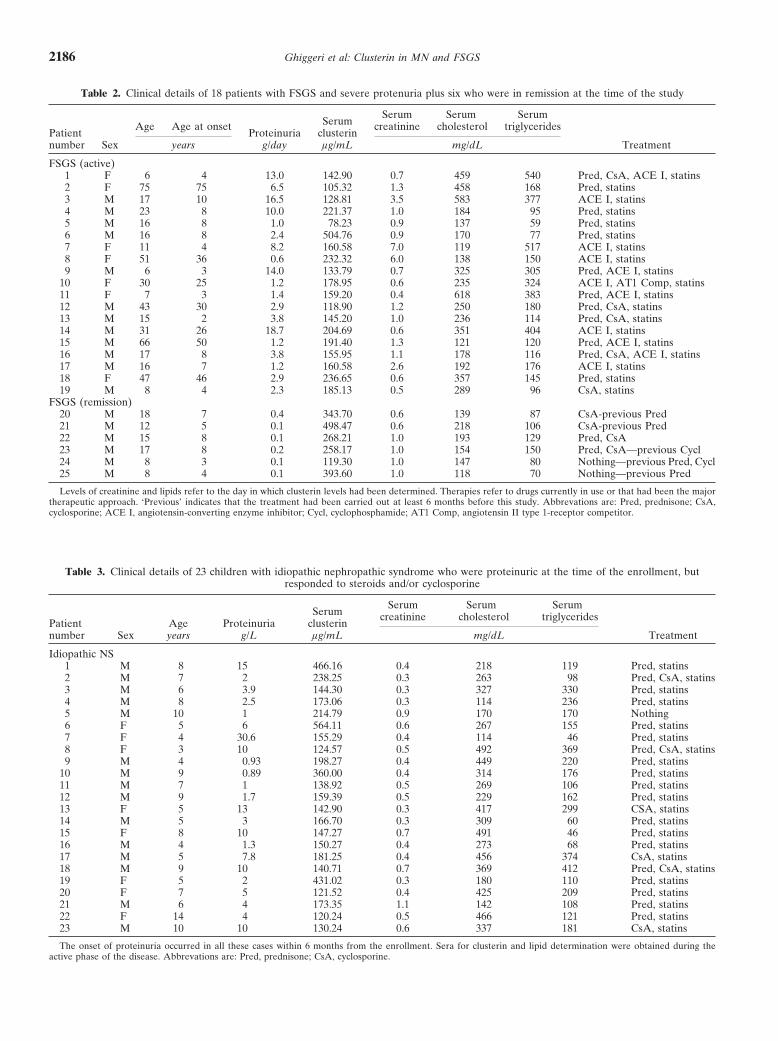
Table 2. Clinical details of 18 patients with FSGS and severe protenuria plus six who were in remission at the time of the study
Serum Serum SerumSerumAge Age at onset creatinine cholesterol triglycerides
Patient Proteinuria clusterinnumber Sex years g/day lg/mL mg/dL Treatment
FSGS (active)1 F 6 4 13.0 142.90 0.7 459 540 Pred, CsA, ACE I, statins2 F 75 75 6.5 105.32 1.3 458 168 Pred, statins3 M 17 10 16.5 128.81 3.5 583 377 ACE I, statins4 M 23 8 10.0 221.37 1.0 184 95 Pred, statins5 M 16 8 1.0 78.23 0.9 137 59 Pred, statins6 M 16 8 2.4 504.76 0.9 170 77 Pred, statins7 F 11 4 8.2 160.58 7.0 119 517 ACE I, statins8 F 51 36 0.6 232.32 6.0 138 150 ACE I, statins9 M 6 3 14.0 133.79 0.7 325 305 Pred, ACE I, statins
10 F 30 25 1.2 178.95 0.6 235 324 ACE I, AT1 Comp, statins11 F 7 3 1.4 159.20 0.4 618 383 Pred, ACE I, statins12 M 43 30 2.9 118.90 1.2 250 180 Pred, CsA, statins13 M 15 2 3.8 145.20 1.0 236 114 Pred, CsA, statins14 M 31 26 18.7 204.69 0.6 351 404 ACE I, statins15 M 66 50 1.2 191.40 1.3 121 120 Pred, ACE I, statins16 M 17 8 3.8 155.95 1.1 178 116 Pred, CsA, ACE I, statins17 M 16 7 1.2 160.58 2.6 192 176 ACE I, statins18 F 47 46 2.9 236.65 0.6 357 145 Pred, statins19 M 8 4 2.3 185.13 0.5 289 96 CsA, statins
FSGS (remission)20 M 18 7 0.4 343.70 0.6 139 87 CsA-previous Pred21 M 12 5 0.1 498.47 0.6 218 106 CsA-previous Pred22 M 15 8 0.1 268.21 1.0 193 129 Pred, CsA23 M 17 8 0.2 258.17 1.0 154 150 Pred, CsA—previous Cycl24 M 8 3 0.1 119.30 1.0 147 80 Nothing—previous Pred, Cycl25 M 8 4 0.1 393.60 1.0 118 70 Nothing—previous Pred
Levels of creatinine and lipids refer to the day in which clusterin levels had been determined. Therapies refer to drugs currently in use or that had been the majortherapeutic approach. ‘Previous’ indicates that the treatment had been carried out at least 6 months before this study. Abbrevations are: Pred, prednisone; CsA,cyclosporine; ACE I, angiotensin-converting enzyme inhibitor; Cycl, cyclophosphamide; AT1 Comp, angiotensin II type 1-receptor competitor.
Table 3. Clinical details of 23 children with idiopathic nephropathic syndrome who were proteinuric at the time of the enrollment, butresponded to steroids and/or cyclosporine
Serum Serum SerumSerum creatinine cholesterol triglycerides
Patient Age Proteinuria clusterinnumber Sex years g/L lg/mL mg/dL Treatment
Idiopathic NS1 M 8 15 466.16 0.4 218 119 Pred, statins2 M 7 2 238.25 0.3 263 98 Pred, CsA, statins3 M 6 3.9 144.30 0.3 327 330 Pred, statins4 M 8 2.5 173.06 0.3 114 236 Pred, statins5 M 10 1 214.79 0.9 170 170 Nothing6 F 5 6 564.11 0.6 267 155 Pred, statins7 F 4 30.6 155.29 0.4 114 46 Pred, statins8 F 3 10 124.57 0.5 492 369 Pred, CsA, statins9 M 4 0.93 198.27 0.4 449 220 Pred, statins
10 M 9 0.89 360.00 0.4 314 176 Pred, statins11 M 7 1 138.92 0.5 269 106 Pred, statins12 M 9 1.7 159.39 0.5 229 162 Pred, statins13 F 5 13 142.90 0.3 417 299 CSA, statins14 M 5 3 166.70 0.3 309 60 Pred, statins15 F 8 10 147.27 0.7 491 46 Pred, statins16 M 4 1.3 150.27 0.4 273 68 Pred, statins17 M 5 7.8 181.25 0.4 456 374 CsA, statins18 M 9 10 140.71 0.7 369 412 Pred, CsA, statins19 F 5 2 431.02 0.3 180 110 Pred, statins20 F 7 5 121.52 0.4 425 209 Pred, statins21 M 6 4 173.35 1.1 142 108 Pred, statins22 F 14 4 120.24 0.5 466 121 Pred, statins23 M 10 10 130.24 0.6 337 181 CsA, statins
The onset of proteinuria occurred in all these cases within 6 months from the enrollment. Sera for clusterin and lipid determination were obtained during theactive phase of the disease. Abbrevations are: Pred, prednisone; CsA, cyclosporine.
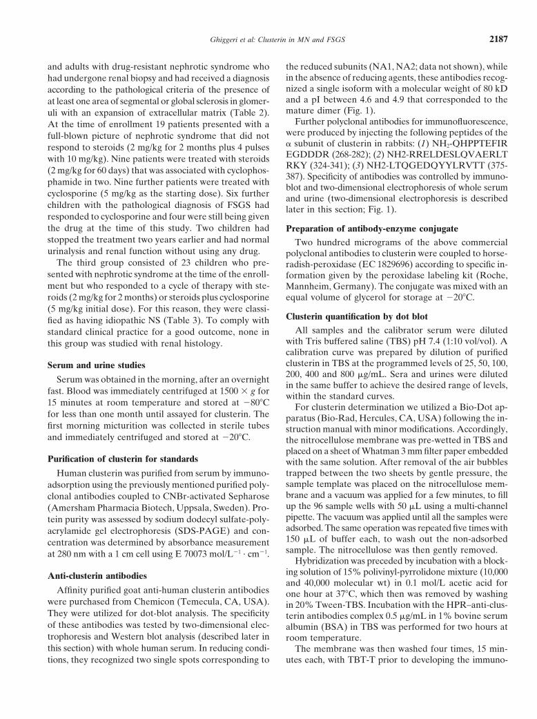
Ghiggeri et al: Clusterin in MN and FSGS 2187
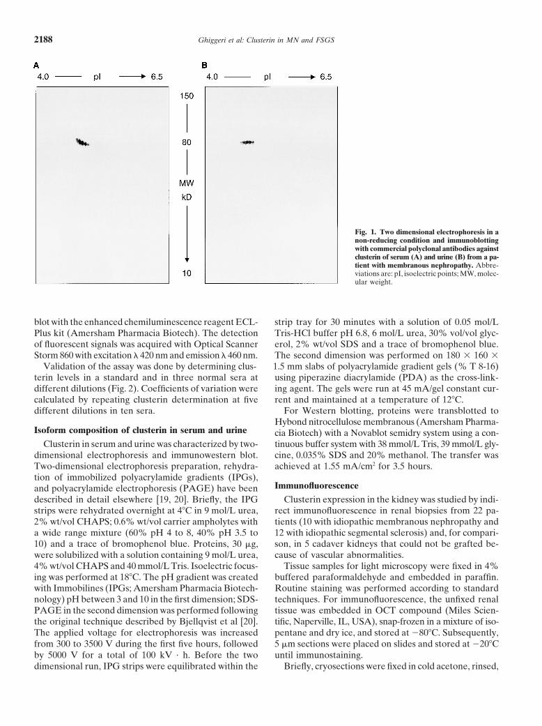
and adults with drug-resistant nephrotic syndrome who the reduced subunits (NA1, NA2; data not shown), whilein the absence of reducing agents, these antibodies recog-had undergone renal biopsy and had received a diagnosisnized a single isoform with a molecular weight of 80 kDaccording to the pathological criteria of the presence ofand a pI between 4.6 and 4.9 that corresponded to theat least one area of segmental or global sclerosis in glomer-mature dimer (Fig. 1).uli with an expansion of extracellular matrix (Table 2).
Further polyclonal antibodies for immunofluorescence,At the time of enrollment 19 patients presented with awere produced by injecting the following peptides of thefull-blown picture of nephrotic syndrome that did not� subunit of clusterin in rabbits: (1) NH2-QHPPTEFIRrespond to steroids (2 mg/kg for 2 months plus 4 pulsesEGDDDR (268-282); (2) NH2-RRELDESLQVAERLTwith 10 mg/kg). Nine patients were treated with steroidsRKY (324-341); (3) NH2-LTQGEDQYYLRVTT (375-(2 mg/kg for 60 days) that was associated with cyclophos-387). Specificity of antibodies was controlled by immuno-
phamide in two. Nine further patients were treated withblot and two-dimensional electrophoresis of whole serum
cyclosporine (5 mg/kg as the starting dose). Six further and urine (two-dimensional electrophoresis is describedchildren with the pathological diagnosis of FSGS had later in this section; Fig. 1).responded to cyclosporine and four were still being giventhe drug at the time of this study. Two children had Preparation of antibody-enzyme conjugatestopped the treatment two years earlier and had normal Two hundred micrograms of the above commercialurinalysis and renal function without using any drug. polyclonal antibodies to clusterin were coupled to horse-
The third group consisted of 23 children who pre- radish-peroxidase (EC 1829696) according to specific in-sented with nephrotic syndrome at the time of the enroll- formation given by the peroxidase labeling kit (Roche,ment but who responded to a cycle of therapy with ste- Mannheim, Germany). The conjugate was mixed with an
equal volume of glycerol for storage at �20�C.roids (2 mg/kg for 2 months) or steroids plus cyclosporine(5 mg/kg initial dose). For this reason, they were classi-
Clusterin quantification by dot blotfied as having idiopathic NS (Table 3). To comply withAll samples and the calibrator serum were dilutedstandard clinical practice for a good outcome, none in
with Tris buffered saline (TBS) pH 7.4 (1:10 vol/vol). Athis group was studied with renal histology.calibration curve was prepared by dilution of purifiedclusterin in TBS at the programmed levels of 25, 50, 100,Serum and urine studies200, 400 and 800 �g/mL. Sera and urines were dilutedSerum was obtained in the morning, after an overnightin the same buffer to achieve the desired range of levels,
fast. Blood was immediately centrifuged at 1500 � g for within the standard curves.15 minutes at room temperature and stored at �80�C For clusterin determination we utilized a Bio-Dot ap-for less than one month until assayed for clusterin. The paratus (Bio-Rad, Hercules, CA, USA) following the in-first morning micturition was collected in sterile tubes struction manual with minor modifications. Accordingly,and immediately centrifuged and stored at �20�C. the nitrocellulose membrane was pre-wetted in TBS and
placed on a sheet of Whatman 3 mm filter paper embeddedPurification of clusterin for standards with the same solution. After removal of the air bubbles
trapped between the two sheets by gentle pressure, theHuman clusterin was purified from serum by immuno-sample template was placed on the nitrocellulose mem-adsorption using the previously mentioned purified poly-brane and a vacuum was applied for a few minutes, to fillclonal antibodies coupled to CNBr-activated Sepharoseup the 96 sample wells with 50 �L using a multi-channel(Amersham Pharmacia Biotech, Uppsala, Sweden). Pro-pipette. The vacuum was applied until all the samples weretein purity was assessed by sodium dodecyl sulfate-poly-adsorbed. The same operation was repeated five times withacrylamide gel electrophoresis (SDS-PAGE) and con-150 �L of buffer each, to wash out the non-adsorbedcentration was determined by absorbance measurementsample. The nitrocellulose was then gently removed.at 280 nm with a 1 cm cell using E 70073 mol/L�1 · cm�1.
Hybridization was preceded by incubation with a block-ing solution of 15% polivinyl-pyrrolidone mixture (10,000Anti-clusterin antibodiesand 40,000 molecular wt) in 0.1 mol/L acetic acid for
Affinity purified goat anti-human clusterin antibodies one hour at 37�C, which then was removed by washingwere purchased from Chemicon (Temecula, CA, USA). in 20% Tween-TBS. Incubation with the HPR–anti-clus-They were utilized for dot-blot analysis. The specificity terin antibodies complex 0.5 �g/mL in 1% bovine serumof these antibodies was tested by two-dimensional elec- albumin (BSA) in TBS was performed for two hours attrophoresis and Western blot analysis (described later in room temperature.this section) with whole human serum. In reducing condi- The membrane was then washed four times, 15 min-
utes each, with TBT-T prior to developing the immuno-tions, they recognized two single spots corresponding to
Ghiggeri et al: Clusterin in MN and FSGS2188
Fig. 1. Two dimensional electrophoresis in anon-reducing condition and immunoblottingwith commercial polyclonal antibodies againstclusterin of serum (A) and urine (B) from a pa-tient with membranous nephropathy. Abbre-viations are: pI, isoelectric points; MW, molec-ular weight.
blot with the enhanced chemiluminescence reagent ECL- strip tray for 30 minutes with a solution of 0.05 mol/LTris-HCl buffer pH 6.8, 6 mol/L urea, 30% vol/vol glyc-Plus kit (Amersham Pharmacia Biotech). The detection
of fluorescent signals was acquired with Optical Scanner erol, 2% wt/vol SDS and a trace of bromophenol blue.The second dimension was performed on 180 � 160 �Storm 860 with excitation � 420 nm and emission � 460 nm.
Validation of the assay was done by determining clus- 1.5 mm slabs of polyacrylamide gradient gels (% T 8-16)using piperazine diacrylamide (PDA) as the cross-link-terin levels in a standard and in three normal sera at
different dilutions (Fig. 2). Coefficients of variation were ing agent. The gels were run at 45 mA/gel constant cur-rent and maintained at a temperature of 12�C.calculated by repeating clusterin determination at five
different dilutions in ten sera. For Western blotting, proteins were transblotted toHybond nitrocellulose membranous (Amersham Pharma-
Isoform composition of clusterin in serum and urine cia Biotech) with a Novablot semidry system using a con-tinuous buffer system with 38 mmol/L Tris, 39 mmol/L gly-Clusterin in serum and urine was characterized by two-
dimensional electrophoresis and immunowestern blot. cine, 0.035% SDS and 20% methanol. The transfer wasachieved at 1.55 mA/cm2 for 3.5 hours.Two-dimensional electrophoresis preparation, rehydra-
tion of immobilized polyacrylamide gradients (IPGs),Immunofluorescenceand polyacrylamide electrophoresis (PAGE) have been
described in detail elsewhere [19, 20]. Briefly, the IPG Clusterin expression in the kidney was studied by indi-rect immunofluorescence in renal biopsies from 22 pa-strips were rehydrated overnight at 4�C in 9 mol/L urea,
2% wt/vol CHAPS; 0.6% wt/vol carrier ampholytes with tients (10 with idiopathic membranous nephropathy and12 with idiopathic segmental sclerosis) and, for compari-a wide range mixture (60% pH 4 to 8, 40% pH 3.5 to
10) and a trace of bromophenol blue. Proteins, 30 �g, son, in 5 cadaver kidneys that could not be grafted be-cause of vascular abnormalities.were solubilized with a solution containing 9 mol/L urea,
4% wt/vol CHAPS and 40 mmol/L Tris. Isoelectric focus- Tissue samples for light microscopy were fixed in 4%buffered paraformaldehyde and embedded in paraffin.ing was performed at 18�C. The pH gradient was created
with Immobilines (IPGs; Amersham Pharmacia Biotech- Routine staining was performed according to standardtechniques. For immunofluorescence, the unfixed renalnology) pH between 3 and 10 in the first dimension; SDS-
PAGE in the second dimension was performed following tissue was embedded in OCT compound (Miles Scien-tific, Naperville, IL, USA), snap-frozen in a mixture of iso-the original technique described by Bjellqvist et al [20].
The applied voltage for electrophoresis was increased pentane and dry ice, and stored at �80�C. Subsequently,5 �m sections were placed on slides and stored at �20�Cfrom 300 to 3500 V during the first five hours, followed
by 5000 V for a total of 100 kV · h. Before the two until immunostaining.Briefly, cryosections were fixed in cold acetone, rinsed,dimensional run, IPG strips were equilibrated within the

Ghiggeri et al: Clusterin in MN and FSGS 2189
Fig. 2. Calibration curve with a purified stan-dard of clusterin and three sera at different di-lutions. The average optical density for thechemiluminescence analysis was plotted againstthe amount of clusterin loaded. Symbols are:(�) ApoJ St; (�) serum 1; (�) serum 2; (�)serum 3. (Inset) A plot that utilizes a logarith-mic scale in the abscissa to visualize the curveat low concentrations.
and sequentially incubated with the primary rabbit poly- Statistical methodsclonal antibody against clusterin, followed by fluorescein The serum concentrations of clusterin in MN, FSGS,isothiocyanate (FITC)-labeled goat anti-rabbit second- idiopathic NS and controls were compared utilizing theary antibody (Histoline; Zymed, Milan, Italy). After wash- one-way analysis of variance (ANOVA). Data are givening, sections were then mounted with an anti-fading mount- as mean � standard deviation.ing medium (Vectashield; Vector, DBA Italia SRL, Milan,Italy). Specificity of labeling was demonstrated by the lack RESULTSof staining after substituting phosphate buffered saline
General features(PBS) and proper control immunoglobulins (Zymed) forThis study was conducted in two cohorts of patientsthe primary antibody.
with histologically-proven MN and FSGS (Tables 1 and 2)Immunofluorescence results were evaluated by an elec-and in a further group of children with nephrotic syn-tronic image analysis system (ETC3000, Graftek; Villan-drome who responded to classical treatments (Table 3).terio, Pavia, Italy). Images were digitalized using a videoOverall, most of them were frankly proteinuric whilecamera (DC200; Leica, Milan, Italy) connected to a Leitzonly few presented mild proteinuria or were in remission.Diaplan microscope (Leica) and to a Pentium III 500In fact, among the MN patients only four had proteinuriacomputer (Maxwel, Rozzano, Italy) equipped with aless than 0.5 g/day, six were frankly proteinuric (5 g/day)frame grabber (Leica). The electronic system was pro-and the remaining thirteen showed levels between 1 andgrammed for glomerular evaluation by selecting the ROI5 g/day. Renal function was normal in all but four sub-
(region of interest mean statistic) analysis and an auto-jects who presented a creatinine level between 1.7 and
mated macro, composed of a planned threshold proce- 2.2 mg/dL. Also, FSGS patients presented variable pro-dure, filtering and Danielsson algorithm, was applied to teinuria: six were in stable remission (proteinuria 0.5all digitized images. A mean of 8 � 1.2 (minimum 6 to g/day), eight were frankly proteinuric (5 g/day) andmaximum 19) glomeruli/biopsy were analyzed. Globally eleven presented intermediate levels between 1 andsclerotic glomeruli were excluded from the evaluation. 5 g/day. Four out the six FSGS patients in remissionResults were expressed as number of positive pixels/ were still receiving drugs at the time of enrollment, whileglomerular area, automatically exported and elaborated another two had drug-induced remission two years be-in an electronic file (SPSS 9.0 for Windows; SPSS Inc., fore and during the study period were without any treat-Chicago, IL, USA) using the mean and standard devia- ment. Serum creatinine levels were normal in 20 out oftion. The significance was set at P 0.05 and was ana- the 24 patients of the FSGS group; in four patients
(1 child, 2 adolescents and 1 adult) creatinine had reachedlyzed using the �2 test.
Ghiggeri et al: Clusterin in MN and FSGS2190
Table 4. Serum and urinary levels of clusterin in the cohorts ofpatients with membranous nephropathy and focal glomerulosclerosis
subdivided according to levels of proteinuria in active disease(1 g/day) and remission (0.5 g/day)
Clusterin serum Clusterin urineCreatinine
Group N lg/mL lg/mg
Controls 50 366.60�62.0 7.06�0.42 7.8�0.47MN (active) 19 220.77�110.9a 1.03�0.75a 1.47�1.07MN (remission) 4 236.60�38.7a 3.31�5.65 3.68�6.28FSGS (active) 19 181.44�89.0a 1.32�2.34a 2.2�3.9FSGS (remission) 6 312.50�130.5 5.27�6.55 6.59�8.2Idiopathic NS 23 210.11�122.0a 0.72�0.99a 1.44�1.98
A group of 23 children with idiopathic nephrotic syndrome who were succes-sively found to respond to steroids or cyclosporine was enrolled to address aspecific role of proteinuria in clusterin levels. Urinary clusterin was determinedon fresh samples obtained in the morning and results are given as concentrationper mL or corrected for urinary creatinine.
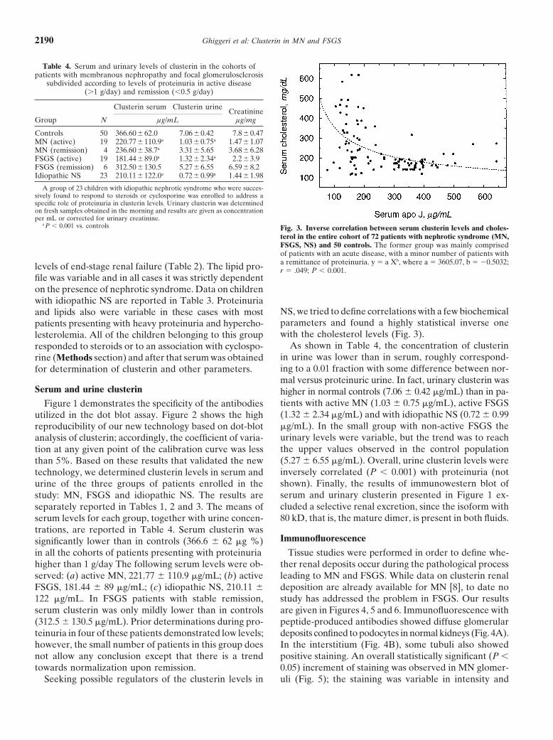
a P 0.001 vs. controls Fig. 3. Inverse correlation between serum clusterin levels and choles-terol in the entire cohort of 72 patients with nephrotic syndrome (MN,FSGS, NS) and 50 controls. The former group was mainly comprisedof patients with an acute disease, with a minor number of patients witha remittance of proteinuria. y � a Xb, where a � 3605.07, b � �0.5032;levels of end-stage renal failure (Table 2). The lipid pro- r � .049; P 0.001.
file was variable and in all cases it was strictly dependenton the presence of nephrotic syndrome. Data on childrenwith idiopathic NS are reported in Table 3. Proteinuria
NS, we tried to define correlations with a few biochemicaland lipids also were variable in these cases with mostparameters and found a highly statistical inverse onepatients presenting with heavy proteinuria and hypercho-with the cholesterol levels (Fig. 3).lesterolemia. All of the children belonging to this group
As shown in Table 4, the concentration of clusterinresponded to steroids or to an association with cyclospo-in urine was lower than in serum, roughly correspond-rine (Methods section) and after that serum was obtaineding to a 0.01 fraction with some difference between nor-for determination of clusterin and other parameters.mal versus proteinuric urine. In fact, urinary clusterin was
Serum and urine clusterin higher in normal controls (7.06 � 0.42 �g/mL) than in pa-tients with active MN (1.03 � 0.75 �g/mL), active FSGSFigure 1 demonstrates the specificity of the antibodies(1.32 � 2.34 �g/mL) and with idiopathic NS (0.72 � 0.99utilized in the dot blot assay. Figure 2 shows the high�g/mL). In the small group with non-active FSGS thereproducibility of our new technology based on dot-bloturinary levels were variable, but the trend was to reachanalysis of clusterin; accordingly, the coefficient of varia-the upper values observed in the control populationtion at any given point of the calibration curve was less(5.27 � 6.55 �g/mL). Overall, urine clusterin levels werethan 5%. Based on these results that validated the newinversely correlated (P 0.001) with proteinuria (nottechnology, we determined clusterin levels in serum andshown). Finally, the results of immunowestern blot ofurine of the three groups of patients enrolled in theserum and urinary clusterin presented in Figure 1 ex-study: MN, FSGS and idiopathic NS. The results arecluded a selective renal excretion, since the isoform withseparately reported in Tables 1, 2 and 3. The means of80 kD, that is, the mature dimer, is present in both fluids.serum levels for each group, together with urine concen-
trations, are reported in Table 4. Serum clusterin wasImmunofluorescencesignificantly lower than in controls (366.6 � 62 �g %)
in all the cohorts of patients presenting with proteinuria Tissue studies were performed in order to define whe-ther renal deposits occur during the pathological processhigher than 1 g/day The following serum levels were ob-
served: (a) active MN, 221.77 � 110.9 �g/mL; (b) active leading to MN and FSGS. While data on clusterin renaldeposition are already available for MN [8], to date noFSGS, 181.44 � 89 �g/mL; (c) idiopathic NS, 210.11 �
122 �g/mL. In FSGS patients with stable remission, study has addressed the problem in FSGS. Our resultsare given in Figures 4, 5 and 6. Immunofluorescence withserum clusterin was only mildly lower than in controls
(312.5 � 130.5 �g/mL). Prior determinations during pro- peptide-produced antibodies showed diffuse glomerulardeposits confined to podocytes in normal kidneys (Fig. 4A).teinuria in four of these patients demonstrated low levels;
however, the small number of patients in this group does In the interstitium (Fig. 4B), some tubuli also showedpositive staining. An overall statistically significant (P not allow any conclusion except that there is a trend
towards normalization upon remission. 0.05) increment of staining was observed in MN glomer-uli (Fig. 5); the staining was variable in intensity andSeeking possible regulators of the clusterin levels in

Ghiggeri et al: Clusterin in MN and FSGS 2191
Fig. 5. Membranous nephropathy showing that the increment in clus-Fig. 4. Normal kidney showing clusterin staining present on glomerularterin staining is variable in different cases from moderate (A) to verypodocytes (A) and on some tubules in the interstitium (B) (IF, �400).intense (B) (IF, �400). Immunofluorescence was done with antibodiesImmunofluorescence (IF) was done with antibodies raised against clus-raised against clusterin peptides.terin peptides.
diffusion from mild, focal and segmental (Fig. 5A) to renal disease. In MN, clusterin competes with C5b-9 forvery intense, global and diffuse (Fig. 5B). the same receptor, that is, megalin in podocytes [11–13],
A focal, but not statistically significant decrease of and limits to some extent the accessibility and the proteo-clusterin staining was detected in FSGS (Fig. 6). In addi- lyses activity of the complement at this level. It is nottion to the glomeruli maintaining a staining similar to surprising that serum depletion of clusterin represents anormal kidney (Fig. 6A), a global (Fig. 6B) or segmental negative event in experimental models of MN [9] and(Fig. 6C) staining absence was found in 30% of glomer- in animals lacking the protein due to genetic manipula-uli. When segmental, it was mainly but not exclusively tion [7]. In fact, after depletion of clusterin, glomerulardetected in sclerotic areas. deposits of complement and immunoglobulins are wider
No differences from normal kidney were observed in and proteinuria is significantly higher than in non-terms of tubular staining in both MN and FSGS patients. depleted animals. Moreover, clusterin-deficient mice de-
The results of the quantitation of clusterin deposits by velop a progressive glomerulopathy with age, character-morphometric analysis are given in Figure 7. MN kidneys
ized by immunocomplex localization in mesangium andpresent a statistically significant mean increase.
glomerulosclerosis that mimics, in some way, FSGS inhumans [10]. The possibility that clusterin plays a protec-
DISCUSSION tive role in FSGS also is supported by in vitro studiesshowing an increased permeability activity in serum ofClusterin is thought to play an important protectivepatients with FSGS when the serum is depleted of clus-function in specific renal diseases such as MN and FSGS,
where it should act at different levels with each type of terin [14]. Since permeability activity in FSGS is linked

Ghiggeri et al: Clusterin in MN and FSGS2192
Fig. 7. Evaluation of clusterin glomerular expression in normal kidneys(NK), membranous nephropathy (MN), and focal segmental sclerosis(FSGS). Results are expressed as number of pixels/glomerular area(mean � SD).
son for this failure rests on the difficulty to measure clus-terin levels in fluids. For this reason we developed a newdot-blot technique that appears to be highly reproducibleand specific. Only one study has investigated the anti-inflammatory and anti-complement activity of clusterinin lupus erythematosus [21]. Another report examinedclusterin levels in alcohol cirrhosis [22], the liver beingthe major site for its synthesis. In both cases, serum clus-terin levels were low and were significantly correlatedwith the disease features.
This study demonstrates that serum clusterin is re-markably low in patients with nephrotic syndrome, whichincludes MN, FSGS and idiopathic NS, than in a controlpopulation composed of age-matched subjects. Urinaryexcretion also is lower in patients than in normal con-trols, with a trend to be higher in those who present withremission after specific treatments. Finally, the compo-sition of clusterin is the same in serum and urine, indicat-ing that renal handling of clusterin is not selective andis a direct function of the serum levels. In the attemptto define the mechanism responsible for depletion ofclusterin in nephrotic syndrome, we looked at severalputative factors, including urinary loss, loss of regulatory
Fig. 6. Focal segmental sclerosis displaying a glomerulus positive for substances and plasma lipids. The first possibility can beclusterin similar to a normal kidney (A), while other glomeruli demon- reasonably ruled out since the concentration of clusterinstrate a global (B) or segmental (C ) decrease in staining (IF, �400).
in urine was low in the advanced stage of NS and theImmunofluorescence was done with antibodies raised against clusterinpeptides. trend for patients showing remittance was for a restora-
tion of normal levels.Instead, a high statistical correlation was found by
to proteinuria, depletion of clusterin should represent a plotting the serum levels of clusterin with cholesterol,factor aggravating the course of the disease [15, 16]. Until suggesting a mechanism for low clusterin to be linkednow, there is little, if any, information about clusterin with hyperlipidemia. The association between hyperlip-
idemia and nephrotic syndrome has been recognized formetabolism in patients with nephrotic syndrome. The rea-

Ghiggeri et al: Clusterin in MN and FSGS 2193
several decades. The classic finding is increased plasma complex regulatory mechanism. According to these ex-perimental studies, low levels of clusterin in serum shouldcholesterol, low- (LDL) and very low-density lipoprotein
(VLDL) cholesterol, while high-density lipoproteins are represent a cause of an unfavorable outcome.unchanged (HDL1) or slightly decreased (HDL2) [23–25].These changes derive from a combination of increased ACKNOWLEDGMENTSproduction, impaired lipolysis, and reduced receptor- The authors acknowledge the financial support of the “Foundation
for Studies on Renal Diseases in Children” and thank Prof. R. Gus-mediated clearance of lipoproteins, while low HDL alsomano for her continuous interest in this research. Partial support wascan be influenced by increased urinary loss. The data pre-given by the “Project Glomerulonephritis in memory of Pippo Neglia.”sented here suggest that other regulatory mechanisms have This study also was supported by a grant from the Italian Ministry of
combined effects on different components such as cho- Health (Progetto finalizzato IRCCS 070: 2/RF00.167).lesterol and clusterin, eventually resulting in low clusterin
Reprint requests to Gian Marco Ghiggeri, M.D., Laboratory on Pa-and high cholesterol levels. It is noteworthy that lipid- thophisiology of Uremia, Istituto Giannina Gaslini, 16148 Genova, Italy.lowering therapy with probucol and low-density lipopro- E-mail: [email protected] apheresis ameliorates proteinuria and reduces glo-merular injuries in MN and FSGS [26, 27]. We speculate REFERENCESthat clusterin provides the logical link between hypercho- 1. Blaschuk O, Burdzy K, Fritz IB: Purification and characteriza-lesterolemia and a poor outcome of glomerulopathy. tion of a cell-aggregating factor (clusterin), the major glycoprotein
in ram rete testis fluid. J Biol Chem 258:7714–7720, 1983The expression of clusterin in both normal and affected2. de Silva HV, Stuart WD, Park YB, et al: Purification and charac-kidneys has received limited attention in the past and terization of apolipoprotein J. J Biol Chem 265:14292–14297, 1990
has been confined to experimental models of nephrosis 3. May PC, Finch CE: Sulfated glycoprotein 2: New relationships ofthis multifunctional protein to neurodegeneration. Trends Neurosci[28, 29] cystic and dysplastic diseases. In 1994, Dvergstan15:391–396, 1992et al found no clusterin staining in normal renal tissue
4. Wilson MR, Easterbrook-Smith SB: Clusterin is a secreted mam-while there was an increased expression in epithelial cells malian chaperone. Trends Biochem Sci 25:95–98, 2000
5. May PC, Lampert-Etchells M, Johnson SA, et al: Dynamicslining cysts and in some immature dysplastic tubulesof gene expression for a hippocampal glycoprotein elevated in[28]. Our data indicate that clusterin localizes in normalAlzheimer’s disease and in response to experimental lesions in rat.
glomeruli. We found an increased glomerular expression Neuron 5:831–839, 19906. Han BH, DeMattos RB, Dugan LL, et al: Clusterin contributesof clusterin in MN patients, whereas in sclerotic areas
to caspase-3-independent brain injury following neonatal hypoxia-typical of the FSGS kidney, which are the prominentischemia. Nat Med 7:338–343, 2001
sites for the accumulation of other lipids such as LDL, 7. McLaughlin L, Zhu G, Mistry M, et al: Apolipoprotein J/clusterinlimits the severity of murine autoimmune myocarditis. J Clin Investit was absent. This point is pertinent to the evolution106:1105–1113, 2000of renal lesions, since it indicates that the mechanisms
8. Murphy BF, Kirszbaum L, Walker ID, d’Apice AJ: SP-40,40, aregulating the glomerular expression of clusterin is unre- newly identified normal human serum protein found in the SC5b-9lated to the serum levels. Indeed, Yamada et al have complex of complement and in the immune deposits in glomerulo-
nephritis. J Clin Invest 81:1858–1864, 1988recently demonstrated that the mesangial expression of9. Saunders JR, Aminian A, McRae JL, et al: Clusterin depletionclusterin is up-regulated during the course of immune- enhances immune glomerular injury in the isolated perfused kid-
mediated renal injures, this representing a critical step ney. Kidney Int 45:817–827, 199410. Rosenberg ME, Girton R, Finkel D, et al: Apolipoproteinin protecting the kidney from complement attacks [30].
J/clusterin prevents a progressive glomerulopathy of aging. MolTherefore, in general it is possible that the glomeru- Cell Biol 22:1893–1902, 2002lar expression of clusterin is dependent on the serum 11. Orlando RA, Kerjaschki D, Farquhar MG: Megalin (gp330)
possesses an antigenic epitope capable of inducing passive Hey-pool, this determining a defect in glomerular clusterin inmann nephritis independent of the nephritogenic epitope in recep-FSGS. In particular cases, such as in MN, other immuno- tor-associated protein. J Am Soc Nephrol 6:61–67, 1995
linked regulatory mechanisms could stimulate mesangial 12. Kounnas MZ, Loukinova EB, Stefansson S, et al: Identificationof glycoprotein 330 as an endocytic receptor for apolipoproteincells to produce clusterin and restore the glomerularJ/clusterin. J Biol Chem 270:13070–13075, 1995pool of the protein. How this different regulation influ- 13. Zlokovic BV, Martel CL, Matsubara E, et al: Glycoprotein
ences the clinical outcome in MN and in FSGS patients 330/megalin: probable role in receptor-mediated transport of apoli-poprotein J alone and in a complex with Alzheimer disease amyloidis currently unknown and clinical trials should focus onbeta at the blood-brain and blood-cerebrospinal fluid barriers. Procthis aspect. The normalization of serum levels of clusterinNatl Acad Sci USA 93:4229–4234, 1996
(and indirectly of cholesterol) represents a primary ob- 14. Candiano G, Musante L, Carraro M, et al: Apolipoproteinsprevent glomerular albumin permeability induced in vitro by serumjective of any therapeutic approach to both diseases.from patients with focal segmental glomerulosclerosis. J Am SocIn conclusion, these results indicate that serum andNephrol 12:143–150, 2001
urinary clusterin is low in proteinuric patients with MN, 15. Savin VJ, Sharma R, Sharma M, et al: Circulating factor associatedwith increased glomerular permeability to albumin in recurrentFSGS and idiopathic NS, but the urinary losses do notfocal segmental glomerulosclerosis. N Engl J Med 334:878–883, 1996account for this reduction and hypercholesterolemia
16. Dantal J, Bigot E, Bogers W, et al: Effect of plasma proteinappears to be the unifying feature. Glomerular clusterin adsorption on protein excretion in kidney-transplant recipients
with recurrent nephrotic syndrome. N Engl J Med 330:7–14, 1994is variably expressed in FSGS and in MN, suggesting a

Ghiggeri et al: Clusterin in MN and FSGS2194
17. Ehrenreich T, Porush JG, Churg J, et al: Treatment of idiopathic syndrome: Causes, consequences, and treatment. Am J Kidney Dis23:331–346, 1994membranous nephropathy. N Engl J Med 295:741–746, 1976
24. Deighan CJ, Caslake MJ, McConnell M, et al: Patients with18. Ponticelli C, Zucchelli P, Imbasciati E, et al: Controlled trial ofnephrotic-range proteinuria have apolipoprotein C and E deficientmethylprednisolone and chlorambucil in idiopathic membranousVLDL1. Kidney Int 58:1238–1246, 2000nephropathy. N Engl J Med 310:946–950, 1984
25. Short CD, Durrington PN, Mallick NP, et al: Serum and urinary19. Musante L, Candiano G, Ghiggeri GM: Resolution of fibronectin high density lipoproteins in glomerular disease with proteinuria.and other uncharacterized proteins by two-dimensional polyacryl- Kidney Int 29:1224–1228, 1986amide electrophoresis with thiourea. J Chromatogr B Biomed Sci 26. Haas M, Kerjaschki D, Mayer G: Lipid-lowering therapy in mem-Appl 705:351–356, 1998 branous nephropathy. Kidney Int 56(Suppl 71):S110–S112, 1999
20. Bjellqvist B, Pasquali C, Ravier F, et al: A nonlinear wide-range 27. Muso E, Mune M, Fujii Y, et al: Low density lipoprotein apheresisimmobilized pH gradient for two-dimensional electrophoresis and therapy for steroid-resistant nephrotic syndrome. Kidney Int 56
(Suppl 71):S122–S125, 1999its definition in a relevant pH scale. Electrophoresis 14:1357–1365,28. Dvergsten J, Manivel JC, Correa-Rotter R, Rosenberg ME:1993
Expression of clusterin in human renal diseases. Kidney Int 45:828–21. Newkirk MM, Apostolakos P, Neville C, Fortin PR: Systemic835, 1994lupus erythematosus, a disease associated with low levels of clus-
29. Correa-Rotter R, Ibarra-Rubio ME, Schwochau G, et al: Induc-terin/apoJ, an antiinflammatory protein. J Rheumatol 26:597–603, tion of clusterin in tubules of nephrotic rats. J Am Soc Nephrol1999 9:33–37, 1998
22. Hogasen K, Homann C, Mollnes TE, et al: Serum clusterin and 30. Yamada K, Hori Y, Hanafusa N, et al: Clusterin is up-regulated invitronectin in alcoholic cirrhosis. Liver 16:140–146, 1996 glomerular mesangial cells in complement-mediated injury. Kidney
Int 59:137–146, 200123. Wheeler DC, Bernard DB: Lipid abnormalities in the nephrotic
Copyright © 2022 FDOKUMEN