Clusterin interacts with Paclitaxel and confer Paclitaxel resistance in ovarian cancer
12
Clusterin Interacts with Paclitaxel and Confer Paclitaxel Resistance in Ovarian Cancer 1,2 Dong Choon Park * ,† , Seung Geun Yeo ‡ , Mark R. Wilson § , Justin J. Yerbury § , Joseph Kwong † , William R. Welch ¶ , Yang Kyu Choi # , Michael J. Birrer ** , Samuel C. Mok †† and Kwong-Kwok Wong †† *Department of Obstetrics and Gynecology, Saint Vincent’s Hospital, The Catholic University of Korea, Suwon, Republic of Korea; † Department of Obstetrics, Gynecology and Reproductive Biology, Division of Gynecologic Oncology, Brigham and Women’s Hospital, Harvard Medical School, Boston, MA, USA; ‡ East-West Medical Research Institute, Kyung Hee University, Seoul, Republic of Korea; § School of Biological Sciences, University of Wollongong, NSW, Australia; ¶ Department of Pathology, Brigham and Women’s Hospital, Harvard Medical School, Boston, MA, USA; # Kunkook University School of Medicine, Seoul, Republic of Korea; **Cell and Cancer Biology Branch, National Cancer Institute, National Institutes of Health, Bethesda, MD, USA; †† Department of Gynecologic Oncology, The University of Texas M.D. Anderson Cancer Center, Houston, TX, USA Abstract Optimal debulking followed by chemotherapy is the standard treatment of managing late-stage ovarian cancer, but chemoresistance is still a major problem. In this study, we compared expression profiles of primary tumor tissue from five long-term (>8 years) and five short-term (<2 years) ovarian cancer survivors and identified clusterin as one of the genes that were significantly up-regulated in short-term survivors. We then evaluated the prognostic significance of clusterin and its possible correlation with chemoresistance in ovarian cancer by immunohistostain- ing of clusterin in 62 tumor samples from patients with stage III, high-grade serous ovarian cancer. After adjusting for debulking status and age, Cox regression analyses showed that high levels of clusterin expression correlate with poor survival (hazard ratio, 1.07; 95% confidence interval, 1.002–1.443; P = .04). We also investigated clusterin in paclitaxel resistance by modulating the endogenous clusterin expression in ovarian cancer cells and treating the cells with purified clusterin. Results indicate that high-clusterin–expressing ovarian cancer cells are more resistant to pac- litaxel. Moreover, exposing ovarian cancer cells to exogenous clusterin increases cells’ resistance to paclitaxel. Fi- nally, using size exclusion chromatography and fluorescently labeled paclitaxel, we demonstrated that clusterin binds to paclitaxel. In summary, our findings suggest that high levels of clusterin expression increase paclitaxel resistance Abbreviations: shRNA, short hairpin RNA; CLU, clusterin; siRNA, small interfering RNA Address all correspondence to: Kwong-Kwok Wong, PhD, Department of Gynecologic Oncology, Unit 1362, The University of Texas M.D. Anderson Cancer Center, 1515 Holcombe Boulevard, Houston, TX 77030. E-mail: [email protected] 1 This study was supported in part by the Dana Farber/Harvard Ovarian Specialized Programs of Research Excellence (SPORE) P50CA105009, the M.D. Anderson Cancer Center SPORE P50CA83639, and R33CA103595 from the National Institutes of Health; the Department of Health and Human Services; the Gillette Center For Women ’ s Cancer; the Adler Foundation, Inc.; the Edgar Astrove Fund; the Ovarian Cancer Research Fund, Inc.; the Catholic Medical Center Research Foundation (from program year 2006); and the 2006 Research Fund from St. Vincent ’ s Hospital. 2 This article refers to supplementary materials, which are designated by Table W1 and Figures W1 to W5 and are available online at www.neoplasia.com. Received 16 May 2008; Revised 17 June 2008; Accepted 20 June 2008 Copyright © 2008 Neoplasia Press, Inc. All rights reserved 1522-8002/08/$25.00 DOI 10.1593/neo.08604 www.neoplasia.com Volume 10 Number 9 September 2008 pp. 964–972 964
-
Upload
independent -
Category
Documents
-
view
0 -
download
0
Transcript of Clusterin interacts with Paclitaxel and confer Paclitaxel resistance in ovarian cancer

AbbreviatioAddress alHolcombe1This studSPORE P5Adler FounResearch F2This articReceived 1
CopyrightDOI 10.1
www.neoplasia.com
Volume 10 Number 9 September 2008 pp. 964–972 964
Clusterin Interacts with Paclitaxeland Confer Paclitaxel Resistancein Ovarian Cancer1,2
ns: shRNA, short hairpin RNA; CLU, clusterin; siRNA, small interfering RNAl correspondence to: Kwong-Kwok Wong, PhD, Department of Gynecologic OBoulevard, Houston, TX 77030. E-mail: [email protected] was supported in part by the Dana Farber/Harvard Ovarian Specialized Program0CA83639, and R33CA103595 from the National Institutes of Health; the Dedation, Inc.; the Edgar Astrove Fund; the Ovarian Cancer Research Fund, Inc.; thund from St. Vincent’s Hospital.le refers to supplementary materials, which are designated by Table W1 and F6 May 2008; Revised 17 June 2008; Accepted 20 June 2008
© 2008 Neoplasia Press, Inc. All rights reserved 1522-8002/08/$25.00593/neo.08604
Dong Choon Park*,†, Seung Geun Yeo‡,Mark R. Wilson§, Justin J. Yerbury§,Joseph Kwong†, William R. Welch¶,Yang Kyu Choi#, Michael J. Birrer**,Samuel C. Mok†† and Kwong-Kwok Wong††
*Department of Obstetrics and Gynecology, Saint Vincent’sHospital, The Catholic University of Korea, Suwon, Republicof Korea; †Department of Obstetrics, Gynecology andReproductive Biology, Division of Gynecologic Oncology,Brigham and Women’s Hospital, Harvard Medical School,Boston, MA, USA; ‡East-West Medical Research Institute,Kyung Hee University, Seoul, Republic of Korea; §Schoolof Biological Sciences, University of Wollongong, NSW,Australia; ¶Department of Pathology, Brigham and Women’sHospital, Harvard Medical School, Boston, MA, USA;#Kunkook University School of Medicine, Seoul, Republicof Korea; **Cell and Cancer Biology Branch, NationalCancer Institute, National Institutes of Health, Bethesda,MD, USA; ††Department of Gynecologic Oncology,The University of Texas M.D. Anderson Cancer Center,Houston, TX, USA
AbstractOptimal debulking followed by chemotherapy is the standard treatment of managing late-stage ovarian cancer, butchemoresistance is still a major problem. In this study, we compared expression profiles of primary tumor tissuefrom five long-term (>8 years) and five short-term (<2 years) ovarian cancer survivors and identified clusterin asone of the genes that were significantly up-regulated in short-term survivors. We then evaluated the prognosticsignificance of clusterin and its possible correlation with chemoresistance in ovarian cancer by immunohistostain-ing of clusterin in 62 tumor samples from patients with stage III, high-grade serous ovarian cancer. After adjustingfor debulking status and age, Cox regression analyses showed that high levels of clusterin expression correlate withpoor survival (hazard ratio, 1.07; 95% confidence interval, 1.002–1.443; P = .04). We also investigated clusterin inpaclitaxel resistance by modulating the endogenous clusterin expression in ovarian cancer cells and treating the cellswith purified clusterin. Results indicate that high-clusterin–expressing ovarian cancer cells are more resistant to pac-litaxel. Moreover, exposing ovarian cancer cells to exogenous clusterin increases cells’ resistance to paclitaxel. Fi-nally, using size exclusion chromatography and fluorescently labeled paclitaxel, we demonstrated that clusterin bindsto paclitaxel. In summary, our findings suggest that high levels of clusterin expression increase paclitaxel resistance
ncology, Unit 1362, The University of Texas M.D. Anderson Cancer Center, 1515
s of Research Excellence (SPORE) P50CA105009, the M.D. Anderson Cancer Centerpartment of Health and Human Services; the Gillette Center For Women’s Cancer; thee Catholic Medical Center Research Foundation (from program year 2006); and the 2006
igures W1 to W5 and are available online at www.neoplasia.com.
Neoplasia Vol. 10, No. 9, 2008 Clusterin Confers Paclitaxel Resistance Park et al. 965
in ovarian cancer cells by physically binding to paclitaxel, which may prevent paclitaxel from interacting with micro-tubules to induce apoptosis. Thus, clusterin is a potential therapeutic target for enhancing chemoresponsiveness inpatients with a high-level clusterin expression.
Neoplasia (2008) 10, 964–972
IntroductionEpithelial ovarian cancer, the most common human ovarian cancer[1], is also the most lethal, partly because 75% of ovarian cancers aredetected as late-stage disease [2] and 25% of tumors do not respondto standard chemotherapy. Nevertheless, after surgical debulking ofthe tumor and standard chemotherapy, approximately 5% to 10%patients experience 5-year progression-free survival and are thereforeconsidered long-term survivors [3]. Identifying new molecular ther-apeutic targets for the treatment of the disease should improve theseoutcomes, but to do so, we must first gain greater insight into thepathogenesis of ovarian cancer, perhaps by better understandingthe molecular differences between tumors from short- and long-termovarian cancer survivors.Transcription profiling, one increasingly standard approach to in-
vestigating global gene expression differences in cancer samples [4–10], has already been used to identify many potential prognosticbiomarkers and therapeutic targets. In this study, our primary pur-pose was to identify a list of genes that are significantly up-regulatedin short-term survivors by transcription profiling. It is hoped thatsome of these genes might be potential therapeutic targets. Clusterinwas found to be one of the genes that are significantly up-regulatedin short-term survivors.Although previous studies have established that high levels of clus-
terin expression correlate with the progression of various cancers,such as cancer of the bladder [11], breast [12], and prostate [13],and that clusterin may also be involved in chemoresistance in thesecancers [14–16], the correlation of high clusterin expression inovarian cancer cells with short-term survival and chemoresistancein ovarian cancer patients has not been established. Therefore, in thisstudy, we sought to clarify the prognostic significance of clusterin inovarian cancer and explore its role in chemoresistance. By manipu-lating clusterin expression and exposure in several ovarian cancer celllines and using size exclusion chromatography and fluorescently la-beled paclitaxel, we found that clusterin increases the resistance ofovarian cancer cells to paclitaxel by binding to it, both in vivo andin vitro. Furthermore, immunohistochemical analyses of 62 indepen-dent ovarian tumor samples from patients with stage III, high-gradeserous ovarian cancer showed that high levels of clusterin expressioncorrelate with poor overall survival rates.
Materials and Methods
Microarray AnalysisAffymetrix U133plus2 GeneChips (Santa Clara, CA) were used to
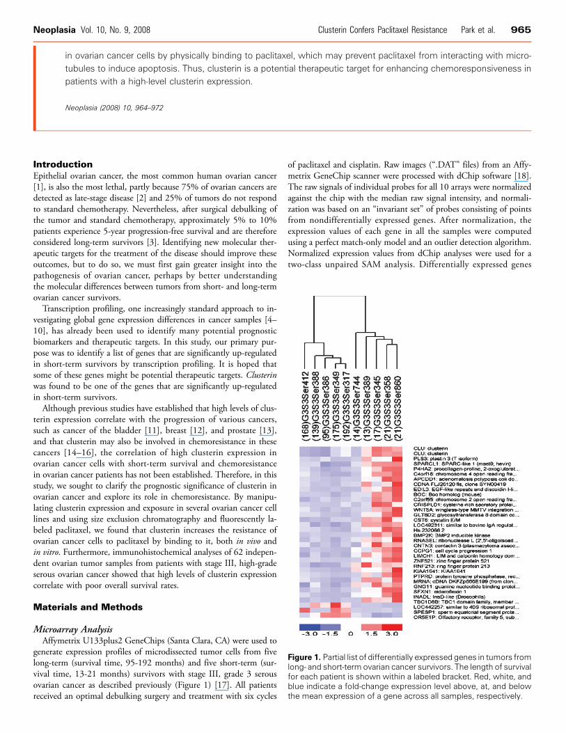
generate expression profiles of microdissected tumor cells from fivelong-term (survival time, 95-192 months) and five short-term (sur-vival time, 13-21 months) survivors with stage III, grade 3 serousovarian cancer as described previously (Figure 1) [17]. All patientsreceived an optimal debulking surgery and treatment with six cycles
of paclitaxel and cisplatin. Raw images (“.DAT” files) from an Affy-metrix GeneChip scanner were processed with dChip software [18].The raw signals of individual probes for all 10 arrays were normalizedagainst the chip with the median raw signal intensity, and normali-zation was based on an “invariant set” of probes consisting of pointsfrom nondifferentially expressed genes. After normalization, theexpression values of each gene in all the samples were computedusing a perfect match-only model and an outlier detection algorithm.Normalized expression values from dChip analyses were used for atwo-class unpaired SAM analysis. Differentially expressed genes
Figure 1. Partial list of differentially expressed genes in tumors fromlong- and short-term ovarian cancer survivors. The length of survivalfor each patient is shown within a labeled bracket. Red, white, andblue indicate a fold-change expression level above, at, and belowthe mean expression of a gene across all samples, respectively.

966 Clusterin Confers Paclitaxel Resistance Park et al. Neoplasia Vol. 10, No. 9, 2008
were identified by supervised analysis with Significance Analysis ofMicroarrays (SAM) software [19], which yields results similar tothose obtained using Student’s t test but includes permutations tocalculate the false discovery rate, or a q value, for each gene. Eachq value represents the probability that a gene is falsely identified asdifferentially expressed, with smaller q values indicating more signif-icant differential expression levels.
Cell Lines and Tissue SamplesAll 13 ovarian cancer cell lines (PEO4, CAOV3, OVCA420,
TOV21G, RMUG-S, RMUG-L, OVCA822, 0V2008, ES2, TOV112,OVCA433, OVCA432, and SKOV3) were grown in a medium ofequal parts M199 and MCDB105 supplemented with 10% fetalbovine serum as described previously [20]. All tumor specimensfrom patients were collected and archived according to protocolsapproved by the institutional review boards of the appropriate insti-tutions that included an informed consent or a waiver. All the pa-tients had stage III and grade III serous ovarian tumors and weretreated uniformly with paclitaxel/carboplatin first-line chemother-apy regimen.
ImmunohistochemistryFive normal ovarian surface epithelial samples and 62 samples
from previously untreated stage III, high-grade primary papillaryserous carcinomas were immunostained using an avidin-biotinmethod with an anti–clusterin α chain mouse monoclonal antibody(5 μg/ml; Upstate Biotechnology, Lake Placid, NY), with clusterinexpression scored as described previously [21]. Cox regression andKaplan-Meier survival analyses were used to compare immunohisto-chemical results with patient survival data. Statistical significance wasdetermined by using the log rank test with statistical software SPSSversion 15.0.
Western Blot AnalysisCell lysates were prepared from the 13 ovarian cancer cell lines
[20] and subjected to Western blot analysis using an anti–clusterinmouse monoclonal antibody (Upstate Biotechnology) and an anti–β-actin monoclonal antibody for normalization of protein loading.Immunoreactivity was detected using the ECL chemiluminescencesystem (Amersham, Piscataway, NJ) and quantified using an imagingdensitometer (Model GS-670; Bio-Rad, Hercules, CA).
IC50 and Cell Proliferation AssayCells (1 × 10⁴ per well) were seeded in a 96-well plate in 0.1 ml of
culture medium and allowed to grow overnight. The next day, themedium was supplemented with either vehicle control or 10−9 to10−4 M paclitaxel (Sigma, St. Louis, MO). The 50% inhibitory drugconcentration (IC50) was determined using the 2,3-bis(2-methoxy-4-nitro-5-sulfophenyl)-2H -tetrazolium-5-carboxanilide (XTT) assay(Roche Molecular Biochemicals, Indianapolis, IN) according to themanufacturer’s instructions. The metabolism of XTTwas quantifiedby measuring the absorbance at 450 nm, and the IC50 of paclitaxelfor each of the cell lines was estimated from the semilogarithmicdose-response curves by linear interpolation.
DNA Fragmentation Assay for ApoptosisCells (1 × 10⁴ per well) were seeded in a 96-well plate in 0.1 ml
of culture medium and allowed to grow overnight. The next day, cellswere treated with 10−7 M paclitaxel for 48 hours at 37°C in the pres-
ence or absence of clusterin. The amounts of cytoplasmic histone-associated DNA fragments (mononucleosome and oligonucleosomes)formed during apoptosis were measured using a cell death detectionELISAPlus kit (Roche Molecular Biochemicals) according to the manu-facturer’s instructions. Each assay was performed in quadruplicate, andeach experiment was repeated three times. The relative percentages ofDNA fragmentation were calculated as the ratios OD405 readings frompaclitaxel-treated to paclitaxel-untreated cells.
Generation of Stably Transfected CellsOverexpressing Clusterin
SKOV3 cells were transfected with full-length clusterin cDNAcloned into pcDNA3.1 expression vectors (pcDNA3.1/CLU) usingFuGENE 6 transfection reagent according to the manufacturer’sprotocol (Roche Molecular Biochemicals). Mock transfection wasperformed the same way, using the vector pcDNA3.1. Stably trans-fected clones were selected 36 hours later by adding G418 (RocheMolecular Biochemicals) at 500 μg/ml. The stable clones were thenevaluated for the expression of clusterin and paclitaxel resistance.
In Vitro Small Interfering RNA–Mediated Gene SilencingClusterin small interfering RNA (siRNA) duplex, consisting of
nucleotides +85 to +96 (where the translation start site was definedas +1; Dharmacon Research, Lafayette, CO), or luciferase GL2Duplex (Dharmacon) at 0.2 nmol/ml was transfected into ovarian can-cer cells expressing high levels of clusterin (PEO4, RMUG-S, andTOV21G) using an oligofectamine reagent (Invitrogen, Carlsbad,CA) as described by the manufacturer’s protocols (Life Technologies,Inc., Gaithersburg, MD). Transfected cells were evaluated for clusterinexpression and sensitivity to paclitaxel.
Exogenous Clusterin TreatmentClusterin was purified from human sera by immunoaffinity chro-
matography, using an anti–clusterin monoclonal antibody undernative conditions at 4°C as described previously [22]. Analysis ofclusterin purity by SDS-PAGE showed a major band (nonreduced)at 75 to 80 kDa [22]. To further ensure the purity of the product,we performed an additional purification step using ion-exchangechromatography. The eluate from the immunoaffinity column wasdialyzed against 50 mM Tris buffer (pH 8.0), loaded onto a Q-Sepharose FF column, and eluted with a linear NaCl gradient in thesame buffer. Clusterin-containing fractions (as judged by SDS-PAGE)were then pooled and concentrated. SKOV3 cells (1 × 10⁴ cells perwell) in 96-well plates were treated with 10−7 M paclitaxel in the pres-ence of 0.5, 2.5, 5, and 7.5 and 15 μg/ml purified clusterin for 48 hoursat 37°C. Cells were then harvested for XTT and apoptosis assays.
In Vivo Short Hairpin RNA–Mediated Gene SilencingUsing a Lentiviral Expression System
A Lentiviral vector expressing short hairpin RNA (shRNA) tar-geting clusterin was constructed using the BLOCK-iT LentiviralRNAi expression system (Invitrogen). DNA oligonucleotides en-coding shRNA of clusterin (CLU) were designed using BLOCK-iT RNAi designer (Invitrogen) and inserted downstream of thehuman U6 pol III promoter and upstream of a pol III terminatorin pENTR/U6 vector (Invitrogen) and then into the vector pLenti-GW/U6 as pLenti-GW/U6-CLUshRNA. Lentiviral particles were
Neoplasia Vol. 10, No. 9, 2008 Clusterin Confers Paclitaxel Resistance Park et al. 967
then generated from pLenti-GW/U6-CLUshRNA by transfecting293 FT cell lines with the shRNA construct and helper plasmids.To investigate the effects of decreased clusterin expression on
paclitaxel resistance in xenografted tumors, forty 6- to 8-week-oldfemale nude mice (SLC; Shizuoka, Japan) were evenly distributed forgenerating xenograft mouse models using control cells (PEO4 trans-duced with Lentiviral particles carrying shRNA for the LacZ gene)or clusterin knockdown cells (PEO4 transduced with Lentiviral parti-cles carrying shRNA for the CLU gene). After being transduced withLentiviral particles carrying shRNA for the LacZ or CLU gene over-night, 5 × 10⁵ paclitaxel-resistant PEO4 cells were used to inoculatesubcutaneously into the posterior neck region of each nude mouse.When the xenografts grew to approximately 100 mm3, mice were in-jected intraperitoneally with 0.2 mg/0.1 ml per 10 g of paclitaxel threetimes per week for 3 to 4 weeks. Tumor volumes were measured twiceevery week and calculated using the formula: length × width × depth ×0.5236. The mice were euthanized at day 27, and the levels of clusterinexpression in xenografted tumors were determined by Western blotanalysis. Animals were housed in pathogen-free units in compliancewith institutional animal care and use committee regulations.
Effects of Clusterin on the Binding of Paclitaxel toMicrotubules in Ovarian Cancer CellsPEO4 and SKOV3 cells were trypsinized, inoculated into cham-
bered coverglasses (Fisher Scientific, Pittsburgh, PA), and incubatedfor 12 hours at 37°C. The cells were washed three times with 2%BSA/PBS, 10−6 M Oregon Green (OG) 488–labeled paclitaxel(paclitaxel-OG; Invitrogen) was added, and the cells were incubatedfor 1 hour at 37°C in PEM buffer (50 mM PIPES, 2 mM EGTA,2 mM MgCl2, pH 7.4), with or without 7.5 mg/ml purified clus-terin. After incubation with the 10−6 mg/ml Hoechst 33258 penta-hydrate (Molecular Probes, Eugene, OR) for 15 minutes at 37°C, thecells were examined using a Leica DMIRE2 inverted fluorescencemicroscope with appropriate filters.
Measurement of Paclitaxel-Clusterin BindingSolutions of clusterin or control protein glutathione-S -transferase
(GST) at 5 mM, alone or in the presence of a 1:1 molar ratio ofpaclitaxel-OG, phalloidin-OG, or OG in PEM were incubated for1 hour at 37°C. Then, solutions were fractionated using size exclusionchromatography with a Biosep SEC S4000 column (Phenomenex,Sydney, Australia) and an AKTA FPLC system (GE Healthcare,Sydney, Australia); the A280 of the eluate, as an indication of pro-tein elution, was measured continuously. Collected fractions (200 mleach) were transferred into the wells of black 96-well plates (Greiner,Frickenhausen, Germany), and the fluorescence of paclitaxel-OG,phalloidin-OG, and OG was measured using a Fluostar microplatereader (BMG Labtech, Melbourne; excitation, 485 ± 5 nm; emis-sion, 520 ± 5 nm). Other control proteins (superoxide dismutaseand hemoglobin) were also tested.
Results
Expression Profiling of Microdissected Tumors fromShort- and Long-term SurvivorsUsing SAM analysis to compare the expression profiles of tumors
from five short- and five long-term ovarian cancer survivors, we iden-
tified 77 probe sets with greater than threefold expression in short-than in long-term survivors (Table W1; q < 15%). Two of theseprobe sets encode clusterin that was up-regulated 3.8-fold more inshort- than in long-term survivors (Figure 1).
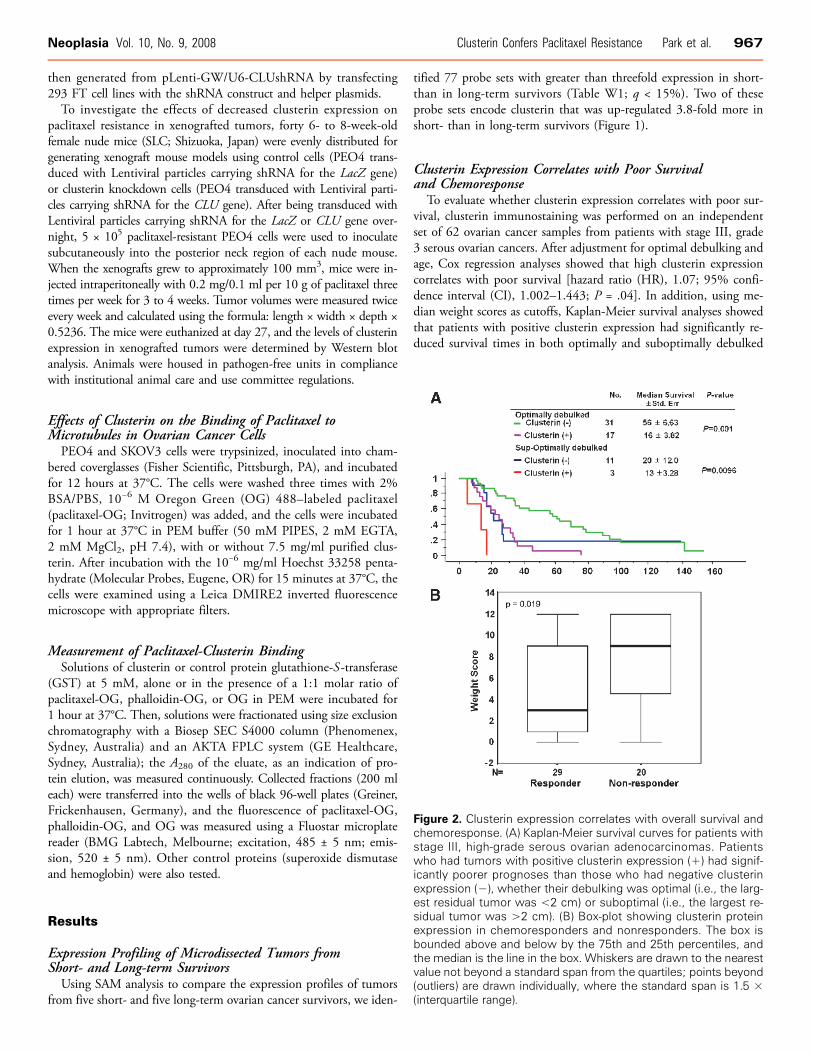
Clusterin Expression Correlates with Poor Survivaland Chemoresponse
To evaluate whether clusterin expression correlates with poor sur-vival, clusterin immunostaining was performed on an independentset of 62 ovarian cancer samples from patients with stage III, grade3 serous ovarian cancers. After adjustment for optimal debulking andage, Cox regression analyses showed that high clusterin expressioncorrelates with poor survival [hazard ratio (HR), 1.07; 95% confi-dence interval (CI), 1.002–1.443; P = .04]. In addition, using me-dian weight scores as cutoffs, Kaplan-Meier survival analyses showedthat patients with positive clusterin expression had significantly re-duced survival times in both optimally and suboptimally debulked
Figure 2. Clusterin expression correlates with overall survival andchemoresponse. (A) Kaplan-Meier survival curves for patients withstage III, high-grade serous ovarian adenocarcinomas. Patientswho had tumors with positive clusterin expression (+) had signif-icantly poorer prognoses than those who had negative clusterinexpression (−), whether their debulking was optimal (i.e., the larg-est residual tumor was <2 cm) or suboptimal (i.e., the largest re-sidual tumor was >2 cm). (B) Box-plot showing clusterin proteinexpression in chemoresponders and nonresponders. The box isbounded above and below by the 75th and 25th percentiles, andthe median is the line in the box. Whiskers are drawn to the nearestvalue not beyond a standard span from the quartiles; points beyond(outliers) are drawn individually, where the standard span is 1.5 ×(interquartile range).
968 Clusterin Confers Paclitaxel Resistance Park et al. Neoplasia Vol. 10, No. 9, 2008
cases (P = .001 and P = .0096, respectively; Figure 2A). Normal ovar-ian surface epithelial cells had no immunostaining of clusterin. Theimmunostaining intensities of clusterin (weight score) in the non-responder group was significantly (P = .019) higher than those inthe responder group (Figure 2B).
Clusterin Expression Correlates with Paclitaxel Resistancein Ovarian Cancer Cell Lines
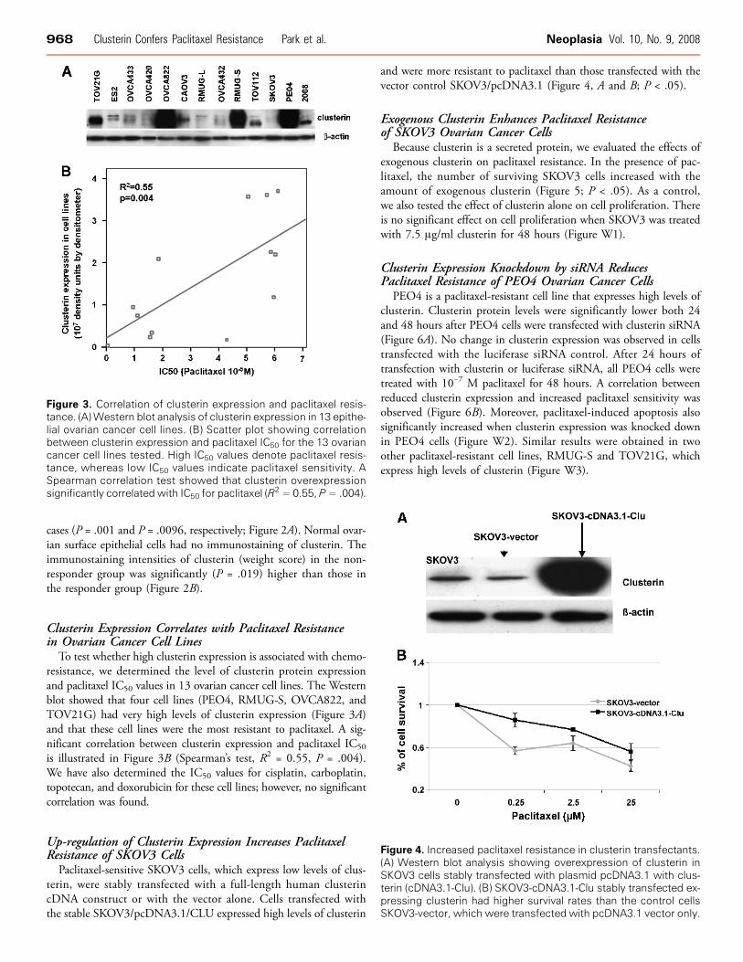
To test whether high clusterin expression is associated with chemo-resistance, we determined the level of clusterin protein expressionand paclitaxel IC50 values in 13 ovarian cancer cell lines. The Westernblot showed that four cell lines (PEO4, RMUG-S, OVCA822, andTOV21G) had very high levels of clusterin expression (Figure 3A)and that these cell lines were the most resistant to paclitaxel. A sig-nificant correlation between clusterin expression and paclitaxel IC50
is illustrated in Figure 3B (Spearman’s test, R2 = 0.55, P = .004).We have also determined the IC50 values for cisplatin, carboplatin,topotecan, and doxorubicin for these cell lines; however, no significantcorrelation was found.
Up-regulation of Clusterin Expression Increases PaclitaxelResistance of SKOV3 Cells
Paclitaxel-sensitive SKOV3 cells, which express low levels of clus-terin, were stably transfected with a full-length human clusterincDNA construct or with the vector alone. Cells transfected withthe stable SKOV3/pcDNA3.1/CLU expressed high levels of clusterin
and were more resistant to paclitaxel than those transfected with thevector control SKOV3/pcDNA3.1 (Figure 4, A and B; P < .05).
Exogenous Clusterin Enhances Paclitaxel Resistanceof SKOV3 Ovarian Cancer Cells
Because clusterin is a secreted protein, we evaluated the effects ofexogenous clusterin on paclitaxel resistance. In the presence of pac-litaxel, the number of surviving SKOV3 cells increased with theamount of exogenous clusterin (Figure 5; P < .05). As a control,we also tested the effect of clusterin alone on cell proliferation. Thereis no significant effect on cell proliferation when SKOV3 was treatedwith 7.5 μg/ml clusterin for 48 hours (Figure W1).
Clusterin Expression Knockdown by siRNA ReducesPaclitaxel Resistance of PEO4 Ovarian Cancer Cells
PEO4 is a paclitaxel-resistant cell line that expresses high levels ofclusterin. Clusterin protein levels were significantly lower both 24and 48 hours after PEO4 cells were transfected with clusterin siRNA(Figure 6A). No change in clusterin expression was observed in cellstransfected with the luciferase siRNA control. After 24 hours oftransfection with clusterin or luciferase siRNA, all PEO4 cells weretreated with 10−7 M paclitaxel for 48 hours. A correlation betweenreduced clusterin expression and increased paclitaxel sensitivity wasobserved (Figure 6B). Moreover, paclitaxel-induced apoptosis alsosignificantly increased when clusterin expression was knocked downin PEO4 cells (Figure W2). Similar results were obtained in twoother paclitaxel-resistant cell lines, RMUG-S and TOV21G, whichexpress high levels of clusterin (Figure W3).
Figure 4. Increased paclitaxel resistance in clusterin transfectants.(A) Western blot analysis showing overexpression of clusterin inSKOV3 cells stably transfected with plasmid pcDNA3.1 with clus-terin (cDNA3.1-Clu). (B) SKOV3-cDNA3.1-Clu stably transfected ex-pressing clusterin had higher survival rates than the control cellsSKOV3-vector, which were transfected with pcDNA3.1 vector only.
Figure 3. Correlation of clusterin expression and paclitaxel resis-tance. (A)Western blot analysis of clusterin expression in 13 epithe-lial ovarian cancer cell lines. (B) Scatter plot showing correlationbetween clusterin expression and paclitaxel IC50 for the 13 ovariancancer cell lines tested. High IC50 values denote paclitaxel resis-tance, whereas low IC50 values indicate paclitaxel sensitivity. ASpearman correlation test showed that clusterin overexpressionsignificantly correlated with IC50 for paclitaxel (R
2 = 0.55, P= .004).

Neoplasia Vol. 10, No. 9, 2008 Clusterin Confers Paclitaxel Resistance Park et al. 969
Clusterin Expression Knockdown in Ovarian Cancer Cells byshRNA Reduces Tumor Size in Xenograft Mouse Models WhenTreated with PaclitaxelPEO4 cells transduced with Lentivirus carrying clusterin or LacZ
shRNA were xenografted into mice, and the effects of paclitaxelwere determined. After 22 days, the tumors that had developedfrom the xenografted cells transduced with clusterin shRNA weresignificantly smaller than those of the control groups (Figure 7A).Decreased clusterin expression in xenografted tumor masses afterautopsy was confirmed by Western blot analysis and densitometerquantification. The difference between shRNA-clusterin–treated tu-mor and shRNA-control was significant (twofold decrease) with P =.037 (Figure 7B).
Effect of Clusterin on Microtubule BindingTo evaluate whether clusterin confers paclitaxel resistance by pre-
venting the binding of paclitaxel to microtubules, both paclitaxel-resistant PEO4 cells and paclitaxel-sensitive SKOV3 cells were treatedwith identical concentrations of paclitaxel-OG. PEO4 cells had min-
imal microtubule staining in the absence or presence of exogenousclusterin (Figure 8, A and B). In contrast, SKOV3 cells had strongcytoplasmic staining in the absence of clusterin, whereas no stain-ing was observed in the presence of exogenous clusterin, suggestingthat exogenous clusterin prevents the binding of paclitaxel to mi-crotubules (Figure 8, C and D). To clarify the role of endogenousclusterin, we infected PEO4 cells with Lentivirus carrying shRNA-clusterin (Lenti-shRNA-clusterin) to knockdown the endogenous clus-terin expression. The result showed that PEO4 cells infected withLenti-shRNA-clusterin had strong cytoplasmic stain by paclitaxel-OG, whereas PEO4 cells infected with Lentivirus vector controlhad undetectable staining similar to Figure 8, C and B, respectively(Figure W4).
Figure 6. PEO4 became sensitive to paclitaxel after silencing clus-terin expression with siRNA. (A) Western blot analyses show thetime course of clusterin silencing in PEO4 cells transfected withsiRNA for clusterin (+) or luciferase (−). After transfection, thecells were cultured for 2 days in medium containing 10−7 M pac-litaxel. (B) Cell survival after paclitaxel treatment as determined byXTT assay.
Figure 7. Effects of decreased clusterin expression on paclitaxelresistance in xenografted tumors. (A) When xenografts in nudemice resulting from the inoculation of paclitaxel-resistant cellstransduced with shRNA for clusterin or the Lacz gene grew to ap-proximately 100 mm3, mice were injected with 0.2 mg/0.1 ml per10 g of paclitaxel intraperitoneally. Tumor volumes were measuredtwice every week. (B) Western blot analysis of clusterin expressionfrom xenografted tumors from mouse autopsies. Tumors fromthree separate mice from both the shRNA-Lacz and shRNA-clusteringroups were analyzed.
Figure 5. Exogenous clusterin enhances the paclitaxel resistance of SKOV3 cells. Cell proliferation was measured by XTT assay inSKOV3 cells treated with 10−7 M paclitaxel (T) for 48 hours in the presence of the indicated concentrations of clusterin. The baselineused for calculating relative cell proliferation rate was set by adjusting the proliferation rate of paclitaxel-treated SKOV3 cells withoutexogenous clusterin to 1.

970 Clusterin Confers Paclitaxel Resistance Park et al. Neoplasia Vol. 10, No. 9, 2008
Binding Interactions between Clusterin and PaclitaxelTo evaluate whether clusterin binds specifically to paclitaxel, equi-
molar mixtures of clusterin or GST as a control and paclitaxel-OG,phalloidin-OG, or OG were incubated together and then fraction-ated by size exclusion chromatography. After incubation with clus-terin, the elution profile of paclitaxel-OG adopted a pattern similarto that of clusterin alone (Figure 9, A and B). In contrast, after in-cubation with GST, the elution profile of paclitaxel-OG was similarto that of paclitaxel-OG alone, implying that under these conditions,the small molecule remained unbound. Similar results were obtainedwith two other control proteins, superoxide dismutase and hemoglo-bin (data not shown), and OG and phalloidin-OG, the control dyes(Figure W5).
DiscussionBy comparing the expression profiles generated from short- and
long-term ovarian cancer survivors, we found that clusterin expres-sion was significantly higher in short- than in long-term survivors(Figure 1). Also, Kaplan-Meier survival analyses of results from clus-terin immunostaining in 62 samples of late-stage, high-grade ovarianserous carcinomas indicated that the overexpression of clusterin cor-relates with poor survival (Figure 2A). This is consistent with reportsthat clusterin is a marker of poor prognosis in other cancer types [23–27]. Moreover, overexpression of clusterin also correlates with che-moresistance (Figure 2B).
Clusterin, also known as testosterone-repressed prostate message-2(TRPM-2), sulfated glycoprotein-2 (SGP-2), SP-40, and apolipopro-tein J (ApoJ), is a secreted heterodimeric glycoprotein encoded by asingle gene on chromosome 8p21-p12. When analyzed by SDS-PAGE, clusterin migrates as a broad band at 70 to 80 kDa but
has an actual mass of approximately 61 kDa (determined by massspectrometry) [28]. Clusterin is present in various biologic fluids[29] and has been implicated in several physiological processes, in-cluding cell adhesion and aggregation [30], complement inhibition[31], lipid transport, membrane protection, sperm maturation, andendocrine secretion [31,32]. Clusterin expression has also been im-plicated in chemoresistance in several other cancer types [14,15].Because the resistance of tumor cells to various available chemother-apeutic agents such as paclitaxel has been one of the major factorsleading to poor survival in ovarian cancer patients, we therefore hy-pothesized that clusterin expression confers chemoresistance to ovar-ian cancer cells.
In this study, we demonstrated that clusterin expression correlatedwith paclitaxel resistance both in vitro and in vivo. We found thathigh levels of clusterin expression in ovarian cancer cell lines corre-lated with paclitaxel resistance, although a few cell lines with lowlevels of clusterin expression were also resistant to paclitaxel (Fig-ure 3). These cell lines might use mechanisms other than overexpres-sion of clusterin to confer paclitaxel resistance such as the expressionof metabolizing enzyme cytochrome P450 or cellular efflux ABCtransporter [33]. To demonstrate the role of clusterin in paclitaxel re-sistance, we manipulated the endogenous level of clusterin expressionin a paclitaxel-sensitive cell line (SKOV3, low level of endogenousclusterin expression) and a paclitaxel-resistant cell line (PEO4, highlevel of endogenous clusterin expression). We found that paclitaxel-sensitive SKOV3 cells becamemore resistant to paclitaxel when endoge-nous clusterin expression level was up-regulated (Figure 4). Conversely,
Figure 9. Coelution of clusterin and OG-labeled paclitaxel (paclitaxel-OG) in size exclusion chromatography. Solutions containing clus-terin and paclitaxel-OG mixture, paclitaxel-OG alone, or a GST andpaclitaxel-OG mixture (all at 5 mM) were fractionated by size exclu-sion chromatography in PEM buffer, and the OG fluorescence andA280 of the eluate were measured. The upper panel shows OGfluorescence as a function of elution time; the lower panel showsthe corresponding A280 profile. The result shown is representativeof three independent experiments.
Figure 8. Exogenous clusterin prevents the binding of paclitaxel tomicrotubules in SKOV3 cells. PEO4 (A and B) and SKOV3 (C and D)cells cultured in chamber coverglasses were treated with 10−6 MOG-labeled paclitaxel in the absence (A and C) or presence (B andD) of 7.5 μg/ml clusterin for 30 minutes. Cells were also simulta-neously incubated with the Hoechst stain for nuclei visualization,and fluorescent images were captured by an inverted fluores-cence microscope equipped with a digital camera using the samesetting. The binding of OG-labeled paclitaxel to microtubules isshown in panel C.

Neoplasia Vol. 10, No. 9, 2008 Clusterin Confers Paclitaxel Resistance Park et al. 971
paclitaxel-resistant PEO4 cells became more sensitive to paclitaxeland more apoptotic in vitro and in vivo when endogenous clusterinexpression level was down-regulated. These results indicated that highlevels of endogenous clusterin expression were involved in the pacli-taxel resistance of ovarian cancer cells.In addition to the endogenous clusterin, the exogenous clusterin
added in our experiments enhanced the paclitaxel resistance ofSKOV3 cells, as indicated by the cells’ increased survival rate andthe reduction in paclitaxel-induced apoptosis (Figure 5). The extentof both the increased survival and the reduced apoptosis correlatedwith the increase in concentration of exogenous clusterin. This sug-gests that exogenous clusterin binds to extracellular paclitaxel andsubsequently prevents it from entering the cells and exerting toxicity.To test the hypothesis that the exogenous clusterin secreted by ovar-ian cancer cells inhibits the binding of paclitaxel to microtubules insidethe cells, we determined the level of microtubule-associated fluo-rescence in paclitaxel-OG–treated ovarian cancer cells with or with-out clusterin. Significant microtubule-associated fluorescence wasobserved in SKOV3 cells (Figure 8C ) but not in PEO4 cells, whichexpressed high levels of endogenous clusterin (Figure 8, A and B).However, when SKOV3 cells were coincubated with both exogenousclusterin and paclitaxel-OG, negligible microtubule-associated fluo-rescence was observed (Figure 8D).Using size exclusion chromatography to directly test whether clus-
terin bound specifically to paclitaxel in solution, we found thatclusterin did bind to paclitaxel-OG but not to phalloidin-OG orOG alone and that GST and other control proteins did not bindto paclitaxel-OG. In addition, ELISA confirmed that clusterin boundwith high affinity to paclitaxel but not at all to other therapeuticagents, such as cisplatin and carboplatin (data not shown). This isthe first time we are aware of that the binding of clusterin and pac-litaxel has been clearly demonstrated, although clusterin’s role in che-moresistance has been suggested by previous reports in other cancertypes [14–16]. Thus, our results suggest that clusterin, which is gen-erally a secreted molecule, may protect cancer cells from paclitaxeltoxicity by binding to the agent in the extracellular microenviron-ment and preventing it from entering the cell to exert its toxic effects.In conclusion, clusterin overexpression significantly correlates with
decreased survival in ovarian cancer patients, and clusterin confers re-sistance to paclitaxel-induced apoptosis in ovarian cancer cells. Themechanism of resistance may involve clusterin (potentially in bothextra- and intracellular locations) complexing with paclitaxel to inac-tivate the agent’s cytotoxic activity. Therefore, targeting the reductionof clusterin expression may be a worthwhile new therapeutic modalityfor treating ovarian tumors expressing high levels of clusterin.
References[1] Boring CC, Squires TS, Tong T, and Montgomery S (1994). Cancer statistics,
1994. CA Cancer J Clin 44, 7–26.[2] Landis SH, Murray T, Bolden S, and Wingo PA (1998). Cancer statistics, 1998.
CA Cancer J Clin 48, 6–29.[3] Omura GA, Brady MF, Homesley HD, Yordan E, Major FJ, Buchsbaum HJ,
and Park RC (1991). Long-term follow-up and prognostic factor analysis in ad-vanced ovarian carcinoma: the Gynecologic Oncology Group experience. J ClinOncol 9, 1138–1150.
[4] Barlund M, Forozan F, Kononen J, Bubendorf L, Chen Y, Bittner ML, TorhorstJ, Haas P, Bucher C, Sauter G, et al. (2000). Detecting activation of ribosomalprotein S6 kinase by complementary DNA and tissue microarray analysis. J NatlCancer Inst 92, 1252–1259.
[5] Wang T, Hopkins D, Schmidt C, Silva S, Houghton R, Takita H, Repasky E,and Reed SG (2000). Identification of genes differentially over-expressed in lungsquamous cell carcinoma using combination of cDNA subtraction and micro-array analysis. Oncogene 19, 1519–1528.
[6] Xu J, Stolk JA, Zhang X, Silva SJ, Houghton RL, Matsumura M, Vedvick TS,Leslie KB, Badaro R, and Reed SG (2000). Identification of differentially ex-pressed genes in human prostate cancer using subtraction and microarray. Can-cer Res 60, 1677–1682.
[7] DeRisi J, Penland L, Brown PO, Bittner ML, Meltzer PS, Ray M, Chen Y, SuYA, and Trent JM (1996). Use of a cDNA microarray to analyse gene expressionpatterns in human cancer [see comments]. Nat Genet 14, 457–460.
[8] DeRisi JL, Iyer VR, and Brown PO (1997). Exploring the metabolic and geneticcontrol of gene expression on a genomic scale. Science 278, 680–686.
[9] Golub TR, Slonim DK, Tamayo P, Huard C, Gaasenbeek M, Mesirov JP, CollerH, Loh ML, Downing JR, Caligiuri MA, et al. (1999). Molecular classificationof cancer: class discovery and class prediction by gene expression monitoring.Science 286, 531–537.
[10] Alizadeh AA, Eisen MB, Davis RE, Ma C, Lossos IS, Rosenwald A, Boldrick JC,Sabet H, Tran T, Yu X, et al. (2000). Distinct types of diffuse large B-cell lym-phoma identified by gene expression profiling. Nature 403, 503–511.
[11] Miyake H, Gleave M, Kamidono S, and Hara I (2002). Overexpression of clus-terin in transitional cell carcinoma of the bladder is related to disease progressionand recurrence. Urology 59, 150–154.
[12] Redondo M, Villar E, Torres-Munoz J, Tellez T, Morell M, and Petito CK(2000). Overexpression of clusterin in human breast carcinoma. Am J Pathol157, 393–399.
[13] Sensibar JA, Sutkowski DM, Raffo A, Buttyan R, Griswold MD, Sylvester SR,Kozlowski JM, and Lee C (1995). Prevention of cell death induced by tumornecrosis factor alpha in LNCaP cells by overexpression of sulfated glycoprotein-2(clusterin). Cancer Res 55, 2431–2437.
[14] GleaveME,MiyakeH,Zellweger T, Chi K, July L,NelsonC, and Rennie P (2001).Use of antisense oligonucleotides targeting the antiapoptotic gene, clusterin/testosterone–repressed prostate message 2, to enhance androgen sensitivity and chemo-sensitivity in prostate cancer. Urology 58, 39–49.
[15] Miyake H, Hara I, Kamidono S, and Gleave ME (2001). Synergistic chemsen-sitization and inhibition of tumor growth and metastasis by the antisense oligo-deoxynucleotide targeting clusterin gene in a human bladder cancer model. ClinCancer Res 7, 4245–4252.
[16] Miyake H, Nelson C, Rennie PS, and Gleave ME (2000). Acquisition of che-moresistant phenotype by overexpression of the antiapoptotic gene testosterone-repressed prostate message-2 in prostate cancer xenograft models. Cancer Res 60,2547–2554.
[17] Bonome T, Lee JY, Park DC, Radonovich M, Pise-Masison C, Brady J, GardnerGJ, Hao K, Wong WH, Barrett JC, et al. (2005). Expression profiling of serouslow malignant potential, low-grade, and high-grade tumors of the ovary. CancerRes 65, 10602–10612.
[18] Li C and Wong WH (2001). Model-based analysis of oligonucleotide arrays:expression index computation and outlier detection. Proc Natl Acad Sci USA98, 31–36.
[19] Tusher VG, Tibshirani R, and Chu G (2001). Significance analysis of micro-arrays applied to the ionizing radiation response. Proc Natl Acad Sci USA 98,5116–5121.
[20] Kim JH, Skates SJ, Uede T, Wong KK, Schorge JO, Feltmate CM, BerkowitzRS, Cramer DW, and Mok SC (2002). Osteopontin as a potential diagnosticbiomarker for ovarian cancer. JAMA 287, 1671–1679.
[21] Mok SC, Chao J, Skates S, Wong KK, Yiu GY, Muto MG, Berkowitz RS, andCramer DW (2001). Prostasin, a potential serum marker for ovarian cancer,identified through microarray technology. J Natl Cancer Inst 93, 1458–1464.
[22] Wilson MR and Easterbrook-Smith SB (2000). Clusterin is a secreted mamma-lian chaperone. Trends Biochem Sci 25, 95–98.
[23] Miyake H, Gleave M, Kamidono S, and Hara I (2002). Overexpression of clus-terin in transitional cell carcinoma of the bladder is related to disease progressionand recurrence. Urology 59, 150–154.
[24] Kruger S, Ola V, Fischer D, Feller AC, and Friedrich M (2007). Prognosticsignificance of clusterin immunoreactivity in breast cancer. Neoplasma 54,46–50.
[25] Miyake H, Hara S, Arakawa S, Kamidono S, and Hara I (2002). Over expres-sion of clusterin is an independent prognostic factor for nonpapillary renal cellcarcinoma. J Urol 167, 703–706.

972 Clusterin Confers Paclitaxel Resistance Park et al. Neoplasia Vol. 10, No. 9, 2008
[26] Miyake H, Yamanaka K, Muramaki M, Kurahashi T, Gleave M, and Hara I(2005). Enhanced expression of the secreted form of clusterin following neo-adjuvant hormonal therapy as a prognostic predictor in patients undergoing rad-ical prostatectomy for prostate cancer. Oncol Rep 14, 1371–1375.
[27] Zhang S, Zhang D, Zhu Y, Guo H, Zhao X, and Sun B (2006). Clusterin ex-pression and univariate analysis of overall survival in human breast cancer. Tech-nol Cancer Res Treat 5, 573–578.
[28] Kapron JT, Hilliard GM, Lakins JN, Tenniswood MP, West KA, Carr SA, andCrabb JW (1997). Identification and characterization of glycosylation sites inhuman serum clusterin. Protein Sci 6, 2120–2133.
[29] Aronow BJ, Lund SD, Brown TL, Harmony JA, and Witte DP (1993). Apo-lipoprotein J expression at fluid-tissue interfaces: potential role in barrier cyto-protection. Proc Natl Acad Sci USA 90, 725–729.
[30] Silkensen JR, Skubitz KM, Skubitz AP, Chmielewski DH, Manivel JC,Dvergsten JA, and Rosenberg ME (1995). Clusterin promotes the aggregationand adhesion of renal porcine epithelial cells. J Clin Invest 96, 2646–2653.
[31] Rodenburg KW, Kjoller L, Petersen HH, and Andreasen PA (1998). Binding ofurokinase-type plasminogen activator–plasminogen activator inhibitor-1 com-plex to the endocytosis receptors alpha2-macroglobulin receptor/low-density li-poprotein receptor–related protein and very-low-density lipoprotein receptorinvolves basic residues in the inhibitor. Biochem J 329, 55–63.
[32] Jenne DE and Tschopp J (1992). Clusterin: the intriguing guises of a widelyexpressed glycoprotein. Trends Biochem Sci 17, 154–159.
[33] DeLoia JA, Zamboni WC, Jones JM, Strychor S, Kelley JL, and Gallion HH(2008). Expression and activity of taxane-metabolizing enzymes in ovarian tu-mors. Gynecol Oncol 108, 355–360.

Table W1. List of Genes Up-regulated in Short-term Survivors.
ProbeSet ID
Gene Name Fold Change q (%)205051_s_at
v-kit Hardy-Zuckerman 4 feline sarcoma viral oncogene homolog 4.2 0 207480_s_at Meis1, myeloid ecotropic viral integration site 1 homolog 2 (mouse) 3.3 0 226977_at similar to bovine IgA regulatory protein 3.7 4.5 230000_at chromosome 17 open reading frame 27 3.1 4.5 212327_at hypothetical protein 3.5 11.6 200795_at SPARC-like 1 (mast9, hevin) 3.4 11.6 204115_at guanine nucleotide binding protein (G protein), gamma 11 3.4 11.6 227354_at phosphoprotein associated with glycosphingolipid microdomains 1 3.3 11.6 209348_s_at v-maf musculoaponeurotic fibrosarcoma oncogene homolog (avian) 3.3 11.6 210346_s_at CDC-like kinase 4 3.2 11.6 227131_at mitogen-activated protein kinase kinase kinase 3 3 11.6 206595_at cystatin E/M 5.2 11.7 223204_at chromosome 4 open reading frame 18 4.4 11.7 238067_at TBC1 domain family, member 8B (with GRAM domain) 4.2 11.7 220940_at KIAA1641 4.2 11.7 213905_x_at biglycan /// teashirt family zinc finger 1 4.1 11.7 211458_s_at GABA(A) receptor–associated protein like 1 /// GABA(A) receptorsassociated protein like 3
4 11.71559060_a_at
KIAA1961 gene 4 11.7 228256_s_at erythrocyte membrane protein band 4.1 like 4A 4 11.7 204777_s_at mal, T-cell differentiation protein 4 11.7 1559360_at Ephrin-A5 3.9 11.7 208791_at clusterin 3.8 11.7 208792_s_at clusterin 3.8 11.7 201261_x_at biglycan 3.8 11.7 233952_s_at zinc finger protein 295 3.7 11.7 241893_at Mannosyl (alpha-1,6-)-glycoprotein beta-1,6-Nacetyl-glucosaminyltransferase
3.6 11.7214705_at
InaD-like (Drosophila) 3.6 11.7 225990_at Boc homolog (mouse) 3.6 11.7 202733_at procollagen-proline, 2-oxoglutarate 4-dioxygenase (proline 4-hydroxylase),alpha polypeptide II
3.6 11.7229285_at
ribonuclease L (2′,5′-oligoisoadenylate synthetase–dependent) 3.5 11.7 214246_x_at misshapen-like kinase 1 (zebrafish) 3.4 11.7 224831_at cytoplasmic polyadenylation element binding protein 4 3.3 11.7 223681_s_at InaD-like (Drosophila) 3.3 11.7 225688_s_at pleckstrin homology-like domain, family B, member 2 3.2 11.7 222154_s_at DNA polymerase-transactivated protein 6 3.1 11.7 202430_s_at phospholipid scramblase 1 3.1 11.7 202007_at nidogen 1 3.1 11.7 218656_s_at lipoma HMGIC fusion partner 3.1 11.7 218353_at regulator of G-protein signaling 5 3 11.7 229795_at Transcribed locus 3 11.7 201865_x_at nuclear receptor subfamily 3, group C, member 1 (glucocorticoid receptor) 3 11.7 236313_at cyclin-dependent kinase inhibitor 2B (p15, inhibits CDK4) 3 11.7 225275_at EGF-like repeats and discoidin I–like domains 3 7 14.3 37170_at BMP2 inducible kinase 5.8 14.3 229831_at contactin 3 (plasmacytoma-associated) 4.8 14.3 230869_at Transcribed locus, moderately similar to NP_775622.1 transmembraneprotein 28 [Mus musculus]
4.6 14.3211675_s_at
MyoD family inhibitor domain containing /// MyoD family inhibitordomain containing4.4
14.3201438_at
collagen, type VI, alpha 3 4.3 14.3 212764_at — 4.2 14.3 230865_at Lix1 homolog (mouse) 4 14.3 228067_at similar to 2010300C02Rik protein 3.9 14.3 223343_at membrane-spanning 4-domains, subfamily A, member 7 3.8 14.3 224724_at sulfatase 2 3.8 14.3 206167_s_at Rho GTPase activating protein 6 3.8 14.3 212328_at hypothetical protein 3.8 14.3 237001_at NIK and IKK{beta} binding protein 3.7 14.3 221538_s_at plexin A1 3.5 14.3 212240_s_at phosphoinositide-3-kinase, regulatory subunit 1 (p85 alpha) 3.5 14.3 230069_at sideroflexin 1 3.5 14.3 212980_at ubiquitin-specific peptidase 34 3.5 14.3 204872_at transducin-like enhancer of split 4 (E(sp1) homolog, Drosophila) 3.5 14.3 212586_at calpastatin 3.4 14.3 236241_at Mediator of RNA polymerase II transcription, subunit 31 homolog(Saccharomyces cerevisiae)
3.4 14.3227955_s_at
CDNA: FLJ22256 fis, clone HRC02860 3.3 14.3 201185_at HtrA serine peptidase 1 3.3 14.3 206101_at extracellular matrix protein 2, female organ and adipocyte-specific 3.3 14.3 231853_at tubulin, delta 1 3.2 14.3
Table W1. (continued )
ProbeSet ID Gene Name Fold Change q (%)
223843_at scavenger receptor class A, member 3 3.2 14.3219489_s_at nucleoredoxin 3.1 14.3212757_s_at calcium/calmodulin–dependent protein kinase (CaM kinase) II gamma 3.1 14.3202878_s_at CD93 molecule 3.1 14.3235085_at homolog of rat pragma of Rnd2 3.1 14.3224565_at trophoblast-derived noncoding RNA 3.1 14.3202086_at myxovirus (influenza virus) resistance 1, interferon-inducible protein p78 (mouse) 3.1 14.3219777_at GTPase, IMAP family member 6 3 14.3224837_at forkhead box P1 3 14.3224694_at anthrax toxin receptor 1 3 14.3
Figure W1. Two ovarian cancer cell lines, OVCA432 and SKOV3 were treated with 7.5 μg/ml clusterin for 48 hours. Cell proliferation wasmeasured with XTT assay.
Figure W2. Relative ratio of paclitaxel-induced DNA fragmentationin PEO4 cells transfected with luciferase siRNA or clusterin siRNA.DNA fragmentation, as assayed by quantitative cell death detec-tion ELISA, indicated the extent of apoptosis. Values displayedare the mean of three experiments, each conducted with quadru-plicated samples.

Figure W3. siRNA knockdown experiments on three additionalovarian cancer cell lines. Cells were transduced with Lentiviral vec-tors with siRNA for luciferase and clusterin. Western blot analysis(upper panel) was used to confirm clusterin silencing in two ovar-ian cancer cell lines with high levels of clusterin expression. Treat-ment with paclitaxel (T) decreased the relative cell proliferation,i.e., the number of viable clusterin siRNA-transfected cells, signif-icantly more than it decreased the number of Luciferase siRNA-transfected cells (lower panel).
Figure W4. Cytoplasmic staining of OG-labeled paclitaxel in PEO4cells after clusterin expression was silenced by Lentivirus carryingshRNA-clusterin. PEO4 cells were infected with Lentivirus withshRNA-clusterin (A) or vector control (B) for 48 hours in chamberslides. Subsequently, cells were treated with 10−6 M OG-labeledpaclitaxel for 2 hours. Cells were counterstained with DAPI for nu-clei and fluorescent images were captured.
Figure W5. Solutions containing clusterin and either OG or phalloidin-OG (all at 5 μM) were fractionated by size exclusion chromatographyin PEM buffer, and the OG fluorescence and A280 of the eluate were measured as described in the Materials and Methods section. Theupper panel shows OG fluorescence as a function of elution time; the lower panel shows the corresponding A280 profile. The identity ofindividual traces is indicated in the corresponding key.