Mesenchymal Stromal Cells Primed with Paclitaxel Provide a New Approach for Cancer Therapy
11
Mesenchymal Stromal Cells Primed with Paclitaxel Provide a New Approach for Cancer Therapy Augusto Pessina 1 * . , Arianna Bonomi 1 , Valentina Cocce ` 1 , Gloria Invernici 2 , Stefania Navone 2 , Loredana Cavicchini 1 , Francesca Sisto 1 , Maura Ferrari 3 , Lucia Vigano ` 4 , Alberta Locatelli 4 , Emilio Ciusani 5 , Graziella Cappelletti 6 , Daniele Cartelli 6 , Caruso Arnaldo 7 , Eugenio Parati 2 , Giovanni Marfia 2 , Roberto Pallini 8 , Maria Laura Falchetti 9 , Giulio Alessandri 2. 1 Department of Public Health, Microbiology, Virology, University of Milan, Milan, Italy, 2 Department of Cerebrovascular Diseases, Fondazione IRCCS Neurological Institute Carlo Besta, Milan, Italy, 3 Istituto Zooprofilattico Sperimentale della Lombardia e dell’ Emilia Romagna, Brescia, Italy, 4 Fondazione IRCCS Istituto Nazionale dei Tumori, Milan, Italy, 5 Department of Diagnostics and Applied Technology, Fondazione IRCCS Neurological Institute Carlo Besta, Milan, Italy, 6 Department of Biology, University of Milan, Milan, Italy, 7 Department of Experimental and Applied Medicine, University of Brescia, Brescia, Italy, 8 Institute of Neurosurgery, Catholic University School of Medicine, Rome, Italy, 9 Institute of Neurobiology and Molecular Medicine, CNR, Rome, Italy Abstract Background: Mesenchymal stromal cells may represent an ideal candidate to deliver anti-cancer drugs. In a previous study, we demonstrated that exposure of mouse bone marrow derived stromal cells to Doxorubicin led them to acquire anti- proliferative potential towards co-cultured haematopoietic stem cells (HSCs). We thus hypothesized whether freshly isolated human bone marrow Mesenchymal stem cells (hMSCs) and mature murine stromal cells (SR4987 line) primed in vitro with anti-cancer drugs and then localized near cancer cells, could inhibit proliferation. Methods and Principal Findings: Paclitaxel (PTX) was used to prime culture of hMSCs and SR4987. Incorporation of PTX into hMSCs was studied by using FICT-labelled-PTX and analyzed by FACS and confocal microscopy. Release of PTX in culture medium by PTX primed hMSCs (hMSCsPTX) was investigated by HPLC. Culture of Endothelial cells (ECs) and aorta ring assay were used to test the anti-angiogenic activity of hMSCsPTX and PTX primed SR4987(SR4987PTX), while anti- tumor activity was tested in vitro on the proliferation of different tumor cell lines and in vivo by co-transplanting hMSCsPTX and SR4987PTX with cancer cells in mice. Nevertheless, despite a loss of cells due to chemo-induced apoptosis, both hMSCs and SR4987 were able to rapidly incorporate PTX and could slowly release PTX in the culture medium in a time dependent manner. PTX primed cells acquired a potent anti-tumor and anti-angiogenic activity in vitro that was dose dependent, and demonstrable by using their conditioned medium or by co-culture assay. Finally, hMSCsPTX and SR4987PTX co-injected with human cancer cells (DU145 and U87MG) and mouse melanoma cells (B16) in immunodeficient and in syngenic mice significantly delayed tumor takes and reduced tumor growth. Conclusions: These data demonstrate, for the first time, that without any genetic manipulation, mesenchymal stromal cells can uptake and subsequently slowly release PTX. This may lead to potential new tools to increase efficacy of cancer therapy. Citation: Pessina A, Bonomi A, Cocce ` V, Invernici G, Navone S, et al. (2011) Mesenchymal Stromal Cells Primed with Paclitaxel Provide a New Approach for Cancer Therapy. PLoS ONE 6(12): e28321. doi:10.1371/journal.pone.0028321 Editor: Marc Tjwa, University of Frankfurt - University Hospital Frankfurt, Germany Received April 15, 2011; Accepted November 5, 2011; Published December 20, 2011 Copyright: ß 2011 Pessina et al. This is an open-access article distributed under the terms of the Creative Commons Attribution License, which permits unrestricted use, distribution, and reproduction in any medium, provided the original author and source are credited. Funding: This research was partially supported by AIRC (Associazione Italiana per la Ricerca sul Cancro) Project AIRC IG-9062, National Italian Grant PUR 2008 and Fondazione Banca del Monte di Lombardia. The funders had no role in study design, data collection and analysis, decision to publish, or preparation of the manuscript. Competing Interests: The authors have declared that no competing interests exist. * E-mail: [email protected] . These authors contributed equally to this work. Introduction The main goal in cancer chemotherapy is to localize the drug effect in the tumor microenvironment in order to kill as many cancer cells as possible while producing the lowest collateral toxicity. To do this, a significant number of technical approaches, from the use of toxic immunoconjugates for targeting tumor specific antigens [1] to the sophisticated use of nanoparticles [2] or manipulated stem cells [3] for drugs delivery, have been investigated and published in the last 20 years. Since Mesenchymal stem cells (MSCs) easily adapt to culture conditions necessary for in vitro manipulation and home to pathological tissues when injected in vivo, these cells seem to represent the best choice to deliver anti-tumor agents [4,5]. Transgenic procedures have been used to induce MSCs to secrete therapeutic cytokines or growth and inhibitory factors [6,7]. Recent data have shown that engineered MSCs produce anti-tumor factors which have the capacity to kill cancer cells both in vitro and in vivo [3,8–12]. Although there are promising results on animals, the genetic manipulation of MSCs in clinical application in humans has some risks [13]. In a previous study, we demonstrated that mouse bone marrow (BM) derived stromal cell (SR4987 cell line) cultured in the PLoS ONE | www.plosone.org 1 December 2011 | Volume 6 | Issue 12 | e28321
-
Upload
independent -
Category
Documents
-
view
1 -
download
0
Transcript of Mesenchymal Stromal Cells Primed with Paclitaxel Provide a New Approach for Cancer Therapy

Mesenchymal Stromal Cells Primed with PaclitaxelProvide a New Approach for Cancer TherapyAugusto Pessina1*., Arianna Bonomi1, Valentina Cocce1, Gloria Invernici2, Stefania Navone2, Loredana
Cavicchini1, Francesca Sisto1, Maura Ferrari3, Lucia Vigano4, Alberta Locatelli4, Emilio Ciusani5, Graziella
Cappelletti6, Daniele Cartelli6, Caruso Arnaldo7, Eugenio Parati2, Giovanni Marfia2, Roberto Pallini8,
Maria Laura Falchetti9, Giulio Alessandri2.
1 Department of Public Health, Microbiology, Virology, University of Milan, Milan, Italy, 2 Department of Cerebrovascular Diseases, Fondazione IRCCS Neurological
Institute Carlo Besta, Milan, Italy, 3 Istituto Zooprofilattico Sperimentale della Lombardia e dell’ Emilia Romagna, Brescia, Italy, 4 Fondazione IRCCS Istituto Nazionale dei
Tumori, Milan, Italy, 5 Department of Diagnostics and Applied Technology, Fondazione IRCCS Neurological Institute Carlo Besta, Milan, Italy, 6 Department of Biology,
University of Milan, Milan, Italy, 7 Department of Experimental and Applied Medicine, University of Brescia, Brescia, Italy, 8 Institute of Neurosurgery, Catholic University
School of Medicine, Rome, Italy, 9 Institute of Neurobiology and Molecular Medicine, CNR, Rome, Italy
Abstract
Background: Mesenchymal stromal cells may represent an ideal candidate to deliver anti-cancer drugs. In a previous study,we demonstrated that exposure of mouse bone marrow derived stromal cells to Doxorubicin led them to acquire anti-proliferative potential towards co-cultured haematopoietic stem cells (HSCs). We thus hypothesized whether freshly isolatedhuman bone marrow Mesenchymal stem cells (hMSCs) and mature murine stromal cells (SR4987 line) primed in vitro withanti-cancer drugs and then localized near cancer cells, could inhibit proliferation.
Methods and Principal Findings: Paclitaxel (PTX) was used to prime culture of hMSCs and SR4987. Incorporation of PTXinto hMSCs was studied by using FICT-labelled-PTX and analyzed by FACS and confocal microscopy. Release of PTX inculture medium by PTX primed hMSCs (hMSCsPTX) was investigated by HPLC. Culture of Endothelial cells (ECs) and aortaring assay were used to test the anti-angiogenic activity of hMSCsPTX and PTX primed SR4987(SR4987PTX), while anti-tumor activity was tested in vitro on the proliferation of different tumor cell lines and in vivo by co-transplanting hMSCsPTXand SR4987PTX with cancer cells in mice. Nevertheless, despite a loss of cells due to chemo-induced apoptosis, both hMSCsand SR4987 were able to rapidly incorporate PTX and could slowly release PTX in the culture medium in a time dependentmanner. PTX primed cells acquired a potent anti-tumor and anti-angiogenic activity in vitro that was dose dependent, anddemonstrable by using their conditioned medium or by co-culture assay. Finally, hMSCsPTX and SR4987PTX co-injectedwith human cancer cells (DU145 and U87MG) and mouse melanoma cells (B16) in immunodeficient and in syngenic micesignificantly delayed tumor takes and reduced tumor growth.
Conclusions: These data demonstrate, for the first time, that without any genetic manipulation, mesenchymal stromal cellscan uptake and subsequently slowly release PTX. This may lead to potential new tools to increase efficacy of cancer therapy.
Citation: Pessina A, Bonomi A, Cocce V, Invernici G, Navone S, et al. (2011) Mesenchymal Stromal Cells Primed with Paclitaxel Provide a New Approach for CancerTherapy. PLoS ONE 6(12): e28321. doi:10.1371/journal.pone.0028321
Editor: Marc Tjwa, University of Frankfurt - University Hospital Frankfurt, Germany
Received April 15, 2011; Accepted November 5, 2011; Published December 20, 2011
Copyright: � 2011 Pessina et al. This is an open-access article distributed under the terms of the Creative Commons Attribution License, which permitsunrestricted use, distribution, and reproduction in any medium, provided the original author and source are credited.
Funding: This research was partially supported by AIRC (Associazione Italiana per la Ricerca sul Cancro) Project AIRC IG-9062, National Italian Grant PUR 2008 andFondazione Banca del Monte di Lombardia. The funders had no role in study design, data collection and analysis, decision to publish, or preparation of themanuscript.
Competing Interests: The authors have declared that no competing interests exist.
* E-mail: [email protected]
. These authors contributed equally to this work.
Introduction
The main goal in cancer chemotherapy is to localize the drug
effect in the tumor microenvironment in order to kill as many
cancer cells as possible while producing the lowest collateral toxicity.
To do this, a significant number of technical approaches, from the
use of toxic immunoconjugates for targeting tumor specific antigens
[1] to the sophisticated use of nanoparticles [2] or manipulated stem
cells [3] for drugs delivery, have been investigated and published in
the last 20 years. Since Mesenchymal stem cells (MSCs) easily adapt
to culture conditions necessary for in vitro manipulation and home to
pathological tissues when injected in vivo, these cells seem to
represent the best choice to deliver anti-tumor agents [4,5].
Transgenic procedures have been used to induce MSCs to secrete
therapeutic cytokines or growth and inhibitory factors [6,7]. Recent
data have shown that engineered MSCs produce anti-tumor factors
which have the capacity to kill cancer cells both in vitro and in vivo
[3,8–12]. Although there are promising results on animals, the
genetic manipulation of MSCs in clinical application in humans has
some risks [13].
In a previous study, we demonstrated that mouse bone marrow
(BM) derived stromal cell (SR4987 cell line) cultured in the
PLoS ONE | www.plosone.org 1 December 2011 | Volume 6 | Issue 12 | e28321
presence of doxorubicin (DXR), a potent anti-cancer compound,
were able to uptake significant amounts of the drug without
showing significant signs of toxicity. In contrast, hematopoietic
stem cells (HSCs) from BM were very sensitive to DXR.
Interestingly, we observed a significant inhibition of HSCs-
induced colony formation units (CFU) if co-cultured with
SR4987 first primed with DXR. We concluded that murine
stromal cells may act as a reservoir for DXR that, subsequently,
may release some DXR metabolites or even DXR in its original
form, leading to HSCs-induced CFU inhibition [14]. Conse-
quently, we hypothesized that, also in vivo, BM stromal cells may
play a dual role in controlling drug toxicity, depending on their
capacity to uptake and release chemotherapeutic drugs [14].
Considering these properties, we here investigate whether human
MSCs (hMSCs) freshly prepared from BM and mouse SR4987,
after priming with the anticancer and anti-angiogenic drug
paclitaxel (PTX), may acquire the capacity to kill tumor cells
(TCs) in their proximity.
Paclitaxel is a highly lipophilic drug (derived from Taxus
brevifolia), very active on many solid tumors and also able to inhibit
endothelial cell proliferation. Paclitaxel affects cytoskeleton by
promoting microtubule polymerization that induces the mitotic
arrest of the cell [15,16].
We demonstrate, for the first time, that, in a time dependent
kinetics, hMSCs and mouse SR4987 primed with PTX
(MSCsPTX, SR4987PTX) release the drug in an amount enough
to affect tumor proliferation, to kill endothelial cells (ECs) in vitro
and, most importantly, to reduce tumor growth in vivo. Our results
are the first demonstration that, through a simple in vitro
procedure, hMSCs and mouse stromal cells can be loaded with
anti-cancer drugs and used in vivo to release them into a tumor
microenvironment.
Results
Characterization of MSCs, P-glycoprotein (P-gp)expression and sensitivity to PTX
The cells used in this study were three different fresh
preparations of hMSCs and the mouse stromal cell line SR4987
[17]. Cultured hMSCs were analyzed to confirm the expression of
MSC markers as well as their multipotential differentiation
capacity. They were positive for CD44+, CD73+, CD90+,
CD105+, and HLA-I and negative for CD142, CD312,
CD342, CD452, CD802 and HLA-II. When cultured under
differentiating conditions, they acquired osteo-adipo and chon-
droblasts markers (Fig. S1). We next assessed the sensitivity of
hMSCs and SR4987 to PTX, in a 24 h cytotoxicity test and in an
anti-proliferation assay at 7 days (Fig. 1A, B). SR4987 and hMSCs
were sensitive to the anti-proliferative activity of PTX, according
to a dose-dependent kinetics with a IC50 value of 25.6611.08 ng/
ml and 4.0761.75 ng/ml respectively. By contrast, both SR4987
and hMSCs were strongly resistant to PTX direct cytotoxicity,
with very little cell death even at concentrations higher than
10.000 ng/ml (Fig. 1A, B). The inhibition of hMSCs and SR4987
proliferation by PTX was confirmed by performing cell cycle
analysis, showing significant accumulation of cells in S, and in a
minor degree, G2/M phase. The viability of both hMSCs and
SR4987 cells was not affected significantly and cells detached,
washed and subcultured in the absence of drug gave a cell
monolayer with a cell viability in the range of controls and a cell
cycle pattern restored after 72 h (Fig. S2). These data have been
confirmed by the percentages of apoptotic/necrotic cells counted
in the different experimental conditions by Annexin assay (Table
S1). The treatment does produce some loss of cells due to chemo-
induced apoptosis, although the only significant increase in
apoptosis (P,0.05) was evidenced in SR4987 cells (at 24 hours
of treatment with PTX) that recovered after drug replacement
(24+24 h). These data are in agreement with the reports of other
authors on the sensitivity of stromal cells to Paclitaxel [18,19].
Both hMSCs and SR4987 cells expressed P-gp that was down-
modulated by treatment with PTX and with verapamil (VP). The
presence of VP increased SR4987 sensitivity to the anti-
proliferation activity of PTX, whereas it was not effective on
hMSCs (Fig. S3).
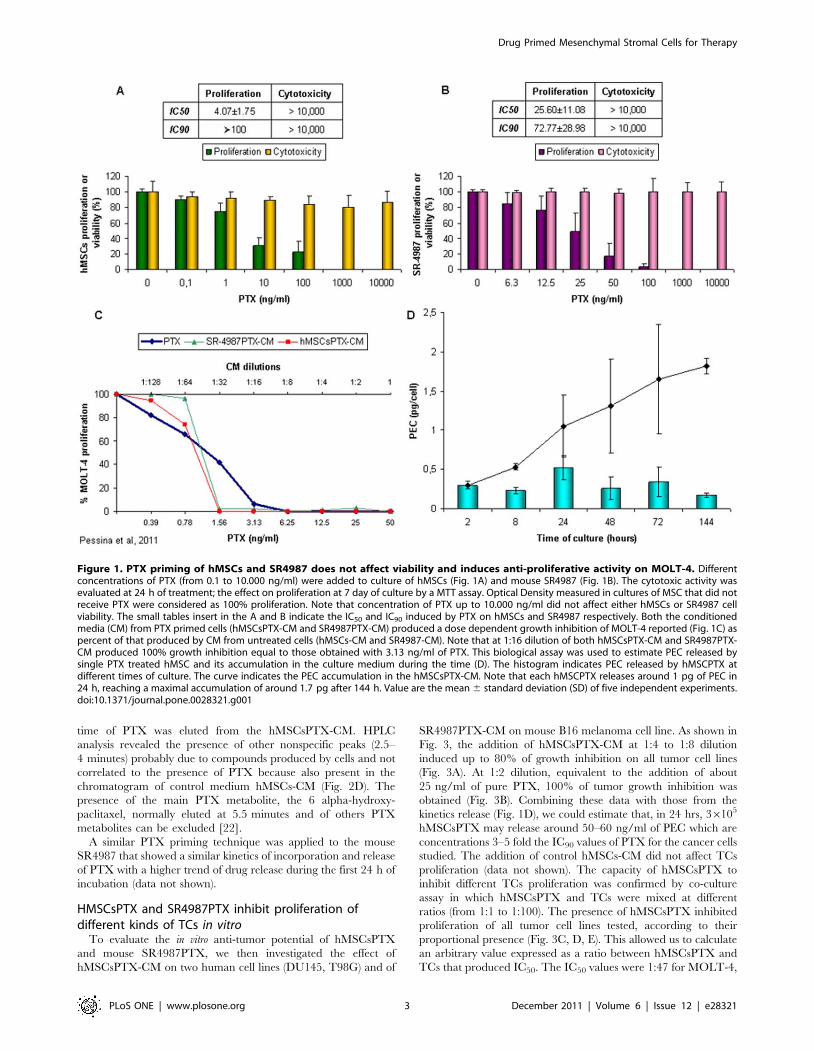
PTX-uptake and release by hMSCsBased on previous studies [14], sub-confluent culture (36105) of
adherent hMSCs were exposed for 24 h to 2.000 ng/ml PTX
(enough to completely block cell proliferation, but not able to
affect cell viability). After several washes and trypsinization,
hMSCsPTX were further cultured for 24 h and their conditioned
medium (CM) was tested on Molt-4, a leukaemia cell line very
sensitive to PTX (IC50 of 1.4861.06 ng/ml) [20]. The CM from
both cultures of hMSCPTX (hMSCsPTX-CM) produced a strong
dose-dependent anti-proliferative effect on MOLT-4, equivalent to
those obtained with pure PTX at doses from 0.39 to 50 ng/ml
(Fig. 1C). By contrast, the CM from control cells (hMSCs-CM)
were not effective. Comparing the inhibitory activity of pure PTX
and CM on Molt-4, we calculated the PTX equivalent
concentration (PEC) in the CM used to estimate the PEC released
by a single cell (PEC pg/cell). The total PEC internalized by
hMSCsPTX in 24 h, evaluated by testing the hMSCsPTX cell
lysates, was 2.6760.8 pg/cell, suggesting that hMSCs in 24 h
could incorporate about 8% of the PTX initially available
(33.3 pg/cell). Results on lysates of hMSCs fixed in formalin
before PTX priming indicated some unspecific binding of PTX,
corresponding to 0.1160.01 of PEC pg/cell (about 4% of total
PEC present in the lysate of living cells).
Thereafter, we calculated the time dependent release of PEC by
hMSCsPTX by replacing and collecting their CM at different
intervals of time. Detectable activity of PEC was present after 2 h of
incubation of hMSCsPTX reaching a PEC of 1 pg/cell during the
first 24 h of culture, and a maximum concentration of about 1.7–
2.0 pg/cell at 144 h (Fig. 1D). Since these values did not increase
with longer incubation, we estimated that around 25–30% of the
total PEC found in cell lysate was retained by the cells and never
released. The internalization of PTX into hMSCs was investigated
by confocal microscopy using Fluorescent PTX (PTX-F) (Fig. 2A).
PTX-F localization into hMSCs was analysed over time. After 1 h
of priming, the internalization of PTX-F by hMSCs was
appreciable. The staining was intense and enriched inside vesicles
at the end of priming (24 h). After 24 h, we observed that the
distribution of PTX-F remained confined to vesicles, many of which
were close to the cell membrane, suggesting a possible secretion. To
assess if PTX-F was enriched in vesicles derived from Golgi
apparatus, co-localization analysis was performed in hMSCs stained
with the specific marker BODIPYH TR ceramide. As seen in the
white spots in Figure 2A mask, we observed a high level of
colocalization between PTX-F and BODIPYH TR ceramide,
meaning that PTX-F was internalized inside Golgi-derived vesicles.
Thus, PTX-F accumulates in vesicles instead of accumulating in
microtubules [15] as many other xenobiotics do [21], and its levels
decrease in hMSCs following the kinetics shown by FACS analysis
(Fig. 2A and Fig. S4).
The presence of PTX in hMSCsPTX-CM was confirmed by
HPLC analysis. The HPLC chromatograms obtained from
hMSCsPTX-CM and from a standard sample of PTX in PBS
(1.000 ng/ml) (Fig. 2B, C) show that a peak of identical retention
Drug Primed Mesenchymal Stromal Cells for Therapy
PLoS ONE | www.plosone.org 2 December 2011 | Volume 6 | Issue 12 | e28321
time of PTX was eluted from the hMSCsPTX-CM. HPLC
analysis revealed the presence of other nonspecific peaks (2.5–
4 minutes) probably due to compounds produced by cells and not
correlated to the presence of PTX because also present in the
chromatogram of control medium hMSCs-CM (Fig. 2D). The
presence of the main PTX metabolite, the 6 alpha-hydroxy-
paclitaxel, normally eluted at 5.5 minutes and of others PTX
metabolites can be excluded [22].
A similar PTX priming technique was applied to the mouse
SR4987 that showed a similar kinetics of incorporation and release
of PTX with a higher trend of drug release during the first 24 h of
incubation (data not shown).
HMSCsPTX and SR4987PTX inhibit proliferation ofdifferent kinds of TCs in vitro
To evaluate the in vitro anti-tumor potential of hMSCsPTX
and mouse SR4987PTX, we then investigated the effect of
hMSCsPTX-CM on two human cell lines (DU145, T98G) and of
SR4987PTX-CM on mouse B16 melanoma cell line. As shown in
Fig. 3, the addition of hMSCsPTX-CM at 1:4 to 1:8 dilution
induced up to 80% of growth inhibition on all tumor cell lines
(Fig. 3A). At 1:2 dilution, equivalent to the addition of about
25 ng/ml of pure PTX, 100% of tumor growth inhibition was
obtained (Fig. 3B). Combining these data with those from the
kinetics release (Fig. 1D), we could estimate that, in 24 hrs, 36105
hMSCsPTX may release around 50–60 ng/ml of PEC which are
concentrations 3–5 fold the IC90 values of PTX for the cancer cells
studied. The addition of control hMSCs-CM did not affect TCs
proliferation (data not shown). The capacity of hMSCsPTX to
inhibit different TCs proliferation was confirmed by co-culture
assay in which hMSCsPTX and TCs were mixed at different
ratios (from 1:1 to 1:100). The presence of hMSCsPTX inhibited
proliferation of all tumor cell lines tested, according to their
proportional presence (Fig. 3C, D, E). This allowed us to calculate
an arbitrary value expressed as a ratio between hMSCsPTX and
TCs that produced IC50. The IC50 values were 1:47 for MOLT-4,
Figure 1. PTX priming of hMSCs and SR4987 does not affect viability and induces anti-proliferative activity on MOLT-4. Differentconcentrations of PTX (from 0.1 to 10.000 ng/ml) were added to culture of hMSCs (Fig. 1A) and mouse SR4987 (Fig. 1B). The cytotoxic activity wasevaluated at 24 h of treatment; the effect on proliferation at 7 day of culture by a MTT assay. Optical Density measured in cultures of MSC that did notreceive PTX were considered as 100% proliferation. Note that concentration of PTX up to 10.000 ng/ml did not affect either hMSCs or SR4987 cellviability. The small tables insert in the A and B indicate the IC50 and IC90 induced by PTX on hMSCs and SR4987 respectively. Both the conditionedmedia (CM) from PTX primed cells (hMSCsPTX-CM and SR4987PTX-CM) produced a dose dependent growth inhibition of MOLT-4 reported (Fig. 1C) aspercent of that produced by CM from untreated cells (hMSCs-CM and SR4987-CM). Note that at 1:16 dilution of both hMSCsPTX-CM and SR4987PTX-CM produced 100% growth inhibition equal to those obtained with 3.13 ng/ml of PTX. This biological assay was used to estimate PEC released bysingle PTX treated hMSC and its accumulation in the culture medium during the time (D). The histogram indicates PEC released by hMSCPTX atdifferent times of culture. The curve indicates the PEC accumulation in the hMSCsPTX-CM. Note that each hMSCPTX releases around 1 pg of PEC in24 h, reaching a maximal accumulation of around 1.7 pg after 144 h. Value are the mean 6 standard deviation (SD) of five independent experiments.doi:10.1371/journal.pone.0028321.g001
Drug Primed Mesenchymal Stromal Cells for Therapy
PLoS ONE | www.plosone.org 3 December 2011 | Volume 6 | Issue 12 | e28321

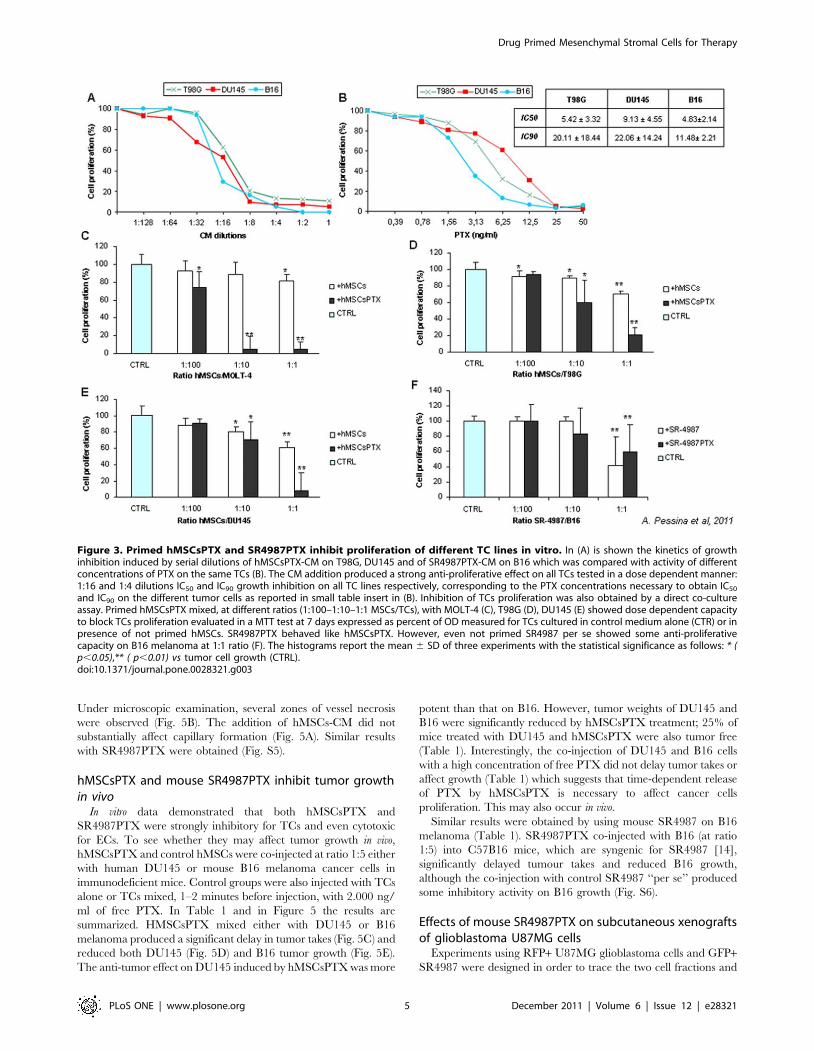
1:5 for T98G and DU145 and only 1:2–3 for B16 tumor cells. The
addition of control hMSCs did not substantially affect TCs
proliferation, although a low significant growth inhibition
(p,0.02) was observed at 1:1 ratio for all the tumor cell lines
tested.
Mouse SR4987PTX worked like hMSCsPTX in uptake and
release of the drug as tested by MTT assay on CM (Fig. 1A).
However, in the co-culture assay, SR4987 cells ‘‘per se’’ produced
a inhibition of B16 melanoma cells ( p,0.05) that did not differ
from that produced by SR4987PTX (Fig. 3F).
hMSCSPTX and mouse SR4987PTX display potent in vitroanti-angiogenic activity
Because PTX is considered an inhibitor of angiogenesis [16,23],
we thus investigated whether hMSCsPTX and SR4987PTX could
affect angiogenesis in vitro (Fig. 4). The anti-angiogenic activity of
pure PTX was initially tested on HUVECs and on microvascular
ECs (HMECs) proliferation. PTX at doses up to 10 ng/ml was
extremely cytotoxic for both HUVECs and HMECs. PTX
produced around 50% of ECs growth inhibition (IC50) at
4.6 ng/ml (Fig. 4A). The effect of hMSCsPTX-CM on HUVECs
and HMECs was extremely cytotoxic at 1:2 and 1:4 dilutions
(Fig. 4B, C). At 1:8 hMSCsPTX-CM inhibited both HUVECs and
HMECs proliferation but less than 5 ng/ml of pure PTX. Similar
results were obtained by co-culture assay (Fig. 4D, E, F). Ratio 1:1
and 1:5 of hMSCsPTX/ECs produced a strong cytotoxic effect on
both HUVECs and HMECs, while at ratio 1:10, no cytotoxicity
but significant ECs growth inhibition was observed (Fig. 4D, E).
Interestingly, at ratio 1:5, hMSCsPTX initiated killing HMECs
during the first 24 h of co-culture; after 72 h incubation very few
HMECs survived (Fig. 4F). Control hMSCs-CM did not affect
ECs proliferation.
The anti-angiogenic potential of hMSCsPTX was also investi-
gated by using the rat aorta ring assay [24]. As shown in Fig. 5A,
the addition of 1:2 and 1:4 dilution of hMSCsPTX-CM strongly
reduced either spontaneous or VEGFa-induced sprouting of
neocapillaries from aorta rings. Dilution 1:8 of hMSCsPTX-CM
that inhibit ECs proliferation did not affect aorta ring capillaries
formation (Fig. 5A). HMSCsPTX-CM was also able to induce
capillary regression if added to aorta rings after 7 days of culture.
Figure 2. PTX-F internalization and release by hMSCs. The internalization of PTX-F was analyzed by confocal microscopy (A) in live hMSCsprimed 1 (1) or 24 (24) h with PTX-F (green) and loaded with the Golgi specific marker BOPIPYHTR ceramide (red). Cells were also observed 24 h afterwashing step (24+24). PTX-F accumulates in cells and co-localizes with Golgi apparatus or Golgi-derived vesicles. Mask panel highlights the co-localization between PTX-F and BODIPYHTR ceramide showing white spots, that indicate those pixels in which both the fluorescent signals aredetectable.. White lines represent the cell boundary and arrows indicate vesicles close to the cell membrane. Scale bar: 20 mm. The release of PTX inthe hMSCsPTX-CM at 24 h was analysed by HPLC. The elution profile (B) was compared to that of pure PTX at 1.000 ng/ml (C). The figure reports achromatogram profile of one typical experiment where hMSCsPTX-CM evidences a peak that clearly identified PTX and that was quantified on a PTXstandard curve as 68.1 ng/ml. Figure 2D reports the profile of the hMSCs-CM cultured in the absence of PTX. The peak labeled as I.S. is the internalstandard Cephalomannine added to all samples for the correct quantification of PTX.doi:10.1371/journal.pone.0028321.g002
Drug Primed Mesenchymal Stromal Cells for Therapy
PLoS ONE | www.plosone.org 4 December 2011 | Volume 6 | Issue 12 | e28321
Under microscopic examination, several zones of vessel necrosis
were observed (Fig. 5B). The addition of hMSCs-CM did not
substantially affect capillary formation (Fig. 5A). Similar results
with SR4987PTX were obtained (Fig. S5).
hMSCsPTX and mouse SR4987PTX inhibit tumor growthin vivo
In vitro data demonstrated that both hMSCsPTX and
SR4987PTX were strongly inhibitory for TCs and even cytotoxic
for ECs. To see whether they may affect tumor growth in vivo,
hMSCsPTX and control hMSCs were co-injected at ratio 1:5 either
with human DU145 or mouse B16 melanoma cancer cells in
immunodeficient mice. Control groups were also injected with TCs
alone or TCs mixed, 1–2 minutes before injection, with 2.000 ng/
ml of free PTX. In Table 1 and in Figure 5 the results are
summarized. HMSCsPTX mixed either with DU145 or B16
melanoma produced a significant delay in tumor takes (Fig. 5C) and
reduced both DU145 (Fig. 5D) and B16 tumor growth (Fig. 5E).
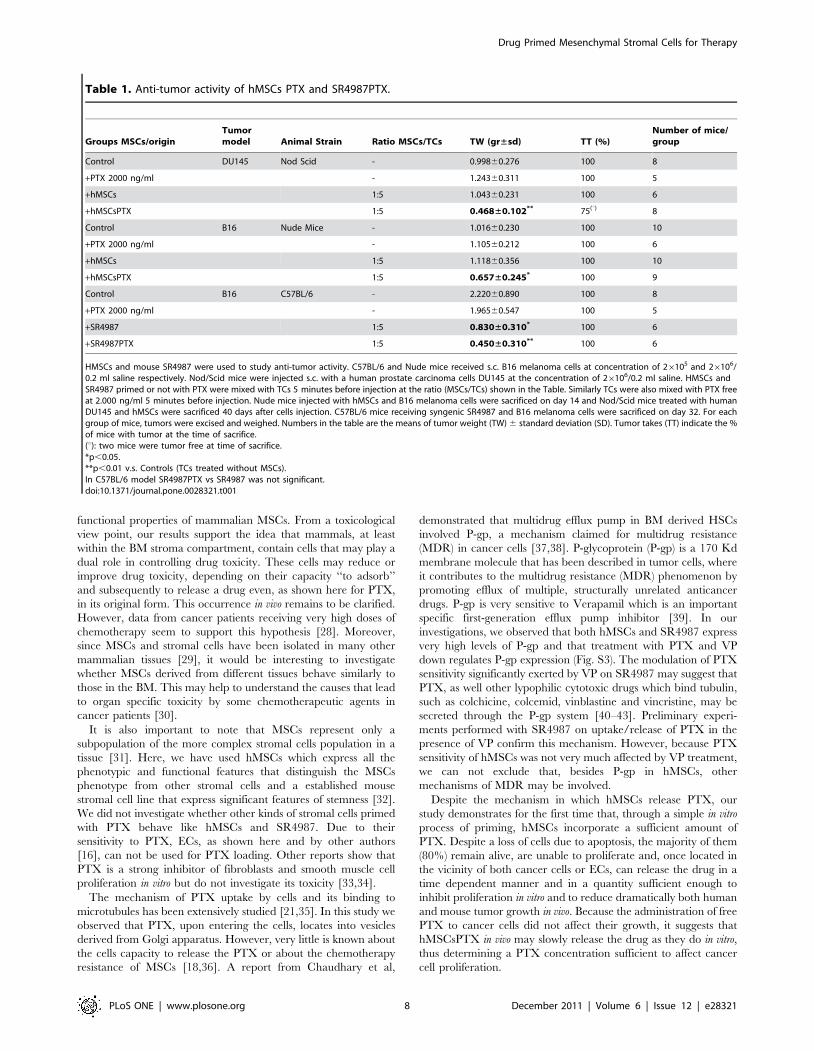
The anti-tumor effect on DU145 induced by hMSCsPTX was more
potent than that on B16. However, tumor weights of DU145 and
B16 were significantly reduced by hMSCsPTX treatment; 25% of
mice treated with DU145 and hMSCsPTX were also tumor free
(Table 1). Interestingly, the co-injection of DU145 and B16 cells
with a high concentration of free PTX did not delay tumor takes or
affect growth (Table 1) which suggests that time-dependent release
of PTX by hMSCsPTX is necessary to affect cancer cells
proliferation. This may also occur in vivo.
Similar results were obtained by using mouse SR4987 on B16
melanoma (Table 1). SR4987PTX co-injected with B16 (at ratio
1:5) into C57B16 mice, which are syngenic for SR4987 [14],
significantly delayed tumour takes and reduced B16 growth,
although the co-injection with control SR4987 ‘‘per se’’ produced
some inhibitory activity on B16 growth (Fig. S6).
Effects of mouse SR4987PTX on subcutaneous xenograftsof glioblastoma U87MG cells
Experiments using RFP+ U87MG glioblastoma cells and GFP+SR4987 were designed in order to trace the two cell fractions and
Figure 3. Primed hMSCsPTX and SR4987PTX inhibit proliferation of different TC lines in vitro. In (A) is shown the kinetics of growthinhibition induced by serial dilutions of hMSCsPTX-CM on T98G, DU145 and of SR4987PTX-CM on B16 which was compared with activity of differentconcentrations of PTX on the same TCs (B). The CM addition produced a strong anti-proliferative effect on all TCs tested in a dose dependent manner:1:16 and 1:4 dilutions IC50 and IC90 growth inhibition on all TC lines respectively, corresponding to the PTX concentrations necessary to obtain IC50
and IC90 on the different tumor cells as reported in small table insert in (B). Inhibition of TCs proliferation was also obtained by a direct co-cultureassay. Primed hMSCsPTX mixed, at different ratios (1:100–1:10–1:1 MSCs/TCs), with MOLT-4 (C), T98G (D), DU145 (E) showed dose dependent capacityto block TCs proliferation evaluated in a MTT test at 7 days expressed as percent of OD measured for TCs cultured in control medium alone (CTR) or inpresence of not primed hMSCs. SR4987PTX behaved like hMSCsPTX. However, even not primed SR4987 per se showed some anti-proliferativecapacity on B16 melanoma at 1:1 ratio (F). The histograms report the mean 6 SD of three experiments with the statistical significance as follows: * (p,0.05),** ( p,0.01) vs tumor cell growth (CTRL).doi:10.1371/journal.pone.0028321.g003
Drug Primed Mesenchymal Stromal Cells for Therapy
PLoS ONE | www.plosone.org 5 December 2011 | Volume 6 | Issue 12 | e28321

to investigate their interactions in vivo. Control mice, grafted
either with U87MG cells or with U87MG cells and SR4987,
demonstrated that the expression of RFP and GFP did not alter
tumorigenicity and survival of these cells in the in vivo condition.
Overall, the SR4987PTX cells had an inhibitory effect on the
growth of glioblastoma xenografts (Fig. S7A–B). At the 2, 4, and
6-week time points, the RFP+ U87MG/GFP+ SR4987PTX
xenografts were significantly smaller than the RFP+ U87MG
xenografts (p,0.01, p,0.05, and p,0.001, respectively; Student’s
t-test). At the same time points, the RFP+ U87MG/GFP+SR4987-PTX xenografts were significantly smaller than RFP+U87MG/GFP+ SR4987 xenografts as well (p,0.02, p,0.05, and
p,0.001, respectively; Student’s t-test). Fluorescence microscopy
showed that, in the context of the tumor, the GFP+ SR4987PTX
cells arranged themselves in strands and columns to form nets
that entrapped the RFP+ U87MG cells (Fig. S7C). Conversely,
the GFP+ SR4987 that had not been primed with PTX were
intermingled with the U87MG cells without any propensity to
organize into secondary structures (Fig. S7C). Histological
examination revealed that the xenografts containing SR4987
cells, irrespective of their PTX priming, did not develop foci of
necrosis, which are typically seen in the U87MG xenografts at the
4 and 6 week survival time (Fig. S7D). Both the size and
histological appearance of U87MG and U87MG/SR4987
xenografts did not differ significantly from those xenografts
generated by injection of the virus transduced RFP+ U87MG
cells and RFP+ U87MG/GFP+ SR4987, respectively (data not
shown).
Discussion
The mouse stromal cell line SR4987 primed first in vitro with
DXR and then co-cultured with HSCs results in a strong
inhibition of CFU formations [14]. Noting this property, we
wondered if hMSCs and SR4987 primed with an anti-cancer drug
could acquire anti-tumor activity and consequently, be used for
cancer therapy.
In order to validate this hypothesis, both mouse SR4987 and
hMSCs were primed with PTX, because it demonstrated both
strong anti-tumor [15,25] and anti-angiogenic activities [16,23].
Once released by primed cells, PTX could affect both tumor and
ECs proliferation.
Figure 4. PTX and primed hMSCsPTX inhibit ECs proliferation in vitro. PTX inhibits ECs proliferation (A). HUVECs and HMECs (26105) werecultured for 72 h in the presence of different concentration of PTX. The IC50 was around 4.69 ng/ml of PTX and at 10 ng/ml PTX significantly blockedboth HUVECs and HMECs proliferation, which at higher doses was cytotoxic. Similarly, the addition of hMSCsPTX-CM greatly inhibit HUVECs (B) andHMECs (C) proliferation. At 1:2 dilution, hMSCsPTX-CM was cytotoxic, while at higher 1:4 and 1:8 dilutions significantly inhibit ECs proliferation.Inhibition of ECs proliferation was obtained by co-culture assay. HMSCsPTX co-cultured at 1:1 and 1:5 ratio (MSCs/ECs) was cytotoxic for both HUVECs(D) and HMECs (E). At 1:10 ratio hMSCsPTX continued to inhibit ECs proliferation. Untreated control hMSCs did not affect ECs. In (F) photographs ofculture of hMSCsPTX mixed 1:5 with HMECs. Cells fixed and stained at different time intervals are shown. hMSCsPTX kill HMECs as early as 24 h afterseeding. White arrows indicate the islands of HMECs still present in the culture. Note that after 72 h most of the HMECs seeded were killed and onlyhMSCsPTX remain in the culture (206 magnification). Bars in the figures are the means 6 SD of three separate experiments done in triplicate. *p,0.05, **p,0.01 vs untreated hMSCs.doi:10.1371/journal.pone.0028321.g004
Drug Primed Mesenchymal Stromal Cells for Therapy
PLoS ONE | www.plosone.org 6 December 2011 | Volume 6 | Issue 12 | e28321

Initial experiments demonstrated that PTX was able to inhibit
hMSCs and SR4987 proliferation but was not cytotoxic; up to
10,000 ng/ml did not affect cell viability. This PTX concentration
was approximately 500 times higher than that necessary to
produce an IC50 on MOLT-4, a human leukemia cell line very
sensitive to PTX activity [20] that we used for monitoring the
hMSCsPTX-CM and SR4987PTX-CM activity. Based on a
previous study [14], we thus established a standard protocol to
prime hMSCs and SR4987 with PTX, consisting of treating
36105 adherent cells with 2.000 ng/ml of PTX for 24 h in
culture. In summary, we found that: a) hMSCs were able to
rapidly incorporate PTX as confirmed by using PTX-F; b)
hMSCsPTX can slowly release PTX, at least in part, in the culture
medium in a time dependent manner, as confirmed by the HPLC
analysis; c) formalin fixed hMSCs and were not effective to uptake
PTX; d) both hMSCsPTX and SR4987PTX acquired a potent
anti-tumor and anti-angiogenic activity in vitro that was dose
dependent, and demonstrable by using their CM or by co-culture
assay; e) hMSCsPTX co-injected in immunodeficient mice with
human DU145 and SR4987PTX co-injected with U87MG or
mouse B16 melanoma, significantly delayed tumor takes and
reduced tumor growth. In the B16 melanoma model we observed
that mouse stromal SR4987 cells are able ‘‘per se’’ to inhibit tumor
cell proliferation at a level similar to SR4987PTX. This finding
seems to be in agreement with the contradictory data reported
from the literature that suggest both the capacity of mesenchymal
cells and stromal cells to favour or to inhibit tumor progression
[26]. In particular, the B16 tumor model represents a condition in
which murine stromal cells anti-tumor activity has been demon-
strated [27]. In this condition it may be that PTX loaded SR4987
cells increase their basal antitumor efficacy without a significant
measurable effect.
Independently of this finding, we can conclude that our results,
taken together, strongly support our initial hypothesis proposing
even hMSCs as a carrier for chemotherapeutic drugs in human. In
our opinion, our results disclose important new insights into
Figure 5. HMSCsPTX inhibit microvessel out-growth in vitro and reduce tumor growth in vivo. In (A) and (B) rat aorta ring assay was usedto test hMSCsPTX-CM microvessel growth inhibition. In (A) hMSCsPTX-CM at different dilutions added to rat aorta rings in the presence or in theabsence of VEGFa induced a great reduction of capillary outgrowth compared to CTRL medium and to hMSCs-CM. VEGFa 20 ng/ml was used aspositive controls (**p,0.01 vs hMSCs-CM). In (B) photographs show hMSCsPTX-CM (at 1:2 dilution), which induced capillary regression asdemonstrated by the presence of vessel rupture and necrotic zones (arrows) (magnifications 206). In (C), (D) and (E) the effects of hMSCsPTX in vivoon tumor takes (C), on DU145 (D) and B16 (E) growth are shown. Around 0.46106 hMSCsPTX and control untreated hMSCs were mixed (at ratio 1:5hMSCs/TCs/) with 26106 DU145 or B16 and then injected subcutaneously (s.c.) into mice. Tumor volumes were calculated by measuring the tumordiameters taken every two days with a calibre. The co-injection of hMSCsPTX with DU145 or B16, produced a significant delay of tumor appearance(C) as well as a great reduction of DU145 (D) and B16 (E) tumor volume, whereas the co-injection with untreated hMSCs did not affect either DU145 orB16 volumes, nor did the injection of TCs mixed 5 minutes before injection with 2.000 ng/ml of PTX (see Table 1). The insert in (D) and (E) is thephoto of tumors of control, hMSCsPTX and hMSCs treated mice at time of sacrifice. *p,0.05 and **p,0.01 vs TCs alone or vs hMSCs treated mice.doi:10.1371/journal.pone.0028321.g005
Drug Primed Mesenchymal Stromal Cells for Therapy
PLoS ONE | www.plosone.org 7 December 2011 | Volume 6 | Issue 12 | e28321
functional properties of mammalian MSCs. From a toxicological
view point, our results support the idea that mammals, at least
within the BM stroma compartment, contain cells that may play a
dual role in controlling drug toxicity. These cells may reduce or
improve drug toxicity, depending on their capacity ‘‘to adsorb’’
and subsequently to release a drug even, as shown here for PTX,
in its original form. This occurrence in vivo remains to be clarified.
However, data from cancer patients receiving very high doses of
chemotherapy seem to support this hypothesis [28]. Moreover,
since MSCs and stromal cells have been isolated in many other
mammalian tissues [29], it would be interesting to investigate
whether MSCs derived from different tissues behave similarly to
those in the BM. This may help to understand the causes that lead
to organ specific toxicity by some chemotherapeutic agents in
cancer patients [30].
It is also important to note that MSCs represent only a
subpopulation of the more complex stromal cells population in a
tissue [31]. Here, we have used hMSCs which express all the
phenotypic and functional features that distinguish the MSCs
phenotype from other stromal cells and a established mouse
stromal cell line that express significant features of stemness [32].
We did not investigate whether other kinds of stromal cells primed
with PTX behave like hMSCs and SR4987. Due to their
sensitivity to PTX, ECs, as shown here and by other authors
[16], can not be used for PTX loading. Other reports show that
PTX is a strong inhibitor of fibroblasts and smooth muscle cell
proliferation in vitro but do not investigate its toxicity [33,34].
The mechanism of PTX uptake by cells and its binding to
microtubules has been extensively studied [21,35]. In this study we
observed that PTX, upon entering the cells, locates into vesicles
derived from Golgi apparatus. However, very little is known about
the cells capacity to release the PTX or about the chemotherapy
resistance of MSCs [18,36]. A report from Chaudhary et al,
demonstrated that multidrug efflux pump in BM derived HSCs
involved P-gp, a mechanism claimed for multidrug resistance
(MDR) in cancer cells [37,38]. P-glycoprotein (P-gp) is a 170 Kd
membrane molecule that has been described in tumor cells, where
it contributes to the multidrug resistance (MDR) phenomenon by
promoting efflux of multiple, structurally unrelated anticancer
drugs. P-gp is very sensitive to Verapamil which is an important
specific first-generation efflux pump inhibitor [39]. In our
investigations, we observed that both hMSCs and SR4987 express
very high levels of P-gp and that treatment with PTX and VP
down regulates P-gp expression (Fig. S3). The modulation of PTX
sensitivity significantly exerted by VP on SR4987 may suggest that
PTX, as well other lypophilic cytotoxic drugs which bind tubulin,
such as colchicine, colcemid, vinblastine and vincristine, may be
secreted through the P-gp system [40–43]. Preliminary experi-
ments performed with SR4987 on uptake/release of PTX in the
presence of VP confirm this mechanism. However, because PTX
sensitivity of hMSCs was not very much affected by VP treatment,
we can not exclude that, besides P-gp in hMSCs, other
mechanisms of MDR may be involved.
Despite the mechanism in which hMSCs release PTX, our
study demonstrates for the first time that, through a simple in vitro
process of priming, hMSCs incorporate a sufficient amount of
PTX. Despite a loss of cells due to apoptosis, the majority of them
(80%) remain alive, are unable to proliferate and, once located in
the vicinity of both cancer cells or ECs, can release the drug in a
time dependent manner and in a quantity sufficient enough to
inhibit proliferation in vitro and to reduce dramatically both human
and mouse tumor growth in vivo. Because the administration of free
PTX to cancer cells did not affect their growth, it suggests that
hMSCsPTX in vivo may slowly release the drug as they do in vitro,
thus determining a PTX concentration sufficient to affect cancer
cell proliferation.
Table 1. Anti-tumor activity of hMSCs PTX and SR4987PTX.
Groups MSCs/originTumormodel Animal Strain Ratio MSCs/TCs TW (gr±sd) TT (%)
Number of mice/group
Control DU145 Nod Scid - 0.99860.276 100 8
+PTX 2000 ng/ml - 1.24360.311 100 5
+hMSCs 1:5 1.04360.231 100 6
+hMSCsPTX 1:5 0.468±0.102** 75(u) 8
Control B16 Nude Mice - 1.01660.230 100 10
+PTX 2000 ng/ml - 1.10560.212 100 6
+hMSCs 1:5 1.11860.356 100 10
+hMSCsPTX 1:5 0.657±0.245* 100 9
Control B16 C57BL/6 - 2.22060.890 100 8
+PTX 2000 ng/ml - 1.96560.547 100 5
+SR4987 1:5 0.830±0.310* 100 6
+SR4987PTX 1:5 0.450±0.310** 100 6
HMSCs and mouse SR4987 were used to study anti-tumor activity. C57BL/6 and Nude mice received s.c. B16 melanoma cells at concentration of 26105 and 26106/0.2 ml saline respectively. Nod/Scid mice were injected s.c. with a human prostate carcinoma cells DU145 at the concentration of 26106/0.2 ml saline. HMSCs andSR4987 primed or not with PTX were mixed with TCs 5 minutes before injection at the ratio (MSCs/TCs) shown in the Table. Similarly TCs were also mixed with PTX freeat 2.000 ng/ml 5 minutes before injection. Nude mice injected with hMSCs and B16 melanoma cells were sacrificed on day 14 and Nod/Scid mice treated with humanDU145 and hMSCs were sacrificed 40 days after cells injection. C57BL/6 mice receiving syngenic SR4987 and B16 melanoma cells were sacrificed on day 32. For eachgroup of mice, tumors were excised and weighed. Numbers in the table are the means of tumor weight (TW) 6 standard deviation (SD). Tumor takes (TT) indicate the %of mice with tumor at the time of sacrifice.(u): two mice were tumor free at time of sacrifice.*p,0.05.**p,0.01 v.s. Controls (TCs treated without MSCs).In C57BL/6 model SR4987PTX vs SR4987 was not significant.doi:10.1371/journal.pone.0028321.t001
Drug Primed Mesenchymal Stromal Cells for Therapy
PLoS ONE | www.plosone.org 8 December 2011 | Volume 6 | Issue 12 | e28321

In summary, to our knowledge this is the first demonstration
that hMSCs can be loaded in vitro with a chemotherapeutic drug
and used for cancer treatment in vivo. Since priming hMSCs with
PTX is a simple procedure that neither requires any genetic
manipulation of cells nor much time, we are confident that, once
the effectiveness of hMSCsPTX on established cancers in mice is
proven, our procedure could be applied in cancer patients,
perhaps in combination with other methods, which use, for
example, nanoparticles or liposome for drug transport.
Materials and Methods
Ethics StatementAll animal experiments were performed according to interna-
tional law and policies (EEC C.D.86/609,OJL358,1987; Guide for
the Care and Use of Laboratory Animals, U.S. National Research
Council, 1996). Experiments and care/welfare were performed in
an animal facility with protocols authorized by Ministero della
Salute (D.I.116/1992,Circ.8/1994, D.M. 60/2003-A, P.C.ID:1/
2009).
Tumor cellsFor the in vitro and in vivo studies three human tumor cell (TC)
lines were used: MOLT-4 (acute lymphoblastic leukemia) [44];
T98G (glioblastoma) [45]; DU-145 (prostate carcinoma) [46] and
B16 mouse melanoma [47]. In vivo studies have also been
performed by using RFP+U87MG glioblastoma cell. (For details
see supporting information text S1).
MSCs expansion and characterizationHMSCs were prepared as previous described [48] from the
mononuclear cell fraction of human bone marrow purchased from
Lonza (USA) who manage human tissue in accordance with
current US regulations governing tissue banking in the Code of
Federal Regulations (CFR), 21 CFR Part 1271. After expansion,
cells were characterized for CD markers expression, differentiating
capacity (osteo-condro-adipocytes) [49] (See supporting informa-
tion Text S1).
Murine stromal cellsFor murine stromal cells, a cell line established from bone
marrow (SR4987) having a high degree of stemness was used. It is
positive for some stem cells markers as vimentin, CD44+,
CD73+,CD105+, CD106+, Sca-1+, CD34+, contain 50%
CD45+ cells and is capable of differentiating into osteocyte and
chondrocytes [17,50]. For some specific in vivo studies, transfected
cells (GFP+SR4987) have been prepared (see Suppoprting
Information Text S1). P-glycoprotein (P-gp) expression was
evaluated by using a mouse monoclonal antibodies anti-human
P-gp (clone C1, Ylem, Italy). Cell were then analyzed by FACS.
Procedure for drug uptake and release by MSCsSR4987 and hMSCs (36105) were treated 24 hours with PTX
(2.000 ng/ml) (Serva, Germany). At the end of the incubation, the
cells were washed twice with PBS, then trypsinized, washed twice
in HBSS and seeded in a new flask. After 24 h of culture, the cell
conditioned medium (CM) was collected and replaced by
repeating this procedure at 48, 72, 96 and 144 hours. The CM
were tested for their anti-tumor proliferation activity in vitro by
using CM from untreated MSCs as negative controls. The passive
membrane drug adsorption and release by fixed cells were also
verified. The internalisation of PTX into MSCs was checked by
Fluorescent PTX (Oregon Green 488 Taxol, Invitrogen, UK).
The effect of 24 h drug treatment on the cell cycle of MSCs was
evaluated by FACS. The presence of PTX in the CM of primed
MSCs was confirmed by HPLC [51] (see supporting information
Text S1).
In vitro anti proliferative assay on tumor cell linesThe effect of both PTX and CM from PTX-treated MSCs
(MSCsPTX-CM) on TC proliferation was studied in 96 multiwell
plates (Sarstedt, Germany) on MOLT-4, T98G and DU145 cells
according to a MTT (3-(4,5-dimethyl-2-thiazolyl)-2,5-diphenyl-2-
H-tetrazoliumbromide) assay [52]. The inhibitory concentrations
(IC50 and IC90) were determined according to the Reed and
Muench formula [53]. The antitumoral activity of MSCsPTX-
CM was compared to that of pure PTX and expressed as PTX
equivalent concentration (PEC) according to the following
algorithm: PEC (ng/ml) = DF50CM6IC50PTX (DF50CM is the
dilution factors (DF) at which the 50% of inhibition was observed
with MSCsPTX-CM; IC50PTX is the concentration (ng/ml) of
pure PTX producing 50% of inhibition). The PTX release (PR) by
a single primed MSC was calculated as ratio between the PEC and
the number of cells seeded: PR (pg/cell) = PEC (ng/ml)6CM
volume (ml)/number of cell seeded (see supporting information
Text S1).
In vitro inhibition of tumor growth by MSCsPTXTo verify the ability of MSCsPTX to inhibit in vitro TC
proliferation, a co-culture system was applied by mixing TCs with
different amounts of MSCs to final ratios of MSCs/TCs of 1-10-
100. Arbitrary values of IC50 and IC90 were calculated as ratio
MSCs/TCs able to inhibit respectively 50% and 90% of TC
proliferation (see supporting information Text S1).
Evaluation of anti-angiogenic properties of SR4987 andhMSCs primed with PTX in vitro
The anti-angiogenic potential of the CM, from MSCs primed or
not with PTX, was tested on the proliferation of Human Umbilical
vein ECs (HUVEC) (purchased from Lonza) and on human derma
microvascular ECs (HMECs) isolated and maintained as previ-
ously described [54]. Both ECs phenotypes were routinely
maintained in EGM bullet kit plus 10% FCS (Lonza). The rat
aorta ring assay was also performed to investigate the anti-
angiogenic potential of control and MSCsPTX-CM according to
the procedure previously described [24,55,56]. Quantification of
angiogenesis was obtained by taking photographs every three days
and by counting the number of microvessels arising from aorta
rings [24] (see supporting information Text S1).
Evaluation of anti-tumor activity of hMSCsPTX andmouse SR4987PTX in vivo
The capacity of control hMSCs and hMSCsPTX to affect
DU145 and B16 melanoma growth were performed on eight-week-
old male NOD/SCID mice (JAX Mice: NOD.CB17-Prkdc scid/j)
and Nude mice (4-week-old male athymic nude-Foxn1 nu/nu from
Harlan, Italy) respectively. The subcutaneous grafting of RFP+U87MG glioblastoma cells mixed with GFP+ SR4987 cells primed
or not with PTX was also performed in nude athymic mice. Eight-
week-old female mice C57Bl6 (Charles River, Italy) were used to
evaluate the effect of mouse control SR4987 and SR4987-PTX on
B16 melanoma (see supporting information Text S1).
Supporting Information
Figure S1 Characterization of Mesenchymal Stem Cellsexpanded from human bone marrow. The MSC feature of
Drug Primed Mesenchymal Stromal Cells for Therapy
PLoS ONE | www.plosone.org 9 December 2011 | Volume 6 | Issue 12 | e28321
the expanded cell population has been confirmed by their capacity
to differentiate into adipocyte (A), osteoblasts (B) and condhro-
blasts (C) under specific stimulation. Figure 1D shows unstimu-
lated MSCs (Negative Control). The box on the right shows the
pattern of CD expression that is confirmed to be typical of MSCs.
(TIF)
Figure S2 Cell cycle analysis by FACS. The histograms
show the effects of PTX treatment on hMSCs and SR4987 cell
cycle after 24 hours of treatment. The percentages of cells counted
in each different cell phase (G0/G1, S and G2/M ) are reported
and compared to these found in untreated cells (CTRL). PTX
24 h = cells after 24 h of PTX treatment; PTX 24+24 h = PTX
treated cells, then subcultured for 24 h without PTX ; PTX
24+72 h = PTX treated cells, then subcultured for 72 h without
PTX. Under the abscissa are reported the percentages of cell
viability evaluated by Trypan Blue.
(TIF)
Figure S3 Modulation of P-gp expression and PTXsensitivity. (A) The histogram shows the basal expression of P-
gp by hMSCs and SR4987 (CTRL), its modulation after 24 hours
of treatment with 2 mg/ml paclitaxel (PTX+) or 20 uM Verapamil
(VP+). P-gp expression was evaluated by FACS and reported as
ratio between fluorescence intensity measured on cells treated with
specific labelled antibody and that of cells treated with isotype
control antibody. (B) The histogram shows the IC50 values (ng/ml)
for PTX determined in a antiproliferation MTT assay in the
absence and in the presence of 20 mM VP. Each point reports the
mean value of three independent experiments.
(TIF)
Figure S4 Internalization and kinetics of FluorescentPTX (PTX-F) decrease in hMSCs. The histogram demon-
strates that PTX-F was internalized by hMSCs. The study was
conducted by treating hMSCs with PTX-F for 24 hrs. Thereafter,
the cells were harvested by trypsin and washed, and then analysed
by FACS immediately (time 0) at 8 and 24 hours while maintained
resuspended in PBS. Violet histogram = cells treated with
unlabelled Taxol; Green histogram = cells treated with FITC-
conjugated Taxol; X axis = green fluorescence intensity; Y
axis = number of cells.
(TIF)
Figure S5 Mouse SR4987PTX-CM inhibit HUVECs andHMECs proliferation. HUVECs (A) and (C) and HMECs (B)
were cultured for 72 hrs in the presence or in the absence of
control SR4987-CM and SR4987PTX-CM at different dilutions.
SR4987PTX-CM at 1:2 and 1:4 dilutions were cytotoxic for
HUVECs, while they induced a significant growth inhibition on
HMECs. In C photographs (206 magnifications) show the
cytotoxic effect of SR4987PTX-CM on HUVECs at 1:2 and 1:4
dilutions. Control SR4987-CM do not inhibit, but even seem to
improve proliferation of both HUVECs and HMECs. The values
in A and B are the means 6 SD of two different experiments
*p,0.05, p,0.01 vs untreated MMSCs.
(TIF)
Figure S6 Mouse SR4987PTX inhibit B16 melanomagrowth in syngeneic C57Bl6 mice. The Figure shows the
effect of control SR4987 and primed SR4987PTX (0.46105)
mixed at 1:5 ratio with B16 melanoma cells (26105) and injected
s.c into syngeneic C57Bl6 mice (see also Table 1). In (A)
SR4987PTX induced a significant reduction of B16 tumor volume
calculated by measuring tumor diameters with a calibre.
Interesting, even the co-injection of control SR4987 with B16
melanoma cells reduced the tumor volumes. In (B) the effect of
SR4987PTX on B16 tumor appearance showing a significative
delay in tumor takes. In (C) the photo of B16 tumors removed
from the s.c of control, SR4987PTX and SR4987 treated mice at
the time of mice sacrifice. * p,0.05 and ** p,0.01 vs control B16
volume.
(TIF)
Figure S7 Effects of GFP+ SR4987-PTX on the growthand histology of subcutaneous RFP+U87MG xenografts.At the 2, 4, and 8 week survival time, the tumor xenografts
generated by co-injection of RFP+U87MG glioblastoma cells and
GFP+SR4987-PTX cells showed significantly smaller diameter
compared with tumor generated by injection of glioblastoma
cells or by co-injection of RFP+U87MG and GFP+SR4987 cells
(* p,0.05; ** p,0.02; *** p,0.001) (A). At two weeks after
grafting: the histology of RFP+U87MG/GFP+SR4987-PTX
xenografts shows reduced cell density with regions of Matrigel
that are not colonized by the tumor cells and vimentin expressing
cells arranged in columns (B). At 4 weeks after grafting : RFP+U87MG xenograft and RFP+ U87MG/GFP+ SR4987 xenograft
show green MSCs intermingled with the red glioblastoma cells.
RFP+ U87MG/GFP+ SR4987-PTX xenograft shows that the
green MSCs arrange themselves to form septi encircling the red
glioblastoma cells (C). At 6 weeks after injection, tumor xenografts
containing GFP+SR4987 do not develop areas of necrosis that are
a typical feature of the RFP+U87MG xenografts at this time point
(D).
(TIF)
Table S1 Evaluation of apoptosis in hMSCs and SR-4987 after PTX treatment. The evaluation of apoptosis was
performed by flow cytometry by the Annexin-V binding assay and
confirmed by the quantification of the sub-G1 cell population.
Data report the percentage of apoptotic/necrotic cells (mean 6
s.d. of three assays). The assays were performed with different
passages of SR4987 and different bone marrow donors).
CTRL = Untreated cells; PTX 24 h = cells after 24 h of PTX
treatment; PTX 24+24 h = treated cells, then subcultured for 24 h
without PTX.
(DOC)
Text S1
(DOC)
Acknowledgments
We thank Mrs Andrea Smith for checking the English of the manuscript.
Author Contributions
Conceived and designed the experiments: AP GA. Performed the
experiments: AB VC GI SN FS LC EC LV AL DC MF AP GC GA
RP MLF. Analyzed the data: AP AB GA VC GI FS LC EC LV AL DC
MF GM. Contributed reagents/materials/analysis tools: AP GI MF LV
AL EC GC CA EP. Wrote the paper: AP GA. Provided endothelial cells:
AC. Supervised in vitro studies: EP.
References
1. Weldon JE, Xiang L, Chertov O, Margulies I, Kreitman RJ, et al. (2009) A
protease-resistant immunotoxin against CD22 with greatly increased activity
against CLL and diminished animal toxicity. Blood 113: 3792–3800.
2. Dhar S, Gu FX, Langer R, Farokhzad OC, Lippard SJ (2008) Targeted delivery
of cisplatin to prostate cancer cells by aptamer functionalized Pt (IV) prodrug-
PLGA-PEG nanoparticles. Proc Natl Acad Sci USA 105: 17356–17361.
Drug Primed Mesenchymal Stromal Cells for Therapy
PLoS ONE | www.plosone.org 10 December 2011 | Volume 6 | Issue 12 | e28321
3. Loebinger MR, Eddaoudi A, Davies D, Janes SM (2009) Mesenchymal stem cell
delivery of TRAIL can eliminate metastatic cancer. Cancer Res 69: 4134–4142.4. Nakamura K, Ito Y, Kawano Y, Kurozumi K, Kobune M, et al. (2004)
Antitumor effect of genetically engineered mesenchymal stem cells in a rat
glioma model. Gene Ther 11: 1155–1164.5. Menon LG, Shi VJ, Carroll RS (2009) Mesenchymal stromal cells as a drug
delivery system, StemBook, ed. The Stem Cell Research Community;doi/10.3824/stembook.1.35.1.
6. Nakamizo A, Marini F, Amano T, Khan A, Studeny M, et al. (2005) Human
bone marrow-derived mesenchymal stem cells in the treatment of gliomas.Cancer Res 65: 3307–3318.
7. Stagg J, Lejeune L, Paquin A, Galipeau J (2004) Marrow stromal cells forinterleukin-2 delivery in cancer immunotherapy. Hum Gene Ther 15: 597–608.
8. Kucerova L, Altanerova V, Matuskova M, Tyciakova S, Altaner C (2007)Adipose tissue-derived human mesenchymal stem cells mediated prodrug cancer
gene therapy. Cancer Res 67: 6304–6313.
9. Studeny M, Marini FC, Champlin RE, Zompetta C, Fidler IJ, et al. (2002) Bonemarrow-derived mesenchymal stem cells as vehicles for interferon-ß delivery into
tumors. Cancer Res 62: 3603–3608.10. Izadpanah R, Trigg C, Patel B, Kriedt C, Dufour J, et al. (2006) Biologic
properties of mesenchymal stem cells derived from bone marrow and adipose
tissue. J Cell Biochem 99: 1285–1297.11. Yong RL, Shinojima N, Fuevo J, Gumin J, Vecil GG, et al. (2009) Human bone
marrow-derived mesenchymal stem cells for intravascular delivery of oncolyticadenovirus Delta24-RGD to human gliomas. Cancer Res 69: 8932–8940.
12. Elzaouk L, Moelling K, Pavlovic J (2006) Anti-tumor activity of mesenchymalstem cells producing IL-12 in a mouse melanoma model. Exp Dermatol 15:
865–874.
13. Zhang XB, Beard BC, Trobridge GD, Wood BL, Sale GE, et al. (2008) Highincidence of leukemia in large animals after stem cell gene therapy with a
HOXB4-expressing retroviral vector. J Clin Invest 118: 1502–1510.14. Pessina A, Piccirillo M, Mineo E, Catalani P, Gribaldo L, et al. (1999) Role of
SR4987 stromal cells in the modulation of doxorubicin toxicity to in vitro
granulocyte-macrophage progenitors (CFU-GM). Life Sci 65: 513–523.15. Schiff PB, Fant J, Horwitz SB (1979) Promotion of microtubule assembly in vitro
by taxol. Nature 277: 665–667.16. Belotti D, Vergani V, Drudis T, Borsotti P, Pitelli MR, et al. (1996) The
microtubule-affecting drug paclitaxel has antiangiogenic activity. Clin CancerRes 2: 1843–1849.
17. Pessina A, Mineo E, Neri MG, Gribaldo L, Colombi R, et al. (1992)
Establishment and characterization of a new murine cell line (SR4987) derivedfrom marrow stromal cells. Cytotechnology 8: 93–102.
18. Li J, Law HKW, Lau YL, Chan GCF (2004) Differential damage and recoveryof human mesenchymal stem cells after exposure to chemotherapeutic agents.
British J Haematol 127: 326–334.
19. Polioudaki H, Kastrinaki MC, Papadaki HA, Theodoropoulos PA (2009)Microtubule-interacting drugs induce moderate and reversible damage to
human bone marrow mesenchymal stem cells. Cell Prolif 42: 434–447.20. Zhang P, Tao DD, Feng YD, Xie DX, Zhou JF, et al. (2006) Paclitaxel induces
apoptosis of acute leukaemia cells in S phase. Ai Zheng 25: 1243–1246.21. Molinari A, Calcabrini A, Meschini S, Stringaro A, Del Bufalo D, et al. (1998)
Detection of P-glycoprotein in the Golgi apparatus of drug-untreated human
melanoma cells. Int J Cancer 75: 885–893.22. Kumar G, Ray S, Walle T, Huang Y, Willigham M, et al. (1995) Comparative
in vitro cytotoxic effects of taxol and its major human metabolite 6 alpha-hydroxytaxol. Cancer Chemother Pharmacol 36: 129–135.
23. Kunstfeld R, Wickenhauser G, Michaelis U, Teifel M, Umek W, et al. (2003)
Paclitaxel encapsulated in cationic liposomes diminishes tumor angiogenesis andmelanoma growth in a ‘‘humanized’’ SCID mouse model. J Invest Dermatol
120: 476–482.24. Nicosia RF, Ottinetti A (1990) Growth of microvessel in serum-free matrix
culture of rat aorta: a quantitative assay of angiogenesis in vitro. Lab Invest 63:
115–122.25. Rennison ME, Handel SE, Wilde CJ, Burgoyne RD (1992) Investigation of the
role of microtubules in protein secretion from lactating mouse mammaryepithelial cells. J Cell Sci 102: 239–247.
26. Ciavarella S, Dominici M, Dammacco F, Silvestris F (2011) Mesenchymal StemCells: A New Promise in Anticancer Therapy. Stem Cells Develop 20: 1–10.
27. Maestroni GJM, Hertens E, Galli P (1999) Factor(s) from non-macrophage bone
marrow stromal cells inhibit Lewis lung carcinoma and B16 melanoma growthin mice. Cell Mol Life Sci 55: 663–667.
28. Breeden JH, Vollmer JT, Twomey PL (1982) Toxicity of very high dosenitrosourea administration. Cancer 50: 1728–1733.
29. Kern S, Eichler H, Stoeve J, Kluter H, Bieback K (2006) Comparative analysis
of mesenchymal stem cells from bone marrow, umbilical cord blood, or adiposetissue. Stem Cells 24: 1294–1301.
30. Gille L, Nohl H (1997) Analyses of the molecular mechanism of adriamycin-
induced cardiotoxicity. Free Radic Biol Med 23: 775–782.31. Tormin A, Brune JC, Olsson E, Valcich J, Neuman U, et al. (2009)
Characterization of bone marrow-derived mesenchymal stromal cells (MSC)
based on gene expression profiling of functionally defined MSC subsets.Cytotherapy 11: 114–128.
32. Comite P, Cobianchi L, Avanzini MA, Zonta S, Mantelli M, et al. (2010)Isolation and ex vivo expansion of bone marrow-derived porcine mesenchymal
stromal cells: potential for application in an experimental model of solid organ
transplantation in large animals. Transplant Proc 42: 1341–1343.33. Zhang J, Melhem M, Kassing W, Kelly B, Wang Y, et al. (2007) In vitro
paclitaxel and radiation effects on the cell types responsible for vascular stenosis:a preliminary analysis. Blood Purif 25: 155–160.
34. Axel DI, Kunert W, Goggelmann C, Oberhoff M, Herdeg C, et al. (1997)Paclitaxel inhibits arterial smooth muscle cell proliferation and migration in vitro
and in vivo using local drug delivery. Circulation 96: 636–645.
35. Singla AK, Garg A, Aggarwal D (2002) Paclitaxel and its formulations.Int J Pharm 235: 179–192.
36. Mueller LP, Luetzkendorf J, Mueller T, Reichelt K, Simon H, et al. (2006)Presence of mesenchymal stem cells in human bone marrow after exposure to
chemotherapy: evidence of resistance to apoptosis induction. Stem Cells 24:
2753–2765.37. Kane SE, Pastan I, Gottesman MM (1990) Genetic basis of multidrug resistance
of tumor cells. J Bioenerg Biomembr 22: 593–618.38. Chaudhary PM, Roninson IB (1993) Induction of multidrug resistance in human
cells by transient exposure to different chemotherapeutic drugs. J Natl CancerInst 85: 632–639.
39. Nobili S, Landini I, Giglioni B, Mini E (2006) Pharmacological strategies for
overcoming multidrug resistance. Curr Drug Targets 7: 861–879.40. Lalande ME, Ling V, Miller RG (1981) Hoechst 33342 dye uptake as a probe of
membrane permeability changes in mammalian cells. Proc Natl Acad Sci USA78: 363–367.
41. Lampidis TJ, Munck JN, Krishan A, Tapiero H (1985) Reversal of resistance to
rhodamine 123 in adriamycin-resistant Friend leukemia cells. Cancer Res 45:2626–2631.
42. Neyfakh AA (1988) Use of fluorescent dyes as molecular probes for the study ofmultidrug resistance. Exp Cell Res 174: 168–176.
43. Neyfakh AA, Dmitrevskaya TV, Serpinskaya AS (1988) The membranetransport system responsible for multidrug resistance is operating in nonresistant
cells. Exp Cell Res 178: 513–517.
44. Minowada J, Onuma T, Moore GE (1972) Rosette-forming human lymphoidcell lines. I. Establishment and evidence for origin of thymus-derived
lymphocytes. J Natl Cancer Inst 49: 891–895.45. Stein G (1979) T98G: an anchorage-independent human tumor cell line that
exhibits stationary phase G1 arrest in vitro. J Cell Physiol 99: 43–54.
46. Mickey DD, Stone KR, Wunderli H, Mickey GH, Vollmer RT, et al. (1977)Heterotransplantation of a human prostatic adenocarcinoma cell line in nude
mice. Cancer Res 37: 4049–4058.47. Riley V (1963) Enzymatic determination of transmissible replicating factors
associated with mouse tumors. Ann N Y Acad Sci 100: 762–790.48. Pessina A, Eletti B, Croera C, Savalli N, Diodovich C, et al. (2004) Pancreas
developing markers expressed on human mononucleated umbilical cord blood
cells. Biochem Biophys Res Commun 323: 315–322.49. Pittenger MF, Mackay AM, Beck SC, Jaiswal RK, Douglas R, et al. (1999)
Multilineage potential of adult human mesenchymal stem cells. Science 284:143–147.
50. Pessina A, Sisto F, Cocce V, Cavicchini E, Ciusani E, et al. (2011) A
mesenchymal stromal cell line resistant to paclitaxel that spontaneouslydifferentiates into osteoblast-like cells. Cell Biol Toxicol 27: 169–180.
51. Gianni L, Kearns CM, Giani A, Capri G, Vigano L, et al. (1995) Nonlinearpharmacokinetic of paclitaxel and its pharmacokinetic/pharmacodynamic
relationships in humans. J Clin Oncol 13: 180–190.
52. Mossman T (1983) Rapid colorimetric assay for cellular growth and survival:application to proliferation and cytotoxicity assays. J Immunol Methods 65:
55–63.53. Reed LJ, Muench H (1938) A simple method of estimating fifty percent
endpoints. The American Journal of Hygiene 27: 493–497.54. Caruso A, Rotola A, Comar M, Favilli F, Galvan M, et al. (2002) HHV-6 infects
human aortic and heart microvascular endothelial cells increasing their ability to
secrete proinflammatory chemokines. J Med Virol 67: 528–533.55. Elsdale T, Bard J (1992) Collagen substrata for study of cell behavior. J Cell Bio
54: 5539–555l.56. Invernici G, Emanueli C, Madeddu P, Cristini S, Gadau S, et al. (2007) Human
fetal aorta contains vascular progenitor cells capable of inducing vasculogenesis,
angiogenesis, and myogenesis in vitro and in a murine model of peripheralischemia. Am J Pathol 170: 1879–1892.
Drug Primed Mesenchymal Stromal Cells for Therapy
PLoS ONE | www.plosone.org 11 December 2011 | Volume 6 | Issue 12 | e28321