Oxidative stress and antioxidants at biosurfaces: plants, skin, and respiratory tract surfaces
Upload
independentCategory
view
0download
0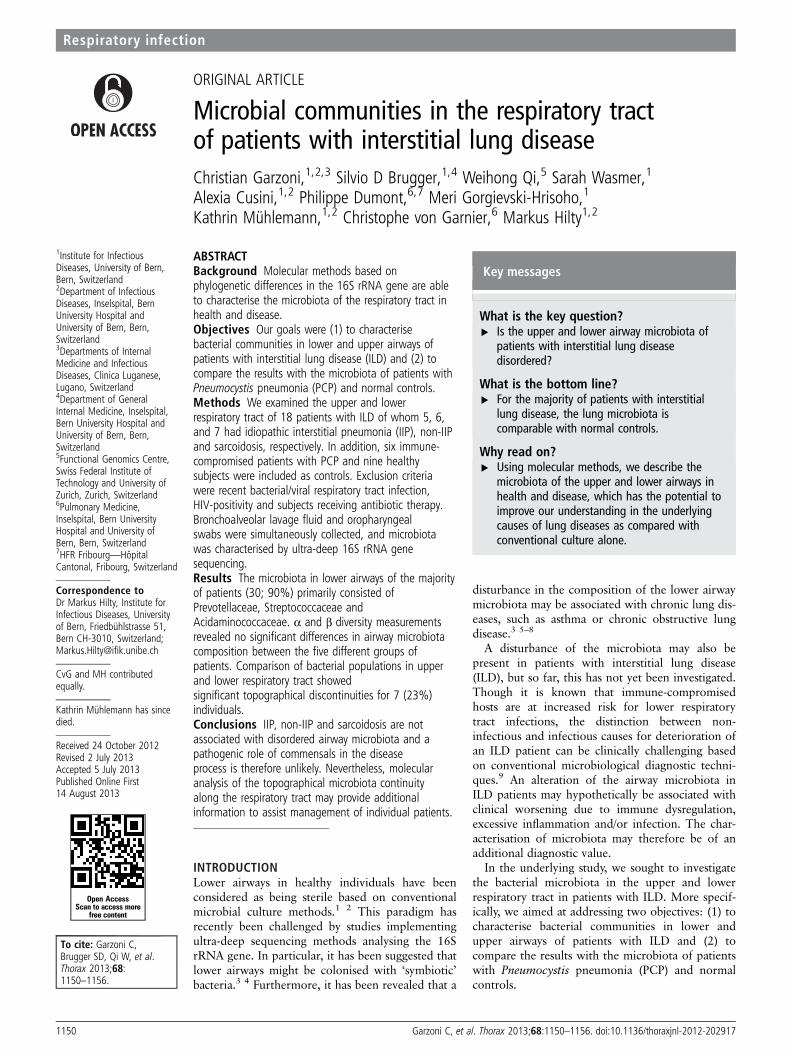
ORIGINAL ARTICLE
Microbial communities in the respiratory tractof patients with interstitial lung diseaseChristian Garzoni,1,2,3 Silvio D Brugger,1,4 Weihong Qi,5 Sarah Wasmer,1
Alexia Cusini,1,2 Philippe Dumont,6,7 Meri Gorgievski-Hrisoho,1
Kathrin Mühlemann,1,2 Christophe von Garnier,6 Markus Hilty1,2
1Institute for InfectiousDiseases, University of Bern,Bern, Switzerland2Department of InfectiousDiseases, Inselspital, BernUniversity Hospital andUniversity of Bern, Bern,Switzerland3Departments of InternalMedicine and InfectiousDiseases, Clinica Luganese,Lugano, Switzerland4Department of GeneralInternal Medicine, Inselspital,Bern University Hospital andUniversity of Bern, Bern,Switzerland5Functional Genomics Centre,Swiss Federal Institute ofTechnology and University ofZurich, Zurich, Switzerland6Pulmonary Medicine,Inselspital, Bern UniversityHospital and University ofBern, Bern, Switzerland7HFR Fribourg—HôpitalCantonal, Fribourg, Switzerland
Correspondence toDr Markus Hilty, Institute forInfectious Diseases, Universityof Bern, Friedbühlstrasse 51,Bern CH-3010, Switzerland;[email protected]
CvG and MH contributedequally.
Kathrin Mühlemann has sincedied.
Received 24 October 2012Revised 2 July 2013Accepted 5 July 2013Published Online First14 August 2013
To cite: Garzoni C,Brugger SD, Qi W, et al.Thorax 2013;68:1150–1156.
ABSTRACTBackground Molecular methods based onphylogenetic differences in the 16S rRNA gene are ableto characterise the microbiota of the respiratory tract inhealth and disease.Objectives Our goals were (1) to characterisebacterial communities in lower and upper airways ofpatients with interstitial lung disease (ILD) and (2) tocompare the results with the microbiota of patients withPneumocystis pneumonia (PCP) and normal controls.Methods We examined the upper and lowerrespiratory tract of 18 patients with ILD of whom 5, 6,and 7 had idiopathic interstitial pneumonia (IIP), non-IIPand sarcoidosis, respectively. In addition, six immune-compromised patients with PCP and nine healthysubjects were included as controls. Exclusion criteriawere recent bacterial/viral respiratory tract infection,HIV-positivity and subjects receiving antibiotic therapy.Bronchoalveolar lavage fluid and oropharyngealswabs were simultaneously collected, and microbiotawas characterised by ultra-deep 16S rRNA genesequencing.Results The microbiota in lower airways of the majorityof patients (30; 90%) primarily consisted ofPrevotellaceae, Streptococcaceae andAcidaminococcaceae. α and β diversity measurementsrevealed no significant differences in airway microbiotacomposition between the five different groups ofpatients. Comparison of bacterial populations in upperand lower respiratory tract showedsignificant topographical discontinuities for 7 (23%)individuals.Conclusions IIP, non-IIP and sarcoidosis are notassociated with disordered airway microbiota and apathogenic role of commensals in the diseaseprocess is therefore unlikely. Nevertheless, molecularanalysis of the topographical microbiota continuityalong the respiratory tract may provide additionalinformation to assist management of individual patients.
INTRODUCTIONLower airways in healthy individuals have beenconsidered as being sterile based on conventionalmicrobial culture methods.1 2 This paradigm hasrecently been challenged by studies implementingultra-deep sequencing methods analysing the 16SrRNA gene. In particular, it has been suggested thatlower airways might be colonised with ‘symbiotic’bacteria.3 4 Furthermore, it has been revealed that a
disturbance in the composition of the lower airwaymicrobiota may be associated with chronic lung dis-eases, such as asthma or chronic obstructive lungdisease.3 5–8
A disturbance of the microbiota may also bepresent in patients with interstitial lung disease(ILD), but so far, this has not yet been investigated.Though it is known that immune-compromisedhosts are at increased risk for lower respiratorytract infections, the distinction between non-infectious and infectious causes for deterioration ofan ILD patient can be clinically challenging basedon conventional microbiological diagnostic techni-ques.9 An alteration of the airway microbiota inILD patients may hypothetically be associated withclinical worsening due to immune dysregulation,excessive inflammation and/or infection. The char-acterisation of microbiota may therefore be of anadditional diagnostic value.In the underlying study, we sought to investigate
the bacterial microbiota in the upper and lowerrespiratory tract in patients with ILD. More specif-ically, we aimed at addressing two objectives: (1) tocharacterise bacterial communities in lower andupper airways of patients with ILD and (2) tocompare the results with the microbiota of patientswith Pneumocystis pneumonia (PCP) and normalcontrols.
Open AccessScan to access more
free content
Key messages
What is the key question?▸ Is the upper and lower airway microbiota of
patients with interstitial lung diseasedisordered?
What is the bottom line?▸ For the majority of patients with interstitial
lung disease, the lung microbiota iscomparable with normal controls.
Why read on?▸ Using molecular methods, we describe the
microbiota of the upper and lower airways inhealth and disease, which has the potential toimprove our understanding in the underlyingcauses of lung diseases as compared withconventional culture alone.
1150 Garzoni C, et al. Thorax 2013;68:1150–1156. doi:10.1136/thoraxjnl-2012-202917
Respiratory infection

MATERIAL AND METHODSStudy population and definitionsThe Epidemiology and Clinical Relevance of Infectious Agentsin Bronchoalveolar Lavage (ECRIBAL) study is a prospective,single centre observational study investigating new diagnosticmethods and potential pathogens in patients with ILD and inimmune-compromised patients with suspicion of lung infection.The study has been approved by the local ethics committee(Kantonale Ethikkommission Bern, approval Nr. 153/08).
For the purpose of the current microbiota study and based onthe ATS/ERS international multidisciplinary consensus classifica-tion,10 we included patients of the following three ILD groupsfrom ECRIBAL: exacerbated idiopathic interstitial pneumonias(IIP) (n=5), exacerbated non-IIP (n=6) and ILD due to sarcoid-osis (n=7). Based on this classification, the non-IIP group con-tained ILD due to known causes, such as collagen vasculardisease, environmental, drug related and ‘other’ ILD, forexample, eosinophilic pneumonia (see table 1 for details).
In addition, we included the following comparative groups:patients with Pneumocystis pneumonia (PCP) (n=6) and healthycontrols without evidence for pulmonary or systemic disease(n=9).
Enrolled individuals with an indication for bronchoscopy atBern University Hospital (Switzerland) had to be ≥18 years ofage and able to provide informed consent. Exclusion criteria forthis study were: recent bacterial/viral respiratory tract infectionwithin 2 weeks prior to bronchoalveolar lavage (BAL),HIV-positivity and subjects that had received antibiotic therapywithin 48 h prior to BAL as this was previously shown to affectthe airway microbiota.11 12 The time difference between BALand lung function was normally 2 weeks. We initially selected41 individuals out of whom 8 individuals from the followinggroups had to be excluded due to absent amplification of lowerairway microbiota from BAL: sarcoidosis (n=3), non-IIP (n=3)and controls (n=3).
BAL procedure and microbiological analysisBAL was performed during bronchoscopy by wedging of thebronchoscope tip in a segmental bronchus of the lobe that dis-played radiological changes, followed by fractional instillationand withdrawal of a total of 150 mL prewarmed saline using50 mL syringes. Microbiological investigations in BAL consistedof routine bacterial, fungal, viral and mycobacterial culture anda panel of 15 respiratory viruses routinely detected (antigendetection and/or PCR).13 Additional immunofluorescence andPCR detection of P. jirovecii were performed as previouslydescribed.14 Finally, oropharyngeal (OP) swabs were taken forsubsequent analysis of viruses and the microbiota of the upperrespiratory tract.
PCR amplification of the 16S rRNA genesDNA extraction was performed using 200 μL of BAL sampleand the OP swab as previously described.15 16 V3–V5 regions ofbacterial 16S rRNA genes were amplified using the primer pairs341F/926R.17 Primer sequences (bold) were modified by add-ition of the Roche 454 Titanium sequencing A or B adaptorsequence (lower cases) and a 10-mer multiplex identifier(A-MID-341F, 50-cgtatcgcctccctcgcgccatcag-[NNNNNNNNNN]-ACTCCTACGGGAGGCAGCAG-30; B-MID-926R,50-ctatgcgccttgccagcccgctcag-[NNNNNNNNNN]-CCGTCAATTCMTTTGAGTTT-30. PCR reactions were performed andpurified using Wizard SV PCR clean-up system (Promega,Madison) as described.17 The final elution step was performedusing 40 mL of double distilled water.
454 Titanium amplicon sequencingSamples were pooled using 10 ng of PCR product of eachsample resulting in eight different pools including every multi-plex identifier (MID) once. The amplicon libraries weresequenced according to the 454 Titanium Amplicon Sequencingprotocols and a series of quality control steps were applied tothe resulting 454 reads.17 454 raw reads were submitted to theNCBI Sequence Read Archive under the sequencing experimentSRA026964 with the accession numbers SRX033134 andSRX193716 (normal control samples).
Calculation of richness and Shannon diversity indices andcommunity comparisons (α and β diversity)PyroTagger was used for the definition of operational taxonomicunits based on 97% sequence identity, estimation of chimaerasand taxonomy assignments.18 α diversity analysis (includingrichness, Shannon and Simpson Diversity indices) was
Table 1 ILD (IIP, non-IIP, sarcoidosis), PCP and control subgroupsused for analysis
IIP (n=5)Age, year (mean±SD) 54.6±22.8Sex, male (n) 2Immunosuppressive therapy (n) 2Detailed diagnosis IPF(2), UIP (2), COP (1),FEV1% (range; mean±SD) 40–108; 74.8±24.5FVC % (range; mean±SD) 42–118; 77.4±29.5
TLC % (range; mean±SD) 43–104; 76.2±27.8Non-IIP (n=6)Age, year (mean±SD) 56.0±17.9Sex, male (n) 5Immunosuppressive therapy (n) 3Detailed diagnosis RA (1), asbestosis (1), EP (3), EAA (1)FEV1% (range; mean±SD) 55–75; 65.5±8.2FVC % (range; mean±SD) 58–85; 74.2±11.7TLC % (range; mean±SD)* 52–94; 76.0±16.0
Sarcoidosis (n=7)Age, year (mean±SD) 48.5±6.5Sex, male (n) 6Immunosuppressive therapy (n) 1FEV1% (range; mean±SD) 60–136; 91.4±26.1FVC % (range; mean±SD) 65–131; 99.6±28.1TLC % (range; mean±SD) 59–122; 94.0±21.6
Pneumocystis pneumonia (PCP) (n=6)Age, year (mean±SD) 58.3±10.6Sex, male (n) 5Immunosuppressive therapy (n) 6FEV1% (range; mean±SD)† 56–101; 80.8±19.6FVC % (range; mean±SD) † 52–115; 84.0±23.0
Normal controls (n=9)Age, year (mean±SD) 57.6±11.4Sex, male (n) 6Immunosuppressive therapy (n) 0
*No TLC data available for patient (ID 12).†No lung function data available for patient (ID 25) with Pneumocystis pneumonia.COP, cryptogenic organising pneumonia; EAA, exogenous allergic alveolitis; EP,eosinophilic pneumonia; FEV1%, forced expiratory volume in 1 s as a percentage ofthe predicted value; FVC %, forced vital capacity as a percentage of the predictedvalue; IIP, idiopathic interstitial pneumonia; ILD, interstitial lung disease;IPF, idiopathic pulmonary fibrosis; non-IIP, non-idiopathic interstitial pneumonia;RA, rheumatoid arthritis; TLC %, total lung capacity as a percentage of the predictedvalue; UIP, usual interstitial pneumonia.
Garzoni C, et al. Thorax 2013;68:1150–1156. doi:10.1136/thoraxjnl-2012-202917 1151
Respiratory infection

performed employing Mothur.19 For the statistical analysis ofthe α diversity results, ordinary one-way ANOVA and Mann–Whitney analyses were performed. Resulting graphs were gener-ated with GraphPad Prism V.5.02 (GraphPad Software, Inc, LaJolla, California, USA).
Community comparisons were calculated using non-metricmultidimensional scaling (nMDS) and the adonis function ofthe vegan package in R as previously described.17 For the cre-ation of the input distance matrices, binary and abundance-based (Horn) calculations were performed as previouslydescribed.17 20 Procrustes function of R was used to comparethe nMDS plots between the upper and lower respiratory tract.
Ordinary one-way ANOVA was performed to investigate thesignificant differences of the relative mean abundances of bacter-ial families of the upper and lower airways.
RESULTSStudy patients, PCR amplification and 16S rRNA genepyrosequencingClinical characteristics of the 33 patients are summarised intable 1. The study subjects had an overall mean age of 55.2±13.7 years and were categorised into the following groups: IIP,non-IIP, sarcoidosis, PCP and healthy controls. Nine (27.3%)were women, sarcoidosis was the most frequent ILD diagnosis(n=7) and none of the 33 study subjects received antibiotictreatment within 48 h before BAL. Though amplification of the33 BAL samples revealed that the bacterial quantity of controlswas lower, this was not statistically significant. Subsequent 16SrRNA gene pyrosequencing was performed and estimatesrevealed that sequencing depth was approximately 80% (datanot shown).
α Diversity measurements in lower airwaysIn order to investigate if microbiota composition varied betweenpatients with different manifestations of ILD, we initially calcu-lated bacterial richness and Shannon diversity indices of thelower airways, that is, in samples obtained from BAL (α diver-sity; figure 1). Both indices are ideal measurements to easily andrapidly investigate alterations in the microbiota as they result ina numeric value that may serve as a surrogate marker for disor-dered microbial communities. This value decreases when asingle bacterial species has ‘overgrown’ and become predomin-ant, replacing the classic ‘commensal’ microbiota.
The mean richness values were 56.2 (±29.2), 83.7 (±20.6),75.1 (±25.9), 60.3 (±16.4) and 63.3 (±20.3) for IIP, non-IIP,sarcoidosis, PCP and normal controls, respectively (figure 1A).The lowest value was therefore found for IIP but this findingwas not statistically significant compared with the other groups.
As for the Shannon diversity indices, again, patients with IIPhad the lowest values (mean 2.0±1.1) (figure 1B), but differ-ences among the indices were again not significantly differentperforming both ordinary one-way ANOVA and Mann–Whitneyanalyses (figure 1A).
β Diversity measurements in lower airwaysWe then measured pairwise distances between microbial com-munities of all 33 samples. For this, Bray–Curtis distance matri-ces were used as input and the amount of variance wascalculated (β diversity calculations). Overall bacterial communi-ties in lower airways did not significantly differ in patients withIIP, non-IIP, sarcoidosis, PCP and controls (figure 2). However,five patients had a clearly different bacterial profile (ID 64, 53,73, 108 and 50; figure 2).
Relative abundance of bacterial families and quantificationSequencing results and subsequent taxa assignments for all 33BAL samples are illustrated in figure 3. The communities ofthree patients harboured solitary abundances of Neisseriaceae(patient ID 50), Streptococcaceae (patient ID 73) andPasteurellaceae (patient ID 64) (figure 3). However, the micro-biota of the 30 remaining patients primarily consisted of bacter-ial families known as commensals, that is, non-pathogenicbacteria colonising the airways. Such commensals includedPrevotellaceae, Streptococcaceae and Acidaminococcaceae. Dueto the nature of the bacterial families present, this again indi-cated that the microbiota may not be disturbed in IIP, non-IIP,sarcoidosis and PCP as compared with healthy controls. In add-ition, a more detailed investigation of the relative differences ofthe bacterial families revealed that the clinical diagnosis (IIP,non-IIP, sarcoidosis, PCP or controls) is only significantly asso-ciated with the relative mean abundance of Flavobacteriaceae(table 2). For all other families, no significant differences weredetected (table 2).
Figure 1 α-Diversity indices of the lower airway microbiota. Indicatedare the Richness (A) and Shannon-diversity (B) indices for subjects withidiopathic interstitial pneumonia (IIP), non-idiopathic interstitialpneumonia (non-IIP), sarcoidosis, Pneumocystis pneumonia (PCP) andnormal controls. No significant differences were observed.
1152 Garzoni C, et al. Thorax 2013;68:1150–1156. doi:10.1136/thoraxjnl-2012-202917
Respiratory infection

Analysis of topographical continuity along the respiratorytractWe subsequently analysed the upper respiratory tract (orophar-ynx) of the 33 subjects (figure 3; right column). As for thelower airways, Prevotellaceae was the most abundant bacterialfamily followed by Streptococcaceae and Acidaminococcaceae.
In a next step, we quantified differences between upper andlower respiratory tract communities in order to analyse topo-graphical continuity. Assuming that a normal respiratory tracthas similar or concordant microbiota profiles between upperand lower airways,4 a divergence would be indicative of thepresence of disordered microbial communities at the site of
Figure 3 Microbial community comparisons and relative abundances of the top 20 bacterial families in 33 bronchoalveolar lavage (BAL) andoropharyngeal samples. A tree illustrating similarities of the microbial communities of the BAL samples are shown on the left. Abundances ofbacterial families for each subject (ID) are indicated by a greyscale value. The five groups consisting of idiopathic interstitial pneumonia (IIP),non-idiopathic interstitial pneumonia (non-IIP), sarcoidosis, Pneumocystis pneumonia (PCP) and normal controls, as well as if immunocompromised(IC) for each study subject are indicated. The quantities of bacterial species derived from conventional culture are provided in colony forming units(CFU). The concentrations of the amplification products (c) are indicated in ng/μL.
Figure 2 Comparison of bacterialcommunities in the lower airways by anon-metric multidimensional scalingordination plot. Shown are the resultsfor subjects with idiopathic interstitialpneumonia (IIP), non-idiopathicinterstitial pneumonia (non-IIP),sarcoidosis, Pneumocystis pneumonia(PCP) and normal controls. Bray–Curtisdistance matrices were used as input.Statistical differences between the fivegroups were non-significant (ie, smalldistances between central points). Fivepatients had different microbiotaillustrated by the large distances to thecentral points (ID 64, 53, 73, 108 and50). nMDS, non-metricmultidimensional scaling.
Garzoni C, et al. Thorax 2013;68:1150–1156. doi:10.1136/thoraxjnl-2012-202917 1153
Respiratory infection

interest. Using procrustes analysis we revealed that 7 of 33patients (ID 17, 38, 50, 53, 64, 73 and 108) displayed differ-ences exceeding the upper quantile of procrustes errors, suggest-ing a significant disordered microbiota between the upper andlower respiratory tract (figure 4).
We then analysed clinical characteristics of the seven patientswith topographical microbiota discontinuity. Patient 38 had mainlycommensal Corynebacteriaceae in the oropharynx, but essentiallyharbouring ‘normal’ Prevotellaceae, Acidaminococcaceae andStreptococcaceae in the BAL. Therefore, this subject could be
Figure 4 Comparison of themicrobiota between upper and lowerairways by procrustes analysis.Pairwise distance matrices werecalculated between lower and uppermicrobial respiratory tract communitiesfrom which non-metric dimension plotswere subsequently derived. Patientswith procrustes residual exceeding theupper quantile (upper dashed line)were considered as containing differentmicrobial communities (ID 64, 50, 73,38, 53, 17 and 108). Study subjectsare presented according to the clinicaldiagnosis: sarcoidosis, idiopathicinterstitial pneumonia (IIP),non-idiopathic interstitial pneumonia(non-IIP), Pneumocystis pneumonia(PCP) and normal controls. ILD,interstitial lung disease.
Table 2 Relative mean abundances of bacterial families in lower and upper airways
Lower airways Upper airways
Relative mean abundance in % Relative mean abundance in %
Bacterial familiesIIP(n=5)
non-IIP(n=6)
Sarcoidosis(n=7)
PCP(n=6)
Control(n=9)
pValue*
IIP(n=5)
non-IIP(n=6)
Sarcoidosis(n=7)
PCP(n=6)
Control(n=9)
pValue*
Moraxellaceae 0.1 0.1 0.0 0.4 1.8 0.068 2.2 0.0 0.0 0.0 1.2 0.456Comamonadaceae 0.1 0.8 0.2 1.3 0.4 0.274 0.8 0.0 0.6 0.0 0.6 0.727Cellulomonadaceae 0.0 0.0 0.0 11.8 0.0 0.357 0.0 0.0 0.0 0.0 0.0 1.000Aerococcaceae 0.8 1.3 0.6 0.8 0.5 0.488 0.4 3.0 0.9 3.1 1.2 0.428Rhizobiaceae 0.1 1.4 0.4 1.4 0.4 0.191 0.2 0.1 0.4 0.0 1.2 0.462Lactobacillaceae 0.1 0.0 0.0 0.0 1.3 0.560 0.8 0.0 0.1 0.0 0.8 0.413Nocardiaceae 0.4 1.2 0.4 1.8 0.1 0.058 0.0 0.2 0.5 0.1 0.1 0.626Capnocytophagaceae 0.1 2.2 0.6 3.1 0.9 0.239 1.9 0.4 1.0 2.2 0.7 0.522
Microbacteriaceae 0.4 1.4 0.5 1.2 0.1 0.061 0.1 0.1 1.0 0.2 0.1 0.585Sphingomonadaceae 0.1 0.5 0.2 0.4 0.2 0.388 0.1 0.2 0.1 0.0 0.3 0.509Flavobacteriaceae 1.1 4.3 1.3 4.7 0.4 0.036 0.2 0.5 1.2 8.1 0.2 0.048Peptostreptococcaceae 1.3 1.0 1.0 0.7 1.1 0.931 0.9 0.6 1.6 0.3 0.3 0.184Fusobacteriaceae 1.9 7.3 5.9 2.0 6.4 0.282 2.1 3.5 6.3 11.8 6.2 0.442Staphylococcaceae 0.0 1.5 1.0 5.9 0.5 0.313 0.4 2.4 3.2 7.8 0.7 0.355Porphyromonadaceae 2.8 5.3 4.1 11.5 2.8 0.586 4.1 2.3 8.1 5.7 2.0 0.147Pasteurellaceae 19.0 4.0 2.5 4.3 1.1 0.345 2.0 3.4 2.7 39.3 1.4 0.412Neisseriaceae 16.2 4.2 4.7 9.2 6.6 0.686 22.5 1.1 6.1 10.5 4.5 0.281Acidaminococcaceae 4.7 15.0 12.1 5.1 6.2 0.114 9.3 10.2 6.6 10.1 5.3 0.771Streptococcaceae 4.3 10.3 21.6 4.2 17.4 0.380 12.6 10.9 9.6 7.6 22.0 0.090Prevotellaceae 39.3 26.8 37.0 24.7 42.0 0.760 32.3 33.9 35.1 24.8 40.9 0.767
*p Values derived by ordinary one-way analysis of variance are indicated. p<0.05 is considered as being significant indicating a difference between the five groups.IIP, idiopathic interstitial pneumonia; non-IIP, non-idiopathic interstitial pneumonia; PCP, Pneumocystis pneumonia.
1154 Garzoni C, et al. Thorax 2013;68:1150–1156. doi:10.1136/thoraxjnl-2012-202917
Respiratory infection

considered as healthy in the absence of clinical signs and symptomsindicative of an upper respiratory tract infection. The high procrus-tes residuals in patients 17 and 64 have their origins in the highabundances of Pasteurellaceae (ie, Haemophilus influenzae as con-firmed by culture) in the upper (patient 17) and lower (patient 64)respiratory tract (figure 3). H. influenzae is frequently detected inmicrobial cultures, but it is often unclear if this reflects non-pathogenic colonisation or infection, given that different strainswith both features are known.21 In our study, we identifiedH. influenzae in patients 17, 25 and 64, but only in patient 64, theoverall microbiota was disordered in the lower respiratory tract,suggesting infection rather than colonisation (figure 3). Patient 64was diagnosed with idiopathic pulmonary fibrosis without evidenceby conventional microbiology for respiratory tract infection anddid not improve under an initial course of systemic corticosteroidtreatment. Based on the microbiota results of the lower airways,H. influenzae may have played a clinically relevant role in thecourse of the exacerbation, and therefore, in the absence of the‘normal’ microbiota, an initial antibiotic treatment may have beenan appropriate treatment option.
The remaining procrustes differences occurred in patients 50,53, 73 and 108. These were based on excess abundance ofStreptococcus pneumoniae (patient 73), Neisseriaceae (patient50 and normal control 108) and Cellulomonadaceae (mainlyTropheryma whipplei, patient 53) and were localised to thelower airways. In patient 73 with a histologically proven sarcoid-osis, microbiota discontinuity was present with an excess ofS. pneumoniae, but without signs for respiratory tract infection.This finding, together with the excess Neisseriaceae in patients50 and 108, remains unclear. Finally, identification ofCellulomonadaceae (mainly T. whipplei) in patient 53 is an intri-guing finding and may be due to hitherto undiagnosedWhipple’s Disease.
DISCUSSIONRecent studies using 16S rRNA gene pyrosequencing techniqueshave explored the microbiota of the lower and upper respiratorytract in health and disease.3 4 22 23 These techniques now alsoenable to investigate disordered microbial communities inpatients with other lung disorders and conditions, such as IIP,non-IIP and sarcoidosis, which to our knowledge, has not beenperformed to date. Within this study, we therefore aimed atcharacterising the bacterial communities in lower and upperairways of patients with IIP, non-IIP and sarcoidosis and tocompare the results with the microbiota of patients withPneumocystis pneumonia (PCP) and normal controls.
Most of the patients with IIP, non-IIP, sarcoidosis and PCP har-boured three bacterial families (Prevotellaceae, Streptococcaceaeand Acidaminococcaceae) in the respiratory tract that were alsofrequently detected in healthy controls, and therefore, indicatedthat no disordering of the microbiota was present. It is worth-while mentioning that the same commensal families have alsobeen reported in other recent studies analysing the healthyrespiratory tract.3 4
α and β diversity measurements are available to investigate ifclinical factors and/or diagnoses are significantly associated withan altered microbiota composition. In our study, neither α nor βdiversity indices revealed any significant changes related to theclinical diagnosis of IIP, non-IIP, sarcoidosis or PCP comparedwith healthy controls.
To date, sound data linking microbiota changes to lung disor-ders are still scarce as this field of research is still in its infancy.In HIV-infected patients, several known or suspected pathogenicorganisms have been detected that may be related to recurrent
pneumonia.11 In patients with non-CF bronchiectasis, character-istics of the lower airways microbiota correlated with clinicalmarkers of disease severity.23 Finally, two studies revealed thatthe microbiota in cystic fibrosis patients is distinct, probably dueto the specific environmental characteristics in the respiratorytract of these patients.24 25
In our study, we simultaneously analysed upper and lowerrespiratory tract, which is an important aspect for two reasons:first, this approach allows the examination for possible contam-ination from the upper respiratory tract.26 Second, it enables tospecifically localise microbial communities in the airways asshown by recent studies.5 27 For the healthy human respiratorytract, topographical continuity of bacterial populations has pre-viously been described.4 Within our study, we now extend thesefindings by demonstrating that continuity of microbiota alsoexists in the majority of patients with underlying ILD. Thisfinding was not applicable for seven individuals revealing signifi-cant divergence between the upper and lower respiratory tract.This may indicate disturbed microbiota in the upper or lowerairways, but has to be investigated for each patient separately.Based on our data, we suggest that the identification of micro-biota discontinuity along the respiratory tract should generallybe interpreted by taking into account the clinical presentation ofthe patient.
Compared with conventional bacterial cultures, utilisation ofpyrosequencing methods generally has two advantages. First, themethods are able to identify unusual pathogenic bacteria nor-mally not detectable by conventional microbiologicalmethods.28–30 Second, these methods can assist to identify thepathogenic role of ‘commensal’ bacteria. Depletion of thenormal microbiota with ‘overgrowth’ of a single bacterial speciesmay indicate that this is clinically relevant, for example, as seenwith H. influenzae in our study. This is an important aspect as theclinician is commonly confronted with the challenge to distin-guish between innocuous commensals, ‘facultative pathogenic’and ‘pathogenic’ bacteria. However, it is crucial to emphasisethat conventional culture and molecular methods should be seenas complimentary, rather than exclusionary, as molecularmethods may detect difficult-to-culture bacteria but may in turnmiss potentially pathogenic bacteria in low abundance, as thesemay not significantly alter microbial communities.
There are several limitations to our study. First, based on theATS/ERS ILD classification into different groups, subjects pre-sented with a certain degree of heterogeneity within this study.Nevertheless, we feel that selecting the five groups, we havecompared clinically relevant populations. Second, we did notperform extensive ultra-deep sequencing of viruses and fungi inthe collection of samples. Although this has recently beenshown to be feasible, fungi identified by this technique weremostly cultivated and known.31 We routinely searched for 15respiratory viruses, for Pneumocystis jirovecii by immunofluores-cence and PCR, measured galactomannan levels and culturedfungi and mycobacteria. We are therefore confident that we didnot neglect clinically relevant and currently known pathogens.Third, the number of patients included in this feasibility study isrelatively low; therefore, a potential added benefit of character-ising microbiota to distinguish between presence of a relevantpathogen and non-infectious clinical worsening in an ILDpatient requires further evaluation in prospective, larger studies.Finally, this study includes patients with exacerbated IIP andnon-IIP. As we did not include samples from IIP and non-IIPpatients during disease stability, we were therefore not able toinvestigate longitudinal microbiota changes during the actualexacerbation within this study. Nevertheless, considering that we
Garzoni C, et al. Thorax 2013;68:1150–1156. doi:10.1136/thoraxjnl-2012-202917 1155
Respiratory infection

did not detect significant microbiota differences in exacerbatedILD patients compared with normal controls, we hypothesisethat changes from an ILD-driven microbiota towards a ‘normal’microbiota during exacerbation is unlikely and that the presenceof a ‘normal’ microbiota during times of stability would bemore probable. In agreement with this hypothesis, significantchanges in community structure or diversity were not observedat the time of exacerbation in adults with cystic fibrosis.32 33
We conclude that 16S rRNA gene-based pyrosequencing isable to characterise microbial communities in upper and lowerairways in ILD patients, but the diagnosis of IIP, non-IIP, sarcoid-osis or during PCP per se did not alter the overall lung micro-biota. Analysis of the topographical continuity along therespiratory tract may provide important additional informationto assist the physician in understanding the underlying cause ofclinical worsening in ILD patients, a process that will alwaysinvolve distinguishing infectious from non-infectious causes ofinflammation. A potential future application of 16S rRNA gene-based pyrosequencing is the search of hitherto insufficientlyknown bacteria that may be relevant in the pathogenesis andcourse of pulmonary disease.
Acknowledgements We thank the Professors Jean-Francois Dufour, Martin Fey,Felix Frey, Thomas Geiser, Paul Mohacsi and Peter Villiger for their help in providingclinical data and Cornelia Wyss, Monika Wüthrich and Liselotte McEvoy for theirassistance with patient recruitment and administrative tasks. We are also grateful toIrène Stutz and Heidi Pfäffli for technical assistance and sample processing.
Contributors Conception and design: CG, SDB, KM, MG-H. Acquisition of data:CG, SDB, WQ, SW, AC, PD, CvG, MG-H. Analysis and interpretation: CG, SDB, CvG,MG-H. Drafting the manuscript: CG, SDB, WQ, MG-H, CvG, MH.
Funding This study was supported by unrestricted grants (AstraZeneca, Roche,MSD, Essex Chemie Switzerland, Wyeth, Astellas to C.G.), the Swiss NationalScience Foundation (M.D.-Ph.D. scholarship 323500-119214 to S.D.B. and SinergiaCRSII3-141875 to M.H. and C.v.G) and the 2012 Award of the Swiss Society ofHospital Hygiene & the Swiss Society for Infectious Diseases (to M.H.).
Competing interests None.
Patient consent Obtained.
Ethics approval Kantonale Ethikkommission Bern, approval Nr. 153/08.
Provenance and peer review Not commissioned; externally peer reviewed.
Open Access This is an Open Access article distributed in accordance with theCreative Commons Attribution Non Commercial (CC BY-NC 3.0) license, whichpermits others to distribute, remix, adapt, build upon this work non-commercially,and license their derivative works on different terms, provided the original work isproperly cited and the use is non-commercial. See: http://creativecommons.org/licenses/by-nc/3.0/
REFERENCES1 Laurenzi GA, Potter RT, Kass EH. Bacteriologic flora of the lower respiratory tract.
N Engl J Med 1961;265:1273–8.2 Pecora DV. A comparison of transtracheal aspiration with other methods of
determining the bacterial flora of the lower respiratory tract. N Engl J Med1963;269:664–6.
3 Hilty M, Burke C, Pedro H, et al. Disordered microbial communities in asthmaticairways. PLoS One 2010;5:e8578.
4 Charlson ES, Bittinger K, Haas AR, et al. Topographical continuity of bacterialpopulations in the healthy human respiratory tract. Am J Respir Crit Care Med2011;184:957–63.
5 Erb-Downward JR, Thompson DL, Han MK, et al. Analysis of the lung microbiomein the “healthy” smoker and in COPD. PLoS One 2011;6:e16384.
6 Han MK, Huang YJ, Lipuma JJ, et al. Significance of the microbiome in obstructivelung disease. Thorax 2012;67:456–63.
7 Pragman AA, Kim HB, Reilly CS, et al. The lung microbiome in moderate and severechronic obstructive pulmonary disease. PLoS One 2012;7:e47305.
8 Sze MA, Dimitriu PA, Hayashi S, et al. The lung tissue microbiome in chronicobstructive pulmonary disease. Am J Respir Crit Care Med 2012;185:1073–80.
9 Carroll KC. Laboratory diagnosis of lower respiratory tract infections: controversy andconundrums. J Clin Microbiol 2002;40:3115–20.
10 American Thoracic Society/European Respiratory Society InternationalMultidisciplinary Consensus Classification of the Idiopathic Interstitial Pneumonias.This joint statement of the American Thoracic Society (ATS), and the EuropeanRespiratory Society (ERS) was adopted by the ATS board of directors, June 2001and by the ERS Executive Committee, June 2001. Am J Respir Crit Care Med2002;165:277–304.
11 Iwai S, Fei M, Huang D,, et al Oral and airway microbiota in HIV-infectedpneumonia patients. J Clin Microbiol 2012;50:2995–3002.
12 Jakobsson HE, Jernberg C, Andersson AF, et al. Short-term antibiotic treatment hasdiffering long-term impacts on the human throat and gut microbiome. PLoS One2010;5:e9836.
13 Garcia LS. Clinical microbiology procedures handbook. 3rd edn. ASM Press, 2010.14 Muhlethaler K, Bogli-Stuber K, Wasmer S, et al. Quantitative PCR to diagnose
Pneumocystis pneumonia in immunocompromised non-HIV patients. Eur Respir J2012;39:971–8.
15 Brugger SD, Frei L, Frey PM, et al. 16S rRNA terminal restriction fragment lengthpolymorphism for the characterization of the nasopharyngeal microbiota. PLoS One2012;7:e52241.
16 Brugger SD, Hathaway LJ, Muhlemann K. Detection of Streptococcus pneumoniaestrain cocolonization in the nasopharynx. J Clin Microbiol 2009;47:1750–6.
17 Hilty M, Qi W, Brugger SD, et al. Nasopharyngeal microbiota in infants with acuteotitis media. J Infect Dis 2012;205:1048–55.
18 Kunin V, Hugenholtz P. PyroTagger: a fast, accurate pipeline for analysis of rRNAamplicon pyrosequence data. The Open Journal 2010:1–8.
19 Schloss PD, Westcott SL, Ryabin T, et al. Introducing mothur: open-source,platform-independent, community-supported software for describing and comparingmicrobial communities. Appl Environ Microbiol 2009;75:7537–41.
20 Frank DN, Feazel LM, Bessesen MT, et al. The human nasal microbiota andStaphylococcus aureus carriage. PLoS One 2010;5:e10598.
21 King P. Haemophilus influenzae and the lung (Haemophilus and the lung). ClinTransl Med 2012;1:10.
22 Cardenas PA, Cooper PJ, Cox MJ, et al. Upper airways microbiota in antibiotic-naivewheezing and healthy infants from the tropics of rural Ecuador. PLoS One 2012;7:e46803.
23 Rogers GB, van der Gast CJ, Cuthbertson L, et al. Clinical measures of disease inadult non-CF bronchiectasis correlate with airway microbiota composition. Thorax2013;68:731–7.
24 Lipuma JJ. The changing microbial epidemiology in cystic fibrosis. Clin Microbiol Rev2010;23:299–323.
25 Rogers GB, Hoffman LR, Carroll MP, et al. Interpreting infective microbiota: theimportance of an ecological perspective. Trends Microbiol 2013;21:271–6.
26 Goddard AF, Staudinger BJ, Dowd SE, et al. Direct sampling of cystic fibrosis lungsindicates that DNA-based analyses of upper-airway specimens can misrepresent lungmicrobiota. Proc Natl Acad Sci USA 2012;109:13769–74.
27 Willner D, Haynes MR, Furlan M, et al. Spatial distribution of microbial communitiesin the cystic fibrosis lung. ISME J 2012;6:471–4.
28 Bousbia S, Papazian L, Auffray JP, et al. Tropheryma whipplei in patients withpneumonia. Emerg Infect Dis 2010;16:258–63.
29 Harris JK, De Groote MA, Sagel SD, et al. Molecular identification of bacteria inbronchoalveolar lavage fluid from children with cystic fibrosis. Proc Natl Acad SciU S A 2007;104:20529–33.
30 Duff RM, Simmonds NJ, Davies JC, et al. A molecular comparison of microbialcommunities in bronchiectasis and cystic fibrosis. Eur Respir J 2013;41:991–3.
31 Charlson ES, Diamond JM, Bittinger K, et al. Lung-enriched organisms and aberrantbacterial and fungal respiratory microbiota following lung transplant. Am J RespirCrit Care Med 2012;186:536–45.
32 Stressmann FA, Rogers GB, van der Gast CJ, et al. Long-termcultivation-independent microbial diversity analysis demonstrates that bacterialcommunities infecting the adult cystic fibrosis lung show stability and resilience.Thorax 2012;67:867–73.
33 Zhao J, Schloss PD, Kalikin LM, et al. Decade-long bacterial community dynamics incystic fibrosis airways. Proc Natl Acad Sci USA 2012;109:5809–14.
1156 Garzoni C, et al. Thorax 2013;68:1150–1156. doi:10.1136/thoraxjnl-2012-202917
Respiratory infection
Copyright © 2022 FDOKUMEN