Striatal D1 and D2 signaling differentially predict learning from positive and negative outcomes
7
UNCORRECTED PROOF Q1 Striatal D1 and D2 signaling differentially predict learning from positive 2 and negative outcomes Q2 Sylvia M.L. Cox a,b , Michael J. Frank c , Kevin Larcher a,b , Lesley K. Fellows a , Crystal A. Clark a , 4 Marco Leyton b , Alain Dagher a, ⁎ 5 a Montreal Neurological Institute, McGill University, 3801 University Street, Montreal, Quebec H3A 2B4, Canada 6 b Department of Psychiatry, McGill University, 1033 Pine Avenue West, Montreal, Quebec H3A 1A1, Canada 7 c Department of Cognitive, Linguistic & Psychological Sciences, Brown Institute for Brain Science, Brown University, 190 Thayer Street, Providence, RI 02912-1821, USA abstract 8 article info 9 Article history: 10 Accepted 27 December 2014 11 Available online xxxx 12 Keywords: 13 Dopamine 14 Reinforcement 15 Learning 16 Positron emission tomography 17 Striatum 18 The extent to which we learn from positive and negative outcomes of decisions is modulated by the neurotrans- 19 mitter dopamine. Dopamine neurons burst fire in response to unexpected rewards and pause following negative 20 outcomes. This dual signaling mechanism is hypothesized to drive both approach and avoidance behaviors. Here 21 we test a prediction deriving from a computational reinforcement learning model, in which approach is mediated 22 via activation of the direct cortico-striatal pathway due to striatal D1 receptor stimulation, while avoidance 23 occurs via disinhibition of indirect pathway striatal neurons secondary to a reduction of D2 receptor stimulation. 24 Using positron emission tomography with two separate radioligands, we demonstrate that individual differences 25 in human approach and avoidance learning are predicted by variability in striatal D1 and D2 receptor binding, 26 respectively. Moreover, transient dopamine precursor depletion improved learning from negative outcomes. 27 These findings support a bidirectional modulatory role for striatal dopamine in reward and avoidance learning 28 via segregated D1 and D2 cortico-striatal pathways. 29 © 2015 Published by Elsevier Inc. 30 31 32 33 34 Introduction 35 While striatal dopamine signaling is widely thought to play an 36 important role in reward learning (Schultz, 2002), its contribution to 37 learning from negative outcomes is more controversial. Dopamine 38 neurons burst fire following the presentation of unexpected rewards, 39 and pause when an expected reward has been withheld, allowing 40 them to encode a reward prediction error (RPE) signal (Montague 41 et al., 1996; Schultz et al., 1997). One class of reinforcement learning 42 models suggests that phasic dopamine bursts support learning from 43 positive outcomes, but, that, due to low dynamic range at the low end 44 of dopamine signaling, other neuromodulators must be involved 45 in learning from negative outcomes (Daw et al., 2002; Bayer and 46 Glimcher, 2005). Another model, tested here, proposes that dopamine 47 modulates both approach and avoidance learning via two segregated 48 pathways in the cortico-striato-thalamocortical circuit (Frank, 2005; 49 Frank and O'Reilly, 2006; Frank et al., 2007a). In this model (Fig. 1), 50 striatal medium spiny neurons of the direct pathway, which express do- 51 pamine D1 receptors (D1R), facilitate the selection of rewarding actions 52 encoded in the cortex. Those belonging to the indirect pathway, which 53 express dopamine D2 receptors (D2R), help to suppress cortical 54 patterns that encode maladaptive or non-rewarding actions (Surmeier 55 et al., 2011). This opponent system model may better account for asym- 56 metrical effects of dopamine manipulations on reward and punishment 57 learning than single-value reinforcement learning models (Collins and 58 Frank, 2014). One of its predictions is that positive reinforcement 59 learning will be modulated by signaling within the D1 direct pathway 60 while negative reinforcement learning will be modulated by signaling 61 within the D2 indirect pathway. 62 In humans, there is indirect evidence that dopamine modulates 63 approach and avoidance learning in opposite directions (Frank et al., 64 2004; Cools et al., 2006; Frank and O'Reilly, 2006; Pessiglione et al., 65 2006; Moustafa et al., 2008). Genetic studies suggest that these opposite 66 modulations are related to D1R and D2R signaling (Frank et al., 2007b, 67 2009; Frank and Hutchison, 2009; Jocham et al., 2009; Doll et al., 68 2011), but direct evidence is lacking. 69 Here we assess the potentially distinct roles of D1R and D2R signal- 70 ing in human learning and decision making by measuring receptor 71 availabilities using positron emission tomography (PET) with separate 72 tracers selective for D1R ([ 11 C]SCH23390) and D2R ([ 11 C]raclopride), 73 and relating these measures to performance on the Probabilistic Selec- 74 tion Task (PST), which simultaneously measures reinforcement and 75 avoidance learning. In a second study, we employed the PST and acute 76 phenylalanine and tyrosine depletion (APTD) to transiently decrease 77 dopamine levels, providing a causal test of the model prediction that 78 this manipulation would improve learning from negative outcomes. NeuroImage xxx (2015) xxx–xxx ⁎ Corresponding author at: Montreal Neurological Institute, 3801 University St., Montréal, QC H3A 2B4, Canada. E-mail address: [email protected] (A. Dagher). YNIMG-11892; No. of pages: 7; 4C: 2, 4, 5 http://dx.doi.org/10.1016/j.neuroimage.2014.12.070 1053-8119/© 2015 Published by Elsevier Inc. Contents lists available at ScienceDirect NeuroImage journal homepage: www.elsevier.com/locate/ynimg Please cite this article as: Cox, S.M.L., et al., Striatal D1 and D2 signaling differentially predict learning from positive and negative outcomes, NeuroImage (2015), http://dx.doi.org/10.1016/j.neuroimage.2014.12.070
-
Upload
independent -
Category
Documents
-
view
1 -
download
0
Transcript of Striatal D1 and D2 signaling differentially predict learning from positive and negative outcomes
1Q1
2
3Q2
4
567
8
91011
121314151617
3031
32
33
34
35
36
37
38
39
40
41
42
43
44
45
46
47
48
49
50
51
52
53
NeuroImage xxx (2015) xxx–xxx
YNIMG-11892; No. of pages: 7; 4C: 2, 4, 5
Contents lists available at ScienceDirect
NeuroImage
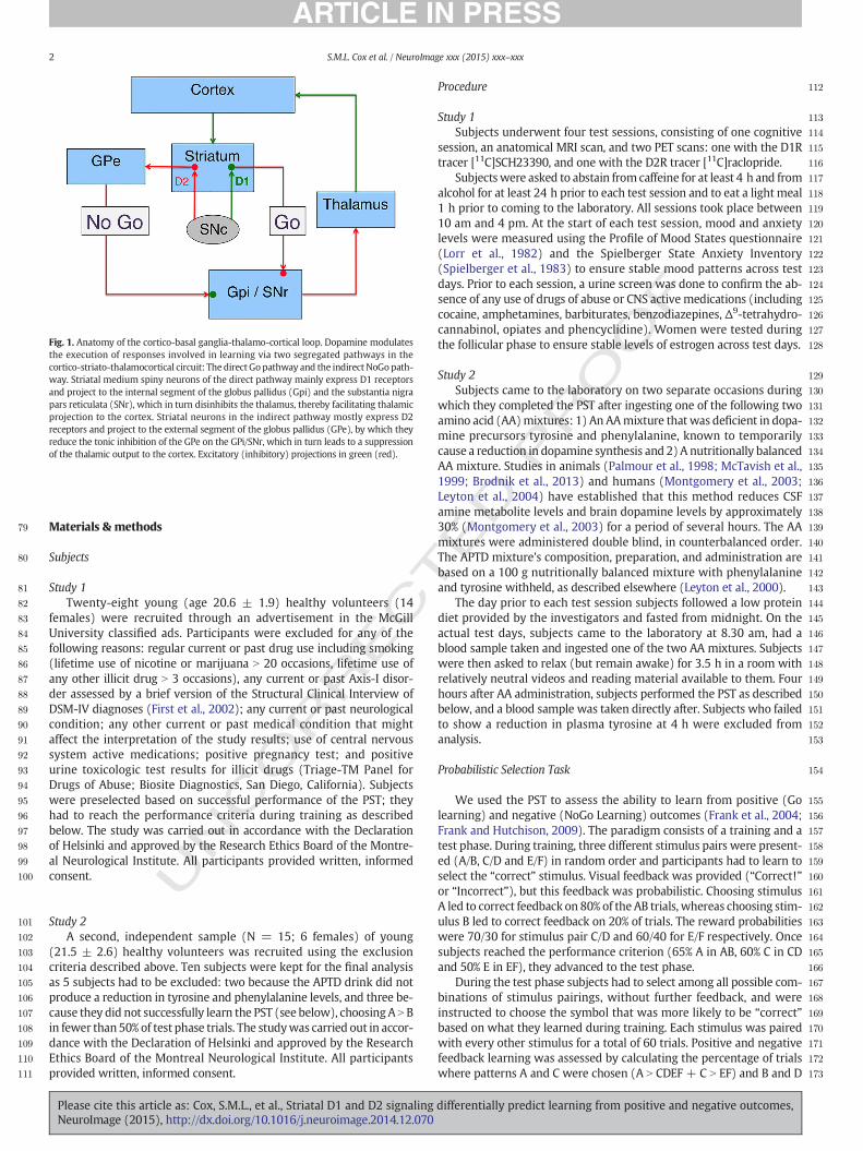
j ourna l homepage: www.e lsev ie r .com/ locate /yn img
Striatal D1 and D2 signaling differentially predict learning from positiveand negative outcomes
OFSylvia M.L. Cox a,b, Michael J. Frank c, Kevin Larcher a,b, Lesley K. Fellows a, Crystal A. Clark a,
Marco Leyton b, Alain Dagher a,⁎a Montreal Neurological Institute, McGill University, 3801 University Street, Montreal, Quebec H3A 2B4, Canadab Department of Psychiatry, McGill University, 1033 Pine Avenue West, Montreal, Quebec H3A 1A1, Canadac Department of Cognitive, Linguistic & Psychological Sciences, Brown Institute for Brain Science, Brown University, 190 Thayer Street, Providence, RI 02912-1821, USA
O⁎ Corresponding author at: Montreal NeurologicalMontréal, QC H3A 2B4, Canada.
E-mail address: [email protected] (A. Dagher).
http://dx.doi.org/10.1016/j.neuroimage.2014.12.0701053-8119/© 2015 Published by Elsevier Inc.
Please cite this article as: Cox, S.M.L., et al.,NeuroImage (2015), http://dx.doi.org/10.101
Ra b s t r a c t
a r t i c l e i n f o18
19
20
21
22
23
24
25
26
Article history:Accepted 27 December 2014Available online xxxx
Keywords:DopamineReinforcementLearningPositron emission tomographyStriatum
27
28
29
TED P
The extent to which we learn from positive and negative outcomes of decisions is modulated by the neurotrans-mitter dopamine. Dopamine neurons burst fire in response to unexpected rewards and pause following negativeoutcomes. This dual signalingmechanism is hypothesized to drive both approach and avoidance behaviors. Herewe test a prediction deriving froma computational reinforcement learningmodel, inwhich approach ismediatedvia activation of the direct cortico-striatal pathway due to striatal D1 receptor stimulation, while avoidanceoccurs via disinhibition of indirect pathway striatal neurons secondary to a reduction of D2 receptor stimulation.Using positron emission tomographywith two separate radioligands,we demonstrate that individual differencesin human approach and avoidance learning are predicted by variability in striatal D1 and D2 receptor binding,respectively. Moreover, transient dopamine precursor depletion improved learning from negative outcomes.These findings support a bidirectional modulatory role for striatal dopamine in reward and avoidance learningvia segregated D1 and D2 cortico-striatal pathways.
© 2015 Published by Elsevier Inc.
C54
55
56
57
58
59
60
61
62
63
64
65
66
67
68
69
70
71
72
73
74
UNCO
RREIntroduction
While striatal dopamine signaling is widely thought to play animportant role in reward learning (Schultz, 2002), its contribution tolearning from negative outcomes is more controversial. Dopamineneurons burst fire following the presentation of unexpected rewards,and pause when an expected reward has been withheld, allowingthem to encode a reward prediction error (RPE) signal (Montagueet al., 1996; Schultz et al., 1997). One class of reinforcement learningmodels suggests that phasic dopamine bursts support learning frompositive outcomes, but, that, due to low dynamic range at the low endof dopamine signaling, other neuromodulators must be involvedin learning from negative outcomes (Daw et al., 2002; Bayer andGlimcher, 2005). Another model, tested here, proposes that dopaminemodulates both approach and avoidance learning via two segregatedpathways in the cortico-striato-thalamocortical circuit (Frank, 2005;Frank and O'Reilly, 2006; Frank et al., 2007a). In this model (Fig. 1),striatalmedium spiny neurons of the direct pathway, which express do-pamine D1 receptors (D1R), facilitate the selection of rewarding actionsencoded in the cortex. Those belonging to the indirect pathway, whichexpress dopamine D2 receptors (D2R), help to suppress cortical
75
76
77
78
Institute, 3801 University St.,
Striatal D1 and D2 signaling6/j.neuroimage.2014.12.070
patterns that encode maladaptive or non-rewarding actions (Surmeieret al., 2011). This opponent systemmodel may better account for asym-metrical effects of dopaminemanipulations on reward and punishmentlearning than single-value reinforcement learning models (Collins andFrank, 2014). One of its predictions is that positive reinforcementlearning will be modulated by signaling within the D1 direct pathwaywhile negative reinforcement learning will be modulated by signalingwithin the D2 indirect pathway.
In humans, there is indirect evidence that dopamine modulatesapproach and avoidance learning in opposite directions (Frank et al.,2004; Cools et al., 2006; Frank and O'Reilly, 2006; Pessiglione et al.,2006;Moustafa et al., 2008). Genetic studies suggest that these oppositemodulations are related to D1R and D2R signaling (Frank et al., 2007b,2009; Frank and Hutchison, 2009; Jocham et al., 2009; Doll et al.,2011), but direct evidence is lacking.
Here we assess the potentially distinct roles of D1R and D2R signal-ing in human learning and decision making by measuring receptoravailabilities using positron emission tomography (PET) with separatetracers selective for D1R ([11C]SCH23390) and D2R ([11C]raclopride),and relating these measures to performance on the Probabilistic Selec-tion Task (PST), which simultaneously measures reinforcement andavoidance learning. In a second study, we employed the PST and acutephenylalanine and tyrosine depletion (APTD) to transiently decreasedopamine levels, providing a causal test of the model prediction thatthis manipulation would improve learning from negative outcomes.
differentially predict learning from positive and negative outcomes,
T
79
80
81
82
83
84
85
86
87
88
89
90
91
92
93
94
95
96
97
98
99
100
101
102
103
104
105
106
107
108
109
110
111
112
113
114
115
116
117
118
119
120
121
122
123
124
125
126
127
128
129
130
131
132
133
134
135
136
137
138
139
140
141
142
143
144
145
146
147
148
149
150
151
152
153
154
155
156
157
158
159
160
Fig. 1. Anatomy of the cortico-basal ganglia-thalamo-cortical loop. Dopamine modulatesthe execution of responses involved in learning via two segregated pathways in thecortico-striato-thalamocortical circuit: The direct Gopathway and the indirect NoGopath-way. Striatal medium spiny neurons of the direct pathway mainly express D1 receptorsand project to the internal segment of the globus pallidus (Gpi) and the substantia nigrapars reticulata (SNr), which in turn disinhibits the thalamus, thereby facilitating thalamicprojection to the cortex. Striatal neurons in the indirect pathway mostly express D2receptors and project to the external segment of the globus pallidus (GPe), by which theyreduce the tonic inhibition of the GPe on the GPi/SNr, which in turn leads to a suppressionof the thalamic output to the cortex. Excitatory (inhibitory) projections in green (red).
2 S.M.L. Cox et al. / NeuroImage xxx (2015) xxx–xxx
NCO
RREC
Materials & methods
Subjects
Study 1Twenty-eight young (age 20.6 ± 1.9) healthy volunteers (14
females) were recruited through an advertisement in the McGillUniversity classified ads. Participants were excluded for any of thefollowing reasons: regular current or past drug use including smoking(lifetime use of nicotine or marijuana N 20 occasions, lifetime use ofany other illicit drug N 3 occasions), any current or past Axis-I disor-der assessed by a brief version of the Structural Clinical Interview ofDSM-IV diagnoses (First et al., 2002); any current or past neurologicalcondition; any other current or past medical condition that mightaffect the interpretation of the study results; use of central nervoussystem active medications; positive pregnancy test; and positiveurine toxicologic test results for illicit drugs (Triage-TM Panel forDrugs of Abuse; Biosite Diagnostics, San Diego, California). Subjectswere preselected based on successful performance of the PST; theyhad to reach the performance criteria during training as describedbelow. The study was carried out in accordance with the Declarationof Helsinki and approved by the Research Ethics Board of the Montre-al Neurological Institute. All participants provided written, informedconsent.
U 161162
163
164
165
166
167
168
169
170
171
172
173
Study 2A second, independent sample (N = 15; 6 females) of young
(21.5 ± 2.6) healthy volunteers was recruited using the exclusioncriteria described above. Ten subjects were kept for the final analysisas 5 subjects had to be excluded: two because the APTD drink did notproduce a reduction in tyrosine and phenylalanine levels, and three be-cause they did not successfully learn the PST (see below), choosingA N Bin fewer than50% of test phase trials. The studywas carried out in accor-dance with the Declaration of Helsinki and approved by the ResearchEthics Board of the Montreal Neurological Institute. All participantsprovided written, informed consent.
Please cite this article as: Cox, S.M.L., et al., Striatal D1 and D2 signalingNeuroImage (2015), http://dx.doi.org/10.1016/j.neuroimage.2014.12.070
ED P
RO
OF
Procedure
Study 1Subjects underwent four test sessions, consisting of one cognitive
session, an anatomical MRI scan, and two PET scans: one with the D1Rtracer [11C]SCH23390, and one with the D2R tracer [11C]raclopride.
Subjectswere asked to abstain fromcaffeine for at least 4 h and fromalcohol for at least 24 h prior to each test session and to eat a light meal1 h prior to coming to the laboratory. All sessions took place between10 am and 4 pm. At the start of each test session, mood and anxietylevels were measured using the Profile of Mood States questionnaire(Lorr et al., 1982) and the Spielberger State Anxiety Inventory(Spielberger et al., 1983) to ensure stable mood patterns across testdays. Prior to each session, a urine screen was done to confirm the ab-sence of any use of drugs of abuse or CNS active medications (includingcocaine, amphetamines, barbiturates, benzodiazepines, Δ9-tetrahydro-cannabinol, opiates and phencyclidine). Women were tested duringthe follicular phase to ensure stable levels of estrogen across test days.
Study 2Subjects came to the laboratory on two separate occasions during
which they completed the PST after ingesting one of the following twoamino acid (AA)mixtures: 1) An AAmixture that was deficient in dopa-mine precursors tyrosine and phenylalanine, known to temporarilycause a reduction in dopamine synthesis and 2)A nutritionally balancedAA mixture. Studies in animals (Palmour et al., 1998; McTavish et al.,1999; Brodnik et al., 2013) and humans (Montgomery et al., 2003;Leyton et al., 2004) have established that this method reduces CSFamine metabolite levels and brain dopamine levels by approximately30% (Montgomery et al., 2003) for a period of several hours. The AAmixtures were administered double blind, in counterbalanced order.The APTD mixture's composition, preparation, and administration arebased on a 100 g nutritionally balanced mixture with phenylalanineand tyrosine withheld, as described elsewhere (Leyton et al., 2000).
The day prior to each test session subjects followed a low proteindiet provided by the investigators and fasted from midnight. On theactual test days, subjects came to the laboratory at 8.30 am, had ablood sample taken and ingested one of the two AA mixtures. Subjectswere then asked to relax (but remain awake) for 3.5 h in a room withrelatively neutral videos and reading material available to them. Fourhours after AA administration, subjects performed the PST as describedbelow, and a blood sample was taken directly after. Subjects who failedto show a reduction in plasma tyrosine at 4 h were excluded fromanalysis.
Probabilistic Selection Task
We used the PST to assess the ability to learn from positive (Golearning) and negative (NoGo Learning) outcomes (Frank et al., 2004;Frank and Hutchison, 2009). The paradigm consists of a training and atest phase. During training, three different stimulus pairs were present-ed (A/B, C/D and E/F) in random order and participants had to learn toselect the “correct” stimulus. Visual feedback was provided (“Correct!”or “Incorrect”), but this feedback was probabilistic. Choosing stimulusA led to correct feedback on 80% of the AB trials, whereas choosing stim-ulus B led to correct feedback on 20% of trials. The reward probabilitieswere 70/30 for stimulus pair C/D and 60/40 for E/F respectively. Oncesubjects reached the performance criterion (65% A in AB, 60% C in CDand 50% E in EF), they advanced to the test phase.
During the test phase subjects had to select among all possible com-binations of stimulus pairings, without further feedback, and wereinstructed to choose the symbol that was more likely to be “correct”based on what they learned during training. Each stimulus was pairedwith every other stimulus for a total of 60 trials. Positive and negativefeedback learning was assessed by calculating the percentage of trialswhere patterns A and C were chosen (A N CDEF + C N EF) and B and D
differentially predict learning from positive and negative outcomes,
T
174
175
176
177
178
179
180
181
182
183
184
185
186
187
188
189
190
191
192
193
194
195
196
197
198
199
200
201
202
203
204
205
206
207
208
209
210
211
212
213
214
215
216
217
218
219
220
221
222
223
224
225
226
227
228
229
230
231
232
233
234
235
236
237
238
239
240
241
242
243
244
245
246
247
248
249
250
251
252
253
254
255
256
257
258
260260
261
262
263
264
265
266
267
268
269
270
271
272
273
274
275
276
277
278
279
280
281
282
283
284
286286
287
288
289
290
291
292
293
294
295
296
3S.M.L. Cox et al. / NeuroImage xxx (2015) xxx–xxx
UNCO
RREC
were avoided (B b CDEF + D b EF), respectively, when presented innovel combinations. This measure is very similar to the “choose-A”/“avoid-B” distinction often used in this task, but is somewhat moresensitive and includes more trials. Results were qualitatively similarwith the standard measures, but there were some participants whoperformed at ceiling on “choose-A”/“avoid-B”, making the data less ap-propriate for statistical inference. Note that, for evaluation of learningperformance in the test phase, A (and C) and B (and D) are always com-pared to a set of stimuli that are on average neutral (mean value of 50%).
Classical reinforcement learningmodels assume that each action hasa single value that reflects the integrated history of both positive andnegative contingencies and that the agent then makes choices basedon the relative difference in values among available actions. Thesemodels cannot predict any difference in choose-A and avoid-B perfor-mance (because the difference between A and neutral is the same asthe difference between neutral and B). However, when these classicalmodels are modified to incorporate opponent valuation systems thatdifferentially represent positive and negative values (summarizing theD1 and D2 cortico-striatal pathways in our model), they exhibit differ-ences in simulated choose-A vs. avoid-B performance when there is anasymmetry in the degree of learning in these two systems (Franket al., 2007a; Collins and Frank, 2014).
Image acquisition and processing
Subjects were scanned twice on a Siemens ECAT high-resolutionresearch tomograph (HRRT) PET camera (207 slice-coveragewith a spa-tial resolution range between 2.3–3.4 mm full width at half maximum).At the beginning of each PET session a 6minute 137Cs transmission scanfor attenuation correction was acquired followed by a bolus injection of8–10 mCi of [11C]SCH23390 or [11C]raclopride. For each scan emissiondata were collected over 60 min in 26 time frames of progressivelylonger duration. No task was administered during scanning; subjectswere instructed to remain awake and rest quietly.
The PET images were reconstructed using the Ordinary PoissonOrdered Subset Expectation Maximization (OP-OSEM) reconstructionalgorithm with 10 iterations and 16 subsets (Comtat et al., 2004),which included correction for scatter, randoms, attenuation, normaliza-tion, resolution degradation and head motion (Costes et al., 2009). Thereconstructed image frameswere composed of 256 × 256 × 207 voxels(voxel side length = 1.21875 mm).
For anatomical coregistration, high-resolution (1 mm3) T1-weighted magnetic resonance images (MRI) were obtained for all par-ticipants on a Siemens Sonata 1.5 T system, using a gradient echopulse sequence (repetition time (TR) = 22 ms, echo time (TE) =9.2 ms, flip angle 30° and matrix size 176 × 256 × 256). Each MRimage was first pre-processed with the CIVET pipeline (version 1.1.9)(wiki.bic.mni.mcgill.ca/index.php/CIVET) developed at the MontrealNeurological Institute (MNI) for fully automated structural image anal-ysis (Ad-Dab'bagh et al., 2006). The native MR volumes were correctedfor image intensity non-uniformity (Sled et al., 1998), and linearly andnon-linearly transformed into standardizedMNI space using automatedfeaturematching to the ICBM152 template (Collins et al., 1994). TheMRimage in MNI space was classified into white matter, gray matter andCSF (Sled et al., 1998), and was automatically segmented using aprobabilistic atlas based approach (Collins and Evans, 1997). Regionsof interest (ROI), including the ventral striatum, caudate and putamen,were defined on each individual's MRI inMNI space using a high resolu-tion template (Fonov et al., 2009).
The spatial rigid-body transformation between the summedPET vol-ume and the native MR image was estimated with normalized mutualinformation, and was used to position the ROI masks into the nativePET space. The resulting registration was visually checked for thewhole brain and at the level of basal ganglia. Since [11C]SCH23390 hasa higher affinity for binding to extrastriatal dopaminergic receptorsthan [11C]raclopride, a more accurate MRI/PET coregistration was
Please cite this article as: Cox, S.M.L., et al., Striatal D1 and D2 signalingNeuroImage (2015), http://dx.doi.org/10.1016/j.neuroimage.2014.12.070
ED P
RO
OF
obtained with the former tracer. In order to improve the MRI/PETcoregistration for the raclopride images, we linearly transformedthe [11C]raclopride volume onto the [11C]SCH23390 volume. Wethen combined these transformation parameters with those of the[11C]SCH23390 to MRI transform to position the ROI masks into nativePET [11C]raclopride space.
For both [11C]raclopride and [11C]SCH23390 scans, time–activitycurves were extracted from the original (non-smoothed) dynamicimage by masking the ROIs onto the native PET space. The ROIs wereeroded in order to reduce partial volume effects on the PET images.Regional [11C]raclopride and [11C]SCH23390 non-displaceable bindingpotential values (BPND) were then computed for each ROI using toolsdeveloped by Turku PET center (http://www.turkupetcentre.net/). Foreach ROI, BPND values were calculated using the Simplified ReferenceTissue Model with the cerebellum as the reference region, which isdevoid of D1/D2/D3 receptors, to describe the kinetics of the freeand specifically bound ligand (Lammertsma and Hume, 1996). BPNDexpresses the relationship between the estimated density of availabledopamine receptors (Bavail), the dissociation constant of its target dopa-mine receptor (KD) and the free fraction of non-specifically bound tracerin the brain (FND) (Mintun et al., 1984):
BPND ¼ FND � Bavail.
KD
� �: ð1Þ
Additionally, voxel-wise BPND images were generated for eachsubject for each tracer, using the time–activity curve at each voxel andthe same Simplified Reference Tissue Model as for the ROI. Thesemaps were used to generate statistical parametric maps of the correla-tions between BPND and task performance (see below).
BPND is proportional to the density of available receptors (Bavail),which is itself a function of total receptor density (Bmax). However,[11C]Raclopride and [11C]SCH23390 BPND are related to receptor densi-ty in differentways (Marcellino et al., 2012). [11C]SCH23390 BPND is lin-early proportional to D1R density or Bmax, and unaffected by variationsin dopamine levels, likely due to the low affinity of dopamine for theD1R, and to the fact that the ligand binds to the receptor equally wellin either affinity state (high or low), or when it is internalized. Severalstudies in primates (Chou et al., 1999) and mice (Thibaut et al., 1996)confirm that SCH23390 binding to dopamine receptors is unaffectedby acute changes in extracellular dopamine. We thus predicted a linearrelationship between [11C]SCH23390 BPND and learning from positiveoutcomes.
[11C]Raclopride BPND, on the other hand, depends on both D2Rdensity and endogenous dopamine levels (Laruelle et al., 1997).Thus [11C]raclopride BPND can be expressed as a function of receptor oc-cupancy (σ, ranging from 0 to 1), which depends on tonic dopaminelevels, and the theoretical number of receptor sites available for binding(BP0, the density of available receptors if there were no dopaminepresent) represented by the following equation (Gjedde et al., 2010).
BPND ¼ 1−σð Þ � BP0 ð2Þ
Since receptor occupancy and the number of available receptor siteshave opposing effects on [11C]raclopride BPND, we predicted a non-linear, quadratic relationship between the [11C]raclopride BPND andlearning from negative outcomes (see below).
Statistical analyses
Study 1To assess the relationship between BPND of the D1R and D2R tracers
and learning from positive and negative outcomes (study 1) linear(Pearson correlation) and non-linear (quadratic) regression modelswere used, respectively. The significance of these regression effectswas tested at the regional level using the BPND values for each ROI, as
differentially predict learning from positive and negative outcomes,
T
297
298
299
300
301
302
304304
305
307307
308
309
310
311
312
313
314
315
316
317
318
319
320
321
322
323
324
325
326
327
328
329
330
331
332
333
334
335
336
337
338
339
340
341
342
343
344
345
346
347
348
349
350
351
352
353
354
355
4 S.M.L. Cox et al. / NeuroImage xxx (2015) xxx–xxx
well as at the level of individual voxels. SPSS Version 20 was used toperform statistical analysis of the ROI data, with significance set atp b 0.05. Additionally, a T-map of voxel-wise linear regression between[11C]SCH23390 BPND and Go learning was generated using Eq. (3)and an F-map of the voxel-wise quadratic regression between BPND[11C]raclopride and NoGo learning was created using Eq. (4).
BPND11Ch i
SCH23390 ¼ α0 þ α1Go α1 ≠ 0 ð3Þ
BPND11Ch i
raclopride ¼ β0 þ β1NoGoþ β2NoGo2 β2≠ 0 ð4Þ
Both statistical parametric maps were generated in MNI space,thresholded at p = 0.05 corrected for multiple comparisons using thestriatum as search volume (Worsley et al., 1996), and superimposedon the average T1-weighted MRI of all participants.
Study 2Amino acid levels in blood were measured using gradient reverse-
phase high-performance liquid chromatography with fluorometricdetection. The availability of dopamine precursors in the brain wasassessed by calculating the ratio of tyrosine and phenylalanine to allother large neutral amino acids (LNAAs) (including valine, methionine,isoleucine, leucine, tryptophan, tyrosine and phenylalanine in thedenominator) at two time points: at baseline, prior to AAmixture inges-tion and 4 h post ingestion. The effect of AA mixture on blood plasmalevels was measured using a 2 × 2 (time × AA condition) repeatedmeasures ANOVA. A paired t-test across sessions was used to measurethe effect of APTD on learning from negative outcomes. Significancewas set at p b 0.05.
UNCO
RREC
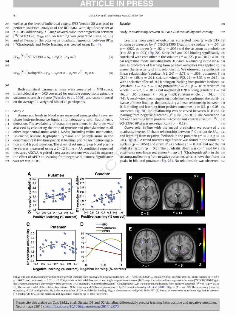
Fig. 2. D1R and D2R availability differentially predict learning from positive and negative outcop=0.002) and putamen (r=0.52, p=0.005) predicts individual differences in learning from pothe striatum and reward learning (p=0.05, corrected). (C) InvertedU relationship between [11C(D) Theoretical model of the relationship between NoGo learning and D2 binding as measuredoccupancy of D2R by dopamine. BP0 is the total number of D2R available for binding. BPND is the[11C]raclopride BPND in the striatum and avoidance learning (p = 0.05 corrected).
Please cite this article as: Cox, S.M.L., et al., Striatal D1 and D2 signalingNeuroImage (2015), http://dx.doi.org/10.1016/j.neuroimage.2014.12.070
ED P
RO
OF
Results
Study 1: relationship between D1R and D2R availability and learning
Learning from positive outcomes correlated linearly with D1Rbinding as assessed by [11C]SCH23390 BPND in the caudate (r = .57,p = .002), putamen (r = .52, p = .005) and the striatum as a whole(r = .55, p = .003) (Fig. 2A). Since D1R and D2R binding significantlycorrelated with each other in the striatum (r2 = 0.23, p = 0.012), a lin-ear regression model including both D1R and D2R binding in the stria-tum as predictors of learning from positive outcomes was applied toassess the selectivity of this relationship. We observed a significantlinear relationship (caudate: F(2, 24) = 5.78, p = .009; putamen: F(2,24) = 4.58, p = .021; striatum whole: F(2, 24) = 5.33, p = .012),with a selective effect of D1Rbinding on learning frompositive feedback(caudate: t = 2.6, p = .016; putamen: t = 2.5, p = .019; striatumwhole: t = 2.7, p = .013), but no effect of D2R binding (caudate: t =.46, p = .65; putamen: t = .42, p = .68; striatum whole: t = .34, p =.74). A voxel-wise linear regression model further confirmed the signif-icance of these findings, demonstrating a linear relationship betweenD1R binding and learning from positive outcomes (t = 6.2, p b 0.05,corrected, Fig. 2B). No relationship was observed between D1R andlearning from negative outcomes (r2 ≤ 0.01, p N 0.6). The correlationbetween learning from positive outcomes and ventral striatum [11C]SCH23390 BPND was non-significant (p = 0.12).
Conversely, in line with the model prediction, we observed aquadratic, inverted U-shape relationship between [11C]raclopride BPNDand learning from negative feedback in the putamen (r2 = .19, p =0.03, Fig. 2C). A trend towards significance was found in the caudatenucleus (p = 0.054) and striatum as a whole (p = 0.058) but not theventral striatum (p = 0.6). The quadratic effect was confirmed by avoxel-wise non-linear regression F-map of [11C]raclopride BPND in thestriatum and learning from negative outcomes, which shows significantpeaks in bilateral putamen (Fig. 2E). No relationship was observed,
mes. (A) [11C]SCH23390 BPND, indicative of D1 receptor density, in the caudate (r = 0.57,sitive outcomes. (B) T-map of voxel-wise linear regression between [11C]SCH23390 BPND in]raclopride BPND in theputamen and learning fromnegative outcomes (r2=0.19, p=0.03).by PET, adapted from Gjedde et al. (2010). BPND = (1− σ) BP0. The occupancy (σ) is themeasured raclopride BP by PET. (E) F-map of voxel-wise non-linear regression between
differentially predict learning from positive and negative outcomes,
T
356
357
358
359
360
361
362
363
364
365
366
367
368
369
370
371
372
373
374
375
376
377
378
379
380
381
382
383
384
385
386
387
388
389
390
391
392
393
394
395
396
397
398
399
400
401
402
403
404
405
406
407
408
409
410
411
412
413
414
415
416
417
418
419
420
421
422
423
424
425
426
427
428
429
430
431
432
433
434
435
436
437
438
439
440
441
442
443
444
445
446
447
5S.M.L. Cox et al. / NeuroImage xxx (2015) xxx–xxx
ORREC
either linear or quadratic, between [11C]raclopride BPND and learningfrom positive outcomes after correcting for the effects of D1R binding(r2 = 0.005, p N 0.7).
Study 2: the effect of dopamine depletion on learning
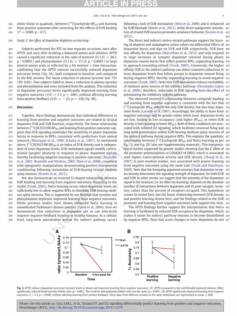
Subjects performed the PST on two separate occasions, once afterAPTD, and once after drinking a balanced amino acid solution. APTDcaused a significant reduction in the ratio of tyrosine (F(1,9) = 28.2,p b 0.0001) and phenylalanine (F(1,9) = 111.9, p b 0.0001) to largeneutral amino acids as reflected by a AA mixture × time interaction,confirming that the APTD mixture successfully reduced dopamineprecursor levels (Fig. 3A), both compared to baseline, and comparedto the BAL session. The mean reduction in plasma tyrosine was 75%(SD: 6.6%). Two subjects failed to show a reduction in plasma tyrosineand phenylalanine and were excluded from the analysis. This reductionin dopamine precursor levels significantly improved learning fromnegative outcomes (t(9) = 2.3, p = .046), without affecting learningfrom positive feedback (t(9) = − .23, p = .556, Fig. 3B).
Discussion
Together, these findings demonstrate that individual differences inlearning from positive and negative outcomes are related to striataldopamine D1R and D2R function, respectively. The linear relationshipbetween [11C]SCH23390 BPND and learning frompositive outcomes sug-gests that D1R signaling modulates the sensitivity to phasic dopaminebursts in response to RPEs, consistent with predictions from animalresearch (Montague et al., 1996; Schultz et al., 1997). As mentionedabove, [11C]SCH23390 BPND is an index of D1R density and is indepen-dent of tonic dopamine levels. D1R-modulated signals modify cortico-striatal synaptic plasticity in response to phasic dopamine signals,thereby facilitating adaptive learning to positive outcomes (Reynoldset al., 2001; Reynolds and Wickens, 2002; Shen et al., 2008), consistentwith optogenetic manipulations demonstrating positive instrumentalconditioning following stimulation of D1R-bearing striatal mediumspiny neurons (Kravitz et al., 2012).
We also demonstrate an inverted U-shaped relationship betweenD2R binding and learning from negative outcomes. According to themodel (Frank, 2005), NoGo learning occurs when dopamine levels aresufficiently low to allow negative RPEs to disinhibit D2R-bearing medi-um spiny neurons. This is supported by our findings that tyrosine andphenylalanine depletion improved learning from negative outcomes.While previous studies have shown enhanced NoGo learning inunmedicated Parkinson's disease patients (Frank et al., 2004), here wedemonstrate that reductions in dopamine per se can selectivelyimprove negative feedback learning in healthy humans. At a cellularlevel, long-term potentiation within the indirect pathway occurs
UNC
Fig. 3. APTD reduces dopamine precursor tyrosine levels in blood and improves learning frosignificantly reduced blood tyrosine/LNAAs ratio (p b 0.001). The result for phenylalanine/LNAAoutcomes (t = 2.3, p = 0.046) without affecting learning from positive feedback. These data, f
Please cite this article as: Cox, S.M.L., et al., Striatal D1 and D2 signalingNeuroImage (2015), http://dx.doi.org/10.1016/j.neuroimage.2014.12.070
ED P
RO
OF
following a lack of D2R stimulation (Shen et al., 2008) and is enhancedby D2R blockade (Beeler et al., 2012), while direct optogenetic stimula-tion of striatal D2R neurons promotes avoidance behavior (Kravitz et al.,2012).
The direct and indirect cortico-striatal pathways support the learn-ing of adaptive and maladaptive action values via differential effects ofdopamine bursts and dips on D1R and D2R, respectively. D1R havelow affinity for dopamine (Marcellino et al., 2012) and only respondto large increases in synaptic dopamine released during phasicdopamine neuron bursts that reflect positive RPEs, supporting learningto approach rewarding stimuli (Frank, 2005). Conversely, the higheraffinity D2R in the indirect pathway can detect transient reductions intonic dopamine levels that follow pauses in dopamine neuron firingduring negative RPEs, thereby supporting learning to avoid negativeoutcomes (Frank, 2005). Note that D2R stimulation reduces excitabilityof medium spiny neuron of the indirect pathway (Hernández-Lópezet al., 2000); therefore, reductions in D2R signaling have the effect ofpotentiating the inhibitory indirect pathway.
The observed inverted U relationship between raclopride bindingand learning from negative outcomes is consistent with the fact that[11C]raclopride BPND reflects not only D2R density, but also tonic dopa-mine levels (Laruelle et al., 1997). Accordingly, the ability to learn fromnegative outcomes will be greater either when tonic dopamine levelsare low, leading to low occupancy (and higher BPND), or when D2Rdensity is low (leading to lower BPND). Both of these conditions are asso-ciated with reduced D2 signaling, which facilitates neuronal firing andlong-term potentiation within D2R-bearing medium spiny neurons ofthe indirect pathway during negative RPEs. This explains the quadraticrelationship between [11C]raclopride BPND and NoGo learning as perEq. (2) and Fig. 2D (also see Supplementary materials). This interpreta-tion is further supported by genetic studies showing that the C allele ofthe promoter polymorphism rs12364283 of DRD2, which is associatedwith higher transcriptional activity and D2R density (Zhang et al.,2007) in post-mortem studies, was associated with poorer learningfrom negative outcomes using the same task (Frank and Hutchison,2009). Note that the foregoing argument assumes that dopamine recep-tor density determines the signaling strength of dopamine, for both D1Rand D2R. In other words, we suggest that the intensity of the dopaminesignal in the striatum (i.e. its effect on learning) depends on the absolutenumber of interactions between dopamine and its post-synaptic recep-tors, rather than the percent of receptors occupied. This hypothesiscannot be tested here, but the linear relationship between D1R densityand positive learning shown here, and the findings related to the D2Rpromoter and learning from negative outcomes both support this view.
The APTD findings further support the interpretation that NoGolearning is facilitated by reduced D2R occupancy by dopamine, whichmakes it easier for indirect pathway neurons to become disinhibitedby negative RPEs. Note that acute changes in tonic dopamine do not
m negative outcomes. (A) APTD compared to the nutritionally balanced mixture (BAL)s ratio was the same (p b 0.001). (B) APTD significantly improved learning from negativerom different sessions in the same individuals, are represented as mean ± SEM.
differentially predict learning from positive and negative outcomes,
T
448
449
450
451
452
453
454
455
456
457
458
459
460
461
462
463
464
465
466
467
468
469
470
471
472
473
474
475
476
477
478
479
480
481
482
483
484
485
486
487
488
489
490
491
492
493
494
495
496
497
498
499
500
501
502
503
504
505
506
507
508
509
510
511
512
513
514
515
516
517
518
519
520
521
522
523
524
525
526
527
528
529
530
531
532
533
534
535
536
537
538
539
540
541
542
543
544
545
546
547
548
549
550Q3
551
552
553
554
555
556
557
558
559
560Q5561562563564565566567568569570571572573574575
6 S.M.L. Cox et al. / NeuroImage xxx (2015) xxx–xxx
UNCO
RREC
lead to changes in dopamine receptor expression (Laruelle et al., 1997),therefore the effect of APTD is assumed to be limited to reduced recep-tor occupancy. APTD did not affect Go learning. According to the model(Frank, 2005), NoGo learning is quite sensitive to changes in baselinetonic dopamine levels, influencing the ability of D2R-bearing mediumspiny neurons to respond to pauses in dopamine firing in response tonegative RPEs. According to recent evidence, APTD affects phasic aswell as tonic dopamine release (Le Masurier et al., 2013). However,modeling of synaptic dopamine signaling suggests that depletingvesicular dopamine may only affect tonic signaling at D2 receptorsand have little effect on phasic signaling (Dreyer et al., 2010). Pleasesee Supplementary materials for details.
These results have implications for our understanding of striataldopamine signaling and its role in pathologies of motivated behavior.First, they support the view that dopamine is not only implicated in pos-itive reinforcement, but that reductions in dopamine are also relevantfor learning from negative outcomes via D2 signaling. Rodent studieshave demonstrated that D2R blockade not only induces motor skilldeficits, but also leads to persistently impaired performance even afternormalization of D2 signaling, implicating aberrant learning (Beeleret al., 2012). The present findings suggest that these same mechanismsare relevant to avoidance learning from negative outcomes in humans.
Our finding that low dopamine facilitates rather than impairslearning from negative outcomes specifically supports the theory thatdopamine acts as a RPE signal rather than a saliency signal. Accordingto the saliency hypothesis low dopamine would be expected to causea reduction in both Go and NoGo learning (Bromberg-Martin et al.,2010). The bidirectional effect of dopamine on processing positive andnegative outcomes, as observed here, forms one of the key distinctionsbetween the RPE and saliency hypotheses, and thus emphasizes therole of dopamine in coding RPE, although this does not exclude theexistence of an additional dopaminergic saliency signal.
The model of basal ganglia processing supported by these resultsprovides a mechanism, at the computational level, for impulse controldisorders. Persistent over-stimulation of striatal D2R should reducethe impact of negative outcomes. This may explain the phenomenonof impulsivity induced by dopamine agonistmedications. Thedopamineagonists that cause impulse control disorders such as pathological gam-bling and compulsive shopping preferentially stimulate the D2R family(Potenza et al., 2007; Voon et al., 2011). In many cases the impulsivebehavior is time-locked to drug administration. Impulsivity, in thiscase, would result from an inability to consider the impact of negativeoutcomes. Further support for this notion comes from two other reportsshowing an inverted U-shaped relationship between [11C]racloprideBPND and measures of impulsive personality, namely the personalityconstruct of sensation seeking (Gjedde et al., 2010), which is itself asso-ciated with impulsive and risky behavior (Zuckerman and Kuhlman,2000), and Negative Urgency (Clark et al., 2012), which is associatedwith problem gambling. Taken together with these findings, our resultssuggest that inherent differences in dopamine D2R signaling maypredispose individuals to addictive and impulsive disorders. This mayrequire re-thinking the labeling of this dopaminergic vulnerability asreflecting “reward deficiency” (Dagher and Robbins, 2009), which im-plies that increased vulnerability results from reduced dopamine signal-ing. In contrast, our findings suggest that increased D2R signaling maybe associated with increased vulnerability to addictive and impulsivedisorders, which is consistent with other studies that have shown thatdecreased activity of the indirect pathway (or increased D2R stimula-tion) predisposes to addictive behaviors (Collins and Woods, 2009;Lobo et al., 2010; Maia and Frank, 2011). Future studies could testwhether impaired NoGo learning and impulsivity correlate acrossindividuals or following dopaminergic manipulations, and investigatethe relationship between negative reinforcement and the urge toengage in reward seeking behavior.
Our results need to be interpreted in light of the following limita-tions. First, the sample size of the APTD study (n = 10) was modest,
Please cite this article as: Cox, S.M.L., et al., Striatal D1 and D2 signalingNeuroImage (2015), http://dx.doi.org/10.1016/j.neuroimage.2014.12.070
ED P
RO
OF
but sufficient to detect a significant effect within-subjects, and withinthe commonly accepted range for assessing pharmacologicalchallenges. Second, our interpretation of the D2 results rests on severalassumptions about D2 signaling and APTD. As highlighted above,measures of [11C]raclopride BPND are unable to differentiate betweenD2R density (Bmax) and receptor occupancy by dopamine. These twomeasures have opposing effects on D2 signaling and thus on learningfrom negative outcomes. Although this accounts for the observedinverted U-shape function, thismethod does not allowus to disentanglethe contribution of endogenous dopamine levels versus receptor densi-ty, nor interactions between the two. Other methods will be needed toaddress this issue. Nevertheless, our findings do show selectivemodula-tion of positive and negative learning by D1 and D2 signaling, respec-tively. Third, the PST, as employed here, is unable to tease apart theexpression of learned associations versus the learning itself. Therefore,an alternative explanation for our findings is that dopamine signalingmediates value-based choice performance rather than learning per se(Smittenaar et al., 2012), an interpretation that is also consistent withrecent refinements to the computational model used here (Collins andFrank, 2014).
Conclusion
Our findings support a modulatory role for striatal dopamine inreward and avoidance based learning via segregated striatal D1R andD2R pathways. Individual differences in D1R and D2R binding predictedlearning from positive and negative outcomes of decisions, respectively.This variability inD1R andD2R signalingmay be responsible for individ-ual differences in the response to reward and punishment relatedsignals, which may underlie differences in vulnerability to drug addic-tion, obesity and other impulse control disorders. We further suggestthat the often-noted association between genetic or PET measures ofD2R and drug addiction or pathological gambling implicates impairedpunishment learning in these disorders.
Acknowledgments
We thank Gabriel Wolf for his help with programming the cognitivetask and Julia Wagner, Lauren Templeton, Helen Hsu, Vera Khramovaand Allison Goodwin-Wilson for their help with data collection andentry. This work was supported by CIHR and NSERC grants to AD andNIH grant (R21DA022630) to LKF.
Conflict of interest
The authors declare no competing financial interests.
Appendix A. Supplementary data
Supplementary data to this article can be found online at http://dx.doi.org/10.1016/j.neuroimage.2014.12.070.
References
Ad-Dab'bagh, Y., Einarson, D., Lyttelton, O., Muehlboeck, J.-S., Mok, K., Ivanov, O., Vincent,R.D., Lepage, C., Lerch, J., Fombonne, E., Evans, A.C., 2006. The CIVET image-processingenvironment: a fully automated comprehensive pipeline for anatomical neuroimag-ing research. Organization for Human Brain Mapping (Florence).
Bayer, H.M., Glimcher, P.W., 2005. Midbrain dopamine neurons encode a quantitativereward prediction error signal. Neuron 47, 129–141.
Beeler, J.A., Frank, M.J., McDaid, J., Alexander, E., Turkson, S., Bernandez, M.S., McGehee,D.S., Zhuang, X., 2012. A role for dopamine-mediated learning in the pathophysiologyand treatment of Parkinson's disease. Cell Rep. 2, 1747–1761.
Brodnik, Z., Double, M., Jaskiw, G.E., 2013. Presynaptic regulation of extracellulardopamine levels in the medial prefrontal cortex and striatum during tyrosinedepletion. Psychopharmacology 227, 363–371.
Bromberg-Martin, E.S., Matsumoto, M., Hikosaka, O., 2010. Dopamine in motivationalcontrol: rewarding, aversive, and alerting. Neuron 68, 815–834.
Chou, Y.H., Karlsson, P., Halldin, C., Olsson, H., Farde, L., 1999. A PET study of D1-likedopamine receptor ligand binding. Psychopharmacology 146, 220–227.
differentially predict learning from positive and negative outcomes,
T
576577578579580581582583584585586587588589590591592593594595596597598599600601602603604605606607608609610611612613614615616617618619620621622623624625626627628629630631632633634635636637638639640641642643644645646647648649650Q6
651652653654655656657658659660661662663664665666667668669670671672673674675676677678679680681682683684685686687688689690691692693694695696697698699700701702703704705706707708709710711712Q7713714715716717718719720721722723
724
7S.M.L. Cox et al. / NeuroImage xxx (2015) xxx–xxx
UNCO
RREC
Clark, L., Stokes, P.R., Wu, K., Michalczuk, R., Benecke, A., Watson, B.J., Egerton, A., Piccini,P., Nutt, D.J., Bowden-Jones, H., Lingford-Hughes, A.R., 2012. Striatal dopamine D2/D3receptor binding in pathological gambling is correlated with mood-related impulsiv-ity. NeuroImage 63, 40–46.
Collins, D.L., Evans, A.C., 1997. Animal: validation and applications of nonlinearregistration-based segmentation. Int. J. Pattern Recognit. Artif. Intell. 11.
Collins, A.G., Frank, M.J., 2014. Opponent actor learning (OpAL): modeling interactiveeffects of striatal dopamine on reinforcement learning and choice incentive. Psychol.Rev. 121, 337–366.
Collins, G.T., Woods, J.H., 2009. Influence of conditioned reinforcement on the response-maintaining effects of quinpirole in rats. Behav. Pharmacol. 20, 492–504.
Collins, D.L., Neelin, P., Peters, T.M., Evans, A.C., 1994. Automatic 3D intersubject registra-tion ofMR volumetric data in standardized Talairach space. J. Comput. Assist. Tomogr.18, 192–205.
Comtat, C., Bataille, F., Michel, C., Jones, J.P., Sibomana, M., Janeiro, L., Trebossen, R., 2004.OSEM-3D reconstruction strategies for the ECAT HRRT. IEEE Nucleur ScienceSymposium Conference Record 6, pp. 3492–3496.
Cools, R., Altamirano, L., D'Esposito, M., 2006. Reversal learning in Parkinson's diseasedepends on medication status and outcome valence. Neuropsychologia 44, 1663–1673.
Costes, N., Dagher, A., Larcher, K., Evans, A.C., Collins, D.L., Reilhac, A., 2009. Motion correc-tion of multi-frame PET data in neuroreceptor mapping: simulation based validation.NeuroImage 47, 1496–1505.
Dagher, A., Robbins, T., 2009. Personality, addiction, dopamine: insights from Parkinson'sdisease. Neuron 61, 502–510.
Daw, N.D., Kakade, S., Dayan, P., 2002. Opponent interactions between serotonin anddopamine. Neural Netw. 15, 603–616.
Doll, B.B., Hutchison, K.E., Frank, M.J., 2011. Dopaminergic genes predict individualdifferences in susceptibility to confirmation bias. J. Neurosci. 31, 6188–6198.
Dreyer, J.K., Herrik, K.F., Berg, R.W., Hounsgaard, J.D., 2010. Influence of phasic and tonicdopamine release on receptor activation. J. Neurosci. 30, 14273–14283.
First, M.B., Spitzer, R.L., Gibbon, M., Williams, J.B.W., 2002. Structural Clinical Interview forDSM-IV-TR Axis I Disorders, Research Version, Non-patient Edition (SCID-I/NP). Bio-metrics Research, New York State Psychiatric Institute, New York (November).
Fonov, V.S., Evans, A.C., McKinstry, R.C., Almli, C.R., Collins, D.L., 2009. Unbiased nonlinearaverage age-appropriate brain templates from birth to adulthood. Organization forHuman Brain Mapping, p. S102.
Frank, M.J., 2005. Dynamic dopamine modulation in the basal ganglia: aneurocomputational account of cognitive deficits in medicated and nonmedicatedParkinsonism. J. Cogn. Neurosci. 17, 51–72.
Frank, M.J., Hutchison, K., 2009. Genetic contributions to avoidance-based decisions:striatal D2 receptor polymorphisms. Neuroscience 164, 131–140.
Frank, M.J., O'Reilly, R.C., 2006. A mechanistic account of striatal dopamine function inhuman cognition: psychopharmacological studies with cabergoline and haloperidol.Behav. Neurosci. 120, 497–517.
Frank, M.J., Seeberger, L.C., O'Reilly, R.C., 2004. By carrot or by stick: cognitive reinforce-ment learning in parkinsonism. Science 306, 1940–1943.
Frank, M.J., Scheres, A., Sherman, S.J., 2007a. Understanding decision-making deficits inneurological conditions: insights from models of natural action selection. Philos.Trans. R. Soc. Lond. B Biol. Sci. 362, 1641–1654.
Frank, M.J., Moustafa, A.A., Haughey, H.M., Curran, T., Hutchison, K.E., 2007b. Genetictriple dissociation reveals multiple roles for dopamine in reinforcement learning.Proc. Natl. Acad. Sci. U. S. A. 104, 16311–16316.
Frank, M.J., Doll, B.B., Oas-Terpstra, J., Moreno, F., 2009. Prefrontal and striatal dopaminer-gic genes predict individual differences in exploration and exploitation. Nat. Neurosci.12, 1062–1068.
Gjedde, A., Kumakura, Y., Cumming, P., Linnet, J., Moller, A., 2010. Inverted-U-shaped cor-relation between dopamine receptor availability in striatum and sensation seeking.Proc. Natl. Acad. Sci. U. S. A. 107, 3870–3875.
Hernández-López, S., Tkatch, T., Perez-Garci, E., Galarraga, E., Bargas, J., Hamm, H.,Surmeier, D.J., 2000. D2 dopamine receptors in striatal medium spiny neurons reduceL-type Ca2+ currents and excitability via a novel PLCβ1–IP3–calcineurin-signalingcascade. J. Neurosci. 20, 8987–8995.
Jocham, G., Klein, T., Neumann, J., von Cramon, D., Reuter, M., Ullsperger, M., 2009. Dopa-mine DRD2 polymorphism alters reversal learning and associated neural activity.J. Neurosci. 29, 3695–3704.
Kravitz, A., Tye, L., Kreitzer, A., 2012. Distinct roles for direct and indirect pathway striatalneurons in reinforcement. Nat. Neurosci. 15, 816–818.
Lammertsma, A.A., Hume, S.P., 1996. Simplified reference tissue model for PET receptorstudies. NeuroImage 4, 153–158.
Laruelle, M., D'Souza, C., Baldwin, R., Abi-Dargham, A., Kanes, S., Fingado, C., Seibyl, J.,Zoghbi, S., Bowers,M., Jatlow, P., Charney, D., Innis, R., 1997. ImagingD2 receptor occu-pancy by endogenous dopamine in humans. Neuropsychopharmacology 17, 162–174.
Le Masurier, M., Zetterström, T., Cowen, P., Sharp, T., 2013. Tyrosine-free amino acidmixtures reduce physiologically-evoked release of dopamine in a selective andactivity-dependent manner. J. Psychopharmacol.
Please cite this article as: Cox, S.M.L., et al., Striatal D1 and D2 signalingNeuroImage (2015), http://dx.doi.org/10.1016/j.neuroimage.2014.12.070
ED P
RO
OF
Leyton, M., Young, S., Pihl, R., Etezadi, S., Lauze, C., Blier, P., Baker, G., Benkelfat, C., 2000.Effects on mood of acute phenylalanine/tyrosine depletion in healthy women.Neuropsychopharmacology 22, 52–63.
Leyton, M., Dagher, A., Boileau, I., Casey, K., Baker, G., Diksic, M., Gunn, R.N., Young, S.,Benkelfat, C., 2004. Decreasing amphetamine-induced dopamine release by acutephenylalanine/tyrosine depletion: a PET/[11C]raclopride study in healthy men.Neuropsychopharmacology 29, 427–432.
Lobo, M.K., Covington III, H.E., Chaudhury, D., Friedman, A.K., Sun, H., Damez-Werno, D.,Dietz, D.M., Zaman, S., Koo, J.W., Kennedy, P.J., Mouzon, E., Mogri, M., Neve, R.L.,Deisseroth, K., Han, M.H., Nestler, E.J., 2010. Cell type-specific loss of BDNF signalingmimics optogenetic control of cocaine reward. Science 330, 385–390.
Lorr, M., McNair, D.M., Fisher, S.U., 1982. Evidence for bipolar mood states. J. Pers. Assess.46, 432–436.
Maia, T.V., Frank, M.J., 2011. From reinforcement learning models to psychiatric andneurological disorders. Nat. Neurosci. 14, 154–162.
Marcellino, D., Kehr, J., Agnati, L.F., Fuxe, K., 2012. Increased affinity of dopamine for D(2)-like versus D(1)-like receptors. Relevance for volume transmission in interpretingPET findings. Synapse 66, 196–203.
McTavish, S., Cowen, P., Sharp, T., 1999. Effect of a tyrosine-free amino acidmixture on re-gional brain catecholamine synthesis and release. Psychopharmacology 141,182–188.
Mintun, M.A., Raichle, M.E., Kilbourn, M.R., Wooten, G.F., Welch, M.J., 1984. A quantitativemodel for the in vivo assessment of drug binding sites with positron emission tomog-raphy. Ann. Neurol. 15, 217–227.
Montague, P., Dayan, P., Sejnowski, T., 1996. A framework for mesencephalic dopaminesystems based on predictive Hebbian learning. J. Neurosci. 16, 1936–1947.
Montgomery, A.J., McTavish, S.F., Cowen, P.J., Grasby, P.M., 2003. Reduction of brain dopa-mine concentration with dietary tyrosine plus phenylalanine depletion: an [11C]raclopride PET study. Am. J. Psychiatry 160, 1887–1889.
Moustafa, A.A., Cohen, M.X., Sherman, S.J., Frank, M.J., 2008. A role for dopamine intemporal decision making and reward maximization in parkinsonism. J. Neurosci.28, 12294–12304.
Palmour, R.M., Ervin, F.R., Baker, G.B., Young, S.N., 1998. An amino acid mixture deficientin phenylalanine and tyrosine reduces cerebrospinal fluid catecholamine metabolitesand alcohol consumption in vervet monkeys. Psychopharmacology 136, 1–7.
Pessiglione, M., Seymour, B., Flandin, G., Dolan, R., Frith, C., 2006. Dopamine-dependentprediction errors underpin reward-seeking behaviour in humans. Nature 442,1042–1045.
Potenza, M., Voon, V., Weintraub, D., 2007. Drug insight: impulse control disorders anddopamine therapies in Parkinson's disease. Nat. Clin. Pract. Neurol. 3, 664–672.
Reynolds, J., Wickens, J., 2002. Dopamine-dependent plasticity of corticostriatal synapses.Neural Netw. 15, 507–521.
Reynolds, J., Hyland, B., Wickens, J., 2001. A cellular mechanism of reward-relatedlearning. Nature 413, 67–70.
Schultz, W., 2002. Getting formal with dopamine and reward. Neuron 36, 241–263.Schultz, W., Dayan, P., Montague, P.R., 1997. A neural substrate of prediction and reward.
Science 275, 1593–1599.Shen, W., Flajolet, M., Greengard, P., Surmeier, D.J., 2008. Dichotomous dopaminergic
control of striatal synaptic plasticity. Science 321, 848–851.Sled, J.G., Zijdenbos, A.P., Evans, A.C., 1998. A nonparametric method for automatic correc-
tion of intensity nonuniformity in MRI data. IEEE Trans. Med. Imaging 17, 87–97.Smittenaar, P., Chase, H., Aarts, E., Nusselein, B., Bloem, B., Cools, R., 2012. Decomposing
effects of dopaminergic medication in Parkinson's disease on probabilistic action se-lection—learning or performance? Eur. J. Neurosci. 35, 1144–1151.
Spielberger, C.D., Gorsuch, R.L., Lushene, R., Vagg, P.R., Jacobs, G.A., 1983. Manual for theState-Trait Anxiety Inventory. Consulting Psychologists Press, Palo Alto, CA.
Surmeier, D., Carrillo-Reid, L., Bargas, J., 2011. Dopaminergic modulation of striatalneurons, circuits, and assemblies. Neuroscience 198, 3–18.
Thibaut, F., Vaugeois, J.-M., Bonnet, J.-J., Costentin, J., 1996. In vivo striatal binding of theD1 antagonist SCH 23390 is not modified by changes in dopaminergic transmission.Neuropharmacology 35, 267–272.
Voon, V., Schoerling, A., Wenzel, S., Ekanayake, V., Reiff, J., Trenkwalder, C., Sixel-D ring, F.,2011. Frequency of impulse control behaviours associated with dopaminergic thera-py in restless legs syndrome. BMC Neurol. 11, 117.
Worsley, K.J., Marrett, S., Neelin, P., Vandal, A.C., Friston, K.J., Evans, A.C., 1996. A unifiedstatistical approach for determining significant signals in images of cerebral activa-tion. Hum. Brain Mapp. 4, 58–73.
Zhang, Y., Bertolino, A., Fazio, L., Blasi, G., Rampino, A., Romano, R., Lee, M.L., Xiao, T., Papp,A., Wang, D., Sadee, W., 2007. Polymorphisms in human dopamine D2 receptor geneaffect gene expression, splicing, and neuronal activity during working memory. Proc.Natl. Acad. Sci. U. S. A. 104, 20552–20557.
Zuckerman, M., Kuhlman, D., 2000. Personality and risk-taking: common biosocial factors.J. Pers. 68, 999–1029.
differentially predict learning from positive and negative outcomes,