Overexpression of Adenosine A2A Receptors in Rats: Effects on Depression, Locomotion, and Anxiety
8
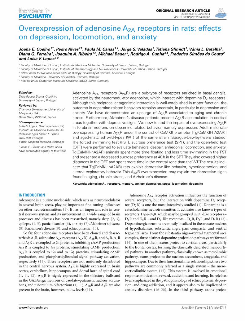
PSYCHIATRY ORIGINAL RESEARCH ARTICLE published: 13 June 2014 doi: 10.3389/fpsyt.2014.00067 Overexpression of adenosine A 2A receptors in rats: effects on depression, locomotion, and anxiety Joana E. Coelho 1† , Pedro Alves 2† , Paula M. Canas 3,4 , Jorge S. Valadas 1 ,Tatiana Shmidt 5 , Vânia L. Batalha 1 , Diana G. Ferreira 1 , Joaquim A. Ribeiro 1,2 , Michael Bader 5 , Rodrigo A. Cunha 3,4 , Frederico Simões do Couto 2 and LuísaV. Lopes 1 * 1 Faculty of Medicine of Lisbon, Instituto de Medicina Molecular, University of Lisbon, Lisbon, Portugal 2 Faculty of Medicine of Lisbon, Institute of Pharmacology and Neurosciences, University of Lisbon, Lisbon, Portugal 3 CNC-Center for Neurosciences and Cell Biology, University of Coimbra, Coimbra, Portugal 4 Faculty of Medicine, University of Coimbra, Coimbra, Portugal 5 Max-Delbrück-Center for Molecular Medicine (MDC), Berlin, Germany Edited by: Silvia Raquel Soares Ouakinin, University of Lisbon, Portugal Reviewed by: Chamindi Seneviratne, University of Maryland, USA David Blum, INSERM, France *Correspondence: Luísa V. Lopes, Neurosciences Unit, Instituto de Medicina Molecular, Av. Professor Egas Moniz 1, Lisbon 1649-028, Portugal e-mail: [email protected] † Joana E. Coelho and Pedro Alves have contributed equally to this work. Adenosine A 2A receptors (A 2A R) are a sub-type of receptors enriched in basal ganglia, activated by the neuromodulator adenosine, which interact with dopamine D 2 receptors. Although this reciprocal antagonistic interaction is well-established in motor function, the outcome in dopamine-related behaviors remains uncertain, in particular in depression and anxiety. We have demonstrated an upsurge of A 2A R associated to aging and chronic stress. Furthermore, Alzheimer’s disease patients present A 2A R accumulation in cortical areas together with depressive signs. We now tested the impact of overexpressing A 2A R in forebrain neurons on dopamine-related behavior, namely depression. Adult male rats overexpressing human A 2A R under the control of CaMKII promoter [Tg(CaMKII-hA2AR)] and aged-matched wild-types (WT) of the same strain (Sprague-Dawley) were studied. The forced swimming test (FST), sucrose preference test (SPT), and the open-field test (OFT) were performed to evaluate behavioral despair, anhedonia, locomotion, and anxiety. Tg(CaMKII-hA2AR) animals spent more time floating and less time swimming in the FST and presented a decreased sucrose preference at 48 h in the SPT.They also covered higher distances in the OFT and spent more time in the central zone than the WT.The results indi- cate that Tg(CaMKII-hA2AR) rats exhibit depressive-like behavior, hyperlocomotion, and altered exploratory behavior. This A 2A R overexpression may explain the depressive signs found in aging, chronic stress, and Alzheimer’s disease. Keywords: adenosine A 2A receptors, memory, anxiety, depression, stress, locomotion, dopamine INTRODUCTION Adenosine is a purine nucleoside, which acts as neuromodulator in several brain areas, playing important fine tuning influences on other neurotransmitters (1). It has an important role in cen- tral nervous system and its involvement in a wide range of brain processes and diseases has been researched, namely sleep (2, 3), epilepsy (4, 5), panic disorder (6), anxiety (7), Alzheimer’s disease (8), Parkinson’s disease (9), and schizophrenia (10). So far, four adenosine receptors have been cloned and charac- terized: A 1 R, adenosine A 2A receptor (A 2A R), A 2B R, and A 3 R. A 1 R and A 3 R are coupled to Gi proteins, inhibiting cAMP production; A 2A R is coupled to Gs proteins, stimulating cAMP production; A 2B R is coupled to Gs and to Gq proteins, stimulating cAMP production, and phosphatidylinositol signal pathway activation, respectively (11). These receptors are not uniformly distributed in the central nervous system. A 1 R is highly expressed in brain cortex, cerebellum, hippocampus, and dorsal horn of spinal cord (1, 11, 12). A 2A R is highly expressed in the olfactory bulb and in the GABAergic neurons of caudate–putamen, nucleus accum- bens, and tuberculum olfactorium (1, 11). A 2B R and A 3 R are also present in the brain, however, in low levels (11). Adenosine A 2A receptor activation influences the function of several receptors, but the interaction with dopamine D 2 recep- tor (D 2 R) is one the most intensively studied (1). Dopamine is a catecholamine neurotransmitter. It activates five known types of receptors, D 1 R–D 5 R, which may be grouped in D 1 -like receptors – D 1 R and D 5 R – and D 2 -like receptors – D 2 R, D 3 R, and D 4 R(13). Dopaminergic neurons are mostly localized in the arcuate nucleus of hypothalamus, substantia nigra pars compacta, and ventral tegmental area. From the substantia nigra–ventral tegmental area complex, three distinct dopamine projection pathways are formed (14). In one of them, axons project to cortical areas, particularly to the frontal cortex, forming the classically described mesocorti- cal pathway. In another pathway, classically known as mesolimbic pathway, axons project to the nucleus accumbens, amygdala, and hippocampus. Due to their functional interrelationships, these two pathways are commonly referred as a single system – the meso- corticolimbic system (15). This system is involved in emotional response, motivation, reward, addiction, and learning. Its role has been emphasized in the pathophysiology of schizophrenia, depres- sion, and drug addiction, and it appears also to be implicated in anxiety disorders (16–20). In the third pathway, axons project www.frontiersin.org June 2014 |Volume 5 | Article 67 | 1
Transcript of Overexpression of Adenosine A2A Receptors in Rats: Effects on Depression, Locomotion, and Anxiety
PSYCHIATRYORIGINAL RESEARCH ARTICLE
published: 13 June 2014doi: 10.3389/fpsyt.2014.00067
Overexpression of adenosine A2A receptors in rats: effectson depression, locomotion, and anxiety
Joana E. Coelho1†, Pedro Alves2†, Paula M. Canas3,4, Jorge S. Valadas1,Tatiana Shmidt 5,Vânia L. Batalha1,Diana G. Ferreira1, Joaquim A. Ribeiro1,2, Michael Bader 5, Rodrigo A. Cunha3,4, Frederico Simões do Couto2
and Luísa V. Lopes1*1 Faculty of Medicine of Lisbon, Instituto de Medicina Molecular, University of Lisbon, Lisbon, Portugal2 Faculty of Medicine of Lisbon, Institute of Pharmacology and Neurosciences, University of Lisbon, Lisbon, Portugal3 CNC-Center for Neurosciences and Cell Biology, University of Coimbra, Coimbra, Portugal4 Faculty of Medicine, University of Coimbra, Coimbra, Portugal5 Max-Delbrück-Center for Molecular Medicine (MDC), Berlin, Germany
Edited by:Silvia Raquel Soares Ouakinin,University of Lisbon, Portugal
Reviewed by:Chamindi Seneviratne, University ofMaryland, USADavid Blum, INSERM, France
*Correspondence:Luísa V. Lopes, Neurosciences Unit,Instituto de Medicina Molecular, Av.Professor Egas Moniz 1, Lisbon1649-028, Portugale-mail: [email protected]†Joana E. Coelho and Pedro Alveshave contributed equally to this work.
Adenosine A2A receptors (A2AR) are a sub-type of receptors enriched in basal ganglia,activated by the neuromodulator adenosine, which interact with dopamine D2 receptors.Although this reciprocal antagonistic interaction is well-established in motor function, theoutcome in dopamine-related behaviors remains uncertain, in particular in depression andanxiety. We have demonstrated an upsurge of A2AR associated to aging and chronicstress. Furthermore, Alzheimer’s disease patients present A2AR accumulation in corticalareas together with depressive signs. We now tested the impact of overexpressing A2ARin forebrain neurons on dopamine-related behavior, namely depression. Adult male ratsoverexpressing human A2AR under the control of CaMKII promoter [Tg(CaMKII-hA2AR)]and aged-matched wild-types (WT) of the same strain (Sprague-Dawley) were studied.The forced swimming test (FST), sucrose preference test (SPT), and the open-field test(OFT) were performed to evaluate behavioral despair, anhedonia, locomotion, and anxiety.Tg(CaMKII-hA2AR) animals spent more time floating and less time swimming in the FSTand presented a decreased sucrose preference at 48 h in the SPT.They also covered higherdistances in the OFT and spent more time in the central zone than the WT.The results indi-cate that Tg(CaMKII-hA2AR) rats exhibit depressive-like behavior, hyperlocomotion, andaltered exploratory behavior. This A2AR overexpression may explain the depressive signsfound in aging, chronic stress, and Alzheimer’s disease.
Keywords: adenosine A2A receptors, memory, anxiety, depression, stress, locomotion, dopamine
INTRODUCTIONAdenosine is a purine nucleoside, which acts as neuromodulatorin several brain areas, playing important fine tuning influenceson other neurotransmitters (1). It has an important role in cen-tral nervous system and its involvement in a wide range of brainprocesses and diseases has been researched, namely sleep (2, 3),epilepsy (4, 5), panic disorder (6), anxiety (7), Alzheimer’s disease(8), Parkinson’s disease (9), and schizophrenia (10).
So far, four adenosine receptors have been cloned and charac-terized: A1R, adenosine A2A receptor (A2AR), A2BR, and A3R. A1Rand A3R are coupled to Gi proteins, inhibiting cAMP production;A2AR is coupled to Gs proteins, stimulating cAMP production;A2BR is coupled to Gs and to Gq proteins, stimulating cAMPproduction, and phosphatidylinositol signal pathway activation,respectively (11). These receptors are not uniformly distributedin the central nervous system. A1R is highly expressed in braincortex, cerebellum, hippocampus, and dorsal horn of spinal cord(1, 11, 12). A2AR is highly expressed in the olfactory bulb andin the GABAergic neurons of caudate–putamen, nucleus accum-bens, and tuberculum olfactorium (1, 11). A2BR and A3R are alsopresent in the brain, however, in low levels (11).
Adenosine A2A receptor activation influences the function ofseveral receptors, but the interaction with dopamine D2 recep-tor (D2R) is one the most intensively studied (1). Dopamine is acatecholamine neurotransmitter. It activates five known types ofreceptors, D1R–D5R, which may be grouped in D1-like receptors –D1R and D5R – and D2-like receptors – D2R, D3R, and D4R (13).Dopaminergic neurons are mostly localized in the arcuate nucleusof hypothalamus, substantia nigra pars compacta, and ventraltegmental area. From the substantia nigra–ventral tegmental areacomplex, three distinct dopamine projection pathways are formed(14). In one of them, axons project to cortical areas, particularlyto the frontal cortex, forming the classically described mesocorti-cal pathway. In another pathway, classically known as mesolimbicpathway, axons project to the nucleus accumbens, amygdala, andhippocampus. Due to their functional interrelationships, these twopathways are commonly referred as a single system – the meso-corticolimbic system (15). This system is involved in emotionalresponse, motivation, reward, addiction, and learning. Its role hasbeen emphasized in the pathophysiology of schizophrenia, depres-sion, and drug addiction, and it appears also to be implicated inanxiety disorders (16–20). In the third pathway, axons project
www.frontiersin.org June 2014 | Volume 5 | Article 67 | 1

Coelho et al. A2A receptors and depression
to the striatum, forming the classically described nigrostriatalpathway. This pathway integrates the neural circuits of the basalganglia responsible for motor control and its malfunctioning isclassically involved in the pathophysiology of Parkinson’s disease(21), although recent evidence also points to a very important rolein the motor changes associated with severe depression (22).
Adenosine A2A receptor and D2R are co-localized in the dorsaland ventral striatum and are reciprocal inhibitors: on one hand,A2AR–D2R heteromers are formed and, when the A2AR is acti-vated, conformational changes are transferred to the D2R – thisleads to a reduction in D2R recognition and signaling (23, 24); onthe other hand, D2R activation inhibits cAMP mediated-effects ofA2AR by inhibiting adenylyl-cyclase (23, 24).
Knowing these possible interactions between A2AR anddopamine, we investigated the impact of A2AR overexpressionin cortical areas onto dopamine-related behavior. Thus, in thepresent work a group of behavioral tests was performed in order toanalyze the effect of A2AR overexpression: (1) sucrose preferencetest (SPT), considered for a behavioral evaluation of anhedonia(25); (2) forced swim test to evaluate motivation and behavioraldespair (26, 27); (3) open-field test (OFT) to study locomotoractivity and anxiety-like behavior (28, 29).
MATERIALS AND METHODSANIMALSAnimal procedures were performed in accordance with theguidelines of the European Community guidelines (Directive2010/63/EU), Portuguese law on animal care (1005/92), andapproved by the Instituto de Medicina Molecular Internal Com-mittee and the Portuguese Animal Ethics Committee (DirecçãoGeral de Veterinária). Transgenic rats with an overexpressionof the human A2AR under the control of the CaMKII promo-tor, tg(CaMKII-hA2AR), were generated by microinjection of alinearized DNA construct into the male pronucleus of Sprague-Dawley rat zygotes with established methods (30). The constructcontained a full-length human A2A cDNA cloned into an expres-sion vector with the 8.5 kb mouse CaMKIIα promoter (31) and apolyadenylation cassette of bovine growth hormone (see Figure 1,top panel). Sprague-Dawley wild-type (WT) rats were used ascontrols. Genotyping : transgenic rats were identified by PCR(30 cycles, 58°C annealing temperature) of their genomic DNAisolated from ear biopsies by the use of the transgene-specificprimers CaMKII-hA2A and rat β-actin primers as an internalcontrol (Invitrogen). According to the performed RNase protec-tion assay, these animals expressed A2AR pre-dominantly in thebrain. qPCR and Western blotting showed that the overexpressionwas mostly in hippocampus and cortex; there was also overex-pression in striatum, however at a lesser extent (Figure 1). Nineweek-old WT and transgenic Sprague-Dawley (CaMKII-hA2AR)male rats were used. They were maintained in groups of three inappropriate cages with food and water ad libitum, temperatureof 21± 0.5°C, humidity of 60± 10%, and 12 h light/dark cyclesbeginning at 8 a.m.
BEHAVIORAL TESTINGThe behavioral testing was performed as before (32), during thelight period of the cycle, in a silent room, under dim light. From
FIGURE 1 | Neuronal overexpression of adenosine A2A receptor (A2AR)inTg(CaMII-hA2AR) rats. A2AR overexpression in the striatum ofTg(CaMKII-hA2AR) rats compared to WT animals was confirmed by westernblotting.
the first to the third day of experiments, the animals were han-dled for approximately 1 min each. On the fourth day, at 2.30 p.m.the OFT was done. On the fifth day, at 1 p.m. the SPT was initi-ated. This test was concluded on the seventh day at 9 a.m. On theseventh and eighth day, both at 2 p.m., the forced swim test wasexecuted.
Open-field testThe rats were placed in a designated corner of a square appa-ratus, surrounded by vertical walls (66 cm× 66 cm× 66 cm) –open-field arena. They freely explored the maze for 5 min. Theirmovements were recorded and analyzed using the video-trackingsoftware – SMART®. The reference point used by the software todetermine the position of the animal was the center of the rat’s dor-sum (also true for the other experiments). Three different zoneswere defined for analysis (29): (1) the area adjacent to the wall(1896 cm2); (2) the central area of the arena (552 cm2); (3) theintermediary area between the two previous ones (1908 cm2). Thepercentage of time spent in each zone, the total distance traveled,the average speed (calculated after the elimination of the restingtime), and the number of rearings and defecations were deter-mined. At the end of the 5 min test, the rat was removed from theopen-field arena and placed into its home cage.
Elevated plus mazeThe maze is shaped like a plus sign and consists of two “open” (nowalls, 5 cm× 29 cm) and two“closed”122 (5 cm× 29 cm× 15 cm)arms, arranged perpendicularly, and elevated 50 cm above thefloor. Each animal was placed on the center of the equipment, fac-ing an open arm. Each test lasted 5 min and all testing sessions wereperformed between 10:00 a.m. and 17:00 p.m. in a sound atten-uated room. The maze was cleaned with a 70% ethanol solutionbetween each animal. The total time spent in the open arms andthe total arms entries (number of entries in open+ closed arms)were used as anxiety and locomotor parameters as before (32).
Sucrose preference testRats were given two previously weighed bottles: with 1% (w/v)sucrose solution (33). The bottles had the same characteristics
Frontiers in Psychiatry | Affective Disorders and Psychosomatic Research June 2014 | Volume 5 | Article 67 | 2

Coelho et al. A2A receptors and depression
and approximately the same volume of liquid and were positionedside-by-side at the rear of the cage. The rats had free access toboth bottles. The position of the bottles in the cage is switchedhalfway through this period. There was no food or water depri-vation before the test. The bottles were weighed again at 48 h andthe consumed weight of each liquid was determined. The sucrosepreference was calculated according to Bekris et al. (34):
SP =sucrose solution intake
(g)
sucrose solution intake(g)+ water intake
(g) × 100.
Forced swim testOn the first of the two test days, all animals were gently placed indi-vidually in a vertical Plexiglas cylinder (height: 45 cm, diameter:19 cm) filled with 23°C tap water at a depth that made it impossiblefor rats to reach the bottom with hind paws (28–30 cm). The ani-mals were removed from the water after 10 min, and dried beforebeing returned to their home cages. The water was changed aftereach session. On the next day, the procedure was repeated with twodifferences: the animals were removed from the water after 5 min,instead of 10 min; the session was video-recorded. An observerblinded to the animal group analyzed the videos. Three differentbehaviors were considered: (1) immobility – according to the cri-teria of Porsolt et al. (35), a rat is judged to be immobile when itfloated passively, making only small movements to keep its noseabove the surface; (2) climbing (or thrashing) – upward-directedmovements with its forepaws, in and out of the water, along theside of the swim chamber; (3) swimming – active movements(usually horizontal) more than necessary to merely maintain itshead above the water (36, 37). Diving and face shaking behaviorswere not considered.
The time (t ) spent in immobility and climbing was measured;the time spent swimming was calculated: t swimming= 5− (tclimbing+ t immobility). Additionally, the latency to the first boutof immobility was determined (38): period of time since the begin-ning of the rat mobilization in the water until the first episode (atleast 1 s) of immobility.
Western blottingThe animals were killed by decapitation after anesthesia underhalothane atmosphere. After decapitation the brain was rapidlyremoved and the striata were dissected rapidly frozen in liquidnitrogen for further analysis. Samples were denatured by heat-ing at 70°C for 30 min for A2AR. Samples and molecular weightmarker were resolved by SDS-PAGE (8 or 10% for resolving anda 5% for stacking gels) in denaturing conditions and electro-transferred to PVDF membranes (Millipore). Membranes wereblocked with 5% non-fat dry milk in TBS-T (Tris buffer salinewith 0.1% Tween-20, 200 nM Tris, 1.5 M NaCl). After washingwith TBS-T, membranes were incubated with primary antibodyin TBS-T with 3% BSA. Secondary antibody incubation was in5% non-fat dry milk in TBS-T. Primary antibody was mouseanti-A2AR (1:2000, Upstate/Millipore – 05-717, Darmstadt, Ger-many), secondary antibodies conjugated with horseradish peroxi-dase were goat anti-mouse (Santa Cruz Biotechnology, Heidelberg,Germany). Chemoluminescent detection was performed with
ECL-PLUS western blotting detection reagent (GE Healthcare)using X-Ray films (Fujifilm).
STATISTICAL ANALYSISThe software used to perform the statistical analysis was Prism5 – GraphPad software®. Unpaired t test with Welch’s correctionwas applied to compare the differences between groups. p < 0.05was considered as statistical significant. Data are expressed asmeans± SEM.
RESULTSWEIGHTThe weight of transgenic CaMKII-hA2AR rats was significantlylower than WT rats (283± 11 vs. 400± 5 g; p < 0.001) (Figure 2).
OPEN-FIELD TESTThe total distance covered in the open-field arena was signifi-cantly higher in Tg(CaMKII-hA2AR) rats [WT: 2956± 160 cm;Tg(CaMKII-hA2AR): 3644± 64 cm; p= 0.0013], and was accom-panied by a significant increase in the number of rearings[WT: 5.9± 0.7; Tg(CaMKII-hA2AR): 10.8± 1.4; p= 0.0390], sug-gesting that Tg(CaMKII-hA2AR) rats display hyperlocomotion,Figure 3.
Transgenic (CaMKII-hA2AR) rats spent less time at the wallzone [WT: 73.97± 1.6%; Tg(CaMKII-hA2AR): 61.59± 1.94%;p < 0.0001] and more time in the central zone of the open-field box [WT: 3.36± 0.48%; Tg(CaMKII-hA2AR): 5.21± 0.40%;p= 0.0083, Figure 4] suggesting that Tg(CaMKII-hA2AR) ratshave increased exploratory behavior. We could not detect sig-nificant changes in the anxious behavior evaluated by EPM(Figure 4C). There were no statistical significant differencesregarding the number of defecations and the average speedbetween the two groups (data not shown).
FIGURE 2 | Weight control ofTg(CaMKII-hA2AR) animals. Results areexpressed as mean±SEM. ***p < 0.001 WT: n=12; Tg(CaMKII-hA2AR):n=4.
www.frontiersin.org June 2014 | Volume 5 | Article 67 | 3

Coelho et al. A2A receptors and depression
SUCROSE PREFERENCE AT 48 HAt 48 h, the preference index for sucrose was significantly higherin WT than in Tg(CaMKII-hA2AR) rats [WT: 91.88± 1.4%;Tg(CaMKII-hA2AR): 44.85± 23.78%, p= 0.0081, Figure 5], sug-gesting that Tg(CaMKII-hA2AR) rats have an anhedonic-likephenotype.
FORCED SWIM TESTTransgenic (CaMKII-hA2AR) rats spent significantly more timefloating than WT rats (2.47± 0.18 vs. 3.04± 0.16 min, p= 0.0452,Figure 6A) indicating that transgenic animals have increasedbehavioral despair. No significant changes were apparent in
FIGURE 3 | Open-field test. (A) Distance covered in centimeter.(B) Number of rearings – results are expressed as mean±SEM.**p < 0.01; ***p < 0.001. WT: n=12; Tg(CAMKII-hA2AR): n=4.
both swimming and climbing times, despite a tendency tolower performance in Tg(CaMKII-hA2AR) animals (Climb-ing: 0.63± 0.08 vs. 0.50± 0.02 min, p= 0.1486; swimming:1.88± 0.16 vs. 1.46± 0.17 min, p= 0.1060).
The latency to the first period of immobility was not dif-ferent between WT and transgenic animals [WT: 25.2± 5.6 s;Tg(CaMKII-hA2AR): 26.6± 3.8 s; p= 0.8304, Figure 6B].
DISCUSSIONWe now report that rats overexpressing A2AR in the hippocampus,cortex and striatum, display depressive-like behavior, increasedlocomotor activity, and altered exploratory behavior.
DEPRESSIVE-LIKE BEHAVIORThe SPT is considered a behavioral test for anhedonia, defined asthe inability to feel pleasure from usually enjoyable activities, acore symptom of depression. Decreased sucrose solution intake,resulting from chronic mild stress, is used as a model of depressionin rats, and can reversed by the administration of antidepressants(25, 39, 40). Thus, a decreased sucrose preference is associated todepressive-like behavior, specifically anhedonia. In our study, ratsoverexpressing A2AR, had a decrease in the preference index forsucrose solution at 48 h.
Additionally, when rats are placed in an inescapable cylinder ofwater – forced swim test – following initial escape-directed move-ments, they develop an immobile posture (27, 36). Immobilityindicates either a failure in the persistence to escape (behavioraldespair) or the act of giving up an active form of coping with thestressful stimuli (36). The immobility period and the latency to thefirst bout of immobility both decrease with the administration ofantidepressants (38, 41, 42). Thus, an increased period of immo-bility and reduced latency to immobility represent depressive-likebehavior. A2AR overexpressing animals spent more time floating,while no significant differences were observed in the time spentswimming, climbing, or latency to immobility. These behaviors,again, suggest a depressive-like phenotype.
FIGURE 4 | Open-field test. Percentage of time spent (A) at the wall zone and (B) in the central zone. (C) Percentage time in the open arms of the elevatedplus maze (EPM). Results are expressed as mean±SEM. **p < 0.01; ***p < 0.001. WT: n=12; Tg(CaMKII-hA2AR): n=4.
Frontiers in Psychiatry | Affective Disorders and Psychosomatic Research June 2014 | Volume 5 | Article 67 | 4

Coelho et al. A2A receptors and depression
FIGURE 5 | Sucrose preference test. (A) Preference index at 48 h and(B) control of amount of solution intake. Results are expressed asmean±SEM. p < 0.05 WT: n= 8; Tg(CaMKII-hA2AR): n=4.
FIGURE 6 | Forced swim test. (A) Time spent floating, swimming, andclimbing; (B) latency to the first period of immobility – results areexpressed as means±SEM. p < 0.05 WT: n=12; Tg(CaMKII-hA2AR): n= 4.
Genetic inactivation and pharmacological blockade of A2ARhave antidepressant-like effects (43). However, selective A2AR ago-nists are also able to decrease the immobility time in the forcedswim test (44, 45). Our observations indicate that A2AR overex-pression in neurons results in a depressive-like phenotype. Inter-estingly, depressive symptoms are found in Alzheimer’s diseasepatients,which have an abnormal accumulation of A2AR in corticalareas (8).
LOCOMOTOR ACTIVITYThe increase in locomotor activity displayed by the Tg(CaMKII-hA2AR) rats is in accordance with the hypolocomotor phenotypeof mice with genetic deletion of A2AR (46, 47). However, thisgenetic manipulation does not reflect the effect of acutely admin-istered A2AR agonists that reduce locomotor activity (48, 49). TheA2AR–D2R interaction hypothesis of reciprocal inhibition is notsuitable to explain the obtained result: with increased amountsof A2AR, we would expect a decreased activation of D2R, whichresults in hypo-locomotion; and it is known that D2R antagonistssuppress locomotion (50, 51).
ANXIOUS-LIKE BEHAVIORSince rat is a gregarious animal, which usually lives in small spaces,its separation from its social group and placing in a large arena trig-ger an anxious behavior. In these situations, they naturally displaya propensity to walk close to the walls and to avoid open spaces,a behavior called thigmotaxis. Based on this, it is considered thatincreased time spent on the central zone of the OFT representsa less anxious behavior (29, 52). Similarly, rats display a pre-disposition toward protected, enclosed areas, which is in conflictwith their innate motivation to explore new environments. TheTg(CaMKII-hA2AR)rats spent more time on the central zone andless time on the wall zone of the OFT. This increase in exploratorybehavior is not a consequence of an anxious-like phenotype of ratsoverexpressing A2AR and is in line is in line with previous studiesof the other known model of rats overexpressing A2AR receptors inwhich no difference was found concerning anxiety-like behavior(53). This could also be due to the postulated differences in thecellular origin of A2AR, which has been highlighted recently in astudy showing that inactivation of striatal A2AR s facilitates Pavlov-ian fear conditioning, whereas inactivation of extrastriatal A2ARsin the forebrain inhibits fear conditioning and also affects anxiety-related behavior (54). Also, there is no evidence for anxiogenic oranxiolytic effects of A2AR agonists or antagonists (49, 55).
The A2AR–D2R interaction hypothesis of reciprocal inhibitiondoes not again provide an explanation for our findings: D2R ago-nists have anxiolytic properties, which are blocked by D2R antago-nists (56, 57); consequently, we would expect an increased anxiousstate in animals overexpressing A2AR. Similar results obtained withanimals overexpressing A2AR but controlled by the widespreadneuronal promoter enolase (NSE) (53), seem to indicate that thestriatal overexpression is not as disturbing as one cortical andhippocampal, dominant in these CaMKII A2A rats.
There could be a mutual influence between anxiety and loco-motor activity behaviors. On one hand, the anxiety state canchange locomotor activity (58). On the other hand, the anxiety-related tests depend on motor activity (59). However, there isevidence that locomotion and anxiety are differentially regulatedby adenosine A2AR: studies using A2AR knockout mice showedthat the hypo-locomotion pattern was equal in homozygous andheterozygous mice, as well as in forebrain selective vs. striatal KO,irrespectively of the effects on anxiety (54, 60).
ADDITIONAL AND INTEGRATIVE EXPLANATIONS FOR THE OBSERVEDPHENOTYPEAs previously mentioned, the A2AR–D2R interaction hypothesis ofreciprocal inhibition is not enough to explain the hyperlocomo-tor and the less anxious-like phenotype of Tg(CaMKII-hA2AR)rats. These findings can be explained by two possibilities: (a)these behaviors are regulated by other neurotransmitters influ-enced by A2AR, whose actions are pre-dominant over dopamineaction; (b) there are alternative interactions between A2AR anddopamine receptors, as some studies about rewarding and habitformation have suggested (46, 61–63). A2AR interacts with severalG protein-coupled (besides D2R), ionotropic, and receptors forneurotrophic factors (1). In this group of interactions, there aresome, which can explain the hyperlocomotor and/or the decreasedanxious-like behaviors: CB1 (56); delta-opioid (56, 64, 65); NMDA
www.frontiersin.org June 2014 | Volume 5 | Article 67 | 5

Coelho et al. A2A receptors and depression
(66, 67); nAch (56, 68); GDNF (69), BDNF (56), or GABAA (70,71) receptors. Additionally, A2AR receptors interact either syner-gistically or antagonistically with A1R receptors (72–74), which,per se, influence several other receptors, increasing the complex-ity of the above mentioned neuromodulation. Furthermore, A2ARare also located at glutamatergic synapses (75) and in astrocytes(76), allowing A2AR to directly control synaptic transmission andplasticity (77), which we have shown before to be directly involvedin adaptive brain behaviors, namely to early-life stress (32). Onehypothesis than cannot be ruled out is the possibility that theseanimals may have adaptive dopamine receptor alterations, whichstill needs to be determined.
In what concerns to the second hypothesis, two possibilities canbe taken into consideration: (1) a single alternative interactionbetween A2AR and dopamine receptors is suitable to explain allbehavioral results; (2) different interactions occur in distinct neu-ronal populations, which control different behavioral processes. Inwhat regards the first possibility, just one of alternative interactionsis able to explain all behavioral results – antagonistic interactionbetween A2AR and D1R occurring at a network level, consideringtheir scarce co-expression, (23, 61, 78). D1R antagonists increaselocomotion when administered chronically (51), whereas D1Ragonists have anxiogenic and antidepressive-like effects (56, 79–81). Therefore, with the A2AR overexpression and the associateddecrease in D1R action, there would be a hyperlocomotor, less anx-ious, and more depressive-like behavior – which perfectly matchesthe results we obtained.
If we consider that different interactions control differentprocesses, then, three alternative interactions can explain theincreased locomotion and the decreased anxious-like behaviors –particular synergism between A2AR and D2R mediated by Gprotein beta/gamma dimers (61, 82), a supposed functional hyper-dopaminergic state in Tg(CaMKII-hA2AR) rats (61, 83); and apresumed increased level of DARPP-32 (a downstream effectormolecule of D2-like receptors) in CaMKII-hA2AR rats (62, 84) –whereas one alternative interaction is able to explain the increaseddepressive-like behavior – synergism between A2AR and mGluR5(46, 85, 86), which were found to be decreased in the NSE A2A
overexpressing rats (53).
CONCLUSIONWe conclude that Tg(CaMKII-hA2AR) rats overexpressing A2ARin hippocampus, cortex, and striatum, have depressive-like behav-ior and increased locomotor activity. Additionally, we found thatthe A2AR–D2R interaction hypothesis of reciprocal inhibition isnot sufficient to explain all the observed behavioral outcomes.Finally, we conclude that and A2AR overexpression in forebrain isassociated with depression, which may explain the depressive signsseen in aging, chronic stress, and Alzheimer’s disease.
ACKNOWLEDGMENTSThe authors acknowledge J. Baião for technical assistance in theanimal house. Joana E. Coelho, Vânia L. Batalha and Diana G.Ferreira were supported by a grant from Fundação para a Ciênciae Tecnologia (FCT); Paula M. Canas and Rodrigo A. Cunha weresupported by FCT (PTDC/SAU-NSC/122254/2010) and DefenseAdvanced Research Projects Agency (DARPA, grant 09-68-ESR-
FP-010). LuísaV. Lopes is an Investigator FCT, funded by Fundaçãopara a Ciência e Tecnologia (PTDC-099853/2009) and Bial.
REFERENCES1. Sebastião AM, Ribeiro JA. Adenosine receptors and the central nervous system.
Handb Exp Pharmacol (2009) (193):471–534.2. Rétey JV, Adam M, Gottselig JM, Khatami R, Dürr R, Achermann P,
et al. Adenosinergic mechanisms contribute to individual differences in sleepdeprivation-induced changes in neurobehavioral function and brain rhythmicactivity. J Neurosci (2006) 26(41):10472–9. doi:10.1523/JNEUROSCI.1538-06.2006
3. Elmenhorst D, Meyer PT, Winz OH, Matusch A, Ermert J, Coenen HH, et al.Sleep deprivation increases A1 adenosine receptor binding in the humanbrain: a positron emission tomography study. J Neurosci (2007) 27(9):2410–5.doi:10.1523/JNEUROSCI.5066-06.2007
4. Boison D. Adenosine dysfunction and adenosine kinase in epileptogenesis. OpenNeurosci J (2010) 4:93–101. doi:10.2174/1874082001004020093
5. Van Dycke A, Raedt R, Dauwe I, Sante T, Wyckhuys T, Meurs A, et al. Con-tinuous local intrahippocampal delivery of adenosine reduces seizure fre-quency in rats with spontaneous seizures. Epilepsia (2010) 51(9):1721–8.doi:10.1111/j.1528-1167.2010.02700.x
6. Lam P, Hong CJ, Tsai SJ. Association study of A2a adenosine receptor geneticpolymorphism in panic disorder. Neurosci Lett (2005) 378(2):98–101. doi:10.1016/j.neulet.2004.12.012
7. Alsene K, Deckert J, Sand P, de Wit H. Association between A2a receptorgene polymorphisms and caffeine-induced anxiety. Neuropsychopharmacology(2003) 28(9):1694–702. doi:10.1038/sj.npp.1300232
8. Albasanz JL, Perez S, Barrachina M, Ferrer I, Martín M. Up-regulation of adeno-sine receptors in the frontal cortex in Alzheimer’s disease. Brain Pathol (2008)18(2):211–9. doi:10.1111/j.1750-3639.2007.00112.x
9. Hurley MJ, Mash DC, Jenner P. Adenosine A(2A) receptor mRNA expressionin Parkinson’s disease. Neurosci Lett (2000) 291(1):54–8. doi:10.1016/S0304-3940(00)01371-9
10. Urigüen L, García-Fuster MJ, Callado LF, Morentin B, La Harpe R, Casadó V,et al. Immunodensity and mRNA expression of A2A adenosine, D2 dopamine,and CB1 cannabinoid receptors in postmortem frontal cortex of subjects withschizophrenia: effect of antipsychotic treatment. Psychopharmacology (2009)206(2):313–24. doi:10.1007/s00213-009-1608-2
11. Fredholm BB, IJzerman AP, Jacobson KA, Klotz KN, Linden J. Internationalunion of pharmacology. XXV. Nomenclature and classification of adenosinereceptors. Pharmacol Rev (2001) 53(4):527–52.
12. Ribeiro JA, Sebastião AM, de Mendonca A. Adenosine receptors in the nervoussystem: pathophysiological implications. Prog Neurobiol (2002) 68(6):377–92.doi:10.1016/S0301-0082(02)00155-7
13. Girault JA, Greengard P. The neurobiology of dopamine signaling. Arch Neurol(2004) 61(5):641–4. doi:10.1001/archneur.61.5.641
14. Bjorklund A, Dunnett SB. Dopamine neuron systems in the brain: an update.Trends Neurosci (2007) 30(5):194–202. doi:10.1016/j.tins.2007.03.006
15. Pierce RC, Kumaresan V. The mesolimbic dopamine system: the final commonpathway for the reinforcing effect of drugs of abuse? Neurosci Biobehav Rev(2006) 30(2):215–38. doi:10.1016/j.neubiorev.2005.04.016
16. Howes OD, Kapur S. The dopamine hypothesis of schizophrenia: version III –the final common pathway. Schizophr Bull (2009) 35(3):549–62. doi:10.1093/schbul/sbp006
17. Pizzagalli DA, Holmes AJ, Dillon DG, Goetz EL, Birk JL, Bogdan R, et al. Reducedcaudate and nucleus accumbens response to rewards in unmedicated individ-uals with major depressive disorder. Am J Psychiatry (2009) 166(6):702–10.doi:10.1176/appi.ajp.2008.08081201
18. Keedwell PA, Andrew C, Williams SC, Brammer MJ, Phillips ML. The neuralcorrelates of anhedonia in major depressive disorder. Biol Psychiatry (2005)58(11):843–53. doi:10.1016/j.biopsych.2005.05.019
19. Wacker J, Dillon DG, Pizzagalli DA. The role of the nucleus accumbens androstral anterior cingulate cortex in anhedonia: integration of resting EEG, fMRI,and volumetric techniques. Neuroimage (2009) 46(1):327–37. doi:10.1016/j.neuroimage.2009.01.058
20. Sadock J, Sadock A, Ruiz P. Kaplan and Sadock’s Comprehensive Textbook of Psy-chiatry. 9th ed. (Vol. 2). Philadelphia: Lippincott Williams & Wilkins (2009). p.1875–6.
Frontiers in Psychiatry | Affective Disorders and Psychosomatic Research June 2014 | Volume 5 | Article 67 | 6

Coelho et al. A2A receptors and depression
21. Korchounov A, Meyer MF, Krasnianski M. Postsynaptic nigrostriatal dopaminereceptors and their role in movement regulation. J Neural Transm (2010)117(12):1359–69. doi:10.1007/s00702-010-0454-z
22. Meyer JH, McNeely HE, Sagrati S, Boovariwala A, Martin K, Verhoeff NP, et al.Elevated putamen D(2) receptor binding potential in major depression withmotor retardation: an [11C]raclopride positron emission tomography study.Am J Psychiatry (2006) 163(9):1594–602. doi:10.1176/appi.ajp.163.9.1594
23. Ferré S, Fredholm BB, Morelli M, Popoli P, Fuxe K. Adenosine-dopaminereceptor-receptor interactions as an integrative mechanism in the basal ganglia.Trends Neurosci (1997) 20(10):482–7. doi:10.1016/S0166-2236(97)01096-5
24. Fuxe K, Ferré S, Genedani S, Franco R, Agnati LF. Adenosine receptor-dopaminereceptor interactions in the basal ganglia and their relevance for brain function.Physiol Behav (2007) 92(1–2):210–7. doi:10.1016/j.physbeh.2007.05.034
25. Muscat R, Willner P. Suppression of sucrose drinking by chronic mild unpre-dictable stress: a methodological analysis. Neurosci Biobehav Rev (1992)16(4):507–17. doi:10.1016/S0149-7634(05)80192-7
26. Rygula R, Abumaria N, Flügge G, Fuchs E, Rüther E, Havemann-Reinecke U.Anhedonia and motivational deficits in rats: impact of chronic social stress.Behav Brain Res (2005) 162(1):127–34. doi:10.1016/j.bbr.2005.03.009
27. Yan HC, Cao X, Das M, Zhu XH, Gao TM. Behavioral animal models of depres-sion. Neurosci Bull (2010) 26(4):327–37. doi:10.1007/s12264-010-0323-7
28. Brooks SP, Dunnett SB. Tests to assess motor phenotype in mice: a user’s guide.Nat Rev Neurosci (2009) 10(7):519–29. doi:10.1038/nrn2652
29. Choleris E, Thomas AW, Kavaliers M, Prato FS. A detailed ethological analy-sis of the mouse open field test: effects of diazepam, chlordiazepoxide and anextremely low frequency pulsed magnetic field. Neurosci Biobehav Rev (2001)25(3):235–60. doi:10.1016/S0149-7634(01)00011-2
30. Popova EA, Krivokharchenko AS, Vil’ianovich LI. In vitro development ofmurine embryos using different types of microinjections. Ontogenez (2002)33:107–10. doi:10.1023/A:1014912128311
31. Mayford M, Bach ME, Kandel E. CaMKII function in the nervous systemexplored from a genetic perspective. Cold Spring Harb Symp Quant Biol (1996)61:219–24. doi:10.1101/SQB.1996.061.01.024
32. Batalha VL, Pego JM, Fontinha BM, Costenla AR,Valadas JS, Baqi Y, et al. Adeno-sine A2A receptor blockade reverts hippocampal stress-induced deficits andrestores corticosterone circadian oscillation. Mol Psychiatry (2013) 18:320–31.doi:10.1038/mp.2012.8
33. Strekalova T, Spanagel R, Bartsch D, Henn FA, Gass P. Stress-induced anhe-donia in mice is associated with deficits in forced swimming and exploration.Neuropsychopharmacology (2004) 29(11):2007–17. doi:10.1038/sj.npp.1300532
34. Bekris S, Antoniou K, Daskas S, Papadopoulou-Daifoti Z. Behavioural and neu-rochemical effects induced by chronic mild stress applied to two different ratstrains. Behav Brain Res (2005) 161(1):45–59. doi:10.1016/j.bbr.2005.01.005
35. Porsolt RD, Le Pichon M, Jalfre M. Depression: a new animal model sensi-tive to antidepressant treatments. Nature (1977) 266(5604):730–2. doi:10.1038/266730a0
36. Cryan JF, Markou A, Lucki I. Assessing antidepressant activity in rodents: recentdevelopments and future needs. Trends Pharmacol Sci (2002) 23(5):238–45.doi:10.1016/S0165-6147(02)02017-5
37. Detke MJ, Rickels M, Lucki I. Active behaviors in the rat forced swimming testdifferentially produced by serotonergic and noradrenergic antidepressants. Psy-chopharmacology (1995) 121(1):66–72. doi:10.1007/BF02245592
38. Castagne V, Porsolt RD, Moser P. Use of latency to immobility improves detec-tion of antidepressant-like activity in the behavioral despair test in the mouse.Eur J Pharmacol (2009) 616(1–3):128–33. doi:10.1016/j.ejphar.2009.06.018
39. Papp M, Willner P, Muscat R. An animal model of anhedonia: attenuationof sucrose consumption and place preference conditioning by chronic unpre-dictable mild stress. Psychopharmacology (1991) 104(2):255–9. doi:10.1007/BF02244188
40. Bessa JM, Ferreira D, Melo I, Marques F, Cerqueira JJ, Palha JA, et al. The mood-improving actions of antidepressants do not depend on neurogenesis but areassociated with neuronal remodeling. Mol Psychiatry (2009) 14(8):764–73, 739.doi:10.1038/mp.2008.119
41. Simões do Couto F, Batalha VL, Valadas JS, Franco J, Ribeiro JA, Lopes LV. Esci-talopram improves memory deficits induced by maternal separation in the rat.Eur J Pharmacol (2012) 695(1–3):71. doi:10.1016/j.ejphar.2012.08.020
42. Cryan JF, Valentino RJ, Lucki I. Assessing substrates underlying the behav-ioral effects of antidepressants using the modified rat forced swimming test.
Neurosci Biobehav Rev (2005) 29(4–5):547–69. doi:10.1016/j.neubiorev.2005.03.008
43. El Yacoubi M, Costentin J, Vaugeois JM. Adenosine A2A receptors anddepression. Neurology (2003) 61(11 Suppl 6):S82–7. doi:10.1212/01.WNL.0000095220.87550.F6
44. Kaster MP, Rosa AO, Rosso MM, Goulart EC, Santos AR, Rodrigues AL. Adeno-sine administration produces an antidepressant-like effect in mice: evidence forthe involvement of A1 and A2A receptors. Neurosci Lett (2004) 355(1–2):21–4.doi:10.1016/j.neulet.2003.10.040
45. Lobato KR, Binfaré RW, Budni J, Rosa AO, Santos AR, Rodrigues AL. Involve-ment of the adenosine A1 and A2A receptors in the antidepressant-like effectof zinc in the forced swimming test. Prog Neuropsychopharmacol Biol Psychiatry(2008) 32(4):994–9. doi:10.1016/j.pnpbp.2008.01.012
46. Castañé A, Soria G, Ledent C, Maldonado R,Valverde O. Attenuation of nicotine-induced rewarding effects in A2A knockout mice. Neuropharmacology (2006)51(3):631–40. doi:10.1016/j.neuropharm.2006.05.005
47. Ledent C, Vaugeois JM, Schiffmann SN, Pedrazzini T, El Yacoubi M, Van-derhaeghen JJ, et al. Aggressiveness, hypoalgesia and high blood pressure inmice lacking the adenosine A2a receptor. Nature (1997) 388(6643):674–8.doi:10.1038/41771
48. Karcz-Kubicha M, Antoniou K, Terasmaa A, Quarta D, Solinas M, Justinova Z,et al. Involvement of adenosine A1 and A2A receptors in the motor effects ofcaffeine after its acute and chronic administration. Neuropsychopharmacology(2003) 28(7):1281–91. doi:10.1038/sj.npp.1300167
49. Jain N, Kemp N, Adeyemo O, Buchanan P, Stone TW. Anxiolytic activity ofadenosine receptor activation in mice. Br J Pharmacol (1995) 116(3):2127–33.doi:10.1111/j.1476-5381.1995.tb16421.x
50. Collins LE, Galtieri DJ, Collins P, Jones SK, Port RG, Paul NE, et al. Interactionsbetween adenosine and dopamine receptor antagonists with different selectivityprofiles: effects on locomotor activity. Behav Brain Res (2010) 211(2):148–55.doi:10.1016/j.bbr.2010.03.003
51. Braun AR, Laruelle M, Mouradian MM. Interactions between D1 and D2dopamine receptor family agonists and antagonists: the effects of chronic expo-sure on behavior and receptor binding in rats and their clinical implications.J Neural Transm (1997) 104(4–5):341–62. doi:10.1007/BF01277656
52. Prut L, Belzung C. The open field as a paradigm to measure the effects of drugson anxiety-like behaviors: a review. Eur J Pharmacol (2003) 463(1–3):3–33.doi:10.1016/S0014-2999(03)01272-X
53. Giménez-Llort L, Schiffmann SN, Shmidt T, Canela L, Camón L, Wassholm M,et al. Working memory deficits in transgenic rats overexpressing human adeno-sine A2A receptors in the brain. Neurobiol Learn Mem (2007) 87(1):42–56.doi:10.1016/j.nlm.2006.05.004
54. Wei CJ, Augusto E, Gomes CA, Singer P, Wang Y, Boison D, et al. Regulation offear responses by striatal and extrastriatal adenosine A2A receptors in forebrain.Biol Psychiatry (2013) 75(11):855–63. doi:10.1016/j.biopsych.2013.05.003
55. El Yacoubi M, Ledent C, Parmentier M, Costentin J, Vaugeois JM. Theanxiogenic-like effect of caffeine in two experimental procedures measuringanxiety in the mouse is not shared by selective A(2A) adenosine receptor antag-onists. Psychopharmacology (2000) 148(2):153–63. doi:10.1007/s002130050037
56. Schiffmann SN, Fisone G, Moresco R, Cunha RA, Ferré S. Adenosine A2Areceptors and basal ganglia physiology. Prog Neurobiol (2007) 83(5):277–92.doi:10.1016/j.pneurobio.2007.05.001
57. Millan MJ, Brocco M, Papp M, Serres F, La Rochelle CD, Sharp T, et al. S32504, anovel naphtoxazine agonist at dopamine D3/D2 receptors: III. Actions in modelsof potential antidepressive and anxiolytic activity in comparison with ropinirole.J Pharmacol Exp Ther (2004) 309(3):936–50.
58. McAuley JD, Stewart AL, Webber ES, Cromwell HC, Servatius RJ, Pang KC.Wistar-Kyoto rats as an animal model of anxiety vulnerability: support for ahypervigilance hypothesis. Behav Brain Res (2009) 204(1):162–8. doi:10.1016/j.bbr.2009.05.036
59. Ramos A. Animal models of anxiety: do I need multiple tests? Trends PharmacolSci (2008) 29(10):493–8. doi:10.1016/j.tips.2008.07.005
60. Bilbao A, Cippitelli A, Martín AB, Granado N, Ortiz O, Bezard E, et al. Absenceof quasi-morphine withdrawal syndrome in adenosine A2A receptor knock-out mice. Psychopharmacology (2006) 185(2):160–8. doi:10.1007/s00213-005-0284-0
61. Brown RM, Short JL, Cowen MS, Ledent C, Lawrence AJ. A differentialrole for the adenosine A2A receptor in opiate reinforcement vs opiate-seeking
www.frontiersin.org June 2014 | Volume 5 | Article 67 | 7

Coelho et al. A2A receptors and depression
behavior. Neuropsychopharmacology (2009) 34(4):844–56. doi:10.1038/npp.2008.72
62. Soria G, Castañé A, Ledent C, Parmentier M, Maldonado R, Valverde O. Thelack of A2A adenosine receptors diminishes the reinforcing efficacy of cocaine.Neuropsychopharmacology (2006) 31(5):978–87. doi:10.1038/sj.npp.1300876
63. Yu C, Gupta J, Chen JF, Yin HH. Genetic deletion of A2A adenosine recep-tors in the striatum selectively impairs habit formation. J Neurosci (2009)29(48):15100–3. doi:10.1523/JNEUROSCI.4215-09.2009
64. Jutkiewicz EM, Baladi MG, Folk JE, Rice KC, Woods JH. The delta-opioid receptor agonist SNC80 [(+)-4-[alpha(R)-alpha-[(2S,5R)-4-allyl-2,5-dimethyl-1-piperazinyl]-(3-met hoxybenzyl)-N,N-diethylbenzamide] synergis-tically enhances the locomotor-activating effects of some psychomotor stimu-lants, but not direct dopamine agonists, in rats. J Pharmacol Exp Ther (2008)324(2):714–24. doi:10.1124/jpet.107.123844
65. Katsuura Y, Taha SA. Modulation of feeding and locomotion through mu anddelta opioid receptor signaling in the nucleus accumbens. Neuropeptides (2010)44(3):225–32. doi:10.1016/j.npep.2009.12.002
66. Kuzmin A, Madjid N, Terenius L, Ogren SO, Bakalkin G. Big dynorphin, aprodynorphin-derived peptide produces NMDA receptor-mediated effects onmemory, anxiolytic-like and locomotor behavior in mice. Neuropsychopharma-cology (2006) 31(9):1928–37. doi:10.1038/sj.npp.1300959
67. Li FW, Deurveilher S, Semba K. Behavioural and neuronal activation aftermicroinjections of AMPA and NMDA into the perifornical lateral hypothalamusin rats. Behav Brain Res (2011) 224(2):376–86. doi:10.1016/j.bbr.2011.06.021
68. Garção P, Szabó EC, Wopereis S, Castro AA, Tomé ÂR, Prediger RD, et al. Func-tional interaction between pre-synaptic α6β2-containing nicotinic and adeno-sine A2A receptors in the control of dopamine release in the rat striatum. BrJ Pharmacol (2013) 169(7):1600–11. doi:10.1111/bph.12234
69. Gomes CA, Simões PF, Canas PM, Quiroz C, Sebastião AM, Ferré S, et al.GDNF control of the glutamatergic cortico-striatal pathway requires tonicactivation of adenosine A receptors. J Neurochem (2009) 108(5):1208–19.doi:10.1111/j.1471-4159.2009.05876.x
70. Zhao H, Ohinata K, Yoshikawa M. Central prostaglandin D(2) exhibitsanxiolytic-like activity via the DP(1) receptor in mice. Prostaglandins Other LipidMediat (2009) 88(3–4):68–72. doi:10.1016/j.prostaglandins.2008.10.001
71. Listos J, Malec D, Fidecka S. Influence of adenosine receptor agonists on ben-zodiazepine withdrawal signs in mice. Eur J Pharmacol (2005) 523(1–3):71–8.doi:10.1016/j.ejphar.2005.07.025
72. Lopes LV, Cunha RA, Ribeiro JA. Cross talk between A(1) and A(2A) adenosinereceptors in the hippocampus and cortex of young adult and old rats. J Neuro-physiol (1999) 82(3):196–203.
73. Lopes LV, Cunha RA, Kull B, Fredholm BB, Ribeiro JA. Adenosine A(2A) recep-tor facilitation of hippocampal synaptic transmission is dependent on tonicA(1) receptor inhibition. Neuroscience (2002) 112:319–29. doi:10.1016/S0306-4522(02)00080-5
74. Ciruela F, Casadó V, Rodrigues RJ, Luján R, Burgueño J, Canals M, et al. Presy-naptic control of striatal glutamatergic neurotransmission by adenosine A1-A2Areceptor heteromers. J Neurosci (2006) 26:2080–7. doi:10.1523/JNEUROSCI.3574-05.2006
75. Rebola N, Rodrigues RJ, Lopes LV, Richardson PJ, Oliveira CR, Cunha RA.Adenosine A1 and A2A receptors are co-expressed in pyramidal neurons andco-localized in glutamatergic nerve terminals of the rat hippocampus. Neuro-science (2005) 133:79–83. doi:10.1016/j.neuroscience.2005.01.054
76. Matos M, Augusto E, Santos-Rodrigues AD, Schwarzschild MA, Chen JF, CunhaRA, et al. Adenosine A2A receptors modulate glutamate uptake in cultured astro-cytes and gliosomes. Glia (2012) 60(5):702–16. doi:10.1002/glia.22290
77. Costenla AR, Diógenes MJ, Canas PM, Rodrigues RJ, Nogueira C, MarocoJ, et al. Enhanced role of adenosine A(2A) receptors in the modulation ofLTP in the rat hippocampus upon ageing. Eur J Neurosci (2011) 34:12–21.doi:10.1111/j.1460-9568.2011.07719.x
78. Short JL, Ledent C, Drago J, Lawrence AJ. Receptor crosstalk: characterization ofmice deficient in dopamine D1 and adenosine A2A receptors. Neuropsychophar-macology (2006) 31(3):525–34. doi:10.1038/sj.npp.1300852
79. Simon P, Panissaud C, Costentin J. Anxiogenic-like effects induced by stimu-lation of dopamine receptors. Pharmacol Biochem Behav (1993) 45(3):685–90.doi:10.1016/0091-3057(93)90525-X
80. D’Aquila PS, Collu M, Pani L, Gessa GL, Serra G. Antidepressant-like effectof selective dopamine D1 receptor agonists in the behavioural despair animalmodel of depression. Eur J Pharmacol (1994) 262(1–2):107–11.
81. Hirano S, Miyata S, Onodera K, Kamei J. Involvement of dopamine D1 recep-tors and alpha1-adrenoceptors in the antidepressant-like effect of chlorpheni-ramine in the mouse tail suspension test. Eur J Pharmacol (2007) 562(1–2):72–6.doi:10.1016/j.ejphar.2007.01.063
82. Yao L, Arolfo MP, Dohrman DP, Jiang Z, Fan P, Fuchs S, et al. beta gammaDimers mediate synergy of dopamine D2 and adenosine A2 receptor-stimulatedPKA signaling and regulate ethanol consumption. Cell (2002) 109(6):733–43.doi:10.1016/S0092-8674(02)00763-8
83. Dassesse D, Massie A, Ferrari R, Ledent C, Parmentier M, Arckens L, et al.Functional striatal hypodopaminergic activity in mice lacking adenosine A(2A)receptors. J Neurochem (2001) 78(1):183–98. doi:10.1046/j.1471-4159.2001.00389.x
84. Svenningsson P, Lindskog M, Ledent C, Parmentier M, Greengard P, FredholmBB, et al. Regulation of the phosphorylation of the dopamine- and cAMP-regulated phosphoprotein of 32 kDa in vivo by dopamine D1, dopamine D2,and adenosine A2A receptors. Proc Natl Acad Sci U S A (2000) 97(4):1856–60.doi:10.1073/pnas.97.4.1856
85. Rodrigues RJ, Alfaro TM, Rebola N, Oliveira CR, Cunha RA. Co-localizationand functional interaction between adenosine A(2A) and metabotropic group5 receptors in glutamatergic nerve terminals of the rat striatum. J Neurochem(2005) 92(3):433–41. doi:10.1111/j.1471-4159.2004.02887.x
86. Li X, Need AB, Baez M, Witkin JM. Metabotropic glutamate 5 receptor antago-nism is associated with antidepressant-like effects in mice. J Pharmacol Exp Ther(2006) 319(1):254–9. doi:10.1124/jpet.106.103143
Conflict of Interest Statement: The authors declare that the research was conductedin the absence of any commercial or financial relationships that could be construedas a potential conflict of interest.
Received: 03 April 2014; accepted: 24 May 2014; published online: 13 June 2014.Citation: Coelho JE, Alves P, Canas PM, Valadas JS, Shmidt T, Batalha VL, FerreiraDG, Ribeiro JA, Bader M, Cunha RA, do Couto FS and Lopes LV (2014) Overexpres-sion of adenosine A2A receptors in rats: effects on depression, locomotion, and anxiety.Front. Psychiatry 5:67. doi: 10.3389/fpsyt.2014.00067This article was submitted to Affective Disorders and Psychosomatic Research, a sectionof the journal Frontiers in Psychiatry.Copyright © 2014 Coelho, Alves, Canas, Valadas, Shmidt , Batalha, Ferreira, Ribeiro,Bader, Cunha, do Couto and Lopes. This is an open-access article distributed under theterms of the Creative Commons Attribution License (CC BY). The use, distribution orreproduction in other forums is permitted, provided the original author(s) or licensorare credited and that the original publication in this journal is cited, in accordance withaccepted academic practice. No use, distribution or reproduction is permitted whichdoes not comply with these terms.
Frontiers in Psychiatry | Affective Disorders and Psychosomatic Research June 2014 | Volume 5 | Article 67 | 8