Preparation and characterization of 6-mercaptopurine-coated magnetite nanoparticles as a drug...
12
© 2013 Dorniani et al, publisher and licensee Dove Medical Press Ltd. This is an Open Access article which permits unrestricted noncommercial use, provided the original work is properly cited. Drug Design, Development and Therapy 2013:7 1015–1026 Drug Design, Development and erapy Preparation and characterization of 6-mercaptopurine-coated magnetite nanoparticles as a drug delivery system Dena Dorniani 1 Mohd Zobir bin Hussein 1 Aminu Umar Kura 2 Sharida Fakurazi 2 Abdul Halim Shaari 3 Zalinah Ahmad 4 1 Materials Synthesis and Characterization Laboratory, Institute of Advanced Technology, 2 Vaccines and Immunotherapeutics Laboratory, 3 Physics Department, Faculty of Science, 4 Chemical Pathology Unit, Department of Pathology, Faculty of Medicine and Health Sciences, Universiti Putra Malaysia, Selangor, Malaysia Correspondence: Mohd Zobir bin Hussein Materials Synthesis and Characterization Laboratory, Institute of Advanced Technology, Universiti Putra Malaysia, 43400 Serdang, Selangor, Malaysia Tel +603 8946 8092 Fax +603 8943 8470 Email [email protected] Background: Iron oxide nanoparticles are of considerable interest because of their use in magnetic recording tape, ferrofluid, magnetic resonance imaging, drug delivery, and treatment of cancer. The specific morphology of nanoparticles confers an ability to load, carry, and release different types of drugs. Methods and results: We synthesized superparamagnetic nanoparticles containing pure iron oxide with a cubic inverse spinal structure. Fourier transform infrared spectra confirmed that these Fe 3 O 4 nanoparticles could be successfully coated with active drug, and thermogravimet- ric and differential thermogravimetric analyses showed that the thermal stability of iron oxide nanoparticles coated with chitosan and 6-mercaptopurine (FCMP) was markedly enhanced. The synthesized Fe 3 O 4 nanoparticles and the FCMP nanocomposite were generally spherical, with an average diameter of 9 nm and 19 nm, respectively. The release of 6-mercaptopurine from the FCMP nanocomposite was found to be sustained and governed by pseudo-second order kinetics. In order to improve drug loading and release behavior, we prepared a novel nanocomposite (FCMP-D), ie, Fe 3 O 4 nanoparticles containing the same amounts of chitosan and 6-mercaptopurine but using a different solvent for the drug. The results for FCMP-D did not demonstrate “burst release” and the maximum percentage release of 6-mercaptopurine from the FCMP-D nanocomposite reached about 97.7% and 55.4% within approximately 2,500 and 6,300 minutes when exposed to pH 4.8 and pH 7.4 solutions, respectively. By MTT assay, the FCMP nanocomposite was shown not to be toxic to a normal mouse fibroblast cell line. Conclusion: Iron oxide coated with chitosan containing 6-mercaptopurine prepared using a coprecipitation method has the potential to be used as a controlled-release formulation. These nanoparticles may serve as an alternative drug delivery system for the treatment of cancer, with the added advantage of sparing healthy surrounding cells and tissue. Keywords: superparamagnetic nanoparticles, 6-mercaptopurine, controlled release, cytotoxic- ity, drug delivery Introduction Nanoparticles, with their well-controlled shapes, sizes, high surface to volume ratio, and magnetic properties 1 are now widely used as drug delivery systems for the treatment of a range of systemic, oral, pulmonary, 2 and kidney diseases. Magnetite nanoparticles, in particular iron oxide nanoparticles, are inorganic materials that can be coated with various polymers and/or loaded with therapeutic agents embedded in polymeric matrices. 3 They have been extensively researched for drug delivery due to their tailor-made properties, strong magnetic response, and ease of preparation. 4 To increase the effect of nanoparticles in biological systems, a variety of polymers, including dextran, poly(ethylene glycol), albumin, poly(ethylene oxide), aspartic acid, Dovepress submit your manuscript | www.dovepress.com Dovepress 1015 ORIGINAL RESEARCH open access to scientific and medical research Open Access Full Text Article http://dx.doi.org/10.2147/DDDT.S43035
Transcript of Preparation and characterization of 6-mercaptopurine-coated magnetite nanoparticles as a drug...
© 2013 Dorniani et al, publisher and licensee Dove Medical Press Ltd. This is an Open Access article which permits unrestricted noncommercial use, provided the original work is properly cited.
Drug Design, Development and Therapy 2013:7 1015–1026
Drug Design, Development and Therapy
Preparation and characterization of 6-mercaptopurine-coated magnetite nanoparticles as a drug delivery system
Dena Dorniani1
Mohd Zobir bin Hussein1
Aminu Umar Kura2
Sharida Fakurazi2
Abdul Halim Shaari3
Zalinah Ahmad4
1Materials Synthesis and Characterization Laboratory, Institute of Advanced Technology, 2Vaccines and Immunotherapeutics Laboratory, 3Physics Department, Faculty of Science, 4Chemical Pathology Unit, Department of Pathology, Faculty of Medicine and Health Sciences, Universiti Putra Malaysia, Selangor, Malaysia
Correspondence: Mohd Zobir bin Hussein Materials Synthesis and Characterization Laboratory, Institute of Advanced Technology, Universiti Putra Malaysia, 43400 Serdang, Selangor, Malaysia Tel +603 8946 8092 Fax +603 8943 8470 Email [email protected]
Background: Iron oxide nanoparticles are of considerable interest because of their use in
magnetic recording tape, ferrofluid, magnetic resonance imaging, drug delivery, and treatment
of cancer. The specific morphology of nanoparticles confers an ability to load, carry, and release
different types of drugs.
Methods and results: We synthesized superparamagnetic nanoparticles containing pure iron
oxide with a cubic inverse spinal structure. Fourier transform infrared spectra confirmed that
these Fe3O
4 nanoparticles could be successfully coated with active drug, and thermogravimet-
ric and differential thermogravimetric analyses showed that the thermal stability of iron oxide
nanoparticles coated with chitosan and 6-mercaptopurine (FCMP) was markedly enhanced.
The synthesized Fe3O
4 nanoparticles and the FCMP nanocomposite were generally spherical,
with an average diameter of 9 nm and 19 nm, respectively. The release of 6-mercaptopurine
from the FCMP nanocomposite was found to be sustained and governed by pseudo-second
order kinetics. In order to improve drug loading and release behavior, we prepared a novel
nanocomposite (FCMP-D), ie, Fe3O
4 nanoparticles containing the same amounts of chitosan
and 6-mercaptopurine but using a different solvent for the drug. The results for FCMP-D did
not demonstrate “burst release” and the maximum percentage release of 6-mercaptopurine from
the FCMP-D nanocomposite reached about 97.7% and 55.4% within approximately 2,500 and
6,300 minutes when exposed to pH 4.8 and pH 7.4 solutions, respectively. By MTT assay, the
FCMP nanocomposite was shown not to be toxic to a normal mouse fibroblast cell line.
Conclusion: Iron oxide coated with chitosan containing 6-mercaptopurine prepared using a
coprecipitation method has the potential to be used as a controlled-release formulation. These
nanoparticles may serve as an alternative drug delivery system for the treatment of cancer, with
the added advantage of sparing healthy surrounding cells and tissue.
Keywords: superparamagnetic nanoparticles, 6-mercaptopurine, controlled release, cytotoxic-
ity, drug delivery
IntroductionNanoparticles, with their well-controlled shapes, sizes, high surface to volume ratio,
and magnetic properties1 are now widely used as drug delivery systems for the
treatment of a range of systemic, oral, pulmonary,2 and kidney diseases. Magnetite
nanoparticles, in particular iron oxide nanoparticles, are inorganic materials that can
be coated with various polymers and/or loaded with therapeutic agents embedded in
polymeric matrices.3 They have been extensively researched for drug delivery due to
their tailor-made properties, strong magnetic response, and ease of preparation.4
To increase the effect of nanoparticles in biological systems, a variety of polymers,
including dextran, poly(ethylene glycol), albumin, poly(ethylene oxide), aspartic acid,
Dovepress
submit your manuscript | www.dovepress.com
Dovepress 1015
O r I g I n A L r E S E A r C H
open access to scientific and medical research
Open Access Full Text Article
http://dx.doi.org/10.2147/DDDT.S43035

Drug Design, Development and Therapy 2013:7
and chitosan, have been used in the coating process, enabling
drug release by controlling diffusion and/or erosion of the
core across the polymeric membrane or matrix. Solubility
and diffusivity of the drug in the polymer membrane is an
important factor in drug release.5 Magnetic nanoparticles
can be used to label specific molecules, structures, and
micro-organisms, and techniques have been developed by
which a magnetic field can be generated by magnetically
labeled targets6 and can be detected directly using a sensitive
magnetometer.
Magnetic nanoparticles consist of a magnetic core and a
protective coating with surface functionality, such that active
biomolecules attached to the surface of these nanoparticles
can be released. Previous research indicates that some
nanoparticles binding antitumor agents have prolonged drug
retention times in tumor tissue and slow tumor growth.7
Chitosan is a natural linear polysaccharide polymer
composed of glucosamine and N-acetylglucosamine residues
derived from alkaline deacetylation of chitin. It is a popular
polymer because of its favorable biological properties, which
include biodegradability, biocompatibility, low toxicity, anti-
carcinogenicity, fungistatic, hemostatic, anticholesterolemic,
and bacteriostatic activity8, its low molecular weight, and
ability to adsorb proteins, peptides and genes.9–12 Chitosan
can also be used in the oral delivery of poorly absorbable
drugs to improve the absorption characteristics of these
drugs.
Purine derivatives, such as 6-mercaptoguanine and
6-mercaptopurine, are the focus of intense interest because
of their antitumor properties, particularly against leuke-
mia,13–15 and their potent acid-base properties, which offer
a variety of metallic bonding sites.15,16 Metal complexes of
these bases appear to have more anticancer activity than the
free ligands.2,17
Recently, 6-mercaptopurine has attracted much atten-
tion as an antineoplastic agent because of good coordina-
tion properties arising from its nitrogen and sulfur donor
sites, which can be bonded at N-1, N-3, N-7, and N-9. In
addition, 6-mercaptopurine has chemotherapeutic activity.
It is believed that the activity of 6-mercaptopurine in cancer
cells is due to its ability to transform the nitrogen donor sites
into the respective ribosides.2
This paper describes the synthesis of a nanocomposite
comprising iron oxide nanoparticles coated with chitosan
and 6-mercaptopurine (FCMP), optimization of their release
behavior by preparing a new nanocomposite (FCMP-D) con-
taining the same amounts of chitosan and FCMP but using a
different solvent for delivery, and the effects on viability in
two cell lines, ie, normal mouse fibroblasts (3T3) and leuke-
mia cells (WEHI-3) when exposed to these compounds.
Materials and methodsMaterialsAll the materials used in this study were of analytical grade,
with no further purification required. Iron (II) chloride
tetrahydrate (FeCl2 ⋅ 4H
2O $99%) and iron (III) chloride
hexahydrate (FeCl3 ⋅ 6H
2O, 99%) were purchased from
Merck KGaA (Darmstadt, Germany). Low molecular weight
chitosan with a degree of deacetylation (75%–85%) was
sourced as a raw material from Sigma-Aldrich (St Louis,
MO, USA). 6-Mercaptopurine monohydrate (99.5%) was
supplied by Acros Organics (Fair Lawn, NJ, USA). Absolute
ethanol solution ($99.5%) was purchased from Sigma
Chemicals (St Louis, MO, USA) and used as a solvent for
6-mercaptopurine. Aqueous acetic acid solution (99.8%)
was used as a solvent for chitosan and obtained from HmbG
Reagent Chemicals (Selangor, Malaysia). All the aqueous
solutions were prepared using distilled deionized water
(18.2 M ⋅ Ωcm−1).
Preparation of magnetite nanoparticles and coating procedureThe iron oxide nanoparticles were prepared as previously
reported by Lee et al.18 A mixture of 2.43 g ferrous chloride
tetrahydrate (FeCl2 ⋅ 4H
2O), 0.99 g ferric chloride hexahy-
drate (FeCl3 ⋅ 6H
2O), and 80 mL of deionized water in the
presence of 6 mL of ammonia hydroxide (25% by mass)
was exposed to ultrasonic irradiation for 1 hour. The pre-
cipitates were centrifuged and washed with deionized water
three times (around 6 minutes for each wash). The washed
precipitates were then dispersed in 100 mL of deionized
water and mixed with 1% chitosan.19 After stirring the
mixture for 24 hours, the black precipitates were collected
using a permanent magnet, washed, and dried in an oven.
A 2% drug solution20,21 was prepared in hot ethanol. To
obtain 6-mercaptopurine-coated magnetite nanoparticles,
the 6-mercaptopurine solution was added to the magnetite
chitosan solution, and the mixture was stirred for 24 hours.
Finally, the coated magnetite was collected using a permanent
magnet, washed with deionized water three times (6 minutes
for each wash), and dried in an oven. In addition, due to the
low percentage loading, another nanocomposite was prepared
using the same amount and methods, except that dimethyl
sulfoxide was used as the solvent for 6-mercaptopurine
(FCMP-D). We compared the two nanocomposites (FCMP
and FCMP-D) in terms of their ability to load and release
submit your manuscript | www.dovepress.com
Dovepress
Dovepress
1016
Dorniani et al
Drug Design, Development and Therapy 2013:7
6-mercaptopurine and performed an in vitro bioassay for
each nanocomposite. Pure 6-mercaptopurine dissolved in
hot ethanol designated as MP and pure 6-mercaptopurine
dissolved in dimethyl sulfoxide was designated as MP-D.
Ultrasonic irradiation resulted in cavitations appearing in
the aqueous medium which produced a high temperature
(around 5,000°C) and over 1,800 kPa, conditions in which
many unusual chemical reactions may occur.22
Cell viability studyCell cultureNormal mouse fibroblast (3T3) and leukemia (WEHI-3)
cell lines were purchased from the American Type Culture
Collection (Manassas, VA, USA) and cultured in RPMI
(Roswell Park Memorial Institute) 1640 medium (Sigma-
Aldrich) supplemented with 10% fetal bovine serum
(Invitrogen, Auckland, New Zealand) and 1% antibiotics
(100 units/mL penicillin and 100 µg/mL streptomycin). The
cells were incubated at 37°C in humidified 5% CO2/95% air,
and then used for seeding and treatment at 90% confluence.
The confluent cell layers were removed enzymatically using
trypsin-ethylenediamine tetra-acetic acid (Gibco, Grand
Island, NY, USA), and resuspended in fresh culture medium.
An MTT assay was performed to observe the cytotoxicity of
FCMP, pure 6-mercaptopurine, and iron oxide nanoparticles
in the two cell lines. In addition, we investigated the cytotox-
icity of the FCMP-D nanocomposite in the two cell lines.
Preparation of nanoparticles for viability assayExperiments were done in triplicate, and a freshly prepared
nanoparticle solution was used to treat the cells each time.
The FCMP, FCMP-D nanocomposites and the corresponding
empty iron oxide carrier were each dispersed in phosphate-
buffered saline. To ensure uniform suspensions, stock suspen-
sions containing 10 mg/mL of each nanoparticle were created
by sonication for 30 minutes and use of culture medium to
obtain the desired concentration via serial dilutions. For
further dispersion, the nanoparticle solutions were vortex-
agitated for 2 minutes each time before use. The cells were
exposed to doses in the range of 0.47–30 µg/mL for 72 hours
to assess the impact of exposure on cell viability. Wells
containing cells and medium only were used as the control.
Cytotoxicity assayThe 3T3 and WEHI-3 cells23 were seeded at a density of
1.0 × 10⁵ cells/well in 96-well plates and kept in 5% CO2 at
37 °C for 24 hours, to promote cell attachment. Using a serial
dilution dose range of 0.47–30 µg/mL and 0 µg/mL as the
control, the cells were left for 72 hours. Cell viability was
determined using a colorimetric assay based on conversion of
the MTT solution to soluble formazan by viable cells. In brief,
20 µL of MTT solution (5 mg/mL in phosphate-buffered
saline) was added to each well and left in the incubator for
3 hours. The medium containing MTT was removed gently
and replaced with dimethyl sulfoxide (100 µL/well) to mix
the formazan crystals until dissolved. Absorbance at 570 nm
and 630 nm (background) was measured using a microplate
enzyme-linked immunosorbent assay reader (ELx800,
BioTek Instruments, Winooski, VT, USA). All experiments
were carried out in triplicate and the results are presented as
the mean ± standard deviation.
Drug-release procedureTo study the release process, two pH levels (7.4 and 4.8) were
used because of their similarity to the pH of blood and that
of the stomach. The pH of blood was adjusted to a narrow
slightly alkaline range of 7.35–7.45, and changing this range
may have fatal consequences. The pH of the stomach varies
from 1–2 to 4–5, and is normally about 5–6 before ingestion
of food. Proteases and hydrochloric acid are released in the
stomach to aid digestion after a meal, and proteases cleave
proteins better in an acidic environment. Therefore, after a
high-protein meal, the pH of the stomach decreases to 1–2.
Meanwhile, buffering increases the pH back to 3–4. After
digestion, pH returns to a resting level of around 4–5.24
To study the controlled-release properties of MP and
MP-D, different anions, including Cl−, HPO42− and H
2PO
4−,
which affect the rate of drug release, were used to observe
the release characteristics of the drug-coated samples.
Therefore, the release profiles of MP and MP-D from the two
nanocomposites, ie, FCMP and FCMP-D, were determined
at 25°C using aqueous buffered solutions of pH 4.825,26 and
pH 7.4,27 and the blank solutions used were pH 4.8 and pH
7.4, respectively. The ultraviolet-visible spectrum of MP and
MP-D shows intense absorbance at 330 nm. Therefore, the
MP and MP-D released were measured at predetermined
time points using an ultraviolet-visible spectrophotometer
at 330 nm. About 10 mg of the FCMP nanocomposite was
added to 25 mL of Na2CO
3 (1 M) and stirred for 24 hours,
and the amount of MP released was measured at a λmax
of
330 nm. For the FCMP-D nanocomposite, the same methods
and amounts of nanocomposite were used.
CharacterizationUsing a diffractometer (XRD-6000, Shimadzu, Tokyo, Japan),
powder X-ray diffraction patterns were recorded to determine
submit your manuscript | www.dovepress.com
Dovepress
Dovepress
1017
6-mercaptopurine-coated magnetite nanoparticles for drug delivery
Drug Design, Development and Therapy 2013:7
the crystal structure of the samples over a temperature range
of 4°C–70°C using CuKα radiation (λ = 1.5406 Å) at 40 kV
and 30 mA. Fourier transform infrared spectroscopy of the
materials was performed over the range of 400–4,000 cm−1
using a spectrophotometer (1752X, Perkin-Elmer, Waltham,
MA, USA) with the KBr disc method. Thermogravimetric
and differential thermogravimetric analyses were carried out
using a Mettler Toledo instrument (Longview, WA, USA)
in 150 µL alumina crucibles in the temperature range of
20°C–1,000°C. The mean particle size, size distribution, and
morphology of the samples were observed using a transmis-
sion electron microscope (H-7100, Hitachi, Tokyo, Japan) at
an accelerating voltage of 100 kV and 150 kV.
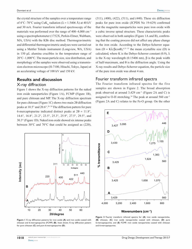
Results and discussionX-ray diffractionFigure 1 shows the X-ray diffraction patterns for the naked
iron oxide nanoparticles (Figure 1A), FCMP (Figure 1B),
and pure chitosan and MP. The X-ray diffraction spectrum
for pure chitosan (Figure 1C) shows two main 2θ diffraction
peaks at 10.5° and 20.6°.28–30 The diffraction pattern for pure
6-mercaptopurine indicated distinct peaks at 2θ = 11.8°,
14.6°, 16.8°, 21.2°, 23.5°, 25.3°, 25.9°, 27.5°, 29.5°, and
30.3° (Figure 1D). Naked iron oxide showed six intense peaks
between 30°C and 70°C that could be assigned as (220),
(311), (400), (422), (511), and (440). These six diffraction
peaks for pure iron oxide (JCPDS No 19-629) confirmed
that the magnetite nanoparticles were pure iron oxide with
a cubic inverse spinal structure. These characteristic peaks
were observed in both samples (Figure 1A and B), confirm-
ing that the coating process did not affect any phase change
in the iron oxide. According to the Debye-Scherrer equa-
tion (D = Kλ/βcosθ),4,31–33 the mean crystallite size (D) is
calculated, where K is the Debye-Scherrer constant (0.9), λ
is the X-ray wavelength (0.15406 nm), β is the peak width
of half-maximum, and θ is the diffraction angle. Using the
X-ray results and Debye-Scherrer equation, the particle size
of the pure iron oxide was about 4 nm.
Fourier transform infrared spectraThe Fourier transform infrared spectra for the f ive
samples are shown in Figure 2. The broad absorption
peak observed at around 3,429 cm−1 (Figure 2A and C) is
assigned to O-H stretching.34 The peak at around 560 cm−1
(Figure 2A and C) relates to the Fe-O group. On the other
10 20 30 40 50 60
Inte
nsity
(au)
2θ/degree
D
C
Inte
nsi
ty (
au)
2θ/degree
220
311
400422
511440
A
B
10 20 30 40 50 60
Figure 1 X-ray diffraction patterns for iron oxide (A) and iron oxide coated with chitosan and 6-mercaptopurine (FCMP) (B). Inset shows X-ray diffraction pattern for pure chitosan (C) and pure 6-mercaptopurine (D).
4,000 3,200 2,400 1,600 800
Wavenumbers (cm--1)
A560
1,070
3,429
B% t
ran
smit
tan
ce
3,425 1,658
560
C
1,624
1,383
3,429
D
1,1551,2753,430
E
3,427 3,095 446
Figure 2 Fourier transform infrared spectra for (A) iron oxide nanoparticles, (B) chitosan, (C) iron oxide nanoparticles coated with chitosan, (D) pure 6-mercaptopurine, and (E) FCMP, iron oxide nanoparticles coated with chitosan and 6-mercaptopurine.
submit your manuscript | www.dovepress.com
Dovepress
Dovepress
1018
Dorniani et al

Drug Design, Development and Therapy 2013:7
hand, the band at 446 cm−1 in the low energy range might be
related to the νFe-S
and νFe-N
vibration modes (Figure 2E). In
Figure 2C, the main characteristic absorption bands appear-
ing at 1,624 cm−1 and 1,383 cm−1 can be assigned to N−H
bending vibration and −C−O stretching of the alcohol group
in chitosan, respectively. Figure 2B shows the characteristic
bands of pure chitosan at around 3,425 cm−1 (O−H stretch-
ing and N-H stretching vibrations), 1,658 cm−1 (amide), and
1,064 cm−1 (C−O−C stretching vibration). The absence
of a band at 1,155 cm−1 (νC = S
/ring vibration) in Figure 2E
suggests participation of an exocyclic (S) atom in metallic
bonding of the heterocyclic ligand in the Fe (II) coordina-
tion compound.16 The peak at 3,095 cm−1 belongs to the νC-H
aromatic (Figure 2E). The peak observed in Figure 2D for
pure 6-mercaptopurine at around 1,275 cm−1 can be assigned
to the C=S group and the absence of this absorption peak at
1,275 cm−1 in the FCMP compound confirms formation of the
6-mercaptopurine complex via the sulfur atom (Figure 2E).16
As could be observed, chitosan and 6-mercaptopurine were
successfully coated to the iron oxide nanoparticles.
Magnetic propertiesFigure 3 shows the hysteresis loops for naked iron oxide
nanoparticles (Figure 3A) and iron oxide nanoparticles
coated with chitosan and 6-mercaptopurine (Figure 3B),
which were characterized using a vibrating sample mag-
netometer at room temperature. The values for saturation
magnetization and remanent magnetization are shown in
Table 1. Using the sonochemical method, the saturation
magnetization of the magnetite nanoparticles was about
29.09 emu/g compared with 17.50 emu/g for FCMP, which
agrees well with previous work.35 It was demonstrated that
both types of magnetic nanoparticles had superparamagnetic
characteristics, meaning that they do not retain any magne-
tism after removal of a magnetic field. The decreased satura-
tion magnetization could be due to the existence of coated
materials on the surfaces of the magnetite nanoparticles.36
A high degree of magnetization and superparamagnetic
properties are in high demand for biomedical applications
because the larger magnetic particles form aggregates after
exposure to a magnetic field.
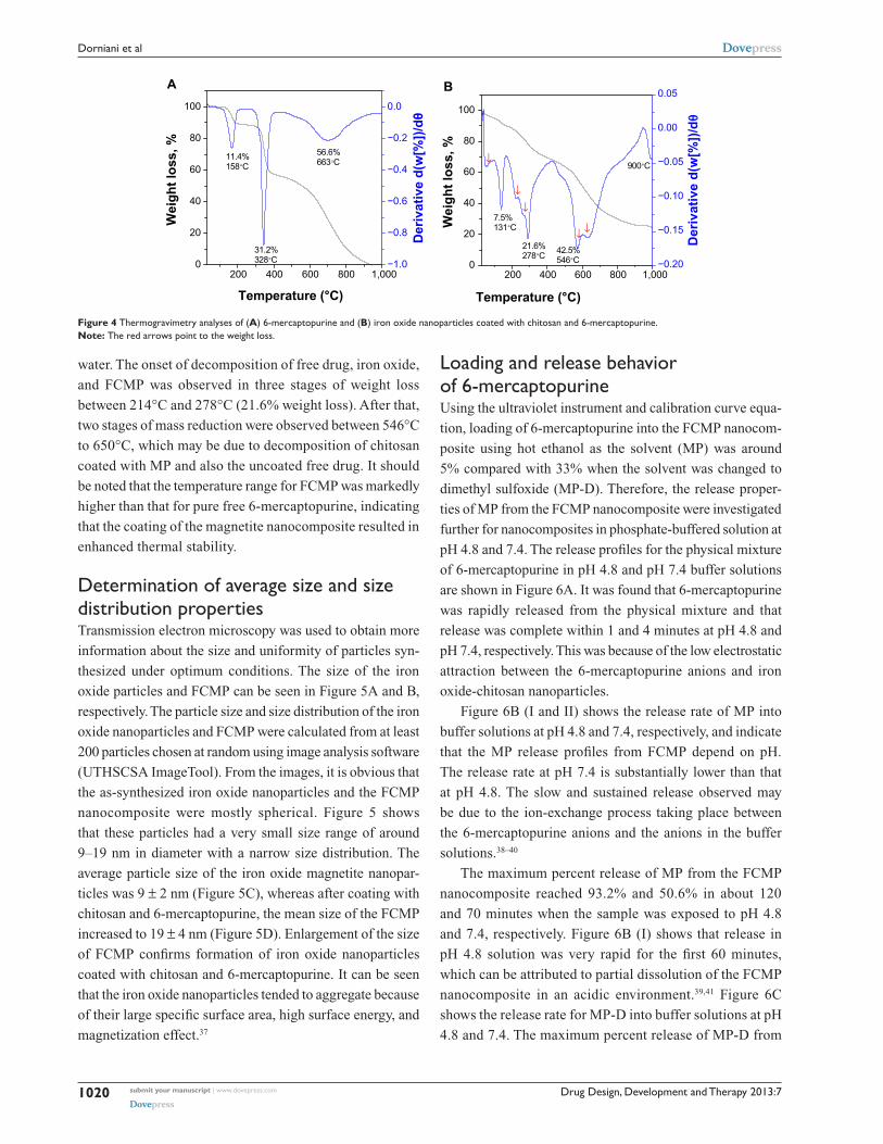
Thermogravimetric analysesThermogravimetric and differential thermogravimetric
analyses (TGA-DTG) is a technique measuring physical
changes in materials. It enables quantitative measurement
of percent weight loss from a sample associated with transi-
tion and thermal degradation in a controlled environment.
Thermal analysis is usually recorded as change in mass from
decomposition, dehydration, and oxidation of a sample,
as related to thermal stability at selected temperatures,
usually between 25°C and 1,000°C. The thermogram data
changed because of the unique physicochemical reactions
that occur in this temperature range. These characteristics
are relevant to the molecular structure of the sample. The
TGA-DTG analyses for pure 6-mercaptopurine and FCMP
nanoparticles was obtained under atmospheric condi-
tions (Figure 4). For free 6-mercaptopurine, the thermal
behavior indicates three stages of weight loss (Figure 4A).
At 158°C, a total weight loss of 11% was observed, and
is attributed to removal of crystalline water. The second
sharp mass reduction step at 328°C (31.2%) corresponds
to decomposition of 6-mercaptopurine. This temperature
agrees well with the value of 320°C–350°C recorded for
the decomposition of 6-mercaptopurine.17 Although the
mass fragmentation process and thermal decomposition is
not exactly the same, the weight loss observed may be due
to loss of an HCS group at this step, corresponding to the
fragmentation of heterocyclic compounds seen on mass
spectroscopy. The differential thermogravimetric curve
shows one peak at 663°C. The curve shape on TGA-DTG
was changed due to the chitosan coating (Figure 4B). The
thermal behavior of FCMP shows that the stages of mass
loss occur at 50°C–900°C. The first stage of mass loss starts
at 4°C–131°C and is attributable to removal of adsorbed
Table 1 Magnetic properties of Fe3O4 magnetite nanoparticles and FCMP
Samples Ms (emu/g) Mr (emu/g)
Fe3O4 29.091 1.098FCMP 17.497 0.365
Abbreviations: FCMP, iron oxide nanoparticles coated with chitosan and 6-mercaptopurine; Mr, remanent magnetization; Ms, saturation magnetization.
−15,000 −10,000 −5,000
−40
−30
−20
−10
0
0
10
20
30
40
5,000
H (Oe)
A
M (
emu
/g)
B
10,000 15,000
Figure 3 Magnetization plots of (A) iron oxide magnetite nanoparticles and (B) iron oxide nanoparticles coated with chitosan and 6-mercaptopurine.Notes: The data is presented in terms of M, mass magnetization (emu/g), versus H, applied magnetic field (Oe).
submit your manuscript | www.dovepress.com
Dovepress
Dovepress
1019
6-mercaptopurine-coated magnetite nanoparticles for drug delivery
Drug Design, Development and Therapy 2013:7
0
20
40
60
80
100
Temperature (°C)
Wei
ght l
oss,
%
A
11.4%158°C
31.2%328°C
56.6%663°C
−1.0
−0.8
−0.6
−0.4
−0.2
0.0
Der
ivat
ive
d(w
[%])/
dθ
200 400 600 800 1,000 200 400 600 800 1,0000
20
40
60
80
100
↓ ↓
↓
↓
↓
Temperature (°C)
Wei
ght l
oss,
%
7.5%131°C
21.6%278°C 42.5%
546°C
900°C
−0.20
−0.15
−0.10
−0.05
0.00
0.05
Der
ivat
ive
d(w
[%])/
dθ
B
Figure 4 Thermogravimetry analyses of (A) 6-mercaptopurine and (B) iron oxide nanoparticles coated with chitosan and 6-mercaptopurine.Note: The red arrows point to the weight loss.
water. The onset of decomposition of free drug, iron oxide,
and FCMP was observed in three stages of weight loss
between 214°C and 278°C (21.6% weight loss). After that,
two stages of mass reduction were observed between 546°C
to 650°C, which may be due to decomposition of chitosan
coated with MP and also the uncoated free drug. It should
be noted that the temperature range for FCMP was markedly
higher than that for pure free 6-mercaptopurine, indicating
that the coating of the magnetite nanocomposite resulted in
enhanced thermal stability.
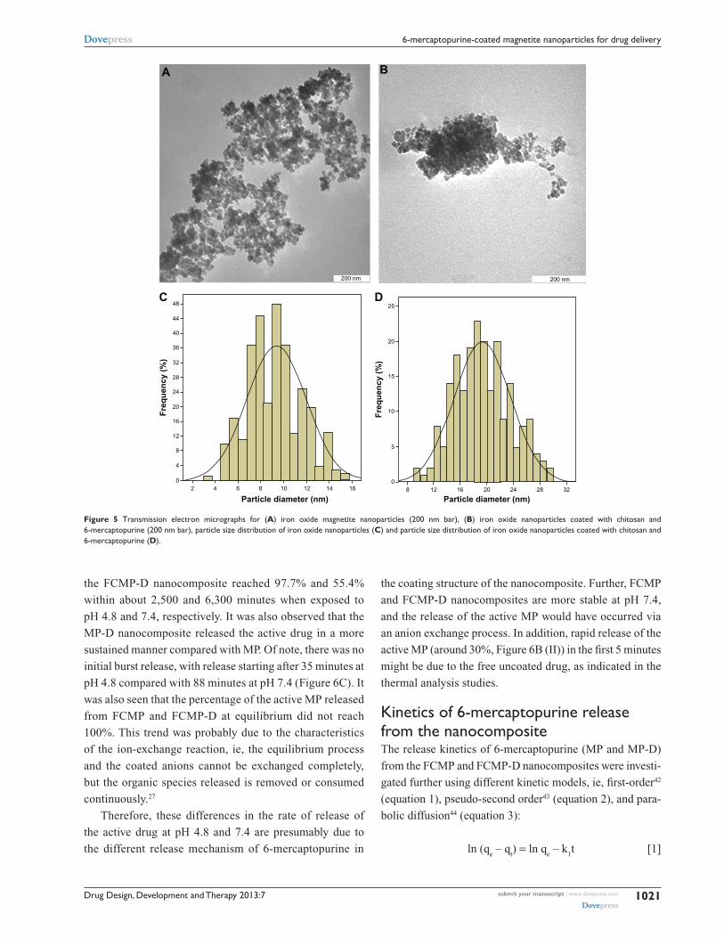
Determination of average size and size distribution propertiesTransmission electron microscopy was used to obtain more
information about the size and uniformity of particles syn-
thesized under optimum conditions. The size of the iron
oxide particles and FCMP can be seen in Figure 5A and B,
respectively. The particle size and size distribution of the iron
oxide nanoparticles and FCMP were calculated from at least
200 particles chosen at random using image analysis software
(UTHSCSA ImageTool). From the images, it is obvious that
the as-synthesized iron oxide nanoparticles and the FCMP
nanocomposite were mostly spherical. Figure 5 shows
that these particles had a very small size range of around
9–19 nm in diameter with a narrow size distribution. The
average particle size of the iron oxide magnetite nanopar-
ticles was 9 ± 2 nm (Figure 5C), whereas after coating with
chitosan and 6-mercaptopurine, the mean size of the FCMP
increased to 19 ± 4 nm (Figure 5D). Enlargement of the size
of FCMP confirms formation of iron oxide nanoparticles
coated with chitosan and 6-mercaptopurine. It can be seen
that the iron oxide nanoparticles tended to aggregate because
of their large specific surface area, high surface energy, and
magnetization effect.37
Loading and release behavior of 6-mercaptopurineUsing the ultraviolet instrument and calibration curve equa-
tion, loading of 6-mercaptopurine into the FCMP nanocom-
posite using hot ethanol as the solvent (MP) was around
5% compared with 33% when the solvent was changed to
dimethyl sulfoxide (MP-D). Therefore, the release proper-
ties of MP from the FCMP nanocomposite were investigated
further for nanocomposites in phosphate-buffered solution at
pH 4.8 and 7.4. The release profiles for the physical mixture
of 6-mercaptopurine in pH 4.8 and pH 7.4 buffer solutions
are shown in Figure 6A. It was found that 6-mercaptopurine
was rapidly released from the physical mixture and that
release was complete within 1 and 4 minutes at pH 4.8 and
pH 7.4, respectively. This was because of the low electrostatic
attraction between the 6-mercaptopurine anions and iron
oxide-chitosan nanoparticles.
Figure 6B (I and II) shows the release rate of MP into
buffer solutions at pH 4.8 and 7.4, respectively, and indicate
that the MP release profiles from FCMP depend on pH.
The release rate at pH 7.4 is substantially lower than that
at pH 4.8. The slow and sustained release observed may
be due to the ion-exchange process taking place between
the 6-mercaptopurine anions and the anions in the buffer
solutions.38–40
The maximum percent release of MP from the FCMP
nanocomposite reached 93.2% and 50.6% in about 120
and 70 minutes when the sample was exposed to pH 4.8
and 7.4, respectively. Figure 6B (I) shows that release in
pH 4.8 solution was very rapid for the first 60 minutes,
which can be attributed to partial dissolution of the FCMP
nanocomposite in an acidic environment.39,41 Figure 6C
shows the release rate for MP-D into buffer solutions at pH
4.8 and 7.4. The maximum percent release of MP-D from
submit your manuscript | www.dovepress.com
Dovepress
Dovepress
1020
Dorniani et al
Drug Design, Development and Therapy 2013:7
the FCMP-D nanocomposite reached 97.7% and 55.4%
within about 2,500 and 6,300 minutes when exposed to
pH 4.8 and 7.4, respectively. It was also observed that the
MP-D nanocomposite released the active drug in a more
sustained manner compared with MP. Of note, there was no
initial burst release, with release starting after 35 minutes at
pH 4.8 compared with 88 minutes at pH 7.4 (Figure 6C). It
was also seen that the percentage of the active MP released
from FCMP and FCMP-D at equilibrium did not reach
100%. This trend was probably due to the characteristics
of the ion-exchange reaction, ie, the equilibrium process
and the coated anions cannot be exchanged completely,
but the organic species released is removed or consumed
continuously.27
Therefore, these differences in the rate of release of
the active drug at pH 4.8 and 7.4 are presumably due to
the different release mechanism of 6-mercaptopurine in
the coating structure of the nanocomposite. Further, FCMP
and FCMP-D nanocomposites are more stable at pH 7.4,
and the release of the active MP would have occurred via
an anion exchange process. In addition, rapid release of the
active MP (around 30%, Figure 6B (II)) in the first 5 minutes
might be due to the free uncoated drug, as indicated in the
thermal analysis studies.
Kinetics of 6-mercaptopurine release from the nanocompositeThe release kinetics of 6-mercaptopurine (MP and MP-D)
from the FCMP and FCMP-D nanocomposites were investi-
gated further using different kinetic models, ie, first-order42
(equation 1), pseudo-second order43 (equation 2), and para-
bolic diffusion44 (equation 3):
ln (qe – q
t) = ln q
e – k
1t [1]
A
C
B
200 nm 200 nm
48
44
40
36
32
28
24
20
16
12
8
4
02
Fre
qu
ency
(%
)
4 6 8 10 12 14 16
Particle diameter (nm)
D25
20
15
10
5
08
Fre
qu
ency
(%
)
12 16 20 24 28 32
Particle diameter (nm)
Figure 5 Transmission electron micrographs for (A) iron oxide magnetite nanoparticles (200 nm bar), (B) iron oxide nanoparticles coated with chitosan and 6-mercaptopurine (200 nm bar), particle size distribution of iron oxide nanoparticles (C) and particle size distribution of iron oxide nanoparticles coated with chitosan and 6-mercaptopurine (D).
submit your manuscript | www.dovepress.com
Dovepress
Dovepress
1021
6-mercaptopurine-coated magnetite nanoparticles for drug delivery

Drug Design, Development and Therapy 2013:7
t/qt = 1/k
2q
e2 + t/q
e [2]
(1 – Mt/M
0)/t = kt−0.5 + b [3]
where qe and q
t are the equilibrium release rate and the release
rate at time t, respectively, k is a constant corresponding to
release amount, and M0 and M
t represent the drug content
remaining in FCMP/FCMP-D at release time 0 and t, respec-
tively, and b is a constant.
Fitting the data for release of the active MP to these three
kinetic models, it was found that the pseudo-second order
model was best able to describe the release kinetic processes
of MP from the FCMP nanocomposite at both pH levels
compared with the other models. Figure 7A shows that, for
phosphate-buffered solution at pH 4.8, the pseudo-second
order model fits better to the data (correlation coefficient,
R2, with K values of 0.9914 and 6.73 × 10⁻⁴ mg per minute,
respectively). At pH 7.4, release of MP from FCMP also
followed the pseudo-second order equation, with a correla-
tion coefficient of 0.9965 (Figure 7B). The release of MP-D
from FCMP-D did not obey either pseudo-second order
(R2=0.1109) or parabolic order kinetics. For the FCMP-D
nanocomposite, it can be observed that the pseudo-second
order model gives a better fit for phosphate-buffered solution
at pH 4.8 (Figure 7C). However, phosphate-buffered solu-
tion at pH 7.4 is better fitted to the first order kinetic model
(Figure 7D). Table 2 indicates the time release profile for
MP and MP-D in phosphate-buffered solutions at pH 7.4
and 4.8 in FCMP and FCMP-D, respectively.
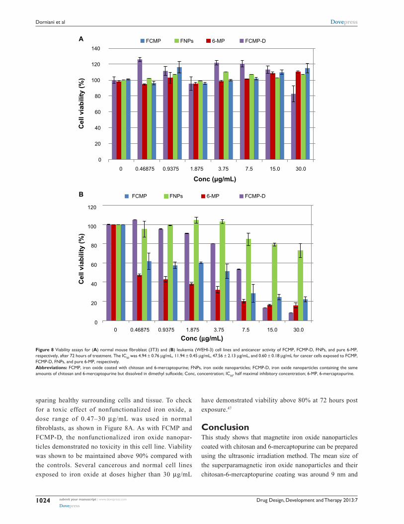
In vitro bioassayFigure 8 shows the viability assays for normal mouse
f ibroblast cells (Figure 8A) and the anticancer activ-
ity of FCMP, FCMP-D, iron oxide nanoparticles,
and pure 6-mercaptopurine, respectively (Figure 8B)
72 hours post treatment. Normal mouse fibroblast cells
exposed to increasing concentrations (0.47–30 µg/mL)
of FCMP, FCMP-D, iron oxide nanoparticles, and pure
6-mercaptopurine did not show a decrease in viability
compared with untreated control cells at 72 hours post-
treatment. Viability of leukemia cells was found to be
reduced to less than 20% following incubation with pure
6-mercaptopurine, FCMP, and FCMP-D at 30 µg/mL after
72 hours. Exposure to the empty carrier, ie, iron oxide
nanoparticles, resulted in sustained viability of the leuke-
mia cells, with more than 70% of cells remaining viable,
even at a concentration of 30 µg/mL after 72 hours. Lower
percent viability was observed following incubation of the
leukemia cells with pure MP than with FCMP and FCMP-
D nanoparticles.
Using this viability (MTT) study, the newly synthesized
FCMP and FCMP-D nanoparticles showed efficacy in the
leukemia cell line in a dose-dependent manner, the pattern
of which resembles that for MP (Figure 8A). On the other
hand, iron oxide nanoparticles had a negligible effect in the
same cancer cell line, with almost 100% of cells remaining
viable at 15 µg/mL compared with about 25% and 15% of
cells remaining viable at 15 µg/mL of FCMP and FCMP-
D, respectively. Thus, the cytotoxicity to leukemia cells is
likely attributable to release of MP from the carrier rather
than the effect of the carrier itself. This result indicates
that the anticancer activity of our new compound is very
similar to that of pure MP and suggests the possibility of
a decreased dosing interval due to the sustained-release
ability of the nanoparticles. With the sustained release and
possible targeted delivery potential of these nanoparticles,
the least amount of active agent (6-mercaptopurine) could
suffice, hence reducing the dosing interval and unnecessary
exposure to large quantities of this hazardous drug. The
results further suggest the possibility of increased anti-
cancer activity with increasing the loading percentage. A
previous study showed good anticancer activity in a HeLa
40
60
80
100
Time (minutes)
% r
elea
seA
pH 7.4
pH 4.8
0
20
40
60
80
100
Time (minutes)%
rel
ease
I
II
B
0 1 2 3 4 5 6 0 40 80 120 160 200
0 1,500 3,000 4,500 6,0000
20
40
60
80
100
Time (minutes)
% r
elea
se
C
III IV
Figure 6 (A) Release profiles for physical mixtures of 6-mercaptopurine and (B) 6-mercaptopurine from iron oxide nanoparticles coated with chitosan and 6-mercaptopurine dissolved in hot ethanol into (I) phosphate-buffered solution at pH 4.8, (II) phosphate-buffered solution at pH 7.4, and (C) release profiles for 6-mercaptopurine from the iron oxide nanoparticles containing the same amounts of chitosan and 6-mercaptopurine dissolved in dimethyl sulfoxide into (III) phosphate-buffered solution at pH 4.8 and (IV) phosphate-buffered solution at pH 7.4.
submit your manuscript | www.dovepress.com
Dovepress
Dovepress
1022
Dorniani et al

Drug Design, Development and Therapy 2013:7
The sustained viability of normal mouse fibroblasts
is shown in Figure 8A. Despite increasing concentra-
tions of FCMP and FCMP-D (using the MTT assay),
there was minimal or no toxicity in this cell line over the
concentration range tested. Thus, FCMP and FCMP-D
likely represent alternative drug delivery systems for
the treatment of cancer, with the added possibility of
0.0
0.5
1.0
t/q
t
t/q
t
Time (minutes)
pH = 4.8Pseudo-second order
R2 = 0.9914
A
0.0
0.7
1.4
Time (minutes)
pH = 7.4Pseudo-second order
R2 = 0.9965
B
0
15
30
45
t/q
t
Time (minutes)
pH = 4.8Pseudo-second orderR2 = 0.9385
C
0 25 50 75 100 125 0 30 60 90
0 800 1,600 2,400 3,200 4,000 0 3,000 6,000
3.9
4.2
4.5
4.8
ln (
qe-
qt)
Time (minutes)
pH = 7.4First order
R2 = 0.87429
D
Figure 7 Fitting the data for 6-mercaptopurine release from iron oxide nanoparticles coated with chitosan and 6-mercaptopurine dissolved in hot ethanol into different solutions to the pseudo-second order kinetics for pH 4.8 (A) and pH 7.4 (B) and fitting data of 6-mercaptopurine released from the iron oxide nanoparticles coated with chitosan and 6-mercaptopurine dissolved in dimethyl sulfoxide into different solutions to the pseudo-second order kinetics for pH 4.8 (C) and first order kinetics for pH 7.4 (D).Abbreviations: t, time; qt, release at time t.
(cervical cancer) cell line using a synthesized complex of
MP and two metals (silver and gold).45 Similar anticancer
activity was demonstrated in an HT29 cell line using
solid lipid nanoparticles, where the trypan blue assay was
used, indicating improved efficacy for doxorubicin-loaded
solid lipid nanoparticles in comparison with the com-
mercially available free counterpart, ie, doxorubicin.46
Table 2 Correlation coefficient, rate constant, and elimination half-life obtained by fitting the data for release of MP and MP-D from FCMP and FCMP-D into phosphate-buffered solution at pH 4.8 and pH 7.4
R2 Rate constant (K)
(mg/min)
t1/2
(min) Sample
Aqueous solution
Saturated release %
Pseudo-first order
Pseudo-second order
Parabolic diffusion
FCMP pH 4.8 93.2 0.9624 0.9914 0.7380 a6.73 × 10−4 14
FCMP pH 7.4 50.6 0.5055 0.9965 0.6839 b3.52 × 10−3 5
FCMP-D pH 4.8 97.7 0.4853 0.9385 0.6255 c2.65 × 10−4 42
FCMP-D pH 7.4 55.4 0.8743 0.1109 0.8154 d1.25 × 10−4 5,529
Notes: a,b,cEstimated using pseudo-second order kinetics and destimated using first order kinetics.Abbreviations: FCMP, iron oxide nanoparticles coated with chitosan and 6-mercaptopurine dissolved in hot ethanol; FCMP-D, iron oxide nanoparticles containing the same amounts of chitosan and 6-mercaptopurine but dissolved in dimethyl sulfoxide; MP, 6-mercaptopurine dissolved in hot ethanol; MP-D, 6-mercaptopurine dissolved in dimethyl sulfoxide; t1/2, elimination half-life.
submit your manuscript | www.dovepress.com
Dovepress
Dovepress
1023
6-mercaptopurine-coated magnetite nanoparticles for drug delivery
Drug Design, Development and Therapy 2013:7
sparing healthy surrounding cells and tissue. To check
for a toxic effect of nonfunctionalized iron oxide, a
dose range of 0.47–30 µg/mL was used in normal
fibroblasts, as shown in Figure 8A. As with FCMP and
FCMP-D, the nonfunctionalized iron oxide nanopar-
ticles demonstrated no toxicity in this cell line. Viability
was shown to be maintained above 90% compared with
the controls. Several cancerous and normal cell lines
exposed to iron oxide at doses higher than 30 µg/mL
have demonstrated viability above 80% at 72 hours post
exposure.47
ConclusionThis study shows that magnetite iron oxide nanoparticles
coated with chitosan and 6-mercaptopurine can be prepared
using the ultrasonic irradiation method. The mean size of
the superparamagnetic iron oxide nanoparticles and their
chitosan-6-mercaptopurine coating was around 9 nm and
Figure 8 Viability assays for (A) normal mouse fibroblast (3T3) and (B) leukemia (WEHI-3) cell lines and anticancer activity of FCMP, FCMP-D, FnPs, and pure 6-MP, respectively, after 72 hours of treatment. The IC50 was 4.94 ± 0.76 µg/mL, 11.94 ± 0.45 µg/mL, 47.56 ± 2.13 µg/mL, and 0.60 ± 0.18 µg/mL for cancer cells exposed to FCMP, FCMP-D, FnPs, and pure 6-MP, respectively.Abbreviations: FCMP, iron oxide coated with chitosan and 6-mercaptopurine; FnPs, iron oxide nanoparticles; FCMP-D, iron oxide nanoparticles containing the same amounts of chitosan and 6-mercaptopurine but dissolved in dimethyl sulfoxide; Conc, concentration; IC50, half maximal inhibitory concentration; 6-MP, 6-mercaptopurine.
0
20
40
60
80
100
120
140
30.015.07.53.751.8750.93750.468750
Cel
l via
bili
ty (
%)
Conc (µg/mL)
FCMP FNPs 6-MP FCMP-DA
0
20
40
60
80
100
120
30.015.07.53.751.8750.93750.468750
Cel
l via
bili
ty (
%)
Conc (µg/mL)
FCMP FNPs 6-MP FCMP-DB
submit your manuscript | www.dovepress.com
Dovepress
Dovepress
1024
Dorniani et al
Drug Design, Development and Therapy 2013:7
19 nm, respectively. The coating process was found to improve
the thermal stability of the resulting nanocomposite compared
with its uncoated counterpart. Release of the active agent from
the nanocomposite was found to occur in a controlled manner
via an anion exchange process, indicating that this nanocom-
posite can be used as a controlled-release formulation. It was
also found that the release behavior of the FCMP-D nanocom-
posite in buffered solutions of pH 4.8 and pH 7.4 was more
sustained than for the FCMP nanocomposite. Further, we did
not observe any burst release for the active MP in FCMP-D,
but did observe this for the FCMP nanocomposite. An in vitro
bioassay study showed that the synthesized nanoparticles may
serve as an alternative drug delivery system for treatment of
cancer, with the added possibility of sparing surrounding
normal cells and tissue.
AcknowledgmentFunding for this research was provided by the Ministry
of Higher Education grant of Malaysia (ERGS/1/11/STG/
UPM/01/18, Vot No 5527050).
DisclosureThe authors report no conflicts of interest in this work.
References 1. Zhu L, Ma J, Jia N, Zhao Y, Shen H. Chitosan-coated magnetic nano-
particles as carriers of 5-fluorouracil: preparation, characterization and cytotoxicity studies. Colloids Surf B Biointerfaces. 2009;68(1):1–6.
2. Selvaraj V, Alagar M, Hamerton I. Analytical detection and biological assay of antileukemic drug using gold nanoparticles. Electrochim Acta. 2006;52(3):1152–1160.
3. Faridi-Majidi R, Sharifi-Sanjani N, Agend F. Encapsulation of mag-netic nanoparticles with polystyrene via emulsifier-free miniemulsion polymerization. Thin Solid Films. 2006;515(1):368–374.
4. Zhao Y, Qiu Z, Huang J. Preparation and analysis of Fe3O
4 magnetic
nanoparticles used as targeted-drug carriers. Chinese J Chem Eng. 2008;16(3):451–455.
5. Mudshinge SR, Deore AB, Patil S, Bhalgat CM. Nanoparticles: emerg-ing carriers for drug delivery. Saudi Pharm J. 2011;19(3):129–141.
6. Xu H, Song T, Bao X, Hu L. Site-directed research of magnetic nanoparticles in magnetic drug targeting. J Magn Magn Mater. 2005;293(1):514–519.
7. Zhang J, Lan CQ, Post M, Simard B, Deslandes Y, Hsieh TH. Design of nanoparticles as drug carriers for cancer therapy. Cancer Genomics Proteomics. 2006;3(3–4):147–157.
8. Hejazi R, Amiji M. Chitosan-based gastrointestinal delivery systems. J Control Release. 2003;89(2):151–165.
9. Amidi M, Romeijn SG, Borchard G, Junginger HE, Hennink WE, Jiskoot W. Preparation and characterization of protein-loaded N-trimethyl chitosan nanoparticles as nasal delivery system. J Control Release. 2006;111(1):107–116.
10. Kim JH, Kim YS, Park K, et al. Antitumor efficacy of cisplatin-loaded glycol chitosan nanoparticles in tumor-bearing mice. J Control Release. 2008;127(1):41–49.
11. Sang Yoo H, Eun Lee J, Chung H, Chan Kwon I, Young Jeong S. Self-assembled nanoparticles containing hydrophobically modified glycol chitosan for gene delivery. J Control Release. 2005;103(1):235–243.
12. Mao C, Zhu JJ, Hu YF, et al. Surface modification using photocross-linkable chitosan for improving hemocompatibility. Colloids Surf B Biointerfaces. 2004;38(1):47–53.
13. Tidd DM, Paterson ARP. A biochemical mechanism for the delayed cytotoxic reaction of 6-mercaptopurine. Cancer Res. 1974;34(4):738–746.
14. Nerstroem VM, Henriksen U, Nielsen PE, Buchardt O, Schmiegelow K, Koch C. Monoclonal antibodies to thioguanine: influence of coupling position on fine specificity. Bioconjug Chem. 1994;5(4):357–363.
15. Sorouraddin MH, Khani MY, Amini K, Naseri A, Asgari D, Rashidi MR. Simultaneous determination of 6-mercaptopurine and its oxidative metab-olites in synthetic solutions and human plasma using spectrophotometric multivariate calibration methods. Bioimpacts. 2011;1(1):53–62.
16. Acevedo-Chávez R, Costas ME, Escudero R. Magnetic study of the novel polynuclear compound [Cu (II)(6-mercaptopurinolate2−) ]
n. J Solid
State Chem. 1997;132(1):78–87. 17. Bariyanga J, Luyt AS. Synthesis, Fourier transform infrared,
nuclear magnetic resonance and thermal analysis of sodium and platinum complexes of 6-mercaptopurine. J Mol Struct. 2001;559(1): 49–54.
18. Lee HS, Shao H, Huang Y, Kwak BK. Synthesis of MRI contrast agent by coating superparamagnetic iron oxide with chitosan. IEEE Trans Magn. 2005;41(10):4102–4104.
19. Genta I, Perugini P, Pavanetto F. Different molecular weight chitosan microspheres: influence on drug loading and drug release. Drug Dev Ind Pharm. 1998;24(8):779–784.
20. Sinha VR, Singla AK, Wadhawan S, et al. Chitosan microspheres as a potential carrier for drugs. Int J Pharm. 2004;274(1):1–33.
21. Ghotbi MY, bin Hussein MZ. Controlled release study of an anti-carcinogenic agent, gallate from the surface of magnetite nanoparticles. J Phys Chem Solids. 2012;73:936–942.
22. Hassanjani-Roshan A, Vaezi MR, Shokuhfar A, Rajabali Z. Synthesis of iron oxide nanoparticles via sonochemical method and their characterization. Particuology. 2011;9(1):95–99.
23. Su C-C, Yang J-S, Lin S-Y, et al. Curcumin inhibits WEHI-3 leukemia cells in BALB/c mice in vivo. In Vivo. 2008;22(1):63–68.
24. Dorniani D, Hussein MZB, Kura AU, Fakurazi S, Shaari AH, Ahmad Z. Preparation of Fe
3O
4 magnetic nanoparticles coated with gallic acid for
drug delivery. Int J Nanomedicine. 2012;7:5745–5756. 25. Xia SJ, Ni ZM, Xu Q, Hu BX, Hu J. Layered double hydroxides as
supports for intercalation and sustained release of antihypertensive drugs. J Solid State Chem. 2008;181(10):2610–2619.
26. Ribeiro C, Arizaga GGC, Wypych F, Sierakowski MR. Nanocomposites coated with xyloglucan for drug delivery: in vitro studies. Int J Pharm. 2009;367(1):204–210.
27. Zhang H, Zou K, Guo S, Duan X. Nanostructural drug-inorganic clay composites: structure, thermal property and in vitro release of captopril-intercalated Mg-Al-layered double hydroxides. J Solid State Chem. 2006;179(6):1792–1801.
28. Zhang B, Wang DF, Li HY, Xu Y, Zhang L. Preparation and properties of chitosan-soybean trypsin inhibitor blend film with anti-Aspergillus flavus activity. Ind Crop Prod. 2009;29(2):541–548.
29. Costa-Júnior ES, Barbosa-Stancioli EF, Mansur AAP, Vasconcelos WL, Mansur HS. Preparation and characterization of chitosan/poly (vinyl alcohol) chemically crosslinked blends for biomedical applications. Carbohydr Polym. 2009;76(3):472–481.
30. Fan M, Hu Q, Shen K. Preparation and structure of chitosan soluble in wide pH range. Carbohydr Polym. 2009;78(1):66–71.
31. Nidhin M, Indumathy R, Sreeram KJ, Nair BU. Synthesis of iron oxide nanoparticles of narrow size distribution on polysaccharide templates. Bull Mater Sci. 2008;31(1):93–96.
32. Ma H, Qi X, Maitani Y, Nagai T. Preparation and characterization of superparamagnetic iron oxide nanoparticles stabilized by alginate. Int J Pharm. 2007;333(1):177–186.
33. Chaparadza A, Rananavare SB, Shutthanandan V. Synthesis and charac-terization of lithium-doped tin dioxide nanocrystalline powders. Mater Chem Phys. 2007;102(2):176–180.
submit your manuscript | www.dovepress.com
Dovepress
Dovepress
1025
6-mercaptopurine-coated magnetite nanoparticles for drug delivery

Drug Design, Development and Therapy
Publish your work in this journal
Submit your manuscript here: http://www.dovepress.com/drug-design-development-and-therapy-journal
Drug Design, Development and Therapy is an international, peer-reviewed open-access journal that spans the spectrum of drug design and development through to clinical applications. Clinical outcomes, patient safety, and programs for the development and effective, safe, and sustained use of medicines are a feature of the journal, which
has also been accepted for indexing on PubMed Central. The manu-script management system is completely online and includes a very quick and fair peer-review system, which is all easy to use. Visit http://www.dovepress.com/testimonials.php to read real quotes from published authors.
Drug Design, Development and Therapy 2013:7
34. Yasin Y, Ismail NM, Hussein MZ, Aminudin N. Synthesis and charac-terization of lawsone-intercalated ZnAl layered double hydroxides. J Biomed Nanotechnol. 2011;7(3):486–488.
35. Bajaj B, Malhotra BD, Choi S. Preparation and characterization of bio-functionalized iron oxide nanoparticles for biomedical application. Thin Solid Films. 2010;519(3):1219–1223.
36. Ge Y, Zhang Y, Xia J, et al. Effect of surface charge and agglomerate degree of magnetic iron oxide nanoparticles on KB cellular uptake in vitro. Colloids Surf B Biointerfaces. 2009;73(2):294–301.
37. Qu JB, Shao H, Jing GL, Huang F. PEG-chitosan-coated iron oxide nanoparticles with high saturated magnetization as carriers of 10-hydroxycamptothecin: preparation, characterization and cytotoxicity studies. Colloids Surf B Biointerfaces. 2013;102: 37–44.
38. Ambrogi V, Fardella G, Grandolini G, Perioli L, Tiralti MC. Intercalation compounds of hydrotalcite-like anionic clays with anti-inflammatory agents, II: uptake of diclofenac for a controlled release formulation. AAPS PharmSciTech. 2002;3(3):77–82.
39. Al Ali SHH, Al-Qubaisi M, Hussein MZ, Ismail M, Zainal Z, Hakim MN. Comparative study of Mg/Al-and Zn/Al-layered double hydroxide-perindopril erbumine nanocomposites for inhibition of angiotensin-converting enzyme. Int J Nanomedicine. 2002;7:4251–4262.
40. Zhang H, Zou K, Sun H, Duan X. A magnetic organic-inorganic com-posite: synthesis and characterization of magnetic 5-aminosalicylic acid intercalated layered double hydroxides. J Solid State Chem. 2005;178(11):3485–3493.
41. Khan AI, O’Hare D. Intercalation chemistry of layered double hydrox-ides: recent developments and applications. J Mater Chem. 2002;12(11): 3191–3198.
42. Hussein-Al-Ali SH, Al-Qubaisi M, Hussein MZ, Ismail M, Zainal Z, Hakim MN. In vitro inhibition of histamine release behavior of cetirizine intercalated into zn/al-and mg/al-layered double hydroxides. Int J Mol Sci. 2002;13(5):5899–5916.
43. Dong L, Yan L, Hou WG, Liu SJ. Synthesis and release behavior of composites of camptothecin and layered double hydroxide. J Solid State Chem. 2010;183(8):1811–1816.
44. Ho YS, Ofomaja AE. Pseudo-second-order model for lead ion sorp-tion from aqueous solutions onto palm kernel fiber. J Hazard Mater. 2006;129(1):137–142.
45. Cuin A, Massabni AC, Pereira GA, et al. 6-Mercaptopurine complexes with silver and gold ions: anti-tuberculosis and anti-cancer activities. Biomed Pharmacother. 2011;65(5):334–338.
46. Serpe L, Catalano MG, Cavalli R, et al. Cytotoxicity of anticancer drugs incorporated in solid lipid nanoparticles on HT-29 colorectal cancer cell line. Eur J Pharm Biopharm. 2004;58(3):673–680.
47. Ankamwar B, Lai TC, Huang JH, et al. Biocompatibility of Fe3O
4
nanoparticles evaluated by in vitro cytotoxicity assays using normal, glia and breast cancer cells. Nanotechnology. 2010;21(7):075102.
submit your manuscript | www.dovepress.com
Dovepress
Dovepress
Dovepress
1026
Dorniani et al