A novel adenoviral vector labeled with superparamagnetic iron oxide nanoparticles for real-time...
6
Laboratory Study A novel adenoviral vector labeled with superparamagnetic iron oxide nanoparticles for real-time tracking of viral delivery Jonathan Yun a , Adam M. Sonabend a , Ilya V. Ulasov c , Dong-Hyun Kim d , Elena A. Rozhkova e , Valentyn Novosad d , Stephen Dashnaw b , Truman Brown b , Peter Canoll a , Jeffrey N. Bruce a , Maciej S. Lesniak c,⇑ a Gabriele Bartoli Brain Tumor Laboratory, Columbia University Medical Center, New York, NY, USA b Hatch Center for MRI Research, Columbia University Medical Center, New York, NY, USA c Brain Tumor Center, Division of Neurosurgery, the University of Chicago, 5841 S. Maryland Avenue, MC 3026, Chicago, IL 60637, USA d Argonne National Laboratory, Materials Science Division, Argonne, IL, USA e Argonne National Laboratory, Center for Nanoscale Materials, Argonne, IL, USA article info Article history: Received 4 December 2011 Accepted 10 December 2011 Keywords: Adenovirus Gene therapy Nanoparticle abstract In vivo tracking of gene therapy vectors challenges the investigation and improvement of biodistribution of these agents in the brain, a key feature for their targeting of infiltrative malignant gliomas. The glioma- targeting Ad5/3-cRGD gene therapy vector was covalently bound to super-paramagnetic iron oxide (Fe 3 O 4 ) nanoparticles (SPION) to monitor its distribution by MRI. Transduction of labeled and unlabeled vectors was assessed on the U87 glioma cell line and normal human astrocytes (NHA), and was higher in U87 compared to NHA, but was similar between labeled and unlabeled virus. An in vivo study was per- formed by intracranial subcortical injection of labeled-Ad5/3-cRGD particles into a pig brain. The labeled vector appeared in vivo as a T2-weighted hyperintensity and a T2-gradient echo signal at the injection site, persisting up to 72 hours post-injection. We describe a glioma-targeting vector that is labeled with SPION, thereby allowing for MRI detection with no change in transduction capability. Ó 2012 Elsevier Ltd. All rights reserved. 1. Introduction The standards of therapy for glioblastoma reveal a need for novel treatment options. 1 Better understanding of the molecular pathogenesis of this disease in recent years has uncovered many potential targets, 2 increasing the promise of virus-mediated gene therapy. Adenoviruses are versatile vectors for gene therapy and are efficient carriers of large transgene constructs. 3–5 In a promis- ing trial, an adenovirus vector was shown to reconstitute wild-type p53 in malignant gliomas following intra-tumoral injection. 6 A limitation of wild-type adenovirus-based gene therapy is the low expression of the coxsackie-associated receptor (CAR) on the surface of glioma cells, 7–9 which limits tropism. We have described a novel adenovirus 5/3 chimera with the tripeptide arginine-gly- cine-aspartic acid (RGD) motif (Ad5/3-cRGD), which allows in- creased glioma-specific transduction by targeting CD46 and integrins. 10–17 Malignant brain tumors infiltrate into the surrounding paren- chyma, 18 and limited distribution of these vectors precludes suc- cessful tumor targeting. Strategies for improved delivery, such as convection-enhanced delivery, 19 may optimize vector distribution, but require the means for in vivo detection to be perfected. Although previous studies have attempted to measure the distribu- tion of vectors with co-infusion of MRI-detectable agents, 20–22 the distribution of the vector and the detectable agent is not identical. The utility of super-paramagnetic iron oxide (Fe 3 O 4 ) nanoparti- cles (SPION) as an MRI contrast agent has been extensively stud- ied. 23–26 Here, we use SPION-labeled Ad5/3-cRGD, providing specificity for MRI detection. Although nanoparticle-labeled adenoviruses have been studied, 27 no in vivo study of direct deliv- ery in a large brain model has been attempted. We provide a proof of principle for the use of SPION-labeled viral vectors for assess- ment of their distribution in the brain. 2. Methods 2.1. Synthesis of SPION and labeled viral capsids SPION (10 nm) were synthesized using the procedures of Sun et al., and using reagents from Sigma-Aldrich (St Louis, MO, USA). 28 The SPION were transferred to aqueous solution via the oleate and oleylamine ligand exchange reaction. A 10 mL hexane solution of 20 mg of SPION and 60 mg (0.36 mmoles) of 3,4-dihy- droxyphenylacetic acid (DOPAC) in dichloromethane was mixed and stirred overnight. The mixture was centrifuged, dried, and 0967-5868/$ - see front matter Ó 2012 Elsevier Ltd. All rights reserved. doi:10.1016/j.jocn.2011.12.016 ⇑ Corresponding author. Tel.: +1 (773) 834 4757; fax: +1 (773) 834 2608. E-mail address: [email protected] (M.S. Lesniak). Journal of Clinical Neuroscience 19 (2012) 875–880 Contents lists available at SciVerse ScienceDirect Journal of Clinical Neuroscience journal homepage: www.elsevier.com/locate/jocn
-
Upload
independent -
Category
Documents
-
view
0 -
download
0
Transcript of A novel adenoviral vector labeled with superparamagnetic iron oxide nanoparticles for real-time...

Journal of Clinical Neuroscience 19 (2012) 875–880
Contents lists available at SciVerse ScienceDirect
Journal of Clinical Neuroscience
journal homepage: www.elsevier .com/ locate/ jocn
Laboratory Study
A novel adenoviral vector labeled with superparamagnetic iron oxidenanoparticles for real-time tracking of viral delivery
Jonathan Yun a, Adam M. Sonabend a, Ilya V. Ulasov c, Dong-Hyun Kim d, Elena A. Rozhkova e,Valentyn Novosad d, Stephen Dashnaw b, Truman Brown b, Peter Canoll a, Jeffrey N. Bruce a,Maciej S. Lesniak c,⇑a Gabriele Bartoli Brain Tumor Laboratory, Columbia University Medical Center, New York, NY, USAb Hatch Center for MRI Research, Columbia University Medical Center, New York, NY, USAc Brain Tumor Center, Division of Neurosurgery, the University of Chicago, 5841 S. Maryland Avenue, MC 3026, Chicago, IL 60637, USAd Argonne National Laboratory, Materials Science Division, Argonne, IL, USAe Argonne National Laboratory, Center for Nanoscale Materials, Argonne, IL, USA
a r t i c l e i n f o a b s t r a c t
Article history:Received 4 December 2011Accepted 10 December 2011
Keywords:AdenovirusGene therapyNanoparticle
0967-5868/$ - see front matter � 2012 Elsevier Ltd. Adoi:10.1016/j.jocn.2011.12.016
⇑ Corresponding author. Tel.: +1 (773) 834 4757; faE-mail address: [email protected]
In vivo tracking of gene therapy vectors challenges the investigation and improvement of biodistributionof these agents in the brain, a key feature for their targeting of infiltrative malignant gliomas. The glioma-targeting Ad5/3-cRGD gene therapy vector was covalently bound to super-paramagnetic iron oxide(Fe3O4) nanoparticles (SPION) to monitor its distribution by MRI. Transduction of labeled and unlabeledvectors was assessed on the U87 glioma cell line and normal human astrocytes (NHA), and was higher inU87 compared to NHA, but was similar between labeled and unlabeled virus. An in vivo study was per-formed by intracranial subcortical injection of labeled-Ad5/3-cRGD particles into a pig brain. The labeledvector appeared in vivo as a T2-weighted hyperintensity and a T2-gradient echo signal at the injectionsite, persisting up to 72 hours post-injection. We describe a glioma-targeting vector that is labeled withSPION, thereby allowing for MRI detection with no change in transduction capability.
� 2012 Elsevier Ltd. All rights reserved.
1. Introduction but require the means for in vivo detection to be perfected.
The standards of therapy for glioblastoma reveal a need fornovel treatment options.1 Better understanding of the molecularpathogenesis of this disease in recent years has uncovered manypotential targets,2 increasing the promise of virus-mediated genetherapy. Adenoviruses are versatile vectors for gene therapy andare efficient carriers of large transgene constructs.3–5 In a promis-ing trial, an adenovirus vector was shown to reconstitute wild-typep53 in malignant gliomas following intra-tumoral injection.6
A limitation of wild-type adenovirus-based gene therapy is thelow expression of the coxsackie-associated receptor (CAR) on thesurface of glioma cells,7–9 which limits tropism. We have describeda novel adenovirus 5/3 chimera with the tripeptide arginine-gly-cine-aspartic acid (RGD) motif (Ad5/3-cRGD), which allows in-creased glioma-specific transduction by targeting CD46 andintegrins.10–17
Malignant brain tumors infiltrate into the surrounding paren-chyma,18 and limited distribution of these vectors precludes suc-cessful tumor targeting. Strategies for improved delivery, such asconvection-enhanced delivery,19 may optimize vector distribution,
ll rights reserved.
x: +1 (773) 834 2608.u (M.S. Lesniak).
Although previous studies have attempted to measure the distribu-tion of vectors with co-infusion of MRI-detectable agents,20–22 thedistribution of the vector and the detectable agent is not identical.
The utility of super-paramagnetic iron oxide (Fe3O4) nanoparti-cles (SPION) as an MRI contrast agent has been extensively stud-ied.23–26 Here, we use SPION-labeled Ad5/3-cRGD, providingspecificity for MRI detection. Although nanoparticle-labeledadenoviruses have been studied,27 no in vivo study of direct deliv-ery in a large brain model has been attempted. We provide a proofof principle for the use of SPION-labeled viral vectors for assess-ment of their distribution in the brain.
2. Methods
2.1. Synthesis of SPION and labeled viral capsids
SPION (10 nm) were synthesized using the procedures of Sunet al., and using reagents from Sigma-Aldrich (St Louis, MO,USA).28 The SPION were transferred to aqueous solution via theoleate and oleylamine ligand exchange reaction. A 10 mL hexanesolution of 20 mg of SPION and 60 mg (0.36 mmoles) of 3,4-dihy-droxyphenylacetic acid (DOPAC) in dichloromethane was mixedand stirred overnight. The mixture was centrifuged, dried, and

Fig. 1. Schematic of the synthesis of nanoparticle (NP)-labeled adenoviral capsids. 1. NP (10 nm) were transferred to the water phase through ligand exchange viachemisorption of catecholate ligand (3,4-dihydroxyphenylacetic acid [DOPAC]) on the NP surface. 2. DOPAC was then functionalized with bi-functional carboxyl-(polyethylene glycol)8-amine (CA[PEG]8) using carbodiimide coupling chemistry. 3. Resulting NP functionalized with PEG8 brushes were linked to the adenoviral capsids asdescribed in Section 2.1 of this article. EDC = 1-ethyl-3-(3-dimethylaminopropyl) carbodiimide, NHS = sulfo-N-hydroxysuccinimide.
876 J. Yun et al. / Journal of Clinical Neuroscience 19 (2012) 875–880
washed with ethanol. The particles were re-dispersed in phosphatebuffered saline (PBS). A solution of Fe3O4-DOPAC (10 mg) in10 mmol PBS, pH 6.3, was mixed with 100 mg (0.1 mmoles) of sul-fo-N-hydroxysuccinimide (NHS) and 20 mg (0.5 mmoles) of 1-ethyl-3-(3-dimethylaminopropyl) carbodiimide (EDC). After onehour of incubation, 2-mercaptoethanol (2-ME) (0.5 lL) was addedto the reaction mixture to quench the unreacted EDC, then 50 mg(0.1 mmoles) of the carboxyl (CA) polyethylene glycol amino acids[CA(PEG)8]) in 100 mmol PBS, pH 7.4, was added. The reaction mix-ture was left overnight with shaking, and then 50 lL of 25 mmolglycine was added to quench remaining activated sites on theparticle surface. The resulting product was dialyzed against 2 L ofthe 10 mmol PBS, pH 7.4, for three hours.
A solution of the Fe3O4–DOPAC–(PEG)8–COOH (150 lL) in PBSwas mixed with 18.5 mg (80 lmoles) of sulfo-NHS and 3.2 mg(16.7 lmoles) of EDC dissolved in 15 lL of 100 mmol PBS, pH
Fig. 2. Ad5-green fluorescent protein (GFP) and rhodamine–dextran distribution in coroin male rats. (A) GFP (green) expression is highest around the site of injection and rhodarhodamine–dextran shows greater spread across the corpus callosum. A smaller area of
6.3, and incubated for one hour with agitation. A total of 0.5 lLof 2-ME was added to quench unreacted EDC, and 400 lL of aden-oviral (Ad) capsids in tris(hydroxymethyl)aminomethane (TRIS)buffer, pH 8, was added. The reaction mixture was left for threehours with gentle shaking, and then 20 lL of 25 mmol glycinewas added. The resulting nanoparticle-labeled viral capsids weredialyzed overnight against 2 L of the 10 mmol PBS, pH 7.4(Fig. 1). Unlabeled and SPION-labeled viral capsids were imagedusing a Philips CM 30 transmission electron microscope (120 kV)(Amsterdam, The Netherlands).
2.2. Co-infusion of wild-type Ad5-GFP with rhodamine-dextran intorat brain
Osmotic pumps (Model 2ML1, ALZET, Cupertino, CA, USA) con-taining a solution of 2 mg rhodamine–dextran (Sigma-Aldrich) and
nal rat brain slices. Ad5-GFP and rhodamine–dextran were co-infused intracraniallymine–dextran (red) is seen throughout the hemisphere. (B) At a more caudal slice,GFP expression is confined to the white matter.

Fig. 3. Histogram of flow cytometry of green fluorescent protein (GFP) expressionwith adenoviral capsid (Ad5/3-cRGD). Infection of U87 glioma cell line and normalhuman astrocytes (NHA) with Ad5/3-cRGD showed a significantly higher percent-age of GFP+ cells in U87 compared to NHA at 5.0 � 104 viral particles (vp)/cell.
Fig. 4. Transmission electron microscopy of unlabeled (A) and super-paramagnetic iron othe SPION-labeled Ad5/3 cRGD there are no unlabeled capsids and/or free particles rem
Fig. 5. U87 cells cultured with super-paramagnetic iron oxide (Fe3O4) nanoparticlenanoparticles. Plates viewed under (A) light (hematoxylin and eosin, �20) and (B) fluunlabeled Ad5/3 cRGD effectively transduces U87 glioma cells, with all clusters expressinare seen with free nanoparticles.
J. Yun et al. / Journal of Clinical Neuroscience 19 (2012) 875–880 877
5.0 � 109 viral particles (vp) of wild-type Ad5-green fluorescentprotein (GFP) (Vector Biolabs; Philadelphia, PA, USA) were im-planted into adult, male Harlan Sprague Dawley rats (n = 3). Infu-sion occurred for 96 hours at a rate of 10 lL/hour. The rats werethen sacrificed and the brains were harvested and fixed. The brainswere sectioned into 150 lm slices using a vibratome.
2.3. Ad5/3-cRGD-GFP transduction in U87 glioma cells and normalhuman astrocytes
U87 glioma cells and normal human astrocytes (NHA) were pla-ted at 5.0 � 105 cells per well in a six-well plate and cultured inDulbecco’s Modified Eagle Medium with 10% fetal bovine serum(Invitrogen; Carlsbad, CA, USA) supplemented with sodium pyru-vate and antibiotics. Virus was added to the wells at increasingconcentrations (0, 1.0 � 104, 5.0 � 104, and 1.0 � 105 vp/cell) intriplicate. To evaluate the effect of labeling on vectors, U87 cellswere infected with Ad5/3cRGD-GFP with or without nanoparticlelabeling at 1.0 � 102 vp/cell and compared to culture in free nano-particles. Fluorescent microscopy (Nikon; Melville, NY, USA) andflow cytometry (BD Biosciences; Franklin Lakes, NJ, USA) were per-formed 48 hours post-infection. Statistics were performed using
xide (Fe3O4) nanoparticles (SPION)-labeled (B) adenoviral capsid (Ad5/3 cRGD). Foraining. Bar represents 100 nm.
s (SPION) unlabeled and labeled adenoviral capsid (Ad5/3 cRGD-GFP) and freeorescent microscopy for green fluorescent protein (GFP) (�20) demonstrate thatg GFP. SPION-labeled Ad5/3 cRGD demonstrates equivalent efficiency. No GFP+ cells
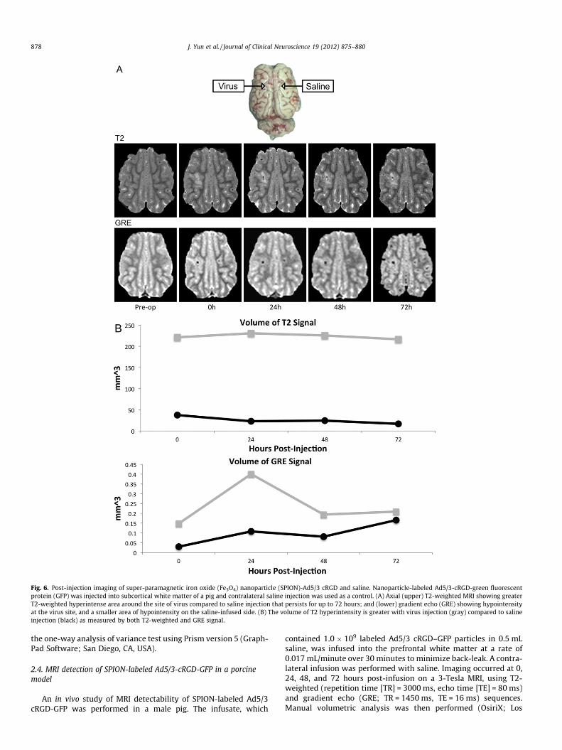
Fig. 6. Post-injection imaging of super-paramagnetic iron oxide (Fe3O4) nanoparticle (SPION)-Ad5/3 cRGD and saline. Nanoparticle-labeled Ad5/3-cRGD-green fluorescentprotein (GFP) was injected into subcortical white matter of a pig and contralateral saline injection was used as a control. (A) Axial (upper) T2-weighted MRI showing greaterT2-weighted hyperintense area around the site of virus compared to saline injection that persists for up to 72 hours; and (lower) gradient echo (GRE) showing hypointensityat the virus site, and a smaller area of hypointensity on the saline-infused side. (B) The volume of T2 hyperintensity is greater with virus injection (gray) compared to salineinjection (black) as measured by both T2-weighted and GRE signal.
878 J. Yun et al. / Journal of Clinical Neuroscience 19 (2012) 875–880
the one-way analysis of variance test using Prism version 5 (Graph-Pad Software; San Diego, CA, USA).
2.4. MRI detection of SPION-labeled Ad5/3-cRGD-GFP in a porcinemodel
An in vivo study of MRI detectability of SPION-labeled Ad5/3cRGD-GFP was performed in a male pig. The infusate, which
contained 1.0 � 109 labeled Ad5/3 cRGD–GFP particles in 0.5 mLsaline, was infused into the prefrontal white matter at a rate of0.017 mL/minute over 30 minutes to minimize back-leak. A contra-lateral infusion was performed with saline. Imaging occurred at 0,24, 48, and 72 hours post-infusion on a 3-Tesla MRI, using T2-weighted (repetition time [TR] = 3000 ms, echo time [TE] = 80 ms)and gradient echo (GRE; TR = 1450 ms, TE = 16 ms) sequences.Manual volumetric analysis was then performed (OsiriX; Los
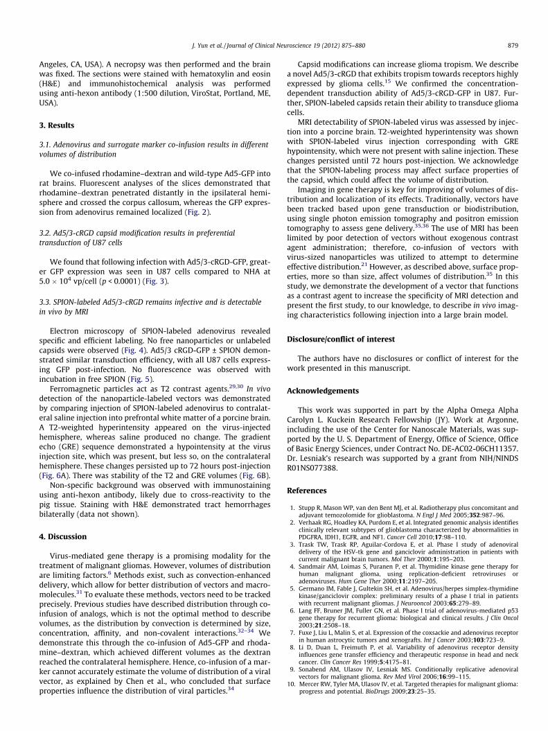
J. Yun et al. / Journal of Clinical Neuroscience 19 (2012) 875–880 879
Angeles, CA, USA). A necropsy was then performed and the brainwas fixed. The sections were stained with hematoxylin and eosin(H&E) and immunohistochemical analysis was performedusing anti-hexon antibody (1:500 dilution, ViroStat, Portland, ME,USA).
3. Results
3.1. Adenovirus and surrogate marker co-infusion results in differentvolumes of distribution
We co-infused rhodamine–dextran and wild-type Ad5-GFP intorat brains. Fluorescent analyses of the slices demonstrated thatrhodamine–dextran penetrated distantly in the ipsilateral hemi-sphere and crossed the corpus callosum, whereas the GFP expres-sion from adenovirus remained localized (Fig. 2).
3.2. Ad5/3-cRGD capsid modification results in preferentialtransduction of U87 cells
We found that following infection with Ad5/3-cRGD-GFP, great-er GFP expression was seen in U87 cells compared to NHA at5.0 � 104 vp/cell (p < 0.0001) (Fig. 3).
3.3. SPION-labeled Ad5/3-cRGD remains infective and is detectablein vivo by MRI
Electron microscopy of SPION-labeled adenovirus revealedspecific and efficient labeling. No free nanoparticles or unlabeledcapsids were observed (Fig. 4). Ad5/3 cRGD-GFP ± SPION demon-strated similar transduction efficiency, with all U87 cells express-ing GFP post-infection. No fluorescence was observed withincubation in free SPION (Fig. 5).
Ferromagnetic particles act as T2 contrast agents.29,30 In vivodetection of the nanoparticle-labeled vectors was demonstratedby comparing injection of SPION-labeled adenovirus to contralat-eral saline injection into prefrontal white matter of a porcine brain.A T2-weighted hyperintensity appeared on the virus-injectedhemisphere, whereas saline produced no change. The gradientecho (GRE) sequence demonstrated a hypointensity at the virusinjection site, which was present, but less so, on the contralateralhemisphere. These changes persisted up to 72 hours post-injection(Fig. 6A). There was stability of the T2 and GRE volumes (Fig. 6B).
Non-specific background was observed with immunostainingusing anti-hexon antibody, likely due to cross-reactivity to thepig tissue. Staining with H&E demonstrated tract hemorrhagesbilaterally (data not shown).
4. Discussion
Virus-mediated gene therapy is a promising modality for thetreatment of malignant gliomas. However, volumes of distributionare limiting factors.6 Methods exist, such as convection-enhanceddelivery, which allow for better distribution of vectors and macro-molecules.31 To evaluate these methods, vectors need to be trackedprecisely. Previous studies have described distribution through co-infusion of analogs, which is not the optimal method to describevolumes, as the distribution by convection is determined by size,concentration, affinity, and non-covalent interactions.32–34 Wedemonstrate this through the co-infusion of Ad5-GFP and rhoda-mine–dextran, which achieved different volumes as the dextranreached the contralateral hemisphere. Hence, co-infusion of a mar-ker cannot accurately estimate the volume of distribution of a viralvector, as explained by Chen et al., who concluded that surfaceproperties influence the distribution of viral particles.34
Capsid modifications can increase glioma tropism. We describea novel Ad5/3-cRGD that exhibits tropism towards receptors highlyexpressed by glioma cells.15 We confirmed the concentration-dependent transduction ability of Ad5/3-cRGD-GFP in U87. Fur-ther, SPION-labeled capsids retain their ability to transduce gliomacells.
MRI detectability of SPION-labeled virus was assessed by injec-tion into a porcine brain. T2-weighted hyperintensity was shownwith SPION-labeled virus injection corresponding with GREhypointensity, which were not present with saline injection. Thesechanges persisted until 72 hours post-injection. We acknowledgethat the SPION-labeling process may affect surface properties ofthe capsid, which could affect the volume of distribution.
Imaging in gene therapy is key for improving of volumes of dis-tribution and localization of its effects. Traditionally, vectors havebeen tracked based upon gene transduction or biodistribution,using single photon emission tomography and positron emissiontomography to assess gene delivery.35,36 The use of MRI has beenlimited by poor detection of vectors without exogenous contrastagent administration; therefore, co-infusion of vectors withvirus-sized nanoparticles was utilized to attempt to determineeffective distribution.21 However, as described above, surface prop-erties, more so than size, affect volumes of distribution.35 In thisstudy, we demonstrate the development of a vector that functionsas a contrast agent to increase the specificity of MRI detection andpresent the first study, to our knowledge, to describe in vivo imag-ing characteristics following injection into a large brain model.
Disclosure/conflict of interest
The authors have no disclosures or conflict of interest for thework presented in this manuscript.
Acknowledgements
This work was supported in part by the Alpha Omega AlphaCarolyn L. Kuckein Research Fellowship (JY). Work at Argonne,including the use of the Center for Nanoscale Materials, was sup-ported by the U. S. Department of Energy, Office of Science, Officeof Basic Energy Sciences, under Contract No. DE-AC02-06CH11357.Dr. Lesniak’s research was supported by a grant from NIH/NINDSR01NS077388.
References
1. Stupp R, Mason WP, van den Bent MJ, et al. Radiotherapy plus concomitant andadjuvant temozolomide for glioblastoma. N Engl J Med 2005;352:987–96.
2. Verhaak RG, Hoadley KA, Purdom E, et al. Integrated genomic analysis identifiesclinically relevant subtypes of glioblastoma characterized by abnormalities inPDGFRA, IDH1, EGFR, and NF1. Cancer Cell 2010;17:98–110.
3. Trask TW, Trask RP, Aguilar-Cordova E, et al. Phase I study of adenoviraldelivery of the HSV-tk gene and ganciclovir administration in patients withcurrent malignant brain tumors. Mol Ther 2000;1:195–203.
4. Sandmair AM, Loimas S, Puranen P, et al. Thymidine kinase gene therapy forhuman malignant glioma, using replication-deficient retroviruses oradenoviruses. Hum Gene Ther 2000;11:2197–205.
5. Germano IM, Fable J, Gultekin SH, et al. Adenovirus/herpes simplex-thymidinekinase/ganciclovir complex: preliminary results of a phase I trial in patientswith recurrent malignant gliomas. J Neurooncol 2003;65:279–89.
6. Lang FF, Bruner JM, Fuller GN, et al. Phase I trial of adenovirus-mediated p53gene therapy for recurrent glioma: biological and clinical results. J Clin Oncol2003;21:2508–18.
7. Fuxe J, Liu L, Malin S, et al. Expression of the coxsackie and adenovirus receptorin human astrocytic tumors and xenografts. Int J Cancer 2003;103:723–9.
8. Li D, Duan L, Freimuth P, et al. Variability of adenovirus receptor densityinfluences gene transfer efficiency and therapeutic response in head and neckcancer. Clin Cancer Res 1999;5:4175–81.
9. Sonabend AM, Ulasov IV, Lesniak MS. Conditionally replicative adenoviralvectors for malignant glioma. Rev Med Virol 2006;16:99–115.
10. Mercer RW, Tyler MA, Ulasov IV, et al. Targeted therapies for malignant glioma:progress and potential. BioDrugs 2009;23:25–35.

880 J. Yun et al. / Journal of Clinical Neuroscience 19 (2012) 875–880
11. Paul CP, Everts M, Glasgow JN, et al. Characterization of infectivity of knob-modified adenoviral vectors in glioma. Cancer Biol Ther 2008;7:786–93.
12. Tyler MA, Ulasov IV, Borovjagin A, et al. Enhanced transduction of malignantglioma with a double targeted Ad5/3-RGD fiber-modified adenovirus. MolCancer Ther 2006;5:2408–16.
13. Ulasov IV, Rivera AA, Han Y, et al. Targeting adenovirus to CD80 and CD86receptors increases gene transfer efficiency to malignant glioma cells. JNeurosurg 2007;107:617–27.
14. Ulasov IV, Tyler MA, Han Y, et al. Novel recombinant adenoviral vector thattargets the interleukin-13 receptor alpha2 chain permits effective gene transferto malignant glioma. Hum Gene Ther 2007;18:118–29.
15. Zheng S, Ulasov IV, Han Y, et al. Fiber-knob modifications enhance adenoviraltropism and gene transfer in malignant glioma. J Gene Med 2007;9:151–60.
16. Steck PA, Moser RP, Bruner JM, et al. Altered expression and distribution ofheparan sulfate proteoglycans in human gliomas. Cancer Res1989;49:2096–103.
17. Maenpaa A, Junnikkala S, Hakulinen J, et al. Expression of complementmembrane regulators membrane cofactor protein (CD46), decay acceleratingfactor (CD55), and protectin (CD59) in human malignant gliomas. Am J Pathol1996;148:1139–52.
18. Barker 2nd FG, Chang SM, Gutin PH, et al. Survival and functional status afterresection of recurrent glioblastoma multiforme. Neurosurgery 1998;42:709–20.discussion 20–3.
19. Lopez KA, Waziri AE, Canoll PD, et al. Convection-enhanced delivery in thetreatment of malignant glioma. Neurol Res 2006;28:542–8.
20. Fiandaca MS, Varenika V, Eberling J, et al. Real-time MR imaging of adeno-associated viral vector delivery to the primate brain. Neuroimage 2009;47:T27–35.
21. Szerlip NJ, Walbridge S, Yang L, et al. Real-time imaging of convection-enhanced delivery of viruses and virus-sized particles. J Neurosurg2007;107:560–7.
22. Su X, Kells AP, Aguilar Salegio EA, et al. Real-time MR imaging with gadoteridolpredicts distribution of transgenes after convection-enhanced delivery of AAV2vectors. Mol Ther 2010;18:1490–5.
23. Laurent S, Bridot JL, Elst LV, et al. Magnetic iron oxide nanoparticles forbiomedical applications. Future Med Chem 2010;2:427–49.
24. Mahmoudi M, Hosseinkhani H, Hosseinkhani M, et al. Magnetic resonanceimaging tracking of stem cells in vivo using iron oxide nanoparticles as a tool
for the advancement of clinical regenerative medicine. Chem Rev2011;111:253–80.
25. Antonelli A, Sfara C, Manuali E, et al. Encapsulation of superparamagneticnanoparticles into red blood cells as new carriers of MRI contrast agents.Nanomedicine (Lond) 2011;6:211–23.
26. Yallapu MM, Othman SF, Curtis ET, et al. Multi-functional magneticnanoparticles for magnetic resonance imaging and cancer therapy.Biomaterials 2011;32:1890–905.
27. Huh YM, Lee ES, Lee JH, et al. Hybrid nanoparticles for magneticresonance imaging of target-specific viral gene delivery. Adv Mater2007;19:3109–12.
28. Sun S, Zeng H, Robinson DB, et al. Monodisperse MFe2O4 (M = Fe, Co, Mn)nanoparticles. J Am Chem Soc 2004;126:273–9.
29. Yang H, Zhuang Y, Sun Y, et al. Targeted dual-contrast T(1)- and T(2)-weightedmagnetic resonance imaging of tumors using multifunctional gadolinium-labeled superparamagnetic iron oxide nanoparticles. Biomaterials2011;32:4584–93.
30. Raty JK, Liimatainen T, Unelma Kaikkonen M, et al. Non-invasive imaging ingene therapy. Mol Ther 2007;15:1579–86.
31. Bobo RH, Laske DW, Akbasak A, et al. Convection-enhanced delivery ofmacromolecules in the brain. Proc Natl Acad Sci U S A 1994;91:2076–80.
32. Kroll RA, Pagel MA, Muldoon LL, et al. Increasing volume of distribution to thebrain with interstitial infusion: dose, rather than convection, might be the mostimportant factor. Neurosurgery 1996;38:746–52.
33. Chen MY, Lonser RR, Morrison PF, et al. Variables affecting convection-enhanced delivery to the striatum: a systematic examination of rate ofinfusion, cannula size, infusate concentration, and tissue-cannula sealingtime. J Neurosurg 1999;90:315–20.
34. Chen MY, Hoffer A, Morrison PF, et al. Surface properties, more than size,limiting convective distribution of virus-sized particles and viruses in thecentral nervous system. J Neurosurg 2005;103:311–9.
35. Li GC, He F, Ling CC. Hyperthermia and gene therapy: potential use of microPETimaging. Int J Hyperthermia 2006;22:215–21.
36. Penuelas I, Boan J, Marti-Climent JM, et al. Positron emission tomography andgene therapy: basic concepts and experimental approaches for in vivo geneexpression imaging. Mol Imaging Biol 2004;6:225–38.