From Benzofurans to Indoles: Palladium-Catalyzed Reductive ...

IOP PUBLISHING NANOTECHNOLOGY
Nanotechnology 19 (2008) 185608 (6pp) doi:10.1088/0957-4484/19/18/185608
Superparamagnetic bimetalliciron–palladium nanoalloy: synthesis andcharacterizationRabia Nazir1, Muhammad Mazhar1, M Javed Akhtar2,M Raza Shah3, Nawazish A Khan4, M Nadeem2,Muhammad Siddique2, Mazhar Mehmood5 and N M Butt6
1 Department of Chemistry, Quaid-i-Azam University, Islamabad 45320, Pakistan2 Physics Division, PINSTECH, PO Nilore, Islamabad 44000, Pakistan3 HEJ Research Institute of Chemistry, University of Karachi, Karachi 75270, Pakistan4 Material Science Laboratory, Department of Physics, Quaid-i-Azam University,Islamabad 45320, Pakistan5 National Centre for Nanotechnology, PIEAS, Islamabad 45650, Pakistan6 Pakistan Science Foundation, Islamabad 44000, Pakistan
E-mail: [email protected]
Received 28 August 2007, in final form 23 January 2008Published 1 April 2008Online at stacks.iop.org/Nano/19/185608
AbstractIron–palladium nanoalloy in the particle size range of 15–30 nm is synthesized by the relativelylow temperature thermal decomposition of coprecipitated [Fe(Bipy)3]Cl2 and [Pd(Bipy)3]Cl2 inan inert ambient of dry argon gas. The silvery black Fe–Pd alloy nanoparticles are air-stable andhave been characterized by EDX-RF, XRD, AFM, TEM, magnetometry, 57Fe Mossbauer andimpedance spectroscopy. This Fe–Pd nanoalloy is in single phase and contains iron sites havingup to 11 nearest-neighboring atoms. It is superparamagnetic in nature with high magneticsusceptibility, low coercivity and hyperfine field.
1. Introduction
Bimetallic nanoparticles are of great significance becauseof the modification of properties observed, compared withmonometallics, not only due to size effects, but also as aresult of the combination of different metals, either as analloy [1] or as a core–shell structure [2, 3]. Catalytic [4],electronic [5], magnetic [6] and optical properties [2] of thebimetallic nanoparticles have been tailored compared withmonometallics. Increased mechanical strength, enhanceddiffusivity, higher specific heat and electrical resistivity havealso been observed [7]. Bimetallic nanoparticles havemajor technological applications in information storage [8]and catalysis [9] which place them on the frontiers ofadvanced materials chemistry. For this purpose, varioussynthetic routes have been approached to obtain bimetallicnanoparticles [10, 11], each of them yielding products withdifferent physicochemical and structural characteristics.
Nanopowders of the Pd-rich Fe–Pd alloys have beensynthesized by ultrasound-assisted electrochemical methods
and pyrolysis of organometallic precursors, the resulting alloysare in the fcc phase with particle sizes ranging between6–11 nm and 5–10 nm, respectively [12]. Severe plasticdeformation and cyclic code formation were used to preparebulk magnetic nanocomposites of ordered L10 Fe–Pd phaseand α-Fe following an atomic ordering and precipitationreaction [13]. Combinations of different stabilizers witholeic acid have been applied to prepare Fe28Pd72 andFe50Pd50 nanoparticles, in the size range 11–16 nm, by thepolyol reduction of Pd(acac)2 and thermal decomposition ofFe(CO)5, respectively. The size control of nanoparticlesis achieved by controlling the concentration and type ofstabilizers and reaction conditions [14, 15]. Nanopowderedalloys of Pd100−xFex , where x = 5.4, 6, 8 and 12,of 5 nm radius have been prepared by ultrasound-assistedelectrochemistry [16]. Recently, synthesis of the orderedalloy nanoparticles, based on chemical techniques, has beenactively developed [17]. Fe–Pd alloy films are considered tobe highly promising shape memory alloys [18] in additionto their remarkable role as hydrogen storage materials [19]
0957-4484/08/185608+06$30.00 © 2008 IOP Publishing Ltd Printed in the UK1
Nanotechnology 19 (2008) 185608 R Nazir et al
and catalysts [20, 21]. Nanoscale bimetallic particles ofFe–Pd have also been used for the treatment of chlorinatedorganic pollutants [22]. Controlling the structure andchemical ordering of bimetallic nanoalloys can be the startingpoint to prepare the building blocks for specifically tailoredcluster-assembled materials [23]. Synthesis of nanoparticles,characterized by a narrow size distribution, is a new challengein solid-state chemistry [24].
As reported in the literature [4, 25] the particle size andthe properties of nanoparticles depend strongly on the specificmethod of preparation and the applied experimental conditions.Hence, there is a need to design a simple and economicalsynthetic route that enables narrow size distribution.
The main purpose of this work is thus twofold: the firstis to develop a simple chemical method for the synthesis of aniron–palladium nanoalloy and secondly to glean informationabout the different electrical and magnetic properties of theiron–palladium system at nanoscale for various technologicalapplications.
2. Experimental details
2.1. Synthesis
All the reagents and chemicals used were of analytical gradeand purchased from Sigma-Aldrich except hydrated ferrouschloride (FeCl2·4H2O) and Triton-X-100, which were suppliedby Merck and Fluka, respectively.
2.1.1. [Fe(Bipy)3]Cl2. 1.98 g (0.1 mol) hydrated ferrouschloride (FeCl2·4H2O) was dissolved in 100 ml of dried THFand placed in a 250 ml round bottomed flask fitted with adropping funnel, reflux condenser and inert gas line. 0.5 mlof Triton-X-100 was added as a capping agent and the solutionwas stirred for 1 h at room temperature to get a homogeneoussolution. 4.71 g (0.6 mol) 2,2′-bipyridin in 50 ml driedTHF was added through a dropping funnel at a drop rate of42 drops h−1 with constant stirring for 24 h. The yellowprecipitates obtained were allowed to settle and filtered througha sintered glass filtering apparatus. The precipitates werewashed several times with THF until free from 2,2′-bipyridinand ferrous chloride. The complex was crystallized frommethanol solution in air. Yield = 86%. m.p. = 168 ◦C; IR(KBr, cm−1): ν (C–Harom) 3050, 2927; ν (C–Narom) 1599;ν (C–Carom) 1493, 1443, 900–600; ν (Fe–N) 416. 1H-NMR (DMSO, ppm): 8.440 (s, 6H, bipyH6,6′); 8.220 (s, 6H,bipyH3,3′); 7.797 (s, 6H, bipyH4,4′); 7.299 (s, 6H, bipyH5,5′).TGA: 187–237 ◦C (22.32% weight loss), 331–449 ◦C (28.22%residual mass).
2.1.2. [Pd(Bipy)3]Cl2. This was synthesized by theprocedure given above for trisbipyridineiron(II) chlorideexcept that 2-propanol was used as solvent. The reactants usedare as follows: 1.77 g (0.1 mol) palladium chloride (PdCl2) and4.71 g (0.6 mol) 2,2′-bipyridin. The grayish brown precipitateswere obtained in yield = 80%. Decomp. = 386.9 ◦C. IR(KBr, cm−1): ν (C–Harom) 3048, 2924; ν (C–Narom) 1601;ν (C–Carom) 1499, 1450, 900–600; ν (Pd–N) 414. 1H-NMR
(DMSO, ppm): 9.09–9.11 (m, 6H2, bipyH6,6′); 8.55–8.58(m, 6H, bipyH3,3′); 8.32–8.37 (m, 6H, bipyH4,4′); 7.78–7.82(m, 6H, bipyH5,5′). TGA: 300–449 ◦C (52.01% residual mass).
2.1.3. Iron–palladium alloy. 3.07 g (4.6 mmol) [Fe(Bipy)3]Cl2 and 3.00 g (4.6 mmol) [Pd(Bipy)3]Cl2 were dissolvedin solution of 180 ml of distilled water and 20 ml of 33%ammonia solution. The solution was slowly evaporated withconstant stirring at 40 ◦C to get the homogeneous powderof both complexes. This powder was then taken into a U-tube containing an argon inlet and outlet and was heated in acube furnace under argon atmosphere. The temperature of thefurnace was raised to 500 ◦C at a heating rate of 0.5 ◦C min−1
and the contents were kept at 500 ◦C for another 24 h, thenallowed to cool to room temperature under an inert atmosphereof argon gas to yield Fe–Pd (1:1) alloy. Yield 85%.
2.2. Characterization
The as-synthesized Fe–Pd nanoalloy was characterized byEDX-RF using a Horiba XRF Analyzer Mensa Bio100, Japan.The XRD data of Fe–Pd was collected, by a Bruker D8discover, Germany, at room temperature by step scanning overthe angular range of 20◦ � 2θ � 90◦ at a step size of0.05◦ and counting time of 6 s/step. The sizes and shapesof nanoparticles were analyzed using transmission electronmicroscopy (TEM) (JEOL 2010), operating at 200 kV andatomic force microscopy (AFM), (Multimode, Nanoscope IIIa,Veeco, CA) in tapping mode which confirmed the alloy to be inthe nanorange. The samples were prepared by spin-coating ofthe dispersion of the alloys in ethanol on a freshly cleaved sheetof mica substrate. AFM images were acquired with samplesimaged at room temperature and repeated several timeswith different concentrations of samples. Silicon cantilevers(Nanoworld, Switzerland; 240 μm long, 30 μm wide, 2.8 μmthick) with an integrated tip, a nominal spring constant of 0.7–3.8 N m−1, tip radius <10 nm and a resonance frequency of70 KHz were plasma-cleaned prior to use. A piezoelectricscanner (Veeco Instruments, CA) with a maximum x , y-scan range of 17.2 μm and a z extension of 3.916 μm wasused. Typically the tip was scanned at a velocity from 0.5 to1 μm s−1. Topography, phase and height images were usedto record the structures. Raw data were modified by applyingthe first-order ‘flatten’ filter in order to achieve scan linesat the same average height and average tilt. AC magneticsusceptibility measurements were taken using a Lock-in-Amplifier SR530 of Standard Research Systems at a lock-infrequency of 270 Hz. For magnetic hysteresis measurements,as-prepared Fe–Pd nanoalloy powder (10–20 mg) was packedin butter paper and measurements were taken at roomtemperature using a vibrating sample magnetometer (VSM).Room temperature Mossbauer measurements were carried outusing a 57Co (Rh-matrix) source, having 25 mCi initial activity,in transmission geometry after the confirmation of single-phase material by XRD. Data analysis was performed usingthe computer program Mos-90 [26], assuming that all thepeaks are Lorentzian in shape. Impedance studies have beenconducted on sintered pellets having 10 mm diameter and
2

Nanotechnology 19 (2008) 185608 R Nazir et al
Figure 1. Powder x-ray diffractogram of Fe–Pd nanoparticlesindicating a single-phase fcc structure.
1.5 mm thickness at room temperature using an Alpha-Nanalyzer (Novocontrol, Germany). Windeta software has beenused for data acquisition, which has been fully automated byinterfacing the analyzer with a PC. The ac amplitude signal thatwas used for these studies has been 1.0 V. Contacts were madeby silver paint on opposite sides of the pellets after cleaningboth surfaces properly.
3. Results
Fe–Pd (1:1) bimetallic alloy nanoparticles of size range15–30 nm were prepared by pyrolysis of coprecipitatedtrisbipyridineiron(II) chloride and trisbipyridinepalladium(II)chloride. The elemental ratio (1:1) was confirmed fromthe energy-dispersive x-ray (EDX) analysis. Powder XRDdata is presented in figure 1, where all peaks were indexedsuccessfully on the basis of a face centered cubic (fcc)structure. The lattice parameter obtained from the least squarefitting procedure is 3.858(1) A, which is in good agreementwith previous findings [12, 17].
The AFM height images of the (a) top view and (b) surfaceplot views of the Fe–Pd alloy are presented in figure 2. The
Figure 3. TEM image of the Fe–Pd nanoparticles with 27 500×magnification.
mean particle size is in the range 15–30 nm, calculated viacross-sectional analysis of the representative height images.The representative TEM plan view image (figure 3) shows Fe–Pd nanoparticles to be of a size ranging from 15 to 20 nm.The particles are well dispersed and separated from eachother, with spherical shape and approximately uniform size,which demonstrates good control over nanoparticle size andmorphology. The TEM and AFM comparison shows the meandiameter obtained from AFM is larger than that of TEM, whichcan be attributed to the tip convolution [27].
A magnetic susceptibility, χ , versus temperature plot forthe Fe–Pd alloy nanoparticles is presented in figure 4a whichshows a substantial increase in χ below 290 K, indicating theonset of magnetic ordering with the lowering of temperature.After attaining the maximum value at TB, i.e. 178 K, withbroad maxima, it rapidly falls off, which is characteristic ofsuperparamagnetic behavior [28]. The plot also features asmall increase in χ at low temperature, i.e. around 104 K up toliquid N2 temperature owing to the role played by the domainboundary exchange interaction. The alloy residing in the fccplane consists of alternating Fe and Pd atomic planes, similarto the tetragonal structure as reported earlier [29]. Pd being a4d transition metal is non-ferromagnetic and does not exhibit
Figure 2. AFM images of the Fe–Pd nanoparticles deposited on mica substrate: (a) top view, (b) 3D view.
(This figure is in colour only in the electronic version)
3

Nanotechnology 19 (2008) 185608 R Nazir et al
Figure 4a. Magnetic susceptibility, χ , measurements with variabletemperature, T (K), showing superparamagnetic behavior.
a magnetic moment, but when alloyed with the ferromagnetictransition metals (e.g. Fe) it shows a magnetic moment [30].On account of its large atomic number Pd is easily polarizedwhen placed in the vicinity of Fe and gives rise to large spin–orbit effects [31]. When more than half the filled orbitals oftwo transition metals, like Fe and Pd, hybridize, their momentscouple parallel to each other [32]. The magnetism in thesematerials is said to be governed by interatomic interactionsbetween 3d and 4d electrons [33]. At the start of magneticordering, the spins of Fe and Pd start aligning parallel toeach other, resulting in enhanced magnetization and increasein χ , until TB, where it attains the maximum value. Asa consequence of the decrease in temperature, the particlesstart magnetically blocking. Further lowering the temperaturebelow TB leads to magnetically frozen particles which isaccompanied by an increase in the magnetic anisotropy. Asa result of this large magnetic anisotropic energy, particlestend to align antiferromagnetically along the easy axis ofmagnetization [34]. The large magnetic anisotropy destabilizesthe system which, in order to stabilize, tries to attain thelow energy state with opposite spins resulting in lowering ofχ . During the magnetic ordering process, magnetic domainsform as a result of coalescence of the particles, which alsolowers the energy of the system. The minimum shown at104 K corresponds to the magnetic moments of the alternatingFe and Pd atoms oriented exactly in opposite directions. Asmall increase in χ , after that point, results from the domainboundaries. At such a low temperature, effects from bothferromagnetic and paramagnetic components vanish and thusthe resultant increase in χ is just due to the influence of Fe andPd present in domain boundaries.
Superparamagnetic behavior of the Fe–Pd nanoparticleswas also confirmed by hysteresis loop studies. Insuperparamagnetic particles the anisotropy barrier of a singleparticle can be easily surmounted by the thermal energy,resulting in zero net magnetization in the absence of anapplied magnetic field. The room temperature hysteresis loopof one such representative sample is shown in figure 4b.This data has shown some coercivity (45.98 Oe) whichapproaches slowly to a saturation value suggesting the
Figure 4b. Magnetization, M (emu g−1), measurements withvariable field, H (Oe), of Fe–Pd nanoalloys showingsuperparamagnetic behavior.
Figure 5. 57Fe Mossbauer spectra of Fe–Pd nanoalloy recorded atroom temperature showing a sextet and a doublet corresponding toferromagnetic and superparamagnetic components, respectively.
existence of superparamagnetism with moderate particlesunblocking [14, 35]. Such behavior of a very small coercivityis a fingerprint of a non-deformed Fe–Pd phase, the coercivityof which can be enhanced by carrying out post-annealing upto 550 ◦C [12]. The magnetic behavior expected for single-domain Fe–Pd nanoparticles had an estimated particle sizeof about 35 nm, which is comparable with the particle sizeobserved in the present studies [16].
Magnetic properties investigated by 57Fe Mossbauerspectroscopy have also shown the increase in magneticmoments of Fe with neighboring Pd atoms. In addition tothat, the 57Fe hyperfine magnetic field (HMF) increases withincreasing Pd concentration [36]. The Mossbauer spectrumof FePd nanoparticles (figure 5), comprising a magneticsextet and a doublet, indicates the presence of a mixtureof ferromagnetic and superparamagnetic components. Thepresence of a quadrupole doublet, with an isomer shift of0.22 mm s−1 and quadrupole splitting of 0.8 mm s−1, ischaracteristic of the presence of small superparamagneticparticles above their blocking temperature [37]. The magneticsextet is typical of the Fe–Pd alloy fine particles below their
4
Nanotechnology 19 (2008) 185608 R Nazir et al
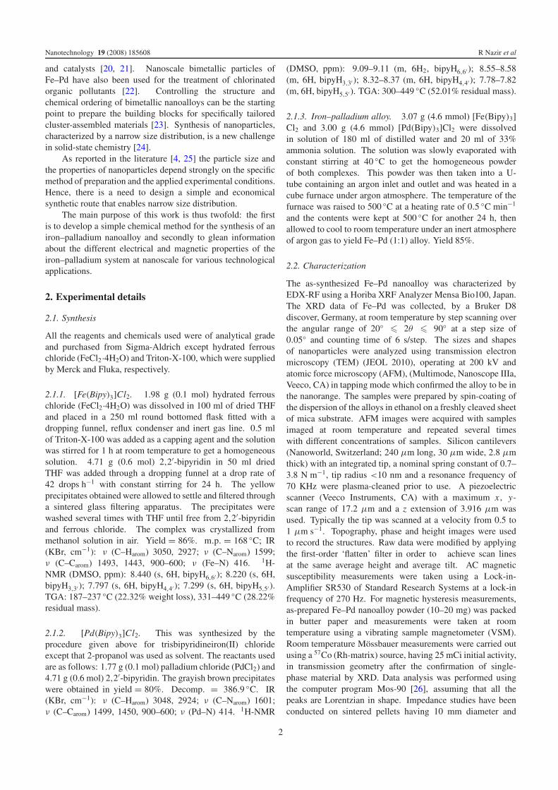
Figure 6. Complex impedance plane plot of Fe–Pd alloy at roomtemperature. Squares are experimental data and the line is a fit, usingan equivalent circuit model –RC–.
blocking temperature. The hyperfine magnetic field (38.7 T)is somewhat high as reported in earlier studies. The linebroadening (linewidth = 1.09 mm s−1 as compared to thenormal linewidth = 0.3–0.4 mm s−1 in our system) is due tothe particle size distribution in the sample. The mean HMF〈Hhf〉 of epitaxial Fe50Pd50 thin films was 34.3 T at 300 Kand 36.8 T at 20 K [38] and appeared significantly largerthan the values reported for Fe50Pd50 [39–41] which are 32 Tat 300 K and 35 T extrapolated at 0 K. However, it wasshown that in disordered fcc Fe–Pd with 25–75 mol% ironconcentration, the HMF distribution can be analyzed assumingdifferent configurations of the immediate environment of theresonance atom. The change in hyperfine field (�Hhf) in thefirst coordination sphere of the 57Fe is 1.6 T per iron atom. Alinear relationship between Hhf and the number of Fe nearestneighbors results in the hyperfine field of 35 T at 300 K forFe0.75Pd0.25. The observation of hyperfine field values as largeas 40 T points out the presence of iron sites with up to 12nearest neighbors [38]. In our sample the Hhf value of 38.7 Tindicates the occurrence of about 11 iron atoms as nearestneighbors.
Figure 6 illustrates a typical Fe–Pd impedance plane plotbetween Z ′ and Z ′′, which are real and imaginary parts ofthe impedance, respectively (Z = Z ′ + jZ ′′) [42]. It showsapparently a perfect semicircle with its center very close to thereal axis having a 1.7◦ dispersion angle, indicating the highquality of the sample and the absence of any disorder in theFe–Pd nanoalloy. The semicircles intersect the Z ′ axis onthe right-hand side, giving a value of resistance correspondingto the dc values whereas the extension of the semicircle onthe left-hand side passes through the origin. To analyze theimpedance spectrum, one unit –RC– equivalent circuit modelhas been employed and the fitting is shown in figure 6. Thefitting parameter, R, indicating the resistance of the Fe–Pdgrain is of the order of 9.65 × 10⁵ � and the value of thecapacitance, C , is 4.30 × 10⁻¹¹ F. Impedance results suggestthat only one type of grain is present, which is the Fe–Pdphase. In order to confirm this finding the electric modulus
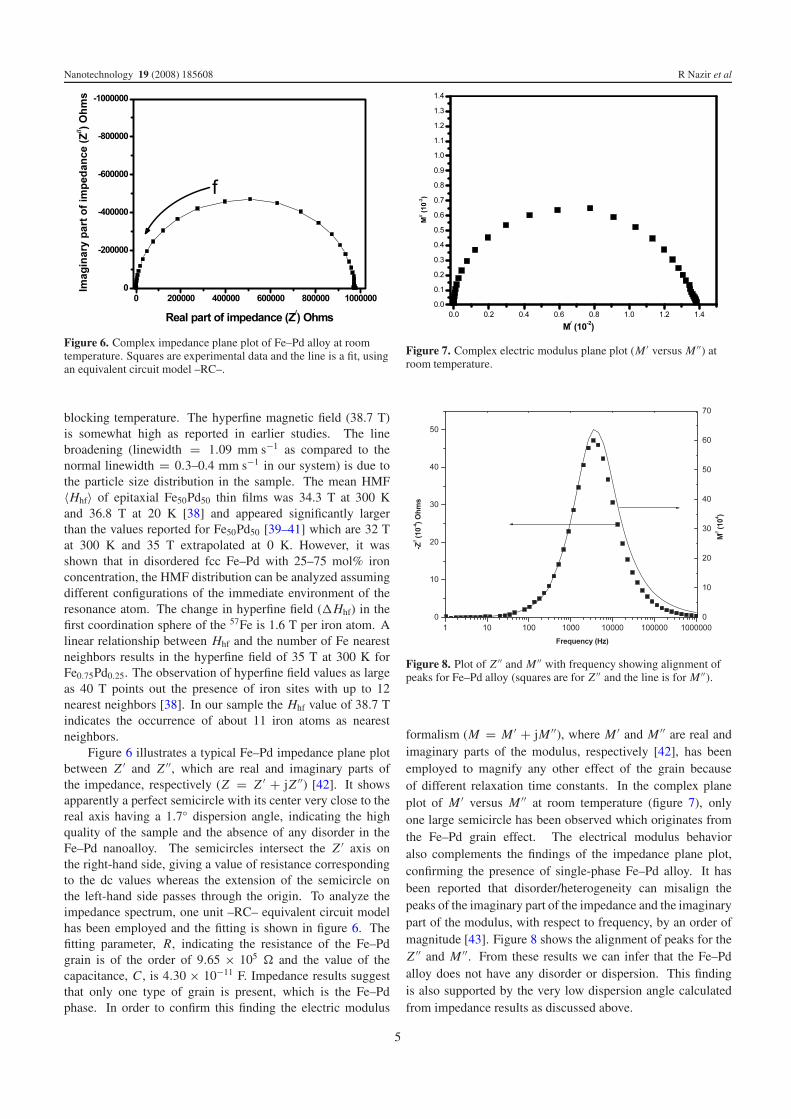
Figure 7. Complex electric modulus plane plot (M ′ versus M ′′) atroom temperature.
Figure 8. Plot of Z ′′ and M ′′ with frequency showing alignment ofpeaks for Fe–Pd alloy (squares are for Z ′′ and the line is for M ′′).
formalism (M = M ′ + jM ′′), where M ′ and M ′′ are real andimaginary parts of the modulus, respectively [42], has beenemployed to magnify any other effect of the grain becauseof different relaxation time constants. In the complex planeplot of M ′ versus M ′′ at room temperature (figure 7), onlyone large semicircle has been observed which originates fromthe Fe–Pd grain effect. The electrical modulus behavioralso complements the findings of the impedance plane plot,confirming the presence of single-phase Fe–Pd alloy. It hasbeen reported that disorder/heterogeneity can misalign thepeaks of the imaginary part of the impedance and the imaginarypart of the modulus, with respect to frequency, by an order ofmagnitude [43]. Figure 8 shows the alignment of peaks for theZ ′′ and M ′′. From these results we can infer that the Fe–Pdalloy does not have any disorder or dispersion. This findingis also supported by the very low dispersion angle calculatedfrom impedance results as discussed above.
5

Nanotechnology 19 (2008) 185608 R Nazir et al
4. Conclusion
The coprecipitated trisbipyridineiron(II) chloride and tris-bipyridinepalladium(II) chloride undergo pyrolysis at 500 ◦Cin an inert atmosphere of argon gas to yield an air-stable,single-phase Fe–Pd (1:1) nanoalloy with particle size range15–30 nm as measured by TEM and AFM. The alloy hasan fcc structure and is superparamagnetic in nature, show-ing enhanced magnetic susceptibility, low coercivity and hy-perfine field. 57Fe Mossbauer measurements confirm that thealloy contains iron sites having up to 11 nearest neighbors.Impedance measurements confirm the absence of any struc-tural disorder in the alloy.
Acknowledgment
RN and MM acknowledge the Higher Education Commissionof Pakistan for financial support through the IndigenousScholarship Scheme for PhD studies in Science andTechnology (300 Sch.).
References
[1] Ennas G, Falqui A, Marras S, Sangregorio C andMorangiu G 2004 Chem. Mater. 16 5659–63
[2] Rodrıguez-Gonzalez B, Burrows A, Watanabe M, Kiely C J andMarzan L M L 2005 J. Mater. Chem. 15 1755–9
[3] Pingali K C, Deng S and Rockstraw D A 2007 Chem. Eng.Commun. 194 780–6
[4] Hernandez-Fernandez P, Rojas S, Ocon P, Gomez de laFuente J L, Fabian J S, Sanza J, Pena M A,Garcıa-Garcıa F J, Terreros P and Fierro J L G 2007J. Phys. Chem. C 111 2913–23
[5] Mulvaney P 1996 Langmuir 12 788–800[6] Crisan O, Angelakeris M, Simeonidis K, Kehagias T,
Komninou P, Giersig M and Flevaris N K 2006 Acta Mater.54 5251–60
[7] Tjong S C and Haydn C 2004 Mater. Sci. Eng. R 45 1–88[8] Suda M, Nakagawa M, Iyoda T and Einaga Y 2007 J. Am.
Chem. Soc. 129 5538–43[9] Pronkin S N, Simonov P A, Zaikovskii V and Savinova E R
2007 J. Mol. Catal. A 265 141–7[10] Cheng G and Walker A R H 2007 J. Magn. Magn. Mater.
311 31–5[11] Basnayake R, Li Z, Katar S, Zhou W, Rivera H, Smotkin E S,
Casadonte D J and Korzeniewski C 2006 Langmuir22 10446–50
[12] Qadri S B, Keller T M, Little C A and Lubitz P 2005Appl. Phys. A 81 587–90
[13] Saha S, Kulovits A, Soffa W A and Barnard J A 2005 J. Appl.Phys. 97 10F301
[14] Hou Y, Kondoh H, Kogure T and Ohta T 2006 Chem. Mater.16 5149–52
[15] Chen M and Nikles D E 2002 J. Appl. Phys. 91 8477–9[16] Guzman M, Delplancke J, Long G J, Delwiche J,
Hubin-Franskin M and Grandjean F 2002 J. Appl. Phys.92 2634–40
[17] Watanabe K, Kura H and Sato T 2006 Sci. Technol. Adv. Mater.7 145–59
[18] Wang F, Doi S, Hosoiri K, Yoshida H, Kuzushima T,Sasadaira M and Watanabe T 2006 Electrochim. Acta51 4250–4
[19] Gonzalez E A, Jasen P V, Castellani N J and Juan A 2004Solid State Commun. 131 81–5
[20] Bachir R, Marecot P, Didillon B and Barbier J 1997Appl. Catal. A 164 313–22
[21] Felicissimo M P, Martyanov O N, Risse T and Freund H 2007Surf. Sci. 601 2105–16
[22] Liu Y, Yang F, Yue P and Chen G 2001 Water Res. 35 1887–90[23] Wu H, Cao Y, Yuan P, Xu H and Wei X 2005 Chem. Phys. Lett.
406 148–53[24] Bradley J S, Tesche B, Busser W, Maase M and Reetz M T
2000 J. Am. Chem. Soc. 122 4631–6[25] Elkins K E, Vedantam T S, Liu J P, Zeng H, Sun S, Ding Y and
Wang Z L 2003 Nano Lett. 3 1647–9[26] Große G 1992 Mos-90 Version 2.2 Manual and Program.
Documentation 2nd edn, March[27] Rasa M and Philipse A P 2002 J. Magn. Magn. Mater.
252 101–3[28] Chakarbarti S, Mandal S K and Chardhuri S 2005
Nanotechnology 16 506–11[29] Clavero C, Garcıa-Martın J M, Armelles G, Cebollada A,
Huttel Y, Estrades S, Arbiol J, Peiro F and Balcells L 2006J. Appl. Phys. 99 073903
[30] Hamad B A and Khalifeh J M 2001 Surf. Sci. 481 33–8[31] MacLaren J M, Duplessis R R, Stern R A and
Willoughby S 2005 IEEE Trans. Magn. 41 4374–9[32] Vogel J, Fontaine A, Cros V, Petroff F, Kappler J, Krill G,
Rogalev A and Goulon J 1997 J. Magn. Magn. Mater.165 96–9
[33] Gross K D, Riegel D and Zeller R 1990 Phys. Rev. Lett.65 3044–7
[34] Li D S, Garmestani H, Yan S, Elawani M B,Schneider-Muntau H J, Liu J P, Saha S and Barnard J A2004 J. Magn. Magn. Mater. 281 272–5
[35] Kang S, Jia Z, Nikles D E and Harrell J W 2004 J. Appl. Phys.95 6744–6
[36] Birsan M, Fultz B and Anthony L 1997 Phys. Rev. B55 11502–6
[37] An S Y, Shim I and Kim C S 2005 J. Appl. Phys. 97 10Q909[38] Gehanno V, Auric P, Marty A and Gilles B 1998 J. Magn.
Magn. Mater. 188 310–8[39] Longworth G 1968 Phys. Rev. 172 572–6[40] Tsurin V A, Yurchikov E E and Menshikov A Z 1976
Sov. Phys. Solid State 17 1942–5[41] Tsurin V A, Ermakov A E, Lebedev Y G and Filippov B N
1976 Phys. Status Solidi a 33 325–32[42] Mcdonald J R and Johnson W B 1987 Impedance
Spectroscopy: Emphasizing Solid Materials and Systemsed J R Mcdonald (New York: Wiley) p 5, 7
[43] Ramzan M and Brydson R 2005 Sensors Actuators A118 322–31
6
Copyright © 2022 FDOKUMEN