PALLADIUM, PLATINUM AND GOLD COMPLEXES - UJ IR
284
PALLADIUM, PLATINUM AND GOLD COMPLEXES: A SYNTHETIC APPROACH TOWARDS THE DISCOVERY OF ANTICANCER AGENTS FRANKLINE KIPLANGAT KETER Thesis submitted in fulfillment of the requirements for the degree Philosophiae Doctor in Chemistry in the Faculty of Science at the University of Johannesburg Supervisor: Professor James Darkwa Co-Supervisor: Professor D. Jasper G. Rees January 2008
-
Upload
khangminh22 -
Category
Documents
-
view
0 -
download
0
Transcript of PALLADIUM, PLATINUM AND GOLD COMPLEXES - UJ IR

PALLADIUM, PLATINUM AND GOLD COMPLEXES: A
SYNTHETIC APPROACH TOWARDS THE DISCOVERY OF
ANTICANCER AGENTS
FRANKLINE KIPLANGAT KETER
Thesis
submitted in fulfillment of the requirements for the degree
Philosophiae Doctor
in
Chemistry
in the
Faculty of Science
at the
University of Johannesburg
Supervisor: Professor James Darkwa
Co-Supervisor: Professor D. Jasper G. Rees
January 2008

ABSTRACT
Ligands bis(pyrazolyl)acetic acid (L1) and bis(3,5-dimethylpyrazolyl)acetic acid (L2)
were synthesised by reacting pyrazoles and dibromoacetic acid under phase transfer
conditions, by using benzyltriethylammonium chloride as the catalyst. Ligands L1 and
L2 were characterised by a combination of 1H, 13C{1H} NMR, IR spectroscopy and
microanalysis. Esterification of L1 and L2 led to formation of bis(pyrazolyl)ethyl acetate
(L3) and bis(3,5-dimethylpyrazolyl)ethyl acetate (L4). Ligands L3 and L4 were also
characterised by a combination of 1H, 13C{1H} NMR, IR spectroscopy and microanalysis.
Subsequently, new pyrazolyl palladium(II) and platinum(II) compounds, [PdCl2(L1)] (1),
[PdCl2(L2)] (2), [PtCl2(L1)] (3a) and [PtCl2(L2)] (4) were prepared by reacting
bis(pyrazolyl)acetic acid ligands (L1-L2) with K2[PdCl4] or K2[PtCl4] respectively. The
structures of complex 1 and 2 reveal distorted square planar geometries. The bond angles
of N-Pd-N, N-Pd-Cl, N-Pd-Cl, for 1 and 2 are between 85.8(3)o and 90.81(4)o). The
platinum compound, K2[Pt4Cl8(L1)2(deprotonated-L1)2].2H2O (3b), crystallised from
aqueous solutions containing 3a when such solutions were left to stand overnight. Each
platinum coordination environment consists of two cis-Cl ligands and one K2-N^N(L1) unit
(L1 = bis(pyrazolyl)acetic acid), with two ligand moieties in 3b that are deprotonated
with two K+ counter ions.
Reaction of bis(pyrazolyl)acetic acid ligands (L1-L2) with [HAuCl4].4H2O gave gold(III)
complexes [AuCl2(L1)]Cl (5a) and [AuCl2(L2)]Cl (6a). The spectroscopic, mass
spectroscopy and microanalysis data were used to confirm the formation of the desired
i
complexes. However, attempts to crystallise 5a and 6a led to formation of
[AuCl2(pz)(pzH)] (5b) and [AuCl2(3,5-Me2pz)(3,5-Me2pzH)] (6b). This was confirmed
by the structural characterisation of 5b, which has a distorted square-planar geometry.
When complexes 1-6a were screened for their anti-tumour activity against CHO-22 cells,
they showed no appreciable biological activities against CHO-22 cells. Substitution
reactions of complexes 1-6a with L-cysteine performed to probe any relationship
between the observed antitumour activities and the rates of ligand substitution of these
complexes were inconclusive.
Dithiocarbamate ligands L5-L8 were synthesised as potassium salts by introducing a CS2
group in positions 1 of pyrazole, 3,5-dimethylpyrazole, indazole and imidazole. The
reaction of L5-L8 with [AuCl(PPh3)], [Au2Cl2(dppe)], [Au2Cl2(dppp)] and
[Au2Cl2(dpph)], led to isolation of complexes [Au(L)(PPh3)] (13-16), [Au2(L)2(dppe)]
(17a-19), [Au2(L)2(dppp)] (20-22) and [Au2(L)2(dpph)] (23-25) (dppe =
bis(diphenylphosphino)ethane, dppp = bis(diphenylphosphino)propane, dpph =
bis(diphenylphosphino)hexane; L = anions of L5-L8). The mononuclear molecular
structure of 15 features a near linear geometry with a P(1)-Au(1)-S(1) angle of
175.36(2) o. The binuclear gold(I) complexes 20-22 and 23-25 have two P-Au-S moieties
as evident in the solid state structure of 25. Attempts to crystallise complex 17a led to the
formation of a gold(I) cluster complex [Au18S8(dppe)6]2+ (17b) as confirmed by X-ray
crystallography. Cluster 17b features weak Au···Au interactions (2.9263(7)-3.1395(7) Å).
ii
Complexes 13-16 and 20-25 were tested in vitro for anticancer activity on HeLa cells.
The activities of gold(I) complexes 13-16 were comparable to that of cisplatin. Dinuclear
gold(I) complexes 20-25 also showed appreciable antitumour activity against HeLa cells.
However, the dpph gold(I) compounds (23-25) were highly active, with 24 showing the
highest activity against HeLa cells (IC50 = 0.1 µM). The tumour specificity (TS) factors
for 23 and 24 were 31.0 and 70.5, respectively.
Excitation measurements of solids and solutions of 13, 14, 15, 17a and 17b at room
temperature showed weak yellow emissions, whereas 16 showed a light blue emission.
Neither solids nor solution of 15 at 77 K luminescenced, but UV-Vis irradiation of
complexes 13, 14, 16, 17a and 17b at 250-800 nm gave weak visible photoluminescence.
Reaction of ligands L5-L7 with K2[PtCl4] formed bis-chelated complexes [Pt(L)2] (31-
33) (L = anions of L5-L7) with three sulfur and one nitrogen atoms coordinated to the
platinum, leaving one uncoordinated sulfur. But when the same ligands were reacted with
[PtCl2(dppe)], complexes of general formula [Pt(L)2(dppe)] (26, 28 and 29) were
obtained. The yellow [Pt(L)2(dppe)] complexes were found to be unstable in solution and
transformed to identical green product, [Pt(S2CO)(dppe)] (27). The structure of complex
27 was confirmed by X-ray crystallography.
Ligands L5-L7 readily oxidises to new bis(dithiocarbamato)disulfide ligands (L9-L11).
The solid state structures of L9 and L10 indicate that the pyrazole rings are connected via
N(I) and N(1A) by a disulfide bridge and are in perpendicular planes. Whereas ligands
iii

L5-L8 and L11 showed no inhibition against growth of HeLa cells, L9 and L10 showed
some activity, with L9 being the most active (IC50 = 3.6 µM, TS = 9.6). Ligands L9-L11
formed unstable products when reacted with [AuCl(tht)], but gave mixed products when
reacted with [(Ph3P)Au]+ salts.
iv

DECLARATION
I declare that the work “Palladium, platinum and gold complexes: a synthetic approach
towards the discovery of anti-cancer agents” is my own work, that it has not been
submitted for any degree or examination in any other university, and that all the sources I
have used or quoted have been indicated and acknowledged by complete references.
FRANKLINE KIPLANGAT KETER
……………………….. ………………………
Signature Date
v
ACKNOWLEDGEMENTS
I wish to express my sincere gratitudes to my principal supervisor Professor James
Darkwa for his invaluable support through the period of my PhD studies. I would also
wish to acknowledge my co-supervisor Professor Jasper Rees (University of the Western
Cape) for his help and advice. I should not forget to thank Dr. Werner (University of
Johannesburg) and Dr. Oyetunji (University of Botswana) for all the fruitful discussion
we had. Many thanks go to Mrs. Nell of Pharmacology department, Pretoria University,
South Africa and Mr. Khanyanda of department of Biotechnology, University of the
Western Cape for some assistance in biochemical assays. I would also like to thank Dr.
Guzei, Mrs. Spencer of Wisconsin University USA, and Dr. Omondi of University of
Johannesburg, South Africa, for having helped in solving the crystal structures reported
herein. I would also like to thank Prof. Mohamed and Ms. Sumitra of University of North
Texas, USA, for their assistance in acquiring luminescence data. Individual contributions
of these people are further acknowledged at the beginning of the respective chapters.
I also take this opportunity to thank the Chemistry department and the University of
Johannesburg as a whole for giving an opportunity to pursue my doctoral studies. Many
thanks go to Organometallic chemistry research group for the group meetings that were
stimulating and informative. I would also like to acknowledge Mintek for the prestigious
bursary awarded to me. Finally I would like to sincerely thank my family for their
invariable support through the entire period. Thank you for believing in me. This was all
possible because of the almighty GOD, and I thank him for his guidance and protection.
vi

DEDICATION
This work is dedicated to my family
vii
ABSTRACT I
DECLARATION V
ACKNOWLEDGEMENTS VI
DEDICATIONS VII
TABLE OF CONTENTS VIII
LIST OF FIGURES XVI
LIST OF TABLES XXIV
ABBREVIATIONS XXVI
Preface XXVII
CHAPTER 1 1
1.0 Introduction 1
1.1. Opening remarks 1
1.2. General DNA-Metal interactions 7
1.3. Non-platinum metal compounds as anti-cancer therapeutic agents 10
1.3.1. Titanium and Vanadium compounds 11
1.3.2. Ruthenium compounds 13
1.3.3. Rhodium compounds 16
1.3.4. Palladium complexes 18
1.3.5. Gold compounds 20
1.4. Bis(pyrazolyl)alkanes and related derivatives 24
1.5. Metal complexes of bis(pyrazolyl)alkanes and other related ligands 29
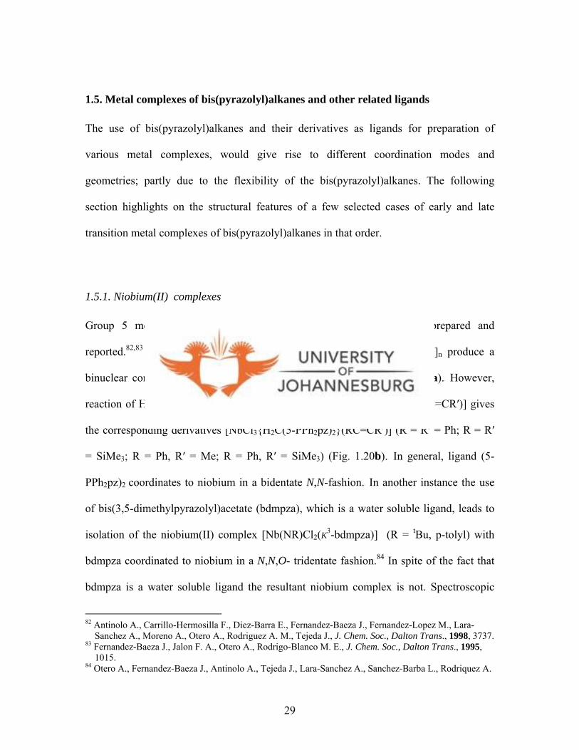
1.5.1. Niobium(II) complexes 29
viii

1.5.2. Manganese(II) and Rhenium(V) complexes 31
1.5.3. Ruthenium complexes 32
1.5.4. Cobalt complexes 33
1.5.5. Palladium platinum and gold complexes 35
1.6. Aims of this study 39
CHAPTER 2 41
BIS(PYRAZOLYL)ACETIC ACID COMPLEXES OF PALLADIUM(II),
PLATINUM(II) AND GOLD(III) 41
2.0. Introduction 41
2.1. Experimental 44
2.1.1 Materials and Instrumentation 44
2.1.2. Synthesis of ligands and metal complexes 46
2.1.2.1. Bis(pyrazol-1-yl)acetic acid (L1) 46
2.1.2.2. Bis(3,5-dimethylpyrazol-1-yl)acetic acid (L2) 46
2.1.2.3. Bis(pyrazol-1-yl)ethyl acetate (L3) 47
2.1.2.4. Bis(3,5-dimethylpyrazol-1yl)ethyl acetate (L4) 47
2.1.2.5. Dichloro-{bis(pyrazol-1-yl)acetic acid}palladium(II) (1) 48
2.1.2.6. Dichloro-{bis(3,5-dimethylpyrazol-1-yl)acetic acid}-
palladium(II) (2) 48
2.1.2.7. Dichloro-{bis(pyrazol-1-yl)acetic acid}palladium(II) (3a) 49
2.1.2.8. K2[Pt4Cl8(L1)2(L1-bpza)2].2H2O (3b) 49
ix
2.1.2.9. Dichloro-{bis(3,5-dimethylpyrazol-1-yl)acetic acid}-
platinum(II) (4) 50
2.1.2.10. Dichloro-{bis(pyrazol-1-yl)acetic acid}gold(III)chloride (5a) 50
2.1.2.11. Dichloro-{bis(3,5-dimethylpyrazol-1-yl)acetic acid}-
gold(III)chloride (6a) 51
2.1.2.12. Dichloro-{bis(3,5-dimethylpyrazol-1-yl)ethyl acetate}-
palladium(II) (7) 51
2.1.3. X-ray crystallography 52
2.1.3.1. Data collection 52
2.1.3.2. Structure solution and refinement 53
2.2. Results and discussion 54
2.2.1. Synthesis of ligands 54
2.2.2. Synthesis of metal complexes 62
2.2.3. Molecular structures of complexes 1, 2, 3b and 5b 69
2.3. Conclusions 85
CHAPTER 3 87
EVALUATION OF ANTI-TUMOUR ACTIVITY OF PALLADIUM(II),
PLATINUM(II) AND GOLD(III) PYRAZOLYL COMPLEXES AGAINST
CHO-22 CELLS AND KINETIC STUDIES 87
3.0. Introduction 87
3.1. Experimental 93
x

3.2 Biological testing 93
3.2.1. Cell culture and drug treatment 93
3.2.2. Complexes and L-cysteine solutions 94
3.2.3. Kinetic measurements 95
3.3. Results and discussion 96
3.3.1. Biological results 96
3.3.2. Kinetics of L-cysteine with palladium(II), platinum(II)
and gold(III) complexes 101
3.4. Conclusions 118
CHAPTER 4 120
PLATINUM(II) AND GOLD(III) DITHIOCARBAMATE COMPLEXES 122
4.0. Introduction 120
4.1. Palladium and platinum thiolate based complexes 122
4.2. Phosphine gold(I) thiolate complexes 126
4.2.1. Monophosphine gold(I) thiolate complexes 127
4.2.2. Diphosphine gold(I) thiolate complexes 130
4.3. Experimental 135
4.3.1. Reagents, starting materials and instrumentation 135
4.3.2. Synthesis and complexation 137
4.3.2.1. Synthesis of potassium-pyrazol-1-yldithiocarbamate (L5) 137
4.3.2.2. Synthesis of potassium-3,5-dimethylpyrazol-1-yl-
dithiocarbamate (L6) 138
xi

4.3.2.3. Synthesis of potassium-indazol-1-yldithiocarbamate (L7) 138
4.3.2.4. Synthesis of potassium-imidazol-1-yldithiocarbamate (L8) 139
4.3.2.5. Synthesis of bis(pyrazol-1-yldithiocarbamato)disulfide (L9) 139
4.3.2.6. Synthesis of bis(3,5-dimethylpyrazol-1-yldithiocarbamato)-
disulfide (L10) 140
4.3.2.7. Synthesis of bis(indazol-1-yldithiocarbamato)disulfide (L11) 140
4.3.2.8. Synthesis of pyrazolyl-1-dithiocarbamato-triphenylphosphino
gold(I) (13) 141
4.3.2.9. Synthesis of 3,5-dimethylpyrazolyl-dithiocarbamato-
triphenylphosphinogold(I) (14) 141
4.3.2.10. Synthesis of indazolyl-1-dithiocarbamato-
triphenylphosphino-gold(I) (15) 142
4.3.2.11. Synthesis of imidazolyl-1-dithiocarbamato-
triphenylphosphino-gold(I) (16) 142
4.3.2.12. Synthesis of bis-(pyrazolyl-1-dithiocarbamato)-bis-
(diphenylphosphino)-ethane gold(I) (17a) and
[Au18S8(dppe)6]2Cl (17b) 143
4.3.2.13. Synthesis of bis(3,5-dimethylpyrazolyl-1-
dithiocarbamato)-bis-(diphenylphosphino)ethane gold(I) (18) 144
4.3.2.14. Synthesis of bis-(indazolyl-1-dithiocarbamato)-bis-
(diphenylphosphino)ethane gold(I) (19) 144
4.3.2.15. Synthesis of binuclear bis(pyrazolyl-1-
dithiocarbamato)-bis-(diphenylphosphino)propane gold(I) (20) 145
xii
4.3.2.16. Synthesis of binuclear bis(3,5-dimethylpyrazolyl-1-
dithiocarbamato)-bis-(diphenylphosphino)propane gold(I) (21) 145
4.3.2.17. Synthesis of binuclear bis(indazolyl-1-
dithiocarbamato)-bis-(diphenylphosphino)propane gold(I) (22) 146
4.3.2.18. Synthesis of binuclear bis(pyrazolyl-1-
dithiocarbamato)-bis-(diphenylphosphino)hexane gold(I) (23) 146
4.3.2.19. Synthesis of binuclear bis(3,5-dimethylpyrazolyl-1-
dithiocarbamato)-bis-(diphenylphosphino)hexane gold(I) (24) 147
4.3.2.20. Synthesis of binuclear bis(indazol-1-yldithiocarbamato)-bis-
(diphenylphosphino)hexane gold(I) (25) 148
4.3.2.21. Synthesis of bis(pyrazol-1-yldithiocarbamato)-bis-
(diphenylphosphino)ethane platinum(II) (26) 148
4.3.2.22. Synthesis of bis(3,5-dimethylpyrazol-1-yldithiocarbamato)-
bis-(diphenylphosphino)ethane platinum(II) (28) 149
4.3.2.23. Synthesis of bis(indazol-1-yldithiocarbamato)-bis-
(diphenylphosphino)ethane platinum(II) (29) 150
4.3.2.24. Synthesis of bis(pyrazol-1-yldithiocarbamato)-bis-
(diphenylphosphino)ethane nickel(II) (30) 150
4.3.2.25. Synthesis of bis(pyrazol-1-yldithiocarbamato)-
platinum(II) (31) 150
4.3.2.26. Synthesis of bis(3,5-dimethylpyrazol-1-yldithiocarbamato)-
platinum(II) (32) 151
4.3.2.27. Synthesis of bis(indazol-1-yldithiocarbamato)platinum(II) (33) 151
xiii
4.3.3. X-ray crystallography 152
4.3.3.1. Data collection 152
4.3.3.2. Structure solution and refinement 153
4.4. Results and discussion 154
4.4.1. Synthesis of ligands and metal complexes 154
4.4.1.1. Dithiocarbamate ligands L5-L8 154
4.4.1.2. Monophosphine gold(I) dithiocarbamato complexes 13-16 156
4.4.1.3. Molecular structure of 15 158
4.4.1.4. Diphosphine gold(I) dithiocarbamato complexes 17a-19 163
4.4.1.5. Molecular structure of 17b 165
4.4.1.6. Diphosphine gold(I) dithiocarbamato complexes 20-25 170
4.4.1.7. Molecular structure of 25 172
4.4.1.8. Diphosphine platinum(II) dithiocarbamato complexes 26a-28 178
4.4.1.9. Molecular structure of 26b 181
4.4.1.10. Transformation of complexes 26a-28 into 26b monitored by
UV-Vis and 31P{1H} NMR spectroscopy 184
4.4.1.11. Platinum(II) dithiocarbamato complexes 29-31 191
4.4.1.12. Molecular structure of 30 194
4.4.2. Synthesis of new bis(dithiocarbamato)disulfide ligands 198
4.4.3. Attempted complexation of ligands L9-L11 205
4.5. Conclusions 207
xiv
CHAPTER 5 210
ANTI-TUMOUR ACTIVITY OF LIGANDS, PLATINUM(II) AND GOLD(I)
DITHIOCARBAMATO COMPLEXES AGAINST HELA CELLS AND
LUMINESCENCE STUDIES 210
5.0. Introduction 210
5.1. Luminescence of gold(I) complexes 212
5.2. Experimental 215
5.2.1. Biological reagents and instrumentation 215
5.2.2. Cell culture and drug treatment 215
5.2.3. Electronic absorption and photoluminescence spectra measurements 217
5.3. Biological results 218
5.3.1. Dithiocarbamate and bis(pyrazolyl-1-dithiocarbamate)disulfide ligands 218
5.3.2. Gold(I) complexes 222
5.3.2.1. Mononuclear gold(I) complexes 13-16 222
5.3.2.2. Dinuclear gold(I) complexes 20-25 227
5.43.3. Platinum(II) complexes 29-31 234
5.4. Electronic absorption and photoluminescence results 235
5.4.1. Absorption spectra 235
5.4.2. Photoluminescence spectra 239
5.5. Conclusions 248
APPENDIX 251
Submitted manuscripts 251
xv
LIST OF FIGURES
Figures Page
Figure 1.1. Estimated numbers of new cases (incidence) and deaths
(mortality), by sex and site. 2
Figure 1.2. Structures of (a) Taxol, (b) Camptothecin, and (c) Tamoxifen 4
Figure 1.3. Structure of dichlorodiammineplatinum(II) complex (cisplatin) 5
Figure 1.4. A Schematic representation of how cisplatin is activated
and subsequently bind to DNA 9
Figure 1.5. Structures of (a) carboplatin, (b) AMD-473
and (c) oxaliplatin 10
Figure 1.6. Structure of (cis-dietoxybis(1-phenylbutane-1,3-dionato)-
titanium(IV), budotitane 12
Figure 1.7. Structures of (a) vanadocene dichloride, (b) vanadium-L-cysteine
complex and (c) vanadium-L-cysteine methyl ester complex 13
Figure 1.8. Structures of (a) [RuCl4(Im)Me2SO)] (NAMI)
and (b) [RuCl2(η6-C6H6)(DMSO)] 14
Figure 1.9. Structures of [RuCl(η6-arene)(en)Cl][PF6]. 15
Figure 1.10. The dirhodium tetraacetate complex, (a) [Rh2(CH3COO)4(H2O)2]
and (b) [Rh2(CF3CONH)4] 17
Figure 1.11. Cyclophosphamide adducts of
(a) rhodium(II) keto-gluconate complex, [Rh2(KG)4], and
(b) rhodium(II) glucuronate complex, [Rh2(GU)4] 18
xvi
Figure 1.12. The trans-[Pd(L)2Cl2] complexes 19
Figure 1.13. A palladium(II) complex of ligands obtained from condensation
of 2-(diphenylphosphino)benzaldehyde and ethyl
hydrazinoacetate 20
Figure 1.14. Structures of (a) auranofin, (b) tetrahedral gold(I) complexes
of 1,2-bis(diphenylphosphino)ethane and
(c) tetrakis((tris(hydroxy-methyl))phosphine)gold(I) complex 21
Figure 1.15. The structure of [AuCl(dien)]Cl2 22
Figure 1.16. Structures of (a) [Au(bipy)(OH)2]PF6 and
(b [Au(bipy*-H)(OH)]PF6 23
Figure 1.17. General structure of bis(pyrazolyl)alkanes, (R2C)n(pz*)2 25
Figure 1.18. Observed coordination with (R2C)n(pz*)2 26
Figure 1.19. Bis(pyrazolyl) ligands. (a) (R')CH(3,5-Me2pz)2,
(b) (py)CH(3,5-Me2pz)2, (c) (CH2OH)CH(3,5-Me2pz)2,
(d) (CO2H)CH(3,5-Me2pz)2, (e) (CS2H)CH(3,5-Me2pz)2 28
Figure 1.20. Structures of (a) [NbCl3{H2C(5-PPh2pz)2}]2,
(b) [NbCl3{H2C(5-PPh2pz)2}(RC=CR’)], (c) [Nb(NR)Cl2(K3-bdmpza],
(d) [NbCl2(K3-bdmpza)(PhC CMe)] 30
Figure 1.21. (a) Structure of [Mn2{H2C(3,5-Me2pz)2}4F2][BF4]2,
(b) [Mn(bdmpza)(CO)3] 31
Figure 1.22. Structural isomers of [Re(O)(bdmpza)Cl2] 32
Figure 1.23. Structure of [Ru(bdmpza)Cl2(PPh3)] 33
xvii
Figure 1.24. (a) Tris(pyrazolyl)methane sulfonate (Tpms), (b) [Co(Tpms)2],
(c) [CoCl2L] 35
Figure 1.25. (a) Six-membered palladacycle [PdCl2{Me2C(pz)2}2],
(b) [Pd{H2C(3,5-R2pz)2}2]2+, (c) [Pd{Me2C(pz)2}2]2+ 36
Figure 1.26. Structure of [PtCl2{H2C(3,5-R2pz)2}] 37
Figure 1.27. Structure of (a) [Pt(IV)I2Me2(R)HC(pz)2], (b) [PtX2Me2{R2C(pz*)2}] 38
Figure 1.28. Cationic gold(III) complex, [Au(Me)2{H2C(3,5-Me2pz)2}][NO3] 38
Figure 2.1. The 1H NMR spectrum of L4 57
Figure 2.2. A molecular drawing of L4 59
Figure 2.3. A molecular packing diagram of L4 along the c axis 60
Figure 2.4. 1H NMR spectrum of 4, [PtCl2(3,5-Me2bpza)] 64
Figure 2.5. ESI-MS spectrum of compound 4a, [PtCl2(bpza)] 65
Figure 2.6. Dichlorogold(III) complexes of bis(1-methyl-2-imidazolyl)ketone
and related ligands 66
Figure 2.7. A molecular drawing of 1 71
Figure 2.8. A molecular packing diagram of 1 viewed along the b axis 72
Figure 2.9. A molecular drawing of 2 73
Figure 2.10. Complex 2 with two palladium units and three solvent molecules
in the cell unit 73
Figure 2.11. Hexanuclear complex 3b with four platinum atoms
and two potassium atoms 77
Figure 2.12. 1H NMR spectrum of 5a and 5b at 300 MHz and
200 MHz respectively 78
xviii
Figure 2.13. A 1H NMR spectroscopic monitoring of hydrolysis process
observed during attempts to crystallize 6a 81
Figure 2.14. 13C{1H} NMR spectrum showing a mixture of 6a
and the hydrolysed products 82
Figure 2.15. A molecular drawing of 5b 83
Figure 2.16. A molecular packing diagram of 5b viewed along c axis 84
Figure 3.1. Cellular viability results showing growth inhibition in
CHO-22 cells by compounds 1, 2, 3a and 4 98
Figure 3.2. Cellular viability results showing growth inhibition in
CHO-22 cells by complexes 5a and 6a 99
Figure 3.3. Kinetic trace for the reaction of complex 10 (5 x 10⁻⁵ M)
with L-cysteine (10 x 10-4M) at 285 nm, T = 298 K 102
Figure 3.4. Plots of kobs vs. [Cys] for 8 (3.0 x 10⁻⁵ M) and 9 (3.8 x 10⁻⁵ M)
at 303 K, I = 0.1 M 106
Figure 3.5. Plots of kobs vs. [Cys] for 10 (5.0 x 10⁻⁵ M) at 293 K, I = 0.1 M 106
Figure 3.6. Kinetic trace for the reaction of complex 11 (5 x 10⁻⁵ M)
with L-cysteine (10 x 10-4M) at 239 nm, T = 298 K 109
Figure 3.7. Plots of kobs vs. [Cys] for 11 (5.0 x 10⁻⁵ M) at 298 K, I = 0.1 M 111
Figure 3.8. Temperature dependence plots for the reaction of 8-11
complexes with L-cysteine 114
Figure 3.9. (a) 1H NMR spectra of [PdCl2(L1)] (1), (b) L-cysteine and
(c) [Pd(L1)(Cys)2] (12a) acquired after 5 min
(solvent, DMSO-d6) 116
Figure 3.10. ESI-MS spectrum of compound 12 118
xix
Figure 4.1. Structures of dithiocarbamate based complexes,
[M(S2CNEt2)(L)]NO3 123
Figure 4.2. Structures of (a) [M(ESDT)(L)Cl], (b) [M(ESDT(L)2]Cl,
(c) [M(ESDT)(en)]Cl 124
Figure 4.3. The structures of planar [M(II)(mnt)2]- complexes 125
Figure 4.4. The structure of auranofin 126
Figure 4.5. Structures of complexes [Au(2-mba)(PR3)] 128
Figure 4.6. A structure of gold(I) 7-azacoumarin complex,
[Au(TS)(PEt3)] 129
Figure 4.7. The structure of [Au(K3TSC)PEt3] 129
Figure 4.8. Structures of (a) [(AuSTg)2(dppe)] and (b) [Au(dppe)2]+ 140
Figure 4.9. Structure of phosphine gold(I) complexes,
[(AuOmS)2(R2P(CH2)nPR2)] 131
Figure 4.10. Phosphinegold(I) thiourea complexes 132
Figure 4.11. 1H NMR spectrum of L7 156
Figure 4.12. A molecular drawing of 15 160
Figure 4.13. A molecular packing diagram of 15 viewed along the c axis 161
Figure 4.14. The 1H NMR spectrum of 17a, with the
31P{1H} NMR spectrum of 17a as an inset 164
Figure 4.15. The molecular diagram of 17b 167
Figure 4.16. The skeletal (Au, P, S) geometry of 17b 168
Figure 4.17. A molecular drawing of 25 174
xx
Figure 4.18. A molecular packing diagram of 25 viewed
along the c axis 175
Figure 4.19. Time of flight ESI-MS spectrum of 22 177
Figure 4.20. IR spectra of 26 and 27 180
Figure 4.21. A molecular drawing of 27 182
Figure 4.22. UV-Vis spectra of 26 and the transformed
product 27 184
Figure 4.23. 31P{1H} NMR spectra overlay showing the transformation
of 26 to 27 over a period of 3 days 185
Figure 4.24. Kinetic plots of transformation of 26 to 27 186
Figure 4.25. First order kinetics plot-determination of the pseudo-first-order
rate constant of transformation of 26 to 27 188
Figure 4.26. 31P{1H} NMR spectra of [NiCl2(dppe)] and
[Ni(L5)2(dppe)] 190
Figure 4.27. The structure of octahedral [NiCl2(pzH)4] complex 191
Figure 4.28. A 1H NMR spectrum of 32 192
Figure 4.29. A molecular drawing of 32 196
Figure 4.30. The 13C{1H} NMR spectrum of L9 with
a 1H NMR spectrum (inset) 199
Figure 4.31. A molecular drawing of L9 201
Figure 4.32. A molecular drawing of L10 201
Figure 4.33. A molecular packing diagram of L10 viewed
along the c axis 203
xxi
Figure 4.34. A 31P{1H} NMR profile showing a mixture of
[Au2(PPh3)2(L9)]2BF4, [(Ph3P)-Au-(PPh3)]+ and
[(Ph3P)-Au-(NCMe)]+ 206
Figure 5.1. Schematic representations of orbital splitting of
gold(I) thiolate compounds 212
Figure 5.2. Structures of [Au2(SR′)2(Ph2PN(R)PPh2)] 214
Figure 5.3. Growth inhibition profile of HeLa cells
by L9 and L10 219
Figure 5.4. Growth inhibition profile of HeLa cells by
gold(I) complex 13 224
Figure 5.5. Growth inhibition profile of HeLa cells by
gold(I) complex 14 224
Figure 5.6. Growth inhibition profile of HeLa cells by
gold(I) complex 15 225
Figure 5.7. Growth inhibition profile of HeLa cells by
gold(I) complex 16 225
Figure 5.8. Growth inhibition profiles of HeLa cells by
gold(I) complex 20 229
Figure 5.9. Growth inhibition profiles of HeLa cells by
gold(I)complex 21 230
Figure 5.10. Growth inhibition profiles of HeLa cells by
gold(I) complex 22 230
xxii

Figure 5.11. Growth inhibition profiles of HeLa cells by
gold(I) complex 23 231
Figure 5.12. Growth inhibition profiles of HeLa cells by
gold(I) complex 24 231
Figure 5.13. Growth inhibition profiles of HeLa cells by
gold(I) complex 25 232
Figure 5.14. UV-Vis spectrum of 15 in CH2Cl2 at 298 K 236
Figure 5.15. UV-Vis spectrum of 17a in CH2Cl2 at 298 K 238
Figure 5.16. UV-Vis spectrum of 20 in CH2Cl2 at 298 K 238
Figure 5.17. UV-Vis spectrum of 23 in CH2Cl2 at 298 K 239
Figure 5.18. Schematic representations of (a) orbital splitting and
(b) energy diagram of 17a and 17b 242
Figure 5.19. Excitation and emission spectra of a frozen
solution of 13 at 77 K 243
Figure 5.20. Excitation and emission spectra of a frozen
solution of 14 at 77 K 244
Figure 5.21. Excitation and emission spectra of a frozen
solution of 16 at 77 K 245
Figure 5.22. Excitation and emission spectra of a frozen
solution of 17a at 77 K 246
Figure 5.23. Excitation and emission spectra of a frozen
solution of 17b at 77 K 247
xxiii
LIST OF TABLES
Table 1.1. Estimated and projected numbers of cancer cases. 2
Table 2.1. Crystal data and structure refinement for L4 61
Table 2.2. Selected bond lengths [Å] and angles [°] for L4 62
Table 2.3. Crystal data and structure refinement for 1, 2, 3b and 5b 74
Table 2.4. Selected bond lengths [Å] and angles [°] for 1, 2, 3b and 5b 75
Table 3.1. Growth inhibition values of compounds 1-6a tested
against CHO-22 cells 97
Table 3.2. Rate constants for the reaction of L-cysteine with 8 104
Table 3.3. Rate constants for the reaction of L-cysteine with 9 105
Table 3.4. Rate constants for the reaction of L-cysteine with 10 105
Table 3.5. Rate constants for the reaction of L-cysteine with 11 110
Table 3.6. Rate constants of the reduction of 11 to gold(I) 110
Table 3.7. Activation parameters determined from the temperature
dependence of the rate constants for the substitution of Pd(II), Pt(II)
and Au(III) complexes, and reduction of Au(III) 113
Table 3.8. Calculation of energy parameters 115
Table 3.9. Variation of k1 and k3 rate constants with temperature
For complexes 1, 3a, 5a and 6a 115
Table 4.1. Selected bond lengths [Å] and angles [°] for 15 159
Table 4.2. Crystal data, data collection and refinement parameters of 15 162
Table 4.3. Selected bond lengths [Å] and angles [°] of 17b 168
Table 4.4. Crystal data, data collection and refinement parameters of 17b 169
xxiv
Table 4.5. Selected bond lengths [Å] and angles [°] for 25 174
Table 4.6. Crystal data, data collection and refinement parameters of 25 176
Table 4.7. Selected bond lengths [Å] and angles [°] for 27 181
Table 4.8. Crystal data, data collection and refinement parameters of 27 183
Table 4.9. Manipulation of integration values 187
Table 4.10. Selected bond lengths [Å] and angles [°] for 32 195
Table 4.11. Crystal data, data collection and refinement parameters of 32 197
Table 4.12. Selected bond lengths [Å] and angles [°] for L9 and L10 202
Table 4.13. Crystal data, data collection and refinement parameters
of L9 and L10 204
Table 5.1. Growth inhibition values of compounds L5-L11 tested
against HeLa cells 220
Table 5.2. Tumour Specificity of L9 221
Table 5.3. Growth inhibition values of compounds 13-16 tested
against HeLa cells 223
Table 5.4. Tumour Specificity of 13-16 226
Table 5.5. Growth inhibition values of compounds 20-25 tested
against HeLa cells 229
Table 5.6. Tumour Specificity of complexes 20-25 233
Table 5.7. Excitation and emission data for 13, 14, 15, 16, 17a and 17b 241
xxv

ABBREVIATIONS
NMR Nuclear magnetic resonance
IR Infra red
ESI-MS Electrospray ionisation mass spectrometry
UV-Vis Ultra violet-visible
CHO Chinese hamster ovary
HeLa Human cervix epitheloid carcinoma
PBS Phosphate buffer saline
MTT 3-(4,5-dimethylthiazol-2yl)-2,5-diphenyltetrazolium bromide
MOMP Mitochondrial outer membrane permeabilisation
Cisplatin cis-dichlorodiammineplatinum(II)
LMCT Ligand metal charge transfer
ca. Approximately
DNA Deoxyribonucleic acid
IC50 Concentration of compound needed to inhibit cell growth by 50% against
a single cell line.
TS Tumour specificity
Nu Nucleophile
bpza Bis(pyrazolyl)acetic acid
dtc Dithiocarbamate
dppe Bis(diphenylphosphino)ethane
dppp Bis(diphenylphosphino)propane
dpph Bis(diphenylphosphino)hexane
xxvi
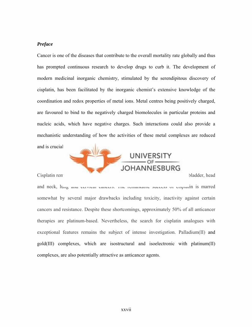
Preface
Cancer is one of the diseases that contribute to the overall mortality rate globally and thus
has prompted continuous research to develop drugs to curb it. The development of
modern medicinal inorganic chemistry, stimulated by the serendipitous discovery of
cisplatin, has been facilitated by the inorganic chemist’s extensive knowledge of the
coordination and redox properties of metal ions. Metal centres being positively charged,
are favoured to bind to the negatively charged biomolecules in particular proteins and
nucleic acids, which have negative charges. Such interactions could also provide a
mechanistic understanding of how the activities of these metal complexes are reduced
and is crucial to the rational design of new compounds.
Cisplatin remains the drug of choice for the treatment of testicular, ovarian, bladder, head
and neck, lung and cervical cancers. The remarkable success of cisplatin is marred
somewhat by several major drawbacks including toxicity, inactivity against certain
cancers and resistance. Despite these shortcomings, approximately 50% of all anticancer
therapies are platinum-based. Nevertheless, the search for cisplatin analogues with
exceptional features remains the subject of intense investigation. Palladium(II) and
gold(III) complexes, which are isostructural and isoelectronic with platinum(II)
complexes, are also potentially attractive as anticancer agents.
xxvii

Thus it is with the intention of linking coordination chemistry and biochemistry in their
widest sense that this project was envisaged. All the same, this area of research is very
broad and thus the main objective for this study was to prepare various palladium(II),
platinum(II), gold(I) and gold(III) complexes with a view to test them as anticancer
agents; while exploring all chemistry aspects of these complexes.
xxviii
List of submitted manuscripts and some in preparation
The work in this thesis is based on reports submitted to “Biomedical applications for
gold: Stages 9-15”, Mintek Communication (SA), C4050M,-C4262M.
1. Bis(pyrazolyl)Palladium(II), Platinum(II) and Gold(III) Complexes: Syntheses,
Molecular Structures and Substitution Reactions with L-cysteine. Frankline K.
Keter, Stephen O. Ojwach, Olayinka A. Oyetunji Ilia A. Guzei and James
Darkwa (Submitted to Inorganica Chimica Acta, 2008)
2. Dichloro-bis-(3,5-dimethyl-pyrazolyl)palladium(II) complex. A DMSO-
d6 solvated complex. Frankline K. Keter, Bernard O. Omondi, and James
Darkwa. (Submitted to Journal of Molecular Structures).
3. The work reported in chapters four and five are currently at various stages of
manuscript preparation for publication (Target journals: Inorganic Chemistry and
Journal of Inorganic Biochemistry).
xxix

CHAPTER 1
1.0 Introduction
1.1. Overview
Cancer is one of the diseases that contribute to the high mortality rate globally. It was
estimated that in 2000, 10 million new cases of cancer were identified;
and about 6 million reported deaths were cancer related.1a,b In the same year, an estimated
22 million people were living with cancer already diagnosed within the previous five
years.1b These statistics depicts a 22% increase in cancer incidence and mortality in the
world in 2000 compared to 1990.1,2 The most common types of cancer in 2000 were
breast (17.2%), colorectal (10.6%) and prostate (6.9%),1 and the highest tumour types
world-wide were lung (12.3%), breast (10.4%) and colorectal (9.4%) tumours (Fig. 1.1).2
The estimated and projected numbers of cancer cases over a period of years is shown in
Table 1.1. Therefore, there is a need for more efficient drugs to curb this scourge.
1 aShibuya K., Mathers C. D., Boschi-Pinto C., Lopez A. D., Murray C. J. L., BMC Cancer, 2002, 2, 37; bSchwartsmann G., Ratain M. J., Cragg G. M., Wong J. E., Saijo N., Parkinson D. R., Fujiwara Y., Pazdur R., Newman D. J., Dagher R., Di Leone L., J. Clin. Oncol. 2002, 20, 47. 2 Parkin D. M., Lancet Oncol. 2001, 2, 533
1

Breast
Stomach
Kidney
Lung
Colon/Rectum
Prostate
Liver
Cervix uteri
OesophagusBladder
Non-Hodgkin lymphoma
Leukaemia
Oral cavityPancreas
Ovary
Men Woman
5,3 million cases4,7 million deaths
4,7 million cases2,7 million deaths
57119112
81
109144
93
260227
204
334
405558
255
810
499
399
543
279
99157
170
116
902
192114
101
97
165
234
370
446
293
1050
318241
47101
7134
337
76
113
133111
471233
86
33
165
12168
(Thousands)
IncidenceMortality
Breast
Stomach
Kidney
Lung
Colon/Rectum
Prostate
Liver
Cervix uteri
OesophagusBladder
Non-Hodgkin lymphoma
Leukaemia
Oral cavityPancreas
Ovary
Breast
Stomach
Kidney
Lung
Colon/Rectum
Prostate
Liver
Cervix uteri
OesophagusBladder
Non-Hodgkin lymphoma
Leukaemia
Oral cavityPancreas
Ovary
Men Woman
5,3 million cases4,7 million deaths
4,7 million cases2,7 million deaths
57119112
81
109144
93
260227
204
334
405558
255
810
499
399
543
279
99157
170
116
902
192114
101
97
165
234
370
446
293
1050
318241
47101
7134
337
76
113
133111
471233
86
33
165
12168
(Thousands)
IncidenceMortalityIncidenceMortality
Figure 1.1. Estimated numbers of new cases (incidence) and deaths (mortality), by sex
and site. (Taken from ref. 1).
Table 1.1. Estimated and projected numbers of cancer cases. (Taken from ref. 1)
Years Region 2000 2010 2022 2050 World 10.06 12.34 15.35 23.83
More developed regions 4.68 5.31 6.03 6.79 Less developed regions 5.38 7.03 9.32 17.04
Africa 0.3 0.79 1.04 2.53 Asia (Japan) 0.52 0.61 0.67 0.65 Asia (Other) 3.94 5.17 6.75 10.74
Europe 2.77 3.06 3.36 3.64 South America 0.83 1.10 1.48 2.88 North America 1.38 1.65 2.03 2.61
Oceania 0.11 0.13 0.16 0.24 NOTE: The number of new cases (millions) of all cancer.
2

There is no doubt that plants have a long history of use in the treatment of cancer3a;
though many of the claims for the efficacy of such treatment have been viewed with
some skepticism because cancer, as a specific disease, is poorly defined in terms of myths
and traditional medicine.1,3a Nevertheless, with the current experience in medicinal
chemistry and pharmacology, in vivo studies done on natural products have resulted in a
number of clinically useful drugs that are now available. Examples include, Taxol (Fig.
1.2a),3b Camptothecin (Fig. 1.2b)3c and Tamoxifen (Fig. 1.2c).3d These are all drugs from
plant sources, generally known as natural products.
Apart from natural products being directly applied as drugs, there are many others that
have served as chemical models or templates for the design and synthesis of other cancer
drugs. Despite significant progress being made in tackling cancer with natural products
derived drugs, it is still a challenge as some of these drugs have side effects. Thus, efforts
to solve this problem continue.
3 aNewman D. J., Cragg G. M., Snader K. M., Nat. Prod. Rep. 2000, 17, 215; bWani M., Taylor H., Wall M., Coggon P., McPhail A.I, J. Am. Chem. Soc., 1971, 93, 2325; cWall M. E., Wani M. C., Cook C. E., Palmer K. H., McPhail A. I., Sim G. A., J. Am. Chem. Soc., 1966, 88, 3888; dJordan V. C., Br. J. Pharmacol., 2006, 147, S269.
3

O
OO
R
R
RO O
R OH
HOO
CCHOH
CH
NHC
C
O
R
CR
O
C
O
O O
(a)
N
N O
O
OOH
H3CH2C
O(CH2)2NMe2
R
(b) (c)
Figure 1.2. Structures of (a) Taxol, (b) Camptothecin, and (c) Tamoxifen.
One area that has gained prominence is the use of inorganic compounds as anticancer
drugs. Since the serendipitious discovery of cis-diamminedichloroplatinum(II) (cisplatin)
by Rosenburg and co-workers in 1965,4 (Fig. 1.3) this compound has become a well
established antineoplastic agent. Cisplatin is one of the most effective drugs in the
treatment of testicular, ovarian, bladder, head and neck cancers.5 The chemotherapeutic
4 Rosenberg B., Van Camp L., Krigas T., Nature 1965, 205, 698. 5 Gumus F., Algul O., Eren G., Eroglu H., Eur. J. Med. Chem 2003, 38, 473
4
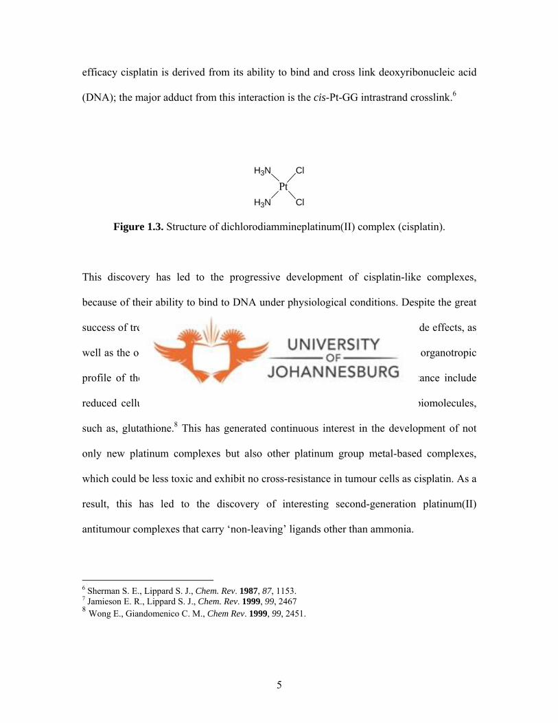
efficacy cisplatin is derived from its ability to bind and cross link deoxyribonucleic acid
(DNA); the major adduct from this interaction is the cis-Pt-GG intrastrand crosslink.6
PtH3N
ClH3N
Cl
Figure 1.3. Structure of dichlorodiammineplatinum(II) complex (cisplatin).
This discovery has led to the progressive development of cisplatin-like complexes,
because of their ability to bind to DNA under physiological conditions. Despite the great
success of treating certain kinds of cancers with cisplatin there are several side effects, as
well as the occurrence of intrinsic and acquired resistances, which limit the organotropic
profile of the drug.7 Some of the reasons that lead to this acquired resistance include
reduced cellular uptake and deactivation of cisplatin by thiol containing biomolecules,
such as, glutathione.8 This has generated continuous interest in the development of not
only new platinum complexes but also other platinum group metal-based complexes,
which could be less toxic and exhibit no cross-resistance in tumour cells as cisplatin. As a
result, this has led to the discovery of interesting second-generation platinum(II)
antitumour complexes that carry ‘non-leaving’ ligands other than ammonia.8
6 Sherman S. E., Lippard S. J., Chem. Rev. 1987, 87, 1153. 7 Jamieson E. R., Lippard S. J., Chem. Rev. 1999, 99, 2467 8 Wong E., Giandomenico C. M., Chem Rev. 1999, 99, 2451.
5
Several platinum complexes with N-heterocyclic ligands such as imidazole, thiazole,
benzimidazole, benzoxazole and bezothiazole, have been reported9 and some of these
compounds have shown significant cytotoxicity.9b,c This is clear evidence that N-
heterocycles impart therapeutic value to platinum complexes in which they feature as
ligands. As such, the use of other nitrogen-donor ligands that are more compatible to the
human system appears to be another way of producing new cancer drugs.10,11
The ability of platinum compounds mentioned above to modulate drug metabolism and
target binding through steric and electronic effects, on the substitution mechanism that
govern the action of these compounds, is of interest.12 For this reason, different strategies
have been employed in a bid to reduce the systematic toxicities of existing drugs, viz. (i)
passive tumour targeting based on the enhanced permeability and retention (EPR) effect;
(ii) receptor mediated targeting; (iii) enzymatically activated prodrugs; (iv) compounds
targeted towards cellular DNA.13 It should be noted that most of these strategies employ
platinum derived complexes because of their ease of syntheses and stabilities. No doubt
that the four strategies mentioned above have added value to the drugs, especially
platinum. However, the targeted drugs are still far from being used to treat patients as the
9 aHe X.-F., Vogels C. M., Decken A., Westcott S. A., Polyhedron, 2004, 23, 155; bMock C., Puscasu I., Rauterkus M. J., Tallen G., Wolff J. E. A., Krebs B., Inorg. Chim. Acta, 2001, 319, 109; cBloemink M. J., Engelking H., Karentzopoulos S., Krebs B., Reedijk J., Inorg. Chem., 1996, 35, 619. 10 Iakovidis A., Hadjiliadis N., Coord. Chem. Rev. 1994, 135, 17 11 Beck, W., Purucker B., Girnth M., Schonenberger H., Seidenberger H., Ruckdeschel G., Z. Naturforsch 1976, 31b, 832. 12 Wang K., Lu J., Li R., Coord. Chem. Rev. 1996, 151, 53. 13 aGabizon A., Shmeeda H., Barenholz Y., Clin. Pharmacokinet., 2003, 42, 419; bBarnes K. R., Kutikov A., Lippard S. J., Chem. Biol., 2004, 11, 557; cHanessian S., Wong J. G., Can. J. Chem. Rev. Can. Chim., 1993, 71, 896; dRen S. M., Cai L. S., Segal B. M., J. Chem. Soc., Dalton Trans., 1999, 1413; eVan Zutphen S., Reedijk J., Coord. Chem. Rev. 2005, 249, 2845.
6

activities are not enormously different from those of cisplatin. Thus, the fourth strategy
remains the most applied at the moment.
1.2. General DNA-metal interactions
DNA is now generally accepted as the target for most anticancer agents.1 3 Out of the four
strategies mentioned (vide supra), it is apparent that synthesising a primary drug targeted
towards the DNA is an appropriate route to anticancer drug discovery. Thus, DNA
remains an attractive target for the design of antitumour agents.14 The reactivity of
cisplatin and its mimics is dependent not only on physiological conditions but also on
structure activity relationships (SARs), viz. (i) a cis geometry with the general formula
cis-[PtX2(amine)2] for platinum(II) compounds and cis-[PtY2X2(amine)2] for
platinum(IV) compounds; (ii) the ligand X should be an anion with intermediate binding
strength, such as, chloride or oxalate; (iii) the amine ligands should possess at least one
NH moiety necessary for hydrogen bonding interactions.8,15 It should, however, be
pointed out that an N-H moiety is not necessarily a requirement when designing
anticancer drugs. This is supported by the fact that some compounds without N-H groups
have shown antitumour activity.16, ,17 18
Within the extracellular milieu the physiological chloride ion concentration is ca. 100
mM, while inside cells it is ca. 4 mM. These conditions facilitate the aquation of cisplatin
14 Budzisz E., Graczyk-Wojciechowska J., Zieba R., Nawrot B., New J. Chem., 2002, 26, 1799. 15 Cleare M. J., Hoeschele J. D., Bioinorg. Chem., 1973, 2, 187. 16 Gund A., Keppler B. K., Angew. Chem., Int. Ed. Engl., 1994, 33, 186. 17 Bloemink M. J., Engelking H., Karentzopoulos S., Krebs B., Reedijk J., Inorg. Chem., 1996, 35, 619. 18 Rauterkus M. J., Fakih S., Mock C., Puscasu I., Krebs B., Inorg. Chim. Acta, 2003, 350, 355.
7
(Fig. 1.4). The aquated species would then easily react with the target molecule, DNA.
19,20 The binding of metal species to DNA is known to occur in two steps, leading to
formation of two forms of adducts; intra-strand and inter-strand cross-links (Fig. 1.4).
The formation of these adducts then blocks DNA transcription and replication causing
cells to undergo G2 cell-cycle arrest.1 9 Under normal circumstances a nucleotide excision
repair (NER) pathway would repair DNA damage.21 However, DNA damage caused by
cisplatin adducts is very poorly repaired in cells. This is due to the fact that cisplatin-
induced intra-strand cross-links serve as binding targets for high-mobility-group proteins
(HMG)22 because of structural similarity between these intra-strand cross-links and the
natural binding sites for HMG.2 1 The binding of HMG to cisplatin-DNA adducts cause
distortion in DNA structure, hence inhibiting DNA repair.19,23 The above findings about
the biological chemistry of cisplatin are informative in drug research as it helps in the
design of new effective metallo-drugs.
19 Chu G., J. Biol. Chem., 1994, 269, 787. 20 Jordan P., Carmo-Fonseca M., Cell Mol. Life Sci., 2000, 57, 1229. 21 Zamble D. B., Mu D., Reardon J. T., Sancer A., Lippard S. J., Biochemistry, 1996, 35, 10004. 22 HMG are small chromosomal-associated proteins that are involved in gene regulation and maintenance of chromosomal structure. 23 Zlatanova J., Yaneva J., Leuba S. H., FASEB J, 1998, 12, 791.
8

PtH3N
ClH3N
ClPt
H3N
ClH3N
OH2
PtH3N
ClH3N
Thiols containinggroups
G
G
Deactivated species
H2O
Cl-
DNA
PtH3N
OH2H3N
G
G
H2O
Cl-
H2
PtH3N
O
H3N
G
G
Figure 1.4. A Schematic representation of how cisplatin is activated and subsequently bind to DNA.
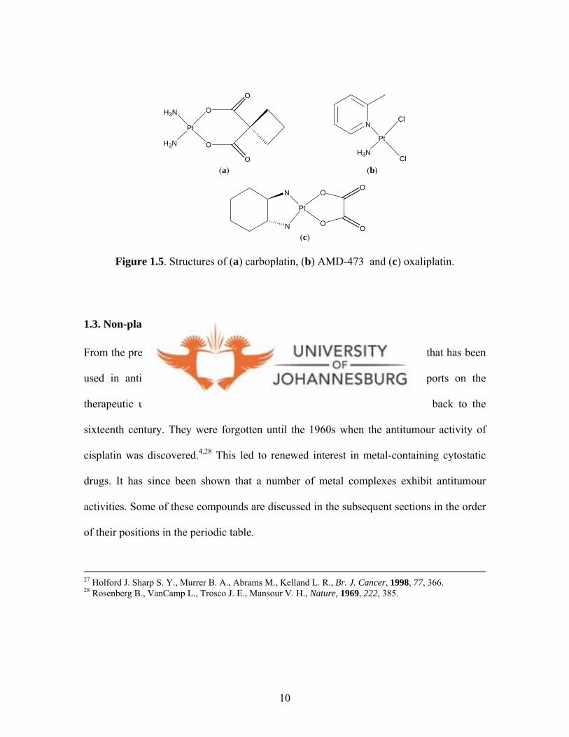
More than 3000 platinum analogues of cisplatin have been synthesised and a few have
made it to the clinical stages.24 Examples include carboplatin (Fig. 1.5a), AMD-473 (Fig.
1.5b) and oxaliplatin (Fig. 1.5c). Carboplatin displays similar activities with cisplatin but
also exhibits bone-marrow toxicity,25,26 On the other hand, AMD-473 showed promising
in vitro activity against cisplatin-resistant A2780cisR cells.25, 27 Despite its low toxicity
profile, the activity of AMD-473 is not great. Oxaliplatin, which is a highly sterically
hindered complex, has good activity and has been approved for clinical use in Europe,
China and United states for colorectal cancer.
24 Marzano C., Trevisan A., Giovagnini L., Fregona D., Toxicology in Vitro, 2002, 16, 413. 25 Kelland L. R., Sharp S. Y., O’Neill C. F., Raynaud F. I., Beale P. J., Judson I. R., Inorg. Biochem., 1999, 77, 111. 26 Cadron I., Leunen K., Amant F., Van Gorp T., Neven P., Vergote I., Gynecol. Oncol., 2007, 106, 354.
9
N
Pt
H3N
Cl
Cl
H3N
Pt
H3N
O
O
O
O
N
N
Pt
O
OO
O
(a) (b)
(c)
Figure 1.5. Structures of (a) carboplatin, (b) AMD-473 and (c) oxaliplatin.
1.3. Non-platinum metal compounds as anti-cancer therapeutic agents
From the preceding section it would appear that platinum is the only metal that has been
used in anticancer therapy. This is far from the truth. The earliest reports on the
therapeutic use of metals or metal containing compounds in cancer date back to the
sixteenth century. They were forgotten until the 1960s when the antitumour activity of
cisplatin was discovered.4,28 This led to renewed interest in metal-containing cytostatic
drugs. It has since been shown that a number of metal complexes exhibit antitumour
activities. Some of these compounds are discussed in the subsequent sections in the order
of their positions in the periodic table.
27 Holford J. Sharp S. Y., Murrer B. A., Abrams M., Kelland L. R., Br. J. Cancer, 1998, 77, 366. 28 Rosenberg B., VanCamp L., Trosco J. E., Mansour V. H., Nature, 1969, 222, 385.
10
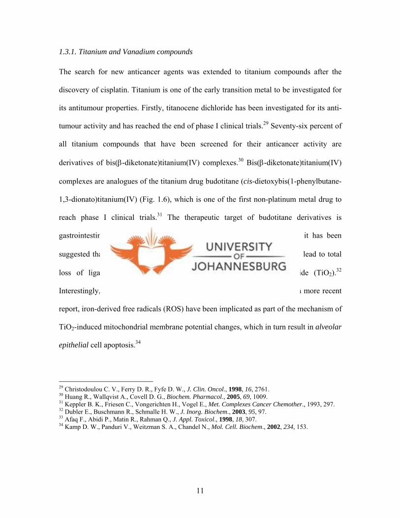
1.3.1. Titanium and Vanadium compounds
The search for new anticancer agents was extended to titanium compounds after the
discovery of cisplatin. Titanium is one of the early transition metal to be investigated for
its antitumour properties. Firstly, titanocene dichloride has been investigated for its anti-
tumour activity and has reached the end of phase I clinical trials.29 Seventy-six percent of
all titanium compounds that have been screened for their anticancer activity are
derivatives of bis(β-diketonate)titanium(IV) complexes.30 Bis(β-diketonate)titanium(IV)
complexes are analogues of the titanium drug budotitane (cis-dietoxybis(1-phenylbutane-
1,3-dionato)titanium(IV) (Fig. 1.6), which is one of the first non-platinum metal drug to
reach phase I clinical trials.31 The therapeutic target of budotitane derivatives is
gastrointestinal tumours, but the mode of action is unknown. However, it has been
suggested that complete hydrolysis of the β-diketonato titanium complexes lead to total
loss of ligands and formation of the water insoluble titanium dioxide (TiO2).32
Interestingly, TiO2 itself has been shown to have antitumour activity.33 In a more recent
report, iron-derived free radicals (ROS) have been implicated as part of the mechanism of
TiO2-induced mitochondrial membrane potential changes, which in turn result in alveolar
epithelial cell apoptosis.34
29 Christodoulou C. V., Ferry D. R., Fyfe D. W., J. Clin. Oncol., 1998, 16, 2761. 30 Huang R., Wallqvist A., Covell D. G., Biochem. Pharmacol., 2005, 69, 1009. 31 Keppler B. K., Friesen C., Vongerichten H., Vogel E., Met. Complexes Cancer Chemother., 1993, 297. 32 Dubler E., Buschmann R., Schmalle H. W., J. Inorg. Biochem., 2003, 95, 97. 33 Afaq F., Abidi P., Matin R., Rahman Q., J. Appl. Toxicol., 1998, 18, 307. 34 Kamp D. W., Panduri V., Weitzman S. A., Chandel N., Mol. Cell. Biochem., 2002, 234, 153.
11

O
O
Ti
O
O
OCH2CH3
OCH2CH3
Figure 1.6. Structure of (cis-dietoxybis(1-phenylbutane-1,3-dionato)titanium(IV), budotitane.
The potential use of vanadium compounds as antitumour agents also received strong
attention after the discovery of cisplatin, even though this speculation has existed since
the beginning of the 20th century.35 Evidence from experimental studies carried out
suggests that vanadium could be considered as a representative of a new class of
anticancer agents.36 Various vanadium complexes, such as, the peroxovanadates36 and
vanadocene dichloride (Fig. 1.7a)37 are amongst the active complexes. Literature survey
reveals that only two vanadium-L-cysteine or L-cysteine methyl ester complexes (Fig.
1.7) have been isolated and shown to significantly prevent lung metastasis in vivo.38
Studies on various cell lines reveal that vanadium exerts its antitumour activity through
inhibition of cellular tyrosine phosphatases and/or activation of tyrosine
phosphorylases.39 Ultimately signal transduction pathways are triggered, hence causing
apoptosis and/or activation of tumour suppressor genes. It is clear from what is known
35 Kieler J., Gromek A., Nissen N. I., Acta Chir. Scand., 1965, 343, 154. 36 Evangelou A. M., Crit. Rev. Oncol./Hematol., 2002, 42, 249. 37 Toney J. H., Murthy M. S., Marks T. J., Chem. Biol. Interact., 1985, 56, 45. 38 Papaioannou A., Manos M., Karkabounas S., Liasko R., Evangelon A. M., Correia I., Kalfakakou V., Pessoa J. C., Kabanos T., J. Inorg. Biochem., 2004, 98, 959. 39 Bevan A. P., Drake P. G., Yale J.-F., Shaver A., Posner B. I., Mol. Cell Biochem., 1995, 153, 49.
12

about the mode of action for titanium and vanadium anticancer agents that they are
different from cisplatin.
V
NH2
OC
O
SH
OC
O
NH2
SH
H2NO
CO
HS
III
V
H2N
O
H2N
O
O
O
S
SIII
(b) (c)
V
Cl
Cl
(a)
Figure 1.7. Structures of (a) vanadocene dichloride, (b) vanadium-L-cysteine complex and (c) vanadium-L-cysteine methyl ester complex.
1.3.2. Ruthenium compounds
Investigation of ruthenium complexes as anticancer agents was stimulated by the reports
of antitumour activity of sodium salt of [RuCl4(Im)Me2SO)], generally referred to as
NAMI (Fig. 1.8a) , which is currently in clinical trials.40 As a result, many ruthenium
complexes have been synthesised and shown to have antitumour activity.41
Organometallic ruthenium(II) complexes with arene ligands represent another group of
ruthenium compounds with antitumour activity. Since the initial discovery that
40 Sanna B., Debidda M., Pintus G., Tadolini B., Posadino A. M., Bennardini F., Sava G., Ventura C., Arch. Biochem. Biophys., 2002, 403, 209. 41 Zhang C. X., Lippard S. J., Curr. Opin. Chem. Biol., 2003, 7, 481.
13

[RuCl2(η6-C6H6)(DMSO)] (Fig. 1.8b) can inhibit topoisomerase II activity,42a three
derivatives have been prepared by replacing the dmso ligand in [RuCl2(η6-
C6H6)(DMSO)] with 3-aminopyridine, p-aminobenzoic acid or aminoguanidine.42b These
analogues show enhanced efficacy of topoisomerase II inhibition and higher cytotoxicity
against breast and colon carcinoma cells. In general, the cytotoxicity of ruthenium
complexes, both in 2+ and 3+ oxidation states is related to its ability to bind DNA. For
ruthenium(III) complexes its reaction with DNA is preceded by reduction from
ruthenium(III) to ruthenium(II).4 1
Ru
O
ClClN
HN
Ru
S
ClCl
Cl Cl
OCH3
CH3
NH
NH
(a) (b)
Figure 1.8. Structures of (a) [RuCl4(Im)Me2SO)] (NAMI) and (b) [RuCl2(η6-C6H6)(DMSO)].
Another set of important compounds are the ruthenium(II) arene complexes of the type
[( 6-arene)Ru(II)(en)Cl][PF ] (en = ethylenediamine) reported by Sadler and co-
workers.
6
43 They include, [( 6-Bip)Ru(II)(en)Cl][PF6] (Bip = biphenyl), [( 6-
THA)Ru(II)(en)Cl][PF ] (THA = 5,8,9,10-tetrahydroanthracene), and [(66-
DHA)Ru(II)(en)Cl][PF6] (DHA = 9,10-dihydroanthracene) (Fig. 1.9). These complexes
42 aGopal Y. N. V., Konuru N., Kondapi A. K., Biochemistry, 1999, 38, 4382; bGopal Y. N. V., Konuru N., Kondapi A. K., Arch. Biochem. Biophys., 2002, 401, 53. 43 Chen H., Parkinson J. A., Parsons S., Goxall R. A., Gould R. O., Sadler S. J., J. Am. Chem. Soc., 2002, 124, 3064.
14

specifically target guanine bases of DNA oligomers and form monofunctional
adducts.44,45 A case in point is where the structures of monofunctional adducts of
complexes [( 6-Bip)Ru(II)(en)Cl][PF6], [( 6-THA)Ru(II)(en)Cl][PF6], and [( 6-
DHA)Ru(II)(en)Cl][PF6] with guanine, were determined.43
Ru
H2N
NH2Cl
R
R =
Biphenyl THA DHA
Figure 1.9. Structures of ruthenium complexes with general formula [RuCl(η6-arene)(en)Cl][PF6].
The hydrophobic interactions between the arene ligand in [(η6-arene)Ru(II)(en)Cl]+ and
DNA could produce a driving force for DNA binding.43,46 In addition, the modeling
studies performed by Chen et al.43 led them to suggest a new potential DNA-binding mode
for ruthenium anticancer drugs. It involves simultaneous intercalation and covalent
coordination with DNA.
44 Morris R. E., Aird R. E., Murdoch P., Chen H., Cummings J, Hughes N. D., Parsons S, Parkin A, Boyd G., Jodrell D. I., Sadler P. J., J. Med. Chem. 2001, 44, 3616. 45 Chen H., Parkinson J. A, Morris R. E., Sadler P. J., J. Am. Chem. Soc., 2003, 125, 173. 46 Ren J., Jenkins T. C., Chaires J. B., Biochemistry, 2000, 39, 8439.
15
1.3.3. Rhodium compounds
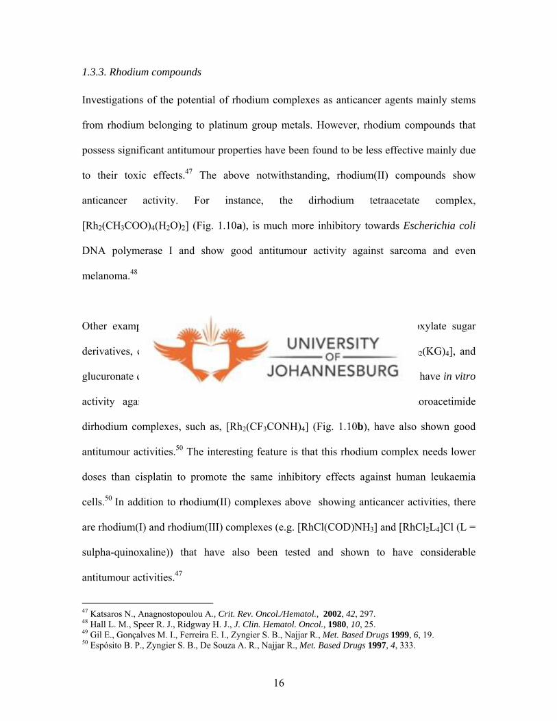
Investigations of the potential of rhodium complexes as anticancer agents mainly stems
from rhodium belonging to platinum group metals. However, rhodium compounds that
possess significant antitumour properties have been found to be less effective mainly due
to their toxic effects.47 The above notwithstanding, rhodium(II) compounds show
anticancer activity. For instance, the dirhodium tetraacetate complex,
[Rh2(CH3COO)4(H2O)2] (Fig. 1.10a), is much more inhibitory towards Escherichia coli
DNA polymerase I and show good antitumour activity against sarcoma and even
melanoma.48
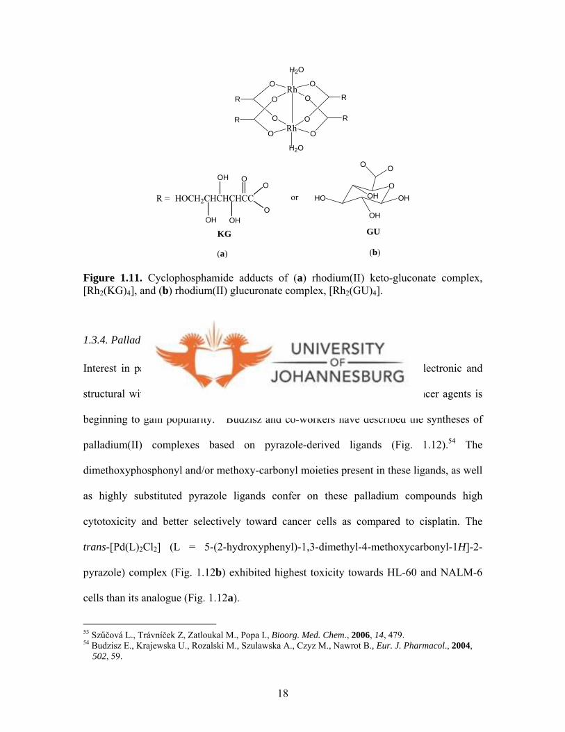
Other examples of rhodium complexes include two new rhodium carboxylate sugar
derivatives, cyclophosphamide adducts of rhodium(II) keto-gluconate, [Rh2(KG)4], and
glucuronate compounds [Rh2(GU)4] (Fig. 1.11), which have been shown to have in vitro
activity against the human leukemia cell line, K-562 cells.49 Trifluoroacetimide
dirhodium complexes, such as, [Rh2(CF3CONH)4] (Fig. 1.10b), have also shown good
antitumour activities.50 The interesting feature is that this rhodium complex needs lower
doses than cisplatin to promote the same inhibitory effects against human leukaemia
cells.50 In addition to rhodium(II) complexes above showing anticancer activities, there
are rhodium(I) and rhodium(III) complexes (e.g. [RhCl(COD)NH3] and [RhCl2L4]Cl (L =
sulpha-quinoxaline)) that have also been tested and shown to have considerable
antitumour activities.47
47 Katsaros N., Anagnostopoulou A., Crit. Rev. Oncol./Hematol., 2002, 42, 297. 48 Hall L. M., Speer R. J., Ridgway H. J., J. Clin. Hematol. Oncol., 1980, 10, 25. 49 Gil E., Gonçalves M. I., Ferreira E. I., Zyngier S. B., Najjar R., Met. Based Drugs 1999, 6, 19. 50 Espósito B. P., Zyngier S. B., De Souza A. R., Najjar R., Met. Based Drugs 1997, 4, 333.
16

Investigations suggest that rhodium compounds may exert their activity by causing
alterations to DNA.51 But a more likely mechanism of action of rhodium complexes may
involve protein binding of rhodium compounds at sulfhydryl sites, thereby inhibiting
enzymes essential for DNA synthesis such as DNA polymerase A, and hence inhibit
DNA synthesis.52
RhO
O
O
O
RhO
O
O
O
H2O
H2O
Rh
HN
O
Rh
O
NH
OH2 H2O
CF3
CF3
HNO
F3C
ONH
CF3
(a) (b)
Figure 1.10. The dirhodium tetraacetate complex, (a) [Rh2(CH3COO)4(H2O)2] and (b) [Rh2(CF3CONH)4]. 51 Clarke M. J., Zhu F., Frasca D. R., Chem. Rev., 1999, 99, 2511. 52 aHoward R. A., Spring T. G., Bear J. L., Cancer Res., 1976, 36, 4402; bTselepi-Kalouli E., Katsaros N., J. Inorg. Biochem. 1990, 40, 95.
17
RhO
O
O
O
RhO
O
O
O
H2O
H2O
R
R
R
R
OHO
OH
OH
O O
OHHOCH2CHCHCHCC
OH
OH
OH
OO
OR = or
KG GU
(a) (b)
Figure 1.11. Cyclophosphamide adducts of (a) rhodium(II) keto-gluconate complex, [Rh2(KG)4], and (b) rhodium(II) glucuronate complex, [Rh2(GU)4].
1.3.4. Palladium complexes
Interest in palladium(II) anticancer agents stems directly from being isoelectronic and
structural with platinum(II). Thus, development of palladium(II) as anticancer agents is
beginning to gain popularity.53 Budzisz and co-workers have described the syntheses of
palladium(II) complexes based on pyrazole-derived ligands (Fig. 1.12).54 The
dimethoxyphosphonyl and/or methoxy-carbonyl moieties present in these ligands, as well
as highly substituted pyrazole ligands confer on these palladium compounds high
cytotoxicity and better selectively toward cancer cells as compared to cisplatin. The
trans-[Pd(L)2Cl2] (L = 5-(2-hydroxyphenyl)-1,3-dimethyl-4-methoxycarbonyl-1H]-2-
pyrazole) complex (Fig. 1.12b) exhibited highest toxicity towards HL-60 and NALM-6
cells than its analogue (Fig. 1.12a).5 4
53 Szűčová L., Trávníček Z, Zatloukal M., Popa I., Bioorg. Med. Chem., 2006, 14, 479. 54 Budzisz E., Krajewska U., Rozalski M., Szulawska A., Czyz M., Nawrot B., Eur. J. Pharmacol., 2004, 502, 59.
18

NN
Me
MeCOOMeOH
N
N
Me
Me
MeOOCHO
Pd
Cl
ClNN
Me
MeP(O)(OMe)2OH
N
N
Me
Me
(MeO)2(O)PHO
Pd
Cl
Cl
(a) (b)
Figure 1.12. The trans-[Pd(L)2Cl2] complexes (L = 5-(2-hydroxyphenyl)-1,3-dimethyl-4-methoxycarbonyl-1H]-2-pyrazole), 5-(2-hydroxyphenyl)-1,3-dimethyl-4-dimethoxyphosphonite]-2-pyrazole)
Another palladium(II) complex of a ligand obtained from the condensation of 2-
(diphenylphosphino)benzaldehyde and ethyl hydrazinoacetate, has been reported (Fig.
1.13).55,56 Its activity against cisplatin-resistant U2-OS/Pt cells and HeLa cells is
attributed to the steric bulkiness of the ligand, and the tridentate nature of the ligand that
enforces a cis-conformation. The 6-membered ring formed around the palladium centre
stabilises the complex, which possibly helps to avoid easy translabilisation in the
biological milieu.
55 Bacchi A., Carcelli M., Costa M., Fochi A., Monici C., Pelagatti P., Pelizzi C., Pelizzi G., Roca L. M. S., J. Organomet. Chem., 2000, 593, 180. 56 Radulovic V., Bacchi A., Pelizzi G., Sladic D., Brceski I., Andjelkovic K., Monatsh. Chem., 2006, 137, 681.
19

PPd
N
ON
O
ClPhPh
H2N
Figure 1.13. A palladium(II) complex utilizing ligand obtained from condensation of 2-(diphenylphosphino)benzaldehyde and ethyl hydrazinoacetate.
1.3.5. Gold compounds
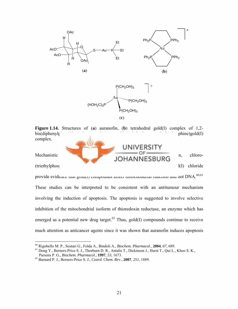
Gold complexes are mainly known for their use in treating rheumatoid arthritis.
Interestingly, gold(I) compounds including auranofin (Fig. 1.14a), have also been
reported to show antitumour activity.57 For instance, tetrahedral gold(I) complexes of 1,2-
bis(diphenylphosphino)ethane ligands (Fig 1.14b) display a wide spectrum of antitumour
activity in vivo, especially in cisplatin-resistant cell lines.58 Very recently, a hydrophilic
tetrakis((tris(hydroxy-methyl))phosphine)gold(I)chloride complex (Fig 1.14c) was
reported to be cytotoxic to several tumour cell lines, for example HCT-15 cells, which is
derived from human colon carcinoma.59
57 McKeage M. J., Maharaj L., Berners-Price S. J., Coord. Chem. Rev., 2002, 232, 127. 58 Urig S., Fritz-Wolf K., Réau R., Herold-Monde C., Tóth K., Davioud-Charvet E., Becker K., Angew. Chem. Int. Ed., 2006, 45, 1881. 59 Pillarsetty N., Katti K. K., Hoffman T. J., Volkert W. A., Katti K. V., Kamei H., Koide T., J. Med. Chem., 2003, 46, 1130.
20
Au
P(CH2OH)3
(HOH2C)3PP(CH2OH)3
P(CH2OH)3
O
H
AcOAcO
OAc
H
HH OAc
S Au P Et
Et
Et
Ph2P PPh2
Au
Ph2P PPh2
(a) (b)
(c)
Figure 1.14. Structures of (a) auranofin, (b) tetrahedral gold(I) complex of 1,2-bis(diphenylphosphino)ethane and (c) tetrakis((tris(hydroxy-methyl))phosphine)gold(I) complex.
Mechanistic studies into antitumour activity of auranofin, chloro-
(triethylphosphino)gold(I) and bis[1,2-bis(diphenylphosphino)ethane]gold(I) chloride
provide evidence that gold(I) compounds affect mitochondrial function and not DNA.60,61
These studies can be interpreted to be consistent with an antitumour mechanism
involving the induction of apoptosis. The apoptosis is suggested to involve selective
inhibition of the mitochondrial isoform of thioredoxin reductase, an enzyme which has
emerged as a potential new drug target.62 Thus, gold(I) compounds continue to receive
much attention as anticancer agents since it was shown that auranofin induces apoptosis
60 Rigobello M. P., Scutari G., Folda A., Bindoli A., Biochem. Pharmacol., 2004, 67, 689. 61 Dong Y., Berners-Price S. J., Thorburn D. R., Antalis T., Dickinson J., Hurst T., Qui L., Khoo S. K., Parsons P. G., Biochem. Pharmacol., 1997, 53, 1673. 62 Barnard P. J., Berners-Price S. J., Coord. Chem. Rev., 2007, 251, 1889.
21

by a mechanism involving inhibition of the mitochondrial enzyme thioredoxin
reductase.63, 64
On the other hand, gold(III) could be stabilised through judicious choice of donor atoms
that can impart stability to this higher oxidation state of gold. Gold(III) complexes are
isoelectronic and isostructural to platinum(II) and are promising candidates as anticancer
agents. Gold(III) compounds investigated for potential antitumour activity are inevitably
four coordinate and feature square planar geometries as found for cisplatin, thus a similar
mechanism of action, i.e. interaction with DNA and disruption of normal cellular
processes, may be operative for these gold(III) compounds.65,66 In this connection,
evidence has been provided that some gold(III) species, e.g. [AuCl(dien)]Cl2 (dien =
diethylendiamine) (Fig. 1.15), bind DNA.6 5
NH2
NH
AuNH2Cl
2Cl-
2+
Figure 1.15. The structure of [AuCl(dien)]Cl2.
Several other gold(III) compounds with multidentate ligands such as ethylenediamine,
diethylenediamine and N-benzyl-N,N-dimethylamine have also been reported to be active
63 Gromer S., Arscott L. D., William Jr. C. H., Schirmer R. H., Becker K., J. Biol. Chem., 1998, 273, 20096. 64 Gromer S., Urig S., Becker K., Med. Res. Rev., 2004, 24, 40. 65 Carotti S., Guerri A., Mazzei T., Messori L., Mini E., Orioli P., Inorg. Chim. Acta, 1998, 281, 90. 66 Carotti S., Marcon G., Marussich M., Mazzei T., Messori L., Mini E., Orioli P., J.Chem.-Biol. Interact., 2000, 125, 29.
22

against human cancer cell lines.67 A recent in vitro cytotoxicity study demonstrated
promising activity of two gold(III) complexes (Fig. 1.16) with bispyridyl ligands,
[Au(bipy)(OH)2]PF6 and [Au(bipy*-H)(OH)]PF6 (bipy = bispyridyl).68 Both complexes
are quite stable under physiological conditions, with [Au(bip*-H)(OH)]PF6 being resistant
to sodium ascorbate reduction.6 8 From this particular study, and contrary to cisplatin,
mechanistic studies indicated that DNA is not the primary cellular target. What is
interesting about one of the gold(III) complexes (Fig. 1.16b) described above is the
stabilisation of the complex by a Au-C bond, thereby prohibiting easy reduction of the
complex. The five-membered chelating ring in which the nitrogen of the amino group and
the carbon of the aryl ring bond to the metal, stabilizes the complex, hence could possibly
prevent translabilisation.69 Other examples of gold(III) compounds that possess
antitumour activities include [AuCl2(ppy)] (ppy = 2-phenylpyridine)70 and [AuCl3(Hpm)]
and [AuCl2(pm)] (Hpm = 2-pyridylmethanol).71
N NAu
HO OH
PF6- N N
Au
HO
CH3
CH3
PF6-
(a) (b)
Figure 1.16. Structures of (a) [Au(bipy)(OH)2]PF6 and (b) [Au(bipy*-H)(OH)]PF6.
67 Messori L., Abbate F., Marcon G., Orioli P., Fontani M., Mini E., Mazzei T., Carotti S., O’Connell T., Zanelo P., J. Med. Chem., 2000, 43, 3541. 68 Marcon G., Carotti S., Coronnello M., Messori L., Mini E., Orioli P., Mazzei T., Cinellu M. A., Minghetti G., J. Med. Chem., 2002, 45, 1672. 69 Zhu Y., Cameron B. R., Skerlj R. T., J. Organomet. Chem., 2003, 677, 57. 70 Fan D., Yang C., Ranford J. D., Lee P. F., Vittal J. J., Dalton Trans., 2003, 2680. 71 Calamai P., Carotti S., Guerri A., Messori L., Mini E., Orioli P., Paolo G., J. Inorg. Biochem., 1997, 66,
23
Judging from the palladium, platinum and gold complexes cited above, it is apparent that
N-containing organic compounds are ligands of choice. In particular, the N-donor ligands
would stabilise gold in its 3+ oxidation state. Bis(pyrazolyl)alkanes are examples of such
N-donor ligands that could be utilised to prepare stable palladium, platinum and gold
compounds. However, there is little information on the use of bis(pyrazolyl)alkanes as
ligands for preparation of potential metal anticancer agents. Thus, the following section
highlights on the preparation of bis(pyrazolyl)alkanes and their derivatives.
Subsequently, their ability to form stable metal complexes is also highlighted.
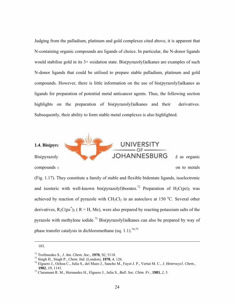
1.4. Bis(pyrazolyl)alkanes and related derivatives
Bis(pyrazolyl)alkanes, (R2C)n(pz*)2, (R = H, Me, tBu), can be described as organic
compounds constituting pyrazoles as sources of N-donors for coordination to metals
(Fig. 1.17). They constitute a family of stable and flexible bidentate ligands, isoelectronic
and isosteric with well-known bis(pyrazolyl)borates.72 Preparation of H2C(pz)2 was
achieved by reaction of pyrazole with CH2Cl2 in an autoclave at 150 oC.7 2 Several other
derivatives, R2C(pz*)2 ( R = H, Me), were also prepared by reacting potassium salts of the
pyrazole with methylene iodide.73 Bis(pyrazolyl)alkanes can also be prepared by way of
phase transfer catalysis in dichloromethane (eq. 1.1).74,75
103. 72 Trofimenko S., J. Am. Chem. Soc., 1970, 92, 5118. 73 Singh H., Singh P., Chem. Ind. (London), 1978, 4, 126. 74 Elguero J., Ochoa C., Julia S., del Mazo J., Sancho M., Fayet J. P., Vertut M. C., J. Heterocycl. Chem., 1982, 19, 1141. 75 Claramunt R. M., Hernandez H., Elguero J., Julia S., Bull. Soc. Chim. Fr., 1983, 2, 5.
24
N
N
R5
R3
R4N
N
R5'
R3'
R4'
C
R1 R2
n
R5 = R5' = H, MeR4 = R4' = H
R3 = R3' = H, MeR1 = H, Me n = 1, 2
Figure 1.17. General structure of bis(pyrazolyl)alkanes, (R2C)n(pz*)2.
N NH
R R
N
N
N
NCH2Cl2+ KOH/K2CO3
∆
R = H, Me, tBu
R
R
R
R
(1.1)
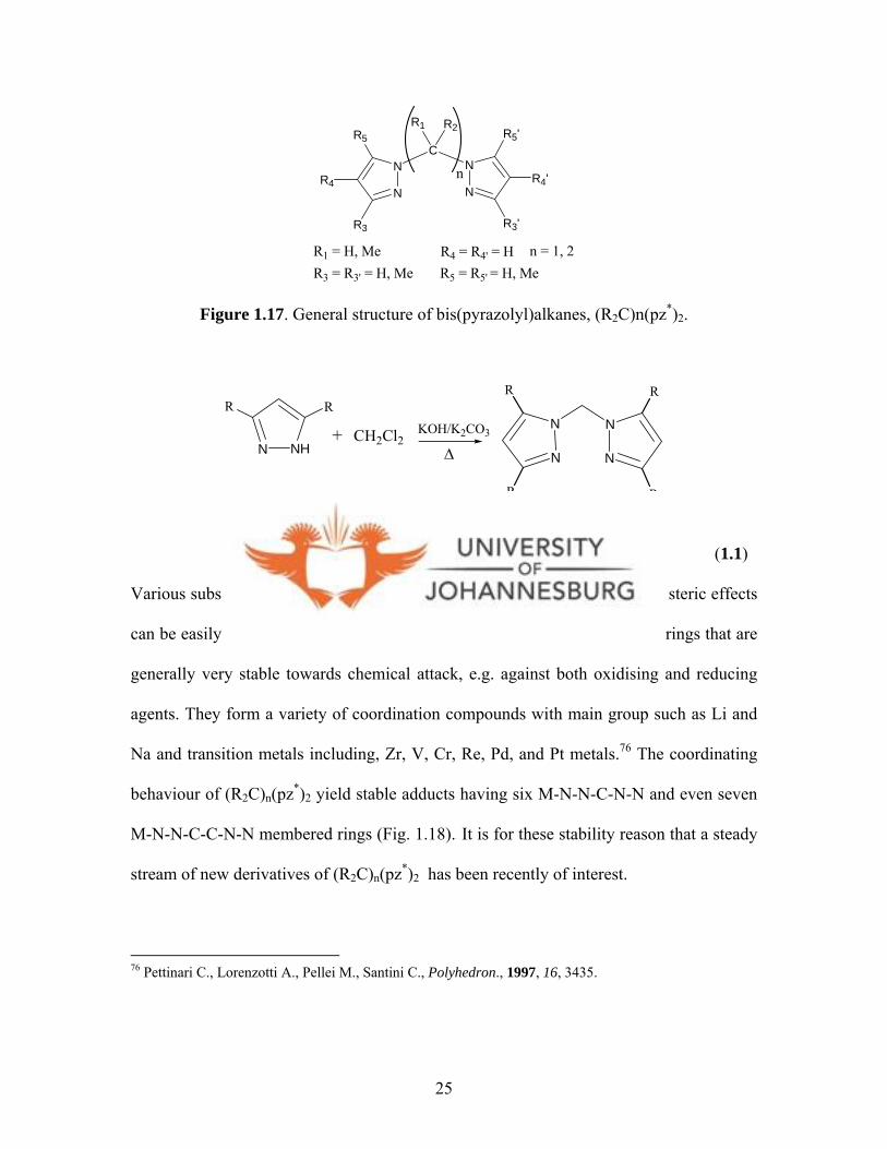
Various substituents may replace each hydrogen atom so that electronic and steric effects
can be easily varied (eq. 1.1). These bis(pyrazolyl)alkanes contain pyrazole rings that are
generally very stable towards chemical attack, e.g. against both oxidising and reducing
agents. They form a variety of coordination compounds with main group such as Li and
Na and transition metals including, Zr, V, Cr, Re, Pd, and Pt metals.76 The coordinating
behaviour of (R2C)n(pz*)2 yield stable adducts having six M-N-N-C-N-N and even seven
M-N-N-C-C-N-N membered rings (Fig. 1.18).76 It is for these stability reason that a steady
stream of new derivatives of (R2C)n(pz*)2 has been recently of interest.
76 Pettinari C., Lorenzotti A., Pellei M., Santini C., Polyhedron., 1997, 16, 3435.
25
N
N
M
N
NC
N
N
CC
N
N
M
Figure 1.18. Observed coordination with (R2C)n(pz*)2.
Using similar procedures of phase transfer catalysis (vide supra), bis(pyrazolyl)ethanes
were also isolated.74,75 This was achieved by reaction of pyrazoles with dibromoethane
under phase transfer catalysis conditions. This procedure has since led to the isolation of
substituted (H2C)n(pzx)2 compounds (X = substituents on pz = H, NO2, 4-Br, 4-NO2,
NH2, 5-NH2; n = 1, 2).75,77
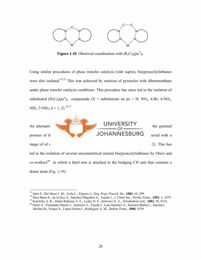
An alternative route to preparation of bis(pyrazolyl)alknes is by lithiating the geminal
protons of the alkane linker.78,79 The resulting carbanions can then be reacted with a
range of of electrophiles to get unsymmetrical bis(pyrazolyl)alkanes (eq. 1.2).79 This has
led to the isolation of several unsymmetrical neutral bis(pyrazolyl)alkanes by Otero and
co-workers80 in which a third arm is attached to the bridging CH unit that contains a
donor atom (Fig. 1.19).
77 Julia S., Del Mazo J. M., Avila L., Elguero J., Org. Prep. Proced. Int., 1984, 16, 299. 78 Diez-Barra E., de la Hoz A., Sánchez-Migallon A., Tejeda J., J. Chem Soc., Perkin Trans., 1993, 1, 1079. 79 Katritzky A. R., Abdel-Rahman A. E., Leahy D. E., Schwarz O. A., Tetrahedron Lett., 1983, 39, 4133. 80 Otero A., Fernandez-Baeza J., Antinolo A., Tejeda J., Lara-Sanchez A., Sanchez-Barba L., Sanchez- Molina M., Franco S., Lopez-Solera I., Rodriguez A. M., Dalton Trans., 2006, 4359.
26

NN
NN
R R
R R
NN
NN
R R
R R
Li
n-BuLi
R = H, Me; E = electrophile
NN
NN
R R
R R
E+E
(1.2)
These unsymmetrical ligands are similar to tris(pyrazolyl)methane ligand, but in this case
one of the pyrazole groups is replaced by a carboxylate, dithiocarboxylate, methoxy or
pyridine group (Fig. 1.19).80 The inclusion of a pyridinyl arm in a (Fig. 2.19) gives a
pyrazolyl ligand that can coordinate to metals not just as in a bidentate fashion but also in
a tridentate fashion. An introduction of a methoxy moiety as the third arm of the lithiated
bis(pyrazolyl)methane (eq. 1.2) gives b (Fig. 1.19), which is an N,N,O- tridentate ligand.
Similarly, addition of an acetamide moiety to a lithiated bis(pyrazolyl)methane leads to
formation of ligand c (Fig. 1.19).80 The delocalisation of electrons on the acetamide unit
would allow the coordination of this ligand in an N,N,N- or N,N,O- tridentate manner.
In order to prepare water-soluble bis(prazolyl) ligands, a new approach to synthesising
bis(pyrazolyl)acetic acid (Fig. 1.19d) was followed. It involves reacting dibromoacetic
acid with two mole equivalents of pyrazole and/or its derivatives to obtain the
carboxylate ligand (Fig. 1.19d).81 Ligand d binds either in an N,N- or N,O- bidentate
fashion or N,N,O- tridentate fashion. Ligand d is a hard acid donor ligand and thus serves
81 Burzlaff N., Hegelmann I., Weibert B., J. Organomet. Chem., 2001, 626, 16.
27
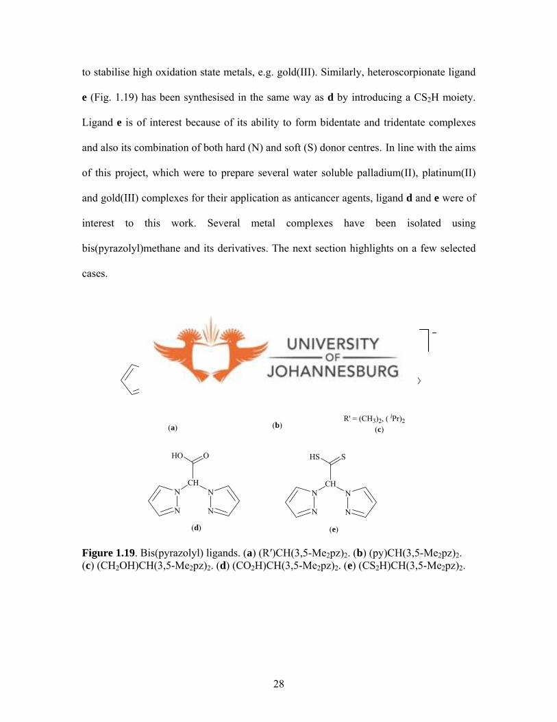
to stabilise high oxidation state metals, e.g. gold(III). Similarly, heteroscorpionate ligand
e (Fig. 1.19) has been synthesised in the same way as d by introducing a CS2H moiety.
Ligand e is of interest because of its ability to form bidentate and tridentate complexes
and also its combination of both hard (N) and soft (S) donor centres.80 In line with the aims
of this project, which were to prepare several water soluble palladium(II), platinum(II)
and gold(III) complexes for their application as anticancer agents, ligand d and e were of
interest to this work. Several metal complexes have been isolated using
bis(pyrazolyl)methane and its derivatives. The next section highlights on a few selected
cases.
N
N
N
NCH
COR'N
N
N
N
NCH
R' = (CH3)2, ( iPr)2
N
(c)(a)
N
N
N
NCH
HO O
N
N
N
NCH
HO
(d)
(b)
N
N
N
NCH
HS S
(e)
Figure 1.19. Bis(pyrazolyl) ligands. (a) (R′)CH(3,5-Me2pz)2. (b) (py)CH(3,5-Me2pz)2. (c) (CH2OH)CH(3,5-Me2pz)2. (d) (CO2H)CH(3,5-Me2pz)2. (e) (CS2H)CH(3,5-Me2pz)2.
28
1.5. Metal complexes of bis(pyrazolyl)alkanes and other related ligands
The use of bis(pyrazolyl)alkanes and their derivatives as ligands for preparation of
various metal complexes, would give rise to different coordination modes and
geometries; partly due to the flexibility of the bis(pyrazolyl)alkanes. The following
section highlights on the structural features of a few selected cases of early and late
transition metal complexes of bis(pyrazolyl)alkanes in that order.
1.5.1. Niobium(II) complexes
Group 5 metals having bis(pyrazolyl)alkanes as ligands have been prepared and
reported.82,83 For instance, reaction of H2C(5-PPh2pz)2 with [NbCl3(dme)]n produce a
binuclear complex [NbCl3{H2C(5-PPh2pz)2}]2 (pz = pyrazolyl) (Fig. 1.20a).82 However,
reaction of H2C(5-PPh2pz)2 with the mononuclear species [NbCl3(dme)(RC=CR′)] gives
the corresponding derivatives [NbCl3{H2C(5-PPh2pz)2}(RC=CR′)] (R = R′ = Ph; R = R′
= SiMe3; R = Ph, R′ = Me; R = Ph, R′ = SiMe3) (Fig. 1.20b).82 In general, ligand (5-
PPh2pz)2 coordinates to niobium in a bidentate N,N-fashion.8 3 In another instance the use
of bis(3,5-dimethylpyrazolyl)acetate (bdmpza), which is a water soluble ligand, leads to
isolation of the niobium(II) complex [Nb(NR)Cl2(K3-bdmpza)] (R = tBu, p-tolyl) with
bdmpza coordinated to niobium in a N,N,O- tridentate fashion.84 In spite of the fact that
bdmpza is a water soluble ligand the resultant niobium complex is not. Spectroscopic
82 Antinolo A., Carrillo-Hermosilla F., Diez-Barra E., Fernandez-Baeza J., Fernandez-Lopez M., Lara- Sanchez A., Moreno A., Otero A., Rodriguez A. M., Tejeda J., J. Chem. Soc., Dalton Trans., 1998, 3737. 83 Fernandez-Baeza J., Jalon F. A., Otero A., Rodrigo-Blanco M. E., J. Chem. Soc., Dalton Trans., 1995, 1015. 84 Otero A., Fernandez-Baeza J., Antinolo A., Tejeda J., Lara-Sanchez A., Sanchez-Barba L., Rodriquez A.
29
studies shows that the [Nb(NR)Cl2(K3-bdmpza)] complex is in an asymmetrical structural
disposition, thus making niobium a chiral centre (Fig. 1.20c). The X-ray structure of the
analogous [NbCl2(K3-bdmpza)(PhC CMe)] complex (Fig. 1.20d) is an indirect
confirmation of isolation of [Nb(NR)Cl2(K3-bdmpza)].85 Complex [NbCl2(K3-
bdmpza)(PhC CMe)] (Fig. 1.20d) exhibits an octahedral geometry where the two
pyrazolyl rings are located in cis- and trans- positions with respect to the (PhC CMe)
moiety.
N N
N N
CH2Nb
Cl
Cl
Cl
RC
CR'PPh2
PPh2
NN
NN
Ph2P
Ph2P
NbCl
Cl
ClN N
N N
PPh2
PPh2
Nb
Cl
R = R' = Ph; R = R' = SiMe3 R = Ph, R' = Me R = Ph, R' = SiMe3
(a)
(b)
N
N
N
NOO
NbCl NR
(c)
N
N
N
NOO
NbCl CPh
MeC
(d)
Cl
Cl
R = tBu, p-tolyl
Figure 1.20. Structures of (a) [NbCl3{H2C(5-PPh2pz)2}]2, (b) [NbCl3{H2C(5-PPh2pz)2}(RC=CR′)], (c) [Nb(NR)Cl2(K3-bdmpza)], (d) [NbCl2(K3-bdmpza)(PhC CMe)].
M., Dalton Trans., 2004, 3963. 85 Otero A., Fernandez-Baeza J., Tejeda J., Antinolo A., Carrillo-Hermosilla F., Diez-Barra E., Lara- Sanchez A., Fernandez-Lopez M., Lanfranchi M., Pellinghelli M. A., J. Chem. Soc., Dalton Trans., 1999, 3537.
30
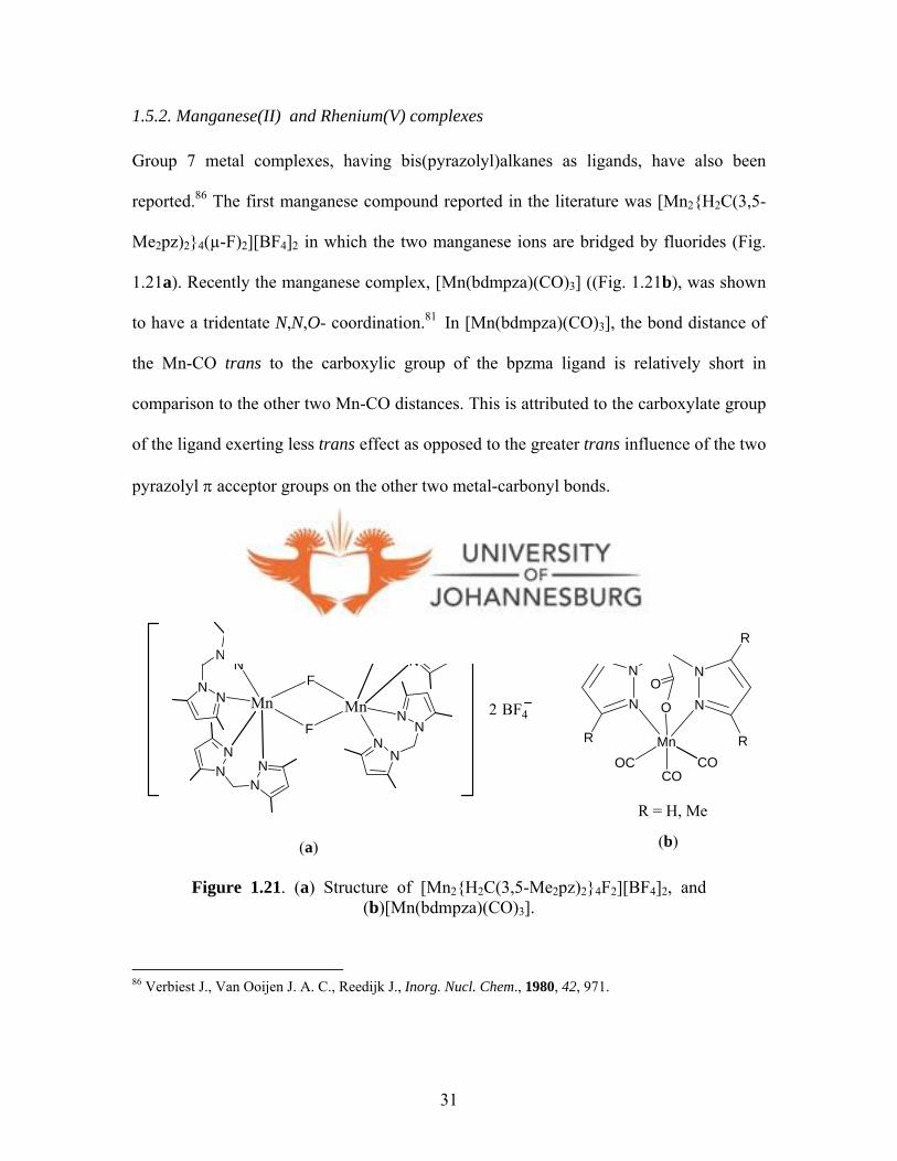
1.5.2. Manganese(II) and Rhenium(V) complexes
Group 7 metal complexes, having bis(pyrazolyl)alkanes as ligands, have also been
reported.86 The first manganese compound reported in the literature was [Mn2{H2C(3,5-
Me2pz)2}4(µ-F)2][BF4]28 6 in which the two manganese ions are bridged by fluorides (Fig.
1.21a). Recently the manganese complex, [Mn(bdmpza)(CO)3] ((Fig. 1.21b), was shown
to have a tridentate N,N,O- coordination.81 In [Mn(bdmpza)(CO)3], the bond distance of
the Mn-CO trans to the carboxylic group of the bpzma ligand is relatively short in
comparison to the other two Mn-CO distances. This is attributed to the carboxylate group
of the ligand exerting less trans effect as opposed to the greater trans influence of the two
pyrazolyl π acceptor groups on the other two metal-carbonyl bonds.81
NN
NN
NN
NN
Mn Mn
F
FBF42
NN
NN
NN
NN
2
N
N
R
R
N
N
R
R
O
O
Mn
COOC CO
R = H, Me
(a) (b)
Figure 1.21. (a) Structure of [Mn2{H2C(3,5-Me2pz)2}4F2][BF4]2, and (b)[Mn(bdmpza)(CO)3].
86 Verbiest J., Van Ooijen J. A. C., Reedijk J., Inorg. Nucl. Chem., 1980, 42, 971.
31
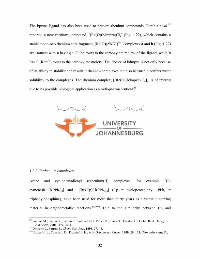
The bpzma ligand has also been used to prepare rhenium compounds. Porchia et al.87
reported a new rhenium compound, [(Re(O)(bdmpza)Cl2] (Fig. 1.22), which contains a
stable mono-oxo rhenium core fragment, [Re(O)(NNO)]2+. Complexes a and b (Fig. 1.22)
are isomers with a having a Cl ion trans to the carboxylate moiety of the ligand, while b
has O (Re=O) trans to the carboxylate moiety.8 7 The choice of bdmpza is not only because
of its ability to stabilise the resultant rhenium complexes but also because it confers water
solubility to the complexes. The rhenium complex, [(Re(O)(bdmpza)Cl2], is of interest
due to its possible biological application as a radiopharmaceutical.88
N
N
N
N
O
O
Re
ClCl O
N
N
N
N
O
O
Re
OCl Cl
(a) (b)
Figure 1.22. Structural isomers of [Re(O)(bdmpza)Cl2].
1.5.3. Ruthenium complexes
Arene and cyclopentadienyl ruthenium(II) complexes, for example [(P-
cymene)RuCl(PPh3)2] and [Ru(Cp)Cl(PPh3)2] (Cp = cyclopentadienyl; PPh3 =
triphenylphosphine), have been used for more than thirty years as a versatile starting
material in organometallic reactions.89,9091 Due to the similarity between Cp and
87 Porchia M., Papini G., Santini C., Lobbia G. G., Pellei M., Tisato F., Bandoli G., Dolmella A., Inorg. Chim. Acta, 2006, 359, 2501. 88 Dilworth J., Parrott S., Chem. Soc. Rev., 1998, 27, 43. 89 aBozec H. L., Touchard D., Dixneuf P. H., Adv. Organomet. Chem., 1989, 29, 163; bGovindaswamy P.,
32

hydrotris(pyrazol-1-yl)borato ligand (Tp), the complex [Ru(Tp)Cl(PPh3)2] has also been
isolated.92 Furthermore, because of the resemblance between Tp and bis(pyrazolyl) acetic
acid (bpza), bpza has recently been used to prepare a stable ruthenium(III) complex,
[Ru(bpza)(Cl)2(PPh3)] (Fig. 1.23).89 The compound exhibits a tridentate N,N,O
coordination of the ligand, with the PPh3 being trans to one of the pyrazolyl rings.
However, there is no information on its application
N
N
N
N
HC
OO
Ru
Ph3PCl
Cl
Figure 1.23. Structure of [Ru(bdmpza)Cl2(PPh3)].
1.5.4. Cobalt complexes
To the best of our knowledge, there are no known examples of cobalt complexes based
on bis(pyrazolyl)acetic acid ligands, but a related ligand, tris(pyrazolyl)methane sulfonate
ligands (Tpms) (Fig. 1.24a), has been used to prepare cobalt complexes as mimics for
coenzymes related to vitamin B12.93 The Tpms ligand has also been used to mimic
metalloenzymes called His-1-carboxylate enzymes,94 which refers to mononuclear non-
Mozharivskyj A. Y., Kollipara M. R., Polyhedron, 2004, 23, 1567; cGovindaswamy P., Mozharivskyj A. Y., Kollipara M. R., Polyhedron, 2004, 23, 3115. 90 Lopez-Hernandez A., Muller R., Kopf H., Burzlaff N., Eur. J. Inorg. Chem., 2002, 671. 91 Lopez-Hernandez A., Muller R., Kopf H., Burzlaff N., Eur. J. Inorg. Chem., 2002, 671. 92 Alcock N. W., Burn I. D., Claire K. S., Hill A. F., Inorg. Chem., 1992, 31, 2906. 93 Parkin G., Chem. Rev., 2004, 104, 699. 94 Papish E. T., Taylor M. T., Jernigan F. E., Rodig M. J., Shawhan R. R., Yap G. P. A., Jove F. A., Inorg. Chem., 2006, 45, 2242.
33

heme iron(II) enzymes responsible for various metabolic process, e.g. biosynthesis of
antibiotics.95 Reactions of Tpms with Co(NO3)2.6H2O gives [Co(Tpms)2] (Fig. 1.24a).
Coordination of Tpms to cobalt(II) ion is through N,N,O- ligating end to give
[Co(Tpms)2] (Fig. 1.24b). Compound [Co(Tpms)2] shows a distorted octahedral
geometry. Other similar cobalt(II) complexes utilising pyrazolyl ligands include
[CoCl2L] (L = bidentate N-donor (2-propargyloxyphenyl)bis(pyrazolyl)methane) (Fig.
1.24c).96 The structure of [CoCl2L] is a five-coordinate complex consisting of a chloro-
bridged dimer of [CoCl2L] units. The coordination around each metal ion is a distorted
trigonal bipyramid. While there is no known native occurrence of cobalt(II) enzymes,
cobalt has been used for preparation of mimics of several enzymes.93,94 because of its
spectroscopic properties that make it an important structural and functional probe with
respect to enzymes functions.97
95 Hegg E. L., Que Jr. L., Eur. J. Biochem., 1997, 250, 625. 96 Mohr F., Cerrada E., Laguna M., Dalton Trans., 2006, 5567 97 Maret W., Vallee B. L., Methods Enzymol., 1993, 226, 52.
34
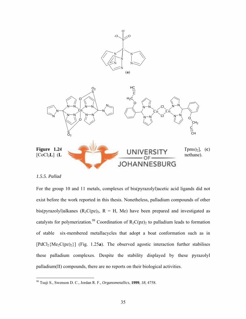
N
N
N
NC
-OS
O
(a)
NN
N NO
H2CC
HC
N N
NN
OCH2
CCH
Co CoCl
ClNN
N N
N N
NN
NN
NN
SO2
O
O2S
O
Co
(b) (c)
N
N
O-
Figure 1.24. (a) Tris(pyrazolyl)methane sulfonate (Tpms), (b) [Co(Tpms)2], (c) [CoCl2L] (L = bidentate N-donor (2-propargyloxyphenyl)-bis(pyrazolyl)methane).
1.5.5. Palladium, platinum and gold complexes
For the group 10 and 11 metals, complexes of bis(pyrazolyl)acetic acid ligands did not
exist before the work reported in this thesis. Nonetheless, palladium compounds of other
bis(pyrazolyl)alkanes (R2C(pz)2, R = H, Me) have been prepared and investigated as
catalysts for polymerization.98 Coordination of R2C(pz)2 to palladium leads to formation
of stable six-membered metallacycles that adopt a boat conformation such as in
[PdCl2{Me2C(pz)2}] (Fig. 1.25a).9 8 The observed agostic interaction further stabilises
these palladium complexes. Despite the stability displayed by these pyrazolyl
palladium(II) compounds, there are no reports on their biological activities.
98 Tsuji S., Swenson D. C., Jordan R. F., Organometallics, 1999, 18, 4758.
35

Structural studies of neutral [PdCl2{H2C(pz)2}], [PdCl2{H2C(3,5-Me2pz)2}] and
[PdCl2{Me2C(pz)2}] and the cationic species [Pd{H2C(pz)2}2]2+, [Pd{H2C(3,5-
Me2pz)2}]2+ and [Pd{Me2C(pz)2}2]2+ (Figs. 1.25b and c) display weak Pd…H-C (agostic)
as reported by Minghetti and co-workers.99 These agostic interactions make the
complexes quite stable.99
NN
N N
N N
NN
Pd
(b)
R R
R R R R
R R 2+
R = H, Me
NN
N N
N N
NN
Pd
2+
(c)
NNNN
C Pd
H2C
Me
H
Cl
Cl
(a)
Figure 1.25. (a) Six-membered palladacycle [PdCl2{Me2C(pz)2}2], (b) [Pd{H2C(3,5-R2pz)2}2]2+, (c) [Pd{Me2C(pz)2}2]2+.
Stable platinum(II) complexes of bis(pyrazolyl)alkanes ligands have also been reported
and include the neutral complexes, [PtCl2{H2C(3,5-R2pz)2}] ( R = H, Me) (Fig. 1.26).99
They have distorted square-planar geometry with the proton (Hendo) of the bridging
methylene in close proximity to the metal centre with a boat conformation of the
99 Minghetti G., Cinellu M. A., Bandini A. L., Banditelli G., De Martin F., Manassero M., J. Organomet. Chem., 1986, 315, 387.
36
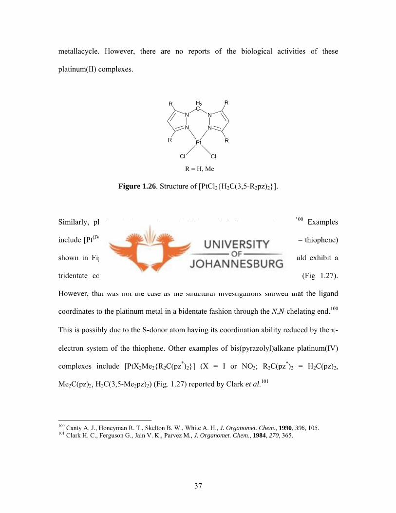
metallacycle. However, there are no reports of the biological activities of these
platinum(II) complexes.
N
N
N
N
H2C
Pt
Cl Cl
R = H, Me
R
R
R
R
Figure 1.26. Structure of [PtCl2{H2C(3,5-R2pz)2}].
Similarly, platinum(IV) complexes of bis(pyrazolyl)alkanes are known.100 Examples
include [Pt(IV)I2Me2(R)HC(pz)2] complexes ( R = py, thi; py = pyridine, thi = thiophene)
shown in Figure 1.27.100 It was expected that [Pt(IV)I2Me2(thi)HC(pz)2] would exhibit a
tridentate coordination mode because of the sulfur atom in thiophine (Fig 1.27).
However, that was not the case as the structural investigations showed that the ligand
coordinates to the platinum metal in a bidentate fashion through the N,N-chelating end.100
This is possibly due to the S-donor atom having its coordination ability reduced by the π-
electron system of the thiophene. Other examples of bis(pyrazolyl)alkane platinum(IV)
complexes include [PtX2Me2{R2C(pz*)2}] (X = I or NO3; R2C(pz*)2 = H2C(pz)2,
Me2C(pz)2, H2C(3,5-Me2pz)2) (Fig. 1.27) reported by Clark et al.101
100 Canty A. J., Honeyman R. T., Skelton B. W., White A. H., J. Organomet. Chem., 1990, 396, 105. 101 Clark H. C., Ferguson G., Jain V. K., Parvez M., J. Organomet. Chem., 1984, 270, 365.
37
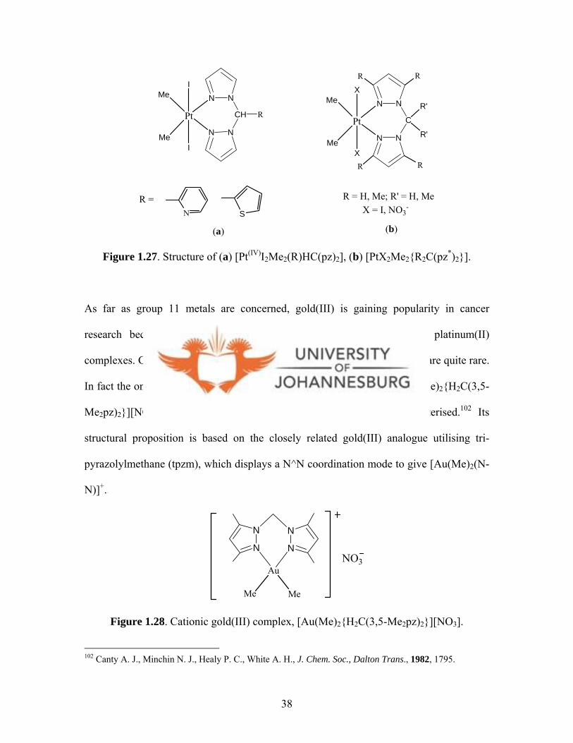
N N
N N
CPt
X
XMe
Me
R'
R'
(b)
N N
N N
CH RPt
I
IMe
Me
(a)
R R
R R
R = H, Me; R' = H, Me X = I, NO3
-SN
R =
Figure 1.27. Structure of (a) [Pt(IV)I2Me2(R)HC(pz)2], (b) [PtX2Me2{R2C(pz*)2}].
As far as group 11 metals are concerned, gold(III) is gaining popularity in cancer
research because of its isoelectronic and isostructural similarities to platinum(II)
complexes. Gold(III) complexes utilising poly(pyrazolyl)alkanes as ligands are quite rare.
In fact the only gold compound described in literature is the cationic [Au(Me)2{H2C(3,5-
Me2pz)2}][NO3] (Fig. 1.28), which has never been structurally characterised.102 Its
structural proposition is based on the closely related gold(III) analogue utilising tri-
pyrazolylmethane (tpzm), which displays a N^N coordination mode to give [Au(Me)2(N-
N)]+.1 0 2
N
N
N
N
Au
Me Me
NO3
Figure 1.28. Cationic gold(III) complex, [Au(Me)2{H2C(3,5-Me2pz)2}][NO3].
102 Canty A. J., Minchin N. J., Healy P. C., White A. H., J. Chem. Soc., Dalton Trans., 1982, 1795.
38
1.6. Aims of this study
From the extensive overview above on bis(pyrazolyl) ligands and their complexation to
various transition metals, it is clear that these ligands lead to isolation of stable metal
complexes. Owing to the interest in developing new metal compounds as anticancer
drugs, the aim was to prepare palladium(II), platinum(II) and gold(III) complexes by
utilising bis(pyrazolyl)acetic acid (R2bpza) compounds as ligands because of their
potential water solubility and flexible coordination. In addition, σ-donor ability of such
ligands was expected to stabilise gold(III) compounds. The choice of gold(III) emanates
from the fact that this ion gives rise to complexes that are isoelectronic and isostructural
with those of platinum(II), whose antitumour activity is well established. The expected
coordination of the bis(pyrazolyl)acetic acid to palladium(II), platinum(II), and gold(III),
would give compounds that are closely related to cisplatin. This is not to insinuate that
the biological activities would be the same. It is also worth mentioning that the
bis(pyrazolyl)acetic acid gold(III) complexes prepared in this thesis, are the second
examples of gold(III) complexes of bis(pyrazolyl)alkanes after [Au(Me)2{H2C(3,5-
Me2pz)2}][NO3] described earlier (vide supra).102 Furthermore, by using
bis(pyrazolyl)acetic acid ligands it is shown that these ligands are able to stabilise both
palladium(II) and gold(III) enough to allow the kinetics of their substitution reactions
with L-cysteine to be studied.
Owing to the insignificant biological activities observed with the new palladium(II),
platinum(II) and gold(III) complexes reported in this thesis, a second initiative involving
39
the use of bis(pyrazolyl) dithioic acid ligands, was pursued. However, attempts to prepare
gold(III) complexes from this type of ligand were unsuccessful as the resultant gold(III)
complexes readily decomposed. As a result of this, no further attempts were made to
isolate the palladium(II) and platinum(II) analogues either. Nonetheless, further
exploration of dithiocarbamate (dtc) as ligands for synthesising gold(I) complexes, was
pursued. Bearing in mind that the scope of this thesis included preparation of gold(I)
complexes, the use of these dtc ligands led to isolation of several mononuclear and
dinuclear gold(I) complexes, which are reported in this thesis.
40

CHAPTER 2
BIS(PYRAZOLYL)ACETIC ACID COMPLEXES OF PALLADIUM(II),
PLATINUM(II) AND GOLD(III)
X-ray structures L4, 1, 3b, and 5b were solved by Dr. Guzei and Mrs. Spencer both of
Wisconsin University (USA), whilst X-ray structure of 2 was solved by the candidate and
Dr. Omondi (University of Johannesburg). The crystal structure discussions are the sole
work of the candidate except for 3b, where Dr. Guzei helped with the discussion.
2.0 Introduction
Substituted pyrazoles constitute an important family of heterocyclic compounds that have
found use in drug development and catalysis.1 Thus there is considerable interest in the
synthesis of pyrazole-based ligands e.g. bis(pyrazolyl)alkanes. Pyrazole-based ligands are
remarkably versatile sources of coordination ligands.2,3 Research on pyrazole-derived
ligands has focused on models of metalloproteins and the applications of
pyrazolylborates as ligands in coordination chemistry.3 The appeal of pyrazole and its
derivatives is the opportunity it offers to alter steric, electronic and solubility properties
of the resultant complexes. It is for this reason that pyrazoles continue to be an important
class of ligands in the syntheses of several metal complexes.
1 Elguero J., Comprehensive Hetercyclic Chemistry II: Pyrazoles, vol. 3, edited by Katrizky A. R., Rees C. W., Scriven E. F. V., 1993, 1-75. 2 Trofimenko S., Chem. Rev., 1972, 72, 492. 3 Esquius G., Pons J., Yanez R., Ros J., Solans X., Font-Bardia M., J. Organomet. Chem., 2000, 605, 226.
41
The ease with which substituted pyrazoles could be achieved is also an added advantage.
This is evident from the extensive overview on the bis(pyrazolyl)alkane ligands (Chapter
1). In this study, pyrazole and 3,5-dimethylpyrazole were used to prepare water soluble
bis(pyrazolyl)acetic acid (L1) and bis(3,5-dimethylpyrazolyl) acetic acid (L2) ligands.
Subsequently, palladium(II), platinum(II) and gold(III) complexes were prepared by
reacting ligands L1 and L2 with the respective metal salts. The results are systematically
reported in the following sections. Schemes 2.1 and 2.2 below show ligands and various
metal complexes reported in this thesis.
42
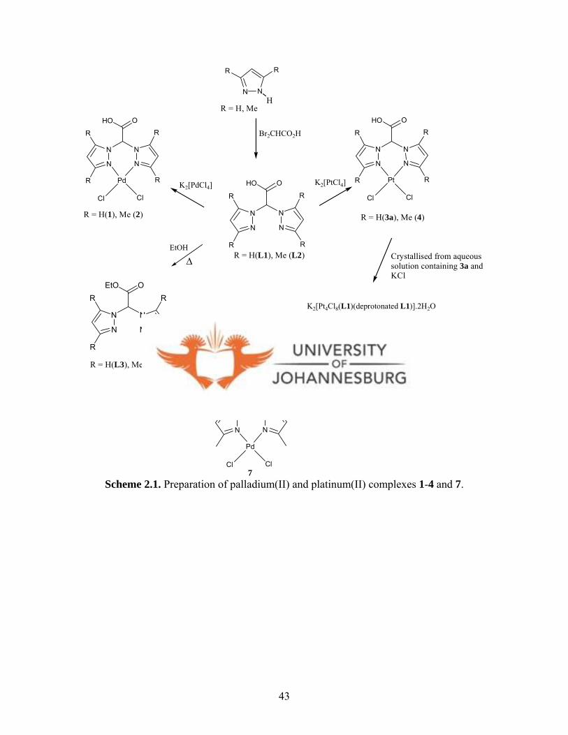
N
N N
N
HO O
R
R
R
R
N
N N
N
EtO OR
R
R
R
N
N N
N
EtO O
Pd
Cl Cl
N
N N
N
HO O
R
R
R
RPd
Cl Cl
[PdCl2(NCMe)2]
K2[PtCl4]
EtOH
∆
K2[PdCl4]
N
N N
N
HO O
R
R
R
RPt
Cl Cl
R = H(L1), Me (L2)
R = H(1), Me (2) R = H(3a), Me (4)
R = H(L3), Me (L4)
7
K2[Pt4Cl8(L1)(deprotonated L1)].2H2O
(3b)
Crystallised from aqueous solution containing 3a andKCl
Br2CHCO2H
N N
RR
HR = H, Me
Scheme 2.1. Preparation of palladium(II) and platinum(II) complexes 1-4 and 7.
43

N
N N
N
HO O
R
R
R
R
N
N N
N
HO O
R
R
R
RAu
Cl Cl
[HAuCl4].4H2O
+
Cl-
R = H (L1), Me (L2)R = H (5a), Me (6a)
N
NH
N
N
AuCl
Cl
Hydrolysis
R
R
R
R
R = H (5b), Me (6b)
Scheme 2.2. Shows preparation of gold(III) complexes 5a and 6a and the hydrolysis products 5b and 6b.
2.1. Experimental
2.1.1. Materials and Instrumentation
All commercial chemicals other than those described below were used as received.
K2[PtCl4], K2[PdCl4], pyrazole, 3,5-dimethylpyrazole, KOH, K2CO3, NaHCO3, MgSO4,
dibromoacetic acid and L-cysteine were purchased from Sigma-Aldrich and used without
further purification. Bis(pyrazolyl)acetic acid (L1) and bis(3,5-dimethylpyrazolyl)acetic
acid (L2) were synthesised according to the literature procedures with minor
modifications.4 The palladium and gold starting materials, [PdCl2(NCMe)2]5 and
4 Burzlaff N., Hegelmann I., Weibert B., J. Organomet. Chem., 2001, 626, 16. 5 King B. R., Eisch J. J., Organometallic syntheses. Elsevier Science Publishers B. V., New York, 1986, 3, 324.
44
[HAuCl4].4H2O,6 were synthesised according to the literature procedures. All the
solvents were also purchased from Sigma-Aldrich and dried using conventional methods.
Tetrahydrofuran (THF) and diethylether (Et2O) were dried over sodium with sodium
benzoate as an indicator, while dichloromethane (CH2Cl2) was dried over phosphorous
pentoxide with sodium benzoate as an indicator. The water used was double distilled. All
manipulations of air-and/or moisture sensitive compounds were performed under dry,
deoxygenated nitrogen atmosphere using Schlenk techniques.
IR spectra were recorded as nujol mulls using NaCl cells and some as KBr pellets on a
Perkin-Elmer, paragon 1000 PC FTIR spectrophotometer. 1H and 13C{1H} NMR spectra
were recorded on a Gemini 2000 instrument (1H, 200 MHz and 13C, 50.3 MHz) and a
Bruker Avance DPX 300 spectrometer (1H, 300 MHz) in CDCl3 and DMSO-d6 at room
temperature. 1H and 13C chemical shifts were referenced to the signals of the residual
protons of the NMR solvents and are quoted in ppm as follows; (i) CDCl3 at 7.26 and
77.0 ppm for 1H and 13C respectively and (ii) for DMSO-d6 at 2.50 and 39.9 ppm for 1H
and 13C respectively. Elemental analyses were performed on a Fisons elemental analyser
at the University of Cape Town, South Africa. ESI-MS spectra were recorded on a
Waters API Quattro Micro spectrotometer at the University of Stellenbosch, South
Africa. The spectra were collected using 3.0 s cyclical scans and applying the sample
cone voltage of 15 V at the source block temperature of 100 oC. Desolvation temperature
was 350 oC and desolvation cone gas flow rate 350L/h.
6 Block B. P., Inorg. Synth., 1953, 4, 14.
45
2.1.2. Synthesis of ligands and metal complexes
2.1.2.1. Bis(pyrazol-1-yl)acetic acid (L1)
To a solution of KOH (1.43 g, 25 mmol) and K2CO3 (3.46 g, 25 mmol) in THF (100 mL)
was added dibromoacetic acid (1.92 g, 8.81 mmol) and pyrazole (1.20 g, 17 mmol).
Benzyltriethylammonium chloride (1.00g, 4.40 mmol) was added to the solution as a
phase transfer catalyst and the mixture refluxed for 15 h. The solvent was then removed
and the residue re-dissolved in 25 mL of water. The pH of the resultant solution was then
adjusted to 7 using conc HCl and unreacted pyrazole removed by extraction with Et2O
(25 mL). The pH of the aqueous phase was further adjusted to 2 and L1 extracted with
Et2O (25 mL) three times. Subsequently L1 was recrystallised from Et2O/acetone
mixture. Yield = 1.01 g (60%). 1H NMR (DMSO-d6, 200 MHz): δ = 7.98 (d, 2H, 3JHH =
2.20 Hz, 5-pz); 7.59 (d, 2H, 3JHH = 2.21 Hz, 3-pz); 7.57(s, 1H, CHCO2H); 6.35 (s, 2H, 4-
pz). 13C{1H} NMR (DMSO-d6, 200 MHz): 166.2 (C(C=O)); 142.3 (C(3-pz); 131.0 (C(4-
pz)); 106.1 (C(5-pz)); 73.2 (C(CHCO2H)). IR (Nujol, cm-1): υO-H = 3439, υC=O = 1720,
υC=N = 1509. Anal. Calc. for C8H8N4O2: C, 50.00; H, 4.20; N, 29.15%. Found: C, 49.95;
H, 3.94; N, 28.59%.
2.1.2.2. Bis(3,5-dimethylpyrazol-1-yl)acetic acid (L2)
The synthesis of L2 was performed in a similar manner as described for L1 using KOH
(1.78 g, 31.26 mmol); K2CO3 (4.32 g, 31.26 mmol); dibromoacetic acid (2.27 g, 10.42
mmol); 3,5-dimethylpyrazole (2.00 g, 20.83 mmol); benzyltriethylammonium chloride
(1.00g, 4.40 mmol). Yield = 1.83 g (71%). 1H NMR (DMSO- d6, 200 MHz): δ = 7.13 (s,
1H, CHCO2H); 5.86 (s, 2H, 4-pz); 2.17 (d, 12H, 6JHH = 20.8 Hz, 5-pz, 3-pz). 13C{1H}
46
NMR (DMSO- d6, 200 MHz): 165.9 (C(C=O)); 146.8 (C(3-pz); 140.5 (C(4-pz)); 106.4
(C(5-pz)); 71.4 (C(CHCO2H)); 13.2 (C(CH3, 5-pz)); 10.8 (C(CH3, 3-pz)). IR (Nujol cm-
1): υO-H = 3402, υC=O = 1743, υC=N = 1558. Anal. Calc. for C12H16N4O2.0.5 H2O: C, 56.02;
H, 6.61; N, 21.78%. Found: C, 55.71; H, 5.75; N, 20.92%.
2.1.2.3. Bis(pyrazol-1-yl)ethyl acetate (L3).
Compound L1 (0.50 g, 2.60 mmol) was refluxed in excess ethanol (40 mL) under acidic
conditions (HCl, 5 mL) for 18 h. The mixture was cooled to room temperature and 50 mL
of deionised water added after which the pH was adjusted to 11 by adding NaHCO3. The
product was extracted with dichloromethane (50 mL) and dried over MgSO4. The solvent
was then removed in vacuo to afford an analytically pure white crystalline L3. Yield =
0.33 g (58%). 1H NMR (CDCl3, 200 MHz): δ 7.74 (d, 2H, pz, 3JHH = 2.2 Hz ); 7.59 (t,
2H, pz, 3JHH = 1.6 Hz,); 7.10 (s, CH, 1H, pz,); 6.33 (t, 2H, pz 2JHH = 1.8 Hz); 4.34 (q 2H,
CH2CH3, 3JHH = 7.4 Hz); 1.26 (t, 3H, CH2CH3, 3JHH = 7.4 Hz). 13C{1H}NMR (CDCl3, 200
MHz): δ 163.8 (C(C=O)); 140.4 (C(5-pz); 129.6 (C(3-pz)); 106.8 (C(4-pz)); 74.1
(C(CHCO2CH2CH3)); 62.7 (C(CH2, CH2CH3 )); 13.4 (C(CH3, CH2CH3 )). IR (Nujol cm-
1): υC=O = 1750. Anal. Calc. for C10H12N4O2: C, 54.54; H, 5.49; N, 25.44%. Found: C,
54.36; H, 5.75; N, 25.09%.
2.1.2.4. Bis(3,5-dimethylpyrazol-1-yl)ethyl acetate (L4).
The synthesis of compound L4 was performed in a similar manner as described for L3
using L2 (1.50 g, 6.05 mmol). Yield = 0.90 g (54%). 1H NMR (CDCl3, 300 MHz): δ 6.91
(s, CH, 1H, pz); 5.84 (s, 1H, pz); 4.36 (q, 2H, CH2CH3, 3JHH = 8.1 Hz); 2.19 (s, 6H, CH3,
47

5-pz); 2.11 (s, 6H, CH3, 3-pz); 1.30 (t, 3H, CH2CH3, 3JHH = 7.8 Hz). 13C{1H}NMR
(CDCl3, 200 MHz): δ 166.1 (C(C=O)); 147.0 (C(5-pz); 140.8 (C(3-pz)); 106.6 (C(4-pz));
71.7 (C(CHCO2CH2CH3)); 63.2 (C(CH2CH3)); 13.4 (C(CH2CH3)); 12.8 (C(CH3, 5-pz));
11.0 (C(CH3, 3-pz)). IR (Nujol cm-1): υC=O = 1735. Anal. Calc. for C14H20N4O2: C, 60.85;
H, 7.30; N, 20.28%. Found: C, 60.76; H, 7.55; N, 19.98%.
2.1.2.5. Dichloro-{bis(pyrazol-1-yl)acetic acid}palladium(II) (1).
To a yellow solution of K2[PdCl4] (0.17 g, 0.52 mmol) in distilled water (15 mL), was
added L1 (0.10 g, 0.52 mmol) and the mixture stirred vigorously at room temperature.
After 1.5 h, a colour change from a yellow solution to an orange suspension was
observed and further stirring for 3 h resulted in the formation of a yellow precipitate. The
precipitate was isolated by filtration and washed with ethanol to afford pure 1. Yield =
0.15 g; (75%). 1H NMR (DMSO-d6, 300 MHz): δ 8.29 (d, 2H, 2JHH = 1.8 Hz, 5-pz); 8.14
(s, 1H, CHCO2H) 8.02 (d, 2H, 2JHH = 1.8 Hz, 3-pz); 6.63 (t, 2H, 2JHH = 2.6 Hz, 4-pz).
13C{1H} NMR (DMSO-d6, 300 MHz): δ 164.9 (C(C=O)); 144.4 (C(5-pz); 136.9 (C(3-
pz)); 108.0 (C(4-pz)); 72.2 (C(CHCO2H)). IR (Nujol cm-1): υO-H = 3441, υC=O = 1738.
Anal. Calc. for C8H8Cl2N4O2Pd: C, 26.00; H, 2.18; N, 15.16%. Found: C, 26.34; H, 2.22;
N, 15.20%.
2.1.2.6. Dichloro-{bis(3,5-dimethylpyrazol-1-yl)acetic acid}palladium(II) (2).
The synthesis of 2 was performed in a similar manner as described for 1 using L2 (0.10
g, 0.40 mmol) and K2[PdCl4] (0.13 g, 0.40 mmol). Yield = 0.10 g; (63%). 1H NMR
(DMSO-d6, 300 MHz): δ 7.59 (s, 1H, CHCO2H); 6.23 (s, 2H, 4-pz); 2.58 (s, 6H, 5-pz);
48
2.51 (s, 6H, 3-pz). 13C{1H} NMR (DMSO-d6, 300 MHz): δ 165.3 (C(C=O)); 149.2 (C(5-
pz); 145.7 (C(3-pz)); 106.2 (C(4-pz)); 72.6 (C(CHCO2H)); 13.6 (C(3-CH3)); 12.9 (C(5-
CH3)). IR (Nujol cm-1): υO-H = 3410, υC=O = 1756. Anal. Calc. for
C12H16Cl2N4O2Pd.0.5CH2Cl2: C, 32.25; H, 3.23; N, 12.04%. Found: C, 32.27; H, 3.41; N,
12.25%.
2.1.2.7. Dichloro-{bis(pyrazol-1-yl)acetic acid}platinum(II) (3a)
To a red solution of K2[PtCl4] (0.43 g, 1.00 mmol) in distilled water (15 mL), was added
L1 (0.20 g, 1.00 mmol) and the mixture stirred vigorously at room temperature. After 2 h
a colour change from red solution to orange suspension was observed and further stirring
resulted in the formation of a yellow precipitate. The precipitate was isolated by filtration
as pure 3a. The filtrate left to stand for 1 day upon which golden yellow single crystals of
3b suitable for X-ray analysis were formed. Yield: 3a = 0.25 g (52%). 1H NMR (DMSO -
d6, 300 MHz): δ 8.34 (d, 2H, 3JHH = 2.20 Hz, 5-pz); 8.12 (d, 2H, 3JHH = 4.80 Hz, 3-pz);
7.96 (s, 1H, CHCO2H); 6.65 (s, 2H, 4-pz). 13C{1H} NMR (DMSO-d6, 300 MHz): δ 166.8
(C(C=O)); 140.1 (C(5-pz); 132.1 (C(3-pz)); 106.5 (C(4-pz)); 73.9 (C(CHCO2H)). IR
(Nujol, cm-1): υO-H = 3460, υC=O = 1763, υC=N = 1517. ESI-MS: m/z 457 (10%)
[PtCl2(bpza)]+ (10%), m/z 490 [PtCl2(bpza)+Na]+ (20%). Anal. Calc. for 3a,
C8H8Cl2N4O2Pt: C, 20.97; H, 1.76; N, 12.23%. Found: C, 20.67; H, 1.60; N, 12.29%.
2.1.2.8. K2[Pt4Cl8(L1)2(deprotonated L1)2].2H2O (3b)
After isolation of 3a, the filtrate was left to stand for 1 day upon which golden yellow
single crystals of 3b suitable for X-ray analysis were formed. Yield: = 0.05 g (11%).
49
Anal. Calc. for 3b, K2[C32H34Cl8N16O10Pt4]: C, 19.76; H, 1.76; N, 11.52%. Found: C,
19.68; H, 1.65; N, 11.89%.
2.1.2.9. Dichloro-{bis(3,5-dimethylpyrazol-1-yl)acetic acid}platinum(II) (4).
The synthesis of 4 was performed in a similar manner as described for 3, but reaction
time was longer (15 h). K2[PtCl4] (0.37 g, 0.81 mmol) and bis(3,5-dimethylpyrazol-1-
yl)acetic acid (0.20g, 0.81 mmol). Yield: 0.38 g (75%). 1H NMR (DMSO-d6, 300 MHz):
δ 7.13 (s, 1H, CHCO2H); 5.86 (s, 2H, 4-pz); 2.17 (s, 6H, 5-pz); 2.07 (s, 6H, 3-pz).
13C{1H} NMR (DMSO-d6, 300 MHz): δ 165.9 (C(C=O)); 146.8 (C(5-pz); 140.5 (C(3-
pz)); 106.4 (C(4-pz)); 71.4 (C(CHCO2H)); 13.2 (C(3-CH3)); 10.8 (C(5-CH3)). IR (Nujol
cm-1): υO-H = 3439, υC=O = 1743, υC=N = 1560. ESI-MS: m/z 519 [PtCl2(3,5-Me2bpza)]+
(10%). Anal. Calc. for C12H14Cl2N4O2Pt: C, 28.03; H, 3.14; N, 10.89%. Found: C, 27.72;
H, 3.25; N, 10.80%.
2.1.2.10. Dichloro-{bis(pyrazol-1-yl)acetic acid}gold(III) chloride (5a).
To a solution of [HAuCl4].4H2O (0.18 g, 0.52 mmol) in distilled water (10 mL) was
added L1 (0.10 g, 0.52 mmol) resulting in the immediate formation of a yellow
precipitate. The reaction mixture was stirred for 45 min at room temperature. The product
was isolated by filtration and dried in vacuo. Yield: 0.15 g (63%). 1H NMR (DMSO-d6,
300 MHz): δ 8.47 (m, 4H, 3-pz,5-pz); 8.11 (1H, CHCO2H); 6.95 (m, 2H, 4-pz). 13C{1H}
NMR (DMSO-d6, 300 MHz): 174.3 (C(C=O)); 149.4 (C(5-pz); 140.2 (C(3-pz)); 115.9
(C(4-pz)); 83.2 (C(CHCO2H)). IR (KBr, cm-1): υO-H = 3492, υC=O = 1761, υC=N = 1509.
50

ESI-MS: m/z 463 [AuCl2(bpza)]+ (5%). Anal. Calc. for C8H8Cl3N4O2Au: C, 24.39; H,
2.52; N, 9.42 %. Found: C, 24.53; H, 2.87; N, 9.53 %.
2.1.2.11. Dichloro-{bis(3,5-dimethylpyrazolyl)acetic acid}gold(III) chloride (6a).
The synthesis of 6a was performed in a similar manner as described for 5a using
[HAuCl4].4H2O (0.10 g, 0.30 mmol) and bis(3,5-dimethylpyrazol-1-yl)acetic acid (0.07
g, 0.30 mmol). Yield: 0.09 g (59%). 1H NMR (DMSO-d6, 300 MHz): δ 7.15 (s, 1H,
CHCO2H); 5.88 (s, 2H, 4-pz); 2.18 (s, 6H, 5-pz); 2.08(s, 6H, 3-pz). 13C{1H} NMR
(DMSO-d6, 300 MHz): 165.8 (C(C=O)); 146.9 (C(3-pz); 140.6 (C(4-pz)); 106.4 (C(5-
pz)); 71.1 (C(CHCO2H)); 13.1 (C(3-CH3)); 10.7 (C(5-CH3)). IR (KBr cm-1): υO-H = 3451,
υC=O = 1765, υC=N = 1518. ESI-MS: m/z 519 [AuCl2(3,5Me2bpza)]+ (5%). Anal. Calc. for
C12H14Cl3N4O2Au: C, 26.13; H, 2.92; N, 10.16%. Found: C, 25.70; H, 3.06; N, 9.99%.
2.1.2.12. Dichloro-{bis(3,5-dimethylpyrazol-1-yl)ethyl acetate}palladium(II) (7).
Compound 7 was prepared by suspending L4 (0.51 g, 1.80 mmol) in CH2Cl2 (20 mL) for
3 min after which it dissolved. [PdCl2(NCMe)2] (0.47 g, 1.80 mmol) was then added and
the mixture stirred for 6 h upon which a yellow precipitate was formed. The precipitate
was filtered, washed with minimum amount of ethanol and the solid dried to afford an
analytically pure complex. Yield = 0.42 g (51%). 1H NMR (CDCl3, 200 MHz): δ 1.35 (t,
3H, CH2CH3, 2JHH = 7.4 Hz); 2.25 (s, 6H, CH3, pz); 2.47 (s, 6H, CH3, pz); 4.31 (q 2H,
CH2CH3, 2JHH = 7.4 Hz); 5.90 (s, 1H, pz); 7.22 (S, CH, 1H, pz). 13C{1H}NMR (CDCl3,
200 MHz): δ 168.5 (C(C=O)); 145.7 (C(5-pz); 141.5 (C(3-pz)); 107.1 (C(4-pz)); 70.9
(C(CHCO2CH2CH3)); 62.4 (C(CH2)); 13.6 (C(CH3))13.4 (C(3-CH3)); 10.0 (C(5-CH3)).
51

IR (Nujol cm-1): υC=O = 1741. Anal. Calc. for C14H20Cl2N4O2Pd: C, 37.07; H, 4.44; N,
12.35%. Found: C, 37.48; H, 4.05; N, 12.62%.
2.1.3. X-ray crystallography
2.1.3.1. Data collection
An orange crystal of [PdCl2(L1)] (1) with approximate dimensions 0.45 x 0.33 x 0.21
mm3 was selected under oil under ambient conditions and attached to the tip of a
Micromount©. The crystal was mounted in a stream of cold nitrogen at 105(2) K and
centreed in the X-ray beam by using a video camera. The crystal evaluation and data
collection were performed on a Bruker CCD-1000 diffractometer with Mo Kα (λ =
0.71073 Å) radiation and the diffractometer to crystal distance of 4.9 cm.
The initial cell constants were obtained from three series of ω scans at different starting
angles. Each series consisted of 20 frames collected at intervals of 0.3º in a 6º range
about ω with the exposure time of 10 seconds per frame. A total of 131 reflections were
obtained. The reflections were successfully indexed by an automated indexing routine
built in the SMART program. The final cell constants were calculated from a set of
12116 strong reflections from the actual data collection.
The data were collected by using the full sphere data collection routine to survey the
reciprocal space to the extent of a full sphere to a resolution of 0.80 Å. A total of 20998
data were harvested by collecting four sets of frames with 0.4º scans in ω and one set of
frames with 0.5º scan in φ. These highly redundant datasets were corrected for Lorentz
52
and polarisation effects. The absorption correction was based on fitting a function to the
empirical transmission surface as sampled by multiple equivalent measurements.7
2.4.3.2. Structure solution and refinement
The systematic absences in the diffraction data were uniquely consistent for the space
group P21/n that yielded chemically reasonable and computationally stable results of
refinement.7 A successful solution by the direct methods provided most non-hydrogen
atoms from the E-map. The remaining non-hydrogen atoms were located in an alternating
series of least-squares cycles and difference Fourier maps. All non-hydrogen atoms were
refined with anisotropic displacement coefficients. All hydrogen atoms except those on
the water molecule were included in the structure factor calculation at idealised positions
and were allowed to ride on the neighbouring atoms with relative isotropic displacement
coefficients. There is also one solvated water molecule per Pd complex in the unit cell.
The water molecules were refined with restraints. The final least-squares refinement of
172 parameters against 3166 data resulted in residuals R (based on F2 for I≥2σ) and wR
(based on F2 for all data) of 0.0180 and 0.0504, respectively. The final difference Fourier
map was featureless. The molecular diagram is drawn with 50% probability ellipsoids.
The solid state structures of [PdCl2(L2)] (2), K2[Pt4Cl8(L1)2(deprotonated L1)2].2H2O
(3b), [AuCl2(pz)(pzH] (5b), and L4 were determined in a similar manner such as
described above for 1.
7 Bruker-AXS. (2000-2003) SADABS V.2.05, SAINT V.6.22, SHELXTL V.6.10 & SMART 5.622 Software Reference Manuals. Bruker-AXS, Madison, Wisconsin, USA.
53

2.2. Results and discussion
2.2.1. Synthesis of ligands
Ligands L1 and L2 were synthesised by reacting pyrazoles and dibromoacetic acid under
phase transfer conditions, by using benzyltriethylammonium chloride as the catalyst (eq.
2.1). Minor modifications to the literature procedure included using NaOH instead of
KOH and the reaction time increased to 15 h from 5 h, which led to improved yields as
compared to those in the literature.4 All the ligands were characterised by a combination
of IR, NMR spectroscopy, microanalysis and in selected cases mass spectrometry and X-
ray crystallography.
N
N N
N
HO O
R
R
R
R
Br2CHCO2H+
R = H (L1), Me (L2)
N N
RR
H
KOH, K2CO3
∆THF,
(2.1)
Ligands L1 and L2 were isolated as white solids in good yields at pH 2. The 1H and
13C{1H} NMR spectra of L1 and L2 are indicative of their isolation. For instance the 1H
NMR spectrum of L1 showed peaks due to the protons of the pyrazolyl rings and the sp3
CH linker at 7.98 ppm (5H, 5′H; pz), 7.59 ppm (3H, 3′H; pz), 7.57 ppm (sp3 CH linker
proton (CHCOOH)) and 6.35 ppm (4H, 4′H; pz). The peaks at 7.98 ppm (5H, 5′H) and
7.59 ppm (3H, 3′H) resonated as doublets as a direct consequence of their coupling with
the protons on the fourth positions of the pyrazole (4H, 4′H). The coupling constants are
54
ca. 2.2 Hz. Pyrazolyl protons appeared downfield as a result of the aromaticity invoked
by the π electron system of the pyrazolyl rings. Despite the fact that the CH linker proton
is an sp3 proton and that would normally appear upfield,8 that was not the case for L1 as
the sp3 proton appeared downfield (7.57 ppm). This is attributed to the fact that the sp3
proton is also de-shielded due to a less electron density chemical environment created by
the π- systems of the pyrazolyl rings. Similarly, the spectrum of L2 showed the same
pattern as that of L1. The only difference was the presence of methyl groups in L2, which
appear upfield at 2.17 and 2.27 ppm for 3-pz and 5-pz, respectively.
Subsequently the 13C{1H} NMR of L1 showed five distinctive peaks that were
characteristic of five carbons of L1. The peak at 166.2 ppm in the 13C{1H} NMR of L1
was characteristic of carbonyl carbon (CHCO2H) of L1, whereas the peaks at 140.1,
131.0 and 106.5 ppm were due to pyrazolyl ring carbons, C-5pz, C-3pz and C-4pz,
respectively. The carbonyl carbon appears downfield at 166.2 due to its de-shielded
environment as a result of the carboxylic moiety. The carboxylic moiety has
delocalisation of electrons around O=C-OH bonds,8 which leads to reduced electron
density around the carbonyl carbon; thus leading to it appearing downfield at 166.2 ppm.
Generally the pyrazolyl ring carbons also appeared downfield (C-5pz = 140.1 ppm, C-3pz
= 131.0 ppm and C-4pz = 106.5 ppm) as a result of the de-shielding effect brought about
by the π electron system of the pyrazolyl rings.8 The sp3 CH linker carbon (CHCO2H)
appears at 73.9 ppm. This is in agreement with the fact that aliphatic carbons are more
8 Morrison R. T., Boyd R. N., Organic Chemistry 6th Ed, 1994, Prentice-Hall International inc., New Delhi India, pp. 604-639.
55
shielded as a result of increased electron density around them and thus would appear
upfield.8 Similar spectral patterns were observed for L2, with sp3 CH linker carbon
(CHCO2H) and carbonyl carbon (CHCO2H) appearing at 71.4 and 165.9 ppm,
respectively.
Infrared spectroscopy was further used to characterise the ligands especially because of
its usefulness in identifying functional groups present on the compounds. The ligands
were run as nujol mulls between NaCl plates and IR spectra acquired. For L1 and L2, the
most important stretching frequencies were those of C=O and O-H, which appeared at
3439 cm-1 υ(O-H), 1720 cm-1 υ(C=O) for L1 and at 3402 cm-1 υ(O-H), 1743 cm-1
υ(C=O) for L2. These frequencies were indicative of the incorporation of carboxylic
moiety in the CH linker of pyrazolyl ligands (eq. 2.1). Microanalysis data for L1 were in
agreement with literature values,4 whilst that of L2 suggests the presence of 0.5 moles of
water.
The acetic acid ligands, L1 and L2, behave like any classical carboxylic acid and have
the propensity to ionise in an aqueous medium. Ligands L1 and L2 could therefore
undergo classical esterification to produce new bis(pyrazolyl)ethyl acetate (L3) and
bis(3,5-dimethylpyrazolyl)ethyl acetate (L4), respectively (Scheme 2.1). Thus L3 and L4
were synthesised by reacting L1 and L2 with excess ethanol under acidic conditions and
isolated in moderate yields. In the 1H NMR spectrum of L4 (Fig. 2.1), the additional
peaks to those of L2 is a triplet at 1.30 ppm and a quartet at 4.36 ppm, which are
characteristic of the methylene and methyl protons of the ethyl moiety, respectively. The
56

peak at 1.64 ppm is due to traces of water in the NMR solvent. The 13C{1H} NMR
spectrum of L4 showed peaks at 63.2 and 13.4 ppm assignable to methylene and methyl
carbons for the ethyl alcohol moiety (CHCO2CH2CH3). The carbonyl carbon (C=O) of
L4 appeared at 166.1 ppm compared to that of the starting material, L2 (165.9 ppm),
signifying the formation of L4. Ligand L3 display similar spectral patterns with the
carbonyl carbon (C=O) resonating at 163.8 ppm in the 13C{1H} NMR spectrum.
Figure 2.1. The 1H NMR spectrum of L4.
To further characterise L3 and L4, their infrared spectra were acquired. Contrary to the
IR spectrum of L1, the IR spectrum of L3 showed that the υ(O-H) band at 3439 cm-1
was absent, thus signifying the conversion of OH moiety in L1 to OCH2CH3 in L3.
Similar observations were made for L4. In addition, the microanalysis data acquired for
L3 and L4 were in agreement with the formulation of the new ligands.
57

The isolation of L4 was further diagnostic evidence that the R2bpza ligands do undergo a
classical esterification process. The molecular structure of L4 is shown in Figure 2.2. The
carbonyl carbon, C(7), exhibits a distorted trigonal geometry. However the geometry
about C(6) is a distorted tetrahedron. The angles vary from 106.2o to 112.49(17)o. The
bond distances and angles are similar to those reported by Burzlaff and coworkers4 (C(4)-
N(12), 1.468(3) Å; C(5)-O(5), 1.215(3) Å; O(4)-C(5)-O(5), 125.4(2)o, C(5)-C(4)-N(22),
109.2(2)o) for bis(3,5-ditert-butylpyrazolyl)acetic acid. A molecular packing diagram of
L4 viewed along the c axis is shown in Figure 2.3. The pyrazolyl rings are oriented
outwards and the acetic acid moieties inwards in the packing diagram; thereby leading to
the formation of an oval shaped cage in the centre. The crystallographic data together
with selected bond lengths and angles are given in Tables 2.1 and Table 2.2 respectively.
58

Figure 2.2. A molecular drawing of L4.
59

Figure 2.3. A molecular packing diagram of L4 viewed along the c axis.
60
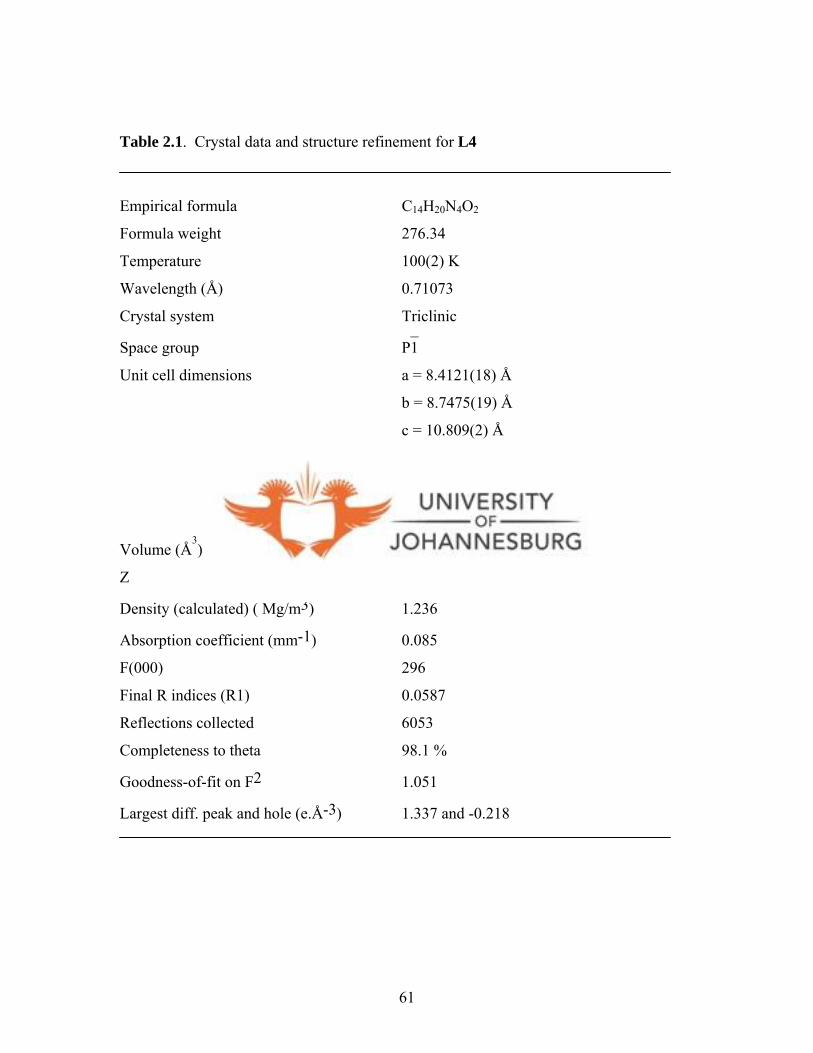
Table 2.1. Crystal data and structure refinement for L4
Empirical formula C14H20N4O2
Formula weight 276.34
Temperature 100(2) K
Wavelength (Å) 0.71073
Crystal system Triclinic
Space group P1̄
Unit cell dimensions a = 8.4121(18) Å
b = 8.7475(19) Å
c = 10.809(2) Å
α = 80.958(3)°
β = 71.135(3)°
γ = 84.365(3)°
Volume (Å3) 742.4(3)
Z 2
Density (calculated) ( Mg/m3) 1.236
Absorption coefficient (mm-1) 0.085
F(000) 296
Final R indices (R1) 0.0587
Reflections collected 6053
Completeness to theta 98.1 %
Goodness-of-fit on F2 1.051
Largest diff. peak and hole (e.Å-3) 1.337 and -0.218
61
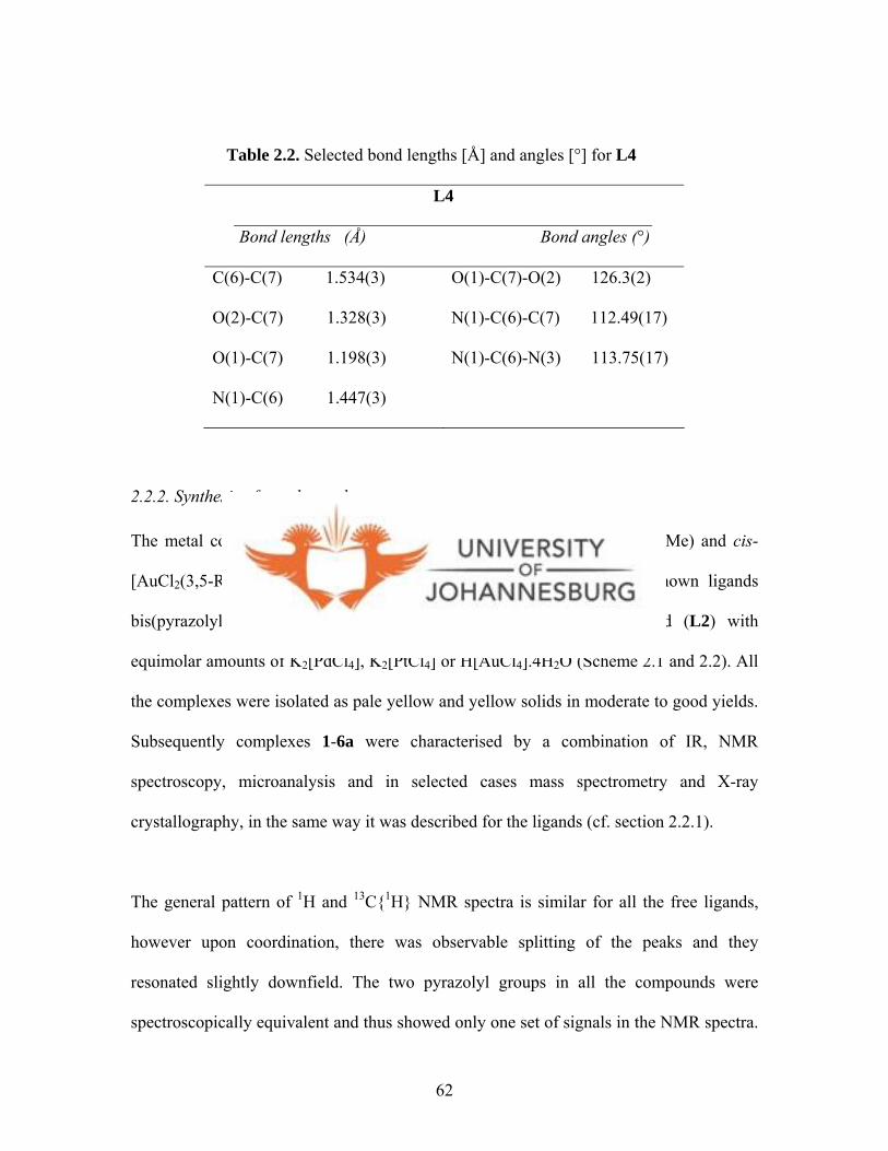
Table 2.2. Selected bond lengths [Å] and angles [°] for L4
L4
Bond lengths (Å) Bond angles (°)
C(6)-C(7) 1.534(3)
O(2)-C(7) 1.328(3)
O(1)-C(7) 1.198(3)
N(1)-C(6) 1.447(3)
O(1)-C(7)-O(2) 126.3(2)
N(1)-C(6)-C(7) 112.49(17)
N(1)-C(6)-N(3) 113.75(17)
2.2.2. Synthesis of metal complexes
The metal complexes 1-6a (cis-[MCl2(3,5-R2bpza)] (M = Pd, Pt, R = H, Me) and cis-
[AuCl2(3,5-R2bpza)]Cl) (R = H, Me) were prepared by reacting the known ligands
bis(pyrazolyl)acetic acid (L1) and bis(3,5-dimethylpyrazolyl)acetic acid (L2) with
equimolar amounts of K2[PdCl4], K2[PtCl4] or H[AuCl4].4H2O (Scheme 2.1 and 2.2). All
the complexes were isolated as pale yellow and yellow solids in moderate to good yields.
Subsequently complexes 1-6a were characterised by a combination of IR, NMR
spectroscopy, microanalysis and in selected cases mass spectrometry and X-ray
crystallography, in the same way it was described for the ligands (cf. section 2.2.1).
The general pattern of 1H and 13C{1H} NMR spectra is similar for all the free ligands,
however upon coordination, there was observable splitting of the peaks and they
resonated slightly downfield. The two pyrazolyl groups in all the compounds were
spectroscopically equivalent and thus showed only one set of signals in the NMR spectra.
62

For instance, the 1H NMR spectrum of [PdCl2(L1)] (1) showed three distinct singlets at
8.29 ppm (5H, 5′H; pz), 8.02 ppm (3H, 3′H; pz) and at 8.14 ppm for the CH linker proton
(CHCOOH) compared to the peaks of L1 at 7.98 ppm (5H, 5′H; pz), 7.59 ppm (3H, 3′H;
pz) and 7.57 (CHCOOH) in the spectrum of L1. This general downfield shift of peaks of
L1 in complex 1 was attributed to L1 being de-shielded as a result of its coordination to
palladium. Similar 1H NMR spectral patterns was observed for analogues of 1, that is,
[PtCl2(L1)] (3a) and [AuCl2(L1)]Cl (5a) complexes. However, there was no significant
changes in the 13C{1H} NMR spectra of complexes 1 and 3a compared to that of L1, with
the diagnostic carbonyl carbon (CHCOOH) appearing at ca. 166 ppm; but in the 13C{1H}
NMR spectrum of gold complex 5a, the carbonyl carbon (CHCOOH) resonated much
downfield at 174 ppm. This was attributed to the coordination of L1 to the gold ion. It
appears that the strong polarising ability of gold(III) ion facilitates the withdrawal of
electrons from the ligand rendering it to be more de-shielded.
In the case of complexes [PdCl2(L2)] (2), [PtCl2(L2)] (4) and [AuCl2(L2)]Cl (6a), their
1H NMR spectra were quite similar to those of complexes 1, 3a and 5a except for the fact
that they showed two additional peaks upfield, which were characteristic of the protons of
the methyl groups in L2. For instance, in the 1H NMR spectrum of complex 4 (Fig. 2.4),
there was a distinct separation of the two methyl group protons, thus resonating at 2.07
and 2.17 ppm for CH3 protons at 3-pz and 5-pz, respectively, as seen in Figure 2.4. This
is in contrast to the two methyl groups of L2 where they overlap at 2.33 ppm, thus giving
a clear indication of formation of complex 4. However, there were no significant shifts in
the 13C{1H} NMR spectra of complexes 2, 4 and 6a compared to that of L2.
63
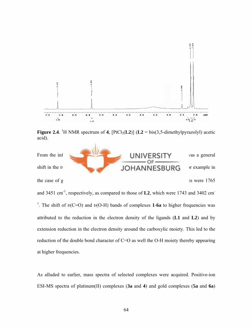
Figure 2.4. 1H NMR spectrum of 4, [PtCl2(L2)] (L2 = bis(3,5-dimethylpyrazolyl) acetic acid).
From the infrared spectra of complexes 1-6a, it was observed that there was a general
shift in the υ(C=O) and υ(O-H) bands of L1 and L2 upon complexation. For example in
the case of gold complex 6a, the υ(C=O) and υ(O-H) stretching frequencies were 1765
and 3451 cm-1, respectively, as compared to those of L2, which were 1743 and 3402 cm-
1. The shift of υ(C=O) and υ(O-H) bands of complexes 1-6a to higher frequencies was
attributed to the reduction in the electron density of the ligands (L1 and L2) and by
extension reduction in the electron density around the carboxylic moiety. This led to the
reduction of the double bond character of C=O as well the O-H moiety thereby appearing
at higher frequencies.
As alluded to earlier, mass spectra of selected complexes were acquired. Positive-ion
ESI-MS spectra of platinum(II) complexes (3a and 4) and gold complexes (5a and 6a)
64

recorded in DMSO:H2O solution, yielded the positive [M]+ ions as the base peaks. In the
spectrum of complex 3a, the molecular ion peak is at m/z 457 (Fig. 2.5). The
fragmentation pattern is, however, complicated and could not be interpreted. The positive
ESI-MS spectra of complexes 4, 5a and 6a, showed molecular ion peaks present at m/z
514 for [PtCl2(L2)], 463 for [AuCl2(L1)]+ and m/z 519 for [AuCl2(L2)]+, respectively.
The molecular ion peak at m/z 542 in the spectra of complex 6a, is related to [M+Na]+
fragment, with the base peak appearing at m/z 249, which correspond to the ligand (L2).
KFK 91
m/z200 220 240 260 280 300 320 340 360 380 400 420 440 460 480 500 520 540
%
0
100KFK_UJ_070413_3 9 (0.182) Cn (Top,4, Ht); Sm (Mn, 3x0.80); Sb (1,40.00 ); Cm (7:14) 2: Scan ES+
8.79e5
257223
215
206
224
253247
242241
259325
269 275
307291 319
405337403
353 381359394
490489407
457455410 440 459481
500 501503
535534513 545 546
N
N N
N
HO O
Pt
Cl Cl3a
Figure 2.5. ESI-MS spectrum of compound 4a, [PtCl2(L1)] (L1 = bis(pyrazolyl) acetic acid).
Complexes 5a and 6a (Scheme 2.2) are cationic gold(III) complexes with chloride
counterions and are similar to [AuCl2(bik)]Cl (bik = bis(1-methyl-2-imidazolylketone)
and [AuCl2(bihm)]Cl (bihm = bis(1-methyl-2-imidazolyl)hydroxymethane) complexes
recently reported by Bulak et al.9 All the three complexes, a, b and c, were found to be
9 Bulak E., Sarper O., Dogan A., Lissner F., Schleid T., Kaim W., Polyhedron, 2006, 25, 2577.
65

nearly square planar gold(III) with the formation of six-membered chelated rings (Fig.
2.6).9
N
N
N
N
O
Au
Cl Cl
N
N
N
N
O
Au
Cl Cl
N
N
N
N
OH
Au
Cl Cl
Cl-
Cl-
AuCl2-
(a) (b)
(c)
Figure 2.6. Dichlorogold(III) complexes of bis(1-methyl-2-imidazolyl)ketone and related ligands (cf. ref. 9).
Whereas the mechanism of coordination of N^N ligands to MCl2 (M = Pd, Pt) to form
complexes similar to 1-4 has been known for a long time, that of AuCl4- has not. For
instance, in the case of palladium(II) and platinum(II) complexes 1-4, the source of MCl2
is their respective K2[MCl4] salts (M = Pd, Pt). The presence of non-bonding electrons
and two vacant coordination sites on square planar d8 complexes, allows them to be
occupied by solvent molecules. Thus, many of the reactions of Pt and Pd complexes
proceed by an associative mechanism.10 One might expect that four-coordinate
10 Cotton F. A., Wilkinson G., Advanced Inorganic Chemistry. 3rd Ed., 1972, pg. 665, 1031.
66

complexes would be more likely than octahedral ones to react by SN2 mechanism,1 0 where
the incoming ligand leads to a formation of a five-coordinate intermediate species
followed by a simultaneous loss of the leaving ligand.
The mechanism for the formation of the gold compounds 5a and 6a is similar and
proceeds as shown in Scheme 2.3. It involves the formation of an intermediate complex
d. Indirect evidence for the formation of d is the pyridine gold(III) compound,
[AuCl3(Hpcp)] (Hpcp = 2-benzoylpyridine), reported by Fuchita et al.11 The availability
of only one N-donor atom in Hpcp leads to formation of a tri-chloro gold complex,
[AuCl3(Hpcp)] (see inset in Scheme 2.3). On the contrary, the availability of two N-donor
atoms in the pyrazolyl ligands (L1-L2), promotes ring closure in intermediate d
involving the second N atom eliminating the Cl trans to the ligand to give 5a and 6a
(Scheme 2.3).
11 Fuchita Y., Ieda H., Tsunemune Y., Kinoshita-Nagaoka J., Kawano H., J. Chem. Soc., Dalton Trans., 1998, 791.
67
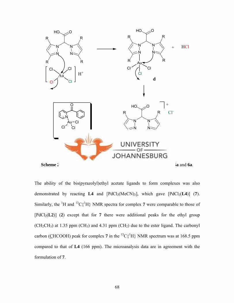
N
N N
N
HO OR
R
R
R
N
N N
N
HO OR
R
R
RAu
ClCl
Cl
AuCl
ClCl
ClH+
+ H
N
N
Cl
N
N
HO OR
R
R
RAu
Cl Cl
+
Cl-
R= H (5a), Me (6a)
d
N
O
AuCl
ClCl
[AuCl3(Hpcp)]
Scheme 2.3. Proposed mechanism of formation of gold(III) compounds 5a and 6a.
The ability of the bis(pyrazolyl)ethyl acetate ligands to form complexes was also
demonstrated by reacting L4 and [PdCl2(MeCN)2], which gave [PdCl2(L4)] (7).
Similarly, the 1H and 13C{1H} NMR spectra for complex 7 were comparable to those of
[PdCl2(L2)] (2) except that for 7 there were additional peaks for the ethyl group
(CH2CH3) at 1.35 ppm (CH3) and 4.31 ppm (CH2) due to the ester ligand. The carbonyl
carbon (CHCOOH) peak for complex 7 in the 13C{1H} NMR spectrum was at 168.5 ppm
compared to that of L4 (166 ppm). The microanalysis data are in agreement with the
formulation of 7.
68
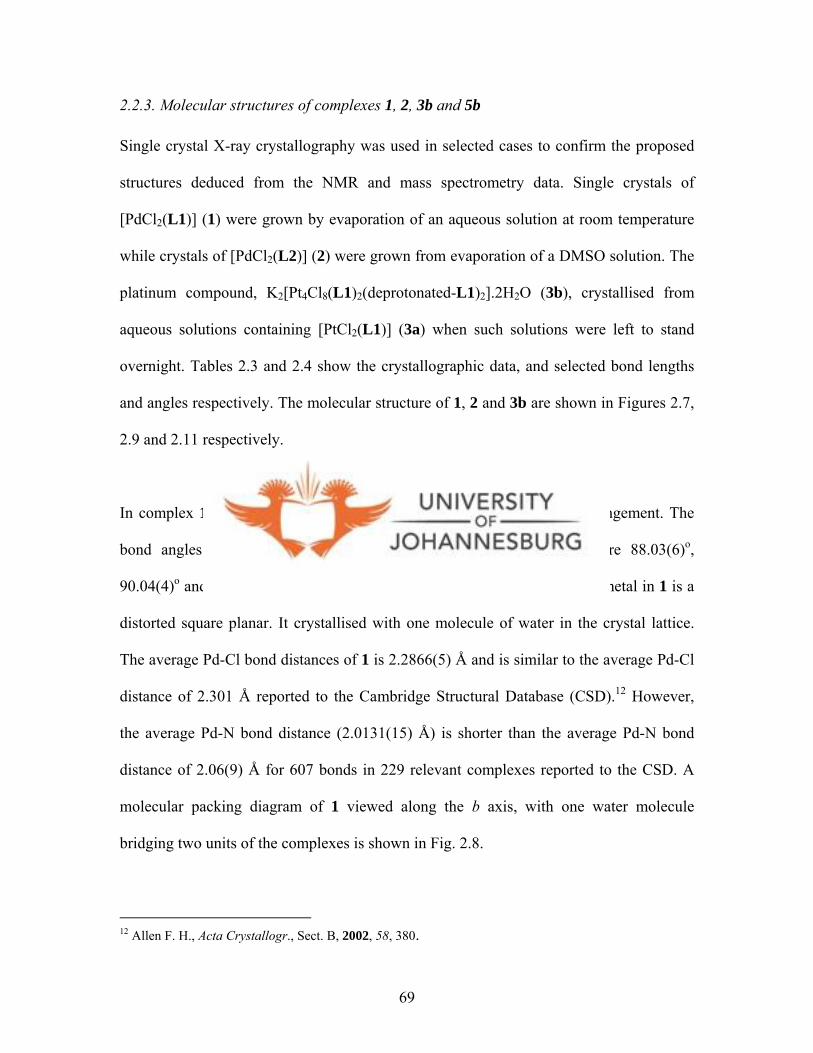
2.2.3. Molecular structures of complexes 1, 2, 3b and 5b
Single crystal X-ray crystallography was used in selected cases to confirm the proposed
structures deduced from the NMR and mass spectrometry data. Single crystals of
[PdCl2(L1)] (1) were grown by evaporation of an aqueous solution at room temperature
while crystals of [PdCl2(L2)] (2) were grown from evaporation of a DMSO solution. The
platinum compound, K2[Pt4Cl8(L1)2(deprotonated-L1)2].2H2O (3b), crystallised from
aqueous solutions containing [PtCl2(L1)] (3a) when such solutions were left to stand
overnight. Tables 2.3 and 2.4 show the crystallographic data, and selected bond lengths
and angles respectively. The molecular structure of 1, 2 and 3b are shown in Figures 2.7,
2.9 and 2.11 respectively.
In complex 1 the two chlorides are coordinated to palladium in a cis-arrangement. The
bond angles of N(1)-Pd(1)-N(4), N(4)-Pd(1)-Cl(1), N(1)-Pd(1)-Cl(2), are 88.03(6)o,
90.04(4)o and 90.81(4)o, respectively. The geometry around the palladium metal in 1 is a
distorted square planar. It crystallised with one molecule of water in the crystal lattice.
The average Pd-Cl bond distances of 1 is 2.2866(5) Å and is similar to the average Pd-Cl
distance of 2.301 Å reported to the Cambridge Structural Database (CSD).12 However,
the average Pd-N bond distance (2.0131(15) Å) is shorter than the average Pd-N bond
distance of 2.06(9) Å for 607 bonds in 229 relevant complexes reported to the CSD. A
molecular packing diagram of 1 viewed along the b axis, with one water molecule
bridging two units of the complexes is shown in Fig. 2.8.
12 Allen F. H., Acta Crystallogr., Sect. B, 2002, 58, 380.
69

Compound [PdCl2(L2)] (2) crystallises with two symmetry independent palladium
complex molecules and three molecules of DMSO solvent in the unit cell. The two
molecules exhibit similar structural features and reside on a crystallographic inversion
centre, with only one half of it being crystallographically independent (Fig. 2.9). Each
palladium coordination environment consists of two cis-Cl ligands. The bond angles of
N(1)-Pd(2)-N(4), N(1)-Pd(2)-Cl(5), Cl(5)-Pd(2)-Cl(3) are 85.8(3)o, 174.9(2)o and
89.87(11)o, respectively. The geometry around the palladium centres is a distorted square
planar. The average Pd-Cl bond distances (2.2928(3) Å) and the average Pd-N bond
distance (2.0475(8) Å) are similar to those of complex 1 described above. There is
intermolecular O-H…O hydrogen bonding observed between two DMSO solvents and
two palladium units (Fig. 2.10). However, there are no intramolecular interactions in the
crystal unit cell.
70

Figure 2.7. A molecular drawing of complex 1. The hydrogen bonding interaction is shown with a dashed line.
71

Figure 2.8. A molecular packing diagram of complex 1 viewed along the b axis. The hydrogen bonding interactions is shown with the dashed lines, with a water molecule bridging two units of the complexes. Selected H atoms are shown.
72
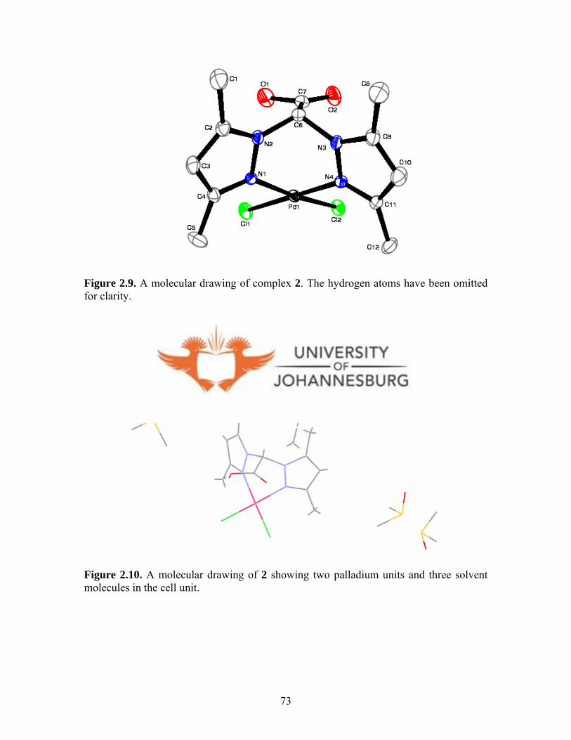
73
Figure 2.10. A molecular drawing of 2 showing two palladium units and three solvent molecules in the cell unit.
Figure 2.9. A molecular drawing of complex 2. The hydrogen atoms have been omitted for clarity.
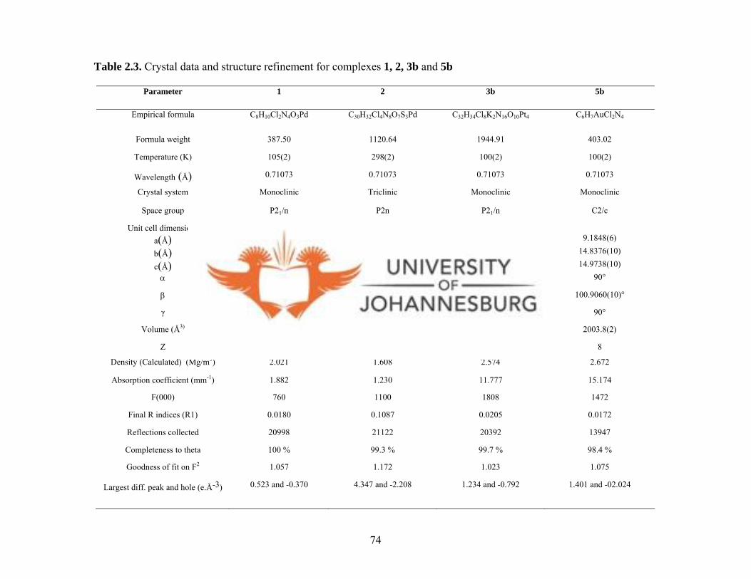
74
Parameter 1 2 3b 5b
Empirical formula C8H10Cl2N4O3Pd C30H32Cl4N8O7S3Pd C32H34Cl8K2N16O10Pt4 C6H7AuCl2N4
Formula weight 387.50 1120.64 1944.91 403.02
Temperature (K) 105(2) 298(2) 100(2) 100(2)
Wavelength (Å) 0.71073 0.71073 0.71073 0.71073
Crystal system Monoclinic Triclinic
Monoclinic Monoclinic
Space group P21/n P2n P21/n C2/c
Unit cell dimensions a(Å) 10.9934(10) 8.7960(14) 10.7762(8) 9.1848(6)
b(Å) 9.7436(9) 16.238(2) 14.4036(11) 14.8376(10)
c(Å) 11.9243(11) 16.430(2) 16.6971(13) 14.9738(10)α 90o 78.038(10)° 90° 90°
β 93.0620(10)o 77.817(11)° 104.494(2)° 100.9060(10)°
γ 90o 89.970(10)° 90° 90°
Volume (Å3) 1275.5(2) 2241.7(6) 2509.2(3) 2003.8(2)
Z 4 4 2 8
Density (Calculated) (Mg/m3) 2.021 1.608 2.574 2.672
Absorption coefficient (mm-1) 1.882 1.230 11.777 15.174
F(000) 760 1100 1808 1472
Final R indices (R1) 0.0180 0.1087 0.0205 0.0172
Reflections collected 20998 21122 20392 13947
Completeness to theta 100 % 99.3 % 99.7 % 98.4 %
Goodness of fit on F2 1.057 1.172 1.023 1.075
Largest diff. peak and hole (e.Å-3) 0.523 and -0.370 4.347 and -2.208 1.234 and -0.792 1.401 and -02.024
Table 2.3. Crystal data and structure refinement for complexes 1, 2, 3b and 5b
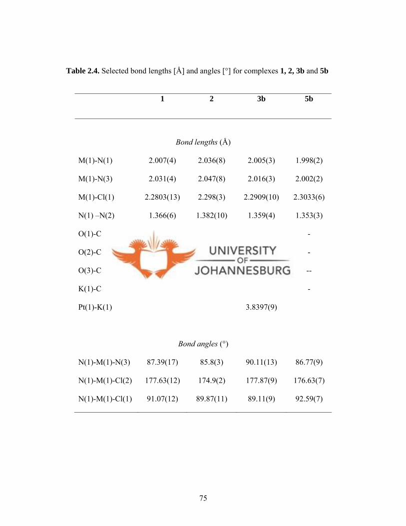
Table 2.4. Selected bond lengths [Å] and angles [°] for complexes 1, 2, 3b and 5b
1 2 3b 5b
Bond lengths (Å)
M(1)-N(1) 2.007(4) 2.036(8) 2.005(3) 1.998(2)
M(1)-N(3) 2.031(4) 2.047(8) 2.016(3) 2.002(2)
M(1)-Cl(1) 2.2803(13) 2.298(3) 2.2909(10) 2.3033(6)
N(1) –N(2) 1.366(6) 1.382(10) 1.359(4) 1.353(3)
O(1)-C(5)
O(2)-C(8)
O(3)-C(16)
1.204(2)
-
-
1.314(14)
-
-
-
1.215(5)
1.224(5)
-
-
--
K(1)-Cl(2) - 3.1822(13) -
Pt(1)-K(1) 3.8397(9)
Bond angles (°)
N(1)-M(1)-N(3) 87.39(17) 85.8(3) 90.11(13) 86.77(9)
N(1)-M(1)-Cl(2) 177.63(12) 174.9(2) 177.87(9) 176.63(7)
N(1)-M(1)-Cl(1) 91.07(12) 89.87(11) 89.11(9) 92.59(7)
75
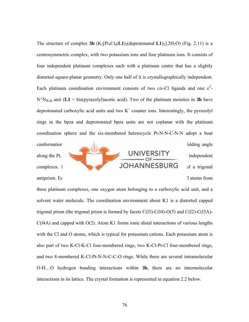
The structure of complex 3b (K2[Pt4Cl8(L1)2(deprotonated L1)2].2H2O) (Fig. 2.11) is a
centrosymmetric complex, with two potassium ions and four platinum ions. It consists of
four independent platinum complexes each with a platinum centre that has a slightly
distorted square-planar geometry. Only one half of it is crystallographically independent.
Each platinum coordination environment consists of two cis-Cl ligands and one K2-
N^N(L1) unit (L1 = bis(pyrazolyl)acetic acid). Two of the platinum moieties in 3b have
deprotonated carboxylic acid units and two K+ counter ions. Interestingly, the pyrazolyl
rings in the bpza and deprotonated bpza units are not coplanar with the platinum
coordination sphere and the six-membered heterocycle Pt-N-N-C-N-N adopt a boat
conformation which in this structure resembles a butterfly; the dihedral folding angle
along the Pt…C(N,N) line averages 48.7(2)º for the two crystallographically independent
complexes. In the centre of 3b is a K2Cl4 fragment that has a shape of a trigonal
antiprism. Each potassium atom is seven-coordinate forming bonds to five Cl atoms from
three platinum complexes, one oxygen atom belonging to a carboxylic acid unit, and a
solvent water molecule. The coordination environment about K1 is a distorted capped
trigonal prism (the trigonal prism is formed by facets C(l3)-C(l4)-O(5) and C(l2)-C(l3A)-
C(l4A) and capped with O(2). Atom K1 forms ionic distal interactions of various lengths
with the Cl and O atoms, which is typical for potassium cations. Each potassium atom is
also part of two K-Cl-K-Cl four-membered rings, two K-Cl-Pt-Cl four-membered rings,
and two 8-membered K-Cl-Pt-N-N-C-C-O rings. While there are several intramolecular
O-H…O hydrogen bonding interactions within 3b, there are no intermolecular
interactions in its lattice. The crystal formation is represented in equation 2.2 below.
76
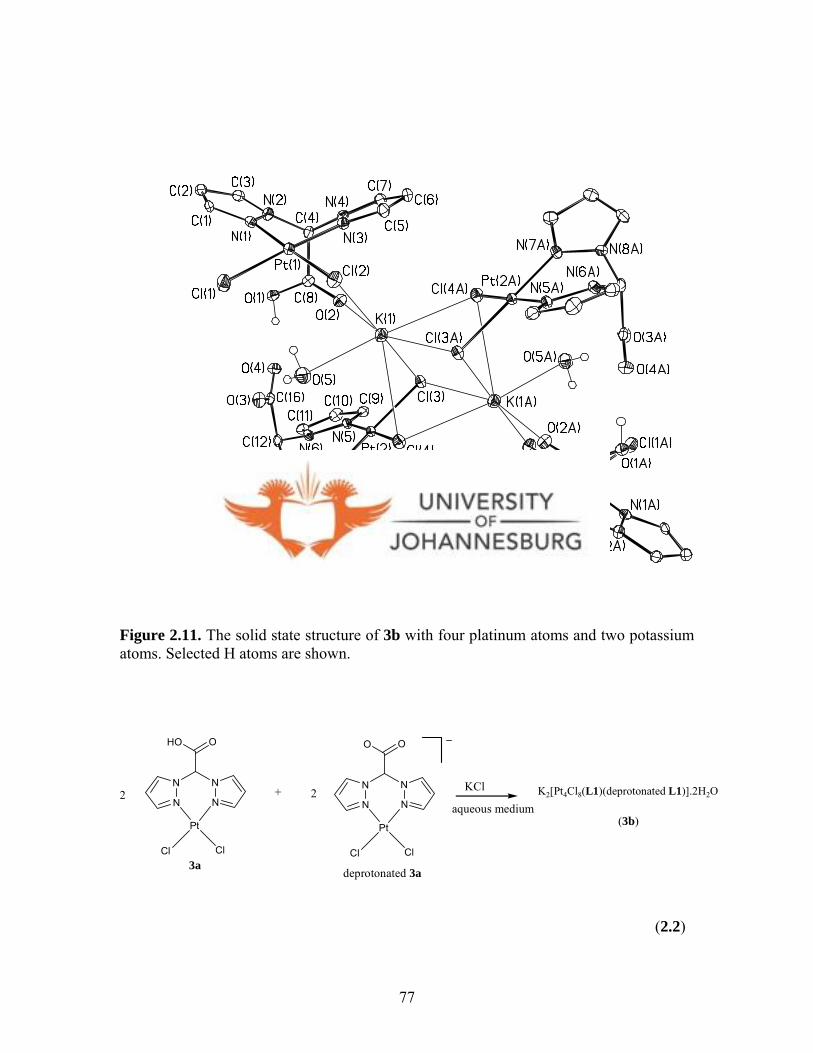
Figure 2.11. The solid state structure of 3b with four platinum atoms and two potassium atoms. Selected H atoms are shown.
N
N N
N
HO O
Pt
Cl Cl3a
K2[Pt4Cl8(L1)(deprotonated L1)].2H2O
(3b)
2 +N
N N
N
O O
Pt
Cl Cl
deprotonated 3a
2aqueous medium
KCl
(2.2)
77
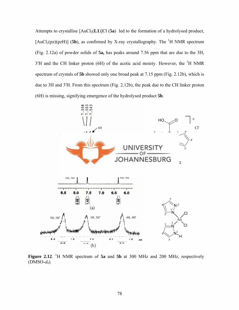
Attempts to crystallise [AuCl2(L1)]Cl (5a) led to the formation of a hydrolysed product,
[AuCl2(pz)(pzH)] (5b), as confirmed by X-ray crystallography. The 1H NMR spectrum
(Fig. 2.12a) of powder solids of 5a, has peaks around 7.56 ppm that are due to the 3H,
3′H and the CH linker proton (6H) of the acetic acid moiety. However, the 1H NMR
spectrum of crystals of 5b showed only one broad peak at 7.15 ppm (Fig. 2.12b), which is
due to 3H and 3′H. From this spectrum (Fig. 2.12b), the peak due to the CH linker proton
(6H) is missing, signifying emergence of the hydrolysed product 5b.
N
N
N
N
AuCl
Cl
H
1'2'
3'
4'
5'
1
23
4
5
N
N N
N
HO O
Au
Cl Cl
+
Cl-
1
2
3
4
5 1'
2'
3'
4'
5'6
4H, 4H′ H
H′ H′ ′
H′
H′
6H
4H, 4H3H, 35H, 5
3H, 3H′ + 6
3H, 3
5H, 5
Figure 2.12. 1H NMR spectrum of 5a and 5b at 300 MHz and 200 MHz, respectively (DMSO-d6).
78

Notably, the peak due to N-H proton at ca. 9 ppm in the 1H NMR spectrum of 5b was
missing. This is possibly due to the proton-deuterium exchange in solution. The
broadness observed in the 1H NMR spectrum of 5b, was attributed to difficulties
encountered in shimming during acquisition of this spectrum. The spectroscopic data as
well as the solid state structure of complex 5b (Fig. 2.15) indicate that complex 5a
hydrolysed in solution to produce 5b (eq. 2.3). Recently Cao et al. 13 reported the amine-
amide hydrolysis of a gold(III) compound that was attributed to the high polarising nature
of the gold(III) centre in this compound. Several reports on the polarising effect of
gold(III) have appeared in the literature.14,15 A similar polarising effect of gold(III) in
complex 5a could explain the hydrolysis observed.
N
NH
N
N
AuCl
ClHydrolysis
R
R
R
R
R = H (5b), Me (6b)
N
N N
N
HO O
R
R
R
RAu
Cl Cl
+
Cl-
R = H (5a), Me (6a)
(2.3)
13 Cao L., Jennings M. C., Puddephat R. J., Inorg. Chem., 2007, 46, 1361. 14 Vicente J., Bermudez M. D., Chicote M. T., Sanchez-Santano M. J., J. Chem. Soc., Dalton Trans., 1990, 1945. 15 Fan D., Yang C. T., Ranford J. D., Vittal J. J., Dalton Trans., 2003, 4749.
79
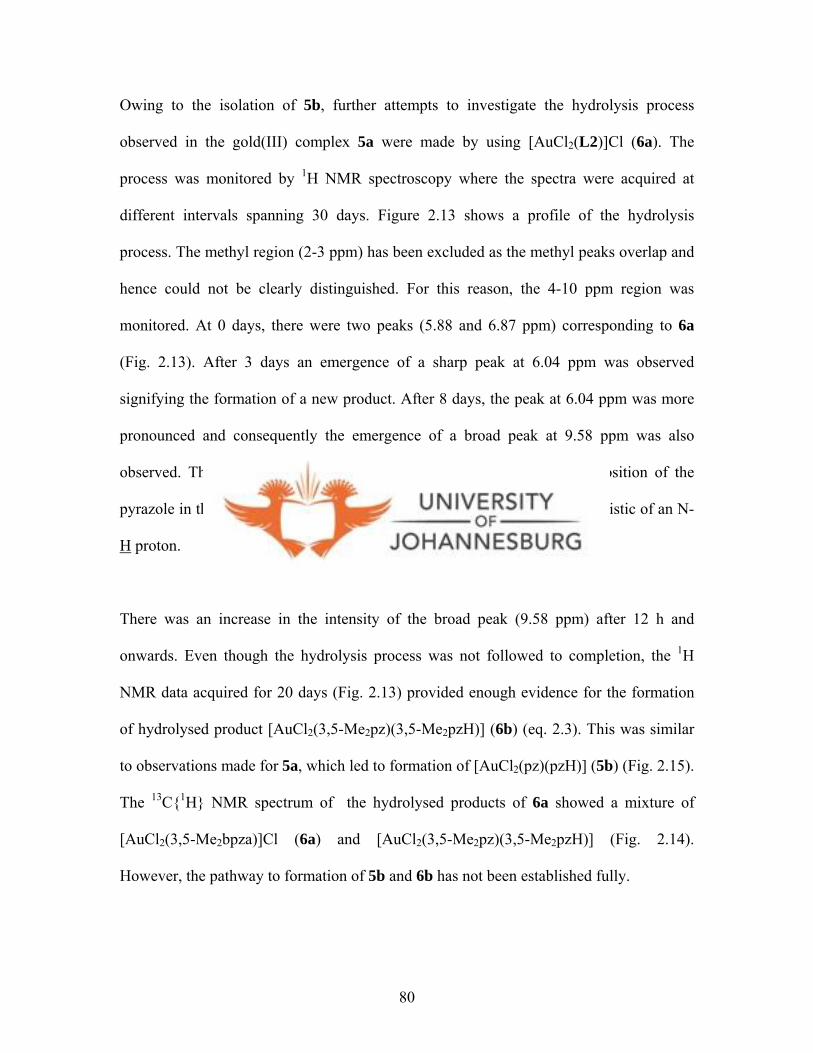
80
Owing to the isolation of 5b, further attempts to investigate the hydrolysis process
observed in the gold(III) complex 5a were made by using [AuCl2(L2)]Cl (6a). The
process was monitored by 1H NMR spectroscopy where the spectra were acquired at
different intervals spanning 30 days. Figure 2.13 shows a profile of the hydrolysis
process. The methyl region (2-3 ppm) has been excluded as the methyl peaks overlap and
hence could not be clearly distinguished. For this reason, the 4-10 ppm region was
monitored. At 0 days, there were two peaks (5.88 and 6.87 ppm) corresponding to 6a
(Fig. 2.13). After 3 days an emergence of a sharp peak at 6.04 ppm was observed
signifying the formation of a new product. After 8 days, the peak at 6.04 ppm was more
pronounced and consequently the emergence of a broad peak at 9.58 ppm was also
observed. The peak at 6.04 ppm is representative of proton at the 4th position of the
pyrazole in the new hydrolysed product, whilst that at 9.58 ppm is characteristic of an N-
H proton.
There was an increase in the intensity of the broad peak (9.58 ppm) after 12 h and
onwards. Even though the hydrolysis process was not followed to completion, the 1H
NMR data acquired for 20 days (Fig. 2.13) provided enough evidence for the formation
of hydrolysed product [AuCl2(3,5-Me2pz)(3,5-Me2pzH)] (6b) (eq. 2.3). This was similar
to observations made for 5a, which led to formation of [AuCl2(pz)(pzH)] (5b) (Fig. 2.15).
The 13C{1H} NMR spectrum of the hydrolysed products of 6a showed a mixture of
[AuCl2(3,5-Me2bpza)]Cl (6a) and [AuCl2(3,5-Me2pz)(3,5-Me2pzH)] (Fig. 2.14).
However, the pathway to formation of 5b and 6b has not been established fully.
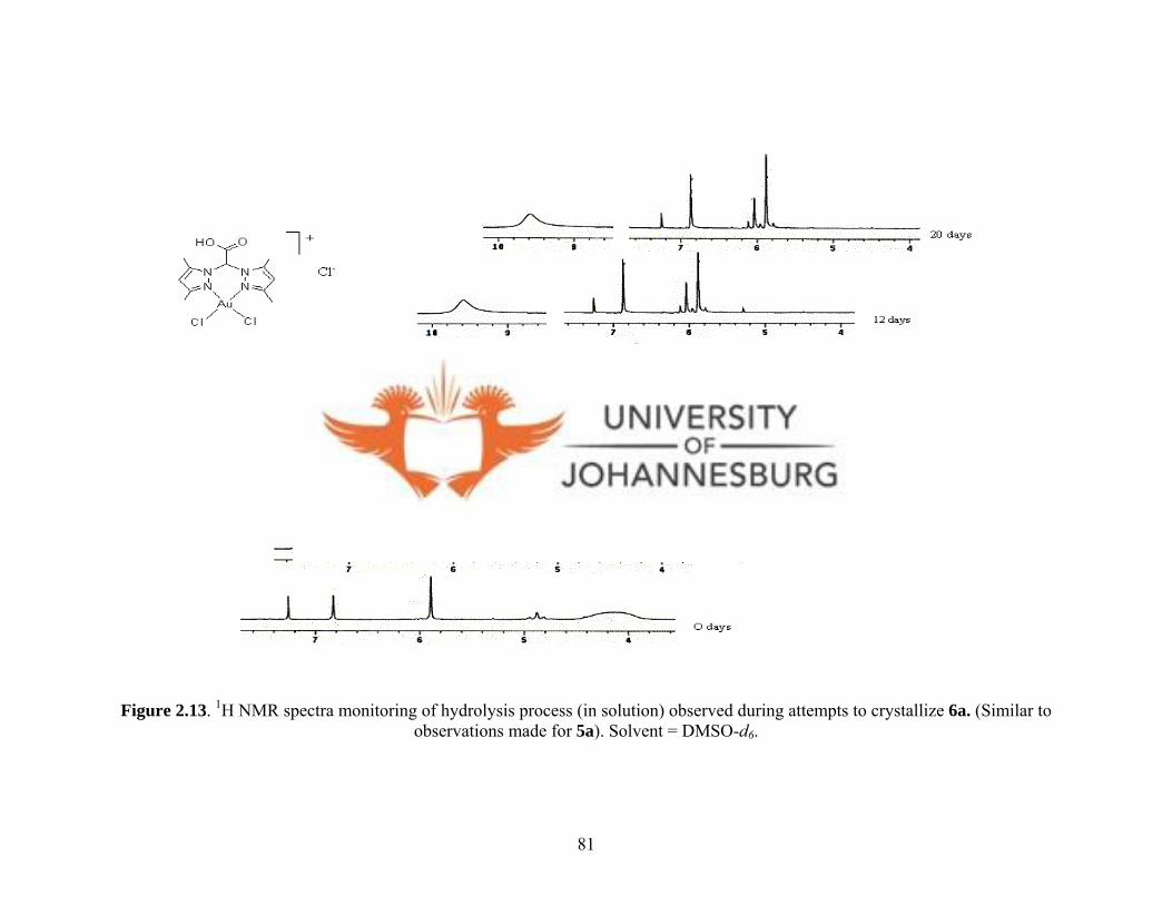
Figure 2.13. 1H NMR spectra monitoring of hydrolysis process (in solution) observed during attempts to crystallize 6a. (Similar to observations made for 5a). Solvent = DMSO-d6.
81
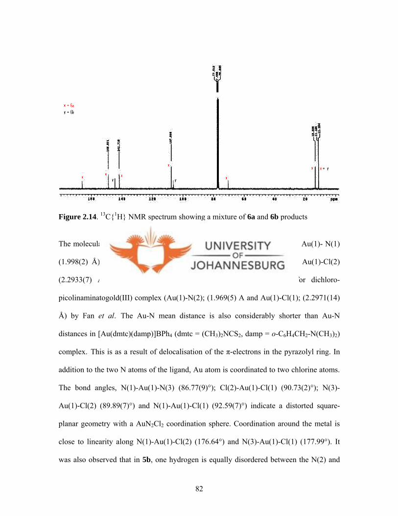
Figure 2.14. 13C{1H} NMR spectrum showing a mixture of 6a and 6b products
The molecular structure of 5b is shown in Figure 2.15. The bond distances, Au(1)- N(1)
(1.998(2) Å), Au(1)- N(3) (2.002(2) Å), Au(1)-Cl(1) (2.3033(6) Å) and Au(1)-Cl(2)
(2.2933(7) Å) are statistically longer compared to those reported for dichloro-
picolinaminatogold(III) complex (Au(1)-N(2); (1.969(5) Å and Au(1)-Cl(1); (2.2971(14)
Å) by Fan et al.15 The Au-N mean distance is also considerably shorter than Au-N
distances in [Au(dmtc)(damp)]BPh4 (dmtc = (CH3)2NCS2, damp = o-C6H4CH2-N(CH3)2)
complex.1 5 This is as a result of delocalisation of the π-electrons in the pyrazolyl ring. In
addition to the two N atoms of the ligand, Au atom is coordinated to two chlorine atoms.
The bond angles, N(1)-Au(1)-N(3) (86.77(9)°); Cl(2)-Au(1)-Cl(1) (90.73(2)°); N(3)-
Au(1)-Cl(2) (89.89(7)°) and N(1)-Au(1)-Cl(1) (92.59(7)°) indicate a distorted square-
planar geometry with a AuN2Cl2 coordination sphere. Coordination around the metal is
close to linearity along N(1)-Au(1)-Cl(2) (176.64°) and N(3)-Au(1)-Cl(1) (177.99°). It
was also observed that in 5b, one hydrogen is equally disordered between the N(2) and
82

N(4) of the pyrazole rings and that Figure 2.15 depicts the structure with hydrogen atom
on the N(2) atom. Figure 2.16 shows the molecular packing diagram of 5b viewed along
the c axis. At the peripheries of the unit cell, formation of pseudo-square cubanes,
defined by N-Au-Cl repetitive units of the neighbouring monomeric complexes, is
evident. Moreover, the π- π interactions of the pyrazole rings at the centre of the unit cell
leads to formation of a figure-eight shape.
Figure 2.15. A molecular drawing of 5b; only one position of the hydrogen equally disordered between N(2) and N(4) is shown.
83

Figure 2.16. A molecular packing diagram of 5b along c axis. It shows formation of cubes by the pyrazolyl rings in the centre, with the dotted line showing H···Cl interactions.
84
2.3. Conclusions
New bis(pyrazolyl)ethyl acetate ligands L3 and L4 were prepared by reacting
bis(pyrazolyl)acetic acid ligands (R2bpza) (L1 and L2) with excess ethanol under acidic
conditions. The isolation of L4 showed that the R2bpza undergo a classical esterification
process. In the solid state structure of L4, the carbonyl carbon, C(7), exhibits a distorted
trigonal geometry. Isolation of [PdCl2(L4)] (7) demonstrates the ability of
bis(pyrazolyl)ethyl acetate ligands to form such complexes.
Structural investigations of the new pyrazolyl palladium(II), platinum(II) and gold(III)
compounds indicates that R2bpza ligands coordinate to these metals in a bidentate fashion
This is evident in the solid state structures of [PdCl2(L1)] (1), [PdCl2(L2)] (2) and
K2[Pt4Cl8(L1)2(deprotonated L1)2].2H2O (3b). Generally, the geometry around the
metals is a distorted square planar. Each palladium coordination environment consists of
two cis-Cl ligands. Owing the acidic nature of R2bpza ligands, its deprotonation leads to a
reproducible formation of platinum(II) complex 3b. Two of the platinum moieties in 3b
have deprotonated carboxylic acid units and two K+ counter ions. Each platinum
coordination environment consists of two cis-Cl ligands and one K2-N^N(L1) unit (L1 =
bis(pyrazolyl)acetic acid). Complex 3b is quite soluble in water and judging from its size,
molecular packing would produce interesting supramolecular compounds that can be
explored for various applications e.g. as pockets for gaseous materials.
85
For the gold(III) complexes [AuCl2(L1)]Cl (5a) and [AuCl2(L2)]Cl (6a), spectroscopic,
mass spectral and microanalysis data were a confirmation of their existence. The
observed hydrolysis of these complexes seems to be due to; (i) the strong polarising
ability of the gold(III) and (ii) the fact that pyrazoles are moderate π-donors; which
means that excessive withdrawal of electron from the pyrazolyl ligands could possibly
lead to their disintegration. Isolation and structural characterisation of 5b points to these
two possibilities being the possible causative reasons for the hydrolysis observed. Overall
the project was a success as it led to isolation of the target complexes.
The major finding is:
• That the flexible coordination and water solubility of bis(pyrazolyl) acetic
acid ligands (R2bpza) (L1-L2) offer the possibility of water soluble
complexes. For instance the platinum(II) and gold(III) complexes obtained by
utilising R2bpza ligands were slightly soluble in water and readily soluble in
DMSO. Furthermore, the σ-donor ability of R2bpza ligands stabilises unstable
gold(III) compounds. This finding confirms that N-donor ligands are best
suited to stabilise gold(III) compounds.
86

CHAPTER 3
EVALUATION OF ANTI-TUMOUR ACTIVITY OF PALLADIUM(II),
PLATINUM(II) AND GOLD(III) PYRAZOLYL COMPLEXES AGAINST CHO-
22 CELLS AND KINETIC STUDIES
Screening of the complexes for their in vitro anticancer activities were performed by Mr
Kanyanda of University of the Western Cape (SA), but the interpretation and discussion
of results is by the candidate.
3.0. Introduction
Platinum group metals compounds are of interest because of their development as
metallodrugs. This includes efforts to improve the effectiveness of existing platinum and
gold compounds as drugs.1 Currently there is also considerable interest in the
coordination chemistry of gold because of biological activities exhibited by these
compounds. Similarly, palladium compounds have also shown activity against cancer
cells. Although the anticancer activity of cis-[PtCl2(NH3)2] (cisplatin) and other related
compounds is by interacting with DNA,2 reactions with other biomolecules like
glutathione, cysteine and metallothionein, in biological milieu are likely to affect the
efficacy of these compounds.3 Palladium and gold compounds react with these
biomolecules too. As a result, the widespread use of cisplatin and related complexes as
1 Hartinger C. G., Platinum Metals Rev., 2008, 52, 96. 2 Takahara P. M., Rosenzweig A. C., Frederick C. A., Lippard S. J., Nature, 1995, 377, 649. 3 El-Khateeb M., Appleton T. G., Gahan L. R., Charles B. G., Berners-Price S. J., Bolton A., J. Inorg. Biochem., 1999, 77, 13.
87

therapeutic agents has engendered great interest in the aqueous solution chemistry of
dichloro-platinum compounds.4
As mentioned before, it has been accepted that the efficiency of cisplatin is based on its
strong binding to DNA nucleobases resulting in a locally unwound and kinked helix.2 The
binding of cisplatin towards DNA has been shown to be kinetically and not
thermodynamically controlled.5 Experiments involving inosine, a is purine nucleoside,
indicate that platinum(II) complexes binds DNA via a solvent-assisted mechanism.6
These experiments suggest that the first step of reaction of cisplatin with DNA involves
replacement of one chloride with a water molecule to give monofunctional adduct, cis-
[PtCl(NH3)2(H2O)]+. This process has been shown by in vitro experiments to be of the
first order, with the rate constant of hydrolysis of cis-[PtCl2(NH3)2] to cis-
[PtCl(NH3)2(H2O)]+ to be ca. 10.2 x 10⁻⁵ s-1.7,8
The mechanism for cisplatin-DNA interactions is presented in Scheme 3.1. The
formation of platinum-DNA monofunctional adducts and the successive formation
platinum-DNA bifunctional adducts display sequence-dependent kinetics (Scheme 3.1). 9
Firstly one chloride in cisplatin is replaced by a water molecule. Subsequently, the first
platinum-N7 bond is formed preferentially on guanines involved in Gn tracts (n ≥ 2) by
displacing the water molecule to give monofunctional adducts.8,9 This step is followed by
4 Erickson E. L., Schneider Y., Takara T. J., Wezeman A. S., Inorg. Chim. Acta., 2007, 360, 3560. 5 Mansy S., Chu G. Y. H., Duncan R. E., Tobias R. S., J. Am. Chem. Soc., 1978, 100, 607 6 Arpalahti J., Mikola M., Mauristo S., Inorg. Chem., 1993, 32, 3327. 7 Bancroft D. P., Lepre C. A., Lippard S. J., J. Am. Chem. Soc., 1990, 112, 6860. 8 Bruhn S. L., Toney J. H., Lippard S. J., Progress in Inorganic Chemistry: Bioinorganic Chemistry, Vol. 38, Edited by Lippard, 1990, 447.
88

replacement of the second chloride with a water molecule. Subsequent closure of
monofunctional adducts to form bifunctional adducts, by loosing the second water
molecule, completes the process of formation of platinum-DNA adduct as shown in
Scheme 3.1.9 Even though the most favoured binding site is N7 position of guanine to
give a GG adduct, other binding sites appear to be guanine-adenine which gives AG
adduct.9 This selectivity of the initial binding step determines the final distribution of the
bifunctional adducts. Previous work has shown that the main adducts are GG (60 - 65%)
and AG (~ 20%) intrastrand chelates.9,10
The substitution of the Cl- in Pt-DNA monofunctional adducts follows first order kinetics
(4-10 ×10-5 s-1 ) at 37 oC.7,8,9 The rates of hydrolysis of the second chloride ligand in
cisplatin suggests that conversion of the chloro-monofunctional adducts to bifunctional
adducts is the rate-limiting step.8,9 However, it is worth mentioning that cisplatin could
also undergo hydrolysis of both chloro ligands before reacting with nuclear DNA.9 Jestin
et al.11 have suggested that the second aquation step could occur within the outersphere
complex formed between cis-[PtCl(NH3)2(H2O)]+ and DNA.
9 Kozelka J., Legendre F., Reeder F., Chottard J., Coord. Chem. Rev., 1999, 190, 61. 10 Saris C. P., van de Vaart P. J. M., Rietbroek R. C., Blommaert F. A., Carcinogenesis, 1996, 17, 2763. 11 Jestin J. L., Lambert B., Chottard J. C., JBIC, 1998, 3, 515.
89
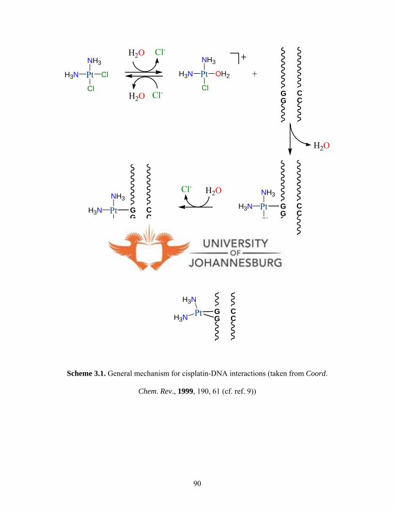
PtH3N
NH3
Cl
Cl
H2O
O
Cl-
H2 Cl-
PtH3N
NH3
Cl
OH2
GG
CC
GG
CC
+
H2
PtH3N
O
GG
CC
PtH3N
NH3NH3
Cl
H2Cl- O
OH2
GG
CC
PtH3N
H3N
H2O
Scheme 3.1. General mechanism for cisplatin-DNA interactions (taken from Coord.
Chem. Rev., 1999, 190, 61 (cf. ref. 9))
90

Reactions of biomolecules present in blood and cells, e.g., glutathione, L-methionine,
metallothionein and L-cysteine, with cisplatin and other metallodrugs is much dependent
on the structures and chemical reactivity of the biomolecules.12,13 These biomolecules
together with other proteins and enzymes have the potential to react with platinum
complexes before they reach the DNA, thereby reducing their activities. Thus,
understanding the dynamics of substitution reactions of potential anticancer compounds
with biological nucleophiles is of relevant and great importance in the current drug
research.
Substitution reactions of d8 transition metals are of importance in cancer chemotherapy.14
This is because it does not just allow for the establishment of reactions rates of various
metal complexes, but also facilitate investigations into whether there is any relationship
with their observed biological activities. Normally guanosine-5′-monophosphate is used
as a model for DNA binding of platinum complexes, and L-cysteine (Cys) as a model for
interaction of metal compounds with proteins that contain Cys residues. In addition, L-
cysteine just like thiourea,14 is a very convenient substitute and is often employed as a
nucleophile to study ligand substitution reactions. Although available data suggest that
mechanism for substitution reactions for platinum(II) may apply to other square-planar
complexes, there is scant information on the substitution reactions of palladium(II)15 and
gold(III) complexes due to their kinetic instability. For instance, gold(III) compounds are
12 Hall M. D., Hambley T. W., Coord. Chem. Rev., 2002, 232, 49. 13 Summa N., Schiess W., Puchta R., Hommes N. E., Van Eldik R., Inorg. Chem., 2006, 45, 2948. 14 Fekl M., van Eldik R.., Eur. J. Inorg. Chem., 1998, 389. 15 Cotton F. A., Wilkinson G., Advanced Inorganic Chemistry, 6th Ed, John Wiley and Sons, New York, 1999, 1064.
91

relatively unstable, light sensitive in solution, and easily reduced to metallic gold under
physiological conditions due to their rapid kinetics and high redox potential than
platinum(II).16 In addition, gold(III) complexes react ca. 103 times faster compared to
platinum(II) analogues, while palladium(II) complexes react 105-106 times faster17,18 and
is not always easy to follow their reactivities. This chapter reports on the investigation of
the possible antitumour activities of dichloro-(bis(pyrazol-1-yl)aceticacid)palladium(II)
([PdCl2(L1)], 1), dichloro-(bis(3,5-dimethylpyrazol-1-yl)acetic acid)palladium(II)
([PdCl2(L2)], 2), dichloro-(bis(pyrazol-1-yl)aceticacid)platinum(II) ([PtCl2(L1)], 3a),
dichloro-(bis(3,5-dimethylpyrazol-1-yl)acetic acid)platinum(II) ([PtCl2(L2)], 4),
dichloro-(bis(pyrazol-1-yl)aceticacid)gold(III)chloride ([AuCl2(L1)]Cl, 5a) and
dichloro-(bis(3,5-dimethylpyrazol-1-yl)aceticacid)gold(III) chloride ([AuCl2(L2)]Cl, 6a)
against mammalian CHO-22 cells. The substitution behaviour, mechanism and associated
kinetics of these complexes with L-cysteine have been performed as part of a study to
establish whether there is any relationship between the observed antitumour activities
against the CHO-22 cells and the kinetic labilities of the complexes.
16 Fan D., Yang C. T., Ranford J. D., Lee P. F., Vittal J. J., J. Chem. Soc., Dalton Trans., 2003, 2680. 17 Schmulling M., Ryabov A. D., Van Eldik R., Dalton Trans., 1994, 1257. 18 Elmroth S., Bugarcic Z., Elding L. I., Inorg. Chem., 1992, 31, 3551.
92

3.1. Experimental
3.1.1 Biological reagents and instrumentation
All commercial chemicals and other reagents, other than those described below, were
used as received. All cell culture reagents were supplied by Invitrogen Ltd. Cisplatin and
neutral red (NR) dye was purchased from Sigma Aldrich. NR dye is a supravital dye used
in histological staining.19 The Hams F-12 medium containing 10% foetal calf serum and
0.2% pn-streptomycin was prepared from the stock Hams F-12 medium. The NR dye was
prepared at 100µg/mL using an elution buffer prepared from an acetic acid:ethanol:water
(50%:1%:49%) solution. Solutions of the six palladium(II), platinum(II) and gold(III)
complexes (1-6a) screened, were prepared as 10 mM stock solutions using 0.2% DMSO
solution. CHO-22 cells were obtained from the Department of Biochemistry, University
of the Western Cape. The absorbance readings of the stained cells were measured
spectrophotometrically at 492 nm.
3.2 Biological testing
3.2.1. Cell culture and drug treatment
CHO cells were cultured in 96 well plates at a population of 2 x 10⁴ cells per well using
Hams F-12 medium. The cells were incubated for 24 h at 37oC in a 5% CO2 atmosphere.
At 80% cell confluence, the Ham F-12 medium was removed, cells washed with
phosphate buffered saline (PBS) and 100 µL of fresh Ham F-12 medium containing the
19 Babich H., Borenfreund E., Appl. Environ. Microbiol., 1991, 57, 2101.
93

test compounds added. After 24 h of incubation at 37 °C in a 5% CO2 atmosphere, the
cells were then washed twice with PBS and 100 µL of serum free Ham F-12 medium
containing NR dye (100 µg/mL) added to each well and further incubated for 3h. Finally
the cells were washed twice with PBS. Thereafter, 50 µL of the elution buffer was used
to lyse the cells and the concentration of accumulated NR dye, which is a marker for cell
viability, measured spectrophotometrically at 492 nm. The percentage (%) cellular
viability values were calculated from the absorbance (Abs) readings using the formula
below; and the IC50 values obtained by extrapolation from the plots of % cellular viability
versus concentrations.
Abs of test reagent
Abs of untreated control 00 x 1
100 -% inhibition =
(3.1)
3.2.2 Complexes and L-cysteine solutions
Dichloro-(bis(pyrazol-1-yl)acetic acid)palladium(II), (1) and dichloro-(bis(3,5-
dimethyl-pyrazol-1-yl)acetic acid)palladium(II) (2), dichloro-(bis(pyrazol-1-yl)-
acetic acid)platinum(II) (3a), dichloro-(bis(3,5-dimethyl-pyrazol-1-yl)acetic
acid)platinum(II) (4), dichloro-(bis(pyrazol-1-yl)acetic acid)gold(III)chloride (5a),
dichloro-(bis(3,5-dimethylpyrazol-1-yl)acetic acid)gold(III)chloride (6a), (Scheme 3.2)
used in this study were synthesised as reported in chapter 2. L-cysteine was purchased
from Sigma-Aldrich and used with no further purification. Diaqua complexes, 8-11, were
prepared in situ by addition of 1.99 molar equivalents of AgClO4 to a 0.2% DMSO
94

solution of known amounts of the palladium(II) and platinum(II) chloro complexes (1, 2
and 3a) and 2.99 molar equivalents of AgClO4 to gold(III) complex, 5a, in 0.1 M HClO4.
The resulting solution was filtered using a 0.5µm Miller-LCR filter and the solution made
up to 50 mL to obtain the desired concentration of the stock solution. The water used was
double-distilled passed through quartz. A 7.5 x 10⁻³ M (pH = 2.13) L-cysteine stock
solution was prepared by dissolving 0.091 g of L-cysteine in 100 mL of double distilled
water. Subsequently, L-cysteine solutions of concentrations, 1 x 10⁻³, 2 x 10⁻³, 3 x 10⁻³, 4
x 10-3 and 5 x 10⁻³ M were prepared from the stock solution.
Caution: Perchlorate salts of metal complexes with organic ligands are potential
explosives.
3.2.3 Kinetic measurements
Kinetic studies were performed using a Shimadzu UV-2501PC UV-Vis
spectrophotometer for the slow reactions and a Hi-Tech SF-61 DX2 stopped-flow
spectrophotometer for the fast reactions. The cell compartments for both instruments
were well thermostatted for constant temperature measurements using a Shimadzu
thermoelectric temperature controller, TCC-240A, with the UV-2501PC UV-Vis
spectrophotometer, while water of regulated temperature was circulated from a Neslab
RTE 7 thermocirculator to the cell compartment of the stopped-flow equipment.
Temperatures were maintained to an accuracy of ±0.1oC, while the ionic strength was
maintained at 0.10M HClO4/LiClO4 in all the reactions monitored. Kinetic data were
collected by measuring absorbance at fixed λmax 315 nm for the palladium(II) complex,
95
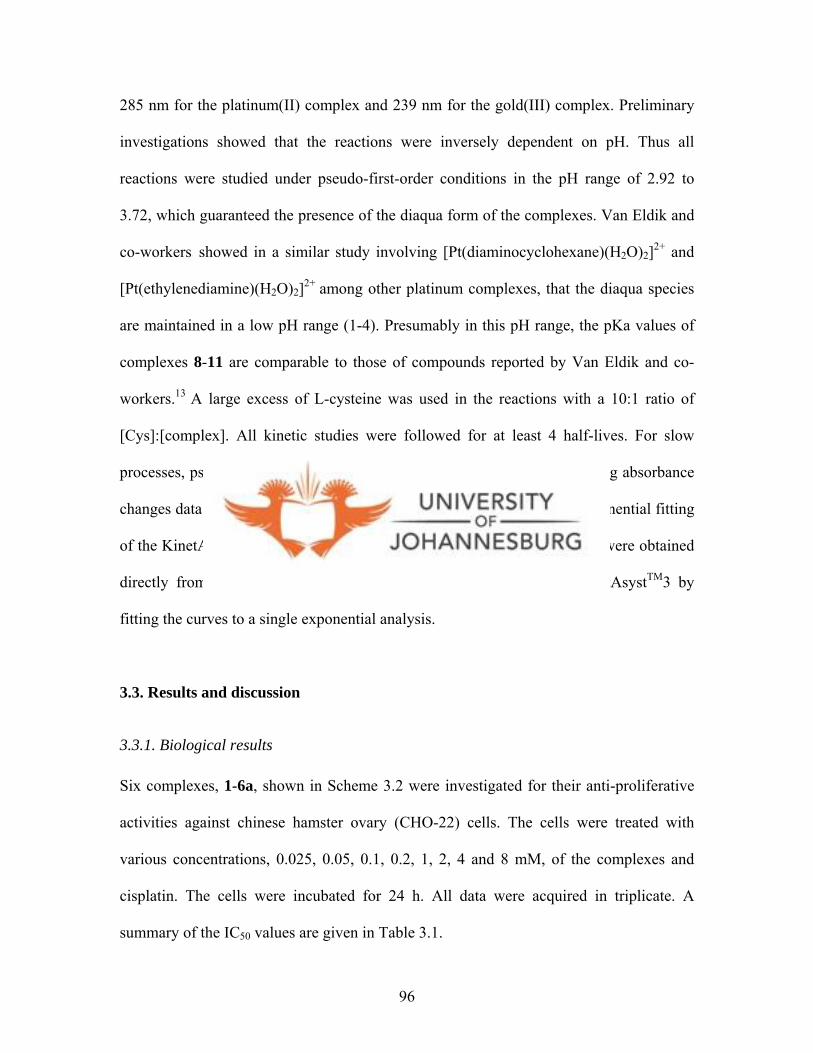
285 nm for the platinum(II) complex and 239 nm for the gold(III) complex. Preliminary
investigations showed that the reactions were inversely dependent on pH. Thus all
reactions were studied under pseudo-first-order conditions in the pH range of 2.92 to
3.72, which guaranteed the presence of the diaqua form of the complexes. Van Eldik and
co-workers13 showed in a similar study involving [Pt(diaminocyclohexane)(H2O)2]2+ and
[Pt(ethylenediamine)(H2O)2]2+ among other platinum complexes, that the diaqua species
are maintained in a low pH range (1-4). Presumably in this pH range, the pKa values of
complexes 8-11 are comparable to those of compounds reported by Van Eldik and co-
workers.13 A large excess of L-cysteine was used in the reactions with a 10:1 ratio of
[Cys]:[complex]. All kinetic studies were followed for at least 4 half-lives. For slow
processes, pseudo first-order rate constants, kobs, were obtained by importing absorbance
changes data measured on a UV-Vis spectrophotometer into the single exponential fitting
of the KinetAsystTM3 software program. The kobs values for fast processes were obtained
directly from the stopped-flow equipment software program using KinetAsystTM3 by
fitting the curves to a single exponential analysis.
3.3. Results and discussion
3.3.1. Biological results
Six complexes, 1-6a, shown in Scheme 3.2 were investigated for their anti-proliferative
activities against chinese hamster ovary (CHO-22) cells. The cells were treated with
various concentrations, 0.025, 0.05, 0.1, 0.2, 1, 2, 4 and 8 mM, of the complexes and
cisplatin. The cells were incubated for 24 h. All data were acquired in triplicate. A
summary of the IC50 values are given in Table 3.1.
96
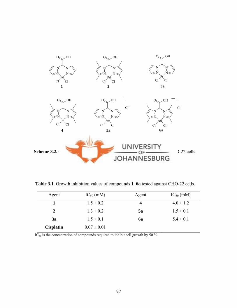
NN N
N
OHO
PdCl Cl
NN N
N
OHO
PdCl Cl
NN N
N
OHO
PtCl Cl
NN N
N
OHO
PtCl Cl
NN N
N
OHO
Au
Cl-
Cl Cl
NN N
N
OHO
Au
Cl-
Cl Cl5a 6a
3a
4
1 2
Scheme 3.2. Complexes tested for anti-tumour their anti-tumour against CHO-22 cells.
Table 3.1. Growth inhibition values of compounds 1–6a tested against CHO-22 cells.
Agent IC50 (mM) Agent IC50 (mM)
1 1.5 ± 0.2 4 4.0 ± 1.2
2 1.3 ± 0.2 5a 1.5 ± 0.1
3a
Cisplatin
1.5 ± 0.1
0.07 ± 0.01
6a 5.4 ± 0.1
IC50 is the concentration of compounds required to inhibit cell growth by 50 %.
97
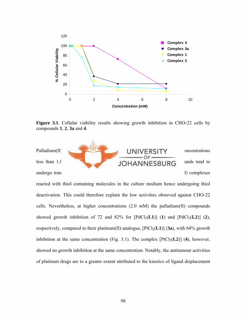
0
20
40
60
80
100
120
0 2 4 6 8
Concentration (mM)
% C
ellu
lar V
iabi
lity
10
Complex 4Complex 3aComplex 1Complex 2
Figure 3.1. Cellular viability results showing growth inhibition in CHO-22 cells by compounds 1, 2, 3a and 4.
Palladium(II) and platinum(II) compounds 1-4 showed no inhibition at concentrations
less than 1.0 mM (Fig. 3.1). This is not surprising as palladium compounds tend to
undergo translabilisation easily. Thus, it is possible that these palladium(II) complexes
reacted with thiol containing molecules in the culture medium hence undergoing thiol
deactivation. This could therefore explain the low activities observed against CHO-22
cells. Nevertheless, at higher concentrations (2.0 mM) the palladium(II) compounds
showed growth inhibition of 72 and 82% for [PdCl2(L1)] (1) and [PdCl2(L2)] (2),
respectively, compared to their platinum(II) analogue, [PtCl2(L1)] (3a), with 64% growth
inhibition at the same concentration (Fig. 3.1). The complex [PtCl2(L2)] (4), however,
showed no growth inhibition at the same concentration. Notably, the antitumour activities
of platinum drugs are to a greater extent attributed to the kinetics of ligand displacement
98
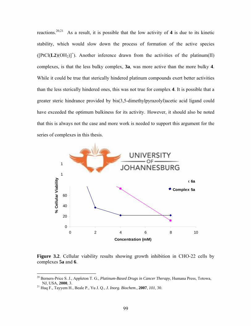
reactions.20,21 As a result, it is possible that the low activity of 4 is due to its kinetic
stability, which would slow down the process of formation of the active species
([PtCl(L2)(OH2)]+). Another inference drawn from the activities of the platinum(II)
complexes, is that the less bulky complex, 3a, was more active than the more bulky 4.
While it could be true that sterically hindered platinum compounds exert better activities
than the less sterically hindered ones, this was not true for complex 4. It is possible that a
greater steric hindrance provided by bis(3,5-dimethylpyrazolyl)acetic acid ligand could
have exceeded the optimum bulkiness for its activity.2 1 However, it should also be noted
that this is always not the case and more work is needed to support this argument for the
series of complexes in this thesis.
0
20
40
60
80
100
120
0 2 4 6 8
Concentration (mM)
% C
ellu
lar V
iabi
lity
10
Complex 6a
Complex 5a
Figure 3.2. Cellular viability results showing growth inhibition in CHO-22 cells by complexes 5a and 6. 20 Berners-Price S. J., Appleton T. G., Platinum-Based Drugs in Cancer Therapy, Humana Press, Totowa, NJ, USA, 2000, 3. 21 Huq F., Tayyem H., Beale P., Yu J. Q., J. Inorg. Biochem., 2007, 101, 30.
99
A similar trend was observed for the gold(III) complexes, which showed no appreciable
growth inhibition of HeLa cells at concentrations less than 1.0 mM (Fig. 3.2). However,
at 2.0 mM compound [AuCl2(L1)]Cl (5a) showed 75% growth inhibition whereas its
dimethyl analogue, [AuCl2(L2)]Cl (6a), had no inhibition at the same concentration in
spite of their structural similarity (Fig. 3.2). The IC50 of compounds 5a and 6a were
found to be 1.6 and 5.4 mM respectively. One of the main problems associated with
gold(III) complexes in comparison with platinum(II), is the observed low stability in
solution under physiological conditions. It has been reported that they undergo a faster
hydrolysis and/or reduction to gold(I) accompanied by the formation of colloidal gold in
some cases, meaning that the compounds are of low clinical usefulness.22 The low
activities of gold(III) complexes 5a and 6a were attributed to their fast hydrolysis rates as
well as their possible low stabilities in physiological conditions. However, there was no
physical evidence of the formation of colloidal gold in the cases of 5a and 6a, as reported
in the literature.22
It is clear that the IC50 values of complexes 1-6a (Table 3.1) are higher than that of
cisplatin (0.07 mM), thus ruling them out as potential anticancer drugs. Nevertheless, the
kinetic labilities of these complexes were investigated in order to find out if there was
any relationship between their ligand substitution reaction rates and the observed low
biological activities. The next section deals with the kinetic studies.
22 aCriado J. J., Manzano J. L., Rodriguez-Fernandez E., J. Inorg. Biochem., 2003, 96, 311; bCalamai P., Carotti S., Guerri A., Mazzei T., Messori L., Mini E., Orioli P., Speroni G. P., J. Inorg. Biochem., 1997, 66, 103.
100

3.3.2 Kinetics of reaction of L-cysteine with palladium(II), platinum(II) and gold(III)
complexes
Reactions of L-cysteine with four selected complexes, 1, 2, 3a and 5a (Scheme 3.2), was
studied by UV-Vis and stopped-flow spectrophotometry. This was achieved by
measuring the absorbance changes with time. Initial investigations showed that the
kinetics of the chloro complexes 1, 2, 3a and 5a were very slow, hence they were
converted to the diaqua forms, [Pd(L1)(OH2)2]2+ (8), [Pd(L2)(OH2)2]2+ (9),
[Pt(L1)(OH2)2]2+ (10) and [Au(L1)(OH2)2]3+ (11) (Scheme 3.3).
NN N
N
HO O
Au
ClCl
+
Cl-
NN N
N
HO O
Au
OH2OH2
3+
3 CIO4-
2+
2 CIO4-
NN N
N
HO O
M
ClCl
R
R
R
R
NN N
N
HO O
M
OH2OH2
R
R
R
R
3AgClO4
2AgClO4
11
M = Pd, R = H (8); M = Pd, R = Me (9); M = Pt, R= H (10)
6a
M = Pd, R = H (1); M = Pd, R = Me (2); M = Pt, R= H (3a)
Scheme 3.3. The preparation of the diaqua complexes 8-10
101
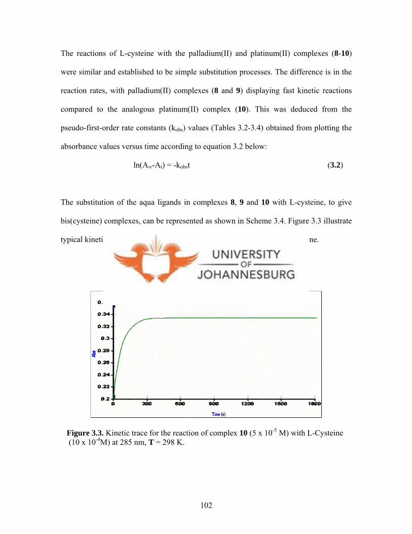
The reactions of L-cysteine with the palladium(II) and platinum(II) complexes (8-10)
were similar and established to be simple substitution processes. The difference is in the
reaction rates, with palladium(II) complexes (8 and 9) displaying fast kinetic reactions
compared to the analogous platinum(II) complex (10). This was deduced from the
pseudo-first-order rate constants (kobs) values (Tables 3.2-3.4) obtained from plotting the
absorbance values versus time according to equation 3.2 below:
ln(A∞-At) = -kobst (3.2)
The substitution of the aqua ligands in complexes 8, 9 and 10 with L-cysteine, to give
bis(cysteine) complexes, can be represented as shown in Scheme 3.4. Figure 3.3 illustrate
typical kinetic trace for the substitution reaction of complex 10 with L-cysteine.
Figure 3.3. Kinetic trace for the reaction of complex 10 (5 x 10⁻⁵ M) with L-Cysteine (10 x 10-4M) at 285 nm, T = 298 K.
102

2+
2 CIO4-
NN N
N
HO O
M
OH2OH2
R
R
R
R
M = Pd, R = H (8); M = Pd, R = Me (9); M = Pt, R= H (10)
S
CO2H
NH2
Cys
k1
kobs NN N
N
HO O
MOH2
+
SHO2C
H2N
NN N
N
HO O
M
SCO2H
NH2
Cys
M = Pd, R = H (12a); M = Pd, R = Me (12b);M = Pt, R = H (12c)
k-1
CIO4-R
R
R
R
R
R
R
R
+
k2
+
Scheme 3.4. The reaction of the palladium(II) and platinum(II) diaqua complexes (8-10) with L-cysteine.
Subsequently, plots of the pseudo-first-order rate constants kobs versus L-cysteine
concentration ([Cys]) were linear according to the two-term rate law:
kobs = k-1 + k1[Cys] (3.3)
This is typical of two-term rate equations for nucleophilic substitution in d8 square planar
complexes, where k1 denotes a second-order rate constant for the direct substitution of
water molecules, and k-1 represents first-order rate constant for the reverse reaction where
a water molecule replaces the coordinated nucleophile.23 Plots of kobs versus L-cysteine
23 Shoukry A., Brindell M., Van Eldik R., Dalton Trans., 2007, 4169.
103
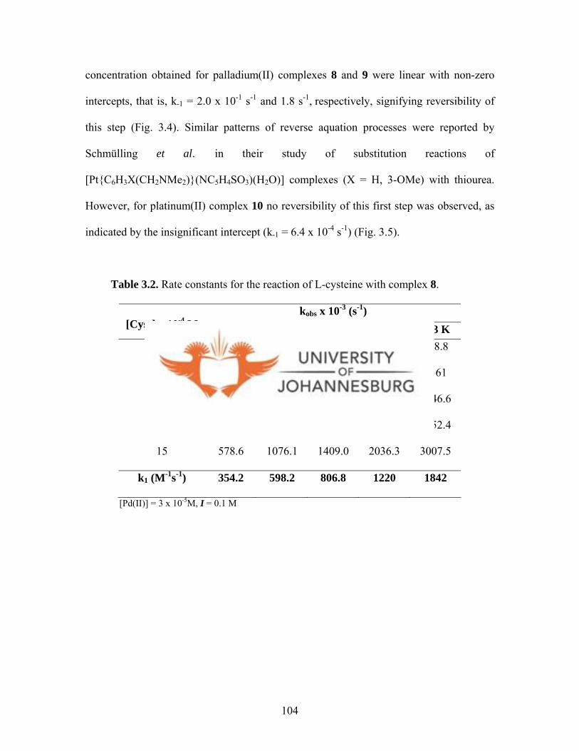
concentration obtained for palladium(II) complexes 8 and 9 were linear with non-zero
intercepts, that is, k-1 = 2.0 x 10⁻¹ s-1 and 1.8 s-1, respectively, signifying reversibility of
this step (Fig. 3.4). Similar patterns of reverse aquation processes were reported by
Schmülling et al.17 in their study of substitution reactions of
[Pt{C6H3X(CH2NMe2)}(NC5H4SO3)(H2O)] complexes (X = H, 3-OMe) with thiourea.
However, for platinum(II) complex 10 no reversibility of this first step was observed, as
indicated by the insignificant intercept (k-1 = 6.4 x 10⁻⁴ s-1) (Fig. 3.5).
Table 3.2. Rate constants for the reaction of L-cysteine with complex 8.
kobs x 10-3 (s-1) [Cyst] x 10-4 M 293 K 298 K 303 K 308 K 313 K
3 153.8 347.5 451.2 595.3 788.8
6 260.1 525.1 667.3 912.6 1361
9 366.4 671.4 940.1 1251.0 1846.6
12 472.6 862.7 1172.2 1690.0 2452.4
15 578.6 1076.1 1409.0 2036.3 3007.5
k1 (M-1s-1) 354.2 598.2 806.8 1220 1842
[Pd(II)] = 3 x 10⁻⁵M, I = 0.1 M
104

Table 3.3. Rate constants for the reaction of L-cysteine with complexe 9.
kobs x 10-3 (s-1) [Cyst] x 10-4 M 293 K 298 K 303 K 308 K 313 K
3 740.4 1479.0 2484.7 3653.6 5791.4
6 1030.3 1881.0 3092.0 4782.7 7044.6
9 1318.6 2402.4 3761.5 5791.0 8689.2
12 1610.5 2810.8 4436.7 6905.4 10410.1
15 1875.0 3226.6 5082.2 8006.5 11710.2
k1 (M-1s-1) 451.9 701.3 1038 1718 2414
[Pd(II)] = 3.8 x 10⁻⁵M, I = 0.1 M
Table 3.4. Rate constants for the reaction of L-cysteine with complex 10.
kobs x 10-3 (s-1) [Cyst] x 10-4 M 293 K 298 K 303 K 308 K 313 K
10 1.4 1.9 2.6 3.3 4.2
20 2.0 3.0 4.5 5.6 7.2
30 2.8 4.0 5.8 7.8 10.0
40 3.6 4.9 7.5 9.5 12.7
50 4.2 6.0 8.7 11.9 14.7
k1 (M-1s-1) 0.7 1.02 1.5 2.1 2.6
[Pt(II)] = 5 x 10⁻⁵M, I = 0.1 M
105
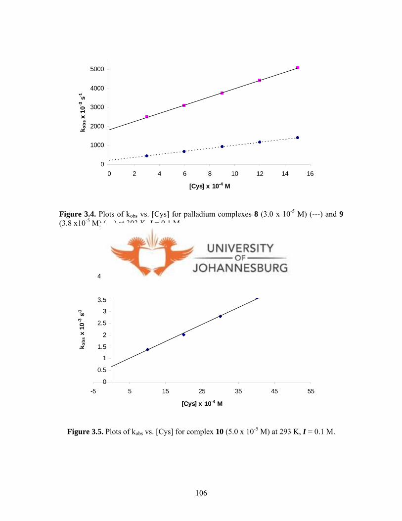
0
1000
2000
3000
4000
5000
0 2 4 6 8 10 12 14 16
[Cys] x 10-4 M
k obs
x 1
0-3 s
-1
Figure 3.4. Plots of kobs vs. [Cys] for palladium complexes 8 (3.0 x 10⁻⁵ M) (---) and 9 (3.8 x10⁻⁵ M) (—) at 303 K, I = 0.1 M.
0
0.5
1
1.5
2
2.5
3
3.5
4
4.5
-5 5 15 25 35 45 5
[Cys] x 10-4 M
k obs
x 1
0-3 s
-1
5
Figure 3.5. Plots of kobs vs. [Cys] for complex 10 (5.0 x 10⁻⁵ M) at 293 K, I = 0.1 M.
106

Contrary to the substitutions reactions of 8-10 with L-cysteine, reactions of L-cysteine
with the gold(III) complex (11) exhibited an initial fast substitution step (Table 3.5, Fig.
3.6) followed by a much slower process. It is possible that this slow process is due to the
reduction of 11 to gold(I) species (Table 3.6, inset in Fig 3.6). The pseudo-first-order rate
constants kobs for the fast substitution process in 11 were obtained in the same way as for
8 and 9, using equation 3.2. Subsequently, the second rate constants for the substitution
reactions were obtained from plotting kobs versus L-cysteine concentration. The plots
were linear according to the two-term rate law given in equation 3.3. The non-zero
intercepts in the plots of kobs vs [Cys] for 11 ( k-1 ≈ 5.0 x 10⁻¹ s-1) signifies the
reversibility of the substitution process, where a coordinated L-cysteine is replaced by a
water molecule. The substitution rates of 11 are three times faster than for platinum
complex 10. For example, the substitution in 11 was fast with a second order rate
constant of 321.8 M-1 s-1 at 298 K compared to that of 10 (1.0 M-1 s-1) at the same
conditions.
The consequent presumptive reduction process of 11 to gold(I) was quite slow and was
captured on the conventional UV-Vis spectrophotometer. The pseudo-first-order rate
constants kobs for this process were obtained by plotting the absorbance values versus
time according to equation 3.2, whilst the second order rate constants (k3) for this process
were obtained from plots of kobs versus [Cys] according to equation 3.4 below. The plots
(Fig. 3.7) were linear with insignificant intercepts (k-1 ≈ 1.0 x 10⁻⁴ s-1), thus indicating the
irreversible nature of the reduction process in 11.
107
kobs = k-1 + k3[Cys] ≈ k3[Cys]
(3.4)
The parameter k3 represents second rate constants for the reduction process, which
follows the substitution reactions of complex 11 with L-cysteine (Scheme 3.5). The kobs
and k3 values for the reduction process in 11 were much smaller than its corresponding
values for the substitution process (Tables 3.5 and 3.6). For example, the second order
rate for the reduction process was found to be 1.0 M-1 s-1 at 298 K (Table 3.6). It is three
times slower than that of the preceding substitution process (321.8 M-1 s-1). From the
results obtained (kobs), this reduction process is linearly dependent on the L-cysteine
concentration.
108

NN N
N
HO O
AuClCl
+
Cl-
NN N
N
HO O
AuOH2OH2
3+
3 CIO4-
3 AgClO4
116a
S
CO2H
NH2
Cys
k1
kobs
NN N
N
HO O
AuOH2
+
SHO2C
H2N
NN N
N
HO O
Au
SCO2H
NH2
Cys
12d
k-1
CIO4-R
R
R
R
R
R
R
R
+
+
CIO4-
Gold reduction products
Reductionk3
k2
+
Scheme 3.5. The reaction of the gold diaqua complex 11 with L-cysteine.
Figure 3.6. Kinetic trace for the reaction of complex 11 (5 x 10⁻⁵ M) with L-Cysteine (10 x 10-4M) at 239 nm, T = 298 K. The slow reduction process is shown in the inset.
109

Table 3.5. Rate constants for the reaction of L-cysteine with 11.
kobs x 10-3 (s-1)
[Cyst] x 10-4 M 293 K 298 K 303 K 308 K 313 K
5 472.7 591.2 917.6 1053.1 1201.3
10 619.2 856.4 1061.3 1251.0 1366.0
20 880.1 1170.2 1405.4 1675.0 1739.2
30 1251.5 1446.0 1766.0 1899.2 2201.4
40 1492.0 1756.0 2062.2 2356.4 2589.7
50 1829.2 2101.4 2462.0 2658.2 2892.5
k1 (M-1s-1) 300.8 321.8 341.6 357.4 387.4
[Au(III)] = 5 x 10⁻⁵ M; I = 0.1 M
Table 3.6. Rate constants of the reduction of 11 to gold(I).
kobs x 10-3 (s-1) [Cyst] x 10-4 M 293 K 298 K 303 K 308 K 313 K
10 1.1 2.1 3.4 4.3 5.0
20 2.0 2.9 4.4 5.5 6.3
30 2.6 3.9 5.3 6.6 7.6
40 3.5 4.9 6.5 8.0 9.7
50 4.4 5.9 7.9 9.7 10.9
k3 (M-1 s-1) 0.8 1.0 1.1 1.3 1.5
[Au(III)] = 5 x 10⁻⁵M, I = 0.1 M.
110

0
500
1000
1500
2000
2500
0 10 20 30 40 50 6
[Cys] x 10-4 M
k obs
x 1
0-3 s
-1
0
1
2
3
4
5
6
7
0 10 20 30 40 50 60
[Cys] x 10-4 M
k 3 x
10-3
s-1
0
Figure 3.7. Plots of kobs vs. [Cys] for 11 (5.0 x 10⁻⁵ M) at 298 K, I = 0.1 M. Inset depicts plots of k1 vs. [Cys] for the reduction process of 11.
In general, the rates of substitution reactions of complexes 8-11 with L-cysteine were
found to be pseudo-first-order. The first step is the substitution of the first aqua ligand,
followed by a second substitution step, which was slower and is attributed to the
substitution of the second aqua ligand. Similar substitution reactions patterns with
different nucleophiles have been observed for many platinum(II) compounds and a few
palladium(II) compounds. For example [Pt(bpy)(H2O)2]2+ (bpy = N,N’-bipyridine),13
[Pt(en)(H2O)2]2+ (en = ethylenediamine)1 3 and [Pd(en)(cbdca)] (en = ethylenediamine,
cbdca = cyclobutane-1,1-dicarboxylate)24 react with thiourea in a similar way as
observed for compounds 8-11 herein (vide supra). However, gold(III) complex 11
undergo a further reduction process presumably to gold(I) species. No efforts were made
24 Shoukry A., Rau T., Shoukry M., Van Eldik R., J. Chem. Soc., Dalton Trans., 1998, 3105.
111
to characterise the reduction products. The rate constants for the substitution reactions of
L-cysteine in palladium(II) complexes 8 and 9 are faster than in platinum(II) (10) and
gold(III) (11) complexes (Tables 3.2-3.5); and in agreement with literature reports that
substitution reactions of square planar palladium(II) complexes are faster than for square
planar gold(III) and platinum(II) complexes respectively.15,17
The rate of substitution in 10 (prepared from 3a) is slower than in complexes 8, 9 and 11
(prepared from 1, 2 and 5a, respectively) (Schemes 3.4 and 3.5). Although these slow
rates suggest that platinum(II) complex 3a would have better chance of reaching the
target in cells compared to palladium(II) complexes (1 and 2) and gold(III) complex 5a,
the IC50 values (Table 3.1) are similar for all the four compounds; suggesting that there is
no relationship between the observed anticancer activities of these complexes with their
kinetic labilities. Nevertheless, the L-cysteine substitution reactions have some
interesting observations.
There are a number of thermodynamic parameters that could be derived from the kinetic
data to shed more light on the mechanism of the L-cysteine substitution reactions. For all
the reactions studied, the rate constants increased with increasing temperature; and from
the second order rate constants at different temperatures, activation parameters ∆H‡ and
∆S‡ were obtained (Table 3.7).
112
Table 3.7. Activation parameters determined from the temperature dependence of the
rate constants for the substitution of Pd(II), Pt(II) and Au(III) complexes, and
reduction of Au(III).
Activation
parameter
8 9 10 11 Reduction of 11 to
gold(I)
∆H‡ (kJ mol-1) 42.6 62.2 48.3 6.8 18.4
∆S‡ (kJ mol-1) -47.8 +18.4 -82.6 -174 -183
The activation entropy (∆S‡) changes for all the substitutions, except for palladium(II)
complex 9, are largely negative and in agreement with the fact that substitution reactions
involving square planar complexes are invariably accompanied by large negative entropy
changes.25 The entropy change for 9 is positive (∆S‡ = +18.4 J K-1 mol-1), but is relatively
low to fit an associative mechanism of substitution. Unlike 8, 10 and 11, compound 9 has
a more bulky ligand, bis(3,5-dimethylpyrazolyl)acetic acid. It is possible that the steric
bulkiness hinders the entrance of L-cysteine; thereby leading to less ordered transition
state. Moreover, such low values (+18.4 J K-1 mol-1) are always regarded as close to zero
and are attributed to associative interchange mechanisms.24, ,26 27 Generally, the data
obtained show that the substitution of aqua ligands in complexes 8-11 is via an
associative mechanism. The data also show that the slower reactions have higher ∆H‡
values, consistent with literature findings that substitution reactions of platinum(II)
25 Wilkins G. R., The Study of Kinetics and Mechanism of Reactions of Transition Metal Complexes, Allyn and Bacon, Inc. Boston, 1974, 227. 26 Rau T., Shoukry M., van Eldik R., Inorg. Chem., 1997, 36, 1454. 27 Hohmann H., Hellquist B., van Eldik R., Inorg. Chem., 1992, 31, 1090.
113

114
complexes are largely ∆H‡-controlled.28 Tables 3.8 and 3.9 show data for calculation of
the energy parameters, whilst Figure 3.8 show the corresponding Eyring plots.
-6
-5.9
-5.8
-5.7
-5.6
-5.5
-5.4
-5.30.00315 0.0032 0.00325 0.0033 0.00335 0.0034 0.00345
1/T (K-1)
ln(k
1/T)
Figure 3.8. Temperature dependence plots for the reaction of complexes with L-cysteine.
28 Bajaj H. C., van Eldik R., Inorg. Chem., 1988, 27, 4052.
(i) 8, 9 and 11, (ii) 10, (iii) reduction of 11.
-0.5
0
0.5
1
1.5
2
2.5
3
0.00315 0.0032 0.00325 0.0033 0.00335 0.0034 0.00345
1/T (K-1)
ln(k
1/T)
Complex 9
Complex 8
Complex 11
-6.5
-6
-5.5
-5
-4.5
-40.00315 0.0032 0.00325 0.0033 0.00335 0.0034 0.00345
1/T (K-1)
ln(k
1/T)
iii
ii
115
8 9 10 11 Reduction of 11
T(K)
1/T (K-1) k1 (M-1s-1) In(k2/T) k1 (M-1s-1) In(k2/T) k1 (M-1s-1) In(k2/T) k1 (M-1s-1) In(k2/T) k3 (M-1s-1) In(k2/T)
293 0.003413 354.2 0.19 451.9 0.43 0.7186 -6.01 300.8 0.03 0.8190 -5.88
298 0.003356 598.2 0.70 701.3 0.86 1.021 -5.67 321.8 0.08 0.9524 -5.75
303 0.003300 806.8 0.98 1038 1.23 1.521 -5.29 341.6 0.12 1.106 -5.61
308 0.003247 1220 1.38 1718 1.72 2.114 -5.98 357.4 0.15 1.319 -5.45
313 0.003195 1842 1.77 2414 2.04 2.639 -4.78 387.4 0.21 1.512 -5.33
8 9 10 11 Reduction of 11
Slope -7054.8 -7489 -5814.2 -817.5 -2543.5
Intercept 24.299 25.987 13.852 2.8166 2.7943
Table 3.9. Variation of k1 and k3 rate constants with temperature for complexes 8, 9, 10 and 11.
Table 3.8. Calculation of energy parameters.
Slope = - ∆H‡/R, Intercept = ln(k/h) + ∆S‡/R

Product formation in the substitution reactions involving complexes 1, 2, 3a and 5a with
L-cysteine could be followed by 1H NMR spectroscopy. In a typical experiment, a direct
reaction of 1 with L-cysteine gave [Pd(L1)(Cys)2] (12a). An attempt was made to follow
the formation of compound 12a by 1H NMR spectroscopy in DMSO-d6. However, the
stepwise formation of 12a could not be captured clearly, probably due to the fast nature
of the substitution process. Nevertheless, the spectrum of solution containing L-cysteine
and 1 shows peaks due to 1 and L-cysteine.
(a)
N
N
(b)
N
N
HO O
Pd
Cl Cl
1
23
4
5 1'
2'3'
4'
5'6
N
N N
N
HO O
Pd
Cys Cys
1
23
4
5 1'
2'3'
4'
5'6
H2N
SH
O
OH
(c)
Figure 3.9. 1H NMR spectra of (a) [PdCl2(L1)] (1), (b) L-cysteine and (c) [Pd(L1)(Cys)2] (12a) acquired after 5 min (solvent, DMSO-d6)
116

The spectrum (Fig. 3.9c) acquired after 5 min shows major upfield shifts of the proton
peaks of the bis(pyrazolyl)acetic acid ligand (bpza) at 7.95 ppm (5H, 5H′), 7.55 ppm (3H,
3H′ + CH linker proton) and 6.32 ppm (4H, 4H′) (Fig. 3.9c) in comparison with that of 1
(Fig. 3.9a), which has the four peaks at 8.29 ppm (5H, 5H′), 8.11 ppm (CH linker
proton), 8.03 ppm (3H, 3H′ ) and at 6.63 ppm (4H, 4H′). The spectrum in Figure 3.9c also
displays five faint humps at 4.18 ppm, 4.81, 5.09, 5.72 ppm and at 8.65 ppm, possibly
due to the intermediate monodentate species. The humps disappeared 30 min thereafter,
pointing to the diminishment of the intermediate species. The spectrum acquired after 48
h display peaks at 7.96, 7.56 and 6.32 ppm corresponding to the bpza ligand (vide supra);
and at 8.52 ppm (COOH), 4.22 ppm (CH), 3.31 ppm (CH2) and 2.92 ppm (NH2), which
correspond to L-cysteine (Fig. 3.9c).
Further efforts to confirm the final products of the substitution reactions were pursued by
setting an experiment on a preparative scale. Compound [Pd(L2)(OH2)2]2+ (9) was
generated from [PdCl2(L2)] (2) as described in Scheme 3.2 and reacted with L-cysteine
to form [Pd(L2)(Cys)2] (12b). The ESI-MS of compound 12b (Fig. 3.10) showed a
molecular ion peak at m/z = 601, which corresponds to formation of 12b. Substitution
reactions in diaqua metal complexes leading to formation of bis(L-cysteine)metal
complexes are known.29 Based on the kinetics, spectroscopic and mass spectrometry data,
it was proposed that the substitution of aqua ligands in complexes 8-11 proceed by the
sequence of reactions in Scheme 3.4 and 3.5.
29 Bose R. N., Ghosh S. K., Moghaddas S., J. Inorg. Biochem., 1997, 65, 199.
117

SHO2C
H2N
NN N
N
HO O
Pd
SCO2H
NH2
12b
Figure 3.10. ESI-MS spectrum of compound 12b.
3.4. Conclusions
As mentioned in chapter 2, the coordination of bis(pyrazolyl)acetic acid ligands to
gold(III), platinum(II) and palladium(II) form complexes that a relatively stable and
closely related to cisplatin. In spite of this advantage, complexes 1-6a had no appreciable
biological activities against CHO-22 cells.
Overall, the kinetics results evidently showed that there was no relationship between the
observed anticancer activities of complexes 1-6a and their reaction rates. Nevertheless,
kinetics study was carried out to establish the probable mechanisms and products formed
from reactions of these complexes and L-cysteine. The substitution reactions of
[Pt(L1)(OH2)2]+ (10) with L-cysteine was found to be slow compared to those of
[Pd(L1)(OH2)2]2+ (8), [Pd(L2)(OH2)2]2+ (9) and [Au(L1)(OH2)2]3+ (11). Furthermore, the
substitution reaction involving gold(III) complex 11 showed a two-step reaction
118

involving the substitution of aqua ligands and a subsequent slow process, possibly due to
reduction of 11 to gold(I). The substitution reactions of L-cysteine with the diaqua metal
complexes 8-11 were found to be temperature dependent and occurred via an associative
mechanism. The largely negative entropy values registered are supportive of an
associative mechanism of substitution. The mass spectrum data points to formation
bis(cysteine)metal complexes, but for gold complex 11, the second slow process
observed suggests that the final products are not necessarily bis(cysteine) complexes.
119

CHAPTER 4
PLATINUM(II) AND GOLD(I) DITHIOCARBAMATE COMPLEXES
X-ray structures 15, 17b, 25 and L10 were solved by Dr. Guzei and Mrs. Spencer both of
Wisconsin University (USA), whilst X-ray structure of L9 was solved by the candidate and
Dr. Omondi (University of Johannesburg). All the crystal structure discussions are the work
of the candidate except for 17b, where Dr. Guzei helped with the discussion.
4.0. Introduction
There has been increasing interest in the synthesis and reactivity of mononuclear metal
complexes with sulfur-containing ligands because of their relevance as models of
biologically redox-active metalloproteins.1, ,2 3 For this reason, the dithiocarbamate ligand
(dtc), first identified during the early days of organosulfur chemistry, has been studied and
applied widely.4 Free dithiocarbamic acids (R2NCS2H) are highly unstable and are normally
isolated as their alkali metal salts.4 Most studied dtcs prepared from secondary amines,
because they are stable and possess interesting electrochemical and optical properties.4,5
Dithiocarbamates are synthesised by reacting amines with carbon disulfide under basic
conditions as illustrated in Scheme 4.1.
1 Power P. P., Shoner S. C., Angew. Chem., Int. Ed. Engl., 1991, 30, 330. 2 Ruiz J., Giner J., Rodríguez V., López G., Casabó J., Molins E., Miravitlles C., Polyhedron,, 2000, 19, 1627. 3 Holm R. H., Kennepohl P., Solomon E. I., Chem. Rev., 1996, 96, 2239. 4 Cookson J., Beer P. D., Dalton Trans., 2007, 1459. 5 Bond A. M., Martin R. L., Coord. Chem. Rev., 1984, 54, 23.
120
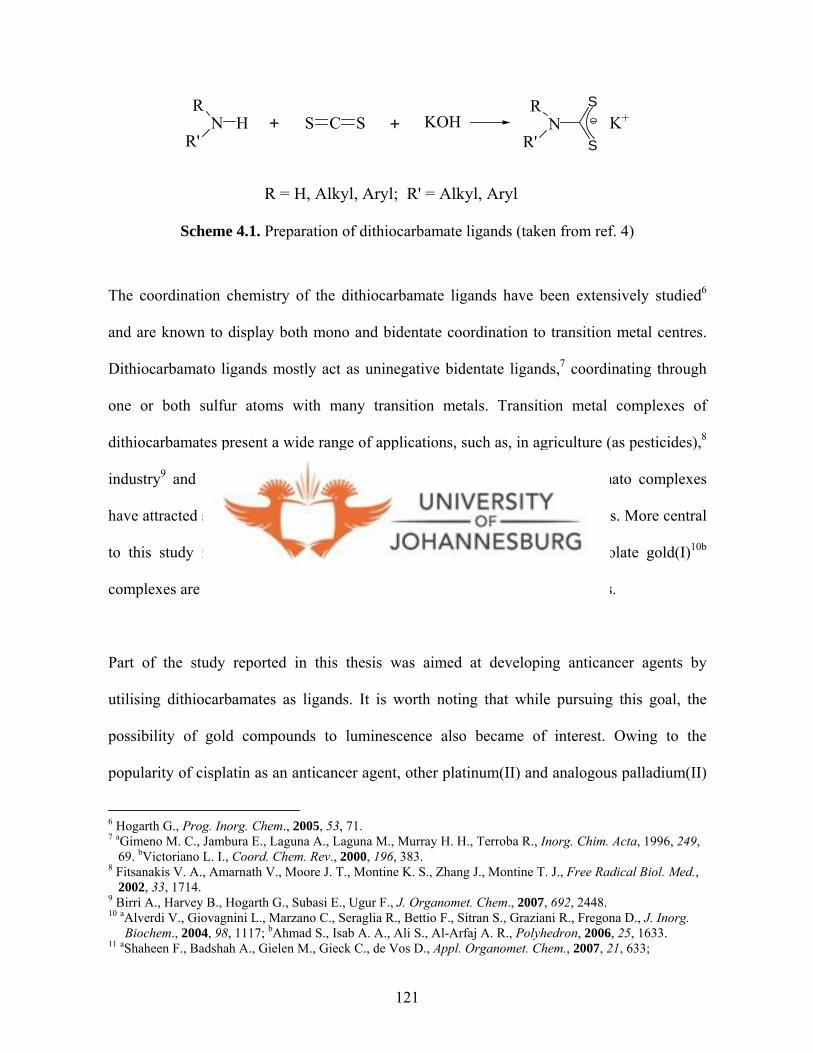
NR
R'H + CS S + N
R
R'
S
S
R = H, Alkyl, Aryl; R' = Alkyl, Aryl
KOH K+
Scheme 4.1. Preparation of dithiocarbamate ligands (taken from ref. 4)
The coordination chemistry of the dithiocarbamate ligands have been extensively studied6
and are known to display both mono and bidentate coordination to transition metal centres.
Dithiocarbamato ligands mostly act as uninegative bidentate ligands,7 coordinating through
one or both sulfur atoms with many transition metals. Transition metal complexes of
dithiocarbamates present a wide range of applications, such as, in agriculture (as pesticides),8
industry9 and in medicine.10 Palladium(II)11a and copper(II)11b dithiocarbamato complexes
have attracted significant attention due to their potential biological applications. More central
to this study is that dithiocarbamate complexes of platinum(II)10a and thiolate gold(I)10b
complexes are known to exhibit antitumour properties and cytotoxic properties.
Part of the study reported in this thesis was aimed at developing anticancer agents by
utilising dithiocarbamates as ligands. It is worth noting that while pursuing this goal, the
possibility of gold compounds to luminescence also became of interest. Owing to the
popularity of cisplatin as an anticancer agent, other platinum(II) and analogous palladium(II)
6 Hogarth G., Prog. Inorg. Chem., 2005, 53, 71. 7 aGimeno M. C., Jambura E., Laguna A., Laguna M., Murray H. H., Terroba R., Inorg. Chim. Acta, 1996, 249, 69. bVictoriano L. I., Coord. Chem. Rev., 2000, 196, 383. 8 Fitsanakis V. A., Amarnath V., Moore J. T., Montine K. S., Zhang J., Montine T. J., Free Radical Biol. Med., 2002, 33, 1714. 9 Birri A., Harvey B., Hogarth G., Subasi E., Ugur F., J. Organomet. Chem., 2007, 692, 2448. 10 aAlverdi V., Giovagnini L., Marzano C., Seraglia R., Bettio F., Sitran S., Graziani R., Fregona D., J. Inorg. Biochem., 2004, 98, 1117; bAhmad S., Isab A. A., Ali S., Al-Arfaj A. R., Polyhedron, 2006, 25, 1633. 11 aShaheen F., Badshah A., Gielen M., Gieck C., de Vos D., Appl. Organomet. Chem., 2007, 21, 633;
121
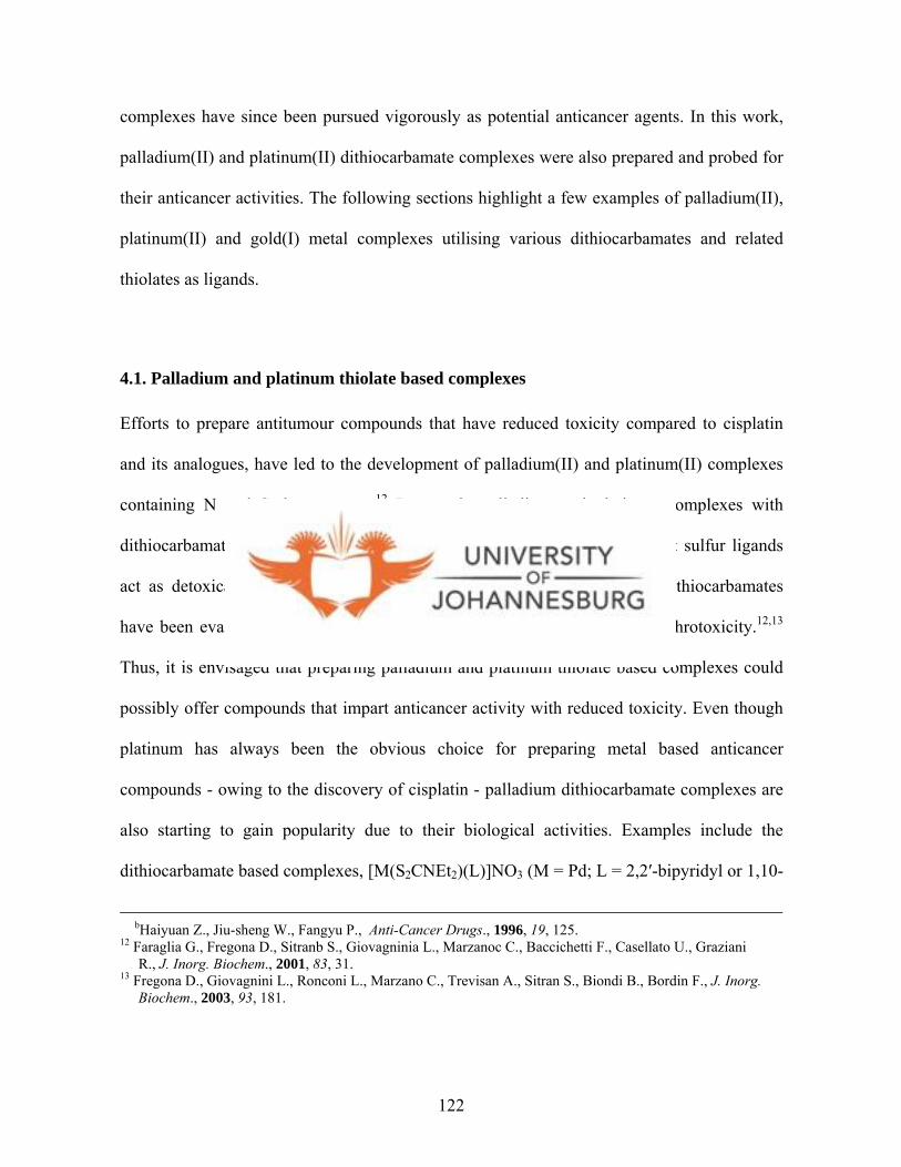
complexes have since been pursued vigorously as potential anticancer agents. In this work,
palladium(II) and platinum(II) dithiocarbamate complexes were also prepared and probed for
their anticancer activities. The following sections highlight a few examples of palladium(II),
platinum(II) and gold(I) metal complexes utilising various dithiocarbamates and related
thiolates as ligands.
4.1. Palladium and platinum thiolate based complexes
Efforts to prepare antitumour compounds that have reduced toxicity compared to cisplatin
and its analogues, have led to the development of palladium(II) and platinum(II) complexes
containing N and S donor atoms.12 Interest in palladium and platinum complexes with
dithiocarbamates and related thiolates emanates from the demonstration that sulfur ligands
act as detoxicant agents against metal containing drugs.13 For instance, dithiocarbamates
have been evaluated for their efficacy as inhibitors of cisplatin induced nephrotoxicity.12,13
Thus, it is envisaged that preparing palladium and platinum thiolate based complexes could
possibly offer compounds that impart anticancer activity with reduced toxicity. Even though
platinum has always been the obvious choice for preparing metal based anticancer
compounds - owing to the discovery of cisplatin - palladium dithiocarbamate complexes are
also starting to gain popularity due to their biological activities. Examples include the
dithiocarbamate based complexes, [M(S2CNEt2)(L)]NO3 (M = Pd; L = 2,2′-bipyridyl or 1,10-
bHaiyuan Z., Jiu-sheng W., Fangyu P., Anti-Cancer Drugs., 1996, 19, 125. 12 Faraglia G., Fregona D., Sitranb S., Giovagninia L., Marzanoc C., Baccichetti F., Casellato U., Graziani R., J. Inorg. Biochem., 2001, 83, 31. 13 Fregona D., Giovagnini L., Ronconi L., Marzano C., Trevisan A., Sitran S., Biondi B., Bordin F., J. Inorg. Biochem., 2003, 93, 181.
122

phenanthroline) (Fig. 4.1), which have shown antitumour activities against leukemia cells.14
The anticancer effect of [M(S2CNEt2)(L)]NO3 complexes is attributed to these complexes
associating to double-stranded DNAs by means of intercalation.14,15
NEt
Et
S
SM
N
N NO3
M = Pd, Pt
NEt
Et
S
SM
NO3N
N
Figure 4.1. Structures of dithiocarbamate based complexes, [M(S2CNEt2)(L)]NO3.
As an extension to [M(S2CNEt2)(L)]NO3 complexes (Fig. 4.1), Faraglia and co-workers12
prepared palladium(II) and platinum(II) complexes with the dithioesters ligands ESDTM
(EtO2CCH2(CH3)NCS2Me) (ESDT = methylamino-acetic acid ethyl ester-dithiocarboxylate)
and ESDTE (EtO2CCH2(CH3)NCS2Et) (Fig. 4.2). However, the anticancer activities of these
complexes against HeLa cells were lower than that of cisplatin (IC50 = 6.33 µM).12,16 Other
closely related examples include [M(DMDT)(Mol)]Cl (M = Pd, Pt; DMDT = Me2NCS2-;
Mol = L-methioninol).17 In all the examples mentioned above, the dithiocarbamate binds to
the metal in an ĸ2-S^S′ chelate fashion.
14 Mital R., Jain N., Srivastava T. S., Inorg. Chim. Acta, 1993, 166, 135. 15 Howe-Grant M., Wu K. C., Bauer W. R., Lippard S. J., Biochemistry, 1976, 15, 4339. 16 Marzano C., Trevisan A., Giovagnini L., Fregona D., Toxicol. In Vitro, 2002, 16, 413. 17 Faraglia G., Fedrigo M. A, Sitran S., Transition Met. Chem., 2002, 27, 200.
123
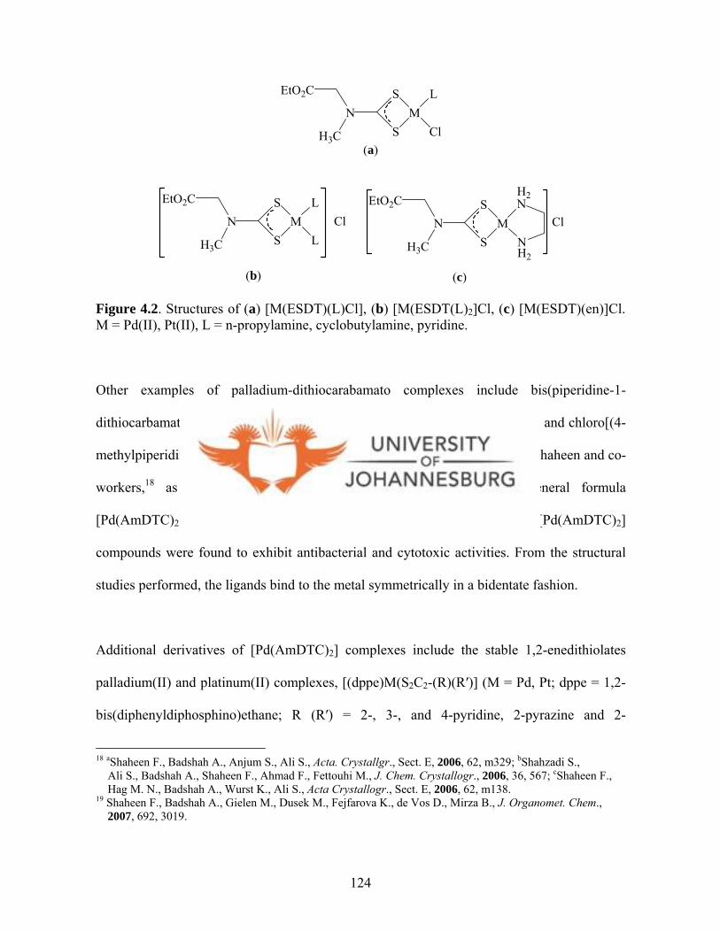
EtO2C
H3C
NS
SM
L
Cl
EtO2C
H3C
NS
SM
L
LCl
EtO2C
H3C
NS
SM
H2N
NH2
Cl
(a)
(b) (c)
Figure 4.2. Structures of (a) [M(ESDT)(L)Cl], (b) [M(ESDT(L)2]Cl, (c) [M(ESDT)(en)]Cl. M = Pd(II), Pt(II), L = n-propylamine, cyclobutylamine, pyridine.
Other examples of palladium-dithiocarabamato complexes include bis(piperidine-1-
dithiocarbamato)Pd(II), bis(4-methylpiperidine-1-S^S′-dithiocarbamato)Pd(II) and chloro[(4-
methylpiperidine-1-S^S′-dithiocarbamato)(PPh3)]Pd(II) recently reported by Shaheen and co-
workers,18 as well as six palladium(II) aminedithiocarbamates of general formula
[Pd(AmDTC)2], where H(AmDTC) = aminedithiocarbamic acid.19 These [Pd(AmDTC)2]
compounds were found to exhibit antibacterial and cytotoxic activities. From the structural
studies performed, the ligands bind to the metal symmetrically in a bidentate fashion.1 9
Additional derivatives of [Pd(AmDTC)2] complexes include the stable 1,2-enedithiolates
palladium(II) and platinum(II) complexes, [(dppe)M(S2C2-(R)(R′)] (M = Pd, Pt; dppe = 1,2-
bis(diphenyldiphosphino)ethane; R (R′) = 2-, 3-, and 4-pyridine, 2-pyrazine and 2-
18 aShaheen F., Badshah A., Anjum S., Ali S., Acta. Crystallgr., Sect. E, 2006, 62, m329; bShahzadi S., Ali S., Badshah A., Shaheen F., Ahmad F., Fettouhi M., J. Chem. Crystallogr., 2006, 36, 567; cShaheen F., Hag M. N., Badshah A., Wurst K., Ali S., Acta Crystallogr., Sect. E, 2006, 62, m138. 19 Shaheen F., Badshah A., Gielen M., Dusek M., Fejfarova K., de Vos D., Mirza B., J. Organomet. Chem., 2007, 692, 3019.
124
quinoxaline.20 The [(dppe)M(S2C2-(R)(R′)] compounds are further stabilised by the dppe
ligand in addition to the chelating 1,2-enedithiolates. However, interest in [(dppe)M(S2C2-
(R)(R′)] complexes concerned physical properties and not biological activities. Other classes
of palladium and platinum thiolate complexes are the salts of [M(II)(mnt)2]- (M = Pd, Pt; mnt
= maleonitriledithiolato) anions (Fig. 4.3).21 The molecular structures of [M(II)(mnt)2]-
exhibit the expected square planar geometry, with the metal bound to four sulfur atoms.
Compounds [M(II)(mnt)2]- were reported to show antiferromagnetic properties.21 In general,
palladium and platinum dithiocarbamate complexes are a promising class of potential
anticancer agents.22
N
N
SM
S
S
S
N
N
SM
S
S
SH2N
H2N NH2
NH2
a b
M = Pd, Pt
Figure 4.3. The structures of planar [M(II)(mnt)2]- complexes.
Similar to palladium(II) and platinum(II) thiolate complexes, gold(I) thiolate based
complexes have also been of interest as potential anticancer agents since it was shown that
the well known antiarthritic agent, auranofin (Fig. 4.4),23,24 and other gold compounds such
20 Kaiwar S. P., Hsu J. K., Liable-Sands L. M., Rheingold A. L., Pilato R. S., Inorg. Chem., 1997, 36, 4234. 21 Bigoli F., Deplano P., Mercuri M. L., Marchio L., Pilia L., Serpe A., Concas G., Congiu F., Sanna S., Chem. Phys. Lett., 2006, 421, 361. 22 Giovagnini L., Marzano C., Bettio F., Fregona D., J. Inorg. Biochem., 2005, 99, 2139. 23 aSimon T. M., Kunishima D. H., Vibert G. J., Lorber A., Cancer, 1979, 44, 1965; bSimon T. M., Kunishima D. H., Vibert G. J., Lorber A., Cancer Res., 1981, 41, 94. 24 Mirabelli C. K., Johnson R. K., Sung C., Faucette L. F., Muirhead K., Crooke S. T., Cancer Res., 1985, 45, 32.
125
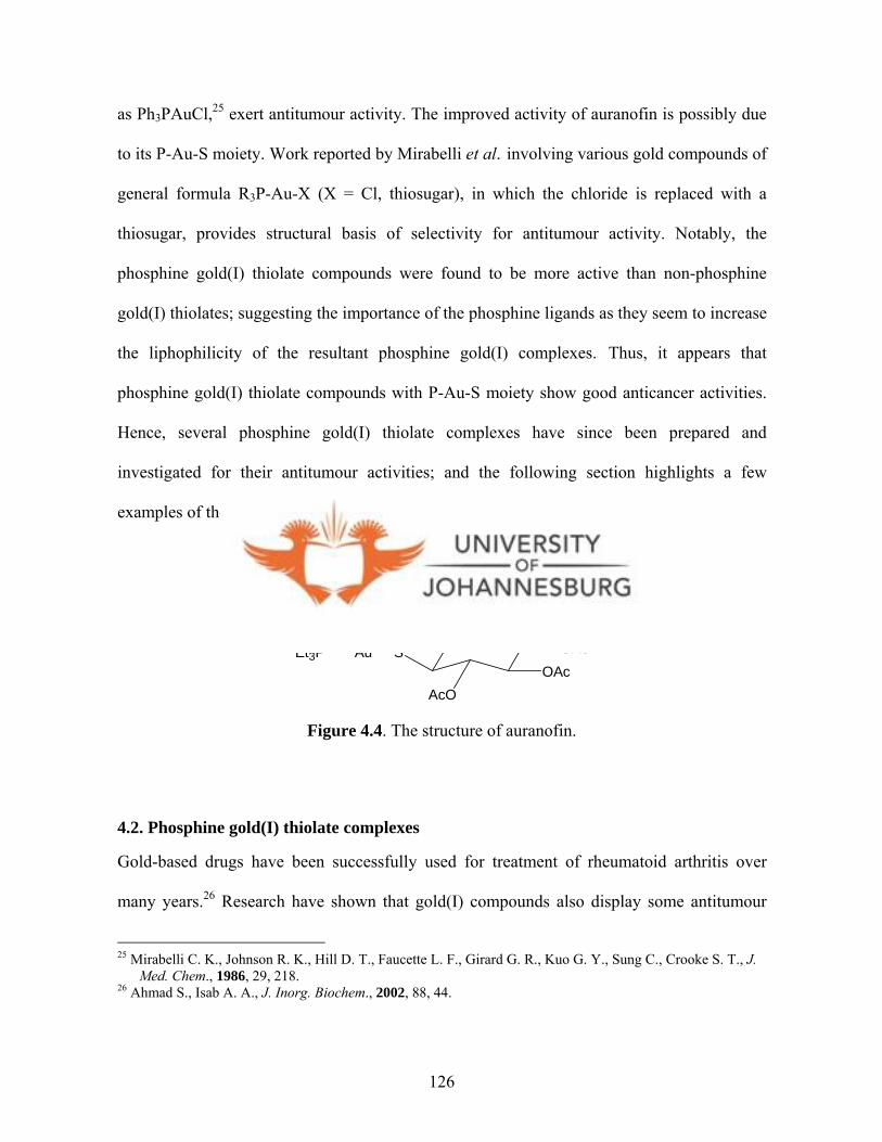
as Ph3PAuCl,25 exert antitumour activity. The improved activity of auranofin is possibly due
to its P-Au-S moiety. Work reported by Mirabelli et al.2 5 involving various gold compounds of
general formula R3P-Au-X (X = Cl, thiosugar), in which the chloride is replaced with a
thiosugar, provides structural basis of selectivity for antitumour activity. Notably, the
phosphine gold(I) thiolate compounds were found to be more active than non-phosphine
gold(I) thiolates; suggesting the importance of the phosphine ligands as they seem to increase
the liphophilicity of the resultant phosphine gold(I) complexes.25 Thus, it appears that
phosphine gold(I) thiolate compounds with P-Au-S moiety show good anticancer activities.
Hence, several phosphine gold(I) thiolate complexes have since been prepared and
investigated for their antitumour activities; and the following section highlights a few
examples of these phosphine gold(I) thiolate complexes.
OSAuEt3P
AcO
AcOOAc
OAc
Figure 4.4. The structure of auranofin.
4.2. Phosphine gold(I) thiolate complexes
Gold-based drugs have been successfully used for treatment of rheumatoid arthritis over
many years.26 Research have shown that gold(I) compounds also display some antitumour
25 Mirabelli C. K., Johnson R. K., Hill D. T., Faucette L. F., Girard G. R., Kuo G. Y., Sung C., Crooke S. T., J. Med. Chem., 1986, 29, 218. 26 Ahmad S., Isab A. A., J. Inorg. Biochem., 2002, 88, 44.
126

activity.27, ,28 29 Currently there is great interest in isolating new gold(I) complexes with
potential anticancer activities and to understand the mechanism through which the gold(I)
compounds exert antitumour activity.2 7 There is an array of gold(I) thiolate complexes
reported in the literature and for the purposes of this thesis few examples are cited, i.e.,
phosphines gold(I) thiolate complexes, since all the new gold(I) complexes reported in this
thesis are phosphine based gold(I) complexes.
4.2.1. Monophosphine gold(I) thiolate complexes
The starting point was the examination of the antitumour activity of auranofin (Fig. 4.4). It
was found to be an effective agent against P388 lymphocytic leukemia.2 4 Literature reports
indicate that auranofin exert its activity by affecting mitochondrial function.24,25,30 Recent
work by Bindoli and co-workers evidently substantiate that auranofin and other gold(I)
compounds exert its activity by affecting the mitochondria.31 They demonstrated that
auranofin inhibits thioredoxin reductase in its mitochondrial environment.3 1 Human
thioredoxin reductase is an homodimeric flavoenzyme closely related to glutathione.32 It
differs from glutathione due to the Cy-SeCys (selenocysteine) sequence fragment, which
makes it a selenoenzyme.32 It is hugely expressed around tumours and thus makes it a target
for the new potential gold(I) compounds.33 It is believed that the binding of Au(I) to the C-
27 Tiekink E. R. T., Crit. Rev. Oncol. Hematol., 2002, 42, 225. 28 Pillarsetty N., Katti K. K., Hoffman T. J., Volkert W. A., Katti K. V., Kamei H., Koide T., J. Med. Chem., 2003, 46, 1130. 29aShaw III C. F., Chem. Rev., 1999, 99, 2589; bGold Progress in Chemistry, Biochemistry and Technology. Shaw III C. F., Ed. Schmidbaur H., John Wiley & Sons, Chichester, England, 1999, 260. 30 McKeage M. J., Maharaj L., Berners-Price S. J., Coord. Chem. Rev., 2002, 232, 127. 31 Rigobello M. P., Scutari G., Boscolo R., Bindoli A., Br. J. Pharmacol., 2002, 136, 1162. bRigobello M. P., Scutari G., Folda A., Bindoli A., Biochem. Pharmacol., 2004, 67, 689. 32 Gromer S., Arscott L. D., Williams Jr. C. H., Schirmer R. H., Becker K., J. Biol. Chem., 1998, 273, 20096. 33 Barnard P. J., Berners-Price S. J., Coord. Chem. Rev., 2007, 251, 1889
127
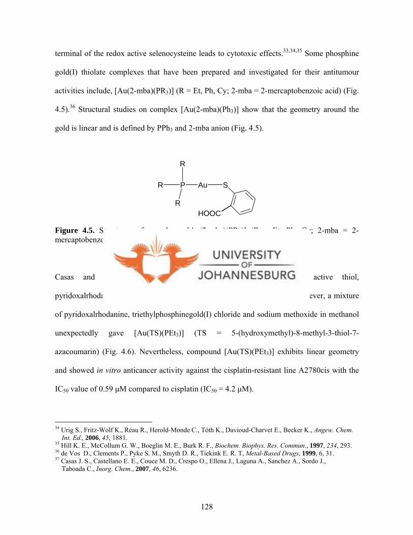
terminal of the redox active selenocysteine leads to cytotoxic effects.33,34,35 Some phosphine
gold(I) thiolate complexes that have been prepared and investigated for their antitumour
activities include, [Au(2-mba)(PR3)] (R = Et, Ph, Cy; 2-mba = 2-mercaptobenzoic acid) (Fig.
4.5).36 Structural studies on complex [Au(2-mba)(Ph3)] show that the geometry around the
gold is linear and is defined by PPh3 and 2-mba anion (Fig. 4.5).
R
R
R
P Au S
HOOC
Figure 4.5. Structures of complexes [Au(2-mba)(PR3)] (R = Et, Ph, Cy; 2-mba = 2-mercaptobenzoic acid).
Casas and co-workers37 have recently explored a biologically active thiol,
pyridoxalrhodanine, to prepare a novel gold(I) 7-azacoumarin complex. However, a mixture
of pyridoxalrhodanine, triethylphosphinegold(I) chloride and sodium methoxide in methanol
unexpectedly gave [Au(TS)(PEt3)] (TS = 5-(hydroxymethyl)-8-methyl-3-thiol-7-
azacoumarin) (Fig. 4.6). Nevertheless, compound [Au(TS)(PEt3)] exhibits linear geometry
and showed in vitro anticancer activity against the cisplatin-resistant line A2780cis with the
IC50 value of 0.59 µM compared to cisplatin (IC50 = 4.2 µM).3 7
34 Urig S., Fritz-Wolf K., Réau R., Herold-Monde C., Tóth K., Davioud-Charvet E., Becker K., Angew. Chem. Int. Ed., 2006, 45, 1881. 35 Hill K. E., McCollum G. W., Boeglin M. E., Burk R. F., Biochem. Biophys. Res. Commun., 1997, 234, 293. 36 de Vos D., Clements P., Pyke S. M., Smyth D. R., Tiekink E. R. T, Metal-Based Drugs, 1999, 6, 31. 37 Casas J. S., Castellano E. E., Couce M. D., Crespo O., Ellena J., Laguna A., Sanchez A., Sordo J., Taboada C., Inorg. Chem., 2007, 46, 6236.
128

NO
HO
S
O
Au P
Figure 4.6. A structure of gold(I) 7-azacoumarin complex, [Au(TS)(PEt3)].
Further efforts to search for more phosphine gold(I) thiolate compounds has been pursued by
preparing phosphine gold(I) complexes of sulfur containing biomolecules. For instance, the
vitamin K3 (menadione) derivative {NaK3TSC (NaK3TSC = menadione sodium bisulfite
thiosemicarbazone)} has been used as a ligand to the prepare gold(I) complex
[AuPEt3(K3TSC)] (Fig. 4.7).38 Compound [AuPEt3(K3TSC)] has the gold(I) atom
coordinated almost linearly to the K3TSC- sulfur and the phosphine P atom.38 In vitro
screening showed significant anticancer activity (IC50 = 0.4 µM) against the cisplatin-
resistant cells, A27780cis. The activity of the ligand, NaK3TSC, was quite insignificant (IC50
= 3732 µM), reiterating the fact that phopshine ligands are necessary for cytotoxic activities.25
PAu
O
NaO3SN
HN
H2N
S
Figure 4.7. The structure of [Au(K3TSC)PEt3].
38 Casas J. S., Castellano E. E., Couce M. D., Ellena J., Sanchez A., Sordo J., Taboada C., J. Inorg. Biochem., 2006, 100, 1858.
129
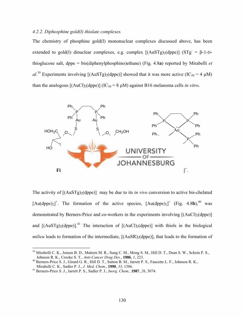
4.2.2. Diphosphine gold(I) thiolate complexes.
The chemistry of phosphine gold(I) mononuclear complexes discussed above, has been
extended to gold(I) dinuclear complexes, e.g. complex [(AuSTg)2(dppe)] (STg- = β-1-D-
thioglucose salt, dppe = bis(diphenylphosphino)ethane) (Fig. 4.8a) reported by Mirabelli et
al.39 Experiments involving [(AuSTg)2(dppe)] showed that it was more active (IC50 = 4 µM)
than the analogous [(AuCl)2(dppe)] (IC50 = 8 µM) against B16 melanoma cells in vitro.3 9
PP
Ph
Ph
Ph
Ph
P P
Ph
Ph
Ph
PhAu
(a) (b)
+
P PPh
Ph
Ph
Ph
Au Au
SOHOH2C
HOOH
OH
SO CH2OH
OHHO
HO
Figure 4.8. The structure of [(AuSTg)2(dppe)] and (b) [Au(dppe)2]+.
The activity of [(AuSTg)2(dppe)] may be due to its in vivo conversion to active bis-chelated
[Au(dppe)2]+. The formation of the active species, [Au(dppe)2]+ (Fig. 4.8b),40 was
demonstrated by Berners-Price and co-workers in the experiments involving [(AuCl)2(dppe)]
and [(AuSTg)2(dppe)].41 The interaction of [(AuCl)2(dppe)] with thiols in the biological
milieu leads to formation of the intermediate, [(AuSR)2(dppe)], that leads to the formation of
39 Mirabelli C. K., Jensen B. D., Mattern M. R., Sung C. M., Mong S. M., Hill D. T., Dean S. W., Schein P. S., Johnson R. K., Crooke S. T., Anti-Cancer Drug Des., 1986, 1, 223. 40 Berners-Price S. J., Girard G. R., Hill D. T., Sutton B. M., Jarrett P. S., Faucette L. F., Johnson R. K., Mirabelli C. K., Sadler P. J., J. Med. Chem., 1990, 33, 1386. 41 Berners-Price S. J., Jarrett P. S., Sadler P. J., Inorg. Chem., 1987, 26, 3074.
130
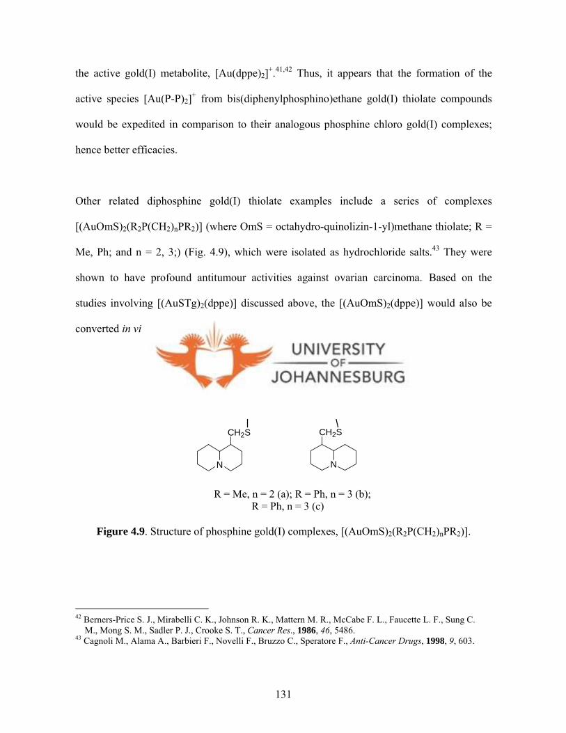
the active gold(I) metabolite, [Au(dppe)2]+.41,42 Thus, it appears that the formation of the
active species [Au(P-P)2]+ from bis(diphenylphosphino)ethane gold(I) thiolate compounds
would be expedited in comparison to their analogous phosphine chloro gold(I) complexes;
hence better efficacies.3 9
Other related diphosphine gold(I) thiolate examples include a series of complexes
[(AuOmS)2(R2P(CH2)nPR2)] (where OmS = octahydro-quinolizin-1-yl)methane thiolate; R =
Me, Ph; and n = 2, 3;) (Fig. 4.9), which were isolated as hydrochloride salts.43 They were
shown to have profound antitumour activities against ovarian carcinoma.4 3 Based on the
studies involving [(AuSTg)2(dppe)] discussed above, the [(AuOmS)2(dppe)] would also be
converted in vivo to the active bis-chelated [Au(dppe)2]+.
P PR
R
R
R
Au Au
N
CH2S
N
CH2S
(CH2)n
R = Me, n = 2 (a); R = Ph, n = 3 (b); R = Ph, n = 3 (c)
Figure 4.9. Structure of phosphine gold(I) complexes, [(AuOmS)2(R2P(CH2)nPR2)].
42 Berners-Price S. J., Mirabelli C. K., Johnson R. K., Mattern M. R., McCabe F. L., Faucette L. F., Sung C. M., Mong S. M., Sadler P. J., Crooke S. T., Cancer Res., 1986, 46, 5486. 43 Cagnoli M., Alama A., Barbieri F., Novelli F., Bruzzo C., Speratore F., Anti-Cancer Drugs, 1998, 9, 603.
131
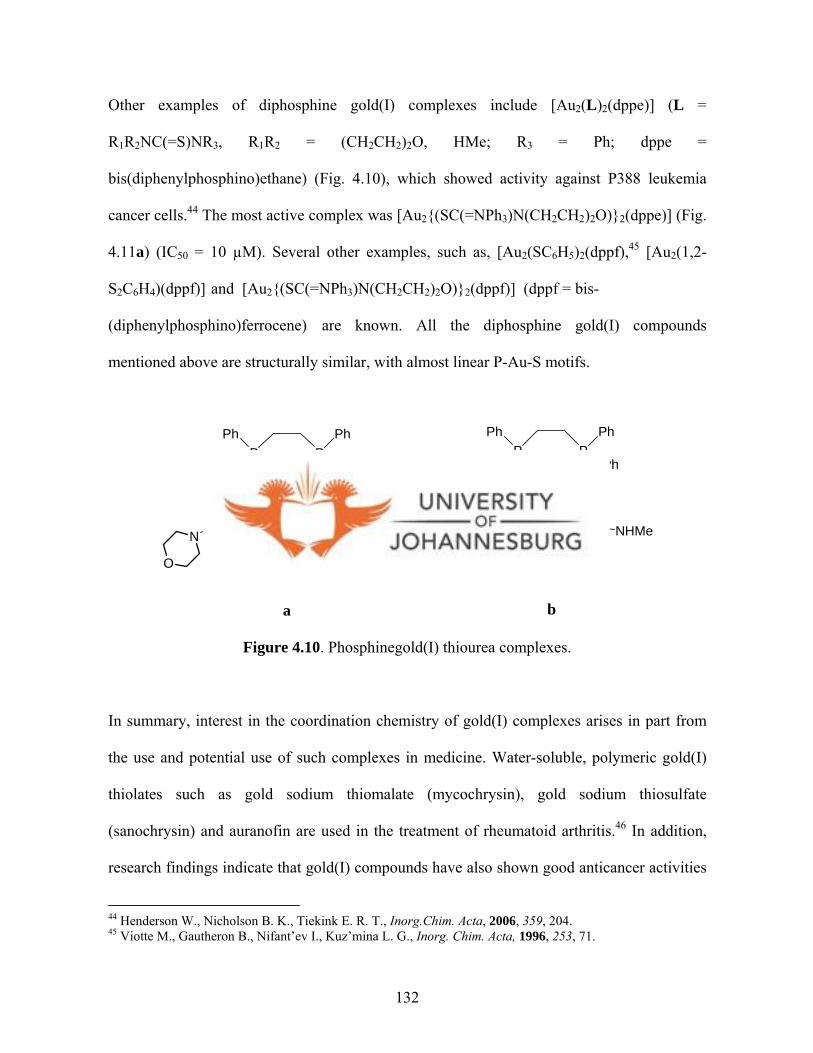
Other examples of diphosphine gold(I) complexes include [Au2(L)2(dppe)] (L =
R1R2NC(=S)NR3, R1R2 = (CH2CH2)2O, HMe; R3 = Ph; dppe =
bis(diphenylphosphino)ethane) (Fig. 4.10), which showed activity against P388 leukemia
cancer cells.44 The most active complex was [Au2{(SC(=NPh3)N(CH2CH2)2O)}2(dppe)] (Fig.
4.11a) (IC50 = 10 µM). Several other examples, such as, [Au2(SC6H5)2(dppf),45 [Au2(1,2-
S2C6H4)(dppf)]4 5 and [Au2{(SC(=NPh3)N(CH2CH2)2O)}2(dppf)] (dppf = bis-
(diphenylphosphino)ferrocene)4 4 are known. All the diphosphine gold(I) compounds
mentioned above are structurally similar, with almost linear P-Au-S motifs.
P
Au Au
PPh
Ph
Ph
Ph
S S
NN
Ph
ONN
PhO
P
Au Au
PPh
Ph
Ph
Ph
S S
NNHMe
CN
NMeHN
CN
a b
Figure 4.10. Phosphinegold(I) thiourea complexes.
In summary, interest in the coordination chemistry of gold(I) complexes arises in part from
the use and potential use of such complexes in medicine. Water-soluble, polymeric gold(I)
thiolates such as gold sodium thiomalate (mycochrysin), gold sodium thiosulfate
(sanochrysin) and auranofin are used in the treatment of rheumatoid arthritis.46 In addition,
research findings indicate that gold(I) compounds have also shown good anticancer activities
44 Henderson W., Nicholson B. K., Tiekink E. R. T., Inorg.Chim. Acta, 2006, 359, 204. 45 Viotte M., Gautheron B., Nifant’ev I., Kuz’mina L. G., Inorg. Chim. Acta, 1996, 253, 71.
132
against numerous cell lines.47 Certain thiosemicarbazones and gold-thiosemicarbazone
complexes have also shown considerable antitumour activity.48
Apart from the fact that phosphine and sulfur containing ligands stabilises gold(I)
compounds, the phosphine ligands in particular, also increase the biological properties of the
resultant gold(I) complex. This is supported by literature reports on various phosphine
gold(I) thiolate compounds described in section 4.2. It is for this reason that this study
embarked on the design of new phosphine gold(I) thiolate complexes possessing P-Au-S
motifs by using triphenylphosphine (PPh3), 1,2-bis(diphenylphosphine)ethane (dppe), 1,2-
bis(diphenylphosphine)propane (dppp), 1,2-bis(diphenylphosphine)hexane (dpph) and the
unexploited pyrazol-1-yl-, indazol-1-yl- and imidazol-1-yl-based dithiocarbamate ligands
(dtcs). Platinum(II) dithiocarbamate compounds were also prepared and their anticancer
activities determined. Details of the syntheses, characterisation and molecular structures are
discussed in this chapter. The compounds discussed in this chapter are given in schemes 4.2
and 4.3 below.
46 Parish R. V., Cotrill S. M., Gold Bull., 1987, 20, 3. 47 aZhang C. X., Lippard S. J., Curr. Opin. Chem. Biol., 2003, 7, 481; bMcKeage M. J., Papathanasiou P., Salem G., Sjaarda A., Swiegers G. F., Warning P., Wild S. B., Met-Based Drugs, 1999, 217. 48 Beraldo H., Gambino D., Mini Rev. Med. Chem., 2004, 4, 159.
133
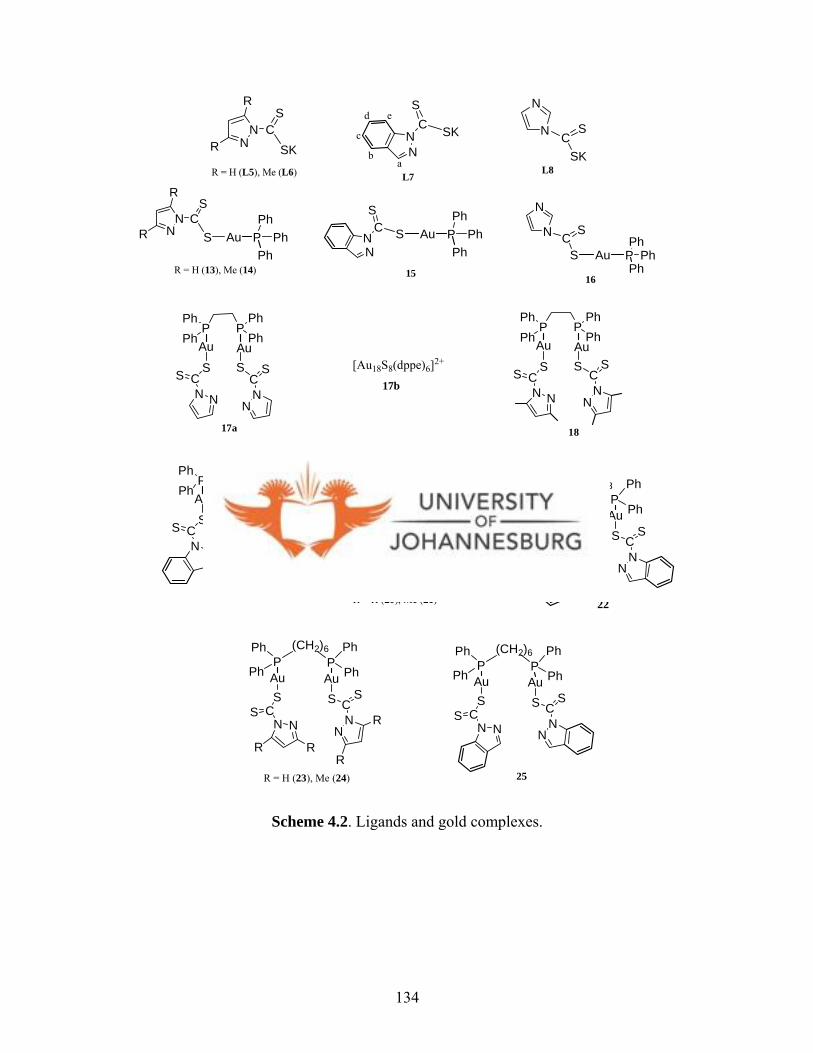
N
N CS
SR = H (13), Me (14)
NN
CS
SP PhPh
AuNN C
S
S
R
R
Ph
P PhPh
AuPh P Ph
PhAu
Ph15 16
P PPhPh
PhPh
Au Au
NNCS S
NNC
SS
17a 18
19
P PPhPh
PhPh
Au Au
NNCS S
NN
CSS
P PPhPh
PhPh
Au Au
NNCS S
NN
CSS
[Au18S8(dppe)6]2+
17b
R = H (20), Me (21)
P PPh
Ph
Ph
PhAu Au
NNCS
S
NNC
SS
R R
R
R
(CH2)3
R = H (23), Me (24)
22
25
P PPh
Ph
Ph
PhAu Au
NNCS
S
NNC
SS
(CH2)3
P PPh
Ph
Ph
PhAu Au
NNCS
S
NNC
SS
R R
R
R
(CH2)6
P PPh
Ph
Ph
PhAu Au
NNCS
S
NNC
SS
(CH2)6
NN C
S
SK
R
R
N
N CS
SKNN
CS
SK
R = H (L5), Me (L6) L8L7
ab
c
d e
Scheme 4.2. Ligands and gold complexes.
134
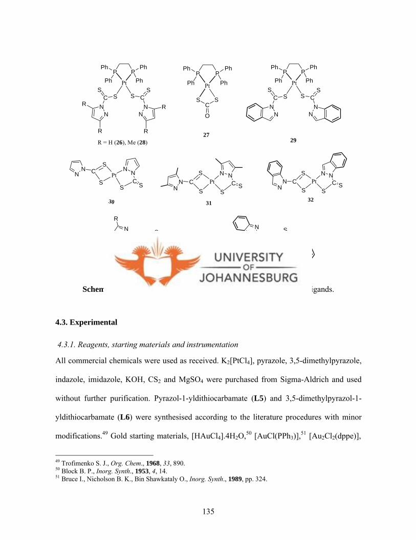
P PPh
Ph
Ph
Ph
NN
CS
S
NN
CS
S
Pt
R
R
R
R
P PPh
Ph
Ph
Ph
O
CS S
Pt
P PPh
Ph
Ph
Ph
NN
CS
S
NN
CS
S
Pt
R = H (26), Me (28) 2927
30
NN C
S
S
N NC
SS
Pt
NN C
S
S
N NC S
S
PtN
N CS
S
N NC S
SPt
3231
NN
CS
S
NN
CS
S
R
RR
RR = H (L9), Me (L10)
NN
CS
S
NN
CS
S
L11
Scheme 4.3. Platinum complexes and bis(dithiocarbamato)disulfide ligands.
4.3. Experimental
4.3.1. Reagents, starting materials and instrumentation
All commercial chemicals were used as received. K2[PtCl4], pyrazole, 3,5-dimethylpyrazole,
indazole, imidazole, KOH, CS2 and MgSO4 were purchased from Sigma-Aldrich and used
without further purification. Pyrazol-1-yldithiocarbamate (L5) and 3,5-dimethylpyrazol-1-
yldithiocarbamate (L6) were synthesised according to the literature procedures with minor
modifications.49 Gold starting materials, [HAuCl4].4H2O,50 [AuCl(PPh3)],51 [Au2Cl2(dppe)],51
49 Trofimenko S. J., Org. Chem., 1968, 33, 890. 50 Block B. P., Inorg. Synth., 1953, 4, 14. 51 Bruce I., Nicholson B. K., Bin Shawkataly O., Inorg. Synth., 1989, pp. 324.
135
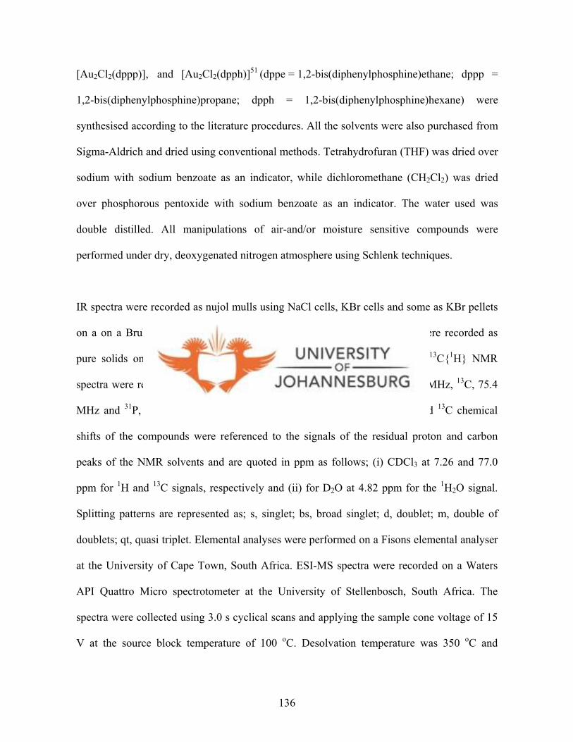
[Au2Cl2(dppp)],5 1 and [Au2Cl2(dpph)]51 (dppe = 1,2-bis(diphenylphosphine)ethane; dppp =
1,2-bis(diphenylphosphine)propane; dpph = 1,2-bis(diphenylphosphine)hexane) were
synthesised according to the literature procedures. All the solvents were also purchased from
Sigma-Aldrich and dried using conventional methods. Tetrahydrofuran (THF) was dried over
sodium with sodium benzoate as an indicator, while dichloromethane (CH2Cl2) was dried
over phosphorous pentoxide with sodium benzoate as an indicator. The water used was
double distilled. All manipulations of air-and/or moisture sensitive compounds were
performed under dry, deoxygenated nitrogen atmosphere using Schlenk techniques.
IR spectra were recorded as nujol mulls using NaCl cells, KBr cells and some as KBr pellets
on a on a Bruker Tensor27 spectrophotometer. In other cases IR spectra were recorded as
pure solids on a Bruker Tensor27 fitted with an ATR-IR probe. 1H and 13C{1H} NMR
spectra were recorded on a Bruker Avance DPX 300 spectrometer (1H, 300 MHz, 13C, 75.4
MHz and 31P, 121.5 MHz) in CDCl3 and D2O at room temperature. 1H and 13C chemical
shifts of the compounds were referenced to the signals of the residual proton and carbon
peaks of the NMR solvents and are quoted in ppm as follows; (i) CDCl3 at 7.26 and 77.0
ppm for 1H and 13C signals, respectively and (ii) for D2O at 4.82 ppm for the 1H2O signal.
Splitting patterns are represented as; s, singlet; bs, broad singlet; d, doublet; m, double of
doublets; qt, quasi triplet. Elemental analyses were performed on a Fisons elemental analyser
at the University of Cape Town, South Africa. ESI-MS spectra were recorded on a Waters
API Quattro Micro spectrotometer at the University of Stellenbosch, South Africa. The
spectra were collected using 3.0 s cyclical scans and applying the sample cone voltage of 15
V at the source block temperature of 100 oC. Desolvation temperature was 350 oC and
136
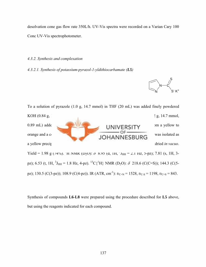
desolvation cone gas flow rate 350L/h. UV-Vis spectra were recorded on a Varian Cary 100
Conc UV-Vis spectrophotometer.
4.3.2. Synthesis and complexation
4.3.2.1. Synthesis of potassium-pyrazol-1-yldithiocarbamate (L5)
N
N C
S
S- K+
To a solution of pyrazole (1.0 g, 14.7 mmol) in THF (20 mL) was added finely powdered
KOH (0.84 g, 14.7 mmol). The mixture was left to stir for 2 min and CS2 (1.12 g, 14.7 mmol,
0.89 mL) added dropwise while stirring the solution. The solution turned from a yellow to
orange and a copious amount of precipitate formed. After 20 min, the product was isolated as
a yellow precipitate by filtration using a G4 frit, washed with diethylether and dried in vacuo.
Yield = 1.98 g (74%). 1H NMR (D2O): δ 8.95 (d, 1H, 3JHH = 2.1 Hz, 5-pz); 7.81 (s, 1H, 3-
pz); 6.53 (t, 1H, 3JHH = 1.8 Hz, 4-pz). 13C{1H} NMR (D2O): δ 218.6 (C(C=S)); 144.3 (C(5-
pz); 130.5 (C(3-pz)); 108.9 (C(4-pz)). IR (ATR, cm-1): υC=N = 1528, υC-S = 1198, υC=S = 843.
Synthesis of compounds L6-L8 were prepared using the procedure described for L5 above,
but using the reagents indicated for each compound.
137
4.3.2.2. Synthesis of potassium-3,5-dimethylpyrazol-1-yldithiocarbamate (L6)
N
N C
S
S- K+
KOH (0.58 g, 10.3 mmol); 3,5-dimethylpyrazole (0.97 g, 10.0 mmol); CS2 (0.99 g, 13.0
mmol, 0.78 mL). Yield = 1.58 g (75%). 1H NMR (D2O): δ 5.92 (s, 1H, 4-pz); 2.38 (s, 3H, 5-
pz); 1.99 (s, 3H, 3-pz). 13C{1H} NMR (D2O): δ 222.2 (C(C=S)); 149.7 (C(5-pz); 142.0 (C(3-
pz)); 109.8 (C(5-pz)); 14.7 (C(CH3, 5-pz); 10.8 (C(CH3, 3-pz). IR (ATR, cm-1): υC=N = 1578,
υC-S = 1224, υC=S = 873.
4.3.2.3. Synthesis of potassium-indazol-1-yldithiocarbamate (L7)
NN
CS- K+
S
HbHc
HdHe
Ha
KOH (0.48 g, 8.46 mmol); indazole (1.0 g, 8.46 mmol); CS2 (0.46 g, 8.46 mmol, 0.36 mL).
Yield = 1.54 g (79%). 1H NMR (D2O): δ 8.75 (d, 1H, 4JHH = 8.7 Hz, (He)); 7.89 (s, 1H, Ha);
7.49 (d, 1H, 3JHH = 8.7 Hz, Hb); 7.24 (t, 1H, 4JHH = 15.0 Hz, Hd); 7.01 (t, 1H, 4JHH = 15 Hz,
Hc). 13C{1H} NMR (D2O): δ 219.2 (C(C=S)); 138.6 (C(5C-pz); 129.0 (C(3C-pz)); 126.9
(C(7C-Ph)); 123.6 (C(4C-Ph); 121.4 (C(8C, 9C-Ph)); 117.2 (C(6C-Ph)). IR (ATR, cm-1):
υC=N = 1608, υC-S = 1297, υC=S = 852.
138

4.3.2.4. Synthesis of potassium-imidazol-1-yldithiocarbamate (L8)
N
NC
S
SK
Imidazole (1.0 g, 14.7 mmol); KOH (0.84 g, 14.7 mmol); CS2 (1.12 g, 14.7 mmol, 0.89 mL).
Yield = 1.89 g (71%). 1H NMR: (D2O): δ 8.79 (s, 1H, 5-Im); 8.12 (s, 1H, 3-Im); 6.99 (s, 1H,
4-Im). 13C{1H} NMR: (D2O): δ 218.5 (C(C=S)); 136.8 (C(5-Im); 128.5 (C(3-Im)); 119.5
(C(4-Im)). IR (KBr, cm-1): υC=N = 1526, υC-S = 1099, υC=S = 837.
4.3.2.5. Synthesis of bis(pyrazol-1-yldithiocarbamato)disulfide (L9)
NN
CS
S
NN
CS
S
To a solution of L5 (0.31 g, 1.45 mmol) in methanol (20 mL), was added iodine pellets (0.18
g, 0.72 mmol). The resultant solution was stirred at room temperature during which a yellow
precipitate was formed. The reaction was left for 15 min and the product isolated as a yellow
solid by filtration. Yield = 0.15 g (75%). 1H NMR (CDCl3): δ 8.54 (s, 2H, 3JHH = 3.6 Hz, 5-
pz); 7.91 (s, 2H, 3JHH = 3.3 Hz, 3-pz); 6.56 (s, 2H, 3JHH = 3.9 Hz, 4-pz). 13C{1H} NMR
(CDCl3): δ 192.3 (C(C=S); 145.6 (C(5, 5′-pz)); 131.5 (C(3, 3′-pz)); 111.6 (C(4, 4′-pz)). IR
(ATR, cm-1): υC=N = 1533, υC-S = 1254, υC=S = 869. Anal. Calc. for C8H6N4S4: C, 33.55; H,
2.11; N, 19.56; S, 44.78%. Found: C, 33.73; H, 2.64; N, 18.95; S, 44.32%.
139

Synthesis of compounds L10 and L11 were prepared using the procedure described for L11
above, but using the reagents indicated for each compound.
4.3.2.6. Synthesis of bis(3,5-dimethylpyrazol-1-yldithiocarbamato)disulfide (L10)
NN
CS
S
NN
CS
S
L6 (0.2 g, 0.95 mmol); iodine (0.12 g, 0.48 mmol). Yield = 0.12 g (73%). 1H NMR (CDCl3):
δ 6.23, 6.16 (s, 2H, 4-pz); 2.83, 2.70 (s, 6H, 5-pz); 2.40, 2.33 (s, 6H, 3-pz). 13C{1H} NMR
(CDCl3): δ 193.2 (C(C=S)); 152.8 (C(5-pz)); 146.6 (C(3-pz); 113.8 (C(4-pz)); 17.1 (C(CH3,
5-pz); 13.8 (C(CH3, 3-pz). IR (ATR, cm-1): υC=N = 1533, υC-S = 1261, υC=S = 879.
4.3.2.7. Synthesis of bis(indazol-1-yldithiocarbamato)disulfide (L11)
NN
C
S
S
ab
c
d e NN
C
S
S
a'b'
c'
d'e'
L7 (0.3 g, 1.29 mmol); iodine (0.16 g, 0.65 mmol). Yield = 0.14 g (56%). 1H NMR
(CDCl3):δ 9.06 (d, 2H, 4JHH = 9.0 Hz, He); 8.35 (s, 1H, Ha); 7.85 (d, 1H, 3JHH = 8.4 Hz, Hb);
7.64 (1H, 3JHH = 15.0 Hz , Hd); 7.47 (t, 1H, 4JHH = 15 Hz, Hc). 13C{1H} NMR (CDCl3): δ
191.8 (C(C=S)); 141.1 (C(5C-pz); 130.9 (C(3C-pz)); 131.2 (C(7C-Ph)); 126.2 (C(4C-Ph);
121.8 (C(8C, 9C-Ph)); 116.8 (C(6C-Ph)). IR (ATR, cm-1): υC=N = 1605, υC-S = 1334, υC=S =
901. Anal. Calc. for C16H10N4S4: C 49.72; H 2.61; N 14.49; 33.18%. Found: C, 48.82; H,
2.46; N, 13.83; S, 33.18%.
140

4.3.2.8. Synthesis of pyrazolyl-1-dithiocarbamato-triphenylphosphinegold(I) (13)
L5 (0.05 g, 0.3 mmol) was dissolved in deionised water (10 mL) and stirred for 2 min. To the
resultant solution was added [AuCl(PPh3)] (0.15 g, 0.3 mmol) in dichloromethane (10 mL)
and the bi-phasic mixture stirred vigorously at room temperature. A colour change in the
organic layer from colourless to red was observed, and after 30 min the two layers were
separated. The dichloromethane solution was dried over anhydrous MgSO4. The solvent was
then removed in vacuo to afford an orange-yellow solid. Yield = 0.17 g (93%). 1H NMR
(CDCl3): δ 7.63 (s, 1H, 5-pz); 7.57 (s, 1H, 3-pz); 7.49 (m, 9H, PPh3); 7.35 (m, 6H, PPh3);
6.27 (s, 1H, 4-pz). 13C{1H} NMR (CDCl3): δ 213.4 (C(C=S)); 144.0 (C(5-pz); 134.2-129.1
(Phenyl region); 140.8 (C(3-pz)); 106.1 (C(4-pz)). IR (KBr, cm-1): υC=N = 1620, υC-S = 1121,
υC=S = 884. 31P{1H} NMR (CDCl3): δ 35.2 (P(PPh3)). Anal. Calc. for C24H22AuN2PS2: C
43.86, H 3.01, N 4.65. Found: C 43.53, H 3.05, N 4.62%.
Synthesis of compounds 14-16 were prepared using the procedure described for 13 above,
but using the reagents indicated for each compound.
4.3.2.9. Synthesis of 3,5-dimethylpyrazolyl-1-dithiocarbamato-triphenylphosphinegold(I)
(14)
L6 (0.1g, 0.47 mmol), [AuCl(PPh3)] (0.24 g, 0.47 mmol). Yield = 0.17 g (57%). 1H NMR
(CDCl3): δ 7.44, 7.29 (m, 15H, PPh3); 6.12, 5.82 (s, 1H, 4-pz); 2.29 (m, 6H, 3-pz, 5-pz).
13C{1H} NMR (CDCl3): δ 215.5 (C(C=S)); 149.7 (C(5-pz); 134.1-128.7 (Phenyl region);
141
142.1 (C(3-pz)); 112.4, 104.1 (C(5-pz)); 13.9 (C(CH3, 5-pz); 12.3 (C(CH3, 3-pz). IR (KBr,
cm-1): υC=N = 1580, υC-S = 1154, υC=S = 854. 31P{1H} NMR (CDCl3): δ 33.8 (P(PPh3)). Anal.
Calc. for C24H22AuN2PS2: C 45.72, H 3.52, N 4.44. Found: C 45.89, H 3.89, N 3.29%.
4.3.2.10. Synthesis of indazolyl-1-dithiocarbamato-triphenylphosphinegold(I) (15)
L7 (0.1 g, 0.4 mmol), [AuCl(PPh3)] (0.2 g, 0.4 mmol). Yield = 0.15 g (58%). 1H NMR
(CDCl3): δ 9.21 (d, 1H, 4JHH = 9.0 Hz, Ha); 8.17 (s, 1H, Hb); 7.73 (d, 1H, 3JHH = 8.4 Hz,
He); 7.61, 7.50 (m, 15H, PPh3); (7.58 (1H, Hd); 7.35 (t, 1H, 4JHH = 15 Hz, Hc). 13C{1H}
NMR (CDCl3): δ 214.2 (C(C=S)); 139.5 (C(5C-pz); 134.7-129.5 (Phenyl region); 129.5
(C(3C-pz)); 126.9 (C(7C-Ph)); 124.6 (C(4C-Ph); 121.4 (C(8C, 9C-Ph)); 118.7 (C(6C-Ph)).
IR (KBr, cm-1): υC=N = 1601, υC-S = 1148, υC=S = 905. 31P{1H} NMR (CDCl3): δ 36.8
(P(PPh3)). Anal. Calc. for C26H20AuN2PS2: C 47.86, H 3.09, N 4.29. Found: C 47.41, H 3.04,
N 3.85%.
4.3.2.11. Synthesis of imidazolyl-1-dithiocarbamato triphenylphosphinogold(I) (16)
L8 (0.1 g, 0.4 mmol), [AuCl(PPh3)] (0.2 g, 0.4 mmol). Yield = 0.15 g (65%). 1H NMR
(CDCl3): δ 7.82 (s, 1H, 5-Im); 7.52 (s, 1H, 3-Im); 7.49 (m, 9H, PPh3); 7.35 (m, 6H, PPh3);
6.71 (s, 1H, 4-Im).13C{1H} NMR (CDCl3): δ 219.4 (C(C=S)); 137.2 (C(2-Im); 133.7-129.4
(Phenyl region); 128.8 (C(5-Im)); 120.1 (C(4(C-Im)). IR (KBr, cm-1): υC=N = 1592, υC-S =
1102, υC=S = 814. 1P{1H} NMR (CDCl3): δ 36.3 (P(PPh3)). Anal. Calc. for C26H20AuN2PS2:
C 47.86, H 3.09, N 4.29. Found: C 47.41, H 3.04, N 3.85%.
142
4.3.2.12. Synthesis of bis-(pyrazolyl-1-dithiocarbamato)-bis-(diphenylphosphino)-
ethanegold(I) (17a) and [Au18S8(dppe)6]2Cl (17b)
To a solution of [Au2Cl2(dppe)] (0.08 g, 0.09 mmol) in CH2Cl2 (10 mL), was added L5 (0.03
g, 0.19 mmol) previously dissolved in water (10 mL). The resultant mixture was stirred at
room temperature for 20 min during which time the CH2Cl2 layer changed colour from a
white suspension to a clear orange solution, whilst the water layer became clear. The mixture
was separated and the organic layer dried over anhydrous MgSO4. The yellow solids were
isolated as products upon in vacuo evaporation of the solvent. Yield = 0.07 g (73%). 1H
NMR (CDCl3): δ 8.78 (d, 2H, 3JHH = 2.7 Hz, 5-pz); 7.80 (s, 2H, 3-pz); 7.78 (m, 8H,
(Ph)2P(CH2)2P(Ph)2); 7.49 (m, 12H, (Ph)2P(CH2)2P(Ph)2); 6.43 (s, 2H, 4-pz); 2.87 (bs, 4H,
CH2, (Ph)2P(CH2)2P(Ph)2). 13C{1H} NMR (CDCl3): δ 214.2 (C(C=S)); 149.7 (C(5-pz);
142.0 (C(3-pz)); 134.1-128.7 (Phenyl region); 111.4 (C(4-pz)); 24.6 (C(Ph2P(CH2)2PPh2). IR
(KBr, cm-1): υC=N = 1590, υC-S = 1190, υC=S = 889. 31P{1H} NMR: (CDCl3): δ 36.1
(Ph2P(CH2)2PPh2). Anal. Calc. for C34H30Au2N4P2S4: C 37.85, H 2.80, N 5.14, S 11.89.
Found: C 38.18, H 3.01, N 4.57, S 11.73%.
Efforts to crystallise 17a in dichloromethane/ether solution led to the formation of a gold(I)
cluster, [Au18S8(dppe)6]2Cl (17b), Anal. Calc. for C26H20AuCl2N2PS2: C 29.71, H 2.32, N
4.10. Found: C 28.90, H 2.00, N 3.80%.
143
Synthesis of compounds 18-25 were prepared using the procedure described for 17a above,
but using the reagents indicated for each compound.
4.3.2.13. Synthesis of bis-(3,5-dimethylpyrazolyl-1-dithiocarbamato)-bis-
(diphenylphosphino)ethanegold(I) (18)
L6 (0.04 g, 0.19 mmol), [Au2Cl2(dppe)] (0.1 g, 0.09 mmol). Yield = 0.09 g (88%). 1H NMR
(CDCl3): δ 7.64 (m, 8H, Ph2P(CH2)2PPh2); 7.35 (m, 12H, Ph2P(CH2)2PPh2); 6.09 (s, 2H, 4-
pz); 2.69 (bs, 4H, Ph2P(CH2)2PPh2); 2.26 (d, 12H, CH3, 3,5-pz). 13C{1H} NMR (CDCl3): δ
216.5 (C(C=S)); 152.7 (C(5-pz); 143.1 (C(3-pz)); 135.1-128.6 (Phenyl region); 112.6 (C(4-
pz)); 14.0 (C(5-pPz); 12.4 (C(3-pz)); 25.7 (C(Ph2P(CH2)2PPh2). IR (KBr, cm-1): υC=N = 1603,
υC-S = 1263, υC=S = 989. 31P{1H} NMR: (CDCl3): δ 35.7 (Ph2P(CH2)2PPh2). Anal. Calc. for
C38H38Au2N4P2S4: C 40.22, H 3.37, N 4.94, S 11.30. Found: C 39.89, H 3.59, N 4.67, S
11.15%.
4.3.2.14. Synthesis of bis-(indazolyl-1-dithiocarbamato)-bis-
(diphenylphosphino)ethanegold(I) (19)
L7 (0.04 g, 0.19 mmol), [Au2Cl2(dppe)] (0.08 g, 0.09 mmol). Yield 0.07 g (63%). 1H NMR
(CDCl3): δ 9.16 (s, 1H, Ha); 8.06 (s, 1H, Hb); 7.78 (m, 8H, (Ph)2P(CH2)2P(Ph)2); 7.49 (m,
12H, (Ph)2P(CH2)2P(Ph)2); 7.67 (d, 1H, 3JHH = 8.4 Hz, He); 7.71 (1H, Hd); 7.43 (t, 1H, 4JHH
= 15 Hz, Hc); 2.87 (bs, 4H, CH2, (Ph)2P(CH2)2P(Ph)2). 13C{1H} NMR (CDCl3): δ 213.9
(C(C=S)); 142.1 (C(5-Pz); 139.0 (C(3-Pz)); 134.1-129.7 (Phenyl region); 126.1 (C(7C-Ph));
123.1 (C(4C-Ph)); 122.2 (C(8C-Ph, 9C-Ph)); 117.5 (C(6C-Ph) 25.2 (C(Ph2P(CH2)2PPh2).
31P{1H} NMR: (CDCl3): δ 34.9 (Ph2P(CH2)2PPh2). Anal. Calc. for C42H34Au2N4P2S4: C
42.79, H 2.91, N 4.75, S 10.88. Found: C 43.24, H 2.82, N 4.65, S 9.97%.
144
4.3.2.15. Synthesis of binuclear bis(pyrazol-1-yldithiocarbamato)-bis-
(diphenylphosphino)propane gold(I) (20)
L5 (0.08 g, 0.46 mmol), [Au2Cl2(dppp)] (0.2 g, 0.23 mmol). Yield = 0.15 g (59%). 1H NMR
(CDCl3): δ 8.73 (s, 2H, 3JHH = 2.7 Hz, 5-pz); 7.73 (m, 8H, Ph2P(CH2)3PPh2); 7.51 (s, 4H,
3JHH = 1.8 Hz, 3-pz); 7.41 (m, 12H, Ph2P(CH2)3PPh2); 6.41 (s, 2H, 3JHH = 2.7 Hz, 4-pz); 2.93
(m, 2H, Ph2PCH2CH2CH2PPh2); 1.99 (m, 4H, Ph2PCH2CH2CH2PPh2). 13C{1H} NMR
(CDCl3): δ 218.9 (C(C=S)); 147.5 (C(5-pz); 140.8 (C(3-pz)); 133.2-129.2 (Phenyl region);
106.2 (C(4-pz)); 28.5 (2C, 1JC-P = 62.7 Hz, P-CH2, Ph2PCH2CH2CH2PPh2); 21.4 (C,
Ph2PCH2CH2CH2PPh2). IR (KBr, cm-1): υC=N = 1628, υC-S = 1088, υC=S = 891. 31P{1H} NMR
(CDCl3): δ 32.8. Anal. Calc. for C35H32Au2N4P2S4: C 38.47, H 2.95, N 5.13, S 11.74. Found:
C 38.99, H 3.08, N 4.09, S 11.45%.
4.3.2.16. Synthesis of binuclear bis(3,5-dimethylpyrazol-1-yldithiocarbamato)-bis-
(diphenylphosphino)propanegold(I) (21)
L6 (0.1 g, 0.46 mmol), [Au2Cl2(dppp)] (0.2 g, 0.23 mmol). Yield = 0.13 g (50%). 1H NMR
(CDCl3): δ = 7.66 (m, 8H, Ph2P(CH2)3PPh2); 7.34 (m, 12H, Ph2P(CH2)3PPh2); 5.85 (s, 2H, 4-
pz); 2.91 (m, 2H, Ph2PCH2CH2CH2PPh2); 1.88 (m, 4H, Ph2PCH2CH2CH2PPh2); 2.24, 2.26
(d, 12H, CH3, 3,5-pz). 13C{1H} NMR (CDCl3): δ 215.9 (C(C=S)); 153.3 (C(5-pz); 144.2
(C(3-pz)); 133.4-129.2 (Phenyl region); 112.5 (C(4-pz)); 27.9 (2C, 1JC-P = 58.4 Hz, P-CH2,
Ph2PCH2CH2CH2PPh2); 20.2 (C, Ph2PCH2CH2CH2PPh2); 13.8 (C(CH3, 5-pz); 13.2 (C(CH3,
3-pz). IR (KBr, cm-1): υC=N = 1653, υC-S = 1099, υC=S = 957. 31P{1H} NMR (CDCl3): δ 29.2.
145

Anal. Calc. for C39H40Au2N4P2S4: C 40.77, H 3.51, N 4.88, S 11.16. Found: C 40.99, H 3.74,
N 4.79, S 11.16%.
4.3.2.17. Synthesis of binuclear bis(indazol-1-yldithiocarbamato)-bis-
(diphenylphosphino)propane gold(I) (22)
L7 (0.04 g, 0.22 mmol), [Au2Cl2(dppp)] (0.1 g, 0.11 mmol). Yield = 0.07 g (54%). 1H NMR
(CDCl3): δ 9.13 (s, 1H, Ha); 8.16 (s, 1H, Hb); 7.73 (1H, Hd); 7.69 (d, 1H, 3JHH = 8.0 Hz,
He); 7.66 (m, 8H, Ph2P(CH2)3PPh2); 7.45 (t, 1H, 4JHH = 16.8 Hz, Hc); 7.42 (m, 12H,
Ph2P(CH2)3PPh2); 2.89(m, 2H, Ph2PCH2CH2CH2PPh2); 1.92 (m, 4H, Ph2PCH2CH2CH2PPh2).
13C{1H} NMR (CDCl3): δ 213.8 (C(C=S)); 140.3 (C(5C-pz); 139.1 (C(3C-pz)); 133.5-129.2
(Phenyl region); 127.9 (C(7C-Ph)); 124.2 (C(4C-Ph)); 121.1 (C(8C-Ph, 9C-Ph)); 118.3
(C(6C-Ph)); 28.6 (2C, 1JC-P = 64.2 Hz, P-CH2, Ph2PCH2CH2CH2PPh2); 19.8
(Ph2PCH2CH2CH2PPh2). IR (KBr, cm-1): υC=N = 1606, υC-S = 1150, υC=S = 993. 31P{1H}
NMR (CDCl3): δ 29.2. ESI-MS: m/z 1190.9 [Au2(L7)2(dppp)]+ (5%). Anal. Calc. for
C43H36Au2N4P2S4: C 43.29, H 3.04, N 4.70, S 10.75. Found: C 43.23, H 3.05, N 4.62, S
10.36%.
4.3.2.18. Synthesis of binuclear bis(pyrazol-1-yldithiocarbamato)-bis-
(diphenylphosphino)hexane gold(I) (23)
L5 (0.02 g, 0.11 mmol), [Au2Cl2(dpph)] (0.05 g, 0.05 mmol). Yield = 0.04 g (70%). 1H NMR
(CDCl3): δ 8.78 (d, 2H, 3JHH = 2.7 Hz, 5-pz); 7.80 (s, 2H, 3-pz); 7.78 (m, 8H,
146

(Ph)2P(CH2)6P(Ph)2); 7.49 (m, 12H, (Ph)2P(CH2)6P(Ph)2); 6.43 (s, 2H, 4-pz); 2.87 (bs, 4H,
Ph2P(CH2)2(CH2)2(CH2)2PPh2); 1.49 (bs, 8H, Ph2P(CH2)2(CH2)2(CH2)2PPh2).. 13C{1H} NMR
(CDCl3): δ 213.8 (C(C=S)); 140.3 (C(5C-pz); 139.1 (C(3C-pz)); 133.5-129.2 (Phenyl
region); 127.9 (C(7C-Ph)); 124.2 (C(4C-Ph)); 121.1 (C(8C-Ph, 9C-Ph)); 118.3 (C(6C-Ph));
30.1 (4C, PPh2(CH2)2(CH2)2(CH2)2PPh2); 25.9 (2C, Ph2P(CH2)2(CH2)2(CH2)2PPh2). IR (KBr,
cm-1): υC=N = 1620, υC-S = 1108, υC=S = 960. 31P{1H} NMR (CDCl3): δ 32.3. Anal. Calc. for
C38H38Au2N4P2S4: C 40.22, H 3.37, N 4.94, S 11.30. Found: C 40.17, H 3.45, N 4.36, S
10.28%.
4.3.2.19. Synthesis of binuclear bis(3,5-dimethylpyrazol-1-yldithiocarbamato)-bis-
(diphenylphosphino)hexane gold(I) (24)
L6 (0.09 g, 0.42 mmol), [Au2Cl2(dpph)] (0.2 g, 0.2 mmol). Yield = 0.12 g (50%). 1H NMR
(CDCl3): δ 7.67 (m, 8H, Ph2P(CH2)6PPh2); 7.40 (m, 12H, Ph2P(CH2)6PPh2); 6.00 (s, 2H, 4-
pz); 2.76 (bs, 4H, Ph2P(CH2)2(CH2)2(CH2)2PPh2); 2.18, 2.27 (d, 12H, CH3, 3,5-pz). 1.60,
1.52 (bs, 8H, Ph2P(CH2)2(CH2)2(CH2)2PPh2). 13C{1H} NMR (CDCl3): δ 215.9 (C(C=S));
153.3 (C(5-pz); 144.2 (C(3-pz)); 133.4-129.2 (Phenyl region); 112.5 (C(4-pz)); 31.2 (4C,
PPh2(CH2)2(CH2)2(CH2)2PPh2); 26.7 ( 2C, Ph2P(CH2)2(CH2)2(CH2)2PPh2); 13.8 (C(5-pz);
13.2 (C(3-pz). IR (KBr, cm-1): υC=N = 1653, υC-S = 1099, υC=S = 957. IR (KBr, cm-1): υC=N =
1657, υC-S = 1099, υC=S = 994. 31P{1H} NMR (CDCl3): δ 32.7. Anal. Calc. for
C42H46Au2N4P2S4: C 42.36, H 3.89, N 4.70, S 10.77. Found: C 41.82, H 3.84, N 4.62, S
10.56%.
147
4.3.2.20. Synthesis of binuclear bis(indazol-1-yldithiocarbamato)-bis-
(diphenylphosphino)hexane gold(I) (25)
L7 (0.1 g, 0.44 mmol), [Au2Cl2(dpph)] (0.2 g, 0.22 mmol). Yield = 0.14 g (52%). 1H NMR
(CDCl3): δ 9.18 (d, 1H, 4JHH = 8.4 Hz, (Ha)); 8.10 (s, 1H, Hb); 7.49 (d, 1H, 3JHH = 8.7 Hz,
He); 7.24 (t, 1H, 4JHH = 15.0 Hz, Hd); 7.32 (t, 1H, 4JHH = 15.0 Hz, Hc); 7.66 (m, 8H,
Ph2P(CH2)6PPh2); 7.42 (m, 12H, Ph2P(CH2)6PPh2); 2.39 (m, 4H,
Ph2P(CH2)2(CH2)2(CH2)2PPh2); 1.57 (m, 8H, Ph2P(CH2)2(CH2)2(CH2)2PPh2). 13C{1H} NMR
(CDCl3): δ 213.8 (C(C=S)); 140.3 (C(5C-pz); 139.1 (C(3C-pz)); 133.5-129.2 (Phenyl
region); 127.9 (C(7C-Ph)); 124.2 (C(4C-Ph)); 121.1 (C(8C-Ph, 9C-Ph)); 118.3 (C(6C-Ph));
28.9 (4C, CH2)2(CH2)2(CH2)2PPh2); 25.6 (2C, Ph2P(CH2)2(CH2)2(CH2)2PPh2). IR (KBr, cm-
1): υC=N = 1657, υC-S = 1099, υC=S = 994. 31P{1H} NMR CDCl3): δ 31.8. Anal. Calc. for
C46H42Au2N4P2S4: C 44.74, H 3.43, N 4.54, S 10.39. Found: C 44.37, H 3.46, N 4.50, S
10.32%.
4.3.2.21. Synthesis of bis-(pyrazol-1-yl-dithiocarbamato)-bis(diphenylphosphino)ethane
platinum(II) (26)
To a solution of L5 (0.06 g, 0.3 mmol) in deionised water (5 mL) was added [PtCl2(dppe)]
(0.1 g, 0.16 mmol) in dichloromethane (10 mL) and the bi-phasic mixture stirred vigorously
at room temperature. A colour change in the organic layer from colourless to yellow was
observed, and after 30 min the two layers were separated. The dichloromethane solution was
dried over anhydrous MgSO4. The solvent was then removed in vacuo to afford a yellow
solid. Efforts to grow crystals of 26, by re-dissolving 26 in dichloromethane and layering it
148

with hexane or diethyl ether, led to formation of green single crystals 27 suitable for X-ray
analysis. Yield 26 = 0.13 g (93%). 1H NMR (CDCl3): δ 8.49 (s, 2H, 5-pz); 7.76 (m, 8H,
Ph2P(CH2)2PPh2); 7.74 (s, 2H, 3-pz); 7.48 (m, 12H, Ph2P(CH2)2PPh2); 6.22 (s, 2H, 4-pz);
3.19 (qt, 4H, 2JP-H + 3JP-H = 18.0 Hz, Ph2P(CH2)2PPh2). 31P{1H} NMR (CDCl3): δ 39.3 (2P,
JPt-P = 3678 Hz, Ph2P(CH2)2PPh2). IR (KBr, cm-1): υC=N = 1609, υC-S = 1099, υC=S = 821.
Anal. Calc. for C34H30N4P2PtS4: C 46.41, H 3.44, N 6.37, S 14.58. Found: C 46.51, H 2.98, N
5.81, S 14.01%.
Synthesis of compounds 28-30 were prepared using the procedure described for 26 above,
but using the reagents indicated for each compound.
4.3.2.22. Synthesis of bis-(3,5-dimethylpyrazol-1-yldithiocarbamato)-bis-
(diphenylphosphino)ethaneplatinum(II) (28)
L6 (0.06 g, 0.3 mmol); [PtCl2(dppe)] (0.1 g, 0.15 mmol). Yield = 0.12 g (86%). 1HNMR
(CDCl3): δ 7.76 (m, 8H, Ph2P(CH2)2PPh2); 7.54 (m, 12H, Ph2P(CH2)2PPh2); 5.91 (s, 2H, 4-
pz); 3.11 (qt, 4H, 2JP-H + 3JP-H = 18.9 Hz, Ph2P(CH2)2PPh2); 2.72 (s, 6H, 5-pz); 2.32 (s, 6H, 3-
pz). 31P{1H} NMR (CDCl3): δ 44.2 (2P, JPt-P = 3897 Hz, Ph2P(CH2)2PPh2). IR (KBr, cm-1):
υC=N = 1591, υC-S = 1149, υC=S = 853. Anal. Calc. for C38H38N4P2PtS4: C 48.76, H 4.09, N
5.99, S 13.70. Found: C 49.21, H 2.98, N 5.81, S 14.01%.
149
4.3.2.23. Synthesis of bis-(indazolyl-1-dithiocarbamato)-bis-
(diphenylphosphino)ethaneplatinum(II) (29)
L7 (0.07 g, 0.3 mmol); [PtCl2(dppe)] (0.1 g, 0.15 mmol). Yield = 0.1 g (67%). 1HNMR
(CDCl3): δ 9.14 (s, 1H, Ha); 8.17 (s, 1H, Hb); 7.74 (m, 8H, Ph2P(CH2)2PPh2); 7.52 (m, 12H,
Ph2P(CH2)2PPh2); 7.67 (d, 1H, 3JHH = 8.4 Hz, He); 7.78 (1H, Hd); 7.43 (t, 1H, 4JHH = 15 Hz,
Hc); 3.11 (qt, 4H, 2JP-H + 3JP-H = 18.9 Hz, Ph2P(CH2)2PPh2). 31P{1H} NMR (CDCl3): δ 44.3
(2P, JPt-P = 3761 Hz, Ph2P(CH2)2PPh2). IR (KBr, cm-1): υC=N = 1634, υC-S = 1187, υC=S = 849.
Anal. Calc. for C42H34N4P2PtS4: C 51.47, H 3.50, N 5.72 %. Found: C 50.82, H 4.01, N
5.19%.
4.3.2.24. Synthesis of bis-(pyrazol-1-yl-dithiocarbamato)-bis(diphenylphosphino)ethane
nickel(II) (30)
L5(0.06 g, 0.3 mmol); [NiCl2(dppe)] (0.08 g, 0.15 mmol). Yield = 0.06 g (55%). Yield =
0.13 g (93%). 31P{1H} NMR (CDCl3): δ 33.8. IR (KBr, cm-1): υC=N = 1617, υC-S = 1183,
υC=S = 835. Anal. Calc. for C34H30N4NiP2S4: C 54.92, H 4.07, N 7.54, S 17.25. Found: C
54.81, H 4.02, N 7.61, S 17.14%.
4.3.2.25. Synthesis of bis(pyrazolyl-1-dithiocarbamato)platinum(II) (31)
To a solution of L5 (0.09 g, 0.48 mmol) in deionised water (10 mL) was added K2[PtCl4]
(0.1 g, 0.24 mmol) in deionised water (10 mL). The solution mixture was stirred at room
temperature for 2 h upon which a colour change of the solution, from yellow to deep red, was
observed. A brown solid was isolated as the product by filtration. Yield = 0.07 g (58%). 1H
150
NMR (CDCl3): δ 8.62, 8.51 (s, 2H, 5-pz); 8.16, 8.02 (s, 2H, 3-pz); 6.69, 6.50 (s, 2H, 4-pz).
IR (KBr, cm-1): υC=N = 1518, υC-S = 1110, υC=S = 855.
Synthesis of compounds 32 and 33 were prepared using the procedure described for 31
above, but using the reagents indicated for each compound.
4.3.2.26. Synthesis of bis(3,5-dimethylpyrazolyl-1-dithiocarbamato)platinum(II) (32)
L6 (0.11 g, 0.52 mmol); K2[PtCl4] (0.11 g, 0.26 mmol). Yield = 0.08 g (54%). 1H NMR
(CDCl3): δ 6.23, 6.16 (s, 2H, 4-pz); 2.83, 2.70 (s, 6H, 5-pz); 2.40, 2.33 (s, 6H, 3-pz).
13C{1H} NMR (CDCl3): δ 226.8 (C(C=S)); 156.5 (C(5-pz)); 143.9 (C(3-pz); 115.3, 113.3
(C(4-pz)); 16.5, 15.3 (C(CH3, 5-pz); 14.6, 14.0 (C(CH3, 3-pz). IR (KBr, cm-1): υC=N = 1580,
υC-S = 1158, υC=S = 887. Anal. Calc. for C12H14N4PtS4: C 26.81, H 2.62, N 10.42. Found: C
26.81, H 2.20, N 10.42%.
4.3.2.27. Synthesis of bis-(indazolyl-1-dithiocarbamato)platinum(II) (33)
L7 (0.1 g, 0.43 mmol); K2[PtCl4] (0.09 g, 0.22 mmol). Yield = 0.06 g (46%). 1H NMR
(CDCl3): δ 9.53 (d, 2H, 4JHH = 8.4 Hz, (Ha)); 8.65 (d, 1H, 3JHH = 8.7 Hz, He); 8.43, 8.31 (s,
2H, Hb); 7.82 (t, 1H, 4JHH = 14.8 Hz, Hd); 7.50 (t, 1H, 4JHH = 15 Hz, Hc). IR (KBr, cm-1):
υC=N = 1606, υC-S = 1172, υC=S = 917.
151

4.3.3. X-ray crystallography
4.3.3.1. Data Collection
An orange crystal of [Au(L7)(PPh3)] (15) with approximate dimensions 0.45 x 0.33 x 0.21
mm3 was selected under oil under ambient conditions and attached to the tip of a
Micromount©. The crystal was mounted in a stream of cold nitrogen at 105(2) K and
centred in the X-ray beam by using a video camera. The crystal evaluation and data
collection were performed on a Bruker CCD-1000 diffractometer with Mo Kα (λ = 0.71073
Å) radiation and the diffractometer to crystal distance of 4.9 cm.
The initial cell constants were obtained from three series of ω scans at different starting
angles. Each series consisted of 20 frames collected at intervals of 0.3º in a 6º range about ω
with the exposure time of 10 seconds per frame. A total of 131 reflections were obtained.
The reflections were successfully indexed by an automated indexing routine built in the
SMART program. The final cell constants were calculated from a set of 12116 strong
reflections from the actual data collection.
The data were collected by using the full sphere data collection routine to survey the
reciprocal space to the extent of a full sphere to a resolution of 0.80 Å. A total of 20998 data
were harvested by collecting four sets of frames with 0.4º scans in ω and one set of frames
with 0.5º scan in φ. These highly redundant datasets were corrected for Lorentz and
152
polarization effects. The absorption correction was based on fitting a function to the
empirical transmission surface as sampled by multiple equivalent measurements.52
4.3.3.2. Structure solution and refinement
The systematic absences in the diffraction data were uniquely consistent for the space group
P21/n that yielded chemically reasonable and computationally stable results of refinement.5 2 A
successful solution by the direct methods provided most non-hydrogen atoms from the E-
map. The remaining non-hydrogen atoms were located in an alternating series of least-
squares cycles and difference Fourier maps. All non-hydrogen atoms were refined with
anisotropic displacement coefficients. All hydrogen atoms except those on the water
molecule were included in the structure factor calculation at idealized positions and were
allowed to ride on the neighboring atoms with relative isotropic displacement coefficients.
There is also one solvated water molecule per Pd complex in the unit cell. The water
molecules were refined with restraints. The final least-squares refinement of 172 parameters
against 3166 data resulted in residuals R (based on F2 for I≥2σ) and wR (based on F2 for all
data) of 0.0180 and 0.0504, respectively. The final difference Fourier map was featureless.
The molecular diagram is drawn with 50% probability ellipsoids.
The solid state structures of (pzCS2)2 (L9), (3,5-pzCS2)2 (L10), [Au18S8(dppe)6]Cl2 (17b),
[Au(L7)(dpph)] (25), [Pt(S2CO)(dppe)] (27), and [Pt(L2)2] (32) were determined in a similar
manner such as described above for 15.
52 Bruker-AXS. (2000-2003) SADABS V.2.05, SAINT V.6.22, SHELXTL V.6.10 & SMART 5.622 Software Reference Manuals. Bruker-AXS, Madison, Wisconsin, USA.
153
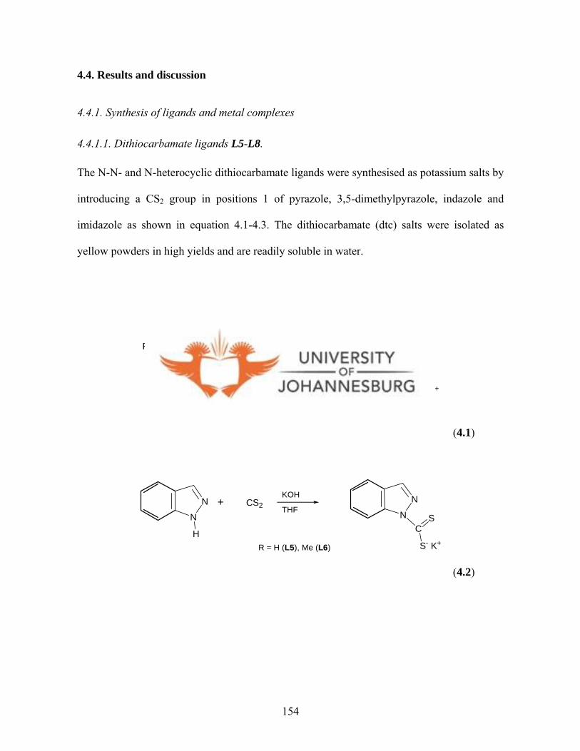
4.4. Results and discussion
4.4.1. Synthesis of ligands and metal complexes
4.4.1.1. Dithiocarbamate ligands L5-L8.
The N-N- and N-heterocyclic dithiocarbamate ligands were synthesised as potassium salts by
introducing a CS2 group in positions 1 of pyrazole, 3,5-dimethylpyrazole, indazole and
imidazole as shown in equation 4.1-4.3. The dithiocarbamate (dtc) salts were isolated as
yellow powders in high yields and are readily soluble in water.
N N
RR
H
+ CS2 N N
RR
CS
S- K+
THF
R = H (L5), Me (L6)
KOH
(4.1)
N
N
H
+ CS2N
NC
S
S- K+
THF
R = H (L5), Me (L6)
KOH
(4.2)
154

N
NH
+ CS2
N
NC
S
S- K+
THF
L8
KOH
(4.3)
The 1H NMR spectrum of L7 showed peaks at 8.75 ppm (doublet), 7.89 ppm (singlet), 7.49
ppm (doublet), 7.24 ppm (triplet) and 7.01 ppm (triplet), which were assigned to He, Ha,
Hb, Hd and Hc protons of the indazole ring, respectively (Fig. 4.11). The 13C{1H} NMR
spectrum of L7 shows the resonance of the diagnostic carbon (C=S) peak at 218.6 ppm.
Similar NMR spectral patterns were observed for L5, L6 and L8, but with different
chemical shifts. Ligands L5 and L6 are known and are reported in the literature.49 The
spectroscopic and microanalysis data for L5 and L6 reported in this thesis are in agreement
with those reported in the literature.49
Subsequently, the dtc salts were reacted with [AuCl(PPh3)], [Au2Cl2(dppe)],
[Au2Cl2(dppp)], [Au2Cl2(dpph)], [PtCl2(dppe)] or K2[PtCl4] to afford several gold(I) and
platinum(II) complexes, respectively. The following sections reports on these new gold(I)
and platinum(II) complexes.
155

NN
CS-K+
S
HbHc
HdHe
Ha
Figure 4.11. 1H NMR spectrum of L7.
4.5.1.2. Monophosphine gold(I) dithiocarbamato complexes 13-16.
Gold(I) complexes [Au(L5)(PPh3)] (13), [Au(L6)(PPh3)] (14), [Au(L7)(PPh3)] (15) and
[Au(L8)(PPh3)] (16) obtained from reaction of [AuCl(PPh3)] and dtc ligands, were isolated
as orange-yellow solids as in Scheme 4.4. The complexes were characterised by a
combination of spectroscopic, mass spectrometry, microanalysis, and in the case of complex
15, X-ray crystallography techniques. For complex 13, the significant upfield shift observed
in the proton peaks of pyrazole indicated complexation. In the 1H NMR spectrum of 13, the
4-pz and 5-pz proton peaks appeared as singlets at 6.27 and 7.63 ppm, respectively,
compared to 6.53 and 8.95 ppm for ligand L5. The 13C{1H} NMR spectrum of complex 13
showed peaks between 128 and 134 ppm, which were assigned to the phenyl carbons of PPh3
ligand. There was no significant shifts of dithio-carbon (C=S) peak of the dtc ligand upon
complexation. For instance, for complex 13, it resonated at 213.4 ppm compared to that of
156
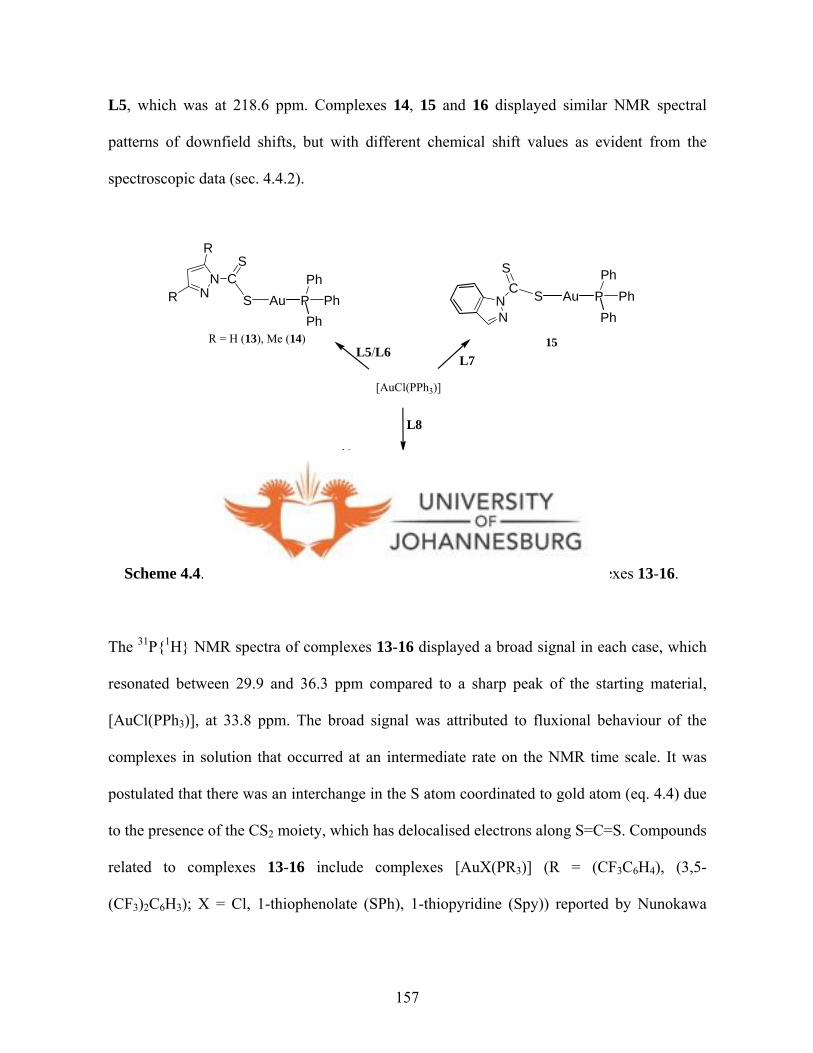
L5, which was at 218.6 ppm. Complexes 14, 15 and 16 displayed similar NMR spectral
patterns of downfield shifts, but with different chemical shift values as evident from the
spectroscopic data (sec. 4.4.2).
N
N CS
S
R = H (13), Me (14)
NN
CS
SP PhPh
AuNN C
S
S
R
R
Ph
P PhPh
AuPh
P PhPh
AuPh
15
16
L5/L6 L7
L8
[AuCl(PPh3)]
Scheme 4.4. Preparation of monophosphine gold(I) dithiocarbamato complexes 13-16.
The 31P{1H} NMR spectra of complexes 13-16 displayed a broad signal in each case, which
resonated between 29.9 and 36.3 ppm compared to a sharp peak of the starting material,
[AuCl(PPh3)], at 33.8 ppm. The broad signal was attributed to fluxional behaviour of the
complexes in solution that occurred at an intermediate rate on the NMR time scale. It was
postulated that there was an interchange in the S atom coordinated to gold atom (eq. 4.4) due
to the presence of the CS2 moiety, which has delocalised electrons along S=C=S. Compounds
related to complexes 13-16 include complexes [AuX(PR3)] (R = (CF3C6H4), (3,5-
(CF3)2C6H3); X = Cl, 1-thiophenolate (SPh), 1-thiopyridine (Spy)) reported by Nunokawa
157

and co-workers,53 and [Au(2-mpa)(PPh3)] (2-mpa = 2-mercaptopropionate)54 where they
exhibit pseudo linear geometries along P-Au-S bond. The geometry of complexes 13-16 was
further established by solid state structure analysis of complex 15.
PPh
Ph
Au
NN
CS
SPh P Ph
Ph
AuNN C
S
S
Ph
(4.4)
4.4.1.3. Molecular structure of [Au(L7)(PPh3)] (15).
Orange crystals of complex 15 suitable for single-crystal X-ray analysis were obtained from
a dichloromethane-hexane mixture. The molecular structure of the crystal is shown in Figure
4.12. The bond lengths Au(1)-S(1) = 2.3272(6), Au(1)-P(1) = 2.2533(6), and N(1)-C(1) =
1.399(3) Å are typical for linear phoshine gold-thiolate complexes.36,37,55 The Au(I) centre
has approximately linear P-Au-S geometry such that the P(1)-Au(1)-S(1) angle = 175.36(2)o
with a maximum deviation from linearity of 4.64o. The S(1)-C(1) bond distance of 1.732(3)
Å is substantially and statistically longer than 1.663(3) Å of S(2)-C(1) which has more
double bond character (C=S). The ligand and the Au atom lies out of the plane as manifested
in the torsional angle of -165.30(17)o for Au(1)-S(1)-C(1)-N(1). The crystal structure
determination of closely related complexes, e.g. [Au(PPh3)(2-mba)] (2-mba = 2-
53 Nunokawa K., Onaka S., Tatematsu T., Ito M., Sakai J., Inorg. Chim. Acta, 2001, 322, 56. 54 Nomiya K., Yamamoto S., Noguchi R., Yokoyama H., Kasuga N. C., Ohyama K., Kato C., J. Inorg. Biochem., 2003, 95, 208.
158

mercaptobenzoic acid) has been reported by de Vos et al.36 and the bond lengths and distances
of 15 correspond to their reported data (Au-P(1) = 2.258(1) Å, Au-S(1) = 2.297(2) Å and
P(1)-Au-S(2) = 177.9(1)o). A molecular packing diagram of 15 viewed along the c axis is
shown in Figure 4.13. It displays the formation of cage-like cuboids defined by repetitive P-
Au-S motifs and the axial phenyl rings of the phosphine ligand. The packing is also
enhanced by the π-π interactions between the indazolyl unit and the phenyl groups of the
phosphine. The selected bond distances and angles with their estimated standard deviations
are listed in Table 4.1. Crystal data, together with the data collection and refinement
parameters are presented in Table 4.2.
Table 4.1. Selected bond lengths [Å] and angles [°] for 15
Bond lengths (Å) Bond angles (°)
Au(1)-P(1) 2.2533(6) P(1)-Au(1)-S(1) 175.36(2)
Au(1)-S(1) 2.3272(6) C(1)-S(1)-Au(1) 100.04(9)
P(1)-C(9) 1.811(2) C(9)-P(1)-Au(1) 118.20(8)
N(1)-C(1) 1.399(3) S(2)-C(1)-S(1) 124.85(15)
S(1)-C(1) 1.732(3)
S(2)-C(1) 1.663(3)
55 aHunks J. W., Jennings C. M., Puddephatt R. J., Inorg. Chim. Acta, 2006, 359, 3605; bLi C. K., Lu X. X., Wong K. M. C., Chan C. L., Zhu N. Y., Yam V. W. W., Inorg. Chem., 2004, 43, 7421; cBalasubramanian R., Guo R., Mills A. J., Murray R. W., J. Am. Chem. Soc., 2005, 127, 8126.
159
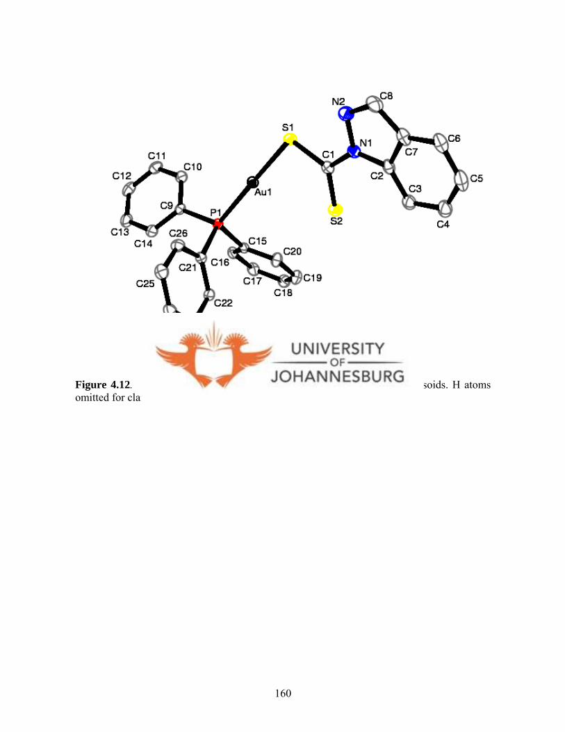
Figure 4.12. A molecular drawing of 15 drawn with 50% probability ellipsoids. H atoms omitted for clarity.
160

Figure 4.13. A molecular packing diagram of 15 viewed along the c axis. The hydrogen atoms omitted for clarity.
161

Table 4.2. Crystal data, data collection and refinement parameters of 15
Parameter 15
Empirical formula C26H20AuN2PS2
Formula weight 652.50
Temperature (K) 100(2)
Wavelength (Å) 0.71073
Crystal system Monoclinic
Space group P21/n
Unit cell dimensions
a(Å) 9.1382(12)
b(Å) 22.487(3) Å
c(Å) 11.3760(14)
α 90o
β 91.541(2)o
γ 90o
Volume (Å3) 2336.8(5)
Z 4
Density (calculated) (Mg/m3) 1.855
Absorption coefficient (mm-1) 6.560
F(000) 1264
Final R indices (R1) 0.0587
Reflections collected 36929
Completeness to theta 99.3 %
Goodness of fit on F2 1.188
Largest diff. peak & hole (e.Å-3) 1.146 and -0.880
162
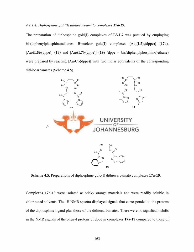
4.4.1.4. Diphosphine gold(I) dithiocarbamato complexes 17a-19.
The preparation of diphosphine gold(I) complexes of L5-L7 was pursued by employing
bis(diphenylphosphino)alkanes. Binuclear gold(I) complexes [Au2(L5)2(dppe)] (17a),
[Au2(L6)2(dppe)] (18) and [Au2(L7)2(dppe)] (19) (dppe = bis(diphenylphosphino)ethane)
were prepared by reacting [Au2Cl2(dppe)] with two molar equivalents of the corresponding
dithiocarbamates (Scheme 4.5).
P PPh
Ph
Ph
PhAu Au
NNCS
S
NN
CSS
[Au2Cl2(dppe)]
17a 18
19
L5 L6
L7
P PPh
Ph
Ph
PhAu Au
NNCS
S
NN
CSS
P PPh
Ph
Ph
PhAu Au
NNCS
S
NN
CSS
[Au18S8(dppe)6]2+
17b
Scheme 4.5. Preparations of diphosphine gold(I) dithiocarbamato complexes 17a-19.
Complexes 17a-19 were isolated as sticky orange materials and were readily soluble in
chlorinated solvents. The 1H NMR spectra displayed signals that corresponded to the protons
of the diphosphine ligand plus those of the dithiocarbamates. There were no significant shifts
in the NMR signals of the phenyl protons of dppe in complexes 17a-19 compared to those of
163

the starting material [Au2Cl2(dppe)]; but there was slight chemical shifts in the NMR peaks
of the ethylene linker protons indicating formation of the desired products. For instance, in
the 1H NMR spectrum of 17a (Fig. 4.14), the CH2 protons in the diphosphine backbone
appeared downfield as a broad singlet at 2.87 ppm compared to that of [Au2Cl2(dppe)] ( 2.76
ppm). The CH2 protons of the dppe ligand appeared as a broad singlet probably due to
unresolved 31P-1H coupling. The 5-pz protons (5H, 5H′) of the pyrazolyl ring in 17a appeared
as a doublet at 8.78 ppm, while the 3-pz protons (3H, 3H′) peak were masked by the phenyl
protons of the dppe ligand (7.80 ppm). This is in contrast to that of L5 where 3-pz and 5-pz
protons appeared at 6.27 and 7.63, respectively.
P PPh
PhPhPh
Au Au
NNCS S
NNC SS
Figure 4.14. The 1H NMR spectrum of 17a (CDCl3), with the 31P{1H} NMR spectrum of 17a as inset.
As mentioned for complexes 13-16, the 31P{1H} NMR spectrum of complex 17a (inset, Fig.
4.14) showed one relatively broad singlet at 36.1 ppm, which is characteristic of compounds
164

with P-Au-S motif.55a,56,57 Similar broad peaks were observed in the spectra of complexes 18
and 19 at 35.7 and 34.9 ppm, respectively. Complexes 17a-19 are comparable to diphosphine
digold(I)-pyridine-2-thiolate complexes, [(AuSN)2(dppm)] and [(AuSN)2(dpph)] (HNS =
pyridine-2-thiolate), reported by Tseng et al.5 7 that show 31P NMR peaks in the range 32.7-
35.9 ppm.
4.4.1.5. Molecular structure of [Au18S8(dppe)6]2Cl (17b).
Interestingly, attempts to crystallise complex 17a in a dichloromethane/diethylether solution
led to the isolation of the cationic gold(I) cluster, [Au18S8(dppe)6]2+ (17b) (Fig. 4.15 and
4.16) as confirmed by X-ray crystallography and microanalysis. The formation of complex
17b is unique in the sense that, there are no reports on the use of dithiocarbamates to make
gold(I) clusters. It appears that there is a similarity between dtc (RCS2-) and Se(SiMe3)2,58
used by Fenske and co-workers, in that they both act as sources of S atoms.
The solid state structure of complex 17b is shown in Figure 4.15 while its skeletal (Au, P, S)
geometry is shown in Figure 4.16. The molecular structure has 18 gold atoms, 8 sulfur atoms
and 6 dppe units. The counterions that should account for the +2 positive charge of the Au
cluster could not be found in the difference map. The complex is isomorphous with the Se
analogue reported by Fenske and co-workers58 and therefore two chloride ions were assumed
to be the anions. This is strongly supported by the macroanalysis data obtained. There were
several partially occupied molecules of some solvent also present in the asymmetric unit.
56 Chen J., Mohamed A. A., Abdou H. E., Krause Bauer J. A., Fackler J. P., Bruce A. E., Bruce M. R. M., Chem. Commun., 2005, 1575. 57 Tzeng B.-C., Liao J.-H., Lee G.-H., Peng S.-M., Inorg. Chim. Acta, 2004, 357, 1405.
165

Bond length restraints were applied to model the molecules but the resulting isotropic
displacement coefficients suggested the molecules were mobile. Option SQUEEZE of
program PLATON59 was used to correct the diffraction data for diffuse scattering effects and
to identify the solvate molecule. PLATON calculated the upper limit of volume that can be
occupied by the solvent to be 3394 Å3, or 19% of the unit cell volume. The program
calculated 3364 electrons in the unit cell for the diffuse species, or 841 electrons in the
asymmetric unit. This approximately corresponded to two chloride ions (36 electrons) and
eight molecules of CHCl3 (464 electrons) and eight molecules of CH2Cl2
(336 electrons) for a
total of 836 electrons. It should be however noted that all derived results are based on the
known contents with the exception of the chloride anions that were included in the formula.
This data was corroborated by the microanalysis data obtained.
The structure of 17b exhibits pseudo-threefold axis that runs through sulfur atoms S1 and S2.
The S1, S2 and other sulfur atoms at the periphery act as µ3 bridges between gold atoms, with
each peripheral sulfur atoms displaying three short Au-S bonds (2.328(4)-2.374(3) Å).5 8 The
Au···Au distances involving the central gold atom fall in the range of 2.9263(7)-3.1395(7) Å,
which is indicative of a d10-d10 closed shell metal interactions. Four different surroundings
are observed for gold atoms in 17b, viz. (i) sulfur bridged non-bonded Au···Au interactions;
(ii) Au-Au non-bridged single bonds; (iii) Au···Au sulfur bridged single bonds; and (iv)
Au···Au phosphine bridged single bonds.5 6 Similar to the [Au18Se8(dppe)6]2+ reported by
Fenske,58 complex 17b has two [Au9S4(dppe)3]2+ fragments that are interlocked in such a
manner that a distorted Au6S2 heterocubane arrangement (Au(1)-Au(6), S(1), S(2)) arises in
58 Fenske D., Langetepe T., Kappes M. M., Hampe O., Weis P., Angew. Chem. Int. Ed. 2000, 39, 1857.
166

the centre of the complex. Cluster 17b features bond angles, S(2)-Au(1)-S(3) = 175.80(12)°
and S(2)-Au(1)-Au(2) = 92.05(8)°. The other bond distances and angles are similar to those
reported in literature.56,60a,b The selected bond distances and angles with their estimated
standard deviations are listed in Table 4.3. Crystal data, together with the data collection and
refinement parameters are presented in Table 4.4.
Figure 4.15. The molecular diagram of 17b drawn with 30% probability ellipsoids. The hydrogen atoms omitted for clarity.
59 Spek A. L., Acta Cryst. Sect. A., 1990, A46, C34. 60 aCanales F., Gimeno M. C., Jones P. G., Laguna A., Angew. Chem., 1994, 106, 811; bHuang S. P., Kanatzidis M. G., Angew. Chem. Int. Ed., 1992, 31, 787.
167
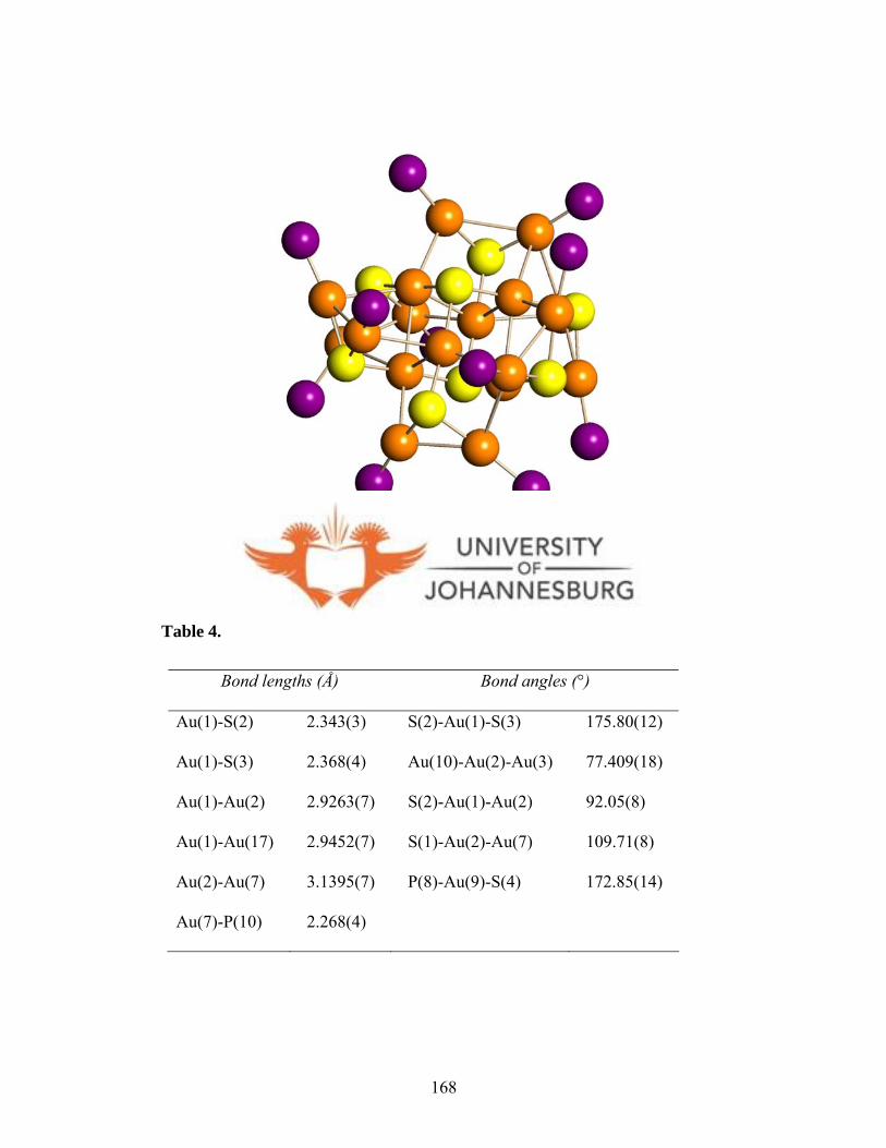
Figure 4.16. The skeletal (Au, P, S) geometry of 17b.
Table 4.3. Selected bond lengths [Å] and angles [°] of 17b
Bond lengths (Å) Bond angles (°)
Au(1)-S(2) 2.343(3) S(2)-Au(1)-S(3) 175.80(12)
Au(1)-S(3) 2.368(4) Au(10)-Au(2)-Au(3) 77.409(18)
Au(1)-Au(2) 2.9263(7) S(2)-Au(1)-Au(2) 92.05(8)
Au(1)-Au(17) 2.9452(7) S(1)-Au(2)-Au(7) 109.71(8)
Au(2)-Au(7) 3.1395(7) P(8)-Au(9)-S(4) 172.85(14)
Au(7)-P(10) 2.268(4)
168
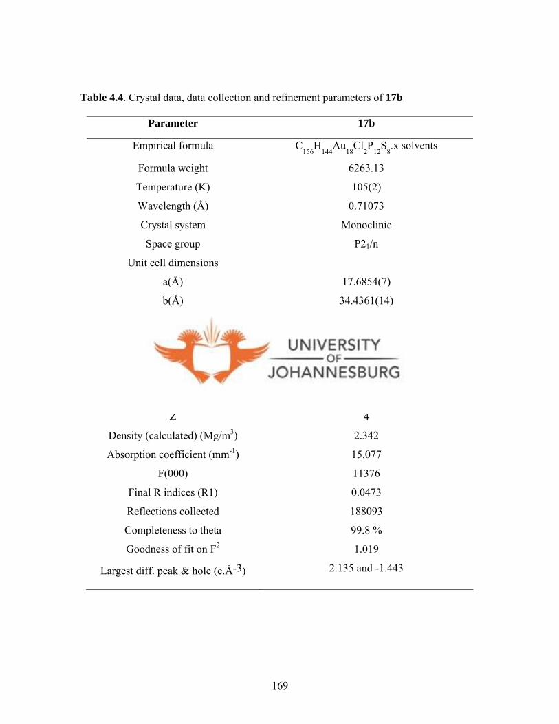
Table 4.4. Crystal data, data collection and refinement parameters of 17b
Parameter 17b
Empirical formula C156
H144
Au18
Cl2P
12S
8.x solvents
Formula weight 6263.13
Temperature (K) 105(2)
Wavelength (Å) 0.71073
Crystal system Monoclinic
Space group P21/n
Unit cell dimensions
a(Å) 17.6854(7)
b(Å) 34.4361(14)
c(Å) 29.1894(12)
α 90°
β 92.4570(10)°
γ 90°
Volume (Å3) 17760.5(12)
Z 4
Density (calculated) (Mg/m3) 2.342
Absorption coefficient (mm-1) 15.077
F(000) 11376
Final R indices (R1) 0.0473
Reflections collected 188093
Completeness to theta 99.8 %
Goodness of fit on F2 1.019
Largest diff. peak & hole (e.Å-3) 2.135 and -1.443
169
The ease of dtcs formation and their wide ranging coordination chemistry has led to the
formation of an array of novel and complex supramolecular architectures.4 For gold
compounds, the process of supramolecular formation seems to be assisted by aurophilicity.
Aurophilicity is the weak gold-gold interactions observed in gold compounds.61 Theoretical
calculations by Pyykko et al.61 show that aurophilicity is amplified by electron correlation
effect (electron interactions in a quantum system) and relativistic effects, which leads to
length contraction between gold atoms. Furthermore, aurophilicity is augmented by soft
ligands, e.g. sulfur, where thiolates compounds of general formula [AuX(PPh3)] (X = RS)
display great aurophilic effects.62 In addition, the observed aurophilicity is enhanced by use
of diphosphines, such as in [(Au-SPh)2(µ-trans-dpen)] (trans-dpen = trans-1,2-
bis(diphenylphosphino)ethylene).63 It is therefore clear that due to close proximity of the P
atoms in the dppe backbone, this property of aurophilicity coupled with the ease of cleavage
of C-N bond in the dithiocarbamate in complex 17a, expedited the process of self-assembly.
It is believed that complex 17b is the first (dppe)-gold(I) dithiocarbamate-generated complex
of high nuclearity that incorporates a range of intramolecular Au···Au interactions.
4.4.1.6. Diphosphine gold(I) dithiocarbamato complexes 20-25.
In order to further establish the phenomenon of cluster formation, the linker spanning
between the two phosphine atoms were increased to (CH2)3 (dppp) and (CH2)6 (dpph).
Complexes [Au2(L5)2(dppp)] (20), [Au2(L6)2(dppp)] (21), [Au2(L7)2(dppp)] (22)
61 Pyykkö P., Zhao Y., Angew. Chem., Int. Ed. Engl., 1991, 30, 604. 62 Pyykkö P., Li J., Runeberg N., Chem. Phys. Lett., 1994, 218, 133. 63 aOnaka S., Katsukawa Y., Shiotsuka M., Kanegawa O., Yamashita M., Inorg. Chim. Acta, 2001, 312, 100;
170

[Au2(L5)2(dpph)] (23), [Au2(L6)2(dpph)] (24), and [Au2(L7)2(dpph)] (25) were synthesised
in the same manner as for complexes 17a-19 by reacting [Au2Cl2(dppp)] or [Au2Cl2(dpph)]
with two molar equivalents of L5, L6 and L7, respectively (Scheme 4.6). In the 1H NMR
spectra of 20-22 in CDCl3, the CH2 protons of the diphosphine ligand appeared upfield (1.88-
2.93 ppm) as second order multiplets. The 13C{1H} NMR spectra of 20-22 showed peaks at
ca. 215 and ca. 129.2-133.5 ppm, which were assigned to C(C=S) and the phenyl carbons,
respectively. Moreover, the 13C{1H} NMR spectra of 20-22 displayed 31P-13C coupling, with
JP-C values between 58.4-64.2 Hz, due to P-CH2. The 31P{1H} NMR spectrum of 22 showed
one broad singlet at 29.2 ppm compared to that of the starting material, [Au2Cl2(dppp)],
which resonated at 27.6 ppm signifying complexation. Broad phosphorous peaks were also
observed for 20 and 21 at 32.8 ppm and 29.2 ppm, respectively.
Compounds 23-25 displayed similar NMR spectral patterns as those of 20-22, with no
significant shifts of the proton peaks in comparison to those of the starting materials
([(AuCl)2(dpph)], L5, L6 and L7). The only difference is that complexes 23-25 did not
exhibit 31P-13C coupling observed for 20-22. The 31P{1H} NMR spectra of 23-25 showed
broad singlet peaks around 32 ppm. The general broadness of the 31P NMR peaks for
compounds 20-25 suggests fluxional behaviour of the complexes in solution.
bPintado-Alba A., de la Riva H., Nieuwhuyzen M., Bautista D., Raithby P. R., Sparkes H. A., Teat S. J., López-de-Luzuriaga J. M., Lagunas M. C., Dalton Trans., 2004, 3459.
171

NNCS
S-K+
RR
R = H (20), Me (21)
NN
CS
S-K+
P PPh
Ph
Ph
PhAu Au
NNCS
S
NN
CSS
R R
R
R
(CH2)3
R = H (23), Me (24)
[Au2Cl2(dpph)]
[Au2Cl2(dppp)]
22
25
[Au2Cl2(dpph)]
[Au2Cl2(dppp)]
P PPh
Ph
Ph
PhAu Au
NNCS
S
NN
CSS
(CH2)3
P PPh
Ph
Ph
PhAu Au
NNCS
S
NN
CSS
R R
R
R
(CH2)6
P PPh
Ph
Ph
PhAu Au
NNCS
S
NN
CSS
(CH2)6
Scheme 4.6. Preparations of diphosphine gold(I) complexes 20-25.
4.4.1.7. Molecular structure of [Au2(L7)2(dpph)] (25).
All the spectroscopic and microanalysis showed that complexes 20-25 were gold(I) binuclear
complexes. This evidence was corroborated by the solid state structure obtained for 25 (Fig.
4.17). Orange crystals suitable for single-crystal X-ray analysis were obtained from a
saturated dichloromethane solution of 25. The bond lengths Au(1)-S(1) = 2.312(3), Au(1)-
P(1) = 2.255(2), and N(1)-C(8) = 1.390(8) Å are typical for linear phosphine gold-thiolate
complexes.36,57 The gold dinuclear compound feature approximately linear gold atom
172

geometries defined by a P-Au-S angle of 173.4(6)o. The S(2)-C(1) bond distance (1.656(6)
Å) was considerably shorter than S(1)-C(1) (1.720(7) Å), which has a double bond character.
The torsional angle of -15.7(4) o for Au(1)-S(1)-C(1)-S(2) signified that the gold atoms and
the dithiocarbamate ligands lie out of the plane defined by the phosphine ligand. There are no
intramolecular or intermolecular Au···Au interaction, probably because of the hexylene linker
((CH2)6) between the two P atoms keeping the gold atoms far apart. The bond angles and
distances the complex 25 are similar to those reported in literature (P-Au-S ≈ 177o, Au-P =
2.25 Å, Au-S = 2.3 Å)64,65 and is similar to [dpph(AuSSNH2)2] (NaSSNH2 = sodium-2-
amino-5-mercapto-1,3,4-thiadiazolate) reported by Tzeng et al.6 4 The molecular packing
diagram of 25 viewed along the c axis is shown in Figure 4.18. The packing of the complex
appears to be governed by the stacking of phenyl groups of the neighbouring molecules with
a combination of π- π contributions of the phenyl rings. At the centre of the unit cell, there is
formation of a hollow cavity defined by two wedged-shapes conjoined with a quasi-circular
one at the centre. The packing displays a dendrimer-like supramolecular spreading outwards
(Fig. 4.18). The selected bond distances and angles with their estimated standard deviations
are listed in Table 4.5. Crystal data, together with the data collection and refinement
parameters are presented in Table 4.6.
64 Tzeng B.-C., Huang Y.-C., Wu W.-M., Lee S. –Y., Lee G..-H., Peng S.-M., Crystal Growth & Design, 2004, 4,3457. 65 Cookson P. D., Tiekink E. R. T., J. Chem. Soc., Dalton Trans., 1993, 259.
173

Table 4.5. Selected bond lengths [Å] and angles [°] for 25
Lengths (Å) Angles(°)
Au(1)-P(1) 2.255(2) P(1)-Au(1)-S(1) 173.41(6)
Au(1)-S(1) 2.312(3) C(1)-S(1)-Au(1) 100.9(2)
P(1)-C(9) 1.818(6) C(15)-P(1)-Au(1) 112.6(8)
N(1)-C(8) 1.390(8) S(2)-C(1)-S(1) 124.1(4)
S(1)-C(1) 1.720(7)
S(2)-C(1) 1.656(6)
Figure 4.17. A molecular drawing of 25 drawn with 50% probability ellipsoids. The hydrogen atoms are omitted for clarity.
174

Figure 4.18. A molecular packing diagram of 25 viewed along the c axis, which forms a hollow cubane in the centre. The hydrogen atoms omitted for clarity.
175

Table 4.6. Crystal data, data collection and refinement parameters of 25
Parameter 25
Empirical formula C46H42Au2N4P2S4
Formula weight 1234.95
Temperature (K) 150(2)
Wavelength (Å) 0.77490
Crystal system Monoclinic
Space group P21/n
Unit cell dimensions
a(Å) 13.499(11)
b(Å) 11.560(19)
c(Å) 15.396(15)
α 90°
β 108.60(6)°
γ 90°
Volume (Å3) 2277(5)
Z 2
Density (calculated) (Mg/m3) 1.801
Absorption coefficient (mm-1) 8.334
F(000) 1196
Final R indices (R1)
0.0404
Reflections collected 31033
Completeness to theta 99.2 %
Goodness of fit on F2 1.021
Largest diff. peak & hole (e.Å-3) 1.454 and -0.856
176

From the solid state structure obtained for complex 25, it is clear that the length of the linker
of the diphosphine ligand plays a crucial role in the formation of cluster compounds
previously observed for complex 17b. The X-ray structure obtained for 25, which is a dpph
derivative, indicates a binuclear gold(I) complex. As the length of the CH2 linker increases,
the two gold centres are kept far apart, hence the Au···Au interaction present in 17a, which
contributed in formation of 17b, was absent in complexes 23-25. Thus, rearrangement to
obtain cluster was not observed for 25 or its analogues. Similarly, complexes 20-22, which
have a propane linker between the phosphine atoms, showed no cluster formation either.
Time of flight ESI-MS data typified by the spectrum of complex 22 (Fig. 4.19) showed
molecular ions corresponding to a binuclear complex. The molecular ion peak is at m/z
1190.88 (Fig. 4.19). However, the fragmentation pattern of 22 seemed complicated and thus
could not be accounted for. Probably this is due to fast disintegration of the complex during
mass spectrum analysis.
m/z1060 1070 1080 1090 1100 1110 1120 1130 1140 1150 1160 1170 1180 1190 1200
%
0
100 1096.98
1088.02
1087.98
1085.77
1096.93
1091.03
1098.99
1100.96
1140.131101.98
1125.551103.72 1110.68 1190.881140.151140.64
P PPh
Ph
Ph
PhAu Au
NN
CS
S
NN
CS
S
(CH2)3
Figure 4.19. Time of flight ESI-MS spectrum of 22.
177

4.4.1.8. Diphosphine platinum(II) dithiocarbamato complexes 26, 28 and 29.
Phosphine platinum(II) dithiocarbamate complexes related to gold(I) complexes discussed
above, were also prepared. Reactions of ligands L5-L7 with [PtCl2(dppe)] in
dichloromethane/water mixture, gave complexes [Pt(L5)2(dppe)] (26), [Pt(L6)2(dppe)] (28)
and [Pt(L7)2(dppe)] (29). However, in dichloromethane or chloroform solutions, these
yellow complexes underwent facile transformation to green complexes at room temperature
(Scheme 4.7). One possible reason for lack of stereochemical rigidity observed in solution is
the thermal motion of the atoms in the ligands.66 These could have been accelerated by
rotation of the pyrazole rings, which would then force the diphosphine ligand to rearrange in
order to minimise steric repulsions due to the bulky phenyl substituents. Nevertheless, initial
1H and 31P{1H} NMR spectroscopic data acquired immediately upon isolation (yellow
solution), indicate formation of the desired products which undergo changes in solution over
time to give an identical green product, [Pt(S2CO)(dppe)] (27) (Scheme 4.7). The 31P{1H}
NMR spectra of 26, 28 and 29 showed only a single resonance, JPt-P, around 3800 Hz
indicating trans-disposition geometries.
178

P PPh
Ph
Ph
Ph
NN
CS
S
NN
CS
S
Pt
R
R
R
R
P PPh
Ph
Ph
Ph
O
CS S
Pt
[PtCl2(dppe)]
Transformationin solution
Yellow
Green
P PPh
Ph
Ph
Ph
NN
CS
S
NN
CS
S
Pt
Yellow
R = H (26), Me (28) 29
Transformationin solution
27
L5/L6 L7
Scheme 4.7. A representation of the proposed mechanism for transformation of complexes 26, 28 and 29 to identical product 27 in dichloromethane solution.
Structural investigation confirms that the complexes 26, 28 and 29 were transformed from
[Pt(L)2(dppe)] (L = L5, L6 and L7) to identical [Pt(S2CO)(dppe)] (27) (Scheme 4.7, Fig.
4.21). Tenorio et al.66 reported on a similar occurrence involving nickel-
dialkyldithiocarbonate complexes. The IR spectrum of 26 exhibited a band at 1609 cm-1 (Fig.
4.20), which was attributed to the stretching frequency of C=N bonds. However, upon
transformation (Scheme 4.7), the C=N motifs were lost and the emergence of C=O observed.
This was evident in the IR spectrum of 27 (Fig. 4.20), with the C=O band at 1680 cm-1,
which is consistent with a bidentate chelating coordination mode of the dithiocarbamate unit
66 Tenorio M. J., Puerta M. C., Valerga P., J. Chem. Soc., Dalton Trans., 1996, 1935.
179

(Fig. 4.20).67 The IR spectra of complexes 28 and 29 display similar pattern as that of
complex 26, with the stretching frequencies of C=N motifs at 1591 and 1634 cm-1,
respectively. Likewise to complex 26, upon transformation, IR spectra of complexes 28 and
29 show the presence of identical C=O band at 1680 cm-1.
P PPh
Ph
Ph
Ph
NN
CS
S
NN
CS
S
Pt
26 27
1680 cm-1 (C=O)
P PPh
Ph
Ph
Ph
O
CS S
Pt
1609 cm-1
(C=N)
Figure 4.20. IR spectra of 26 and 27 depicting the stretching frequencies of υ(C=N) band (1609 cm-1) in 26 and υ(C=O) band (1680 cm-1) in 27. 67 Ruiz J., Rodriguez V., Vicente C., Perez J., Lopez G., Chaloner P. A., Hitchcock P. B., Inorg. Chim. Acta, 2003, 351, 114.
180

4.4.1.9. Molecular structure of [Pt(S2CO)(dppe)] (27).
Single crystals of complex 27 were grown by slow evaporation of a neat chloroform solution
of 26 at room temperature. The solid state structure of 27 is shown in Figure 4.21. It
crystallised with one molecule of solvent in the crystal lattice. It displays a distorted square
planar geometry as indicated by the angles P(4)-Pt(1)-P(1) = 86.17(1)o, S(2)-Pt(1)-S(1) =
76.77(3)o. The dithiocarbonate ligand is considerably more planar, with a mean deviation
from the S2CO plane of 0.001 Å and is closely related to those of bis(diphenylphosphine)-
ligated Ni(II) compounds.66 The bond angles around C(1) are attributed to the sp2
hybridisation for C(1) that is, S(2)-C(1)-S(1) = 108.12(18)o, O(1)-C(1)-S(2) = 126.5(3)o and
O(1)-C(1)-S(1) = 125.3(3)o. The selected bond distances and angles with their estimated
standard deviations are listed in Table 4.7 Crystal data, together with the data collection and
refinement parameters are presented in Table 4.8.
Table 4.7. Selected bond lengths [Å] and angles [°] for 27
Lengths Angles
Pt(1)-P(4) 2.2482(8) P(4)-Pt(1)-P(1) 86.17(3)
Pt(1)-P(1) 2.2532(7) P(1)-Pt(1)-S(2) 177.48(3)
Pt(1)-S(2) 2.3353(7) S(2)-Pt(1)-S(1) 76.77(3)
Pt(1)-S(1) 2.3425(7) S(2)-C(1)-S(1) 108.12(18)
S(1)-C(1) 1.808(3) O(1)-C(1)-S(1) 125.3(3)
S(2)-C(1) 1.779(4) O(1)-C(1)-S(2) 126.5(3)
181

Figure 4.21. A molecular drawing of 27 with 50% probability ellipsoids. The hydrogen atoms are omitted for clarity.
182

Table 4.8. Crystal data, data collection and refinement parameters of 27
Parameter 27
Empirical formula C28H25Cl3OP2PtS2
Formula weight 804.98
Temperature (K) 105(2)
Wavelength (Å) 0.71073
Crystal system Orthorhombic
Space group P212121
Unit cell dimensions
a(Å) 10.1025(5)
b(Å) 13.8660(6)
c(Å) 20.6850(9)
α 90°
β 90°
γ 90°
Volume (Å3) 2897.6(2)
Z 4
Density (calculated) (Mg/m3) 1.845
Absorption coefficient (mm-1) 5.396
F(000) 1568
Final R indices (R1) 0.0198
Reflections collected 38869
Completeness to theta 99.8 %
Goodness of fit on F2 1.027
Largest diff. peak & hole (e.Å-3) 1.518 and -0.406
183

As mentioned earlier, keeping complexes 26, 28 and 29 in solution (dichloromethane or
chloroform) led to their transformation to one identical green product, 27, [Pt(S2CO)(dppe)]
(Scheme 4.5). This transformation was followed in solution by acquiring UV-Vis and
31P{1H} NMR spectra at different intervals. Complex [Pt(L5)2(dppe)] (26) was chosen as a
representative complex for this study. The transformation of 26 was monitored for three days
at room temperature. The UV-Vis spectra acquired showed spectral changes at λmax 283 and
385 nm (Fig. 4.22). The 31P{1H} NMR spectral data shown in Figure 4.23 is indicative of the
transformation process as described below.
184
385 nm
283 nm
Time, 0-36 h Time, 72 h
240 nm 227 nm
4.4.1.10. Transformation of complexes 26, 28 and 29 into 27 monitored by UV-Vis and
31P{1H} NMR spectroscopy.
Figure 4.22. UV-Vis spectra of 26a and the transformed product 26b.
[Pt(pzCS2)2(dppe) 26
CH2Cl2
27
[Pt(S2CO)2(dppe)]

Intermediate
26
27
2627
P PPh
Ph
Ph
Ph
O
CS S
Pt
Green27
P PPh
Ph
Ph
Ph
NN
CS
S
NN
CS
S
Pt
Yellow26
Figure 4.23. 31P{1H} NMR spectra showing the transformation of 26 to 27 over a period of 3 days, solvent = CDCl3.
185

From the 31P{1H} NMR profile (Fig. 4.23), it was observed that after 1 h, there was an
emergence of two additional peaks at 44.2 and 42.2 ppm. These peaks were attributed to the
formation of an intermediate species and [Pt(S2CO)(dppe)] (27), respectively. This indicated
the onset of the transformation process proposed in Scheme 4.6. Between 4 and 34 h, a
gradual disappearance of the product [Pt(L5)2(dppe)] (26) peak at 39.1 ppm was observed. A
similar profile of peak disappearance was observed for the intermediate species (44.2 ppm),
even though it was much slower than that of 26. Eventually, at 34 h, the peaks due to 26 and
the intermediate species were diminished completely.
26
27 Intermediat
01020304050607080
0 20 40 60 80 100 120 140
Time (h)
% in
crea
se o
f pea
k at
42.
2 pp
m
Figure 4.24. Kinetic plots of transformation of 26 to 27 followed by 31P {1H} NMR spectroscopy at 298 K.
The kinetic plots of the transformation process are shown in Figure 4.24. This process is
characteristic of unimolecular reactions, involving transformation of compound 26 to 27. For
the purposes of establishing the reaction rate for this transformation process, the percentage
(%) increase of the 31P NMR peak at 44.2 ppm, which is due to [Pt(S2CO)(dppe)] (27), was
186

monitored. It was arbitrarily assumed that 100% of compound 27 would be formed when the
transformation is complete. Since the % increase in formation of 27 was proportional to time,
the kinetic rate was determined by measuring the % values as a function of time. The various
arbitrary % values (A) (Table 4.9) were obtained from the integration values of the peaks in
the 31P NMR spectra at different times. Subsequently, these values were used to calculate rate
constant according to the equation 4.5 below. The parameter A represents integration value
presumably after complete transformation (100%), whilst At represents integration value at
time t.
ln(A-At) = -kt (4.5)
Table 4.9. Manipulation of integration values
Time (h) % value (A) 100%-A ln(100%- A)
0 0 100 4.61
1 23.05 76.95 4.34
2 28.10 71.90 4.27
21 35.20 64.80 4.17
30 43.62 56.38 4.03
45 57.19 42.81 3.76
54 65.27 34.73 3.55
69 71.81 28.19 3.34
140 89.01 11.99 2.40
* A = Arbitrary NMR integration value.
187

A plot of ln(A-At) versus time (Fig. 4.25) gave a straight line from which the rate constant
was obtained. The transformation process for the formation of [Pt(S2CO)(dppe)] (27) was
found to be pseudo-first-order, with a rate constant of 1.4 x 10⁻² h-1.
y = -0.0142x + 4.3771R2 = 0.9902
2
2.5
3
3.5
4
4.5
0 50 100
Time (h)
ln(A
-At)
150
Figure 4.25. First order kinetics plot-determination of the pseudo-first order rate constant of transformation of 26 to 27.
A postulated mechanism as to how complexes 26, 28 and 29 transform into complex 27 is
given in Scheme 4.8. It involves the intramolecular nucleophilic attack of the platinum(II)
centre by the uncoordinated sulfur, and a subsequent displacement of one mole of
dithiocarbamate ligand to give b (Scheme 4.8). A further nucleophilic attack by H2O in b
leads to an intermediate (c), which finally transforms to [Pt(S2CO)(dppe)] (Scheme 4.8d). It
is suggested that, (i) the rearrangement of the phosphine ligands to minimise steric hindrance
188

due to its phenyl groups and (ii) the ease with which L5-L8 dissociate, accelerates the
transformation to 27.
P PPh
Ph
Ph
Ph
NN
CS
S
NN
CS
S
Pt
R
R
R
R
P PPh
Ph
Ph
Ph
O
CS S
Pt
Yellow
Green
P PPh
Ph
Ph
Ph
NN
CSS
Pt
R
R
N NC
S
-S
RR+
traces of H2O
P PPh
Ph
Ph
Ph
N N
CSS
Pt
RR
N NC
S
HS
RR
O
H+OH-
H
N NH
RR
R = H, Me
a b
c
d
Scheme 4.8. Proposed mechanism of formation of 27 from 26. This applies to
complexes 28 and 29 as well.
Further investigations were pursued by using Ni(II), which gave analogous compounds to the
Pt(II) complexes (26, 28 and 29). [Ni(L5)2(dppe)] (30) was isolated as brown powder by
reacting [NiCl2(dppe)] with two molar equivalents of L5. The 31P{1H} NMR data (Fig. 4.26)
and microanalysis data were a confirmation of the formation of the proposed product.
189
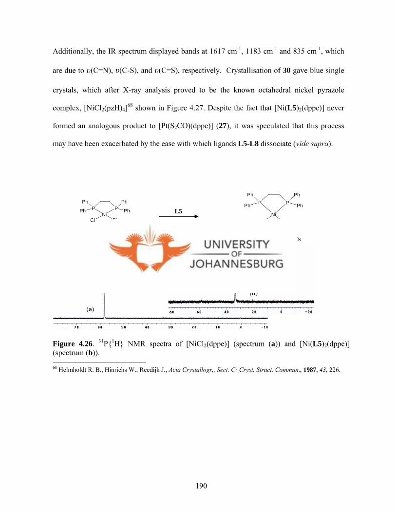
Additionally, the IR spectrum displayed bands at 1617 cm-1, 1183 cm-1 and 835 cm-1, which
are due to υ(C=N), υ(C-S), and υ(C=S), respectively. Crystallisation of 30 gave blue single
crystals, which after X-ray analysis proved to be the known octahedral nickel pyrazole
complex, [NiCl2(pzH)4]68 shown in Figure 4.27. Despite the fact that [Ni(L5)2(dppe)] never
formed an analogous product to [Pt(S2CO)(dppe)] (27), it was speculated that this process
may have been exacerbated by the ease with which ligands L5-L8 dissociate (vide supra).
P PPh
Ph
Ph
PhNi
Cl Cl
P PPh
Ph
Ph
Ph
NN
C
S
SN
N
C
S
S
Ni
Brown
(a)
24.5 ppm
(b)
L5
Figure 4.26. 31P{1H} NMR spectra of [NiCl2(dppe)] (spectrum (a)) and [Ni(L5)2(dppe)] (spectrum (b)). 68 Helmholdt R. B., Hinrichs W., Reedijk J., Acta Crystallogr., Sect. C: Cryst. Struct. Commun., 1987, 43, 226.
190

N
NH
HN N
NNH
N
HN
Ni
Cl
Cl
Figure 4.27. The structure of octahedral [NiCl2(pzH)4] complex.
4.4.1.11. Platinum(II) dithiocarbamato complexes 31-33.
Non-phosphine platinum(II) dithiocarbamato complexes 31-33 were also prepared.
Complexes [Pt(L5)2] (31), [Pt(L6)2] (32) and [Pt(L7)2] (33) were isolated as microcrystalline
reddish-brown solid materials in moderate yields by reacting K2[PtCl4] in deionised water
with L5, L6 and L7, respectively (Scheme 4.9). The 1H NMR spectrum of 32 showed
protons that belong to the methyl groups of 3,5-dimethylpyrazolyl in four different chemical
environments, hence resonated at four different frequencies, that is, at 2.83, 2.70, 2.40 and
2.33 ppm (Fig. 4.28). The 13C{1H} NMR spectrum of 32 showed a diagnostic peak at 226.8
ppm assignable to C(C=S), which was slightly more downfield than that the corresponding
ligand L6 (222.0 ppm). X-ray crystallography was used in the case of 32 to confirm the
proposed formulation of the complex (Fig. 4.29). The structural features are discussed in
detail in section 4.4.1.12 below.
191

a
b
c d
e
31
K2[PtCl4]
NN C
S
S
N NC S
S
PtN
N CS
S
N NC S
S
Pt
L6L5
L7
NN C
S
S
N NC S
S
Pt
33
32
a'
b' c'
d'
e'
Scheme 4.9. Synthesis of platinum(II) dithiocarbamato complexes 31-33.
Figure 4.28. A 1H NMR spectrum of 32. The four methyl peaks resonate at different frequencies between 2.2 and 2.9 ppm indicating that they are in different chemical environments in solution.
192

The 1H NMR spectra of 31 displayed a similar pattern to that 32, with all the peaks
resonating in pairs. The peaks due to 5H, 5H′ protons of the pyrazolyl unit appeared at 8.62
and 8.51 ppm. This was indicative of two L5 ligands in different chemical environments.
Other peaks were (3H, 3H′) at 8.16, 8.02 ppm and (4H, 4H′) at 6.69, 6.50 ppm. However, a
similar pattern was not very pronounced in 33. Except for the fact that Hb and H′b protons
appeared as two different protons at 8.43 and 8.31 ppm, respectively, all the other peaks did
not resonate in pairs as observed for complexes 31 and 32. The 13C{1H} NMR spectra of 31
and 33 could not be obtained because of the poor solubility in common polar solvents, such
as, CDCl3, CD2Cl2 and DMSO-d6. The similarity in the patterns of 1H NMR spectra of
compounds 31-33, led to postulating that they have analogous structures.
The IR spectra of complexes 31-33 showed bands in the 1110-1172 cm-1 region assignable to
υ(CS2) vibration expected for a ĸ2-S^S coordination of the dtc ligands. The insignificant shift
of υ(C=S) band at ca. 843 cm-1 in the IR spectra of complexes 31-33 compared to the υ(C=S)
band in the spectra of the free ligands L5-L7, indicated the presence of a non-coordinated
C=S moiety.69 The pyrazole ring vibrational frequencies for all the complexes occurred
between 1518 and 1606 cm-1, which were generally lower compared to those of the free
ligands L5-L7. The spectroscopic as well as microanalysis data suggest that compounds 31-
33 have similar structural features. X-ray crystallography was used in the case of 32, to
verify the proposed structures (cf. sec. 4.4.1.7 below).
69 Mukhopadhyay S., Mukhopadhyay U., Mak T. C. W., Ray D., Inorg. Chem., 2001, 40, 1057.
193
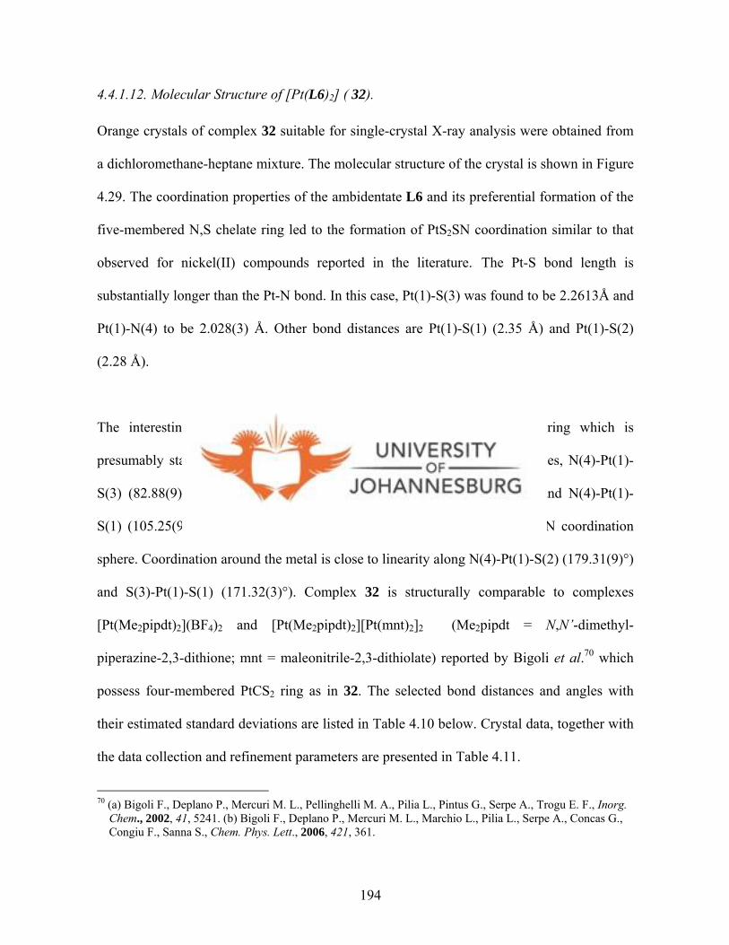
4.4.1.12. Molecular Structure of [Pt(L6)2] ( 32).
Orange crystals of complex 32 suitable for single-crystal X-ray analysis were obtained from
a dichloromethane-heptane mixture. The molecular structure of the crystal is shown in Figure
4.29. The coordination properties of the ambidentate L6 and its preferential formation of the
five-membered N,S chelate ring led to the formation of PtS2SN coordination similar to that
observed for nickel(II) compounds reported in the literature.6 9 The Pt-S bond length is
substantially longer than the Pt-N bond. In this case, Pt(1)-S(3) was found to be 2.2613Å and
Pt(1)-N(4) to be 2.028(3) Å. Other bond distances are Pt(1)-S(1) (2.35 Å) and Pt(1)-S(2)
(2.28 Å).
The interesting feature is the formation of a five-membered PtSCN2 ring which is
presumably stable than the rigid four-membered PtCS2 ring. The bond angles, N(4)-Pt(1)-
S(3) (82.88(9)°); S(2)-Pt(1)-Sl(1) (74.18(3)°); S(3)-Pt(1)-S(2) (97.67(3)°) and N(4)-Pt(1)-
S(1) (105.25(9)°) indicate a distorted square-planar geometry with a PtS2SN coordination
sphere. Coordination around the metal is close to linearity along N(4)-Pt(1)-S(2) (179.31(9)°)
and S(3)-Pt(1)-S(1) (171.32(3)°). Complex 32 is structurally comparable to complexes
[Pt(Me2pipdt)2](BF4)2 and [Pt(Me2pipdt)2][Pt(mnt)2]2 (Me2pipdt = N,N’-dimethyl-
piperazine-2,3-dithione; mnt = maleonitrile-2,3-dithiolate) reported by Bigoli et al.70 which
possess four-membered PtCS2 ring as in 32. The selected bond distances and angles with
their estimated standard deviations are listed in Table 4.10 below. Crystal data, together with
the data collection and refinement parameters are presented in Table 4.11.
70 (a) Bigoli F., Deplano P., Mercuri M. L., Pellinghelli M. A., Pilia L., Pintus G., Serpe A., Trogu E. F., Inorg. Chem., 2002, 41, 5241. (b) Bigoli F., Deplano P., Mercuri M. L., Marchio L., Pilia L., Serpe A., Concas G., Congiu F., Sanna S., Chem. Phys. Lett., 2006, 421, 361.
194
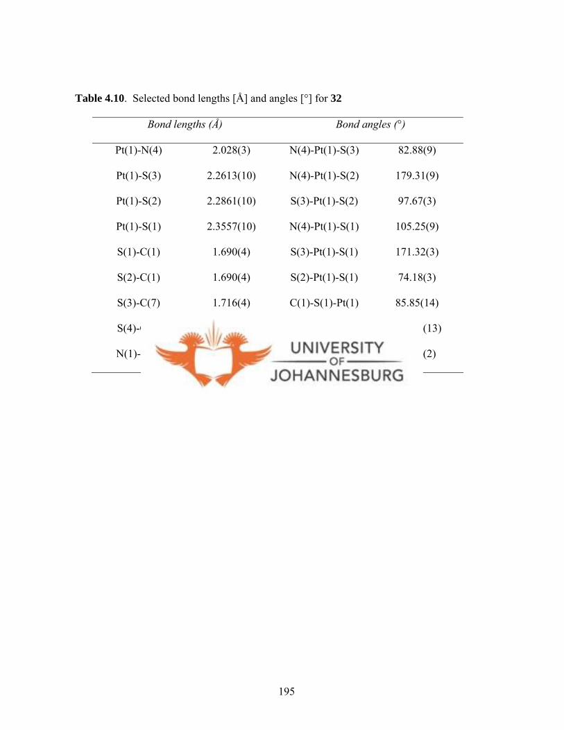
Table 4.10. Selected bond lengths [Å] and angles [°] for 32
Bond lengths (Å) Bond angles (°)
Pt(1)-N(4) 2.028(3) N(4)-Pt(1)-S(3) 82.88(9)
Pt(1)-S(3) 2.2613(10) N(4)-Pt(1)-S(2) 179.31(9)
Pt(1)-S(2) 2.2861(10) S(3)-Pt(1)-S(2) 97.67(3)
Pt(1)-S(1) 2.3557(10) N(4)-Pt(1)-S(1) 105.25(9)
S(1)-C(1) 1.690(4) S(3)-Pt(1)-S(1) 171.32(3)
S(2)-C(1) 1.690(4) S(2)-Pt(1)-S(1) 74.18(3)
S(3)-C(7) 1.716(4) C(1)-S(1)-Pt(1) 85.85(14)
S(4)-C(7) 1.648(4) C(7)-S(3)-Pt(1) 102.56(13)
N(1)-C(1) 1.378(5) S(2)-C(1)-S(1) 111.8(2)
195
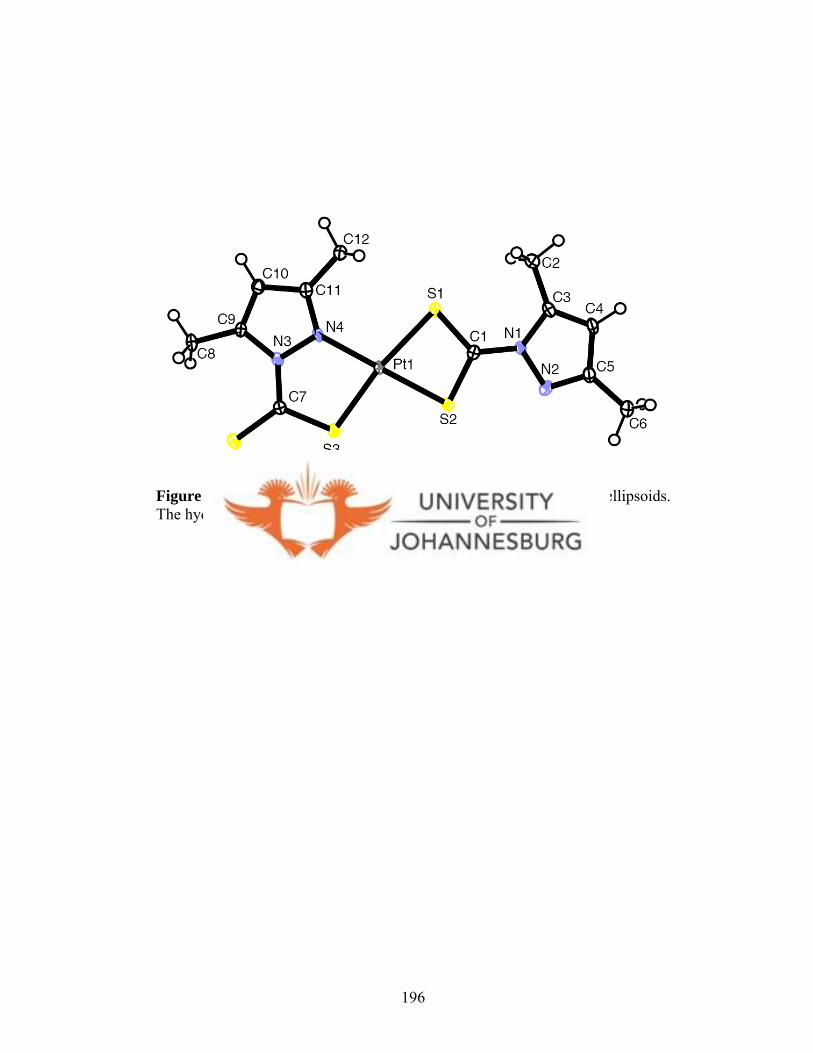
Figure 4.29. A molecular drawing of 32 drawn with 50% probability ellipsoids. The hydrogen atoms omitted for clarity.
196
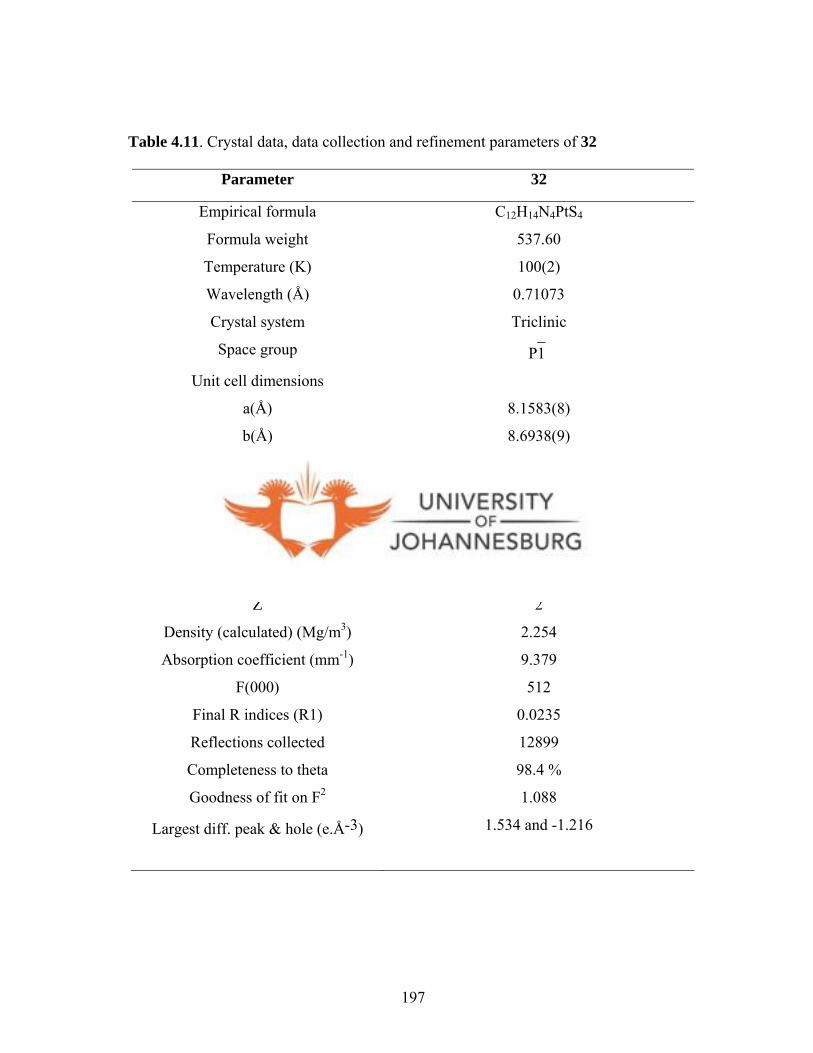
Table 4.11. Crystal data, data collection and refinement parameters of 32
Parameter 32
Empirical formula C12H14N4PtS4
Formula weight 537.60
Temperature (K) 100(2)
Wavelength (Å) 0.71073
Crystal system Triclinic
Space group P1̄
Unit cell dimensions
a(Å) 8.1583(8)
b(Å) 8.6938(9)
c(Å) 11.5863(11)
α 75.211(2)°
β 85.768(2)°
γ 89.997(2)°
Volume (Å3) 792.24(14)
Z 2
Density (calculated) (Mg/m3) 2.254
Absorption coefficient (mm-1) 9.379
F(000) 512
Final R indices (R1) 0.0235
Reflections collected 12899
Completeness to theta 98.4 %
Goodness of fit on F2 1.088
Largest diff. peak & hole (e.Å-3) 1.534 and -1.216
197
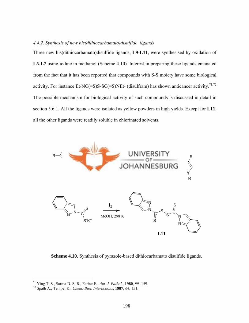
4.4.2. Synthesis of new bis(dithiocarbamato)disulfide ligands
Three new bis(dithiocarbamato)disulfide ligands, L9-L11, were synthesised by oxidation of
L5-L7 using iodine in methanol (Scheme 4.10). Interest in preparing these ligands emanated
from the fact that it has been reported that compounds with S-S moiety have some biological
activity. For instance Et2NC(=S)S-SC(=S)NEt2 (disulfram) has shown anticancer activity.71,72
The possible mechanism for biological activity of such compounds is discussed in detail in
section 5.6.1. All the ligands were isolated as yellow powders in high yields. Except for L11,
all the other ligands were readily soluble in chlorinated solvents.
NN C
S
S-K+
I2
MeOH, 298 K
N
NC
S
S
N
NC
S
S
R
R
R
R
R
RR = H (L9), Me (L10)
NN C
S
S-K+
I2
MeOH, 298 K
N
NC
S
S
N
NC
S
S
L11
Scheme 4.10. Synthesis of pyrazole-based dithiocarbamato disulfide ligands.
71 Ying T. S., Sarma D. S. R., Farber E., Am. J. Pathol., 1980, 99, 159. 72 Spath A., Tempel K., Chem.-Biol. Interactions, 1987, 64, 151.
198
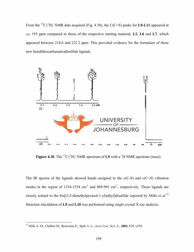
From the 13C{1H} NMR data acquired (Fig. 4.30), the C(C=S) peaks for L9-L11 appeared at
ca. 193 ppm compared to those of the respective starting material, L5, L6 and L7, which
appeared between 218.6 and 222.2 ppm. This provided evidence for the formation of these
new bis(dithiocarbamato)disulfide ligands.
Figure 4.30. The 13C{1H} NMR spectrum of L9 with a 1H NMR spectrum (inset).
The IR spectra of the ligands showed bands assigned to the υ(C-S) and υ(C=S) vibration
modes in the region of 1334-1254 cm-1 and 869-901 cm-1, respectively. These ligands are
closely related to the bis[(3,5-dimethylpyrazol-1-yl)ethyl]disulfide reported by Mills et al.73
Structure elucidation of L9 and L10 was performed using single crystal X-ray analysis.
73 Mills A. M., Chalbot M., Bouwman E., Spek A. L., Acta Cryst. Sect. E., 2003, E59, o258.
199

Orange crystals suitable for single crystal X-ray analysis were obtained from slow evaporation
of THF solutions of L9 and L10. The molecular structures of the crystals are shown in Figures
4.31 and 4.32. In L9 and L10, the two pyrazolyl rings are connected via N(1) and N(1A) by a
disulfide bridge and are in perpendicular planes. The bond lengths and angles (S(2)-S(2)#1 =
2.0309(6) Å, S(2)-C(6) = 1.7958(11) Å, C(6)-S(2)-S(2)#1 = 102.78(4)°) are consistent with
those of disulfide compounds.73,74 The molecular packing diagram of L10 viewed along c axis
is shown in Figure 4.33. There is formation of cuboids, which are defined by two ligand units
and have no intermolecular interactions. The selected bond distances and angles with their
estimated standard deviations, for L9 and L10, are listed in Table 4.12, whilst the crystal data,
together with the data collection and refinement parameters of L9 and L10 are presented in
Table 4.13.
74 Mullica D. F., Trawick M. L., Wu P. W. N., Sappenfield E. L., J. Chem. Crystallogr., 1998, 28, 761.
200
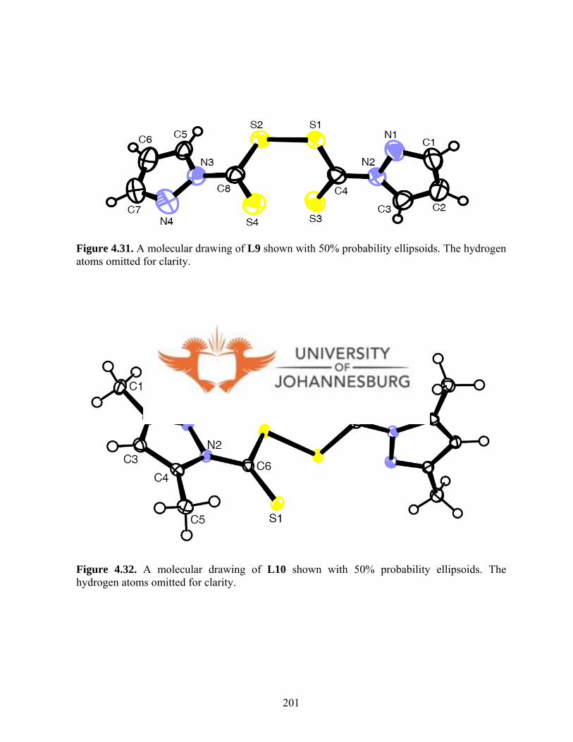
Figure 4.31. A molecular drawing of L9 shown with 50% probability ellipsoids. The hydrogen atoms omitted for clarity.
Figure 4.32. A molecular drawing of L10 shown with 50% probability ellipsoids. The hydrogen atoms omitted for clarity.
201

Table 4.12. Selected bond lengths [Å] and angles [°] for L9 and L10
L9 L10
Bond lengths (Å)
S(1)-C(6) 1.621(2) 1.6294(11)
S(2)-C(6) 1.778(3) 1.7958(11)
S(2)-S(2)#1 2.0194(10) 2.0309(6)
N(1)-N(2) 1.370(3) 1.3884(12)
N(2)-C(6) 1.386(3) 1.3825(13)
Bond angles (°)
C(6)-S(2)-S(2)#1 102.49(9) 102.78(4)
C(2)-N(1)-N(2) 111.8(2) 105.28(9)
C(6)-N(2)-N(1) 104.1(2) 91.07(12)
202

Figure 4.33. A molecular packing diagram of L10 viewed along c axis. The hydrogen atoms omitted for clarity.
203
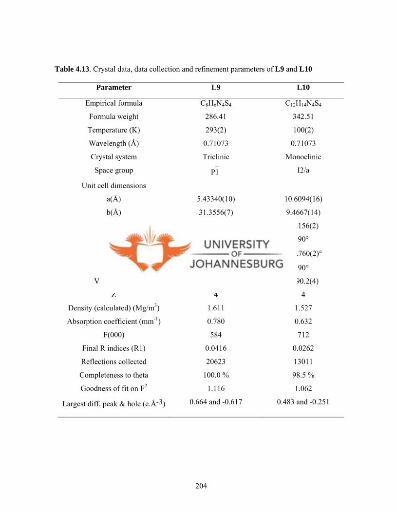
Table 4.13. Crystal data, data collection and refinement parameters of L9 and L10
Parameter L9 L10
Empirical formula C8H6N4S4 C12H14N4S4
Formula weight 286.41 342.51
Temperature (K) 293(2) 100(2)
Wavelength (Å) 0.71073 0.71073
Crystal system Triclinic Monoclinic
Space group P1̄ I2/a
Unit cell dimensions
a(Å) 5.43340(10) 10.6094(16)
b(Å) 31.3556(7) 9.4667(14)
c(Å) 7.2893(2) 15.156(2)
α 90° 90°
β 107.9860(10)° 101.760(2)°
γ 90° 90°
Volume (Å3) 1181.17(5) 1490.2(4)
Z 4 4
Density (calculated) (Mg/m3) 1.611 1.527
Absorption coefficient (mm-1) 0.780 0.632
F(000) 584 712
Final R indices (R1) 0.0416 0.0262
Reflections collected 20623 13011
Completeness to theta 100.0 % 98.5 %
Goodness of fit on F2 1.116 1.062
Largest diff. peak & hole (e.Å-3) 0.664 and -0.617 0.483 and -0.251
204
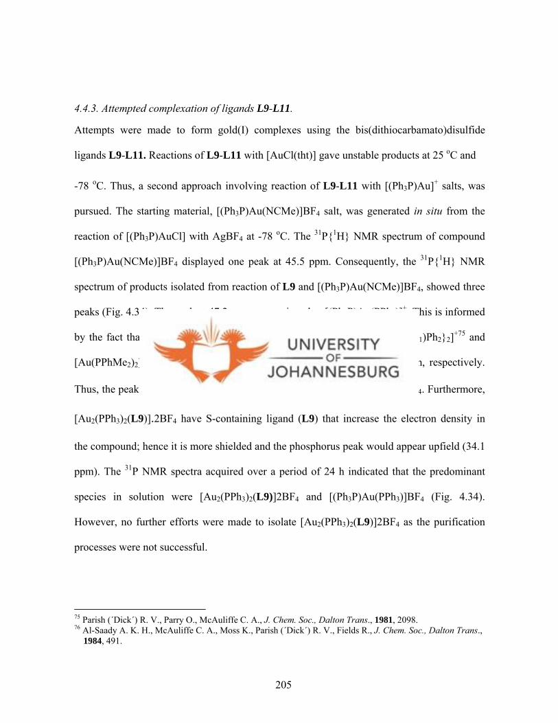
4.4.3. Attempted complexation of ligands L9-L11.
Attempts were made to form gold(I) complexes using the bis(dithiocarbamato)disulfide
ligands L9-L11. Reactions of L9-L11 with [AuCl(tht)] gave unstable products at 25 oC and
-78 oC. Thus, a second approach involving reaction of L9-L11 with [(Ph3P)Au]+ salts, was
pursued. The starting material, [(Ph3P)Au(NCMe)]BF4 salt, was generated in situ from the
reaction of [(Ph3P)AuCl] with AgBF4 at -78 oC. The 31P{1H} NMR spectrum of compound
[(Ph3P)Au(NCMe)]BF4 displayed one peak at 45.5 ppm. Consequently, the 31P{1H} NMR
spectrum of products isolated from reaction of L9 and [(Ph3P)Au(NCMe)]BF4, showed three
peaks (Fig. 4.34). The peak at 47.2 ppm was assigned to [(Ph3P)Au(PPh3)]+. This is informed
by the fact that the 31P NMR studies of closely related species, [Au{P(C6H11)Ph2}2]+75 and
[Au(PPhMe2)2]+,76 showed phosphorous peaks at around 54.0 and 62.0 ppm, respectively.
Thus, the peak at 34.1 ppm was tentatively assigned to [Au2(PPh3)2(L9)].2BF4. Furthermore,
[Au2(PPh3)2(L9)].2BF4 have S-containing ligand (L9) that increase the electron density in
the compound; hence it is more shielded and the phosphorus peak would appear upfield (34.1
ppm). The 31P NMR spectra acquired over a period of 24 h indicated that the predominant
species in solution were [Au2(PPh3)2(L9)]2BF4 and [(Ph3P)Au(PPh3)]BF4 (Fig. 4.34).
However, no further efforts were made to isolate [Au2(PPh3)2(L9)]2BF4 as the purification
processes were not successful.
75 Parish (´Dick´) R. V., Parry O., McAuliffe C. A., J. Chem. Soc., Dalton Trans., 1981, 2098. 76 Al-Saady A. K. H., McAuliffe C. A., Moss K., Parish (´Dick´) R. V., Fields R., J. Chem. Soc., Dalton Trans., 1984, 491.
205

Ph3P Au NCMe BF4
5 min
Figure 4.34. A 31P{1H} NMR profile showing a mixture of desired product [Au2(PPh3)2(L9)]2BF4, [(Ph3P)-Au-(PPh3)]+ and [Ph3P-Au-NCMe]+.
N
NC
S
S
N
NC
S
S
Au
Au
PPh3
Ph3P
BF42
2+
Ph3P Au PPh3 BF4
3 h
24 h
206
4.5. Conclusions
The reaction of dithiocarbamate ligands L5-L8 with [AuCl(PPh3)], [Au2Cl2(dppp)] and
[Au2Cl2(dpph)] led to successful isolation of mononuclear and binuclear gold(I) complexes.
This was confirmed from the spectroscopic and microanalysis data. Furthermore, the
molecular structure of [Au(L7)(PPh3)] (15) and [(AuL7)2(dpph)] (25) which features
approximately linear gold atom geometry, is a further confirmation of existence of
complexes 13-25. Notably, attempts to crystallise complex [(AuL5)2(dppe)] (17a) led to the
formation of gold(I) cluster compound, [Au18S8(dppe)6]Cl2 (17b). The span between the P
atoms of the dppe ligand in 17a seems to play a role in the formation of 17b. The close
proximity of the P atoms in dppe back bone, allows for the gold atoms coordinated to them to
have aurophilic interactions. The presence of these interactions would bring the two gold
centres closer, thus contributing to cluster formation; but it is not clear how this leads to the
re-arrangement and formation of 17b. The X-ray structure of 17b displays Au···Au
interactions (2.9263(7)-3.1395(7) Å).
Reaction of ligands L5-L7 with K2[PtCl4] gave bis-chelated complexes (31-33) having three
sulfur atoms and one nitrogen atom coordinated to the platinum metal, leaving one
uncoordinated sulfur. These complexes have distorted square-planar geometries as revealed
from the solid state structure of 32. Ligand L6 coordinated to platinum atom to form a five-
membered N,S chelate ring (PtS2SN). On the other hand, reactions of [PtCl2(dppe)] with L5-
L7 gave complexes 26, 28 and 29 with a general formula [Pt(L)2(dppe)]. These yellow
complexes were found to be unstable in solution and transformed to an identical green
product, [Pt(S2CO)(dppe)] (27), at room temperature. This has been clearly demonstrated
207
from the UV-Vis and 31P{1H} NMR spectroscopic studies. A pseudo-first-order
transformation process was proposed, with a rate constant of 1.4 x 10⁻² h-1. A further
conformation of this transformation was the solid state structure of 27. Once again the ease
with which L5-L7 disintegrate is speculated to be the cause for this occurrence. Moreover,
there is no doubt that the rearrangement of the phenyl rings in the diphosphine ligands causes
the compound to be rather unstable in solution. Experiments utilising various diphosphine
ligands would help prove this concept.
Isolation of new bis(dithiocarbamato)disulfide ligands (L9-L11) was indicative of how L5-
L7 can undergo easy oxidation. The solid state structures of L9 and L10 is a confirmation of
the oxidation products. Strategies to isolate gold(I) complexes of
bis(dithiocarbamato)disulfide ligands L9-L11 were unsuccessful as they led to formation of
very unstable products. Just as dithiocarbamate ligands L5-L7 undergo oxidation easily, the
resultant disulfides (L9-L11) can also be easily reduced back to L5-L7. Thus, it is possible
that the unstable nature of gold products from L9-L11 is due to reduction of the disulfide
ligands. No further efforts were made to isolate [(PPh3)2L].2BF4 because of purification
problems.
208
The major findings are:
1. dithiocarbamate ligands (L5-L8) could act as sources of sulfur for the isolation of
gold(I) cluster compounds. This is clearly demonstrated from the isolation of the cluster
compound [Au18S8(dppe)6]2+ (17b). This inference is drawn from the fact that the
analogous [Au18Se8(dppe)6]2+ was prepared by reacting [(AuCl)2(dppe)] with
Se(SiMe3)2. However, the isolation of 17b from L5 is unique as there are no reports of
gold(I) cluster obtained from dithiocarbamates.
2. that while the biphasic synthetic procedure for the monophopshine and diphosphine
gold(I) dithiocarbamato complexes works, it is not ideal for the synthesis of the
analogous diphosphine platinum(II) dithiocarbamato complexes. The slightest traces of
water could lead to transformation of the desired compounds. Thus for synthesis of
diphosphine platinum(II) dithiocarbamato compounds, a dry solvent synthetic
procedure is advised.
209

CHAPTER 5
ANTI-TUMOUR ACTIVITY OF LIGANDS, PLATINUM(II) AND GOLD(I)
DITHIOCARBAMATO COMPLEXES AGAINST HELA CELLS AND
LUMINESCENCE STUDIES
Screening of the complexes for their in vitro anticancer activities were performed by Mrs.
Nell of Pretoria University (SA), whilst the photoluminescence experiments were
performed by Ms. Sumitra of University of North Texas. The interpretation and
discussion of the results is by the candidate.
5.0. Introduction
The biochemistry of gold has developed primarily in response to the extensive use of
gold compounds in treating rheumatoid arthritis and in response to efforts to develop
complexes with antitumour, and to some extent, anti-HIV activity.1 These efforts are as a
result of the realisation of the activity of 2,3,4,6-tetra-O-acetylglucopyranosato-S-
(triethyl-phosphine)gold(I) (auranofin) as an antiarthritic agent.2 Thus, gold(I)
coordination compounds are receiving considerable attention because of their potential
applications in medicine, particularly as antitumour agents.3 For instance, auranofin,
although an antiarthritic agent, has also been found to be an effective agent against P388
lymphocytic leukemia.4 This led to the examination of the antitumour activity of
1 Gold Progress in Chemistry, Biochemistry and Technology. Shaw III C. F., Ed. Schmidbaur H., John Wiley & Sons, Chichester, England, 1999, 260. 2 Sutton B. M., McGusty E., Walz D. T., DiMartino M. J., J. Med. Chem., 1972, 15, 1095. 3 Kostova I., Anti-cancer Agents Med. Chem., 2006, 6, 19.
210
phosphine gold(I) thiolate complexes containing the P-Au-S motif found in auranofin.5
This has been achieved by using various phosphines and S-containing ligands.
Researchers have also sought to understand the mechanism(s) by which phosphine
gold(I) thiolate complexes exhibit their antitumour activities. This is informed by the fact
that, unlike platinum complexes, DNA is not the primary target. It has been shown that
the mitochondria act as a potential target of anticancer drugs.6, , , ,7 8 9 10 The mitochondria are
also known to play a decisive role in apoptosis or programmed cell death.7 Apoptosis is a
distinct form of cell death that proceeds along a genetically determined execution
program.
Several studies now show that mitochondria and thioredoxin reductase, which is a
homodimeric selenoenzyme, are the targets for the phosphine gold(I) compounds.7,11, ,12 13
The mechanism is believed to involve the binding of Au(I) to the selenocysteine (Cys-
SeCsy) residue in the mitochondrial enzyme thioredoxin reductase.7,14 From discussions
4 aSimon T. M., Kinishima D. H., Vilbert G. J., Lorber A., Cancer Res., 1981, 41, 94; bMirabelli C. K., Johnson R. K., Sung C., Faucette L. F., Muirhead K., Crooke S. T., Cancer Res., 1985, 45, 32. 5 Mirabelli C. K., Johnson R. K., Hill D. T., Faucette L. F., Girard G. R., Kuo G. Y., Sung C., Crooke S. T., J. Med. Chem., 1986, 29, 218. 6 McKeage M. J., Maharaj L., Berners-Price S. J., Coord. Chem. Rev., 2002, 232, 127. 7 Barnard P. J., Berners-Price S. J., Coord. Chem. Rev., 2007, 25, 1889. 8 Farrell N., Coord. Chem. Rev., 2002, 232, 1 9 Bouchier-Hayes L., Lartigue L., Newmeyer D. D., J. Clin. Invest., 2005, 11, 240. 10 Weissig V., Boddapati S. V., D'Souza G. G. M., Cheng S. M., Drug Design Reviews, 2004, 1, 15. 11 aBragadin M., Scutari G., Folda A., Bindoli A., Rigobello M. P., Ann. N. Y. Acad. Sci., 2004, 1030, 348; bRigobello M. P., Folda A., Dani B., Menabo R., Scutari G., Bindoli A., Eur. J. Pharmacol., 2008, 582, 26. 12 aRigobello M. P., Scutari G., Folda A., Bindoli A., Biochem. Pharmacol., 2004, 67, 689; bRigobello M. P., Scutari G., Boscolo R., Bindoli A., Br. J. Pharmacol., 2002, 136, 1162. 13 Hill K. E., McCollum G. W., Boeglin M. E., Burk R. F., Biochem.Biophys. Res. Commun., 1997, 234, 293. 14 Gromer S., Arscot L. D., Williams Jr. C. H.,Schirmer R. H., Becker K., J. Biol. Chem., 1998, 273, 20096.
211

in chapter 4, it is clear that any attempts to prepare new gold(I) anticancer agents could
make use of a phosphine-gold motif in its design. This is what this project took advantage
of and the biological activities are reported in this chapter.
5.1. Luminescent gold(I) complexes
Apart from the biological activities of gold(I) thiolate complexes, their photophysical and
photochemical properties have potential applications as luminescent materials.15
Luminescence originates from the valence orbitals of free gold(I) ion, which are the filled
5d orbitals, empty 6s and 6p orbitals.16 Since Au(I) is oxidising as well as reducing,
LMCT and MLCT transitions (Fig. 5.1) could occur at reasonable energies.1 6 Low-energy
MLCT absorptions for gold(I) complexes with π-acceptor ligands is dominant; thus,
Au(I) complexes with simple π-acceptor ligands, such as phosphines and isocyanides,
display MLCT absorptions in their electronic spectra.16
Au (6pz)
S (pπ)
Au (5dz2)
Emission derived from LMCT origin
p
p
S(p
dd
σ
σ
σσ
*
*
π)
Emission derived from LMMCT origin
Figure 5.1. Schematic representations of orbital splitting of gold(I) thiolate compounds (Taken from ref. 18).
15 Yam V. W. W., Lo K. K. W., Chem. Soc. Rev., 1999, 28, 323. 16 Vogler A., Kunkely H., Coord. Chem. Rev., 2001, 219, 489.
212

Even though complete knowledge of the nature of luminescence in gold(I) compounds is
lacking, a variety of different excited states suggests that they are emissive. It is assumed
that the lowest-energy excited state, which is populated in absorption, is the emissive
state.16,17 For instance, for mononuclear gold(I) thiolate complexes like [Au(SR′)(PR3)]
(R = Et, Ph; R′ = Ph), solid-state emissions occur at 77 K over a wide energy range of
413-702 nm.18 The origin of the emission has been ascribed to a LMCT (S→Au) triplet
state. Other examples include complexes [Au2(SR′)2(Ph2PN(R)PPh2)] (R′ = C6H4Cl-p >
C6H4F-p > C6H4Me-p; R = hexyl) (Fig 5.2), which shows absorption energy trend in line
with the σ-donating ability of the thiolate ligands.19 This low-energy absorption band has
been assigned to a thiolate-to-gold LMCT transition. The high energy absorptions of
[Au2(SR′)2(Ph2PN(R)PPh2)] at ca. 262-305 nm were tentatively assigned as intraligand
transitions characteristic of the diphosphine ligands.19 Excitation of solids and fluid CHCl3
solutions of [Au2(SR′)2(Ph2PN(R)PPh2)] complexes are dominated by broad and intense
blue-green emission, typical of a metal perturbed ligand-centred emission of the
phosphine ligand.19
17 Kunkely H., Vogler A., Naturforsch., 1996, 51b, 1067. 18 Yam V. W., Chan C., Li C., Wong K. M., Coord. Chem. Rev., 2001, 216, 173. 19 Yam V. W, Chan C. L., Cheung K. K., J. Chem. Soc., Dalton Trans., 1996, 4019.
213

NPh2P PPh2
Au Au
SR' SR'
Cl F MeR' =, ,
Figure 5.2. Structures of [Au2(SR′)2(Ph2PN(R)PPh2)].
The low energy emissions of [Au2(SR′)2(Ph2PN(R)PPh2)] complexes observed at ca. 600
nm originate from emissive states derived from thiolate-to-gold LMCT transitions.19 The
large Stokes shifts (difference between the λmax of the absorption spectrum and λmax of the
emission spectrum) and the observed lifetimes in microseconds range are suggestive of a
triplet parentage (3LMCT).19,20 There is also a relationship between the luminescence
properties and the extent of Au···Au association in the compounds concerned.21 The
excited state for those complexes with short Au···Au contacts has been suggested to show
ligand-to-metal-metal charge-transfer (LMMCT) (Fig. 5.1).18
In this project, the new phosphine gold(I) complexes and platinum(II) complexes were
investigated for their anticancer activities against human cervix epithelial carcinoma
(HeLa) cells. Some of the gold compounds were further investigated for luminescence.
The results are presented in the following sections.
20 Lebedkin S., Langetepe T., Sevillano P., Dieter F., Kappes M. M., J. Phys. Chem. B, 2002, 106, 9019. 21 A. G., Staples R. J., Fackler J. P., Jr, Inorg. Chem., 1995, 34, 75.
214

5.2. Experimental
5.2.1 Biological reagents and instrumentation
All commercial reagents were used as received. Phosphate Buffered Saline (PBS),
Eagle’s RPMI-1640 medium, 3-(4,5-dimethylthiazol-2-yl)-2,5-diphenyltetrazolium
bromide (MTT) kit, phytohemagglutinin-protein form (PHA-P) and the 96-well flat-
bottomed culture plates were all purchased from BD Biosciences Ltd. Cisplatin was
purchased from Sigma Aldrich. Eagle’s medium with 0.1 mM non-essential amino acids
was prepared by adding 2 mM L-glutamine, 1.0 mM sodium pyruvate and 5% bovine
fetal calf serum to the pure Eagle’s RPMI-1640 medium. Twenty compounds, L5-L11
(Scheme 5.1), 13-16 (Scheme 5.2), 20-25 (Scheme 5.3) and 31-33 (Scheme 5.4) were
screened for their antitumour activities against human cervix epithelial carcinoma (HeLa)
cells. HeLa cells and human lymphocytes (PBMCs) from preservative free heparinised
peripheral blood were obtained from the Department of Pharmacology and Pretoria
Medical Hospital, University of Pretoria, South Africa. The absorbance values were
recorded on a Whittaker Microplate Reader 2001 spectrophotometer at 570 nm and the
reference wavelength of 630 nm.
5.2.2. Cell culture and drug treatment
HeLa cells were cultured in Eagle’s medium with 0.1 mM non-essential amino acids, 2
mM L-glutamine, 1.0 mM sodium pyruvate and 5% bovine fetal calf serum, at 37 oC in
an atmosphere of 5% CO2. Cells were plated in 96-well sterile plates at a density of 1 x
215
104 cells/well in 100 µL of medium, and incubated for 1 h. Subsequently, compounds
were added with concentrations from 0 to 50 µM. Cytotoxicity was determined by using
MTT to stain treated HeLa cells after 7 days according to literature methods22. MTT dye
is reduced by living cells to yield a soluble formazan product that can be assayed
colourimetrically.22 A 20 µL volume of freshly prepared MTT (5 mg/mL) was added to
each well and the cells incubated for another 4 h. Cell survival was evaluated by
measuring absorbance at 570 nm, using a Whittaker Microplate Reader 2001. The IC50
values were calculated with the Graphpad programme. All experiments were performed
in triplicate.
The inhibition of the growth of normal cells by the complexes tested was also measured
by employing human lymphocytes (PBMC) cells. The same procedure described above
was used, except for the fact that the treated PBMC cells were incubated for 3 days as
opposed to 7 days for HeLa cells. The aim of testing these compounds on normal cells
was to determine whether the compounds could target the cancerous (HeLa) cells
specifically and not the normal cells. While that was the primary objective, it also
allowed investigation into whether there was any relationship between the observed
activities and the growth rate of the cells. In performing these experiments, the
lymphocytes were divided into two namely; (i) normal cells that were stimulated using
PHA-P so as to increase their proliferation rate (stimulated lymphocytes) and (ii) un-
stimulated normal cells (resting lymphocytes).
22 Mossman T., J. Immunol. Methods, 1983, 65, 55.
216
NOTE: In all the cases, the compounds were tested in different batches and therefore the
IC50 values for the reference compound (cisplatin) had to be established for every batch
of compounds tested. The reader is advised to take note of this.
5.2.3. Electronic absorption and photoluminiscence spectra measurements
UV-Vis spectra of [Au(L7)(PPh3)] (15), [(AuL5)2(dppe)] (17a), [(AuL5)2(dppp)] (20)
and [(AuL5)2(dpph)] (23) (Scheme 5.5) were recorded on a Perkin-Elmer Lambda 900
spectrophotometer at 298 K as dichloromethane solutions. Photoluminescence
measurements were performed on both solid materials and frozen dichloromethane
solutions of complexes [Au(L7)(PPh3)] (13), [Au(L7)(PPh3)] (14), 15, [Au(L7)(PPh3)]
(16), 17a and [Au18S8(dppe)6]2Cl (17b) (Scheme 5.6). Photoluminescence measurements
were carried out at the University of Texas, USA, on a Spex Fluorolog-3 spectrometer
equipped with a 450 W Xe-lamp and double grating monochromators in the excitation
(λexc ~ 250-1000 nm) and two emission channels. Three photo-detectors R5108
Hamamatsu photomultiplier tubes (PMT) and a liquid nitrogen-cooled germanium
photodiode (Edinburgh Instruments) covered the emission ranges of λem ~ 400-800 (UV-
Vis emission channel). All emission spectra were corrected for the wavelength-dependent
instrumental response. The latter was determined using a standard calibration lamp
(Oriel) and a two-inch Spectralon intergrating sphere (Labsphere) installed in the sample
chamber of the Fluorolog-3 spectrometer.
217
5.3. Biological results
Twenty compounds, dithiocarbamate and disulfide ligands (Scheme 5.1), ten gold(I)
complexes (Scheme 5.2 and 5.3) and three platinum(II) dithiocarbamato complexes
(Scheme 5.4), were investigated for their ability to inhibit cell growth on tumour cells in
vitro. The human cervical epitheloid carcinoma (HeLa) cell-line was chosen in this case.
All data were acquired in triplicate and the final values recorded as averages. Tables 5.1-
5.5 list the dose values that caused 50% inhibition of cell growth (IC50). The syntheses of
the ligands, gold(I) and platinum(II) complexes tested are reported in chapter 4. The
biological results are discussed in the following sections systematically starting with the
activities of the ligands, the gold(I) compounds, and finally platinum(II) compounds.
5.3.1. Dithiocarbamate and bis(pyrazolyldithiocarbamate)disulfide ligands
In order to establish the activities of phosphine gold(I) complexes 20-25 and platinum(II)
complexes 31-33, it was important to establish the activities of the ligands first. This was
paramount as it helped in determining whether activities of the metal complexes could be
due to the presence of the dithiocarbamate ligands. Thus, the dithiocarbamate ligands L5-
L8 (Scheme 5.1) were tested for their antitumour activities against cancerous HeLa cells.
They were all found to be inactive against HeLa cells (Table 5.1). However, the new
disulfide ligands L9-L11 (Scheme 5.1), derivatised from L5-L7, showed antitumour
activities against HeLa cells. Ligand L9 had an IC50 value of 3.7 µM (Fig. 5.3, Table 5.1),
which was comparable to that of cisplatin (1.1 µM), while that of L10 was ca. 16 times
less active than cisplatin (17.8 µM).
218
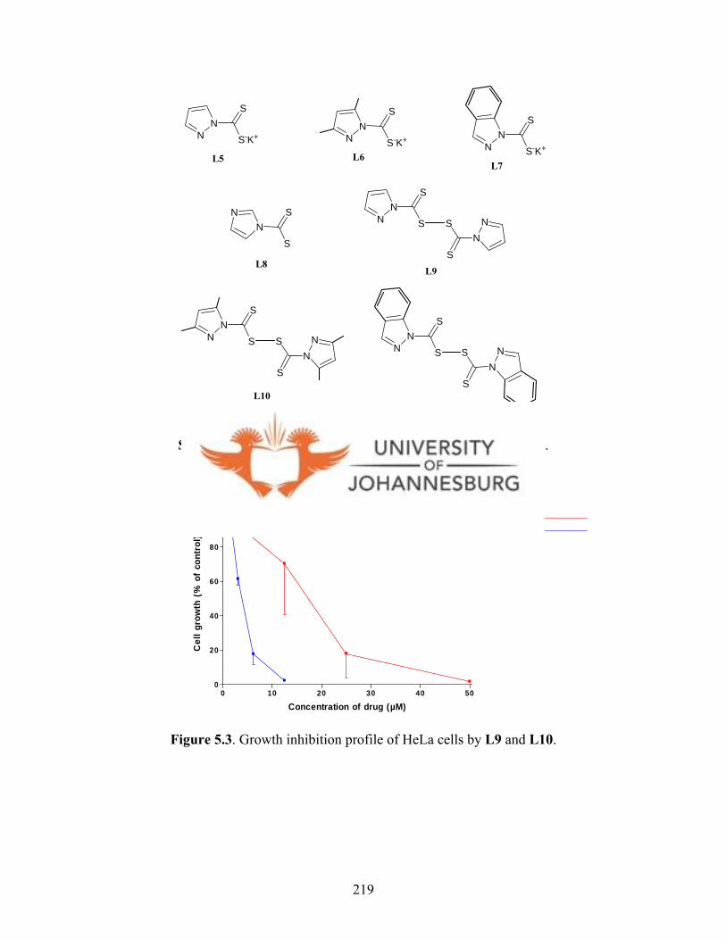
NN
S
S
L11
L10
L9
NN
S
SN
NS
S NN
S
S
NN
S
S NN
S
S
NN
S
S-K+ NN
S
S-K+N
NS
S-K+
NN S
S
L5L7
L6
L8
Scheme 5.1. Dithiocarbamate salts and their disulfide analogues.
0 10 20 30 40 50
0
20
40
60
80
100 KFK 128KFK 165
Concentration of drug (µM)
Cel
l gro
wth
(%
of
cont
rol)
L9 L10
Figure 5.3. Growth inhibition profile of HeLa cells by L9 and L10.
219
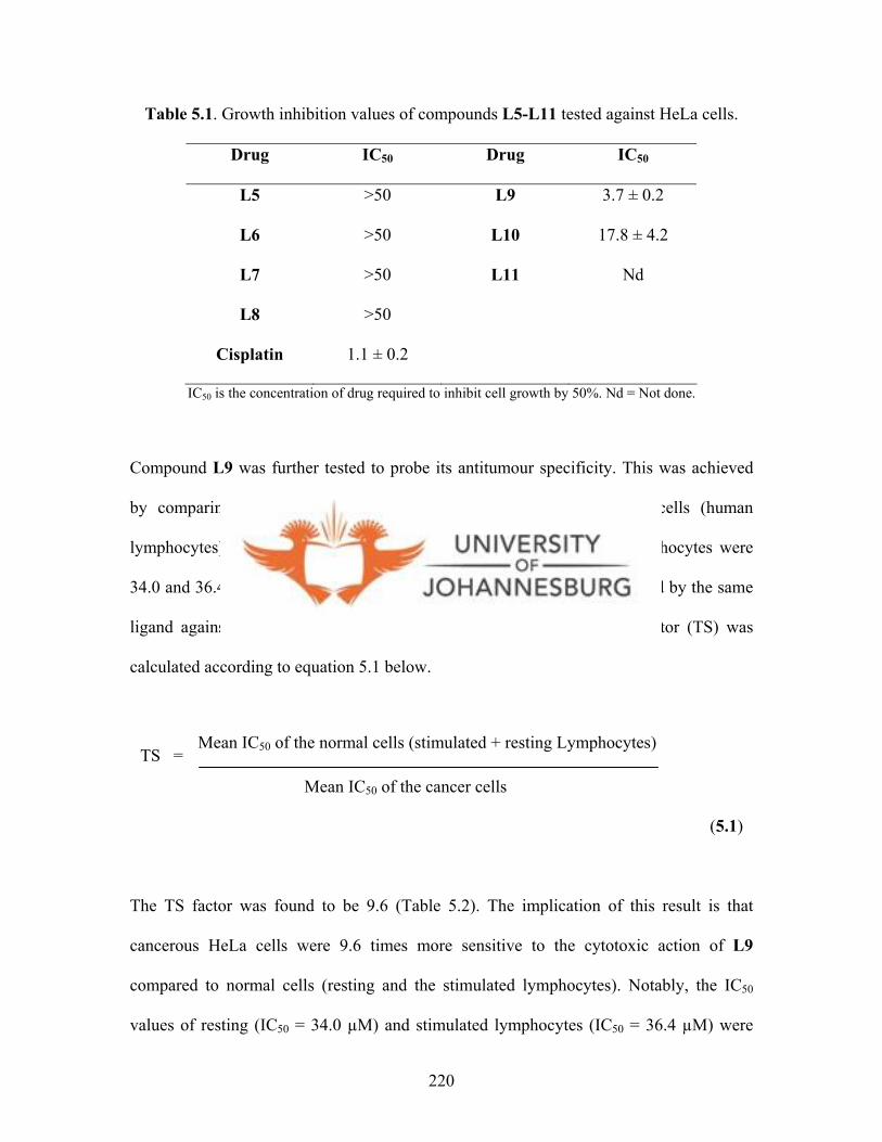
Table 5.1. Growth inhibition values of compounds L5-L11 tested against HeLa cells.
Drug IC50 Drug IC50
L5 >50 L9 3.7 ± 0.2
L6 >50 L10 17.8 ± 4.2
L7 >50 L11 Nd
L8 >50
Cisplatin 1.1 ± 0.2
IC50 is the concentration of drug required to inhibit cell growth by 50%. Nd = Not done.
Compound L9 was further tested to probe its antitumour specificity. This was achieved
by comparing its activity against cancerous HeLa cells and normal cells (human
lymphocytes). The IC50 values registered for resting and stimulated lymphocytes were
34.0 and 36.4 µM (Table 5.2), respectively as opposed to 3.7 µM displayed by the same
ligand against HeLa cells. From these results, the tumour specificity factor (TS) was
calculated according to equation 5.1 below.
TS = Mean IC50 of the cancer cells
Mean IC50 of the normal cells (stimulated + resting Lymphocytes)
(5.1)
The TS factor was found to be 9.6 (Table 5.2). The implication of this result is that
cancerous HeLa cells were 9.6 times more sensitive to the cytotoxic action of L9
compared to normal cells (resting and the stimulated lymphocytes). Notably, the IC50
values of resting (IC50 = 34.0 µM) and stimulated lymphocytes (IC50 = 36.4 µM) were
220

comparable. This suggests that increased growth rate of cells in stimulated lymphocytes,
did not reduce the activity of L9 significantly (36.4 µM). From this result, it was
concluded that the growth rate of cells in this instance had little influence in the observed
activities.
Ligand L9 is similar to Et2NC(=S)S-SC(=S)NEt2 (disulfram), which has anticancer
activity.23,24, ,25 26 Many proposals have been given for the mechanism of action of
disulfram, which ranges from inhibiting DNA topoisomerase,27 to reducing
angiogenesis28 and also inhibiting proteasome signalling pathways.29 Even though there
are no clear structural activity relationships of disulfram, the anticancer activity of
disulfram has been suggested to be due its disulfide nature (S-S moiety), which allows for
the formation of diethyldithiocarbamate that react with sulfhydryl groups of several
enzymes.30,31 Compound L9 could be acting in a similar manner as disulfram.
Table 5.2. Tumour Specificity of L9.
Drug HeLa IC50 (µM) Lymph (resting)
IC50 (µM)
Lymph (stimulated)
IC50 (µM)
Tumour
Specificity
L9 3.7 34.0 36.4 9.6
IC50 is the concentration of drug required to inhibit cell growth by 50%.
23 Spath A., Tempel K., Chem.-Biol. Interactions, 1987, 64, 151. 24 Grafstroem R., Greene F. E., Biochem. Pharmacol., 1980, 29, 1517. 25 Sauna Z. E., Shukla S., Ambudkar S. V., Mol. BioSyst., 2005, 1, 127. 26 Wickström M., Danielsson K., Rickardson L., Gullbo J., Nygren P., Isaksson A., Larsson R., Lövborg H., Biochem. Pharmacol., 2007, 73, 25. 27 Yakisich J. S., Siden A., Eneroth P., Cruz M., Biophys. Res. Commun., 2001, 289, 586. 28 Shiah S. G., Kao Y. R. Wu F. Y. H., Wu C. W., Mol. Pharmacol., 2003, 64, 1076. 29 Lövborg H., Oberg F., Rickardson L., Gullbo J., Nygren P., Larsson R., Int. J. Cancer, 2006, 118, 1577. 30 Hacker M. P., Ershler W. B., Newman R. A., Gamelli R. L., Cancer Res., 1982, 42, 4490. 31 Neims A. H., Coffey D. S., Hellerman L., J. Biol. Chem., 1966, 241, 5941.
221
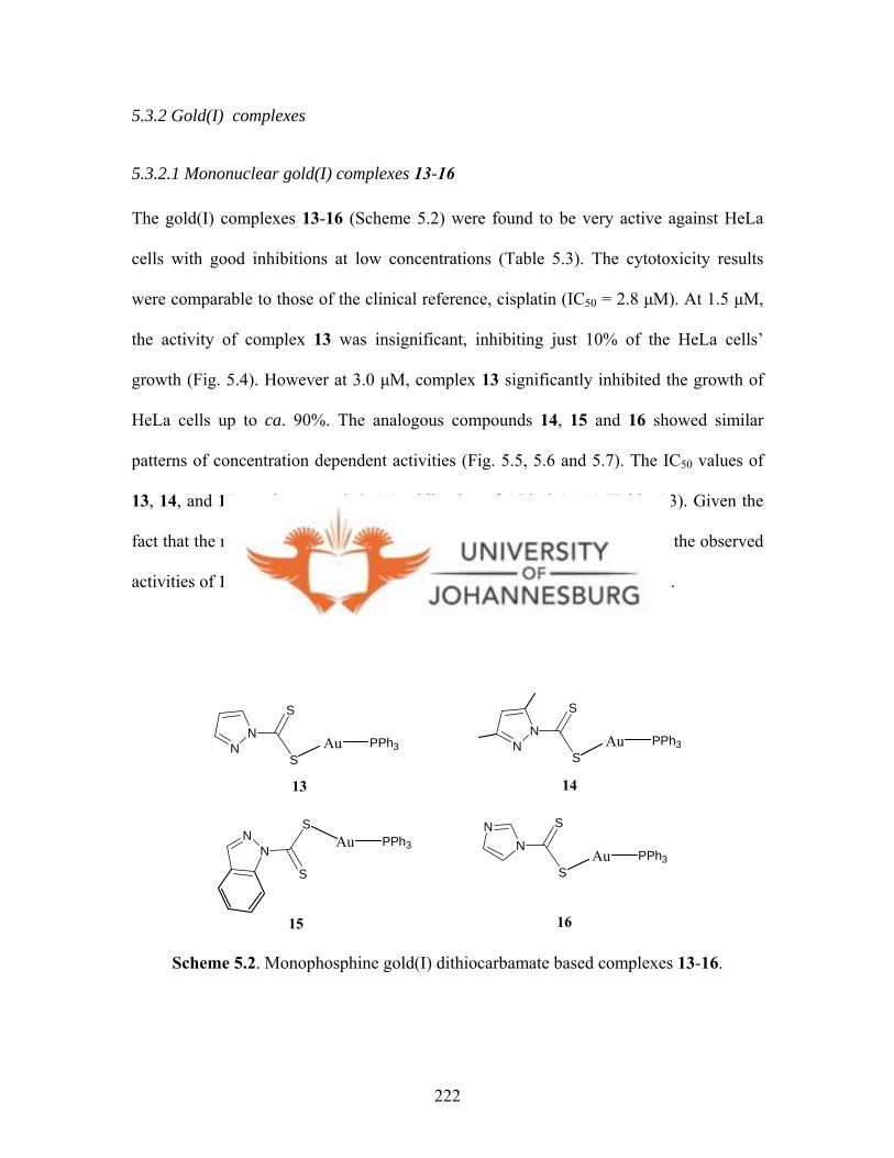
5.3.2 Gold(I) complexes
5.3.2.1 Mononuclear gold(I) complexes 13-16
The gold(I) complexes 13-16 (Scheme 5.2) were found to be very active against HeLa
cells with good inhibitions at low concentrations (Table 5.3). The cytotoxicity results
were comparable to those of the clinical reference, cisplatin (IC50 = 2.8 µM). At 1.5 µM,
the activity of complex 13 was insignificant, inhibiting just 10% of the HeLa cells’
growth (Fig. 5.4). However at 3.0 µM, complex 13 significantly inhibited the growth of
HeLa cells up to ca. 90%. The analogous compounds 14, 15 and 16 showed similar
patterns of concentration dependent activities (Fig. 5.5, 5.6 and 5.7). The IC50 values of
13, 14, and 15 are the same, 2.6 µM, whilst that of 16 is 3.1 µM (Table 5.3). Given the
fact that the respective ligands (L5-L8) were inactive, it was concluded that the observed
activities of 13-16 were due to the P-Au fragments of the gold(I) compounds.
NN
S
SAu PPh3 N
N
S
SAu PPh3
NN
S
SAu PPh3 N
N S
SAu PPh3
13 14
15 16
Scheme 5.2. Monophosphine gold(I) dithiocarbamate based complexes 13-16.
222
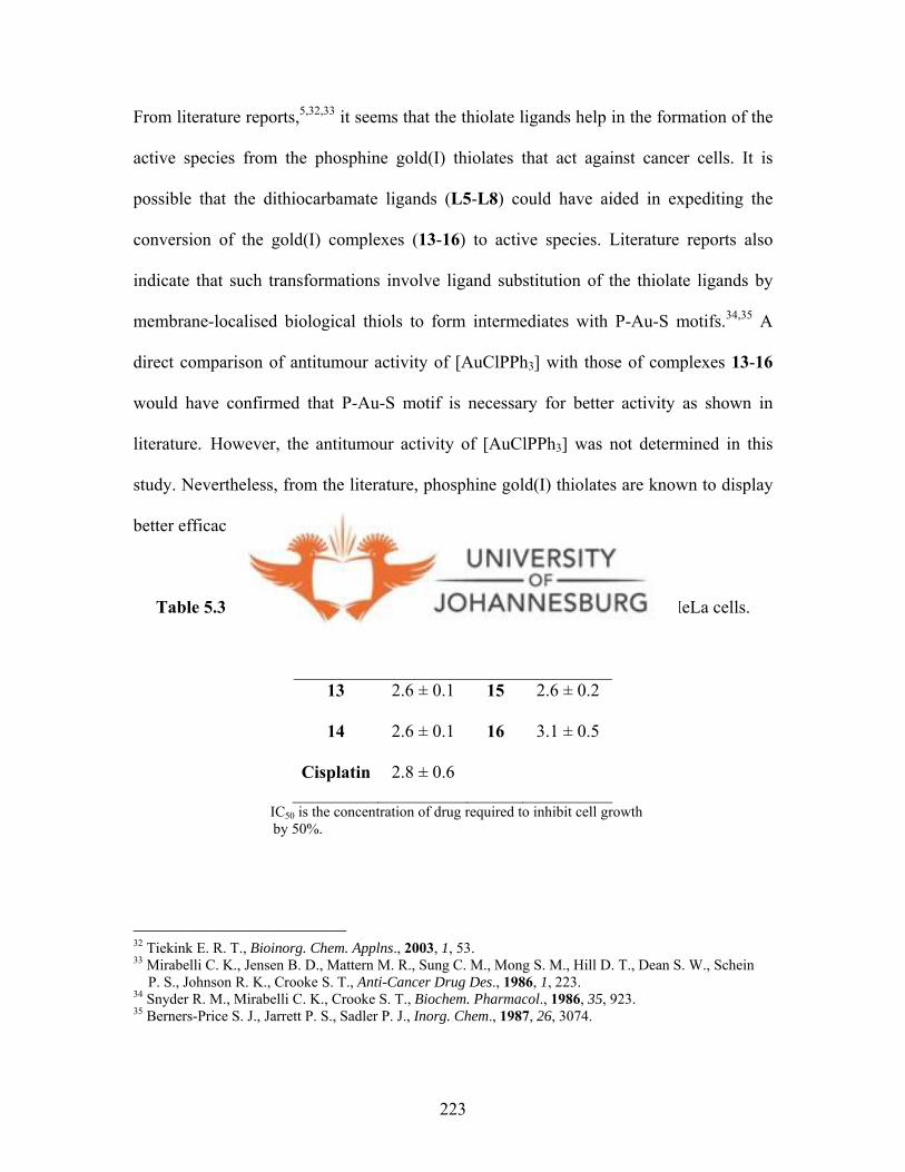
From literature reports,5, ,32 33 it seems that the thiolate ligands help in the formation of the
active species from the phosphine gold(I) thiolates that act against cancer cells. It is
possible that the dithiocarbamate ligands (L5-L8) could have aided in expediting the
conversion of the gold(I) complexes (13-16) to active species. Literature reports also
indicate that such transformations involve ligand substitution of the thiolate ligands by
membrane-localised biological thiols to form intermediates with P-Au-S motifs.34,35 A
direct comparison of antitumour activity of [AuClPPh3] with those of complexes 13-16
would have confirmed that P-Au-S motif is necessary for better activity as shown in
literature.32 However, the antitumour activity of [AuClPPh3] was not determined in this
study. Nevertheless, from the literature, phosphine gold(I) thiolates are known to display
better efficacies compared to their chloro phosphine gold(I) analogues.32
Table 5.3. Growth inhibition values of compounds 13-16 tested against HeLa cells.
Drug IC50 (µM) Drug IC50 (µM)
13 2.6 ± 0.1 15 2.6 ± 0.2
14 2.6 ± 0.1 16 3.1 ± 0.5
Cisplatin 2.8 ± 0.6
IC50 is the concentration of drug required to inhibit cell growth by 50%.
32 Tiekink E. R. T., Bioinorg. Chem. Applns., 2003, 1, 53. 33 Mirabelli C. K., Jensen B. D., Mattern M. R., Sung C. M., Mong S. M., Hill D. T., Dean S. W., Schein P. S., Johnson R. K., Crooke S. T., Anti-Cancer Drug Des., 1986, 1, 223. 34 Snyder R. M., Mirabelli C. K., Crooke S. T., Biochem. Pharmacol., 1986, 35, 923. 35 Berners-Price S. J., Jarrett P. S., Sadler P. J., Inorg. Chem., 1987, 26, 3074.
223

0 1 2 3 4 5 6 70
20
40
60
80
100
Concentration of drug (µM)
Cel
l gro
wth
(%
of
cont
rol)
Figure 5.4. Growth inhibition profile of HeLa cells by gold(I) complex 13.
0 1 2 3 4 5 6 70
20
40
60
80
100
Concentration of drug (µM)
Cel
l gro
wth
(%
of
cont
rol)
Figure 5.5. Growth inhibition profile of HeLa cells by gold(I) complex 14.
224
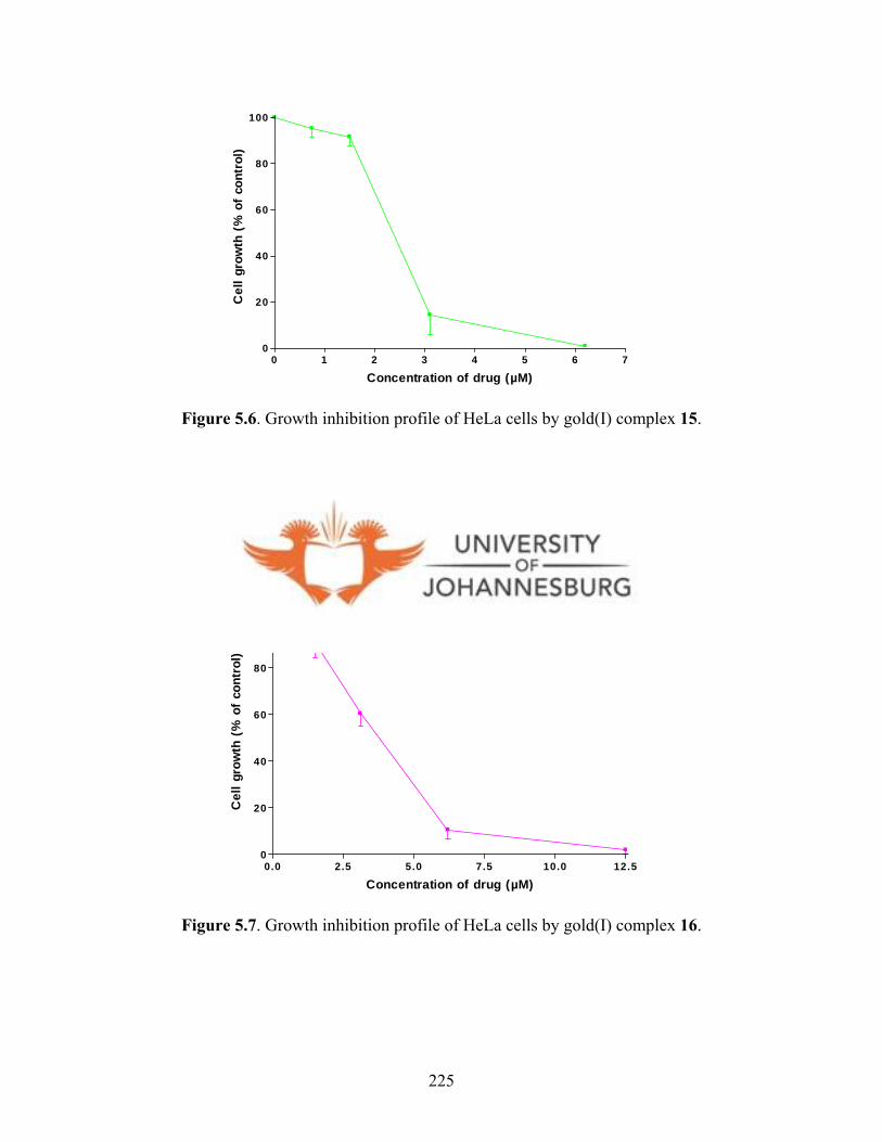
0 1 2 3 4 5 6 70
20
40
60
80
100
Concentration of drug (µM)
Cel
l gro
wth
(%
of
cont
rol)
Figure 5.6. Growth inhibition profile of HeLa cells by gold(I) complex 15.
0.0 2.5 5.0 7.5 10.0 12.50
20
40
60
80
100
Concentration of drug (µM)
Cel
l gro
wth
(%
of
cont
rol)
Figure 5.7. Growth inhibition profile of HeLa cells by gold(I) complex 16.
225

The cytotoxicities of gold(I) complexes 13-16 (Scheme 5.2) against normal cells (human
lymphocytes) were also investigated. This was one of the key objectives of the project; to
establish whether these compounds were specific to killing tumour cells and not normal
cells (tumour specificity). All the complexes displayed tumour specific activities.
Complex 13 was cytotoxic against normal cells (resting and stimulated lymphocytes)
with IC50 values 9.1 and 8.6 µM, respectively, compared to it activity against cancerous
HeLa cells (2.6 µM). The tumour specificity values (TS) were obtained using equation
5.1. However, the TS values were not significantly high and were in the range 3.5-6.1 as
given in Table 5.4.
Table 5.4. Tumour Specificity of 13-16.
Drug HeLa IC50 (µM) Lymph (resting)
IC50 (µM)
Lymph (stimulated)
IC50 (µM)
Tumour
Specificity
13 2.6 9.1 8.5 3.4
14 2.6 9.5 8.6 3.5
15 2.6 14.0 5.1 3.7
16 3.1 15.5 18.5 6.1
Cisplatin 2.8 IC50 is the concentration of drug required to inhibit cell growth by 50%.
There was no clear trend between the activities of compounds 13-16 against stimulated
and resting lymphocytes, contrary to the expectations that stimulated cells would be less
susceptible to the compounds. A case in point is compound 15 with its activities against
resting and stimulated lymphocytes being 14.0 µM and 5.1 µM, respectively. It is
possible that phytohemagglutinin-protein (PHA-P), which was used to stimulate
lymphocytes, could have made the cells more susceptible to these compounds. However,
226

it should be noted that this is an inference drawn from the observed activities and further
experiments would be necessary to ascertain this point. Overall, the increased growth rate
of stimulated cells as opposed to the resting cells did not play a significant role at all. It
was then concluded that the activities observed were independent of the cells’ growth
rate. Complex 16 was found to show the highest TS factor of 6.1, which meant 16
inhibited tumour cells six times more specifically than it inhibited normal cells. As for
complexes 13, 14 and 15, the low TS factors suggested that these compounds killed both
tumour and normal cells indiscriminately.
It is clear that gold(I) compounds 13-16 exhibit good antitumour activity against HeLa
cells. However, there was no clear trend on the dependence of the activities on the
bulkiness of the compounds, i.e. in the order of imidazole, pyrazole, 3,5-
dimethylpyrazole, indazole. It is possible that the dithiocarbamate ligands (L5-L8) are
readily substituted in solution irrespective of their bulkiness. Further stability studies of
complexes 13-16, in solution, should shed some light on this matter.
5.3.2.2 Dinuclear gold(I) complexes 20-25
Binuclear gold(I) complexes 20-25 (Scheme 5.3) were also investigated for their
antitumour activities against cancerous HeLa cells. The IC50 results are given in Table 5.5
below. Of all the six diphosphine gold(I) complexes (20-25) tested, the dpph-based
gold(I) compounds 23-25 were the most active (Scheme 5.3, Fig. 5.11-5.13) with
complex 24 showing the highest activity against HeLa cells (IC50 = 0.1 µM). In the
growth inhibition profile of 24 (Fig. 5.12), there was no significant inhibition of the cells’
227
growth (ca. 20%) at 0.05 µM, but at 0.18 µM, a 80% growth inhibition was registered. A
similar profile was observed for 23 with an IC50 of 0.4 µM. The activities of 24 (IC50 =
0.1 µM ) and 23 (IC50 = 0.4 µM) were comparable to that of the reference compound
cisplatin (IC50 = 0.5 µM). However, complex 25 was about seven times less active (IC50 =
3.7 µM) than the reference compound cisplatin. As for the dppp-based gold(I)
compounds 20-22, their IC50 values were 2.2, 7.0 and 3.4 µM for 20, 21 and 22,
respectively (Table 5.5, Fig. 5.8-5.10).
P PPhPh
PhPh
Au Au
P PPhPh
PhPh
Au AuP PPh
Ph
PhPh
Au Au
P PPh
PhPhPh
Au Au
P PPh
PhPhPh
Au Au
20 21
22 23
24 25
N NS
S
N NS
S
P PPh
PhPhPh
Au Au
N NS
S
N NS
S
N NS
S
N NS
S
N NS
SNN
SS
N NS
SNN
SS
N NS
SNN
SS
Scheme 5.3. Diphosphine gold(I) dithiocarbamate based complexes 20-25, screened for their anti-tumour activities against HeLa cells.
228

Table 5.5. Growth inhibition values of compounds 20-25 tested against HeLa cells.
dppp derivatives dpph derivatives
Drug IC50 (µM) Drug IC50 (µM)
20 2.2 ± 0.5 23 0.4 ± 0.1
21 7.0 ± 0.5 24 0.1 ± 0.01
22 3.4 ± 0.3 25 3.7 ± 0.5
Cisplatin 0.5 ± 0.1
IC50 is the concentration of drug required to inhibit cell growth by 50%.
0.0 2.5 5.0 7.5 10.0 12.50
20
40
60
80
100
Concentration of drug (µM)
Cel
l gro
wth
(%
of
cont
rol)
Figure 5.8. Growth inhibition profiles of HeLa cells by complex 20.
229
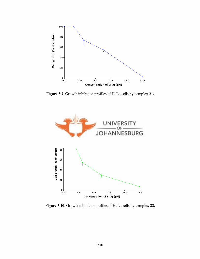
0.0 2.5 5.0 7.5 10.0 12.50
20
40
60
80
100
Concentration of drug (µM)
Cel
l gro
wth
(%
of
cont
rol)
Figure 5.9. Growth inhibition profiles of HeLa cells by complex 21.
0.0 2.5 5.0 7.5 10.0 12.50
20
40
60
80
100
Concentration of drug (µM)
Cel
l gro
wth
(%
of
cont
rol)
Figure 5.10. Growth inhibition profiles of HeLa cells by complex 22.
230

0.00 0.25 0.50 0.75 1.00 1.25 1.500
20
40
60
80
100
Concentration of drug (µM)
Cel
l gro
wth
(%
of
cont
rol)
Figure 5.11. Growth inhibition profiles of HeLa cells by complex 23.
0.0 0.1 0.2 0.3 0.40
20
40
60
80
100
Concentration of drug (µM)
Cel
l gro
wth
(%
of
cont
rol)
Figure 5.12. Growth inhibition profiles of HeLa cells by complex 24.
231
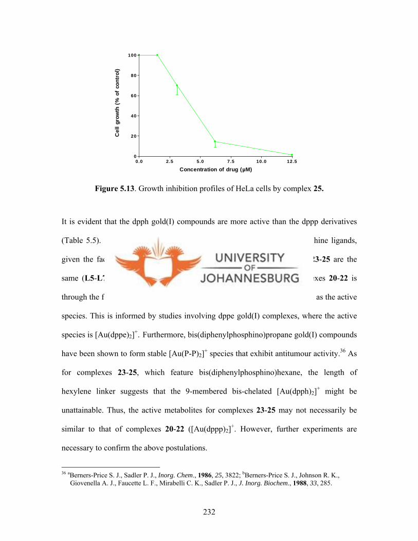
0.0 2.5 5.0 7.5 10.0 12.50
20
40
60
80
100
Concentration of drug (µM)
Cel
l gro
wth
(%
of
cont
rol)
Figure 5.13. Growth inhibition profiles of HeLa cells by complex 25.
It is evident that the dpph gold(I) compounds are more active than the dppp derivatives
(Table 5.5). This observation is attributed to the difference in the diphosphine ligands,
given the fact that the dithiocarbamate ligands of complexes 20-22 and 23-25 are the
same (L5-L7) and were inactive. It is possible that the action of complexes 20-22 is
through the formation of 6-membered bis-chelated compound, [Au(dppp)2]+, as the active
species. This is informed by studies involving dppe gold(I) complexes, where the active
species is [Au(dppe)2]+.3 5 Furthermore, bis(diphenylphosphino)propane gold(I) compounds
have been shown to form stable [Au(P-P)2]+ species that exhibit antitumour activity.36 As
for complexes 23-25, which feature bis(diphenylphosphino)hexane, the length of
hexylene linker suggests that the 9-membered bis-chelated [Au(dpph)2]+ might be
unattainable. Thus, the active metabolites for complexes 23-25 may not necessarily be
similar to that of complexes 20-22 ([Au(dppp)2]+. However, further experiments are
necessary to confirm the above postulations.
36 aBerners-Price S. J., Sadler P. J., Inorg. Chem., 1986, 25, 3822; bBerners-Price S. J., Johnson R. K., Giovenella A. J., Faucette L. F., Mirabelli C. K., Sadler P. J., J. Inorg. Biochem., 1988, 33, 285.
232

The tumour specificity of complexes 20-25 was also investigated. From the results
obtained, dppp derived complexes (20-22) not only killed HeLa cells but also killed
normal cells at comparable concentrations. For example, compound 21 killed HeLa cells
at 7 µM and resting lymphocytes at 6.1 µM. It is noteworthy that the same complex had
an activity of IC50 = 2.9 µM against stimulated lymphocytes. There was no specific trend
derived from these results apart from the fact that the IC50 values registered for
compounds 20-22 against stimulated lymphocytes were generally lower compared to the
respective IC50 values against resting lymphocytes (Table 5.6). Similar observations were
made for complexes 23-25. Notably, the registered IC50 values for 23-25 against
stimulated lymphocytes are significantly lower than the registered IC50 values against
resting lymphocytes (Table 5.6). For instance, the activity of 24 against stimulated
lymphocytes (1.4 µM) is thirteen times higher than its activity against resting
lymphocytes (18.2 µM). At the moment, there are no facts to explain this phenomenon.
Table 5.6. Tumour Specificity of complexes 20-25.
Drug HeLa IC50 (µM) Lymph (resting)
IC50 (µM)
Lymph (stimulated)
IC50 (µM)
Tumour
Specificity
dppp derivatives
20 2.2 7.1 5.0 2.8
21 7.0 6.1 2.9 0.6
22 3.4 18.5 8.2 3.8
dpph derivatives
23 0.4 18.9 6.7 31.0
24 0.1 18.2 1.4 70.5
25 3.7 37.7 30.3 9.2
Cisplatin 0.5 IC50 is the concentration of drug required to inhibit cell growth by 50%.
233
Similar to 13-16, it is possible that the stimulant used, PHA-P, could have had an
influence in the activities observed. This is a mere postulation at the moment and
mechanistic studies involving reaction of PHA-P and compounds 20-25 would be
necessary to confirm or negate this argument.
Tumour specificity (TS) factors were also calculated for complexes 20-25 using equation
5.1 (cf. sec. 5.5.1). The results are presented in Table 5.6 above. In this batch of
compounds, complexes 23 and 24 were found to be more specific in inhibiting the growth
of tumour cells than in normal cells, with TS factors being 31.0 and 70.5, respectively.
The significance of this result is that 23 and 24 are very specific to inhibiting HeLa
cancer cells and not normal cells.
While the analogous dppe gold(I) complexes 17a-19 were synthesised, their biological
activities were, however, not evaluated. This was because of the observed self-
aggregation of the dinuclear complex 17a in solution to form the cluster 17b as reported
in chapter 4.
5.3.3. Platinum(II) complexes 31-33
The dithiocarbamato platinum(II) complexes 31-33 (Scheme 5.4) were also investigated
for their antitumour activities. Compounds 32 and 33 showed insignificant inhibition
activities towards HeLa cells, whilst 31 was not tested because of its limited solubility.
The IC50 values of 32 and 33 were greater than 50 µM.
234
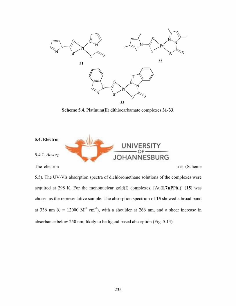
NN
S
S
31 32
33
NN
SS
Pt NN
S
S
NN
SS
Pt
NN
S
S
NN
SS
Pt
Scheme 5.4. Platinum(II) dithiocarbamate complexes 31-33.
5.4. Electronic absorption and photoluminescence results
5.4.1. Absorption spectra
The electronic absorption measurements were performed for four complexes (Scheme
5.5). The UV-Vis absorption spectra of dichloromethane solutions of the complexes were
acquired at 298 K. For the mononuclear gold(I) complexes, [Au(L7)(PPh3)] (15) was
chosen as the representative sample. The absorption spectrum of 15 showed a broad band
at 336 nm (є = 12000 M-1 cm-1), with a shoulder at 266 nm, and a sheer increase in
absorbance below 250 nm; likely to be ligand based absorption (Fig. 5.14).
235
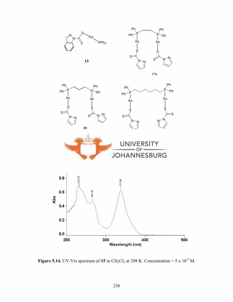
PPPh
Ph
Ph
Ph
N NS
S
NN
SS
P PPh
PhPh
Ph
N NS
S
N NS
S
2023
NN
S
SAu
PPh3
15
P PPh
PhPh
Ph
N NS
S
N NS
S
17a
Au Au
Au Au Au Au
Scheme 5.5. Gold(I) complexes 15, 17a, 20 and 23.
Figure 5.14. UV-Vis spectrum of 15 in CH2Cl2 at 298 K. Concentration = 5 x 10⁻⁵ M.
236
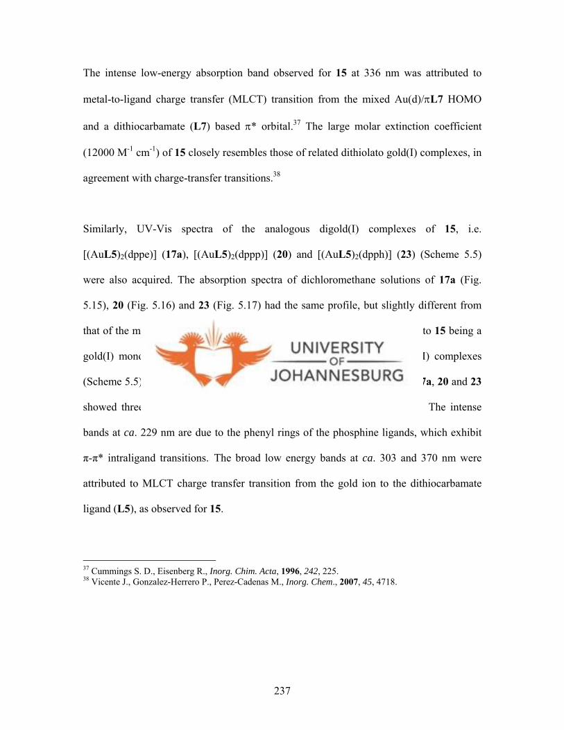
The intense low-energy absorption band observed for 15 at 336 nm was attributed to
metal-to-ligand charge transfer (MLCT) transition from the mixed Au(d)/πL7 HOMO
and a dithiocarbamate (L7) based π* orbital.37 The large molar extinction coefficient
(12000 M-1 cm-1) of 15 closely resembles those of related dithiolato gold(I) complexes, in
agreement with charge-transfer transitions.38
Similarly, UV-Vis spectra of the analogous digold(I) complexes of 15, i.e.
[(AuL5)2(dppe)] (17a), [(AuL5)2(dppp)] (20) and [(AuL5)2(dpph)] (23) (Scheme 5.5)
were also acquired. The absorption spectra of dichloromethane solutions of 17a (Fig.
5.15), 20 (Fig. 5.16) and 23 (Fig. 5.17) had the same profile, but slightly different from
that of the mononuclear gold(I) 15 (Fig. 5.14). This difference is attributed to 15 being a
gold(I) mononuclear complex, while 17a, 20 and 23 are dinuclear gold(I) complexes
(Scheme 5.5). As depicted in the respective Figures 5.15-5.17, complexes 17a, 20 and 23
showed three absorption bands at ca. 229, 303 and 370 nm, respectively. The intense
bands at ca. 229 nm are due to the phenyl rings of the phosphine ligands, which exhibit
π-π* intraligand transitions.18 The broad low energy bands at ca. 303 and 370 nm were
attributed to MLCT charge transfer transition from the gold ion to the dithiocarbamate
ligand (L5), as observed for 15.
37 Cummings S. D., Eisenberg R., Inorg. Chim. Acta, 1996, 242, 225. 38 Vicente J., Gonzalez-Herrero P., Perez-Cadenas M., Inorg. Chem., 2007, 45, 4718.
237
Figure 5.15. UV-Vis spectrum of 17a in CH2Cl2 at 298 K. [17a] = 5 x 10⁻⁵ M.
Figure 5.16. UV-Vis spectrum of 20 in CH2Cl2 at 298 K. [20] = 5 x 10⁻⁵ M.
238
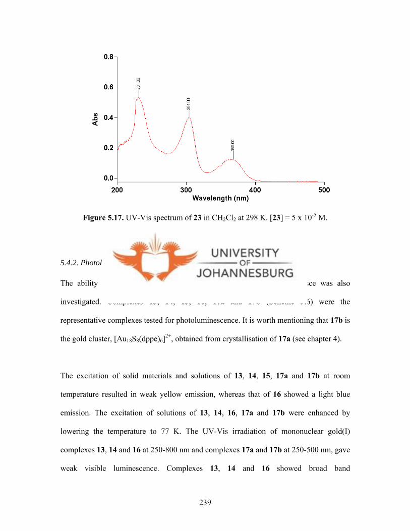
Figure 5.17. UV-Vis spectrum of 23 in CH2Cl2 at 298 K. [23] = 5 x 10⁻⁵ M.
5.4.2. Photoluminescence spectra
The ability of the gold(I) complexes reported in this study to luminesce was also
investigated. Complexes 13, 14, 15, 16, 17a and 17b (Scheme 5.6) were the
representative complexes tested for photoluminescence. It is worth mentioning that 17b is
the gold cluster, [Au18S8(dppe)6]2+, obtained from crystallisation of 17a (see chapter 4).
The excitation of solid materials and solutions of 13, 14, 15, 17a and 17b at room
temperature resulted in weak yellow emission, whereas that of 16 showed a light blue
emission. The excitation of solutions of 13, 14, 16, 17a and 17b were enhanced by
lowering the temperature to 77 K. The UV-Vis irradiation of mononuclear gold(I)
complexes 13, 14 and 16 at 250-800 nm and complexes 17a and 17b at 250-500 nm, gave
weak visible luminescence. Complexes 13, 14 and 16 showed broad band
239
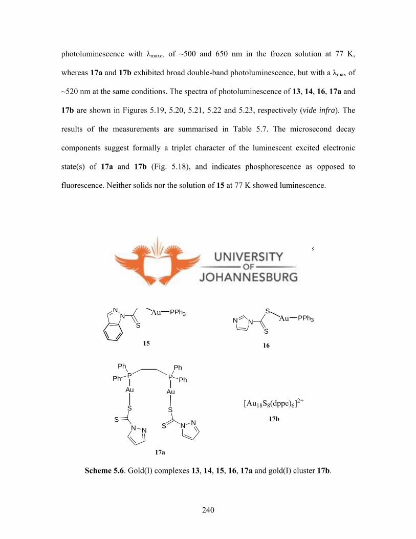
photoluminescence with λmaxes of ~500 and 650 nm in the frozen solution at 77 K,
whereas 17a and 17b exhibited broad double-band photoluminescence, but with a λmax of
~520 nm at the same conditions. The spectra of photoluminescence of 13, 14, 16, 17a and
17b are shown in Figures 5.19, 5.20, 5.21, 5.22 and 5.23, respectively (vide infra). The
results of the measurements are summarised in Table 5.7. The microsecond decay
components suggest formally a triplet character of the luminescent excited electronic
state(s) of 17a and 17b (Fig. 5.18), and indicates phosphorescence as opposed to
fluorescence. Neither solids nor the solution of 15 at 77 K showed luminescence.
240
NN
Scheme 5.6. Gold(I) complexes 13, 14, 15, 16, 17a and gold(I) cluster 17b.
S
SAu PPh3
15
P PPh
PhPh
Ph
N NS
S
N NS
S
17a
NN
S
SAu PPh3
Au Au
NN
S
SAu PPh3
N NS
SAu PPh3
13 14
16
[Au18S8(dppe)6]2+
17b
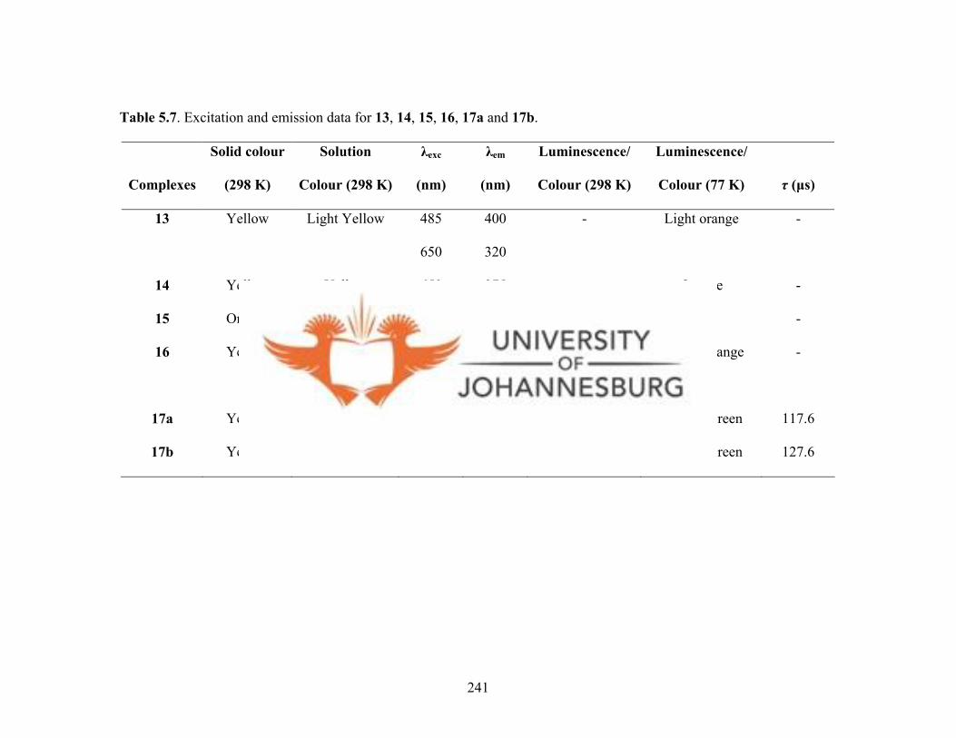
Complexes
Solid colour
(298 K)
Solution
Colour (298 K)
λexc
(nm)
λem
(nm)
Luminescence/
Colour (298 K)
Luminescence/
Colour (77 K)
τ (µs)
13 Yellow Light Yellow 485
650
400
320
- Light orange -
14 Yellow
Yellow 650 375 - Orange -
15 Orange Orange - - - No -
16 Yellow Light Yellow 480
650
320
-
Light blue Intense orange
-
17a Yellow Yellow 470 520 - Yellow/Green 117.6
17b Yellow Yellow 471 522 - Yellow/Green 127.6
Table 5.7. Excitation and emission data for 13, 14, 15, 16, 17a and 17b.
241
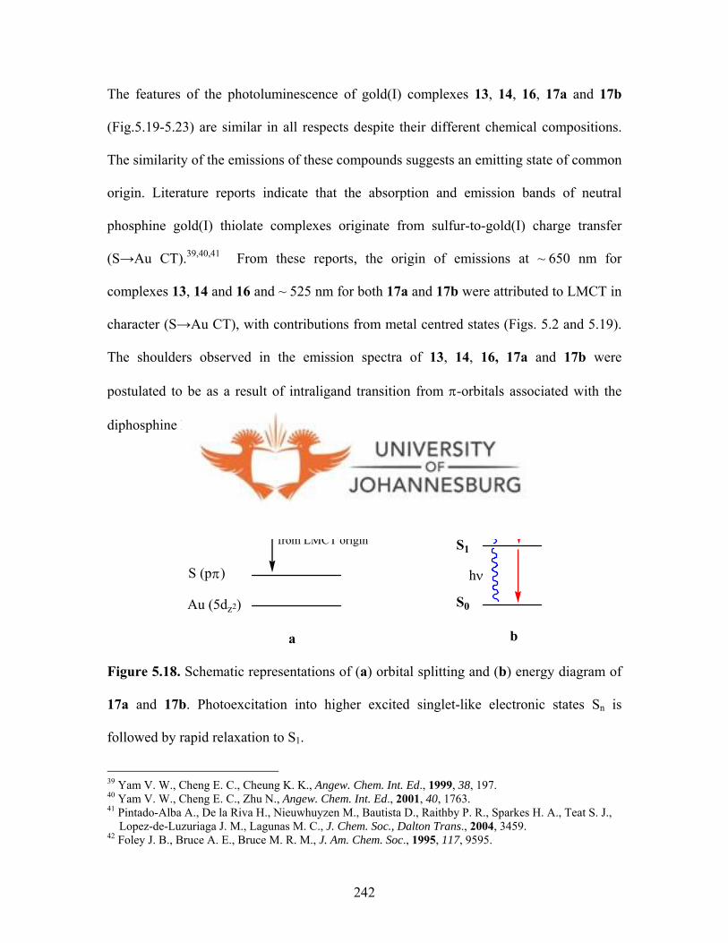
242
The features of the photoluminescence of gold(I) complexes 13, 14, 16, 17a and 17b
(Fig.5.19-5.23) are similar in all respects despite their different chemical compositions.
The similarity of the emissions of these compounds suggests an emitting state of common
origin. Literature reports indicate that the absorption and emission bands of neutral
phosphine gold(I) thiolate complexes originate from sulfur-to-gold(I) charge transfer
(S→Au CT).39, ,40 41 From these reports, the origin of emissions at ~ 650 nm for
complexes 13, 14 and 16 and ~ 525 nm for both 17a and 17b were attributed to LMCT in
character (S→Au CT), with contributions from metal centred states (Figs. 5.2 and 5.19).
The shoulders observed in the emission spectra of 13, 14, 16, 17a and 17b were
postulated to be as a result of intraligand transition from π-orbitals associated with the
diphosphine ligands.16,42
Au (6pz)
S (pπ)
Au (5dz2)
Emission derived from LMCT origin
S0
S1
Sn
h
a
ν
b
Figure 5.18. Schematic representations of (a) orbital splitting and (b) energy diagram of
17a and 17b. Photoexcitation into higher excited singlet-like electronic states Sn is
followed by rapid relaxation to S1.
39 Yam V. W., Cheng E. C., Cheung K. K., Angew. Chem. Int. Ed., 1999, 38, 197. 40 Yam V. W., Cheng E. C., Zhu N., Angew. Chem. Int. Ed., 2001, 40, 1763. 41 Pintado-Alba A., De la Riva H., Nieuwhuyzen M., Bautista D., Raithby P. R., Sparkes H. A., Teat S. J., Lopez-de-Luzuriaga J. M., Lagunas M. C., J. Chem. Soc., Dalton Trans., 2004, 3459. 42 Foley J. B., Bruce A. E., Bruce M. R. M., J. Am. Chem. Soc., 1995, 117, 9595.
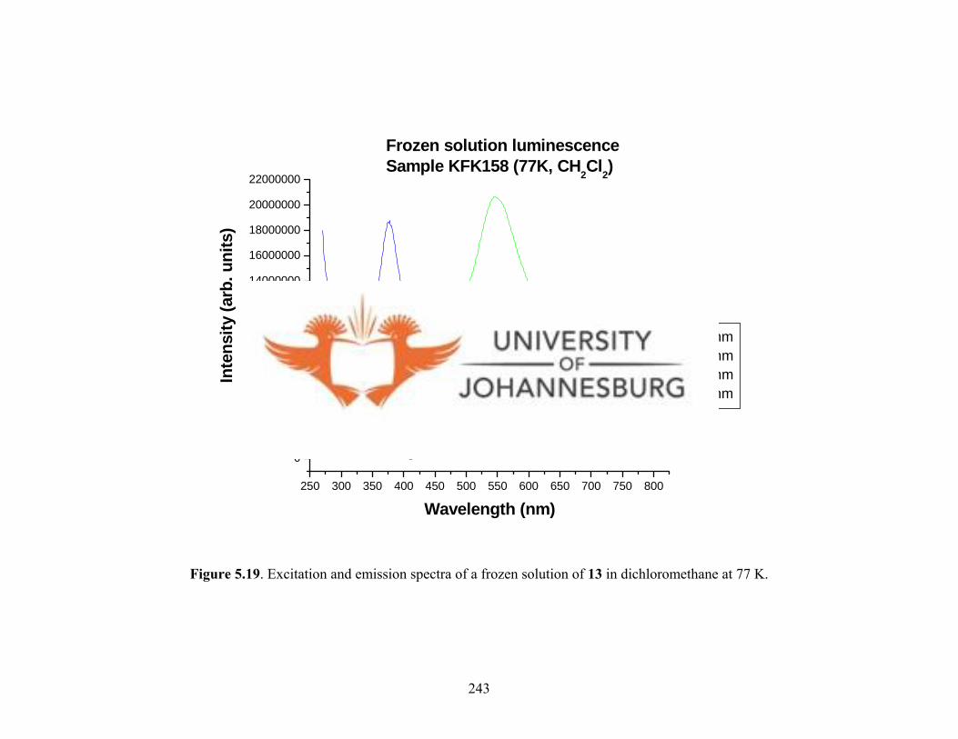
250 300 350 400 450 500 550 600 650 700 750 800
0
2000000
4000000
6000000
8000000
10000000
12000000
14000000
16000000
18000000
20000000
22000000
Frozen solution luminescenceSample KFK158 (77K, CH2Cl2)
Inte
nsity
(arb
. uni
ts)
Wavelength (nm)
λexc = 650 nm λemi = 320 nm λemi = 400 nm λexc = 485 nm
Figure 5.19. Excitation and emission spectra of a frozen solution of 13 in dichloromethane at 77 K.
243

250 300 350 400 450 500 550 600 650 700 750
0
1000000
2000000
3000000
4000000
5000000Frozen solution luminescenceSample KFK121 (77K, CH2Cl2)
Inte
nsity
(arb
. uni
ts)
Wavelength (nm)
λexc = 650 nm λemm = 375 nm
Figure 5.20. Excitation and emission spectra of a frozen solution of 14 in dichloromethane at 77 K.
244

300 400 500 600 700 800
-500000
0
500000
1000000
1500000
2000000
2500000
3000000
3500000
4000000
4500000
Frozen solution luminescenceSample KFK188 (77K, CH2Cl2)
Inte
nsity
(arb
. uni
ts)
Wavelength (nm)
λexc = 650 nm λemm = 320 nm λexc = 480 nm
Figure 5.21. Excitation and emission spectra of a frozen solution of 16 in dichloromethane at 77 K.
245

2 0 0 2 5 0 3 0 0 3 5 0 4 0 0 4 5 0 5 0 0 5 5 0 6 0 0 6 5 0 7 0 0 7 5 0
0
5 0 0 0 0 0
1 0 0 0 0 0 0
1 5 0 0 0 0 0
2 0 0 0 0 0 0
2 5 0 0 0 0 0
3 0 0 0 0 0 0
3 5 0 0 0 0 0
4 0 0 0 0 0 0
Frozen solu tion lum inescence Sam ple K FK 169 (77K , C H 2C l2)
λ exc= 4 7 0 n m λ em m = 5 2 0 n m
Inte
nsity
(arb
. uni
ts)
W avelen gth (n m )
Figure 5.22. Excitation and emission spectra of a frozen solution of 17a in dichloromethane at 77 K.
246
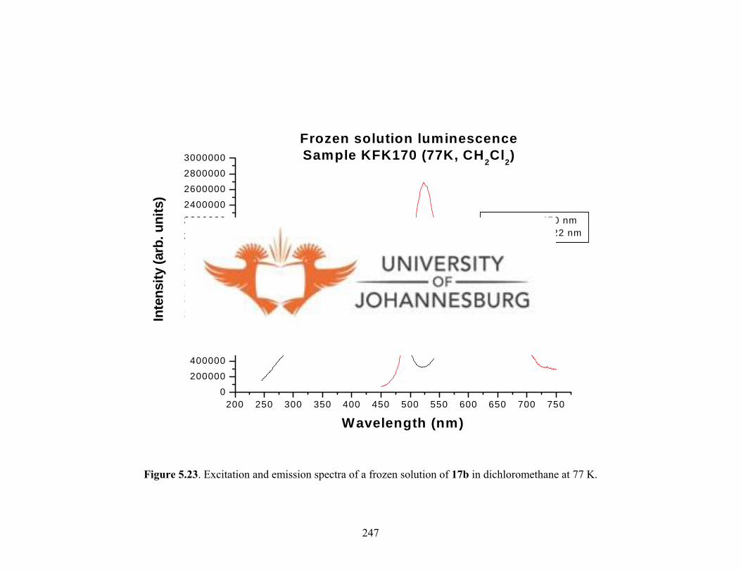
200 250 300 350 400 450 500 550 600 650 700 7500
200000
400000
600000800000
1000000
1200000
1400000
1600000
1800000
2000000
22000002400000
2600000
2800000
3000000
Frozen solution luminescenceSample KFK170 (77K, CH2Cl2)
Inte
nsity
(arb
. uni
ts)
Wavelength (nm)
λexc = 470 nm λemm = 522 nm
Figure 5.23. Excitation and emission spectra of a frozen solution of 17b in dichloromethane at 77 K.
247

5.5. Conclusions
The biological screening of ligands L5-L11, gold(I) complexes 13-16, 20-25 and
platinum(II) complexes 31-33 for their in vitro anticancer activity were performed on
HeLa cells. Compounds 31-33 were inactive. Whereas ligands L5-L8 and L11 showed
no inhibition against HeLa cells, L9 and L10 showed activity, with L9 being the most
active (IC50 = 3.7 ± 0.2 µM, TS = 9.6). It is seems that L9 exert it activity in the same
way as disulfram, by reducing in vivo to pyrazolyldithiocabarmic acid species, which
would react with several enzymes and cause apoptosis. But judging from the various
mechanisms of action reported for disulfram, various mechanistic studies involving L9
are needed to confirm its exact mechanism of action.
The obtained biological data of the gold(I) complexes indicated their appreciable
activities against HeLa cells. Mononuclear gold(I) complexes 13-16 had good antitumour
activities. It was worth mentioning that the activities for 13, 14 and 15 were the same
(IC50 ≈ 2.6 µM). This suggests that the ligands used (L5-L7) are readily substituted in
solution by other biological thiols and thus their bulkiness do not necessarily contribute
in regulating the metabolic rates for conversion of these complexes to active species.
Similarly, the dinuclear gold(I) complexes 20-25 showed appreciable growth inhibitions
against HeLa cells. The dpph based gold compounds 23-25 were the most active, with 24
showing the highest activity against HeLa cells (IC50 = 0.1 ± 0.01 µM). The difference in
activities of complexes 20-22 and 23-25 was attributed to the nature of diphosphine
ligands. While it is necessary to perform further experiments to establish the active
metabolites of 20-25, it is possible to postulate what the active metabolites could be
248

based on literature reports (in chapter 4); that the dppp-based complexes 20-22 would
easily form the possible 6-membered bis-chelated active species, [Au(dppp)2]+; but the
formation of a 9-membered bis-chelated [Au(dpph)2]+ species from dpph-based
compounds 23-25, is highly unlikely. Thus, it appears that the above structural
postulation could be the basis for the difference in the anticancer activities of 20-22 and
23-25.
By testing the potency of gold(I) complexes 13-16, 20-22 and 23-25 on normal cells
(resting and stimulated lymphocytes), the tumour specificity values (TS) were obtained.
Complexes 23 and 24 had the highest TS factors, 31.0 and 70.5, respectively. This means
these two complexes were more specific in inhibiting the growth of tumour cells and not
normal cells.
Two fundamental observations were made from experiments involving normal cells
(resting and stimulated lymphocytes) namely; (i) the growth rate of cells did not hamper
the antitumour activities of these complexes. This is evident from the registered activities
of these gold(I) complexes against stimulated and resting lymphocytes, where there was a
general susceptibility of stimulated lymphocytes to the test compounds compared to un-
stimulated (resting) lymphocytes. This negated the anticipation that stimulated
lymphocytes should have been less susceptible to the gold(I) compounds. (ii) It is
possible that the PHA-P, used to stimulate the growth of normal cells, had an influence
on the observed activities of the gold(I) complexes. It is proposed that PHA-P may have
249
made the cells more susceptible to the compounds. Further studies are needed to probe
this possibility and forms part of the future work.
Electronic absorption measurements were performed in four gold(I) complexes 15, 17a,
20 and 23. The intense lowest-energy absorption band observed for these complexes at
ca. 304, 338 and 368 nm were attributed to the metal-to-ligand charge transfer (MLCT)
transition. The intense bands at ca. 229 nm are due to the phenyl rings of the phosphine
ligands, which feature π intraligand interactions. The broad low energy bands at ca. 303
and 370 nm are attributed to charge transfer transition from gold to the dithiocarbamate
ligand (L5). The excitation of solid materials and solutions of 13, 14, 15, 17a and 17b at
room temperature resulted in weak yellow emission, whereas that of 16 showed a light
blue emission. Generally, the visible luminescence of complexes 13, 14, 16, 17a and 17b
were weak characterised by broad bands photoluminescence. The double-band
photoluminescence for 17a and 17b suggests that the LMCT emissions observed is
influenced by gold-gold interactions (aurophilicity). Overall, the emissions of the
complexes are proposed to originate from a MLCT triplet state, indicating
phosphorescence.
250

APPENDIX
251
Bis(pyrazolyl) Palladium(II), Platinum(II) and Gold(III) Complexes: Syntheses,
Molecular Structures and Substitution Reactions with L-cysteine.
Frankline K. Ketera, Stephen O. Ojwacha, Olayinka A. Oyetunjib, Ilia A. Guzeic
James Darkwaa
aDepartment of Chemistry, University of Johannesburg, P.O. Box 524 Auckland Park
2006, South Africa
bDepartment of Chemistry, University of Botswana, Private Bag UB 00704, Gaborone,
Botswana
cDepartment of Chemistry, University of Wisconsin-Madison, 1101 University Avenue,
Madison, WI 53706, USA.
(Submitted to Inorganica Chimica Acta)
1
Abstract
The reactivity of L-cysteine with pyrazolyl gold(III), platinum(II) and palladium(II)
complexes, [AuCl2(3,5-R2bpza)]Cl {R = H (1)}, [PtCl2(3,5-R2bpza)] {R = H (2)},
[PdCl2(3,5-R2bpza)] {R = H (3), R = Me (4)} (bpza = bis-pyrazolyl acetic acid), was
investigated at 20 – 40 oC in an aqueous medium with an ionic strength of 0.1 M,
between pH 2.92 and 3.72, using conventional UV-Vis and stopped-flow
spectrophotometry. The reactions were performed with the pre-activated species,
[Au(OH2)2(3,5-R2bpza)]Cl {R = H (5)}, [Pt(OH2)2(3,5-R2bpza)] {R = H (6)} and
[Pd(OH2)2(3,5-R2bpza)] {R = H (7), R = Me (8)} and L-cysteine. These substitution
reactions followed the second-order rate law d[Mn+]/dt = k[Cys][Mn+] (n = 2, 3), where k
is a pH-independent rate constant. The entropies of activation obtained are largely
negative (-52.7 and -174 J mol-1K-1), except for 8, which is +18.4 kJ mol-1. Further
reduction of the gold(III) complex (1) to gold(I) species was observed with an entropy
change of -183 J mol-1K-1. The substitution reactions take place in two steps, involving
two incoming cysteine molecules in a sequential manner, with a rapid process second
step. The reactivity of complexes 5 – 8 towards L-cysteine, under pseudo-first-order
conditions, showed that reactions of L-cysteine with 7 and 8 are 103 and 106 times faster
than those of 5 and 6 respectively. Mass spectrometry was used to confirm reaction
product of 8 with L-cysteine and found to be [Pd(Cys)2(3,5-Me2bpza)].
Key words: Kinetics and mechanism; Substitution; L-cysteine; Palladium; Platinum;
Gold; Complexes.
2

Dichloro-bis(3,5-dimethylpyrazolyl)Palladium(II) complex. A DMSO-d6 solvated
complex
Frankline K. Keter, Bernard O. Omondi, James Darkwa
aDepartment of Chemistry, University of Johannesburg, P.O. Box 524 Auckland Park
2006, South Africa
(Submitted to Journal of Molecular Structures)
3