E ciency of lactoferrin to eradicate multidrug resistant ...
Upload
independentCategory
view
0download
0
Biomaterials 25 (2004) 3029–3040
ARTICLE IN PRESS
*Correspondin
G51 2DH, Scotla
3730.
E-mail addres
0142-9612/$ - see
doi:10.1016/j.bio
Lactoferrin and ceruloplasmin derivatized superparamagnetic ironoxide nanoparticles for targeting cell surface receptors
Ajay Kumar Gupta*, Adam S.G. Curtis
Centre for Cell Engineering, Institute of Biomedical and Life Sciences, Joseph Black Building, University of Glasgow,
Glasgow G12 8QQ, Scotland, United Kingdom
Received 11 August 2003; accepted 22 September 2003
Abstract
Tissue and cell-specific drug targeting can be achieved by employing nanoparticle coatings or carrier-drug conjugates that contain
a ligand recognized by a receptor on the target cell. Superparamagnetic iron oxide nanoparticles have been used for many years in
various biomedical applications. In this study, superparamagnetic nanoparticles with specific shape and size have been prepared and
coupled to various proteins. These particles are characterized in vitro and their influence on human dermal fibroblasts is assessed in
terms of cell adhesion, viability, morphology and cytoskeleton organization using various techniques to observe cell–nanoparticle
interaction, including light, fluorescence, scanning and transmission electron microscopy. The results showed that each nanoparticle
type with different surface characteristics caused a distinctly different cell response. The underivatized magnetic particles were
internalized by the fibroblasts probably due to endocytosis, which resulted in disruption of the cell membrane and disorganized cell
cytoskeleton. In contradiction, lactoferrin or ceruloplasmin coated nanoparticles attached to the cell membrane, most likely to the
cell expressed receptors and were not endocytosed. One major problem with uncoated magnetic nanoparticles has been the
endocytosis of particles leading to irreversible entry. These experiments provide a route to prevent this problem, suggesting that cell
response can be directed via specifically engineered particle surfaces.
r 2003 Elsevier Ltd. All rights reserved.
Keywords: Magnetic nanoparticle; Surface modification; Cell adhesion; TEM; Cytotoxicity
1. Introduction
Superparamagnetic iron oxide nanoparticles withtailored surface chemistry have been widely usedexperimentally for numerous in vivo applications suchas magnetic resonance imaging contrast enhancement,tissue repair, immunoassay, detoxification of biologicalfluids, hyperthermia, drug delivery and in cell separa-tion, etc. [1–4]. All these biomedical and bioengineeringapplications require that these nanoparticles have highmagnetization values and size smaller than 20 nm withoverall narrow particle size distribution so that theparticles have uniform physical and chemical properties.In addition, these applications need special surfacecoating of the magnetic particles, which has to be not
g author. 3/2, 15 Southcroft Street, Govan, Glasgow
nd, UK. Tel.: +44-141-425-1938; fax: +44-141-330-
s: [email protected] (A.K. Gupta).
front matter r 2003 Elsevier Ltd. All rights reserved.
materials.2003.09.095
only non-toxic and biocompatible but also allow atargetable delivery with particle localization in a specificarea [5].
Cell labelling with ferro/paramagnetic substances isan increasingly common method for in vivo cellseparation [6] as the labelled cells can be detected bymagnetic resonance imaging [7]. Most current labellingtechniques utilize either of two approaches: (a) attachingmagnetic particles to the cell surface [8] or (b)internalizing biocompatible magnetic particles by fluidphase endocytosis [9], receptor mediated endocytosis[10] or phagocytosis [11]. One strategy to realize efficientand specific cell labelling of magnetic particles is tomodify the nanoparticle surface with a ligand that isefficiently taken up by target cells via receptor-mediatedendocytosis [10]. A variety of potential ligands havebeen conjugated to nanoparticle surfaces to facilitatereceptor-mediated endocytosis of the particles, includingmonoclonal antibodies (Mabs) [11]. Targeting agentssuch as transferrin, albumin, insulin and growth factors

ARTICLE IN PRESSA.K. Gupta, A.S.G. Curtis / Biomaterials 25 (2004) 3029–30403030
etc. have been demonstrated to preferentially target cellsurface, because the receptors for these ligands arefrequently overexpressed on the surface of mammaliancells [12–13]. These receptors are not only cellularmarkers, but also have been shown to efficientlyinternalize molecules coupled to these receptors [12].Furthermore, many of these ligands are stable, andgenerally poorly immunogenic. Despite the well-knownability of these receptors to facilitate internalization ofnanoparticles, little effort has been made on delivery ofmagnetic nanoparticles modified with such ligands.
Lactoferrin (Lf) is an iron binding milk glycoprotein(Mw ¼ 90 kDa) occurring naturally in numerous bodysecretions including milk, tears, mucus blood and saliva[14]. It is structurally similar to transferrin, the plasmairon protein; but lactoferrin has a much higher affinityfor iron (250-fold). Lf acts as an anti-infective agent, amodulator of the inflammatory response and ironabsorption and an immuno-regulatory protein [14]. Onthe other hand, ceruloplasmin (Cp) is a 135 kDa protein,the principal carrier of copper in plasma, which plays animportant role in iron homeostasis and is also aneffective anti-oxidant for a variety of free radicals [15].
Superparamagnetic iron oxide nanoparticles of nar-row size ranges are easily produced and coupled toproteins, providing convenient, readily targetable mag-netic resonance imaging agents. In this study, magneticiron oxide nanoparticles of specific shape and size withtailored surface chemistry (coated with lactoferrin andceruloplasmin) have been prepared and characterized byvarious physicochemical means in vitro for exactdelivery of drugs to target tissues. The influence of thesenanoparticles on human dermal fibroblasts in vitro hasbeen assessed, as compared to those underivatizedparticles, in terms of cell adhesion, cytotoxicity, lightmicroscopy, scanning electron microscopy (SEM),transmission electron microscopy (TEM) and fluores-cent observation of cytoskeleton (F-actin and tubulin).From the results, we observed that lactoferrin orceruloplasmin derivatized superparamagnetic iron oxidenanoparticles are targeted at the surface of the cells withlittle effect on the cytoskeleton of the cells and thus mayserve as a non-toxic and improved way of drugtargeting. These nanoparticles also may serve as reagentfor magnetic resonance imaging of cell trafficking and/or magnetic separation of in vivo homed cells.
2. Materials and methods
2.1. Materials
All the chemicals were of reagent grade and were usedwithout further purification. Ferric chloride hexahy-drate (FeCl3 � 6H2O>99%), ferrous chloride tetrahy-drate (FeCl2 � 4H2O), 1-ethyl-3-(3-dimethylaminopropyl)-
carbodiimide (EDCI), lactoferrin (from human), cer-uloplasmin, (3-(4,5-dimethylthiazol-2-yl)-2,5-diphenyl-tetrazolium bromide) MTT, and sodium oleate(C18H33NaO2>99%) were obtained from Sigma, UKwhile sodium hydroxide (NaOH>99%) and hydrochlo-ric acid (HCl>37% v/v) were obtained from Fluka,U.K. Double distilled water was used for all theexperiments.
2.2. Synthesis of magnetic nanoparticles
The ferric and ferrous salts (molar ratio 2:1) weredissolved in deoxygenated water at a concentration of0.1m of iron ions. Chemical precipitation was achievedby using a 1m deoxygenated solution of sodiumhydroxide. The reaction was carried out in nitrogenatmosphere at low temperature (4–6�C) with vigorousstirring. Particles were washed by dialysis using 12 kDacut off dialysis membrane against double distilled waterto remove unreacted water salts. They were thenprecipitated with acetone and dried in vacuum oven at70–80�C. The surface of the particles was neutralizedwith 0.01m HCl. Particles were coated with sodiumoleate to form the stable dispersion of the magnetitenanoparticles. A solution of 1m sodium oleate wasprepared for coating. To the aqueous suspension ofmagnetic particles, sodium oleate solution was addeddropwise with continuous and vigorous stirring at60–70�C under nitrogen atmosphere. Excess sodiumoleate was removed through rigorous dialysis using12 kDa cut off dialysis membrane as above.
2.3. Derivatization of magnetic particles with lactoferrin
and ceruloplasmin
Lactoferrin and ceruloplasmin were coupled cova-lently at the nanoparticle surface by using 1-ethyl-3-(3-dimethylaminopropyl)-carbodiimide (EDCI) couplingmethods. Ten milligrams of magnetic nanoparticleswere suspended in 2 ml phosphate buffer (pH=4.5)with vortexing. To this solution a freshly preparedEDCI solution (2% w/v in phosphate buffer, pH-4.5)was added dropwise with shaking. The mixture wasallowed to stir at room temperature for 3–4 h. Theparticles were then washed twice by centrifugation at10,000 rpm followed by resuspension in phosphatebuffer. Finally, particles were centrifuged and resus-pended in borate buffer (pH-8.5). About 300–400 mg ofthe protein, lactoferrin or ceruloplasmin (2 mg/ml inPBS, pH-7.4) was then added and mixed gently over-night at room temperature on an end-to-end mixer. Thesolution was then centrifuged for 10 min at 10,000 rpm.The supernatant was used for protein determination.Amount of protein coupled was determined usingLowry’s method (BIO-RAD DC protein assay kit,USA) by calculating the difference between the total

ARTICLE IN PRESSA.K. Gupta, A.S.G. Curtis / Biomaterials 25 (2004) 3029–3040 3031
amount of protein added and the amount present in thesupernatant. The particles were finally washed withwater and kept for future use.
2.4. Characterization of magnetic particles
2.4.1. TEM studies
The average particle size, size distribution andmorphology were examined using a Zeiss 902 trans-mission electron microscope at a voltage of 80 kV.The aqueous dispersion of the particles was drop-castonto a carbon coated copper grid and grid was air driedat room temperature before loading into the micro-scope.
2.4.2. Fourier transformed infrared (FTIR) spectral
studies
The FTIR spectrum was recorded in the transmissionmode on a Nicolet Impact 410 spectrometer. The driedsample of magnetite was grounded with KBr andmixture was compressed into a pellet. The spectrumwas taken from 4000 to 400 cm�1.
2.4.3. Vibrating sample magnetometry (VSM) analysis
Magnetic properties and magnetic particle sizemeasurements have been done using a vibratingsample magnetometer (VSM) (155 vibrating samplemagnetometer, Princeton Applied Research) on liquidsamples.
2.4.4. UV/visible spectroscopy studies for total iron
determination
Total iron concentration determination needs com-plete dissolution of particles. The iron concen-tration was determined after a 2-h digestion of particlesin 30% v/v HCl at elevated temperatures (50–60�C) byspectrophotometric measurements at 340 nm using aShimadzu UV-160A UV-visible recording spectrophot-ometer.
2.5. Cell culture
Infinityt telomerase-immortalized primary humanfibroblasts (h-TERT BJ1, Clonetech Laboratories,Inc., USA) were seeded onto 13-mm glass coverslips ina 24 well plate at a density of 1� 104 cells per well in1 ml of complete medium for 24 h after which thegrowth medium was removed and replaced with themedium containing nanoparticles. For control experi-ments, medium having no particle was used. Themedium used was 71% Dulbecco’s modified Eagle’smedium (DMEM) (Sigma, UK), 17.5% Medium 199(Sigma, UK), 9% foetal calf serum (FCS) (LifeTechnologies, UK), 1.6% 200 mm l-glutamine (LifeTechnologies, UK), and 0.9% 100 mm sodium pyruvate
(Life Technologies, UK). The cells were incubated at37�C in a 5% CO2 atmosphere.
2.6. Cell adhesion assay
The effect of nanoparticles on cell adhesion wasdetermined with cell suspension incubated with orwithout nanoparticles. Fibroblasts (h-TERT BJ1) wereexpanded in monolayer tissue culture. The cells weredetached using trypsin-EDTA solution and divided intotwo individual populations. Cells were seeded with orwithout nanoparticles at concentration 0.1 mg/ml for 24 honto coverslips (13 mm diameter; in triplicate) at 37�C in5% CO2. The cells were washed twice with PBS, fixed in4% formaldehyde/PBS (15 min, 37�C), washed with PBSagain and finally stained for 2 min in 1.0% Coomassieblue in acetic acid/methanol mixture at room tempera-ture. The adherent cells were counted in three separatefields under a light microscope using an eyepiece. Thestained samples were observed by light microscopy anddigital images of the fibroblasts were captured using aHamamatsu Argus 20 for image processing.
2.7. In vitro cell viability/cytotoxicity studies
The MTT (3-(4,5-dimethylthiazol-2-yl)-2,5-diphenyl-tetrazolium bromide) assay is a simple non-radioactivecolorimetric assay to measure cell cytotoxicity, prolif-eration or viability. MTT is a yellow, water-soluble,tetrazolium salt. Metabolically active cells are able toconvert this dye into a water-insoluble dark blueformazan by reductive cleavage of the tetrazolium ring[16]. Formazan crystals, then, can be dissolved in anorganic solvent such as dimethylsulphoxide (DMSO)and quantified by measuring the absorbance of thesolution at 550 nm, and the resultant value is related tothe number of living cells. To determine cell cytotoxi-city/viability, the cells were plated at a density of 1� 104
cells/well in 96 well plate at 37�C in 5% CO2 atmo-sphere. After 24 h of culture the medium in the wells wasreplaced with the fresh medium containing nanoparti-cles in varying concentrations. After 24 h, 20 ml of MTTdye solution (5 mg/ml in phosphate buffer pH-7.4, MTTSigma) was added to each well. After 4 h of incubationat 37�C and 5% CO2 for exponentially growing cellsand 15 min for steady-state confluent cells, the mediumwas removed and formazan crystals were solubilizedwith 200 ml of DMSO and the solution was vigorouslymixed to dissolve the reacted dye. The absorbance ofeach well was read on a microplate reader (DYNA-TECH MR7000 instruments) at 550 nm. The spectro-photometer was calibrated to zero absorbance usingculture medium without cells. The relative cell viability(%) related to control wells containing cell culturemedium without nanoparticles was calculated by½A�test=½A�control�100:
ARTICLE IN PRESS
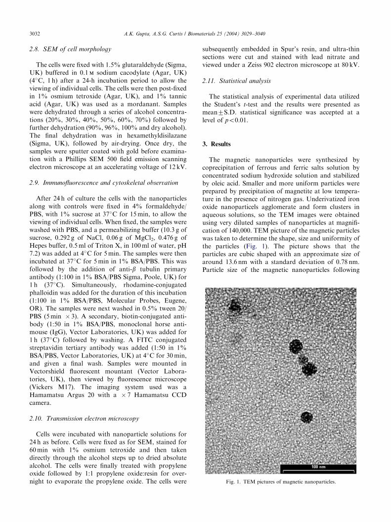
Fig. 1. TEM pictures of magnetic nanoparticles.
A.K. Gupta, A.S.G. Curtis / Biomaterials 25 (2004) 3029–30403032
2.8. SEM of cell morphology
The cells were fixed with 1.5% glutaraldehyde (Sigma,UK) buffered in 0.1m sodium cacodylate (Agar, UK)(4�C, 1 h) after a 24-h incubation period to allow theviewing of individual cells. The cells were then post-fixedin 1% osmium tetroxide (Agar, UK), and 1% tannicacid (Agar, UK) was used as a mordanant. Sampleswere dehydrated through a series of alcohol concentra-tions (20%, 30%, 40%, 50%, 60%, 70%) followed byfurther dehydration (90%, 96%, 100% and dry alcohol).The final dehydration was in hexamethyldisilazane(Sigma, UK), followed by air-drying. Once dry, thesamples were sputter coated with gold before examina-tion with a Phillips SEM 500 field emission scanningelectron microscope at an accelerating voltage of 12 kV.
2.9. Immunofluorescence and cytoskeletal observation
After 24 h of culture the cells with the nanoparticlesalong with controls were fixed in 4% formaldehyde/PBS, with 1% sucrose at 37�C for 15 min, to allow theviewing of individual cells. When fixed, the samples werewashed with PBS, and a permeabilizing buffer (10.3 g ofsucrose, 0.292 g of NaCl, 0.06 g of MgCl2, 0.476 g ofHepes buffer, 0.5 ml of Triton X, in 100 ml of water, pH7.2) was added at 4�C for 5 min. The samples were thenincubated at 37�C for 5 min in 1% BSA/PBS. This wasfollowed by the addition of anti-b tubulin primaryantibody (1:100 in 1% BSA/PBS Sigma, Poole, UK) for1 h (37�C). Simultaneously, rhodamine-conjugatedphalloidin was added for the duration of this incubation(1:100 in 1% BSA/PBS, Molecular Probes, Eugene,OR). The samples were next washed in 0.5% tween 20/PBS (5 min � 3). A secondary, biotin-conjugated anti-body (1:50 in 1% BSA/PBS, monoclonal horse anti-mouse (IgG), Vector Laboratories, UK) was added for1 h (37�C) followed by washing. A FITC conjugatedstreptavidin tertiary antibody was added (1:50 in 1%BSA/PBS, Vector Laboratories, UK) at 4�C for 30 min,and given a final wash. Samples were mounted inVectorshield fluorescent mountant (Vector Labora-tories, UK), then viewed by fluorescence microscope(Vickers M17). The imaging system used was aHamamatsu Argus 20 with a � 7 Hamamatsu CCDcamera.
2.10. Transmission electron microscopy
Cells were incubated with nanoparticle solutions for24 h as before. Cells were fixed as for SEM, stained for60 min with 1% osmium tetroxide and then takendirectly through the alcohol steps up to dried absolutealcohol. The cells were finally treated with propyleneoxide followed by 1:1 propylene oxide:resin for over-night to evaporate the propylene oxide. The cells were
subsequently embedded in Spur’s resin, and ultra-thinsections were cut and stained with lead nitrate andviewed under a Zeiss 902 electron microscope at 80 kV.
2.11. Statistical analysis
The statistical analysis of experimental data utilizedthe Student’s t-test and the results were presented asmean7S.D. statistical significance was accepted at alevel of po0.01.
3. Results
The magnetic nanoparticles were synthesized bycoprecipitation of ferrous and ferric salts solution byconcentrated sodium hydroxide solution and stabilizedby oleic acid. Smaller and more uniform particles wereprepared by precipitation of magnetite at low tempera-ture in the presence of nitrogen gas. Underivatized ironoxide nanoparticels agglomerate and form clusters inaqueous solutions, so the TEM images were obtainedusing very diluted samples of nanoparticles at magnifi-cation of 140,000. TEM picture of the magnetic particleswas taken to determine the shape, size and uniformity ofthe particles (Fig. 1). The picture shows that theparticles are cubic shaped with an approximate size ofaround 13.6 nm with a standard deviation of 0.78 nm.Particle size of the magnetic nanoparticles following

ARTICLE IN PRESS
Fig. 3. Relative magnetization M=Ms and magnetization versus
applied magnetic field for uncoated magnetic nanoparticles.
A.K. Gupta, A.S.G. Curtis / Biomaterials 25 (2004) 3029–3040 3033
protein attachment was found to increase by 2–3 nm.Magnetic particles formed were characterized by infra-red spectroscopy as shown in Fig. 2. The IR spectra ofiron oxide exhibit strong bands in the low frequencyregion (1000–400 cm�1) due to the iron oxide skeletonand the spectrum is highly consistent with magnetite(Fe3O4) spectrum (bands at 408.9, 571.5 and 584.5 cm�1)[17]. In other regions, the spectra of iron oxide haveweak bands.
Fig. 3 shows the relative magnetization curve as afunction of magnetic field for the uncoated particles.From the figure, no hysteresis curve was observed whichindicates the characteristic superparamagnetic beha-viour of the particles. The saturation magnetizationvalue of the magnetite nanoparticles was found between45 and 50 emu/g. The size distribution was calculated bythe equation based on a log-normal function [18]. Theaverage particle size was found to be around12.9270.48 nm, which is in close agreement with thesize obtained from TEM measurements.
Iron content in the nanoparticles was determinedspectrophotometrically and was found to be more than90% of the original iron salts. It was calculated that atotal of 1.71� 1017 particles are present in 1 g ofmagnetic nanoparticles and each iron oxide nanoparticlecontained 62896 iron atoms. Amount of protein boundto the nanoparticles was determined by calculating thedifference between the total amount of protein addedand the amount present in the supernatant. Thepercentage of protein binding to nanoparticles by twostep EDCI coupling process was found to be around60% of the total protein added initially for binding. Thenumber of lactoferrin and ceruloplasmin moleculesbound per particle were calculated by assuming amolecular weight of 90 kDa for lactoferrin and135 kDa for ceruloplasmin. On average, one lactoferrin(or ceruloplasmin) molecule was conjugated per ironoxide particle. The colloidal solution of magneticnanoparticles coated with sodium oleate and furtherfunctionalized with lactoferrin and ceruloplasminshowed very high stability at neutral pH and nosedimentation was observed even after 2 months ofstorage at room temperature. Whereas, uncoated
Fig. 2. FT-IR spectrum of magnetic nanoparticles.
magnetic particles did not form a stable colloidalsuspension and sedimented within 24 h of storage.
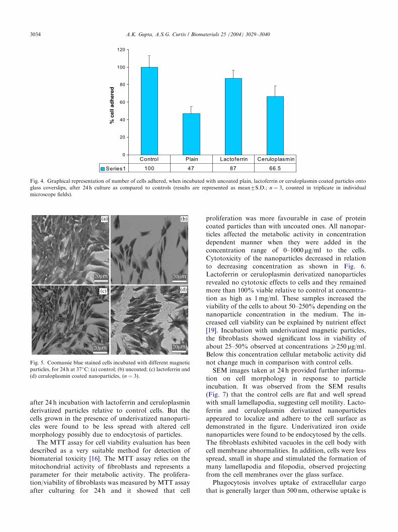
The effect of incubating cells with nanoparticles oncell adhesion to glass coverslips, as compared to controlcells (without particles) was determined and the resultsare shown in Fig. 4. It was observed that differentcoatings on nanoparticles surface give rise to changes inadhesion capacity of the fibroblasts on glass. The figureshows that the number of attached cells was decreasedsignificantly upto 53% in case of underivatized nano-particles compared to the corresponding control cellnumber (no particle). Growing the cells with cerulo-plasmin derivatized nanoparticles decreased the numberof cells attached to glass surface by one-third, whereas,incubating cells with lactoferrin derivatized samplesproduced no significant difference compared to that ofcontrol cell population.
The general morphology of the fibroblasts incubatedwith nanoparticles after staining with Coomassie blue isshown in Fig. 5. The figure shows that the cells were wellspread and there was no distinct change in morphology
ARTICLE IN PRESS
0
20
40
60
80
100
120
% c
ell a
dh
ered
Series1 100 47 87 66.5
Control Plain Lactoferrin Ceruloplasmin
Fig. 4. Graphical representation of number of cells adhered, when incubated with uncoated plain, lactoferrin or ceruloplasmin coated particles onto
glass coverslips, after 24 h culture as compared to controls (results are represented as mean7S.D.; n ¼ 3; counted in triplicate in individual
microscope fields).
Fig. 5. Coomassie blue stained cells incubated with different magnetic
particles, for 24 h at 37�C: (a) control; (b) uncoated; (c) lactoferrin and
(d) ceruloplasmin coated nanoparticles, (n ¼ 3).
A.K. Gupta, A.S.G. Curtis / Biomaterials 25 (2004) 3029–30403034
after 24 h incubation with lactoferrin and ceruloplasminderivatized particles relative to control cells. But thecells grown in the presence of underivatized nanoparti-cles were found to be less spread with altered cellmorphology possibly due to endocytosis of particles.
The MTT assay for cell viability evaluation has beendescribed as a very suitable method for detection ofbiomaterial toxicity [16]. The MTT assay relies on themitochondrial activity of fibroblasts and represents aparameter for their metabolic activity. The prolifera-tion/viability of fibroblasts was measured by MTT assayafter culturing for 24 h and it showed that cell
proliferation was more favourable in case of proteincoated particles than with uncoated ones. All nanopar-ticles affected the metabolic activity in concentrationdependent manner when they were added in theconcentration range of 0–1000 mg/ml to the cells.Cytotoxicity of the nanoparticles decreased in relationto decreasing concentration as shown in Fig. 6.Lactoferrin or ceruloplasmin derivatized nanoparticlesrevealed no cytotoxic effects to cells and they remainedmore than 100% viable relative to control at concentra-tion as high as 1 mg/ml. These samples increased theviability of the cells to about 50–250% depending on thenanoparticle concentration in the medium. The in-creased cell viability can be explained by nutrient effect[19]. Incubation with underivatized magnetic particles,the fibroblasts showed significant loss in viability ofabout 25–50% observed at concentrations X250 mg/ml.Below this concentration cellular metabolic activity didnot change much in comparison with control cells.
SEM images taken at 24 h provided further informa-tion on cell morphology in response to particleincubation. It was observed from the SEM results(Fig. 7) that the control cells are flat and well spreadwith small lamellapodia, suggesting cell motility. Lacto-ferrin and ceruloplasmin derivatized nanoparticlesappeared to localize and adhere to the cell surface asdemonstrated in the figure. Underivatized iron oxidenanoparticles were found to be endocytosed by the cells.The fibroblasts exhibited vacuoles in the cell body withcell membrane abnormalities. In addition, cells were lessspread, small in shape and stimulated the formation ofmany lamellapodia and filopodia, observed projectingfrom the cell membranes over the glass surface.
Phagocytosis involves uptake of extracellular cargothat is generally larger than 500 nm, otherwise uptake is

ARTICLE IN PRESS
0
50
100
150
200
250
300
350
400
0 200 400 600 800 1000 1200
Concentration (µg/ml)
Lactoferrin
Ceruloplasmin
Uncoated
% v
iabi
lity
Fig. 6. Cytotoxicity profiles of magnetic nanoparticles when incubated with human fibroblasts as determined by MTT assay. Percent viability of
fibroblasts was expressed relative to control cells (n ¼ 6). Results are represented as mean7S.D.
Fig. 7. SEM pictures of human fibroblasts incubated with magnetic nanoparticles: (a) control cells; (b) plain uncoated particles; (c) lactoferrin coated
and (d) ceruloplasmin coated nanoparticles. The picture shows that lactoferrin and ceruloplasmin coated nanoparticles adhere to the cell surface
whereas plain uncoated particles were found to be phagocytosed by the cells.
A.K. Gupta, A.S.G. Curtis / Biomaterials 25 (2004) 3029–3040 3035
due to the endocytosis or pinocytosis. Upon endocytosisthe particles may effect the overall cytoskeleton of thecells by forming the vacuoles in the cell body which mayresult in disrupted cytoskeleton and cell membraneprotrusions [20]. From SEM and light microscopyresults, we observed that the surface functionalized
particles did not change the cell morphology to a greaterextent as compared to underivatized ones. Furtherinformation on the data obtained from SEM and lightmicroscopy with cytoskeletal dynamics, immunofluor-escent staining was performed. Immunofluoroscentimages (Fig. 8) were taken by staining for F-actin using

ARTICLE IN PRESS
Fig. 8. Cells incubated with different coated particles and stained for tubulin (green), F-actin (red) and nucleus (blue) (n ¼ 2).
A.K. Gupta, A.S.G. Curtis / Biomaterials 25 (2004) 3029–30403036
rhodamine-phalloidin and for tubulin using anti-tubulinantibodies. The cells were also stained for cell nucleususing 4,6-diamidino-2-phenylindole (DAPI). In thecontrol cells, the microfilaments are well organized inthick bundles forming stress fibres. These fibres arestretched between cell surface and cytoplasm. Themicrotubules also form a dense network equallydistributed around the nucleus in the whole cell volume.In the case of cells incubated with lactoferrin andceruloplasmin derivatized nanoparticles, the F-actincytoskeleton appeared less defined and little disorga-nized. It could be seen, in this case, that the b-tubulinwere slightly disrupted and was dispersed in someundefined regions of the cell. The cells incubated withthe underivatized nanoparticles, however, showed cleardisruption in the cell cytoskeleton. The F-actin cytoske-leton appeared less defined and is seen to be visiblydisorganized in comparison to control cells, with b-tubulin formation appearing disrupted. The b-tubulincould be seen throughout the cell body.
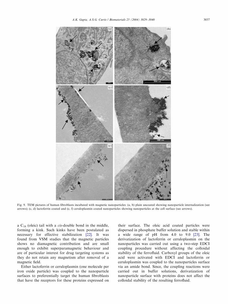
The results obtained from SEM and immunofluor-escene studies were confirmed with TEM studies asshown in Fig. 9. The pictures showed that the under-ivatized plain magnetic nanoparticles are internalizedwithin the fibroblast as a result of endocytosis. The
particles can be seen in the cytoplasm of the fibroblastsforming the vacuoles. It is apparent from the picturesthat derivatization with either lactoferrin or ceruloplas-min made the nanoparticles strongly cell surfaceadhesive and the particles could be seen on the surfaceof the fibroblasts with no particle internalization even athigh particle number adhered to the cells.
4. Discussion
The magnetic particles synthesized in solution undercontrolled conditions of temperature or oxygen haveshown fairly narrow size distribution. The synthesis ofmagnetic nanoparticles in oxygen free environment notonly protects the oxidation of iron oxide particles butalso reduces the size of the particles as compared withmethods without removing oxygen [21].
Magnetic nanoparticles have a large ratio of surface-area to volume and therefore tend to agglomerate inorder to reduce their surface energy by strong magneticdipole–dipole attractions between particles. The colloi-dal suspension of magnetite particles, however can bestabilized by adding long chain surfactant such as oleicacid, (CH3(CH2)7�CH=CH(CH2)7COOH), which has
ARTICLE IN PRESS
Fig. 9. TEM pictures of human fibroblasts incubated with magnetic nanoparticles: (a, b) plain uncoated showing nanoparticle internalization (see
arrows); (c, d) lactoferrin coated and (e, f) ceruloplasmin coated nanoparticles showing nanoparticles at the cell surface (see arrows).
A.K. Gupta, A.S.G. Curtis / Biomaterials 25 (2004) 3029–3040 3037
a C18 (oleic) tail with a cis-double bond in the middle,forming a kink. Such kinks have been postulated asnecessary for effective stabilization [22]. It wasfound from VSM studies that the magnetic particlesshows no diamagnetic contribution and are smallenough to exhibit superparamagnetic behaviour andare of particular interest for drug targeting systems asthey do not retain any magnetism after removal of amagnetic field.
Either lactoferrin or ceruloplasmin (one molecule periron oxide particle) was coupled to the nanoparticlesurfaces to preferentially target the human fibroblaststhat have the receptors for these proteins expressed on
their surface. The oleic acid coated particles weredispersed in phosphate buffer solution and stable withina wide range of pH from 4.0 to 9.0 [23]. Thederivatization of lactoferrin or ceruloplasmin on thenanoparticles was carried out using a two-step EDCIcoupling procedure without affecting the colloidalstability of the ferrofluid. Carboxyl groups of the oleicacid were activated with EDCI and lactoferrin orceruloplasmin was coupled to the nanoparticles surfacevia an amide bond. Since, the coupling reactions werecarried out in buffer solutions, derivatization ofnanoparticle surface with proteins does not affect thecolloidal stability of the resulting ferrofluid.

ARTICLE IN PRESSA.K. Gupta, A.S.G. Curtis / Biomaterials 25 (2004) 3029–30403038
Nanoparticle–cell interaction depends on the sur-face aspects of materials, which may be describedaccording to their chemistry, hydrophilic/hydrophobiccharacteristics or surface energy. These surface char-acteristics determine how the nanoparticles will adsorbto the cell surface and more particularly determine thecell behaviour on contact. Cells in the presence ofnanoparticles first attach, adhere and spread on thesurfaces. Thereafter, the quality of cell adhesion willinfluence their morphology, cytoskeleton organizationand their capacity for proliferation and differentiation.It is known that cell adhesion is mediated by theinteraction of surface proteins such as integrinswith proteins in the extracellular matrix or on thesurface of other cells or particles [24]. The phenomenonof cell adhesion is of crucial importance in governing arange of cellular functions including cell growth,migration, differentiation, survival and tissue organiza-tion [24].
It was observed from cell culture studies that theunderivatized nanoparticles reduced cell adhesion andviability significantly as compared to the cells that werenot exposed to the nanoparticles. One possible explana-tion for this large decrease in cell adhesion and viabilityis that these nanoparticles are taken up by the cells as aresult of endocytosis [25] due to weak cell adhesiveinteractions with the nanoparticles [26]. The low toxicityof nanoparticles derivatized with lactoferrin or cerulo-plasmin may be attributed to the fact that these ligandsact as cellular markers that are targeted at the surfacereceptors expressed on the cell surface without beinginternalized. Receptors are highly regulated cell surfaceproteins, which mediate specific interactions between thecells and their extracellular milieu and they are generallylocalized on the plasma membrane. Lactoferrin is aniron binding glycoprotein of the transferrin family and isimportant in host defence against infection and excessiveinflammation. Also, lactoferrin has very high bindingefficiency for its receptors on the cellular membranesand its activity is mediated through iron sequestrationand interactions with cellular receptors through itspositively charged N-terminus. Ceruloplasmin, on theother hand, is a copper binding serum protein, whichpossesses significant oxidase activity and acts through itsperoxidase or copper binding sites. Because of thesestructural and functional differences between the twoproteins, their derivatization on nanoparticle surfacesgives rise to differences in their cellular responsestowards cell adhesion/viability and binding to surfacereceptors.
The SEM and cytoskeleton studies also verified theabove results. These studies showed that the eachnanoparticle type with different surface characteristicscaused a distinct cell response. The underivatizedparticles were endocytosed by the fibroblasts duringthe 24 h incubation, thereby causing cell death possibly
through apoptosis due to internalization [25]. Endocy-tosis of the particles resulted in disruption of the cellmembrane and disorganized cell cytoskeleton. Cellswere found to be less spread, small in size andstimulated the formation of many lamellipodia andfilopodia, observed projecting from cell membranes overthe glass surface. From SEM studies, it could be seenthat lactoferrin and ceruloplasmin derivatized particlesare highly adhesive to the cell surface receptors. Sincethe derivatized nanoparticles are not endocytosed, onlya little effect on the cytoskeletal organization of thefibroblasts was observed possibly due to the fact that thereceptors mediated delivery takes place through changein F-actin/b-tubulin organization. It is well known thatcytoskeleton reorganization, for example, the formationof lamellipodia, membrane ruffling and contraction ofcells after particle internalization, is required for cellproliferation, migration and in its metabolic actions [27].Cytoskeleton reorganization is controlled by smallGTP-binding proteins [28]. For example, Ras is involvedin membrane ruffling, pinocytosis [29] and the formationof stress fibres [30]. Rho and Rac proteins are alsocritical for cytoskeleton reorganization, the formerbeing responsible for the formation of stress fibres andthe latter for the formation of ruffles [30].
The TEM studies indicated that a substantial numberof underivatized particles were internalized by the cellsconfirming the above SEM and cytoskeletal organiza-tion studies. It was also concluded from the TEMpictures that the lactoferrin and ceruloplasmin deriva-tized particles are not internalized but found to adhereat the cell surface. These results are in close agreementwith the results obtained by Berry et al [31]. Lagrouxand Figarella have found that transferrin would enterinto the cell probably by a relatively classic pathway ofreceptor-mediated endocytosis whereas, lactoferrin andits specific receptor would not be internalized. Theirresults indicate that lactoferrin acts by releasing its ironat the plasma membranes without being itself inter-nalized [32].
In the absence of any system to inhibit phagocytosis,most nanoparticles are endocytosed by cells andeventually sequestered in digestive vacuoles in the cell.Once the particles are endocytosed they are probablyremoved from contact with specific cell surface receptorsand become effectively ineffective. As a result of theseevents, the cells are at high risk of toxic effects fromparticles overload. If the particles can be prevented fromleaving the cell surface they will remain in contact withtheir specific receptors and would be expected to leavethe cell in a state of prolonged stimulation whileprotecting the cells from side effects due to endocytosis.In the present study, we have discovered a route toderivatising superparamagnetic nanoparticles with var-ious proteins that bind strongly to surface receptors thatphagocytosis is inhibited. Confinement to the cell

ARTICLE IN PRESSA.K. Gupta, A.S.G. Curtis / Biomaterials 25 (2004) 3029–3040 3039
surface would provide a route that might allow removalof the particles from the cells after an appropriateresidence time.
5. Conclusions
Superparamagnetic nanoparticles with distinct sur-face characteristics induces either endocytosis or adhe-sion to cell membrane. Surface functionalizednanoparticles with lactoferrin and ceruloplasminshowed high affinity for cell surface receptor mainlydue to ligand–receptor interactions. Their specificattachment to cell surface offers the opportunity tolabel the cells with magnetic particles while reducingnon-specific endocytosis. These studies, therefore, en-abled us to understand the interactions of thesenanoparticles with cells in vitro conditions in moredetails prior to use in in vivo situations. Further studieson the pharmacokinetics and toxicity of these magneticnanoparticles coupled to lactoferrin or ceruloplasmin insuitable in vivo models is in progress.
Acknowledgements
This work was supported by EC contract GRD5-CT2000-00375 project acronym: MAGNANOMED.The authors would like to thank Dr. Stephen Wells(Liquid Research Ltd., Wales, UK) for magneticmeasurements on the samples and Mr. Eoin Robertson(Electron Microscopy Unit) for his assistance. Also, thehelp and advice from Dr. Catherine C. Berry is greatlyappreciated.
References
[1] Weissleder R, Bogdanov A, Neuwelt EA, Papisov M. Long-
circulating iron oxides for MR imaging. Adv Drug Delivery Rev
1995;16:321–34.
[2] Reimer P, Weissleder R. Development and experimental applica-
tion of receptor-specific MR contrast media. Radiology 1996;
36:153–63.
[3] Chouly C, Pouliquen D, Lucet L, Jeune JJ, Jallet P. Development
of superparamagnetic nanoparticles for MRI: effect of particle
size, charge and surface nature on biodistribution. J Micro-
encapsulation 1996;13:245–55.
[4] Gupta PK, Hung CT. Magnetically controlled targeted micro-
carrier systems. Life Sci 1989;44:175–86.
[5] Zhang Y, Kohler N, Zhang M. Surface modification of super-
paramagnetic magnetite nanoparticles and their intracellular
uptake. Biomaterials 2002;23(7):1553–61.
[6] Olsvik O, Popovic T, Skjerve E, Cudjoe KS, Hornes E, Ugelstad
J, Uhlen M. Magnetic separation techniques in diagnostic
microbiology. Clin Microbiol Rev 1994;7:43–54.
[7] Yeh TC, Zhang W, Ldstad ST, Ho C. Intracellular labeling of
T-Cells with superparamagnetic contrast agents. Magn Reson
Med 1993;30:617–25.
[8] Handgretinger R, Lang P, Schumm M, Taylor G, Neu S,
Koscielnak E, Niethammer D, Klingebiel T. Isolation and
transplantation of autologous peripheral CD34+progenitor cells
highly purified by magnetic-activated cell sorting. Bone Marrow
Transpl 1998;21:987–93.
[9] Schoepf U, Marecos E, Jain R, Weissleder R. Intracellular
magnetic labelling of lymphocytes for in vivo trafficking studies.
BioTechniques 1998;24:642–51.
[10] Moore A, Basilion J, Chiocca EA, Weissleder R. Measuring
transferrin receptor gene expression by NMR imaging. Biochim
Biophys Acta 1998;1402:239–49.
[11] Weissleder R, Cheng HC, Bogdanova A, Bogdanov A. Magne-
tically labelled cells can be detected by MR imaging. J Magn
Reson Imaging 1997;7:258–63.
[12] Qian ZM, Li H, Sun H, Ho K. Targeted drug delivery via
transferrin receptor-mediated endocytosis pathway. Pharmacol
Rev 2002;54(4):561–87.
[13] Lauffenburger DA. In: Lauffenburger DA, Linderman JJ, editors.
Receptors: models for binding, trafficking, and signaling. New
York, Oxford: Oxford University Press; 1993.
[14] Levy P, Viljoen M. Lactoferrin: a general review. Hematologica
1995;80:252–67.
[15] Floris G, Medda R, Padigalia A, Musci G. The physiopatholo-
gical significance of ceruloplasmin: a possible therapeutic
approach. Biochem pharmacol 2000;60:1735–41.
[16] Mosmann T. Rapid colorimetric assay for cellular growth and
survival: application to proliferation and cytotoxic assay.
J Immunol Methods 1993;95:55–63.
[17] Schwertmann U, Cornell RM. Iron oxides in the laboratory:
preparation and characterization. Weinheim, Cambridge: VCH;
1991.
[18] Granqvist CG, Buhrman RH. Ultrafine metal particles. J Appl
Phys 1976;47:2200–19.
[19] Fischer D, Li YX, Ahlemeyer B, Krieglstein J, Kissel . In vitro
cytotoxicity testing of polycations: influence of polymer
structure on cell viability and hemolysis. Biomaterials 2003;24(7):
1121–31.
[20] Gagesu R, Gruenberg J, Smyte E. Membrane dynamics in
endocytosis: sturcture–function relationship. Traffic 2000;1:84–8.
[21] Kim DK, Zhang Y, Voit W, Rao KV, Muhammed M. Synthesis
and characterization of surfactant-coated superparamagnetic
monodispersed iron oxide nanoparticles. J Magn Magn Mater
2001;225(1–2):30–6.
[22] Tadmor R, Rosensweig RE, Frey J, Klein J. Resolving the puzzle
of ferrofluid dispersants. Langmuir 2000;16:9117–20.
[23] Hallab NJ, Bundy KJ, O’Connor K, Clark R, Moses RL. Cell
adhesion to Biomaterials: correlations between surface charge,
surface roughness, adsorbed protein and cell morphology. J Long
term Effects Med Implants 1995;5(3):209–31.
[24] Fan J, Lu L, Xu R, Jiang R, Gao Y. Use of water-dispersible
Fe2O3 nanoparticles with narrow size distributions in isolating
avidin. J Colloid Interface Sci 2003;266(1):215–8.
[25] Berry CC, Wells S, Charles S, Curtis ASG. Dextran and albumin
derivatised iron oxide nanoparticles: influence on fibroblasts
in vitro. Biomaterials 2003;24(25):4551–7.
[26] Weissleder R, Stark DD, Engelstad BL, Bacon BR, Compton CC,
White DL, Jacobs P, Lewis J. Superparamagnetic iron oxide:
pharmacokinetics and toxicity. AJR Am J Roentgenol 1989;152:
167–73.
[27] Mitchison TJ, Cramer LP. Actin-based cell motility and cell
locomotion. Cell 1996;84:371–9.
[28] Bar-Sagi D, Hall A. Induction of membrane ruffling and fluid-
phase pinocytosis in quiescent fibroblasts by ras proteins. Cell
2000;103:227–38.
[29] Bar-Sagi D, Feramisco JR. Ras and Rho GTPases: a family
reunion. Science 1986;233:1061–8.

ARTICLE IN PRESSA.K. Gupta, A.S.G. Curtis / Biomaterials 25 (2004) 3029–30403040
[30] Ridley AJ, Hall A. The small GTP-binding protein rho regulates
the assembly of focal adhesions and actin stress fibres in response
to growth factors. Cell 1992;70:389–99.
[31] Berry CC, Wells S, Charles S, Curtis ASG. The influence
of uncoated and coated magnetic nanoparticles on human
fibroblasts in culture. Eur Cell Mater 2002;4(Suppl. 2):
67–8.
[32] Lagroux DR, Figarella C. Evidence for a different mechanism of
lactoferrin and transferrin translocation on HT 29-D4 cells.
Biochem Biophys Res Commun 1990;170(2):837–42.
Copyright © 2022 FDOKUMEN


![Self-assembled monolayers of parent and derivatized [n]staffane-3,3(n-1)-dithiols on polycrystalline gold electrodes](https://static.fdokumen.com/doc/165x107/6328c589ce2aaeab5c0256a0/self-assembled-monolayers-of-parent-and-derivatized-nstaffane-33n-1-dithiols.jpg)