Spectroscopic investigation of interaction between bovine gamma globulin and gold nanoparticles
Advanced Drug Delivery Reviews 60 (2008) 1307–1315
Contents lists available at ScienceDirect
Advanced Drug Delivery Reviews
j ourna l homepage: www.e lsev ie r.com/ locate /addr
Gold nanoparticles in delivery applications☆
Partha Ghosh, Gang Han, Mrinmoy De, Chae Kyu Kim, Vincent M. Rotello ⁎Department of Chemistry, University of Massachusetts, 710 North Pleasant Street, Amherst, MA, 01003, USA
☆ This review is part of the Advanced Drug Delivery RevNanoparticles in Drug Delivery”.⁎ Corresponding author. Tel.: +1 413 545 2058; fax: +
E-mail address: [email protected] (V.M. Rote
0169-409X/$ – see front matter © 2008 Elsevier B.V. Aldoi:10.1016/j.addr.2008.03.016
A B S T R A C T
A R T I C L E I N F OArticle history:
Gold nanoparticles (AuNPs) Received 12 January 2008Accepted 12 March 2008Available online 10 April 2008Keywords:GoldDeliveryDNADrug deliveryGlutathioneNanoparticlesSelf-assembled monolayerPhotochemistryRelease
provide non-toxic carriers for drug and gene delivery applications. With thesesystems, the gold core imparts stability to the assembly, while the monolayer allows tuning of surfaceproperties such as charge and hydrophobicity. An additional attractive feature of AuNPs is their interactionwith thiols, providing an effective and selective means of controlled intracellular release.
© 2008 Elsevier B.V. All rights reserved.
Contents
1. Introduction . . . . . . . . . . . . . . . . . . . . . . . . . . . . . . . . . . . . . . . . . . . . . . . . . . . . . . . . . . . . . . 13072. Synthesis of gold nanoparticles . . . . . . . . . . . . . . . . . . . . . . . . . . . . . . . . . . . . . . . . . . . . . . . . . . . . . 13083. Drug delivery using gold nanoparticles . . . . . . . . . . . . . . . . . . . . . . . . . . . . . . . . . . . . . . . . . . . . . . . . . 1308
3.1. Glutathione-mediated release of small molecules . . . . . . . . . . . . . . . . . . . . . . . . . . . . . . . . . . . . . . . . . 13083.2. Release of nitric oxide and singlet oxygen from gold nanoparticles . . . . . . . . . . . . . . . . . . . . . . . . . . . . . . . . . 1309
4. Gold nanoparticles for delivery of biomolecules . . . . . . . . . . . . . . . . . . . . . . . . . . . . . . . . . . . . . . . . . . . . . 13114.1. Gold nanoparticles for nucleic acids delivery via non-covalent conjugation . . . . . . . . . . . . . . . . . . . . . . . . . . . . . 13114.2. Gold nanoparticles for nucleic acid delivery by covalent linking . . . . . . . . . . . . . . . . . . . . . . . . . . . . . . . . . . 13134.3. Functionalized gold nanoparticles for protein delivery . . . . . . . . . . . . . . . . . . . . . . . . . . . . . . . . . . . . . . . 1313
5. Photothermal effect of gold nanoparticles in therapy . . . . . . . . . . . . . . . . . . . . . . . . . . . . . . . . . . . . . . . . . . . 13136. Gold nanoparticle targeting in vivo . . . . . . . . . . . . . . . . . . . . . . . . . . . . . . . . . . . . . . . . . . . . . . . . . . . 13147. Conclusions . . . . . . . . . . . . . . . . . . . . . . . . . . . . . . . . . . . . . . . . . . . . . . . . . . . . . . . . . . . . . . 1314Acknowledgements . . . . . . . . . . . . . . . . . . . . . . . . . . . . . . . . . . . . . . . . . . . . . . . . . . . . . . . . . . . . . 1314References . . . . . . . . . . . . . . . . . . . . . . . . . . . . . . . . . . . . . . . . . . . . . . . . . . . . . . . . . . . . . . . . . 1314
1. Introduction
Nanocarriers have provided a novel platform for target-specificdelivery of therapeutic agents [1]. Over the past decade, several
iews theme issue on “Inorganic
1 413 545 4490.llo).
l rights reserved.
delivery vehicles have been designed based on different nanomater-ials, such as polymers [2], dendrimers [3], liposomes [4], nanotubes[5], and nanorods [6]. Gold nanoparticles (GNPs) have recentlyemerged as an attractive candidate for delivery of various payloadsinto their targets [7,8]. The payloads could be small drug molecules orlarge biomolecules, like proteins, DNA, or RNA (vide post). Efficientrelease of these therapeutic agents is a prerequisite for effectivetherapy. The release could be triggered by internal (e.g. glutathione(GSH) [9], or pH [10]) or external (e.g. light [11]) stimuli. Significantly

Table 1Synthetic methods and capping agents for GNPs of varying core size
Coresize (d)
Synthetic methods Cappingagents
References
1–2 nm Reduction of AuCl(PPh3) with diborane orsodium borohydride
Phosphine [16]
1.5–5 nm Biphasic reduction of HAuCl4 by sodiumborohydride in the presence of thiolcapping agents
Alkanethiol [17,18]
10–150 nm Reduction of HAuCl4 with sodium citratein water
Citrate [19–21]
Fig. 1. Various applications of gold nanoparticles in therapy.
1308 P. Ghosh et al. / Advanced Drug Delivery Reviews 60 (2008) 1307–1315
the internal stimuli operate in a biologically control manner, whereasthe external stimuli provide spatio-temporal control over the release.
Gold nanoparticles exploit their unique chemical and physicalproperties for transporting and unloading the pharmaceuticals. First,the gold core is essentially inert and non-toxic [12]. A secondadvantage is their ease of synthesis; monodisperse nanoparticlescan be formed with core sizes ranging from 1 nm to 150 nm (Table 1).Further versatility is imparted by their ready functionalization,generally through thiol linkages (vide post). Moreover, their photo-physical properties could trigger drug release at remote place [13].
Several reviews describing nanoparticle–biomacromolecule inter-actions have recently been published, generally focusing on biosen-sing [14] and diagnostic [15] applications. This review will focus ondrug, gene, and protein delivery using GNPs (Fig. 1).
2. Synthesis of gold nanoparticles
Significant efforts have been devoted over the past forty years tothe fabrication of GNPswithmonodispersity and controlled size. GNPswith varying core sizes are prepared by the reduction of gold salts inthe presence of appropriate stabilizing agents that prevent particleagglomeration. Some common synthetic methods of core–shell GNPsare summarized in Table 1.
Several research groups have fabricated delivery systems based onGNPs bearing functional moieties, which are anchored with thiol-linkers, in their monolayers. A wide variety of monolayer protectedclusters (MPCs) can be formed rapidly and in scalable fashion usingthe one-pot protocol developed by Schiffrin et al. in 1994 (Scheme 1)[17]. In this preparation, AuCl4− salts are reduced with NaBH4 in thepresence of the desired thiol capping ligand or ligands. The core size ofthe particles can be varied from 1.5 nm to ∼6 nm by varying the thiol–gold stoichiometry.
The functional diversity of MPCs can be extended through theformation of mixedmonolayer protected clusters (MMPCs) that can besynthesized directly or through post-functionalization of MPCs. Aversatile method for the creation of MMPCs is the place-exchangereaction developed by Murray (Scheme 1) [18]. In this protocol,foreign thiols displace the native ligands of MPCs in an equilibriumprocess. Taken together, the control of monolayer structure providedby nanoparticle synthesis, place displacement, and other post-synthetic modification methods [22] can be used to display a widerange of functionality at the particle surface, including biocompatibleoligo(ethylene glycol) (OEG) and poly(ethylene glycol) (PEG) moieties[23–30]. One obvious benefit of these combinedmethodologies is thatthey are highly amenable to divergent synthesis, an important aspectin the creation of delivery vehicles.
3. Drug delivery using gold nanoparticles
Drug delivery systems (DDSs) provide positive attributes to a ‘free’drug by improving solubility, in vivo stability, and biodistribution.They can also alter unfavorable pharmacokinetics of some ‘free’ drugs.Moreover, huge loading of pharmaceuticals on DDSs can render ‘drugreservoirs’ for controlled and sustained release to maintain the drug
level within therapeutic window. For example, a gold nanoparticlewith 2 nm core diameter could be, in principle, conjugated with ∼100molecules to available ligands (n=∼108) in the monolayer [31].Zubarev et al. have recently succeeded in coupling of ∼70molecules ofpaclitaxel, a chemotherapeutic drug, to a GNP with 2 nm corediameter [32].
3.1. Glutathione-mediated release of small molecules
GSH-mediated release represents an alternative non-enzymaticstrategy for the selective intracellular activation of prodrugs. Thismethodology relies on the dramatic differential in intracellular GSHconcentration (1–10 mM) [33,34] versus extracellular thiol levels [35],where the predominant thiols in blood plasma are glutathione (2 μM)and cysteine (8 μM) [36]. Current methodologies rely on disulfidelinkages between the drugs and carriers [37–42]. While this approachcan be effective, including an example currently in the clinic [37], it isdifficult to tune the reactivity of the disulfide linkage. Additionally,thiol–disulfide exchange can occur with surface cysteines of proteinsin the bloodstream, potentially creating protein–carrier conjugateswith altered bioavailability and pharmacokinetic profiles. We expectour nanoparticles to be resistant to exchange with proteins due to thesteric shielding of the gold–thiol interface.
In recent studies, we have demonstrated cellular delivery and GSH-mediated release of a hydrophobic dye (BODIPY), as a model ofhydrophobic drugs, using functionalized gold nanoparticles (fGNPs)[9]. The particles (core d=∼2 nm) featured a mixed monolayercomposed of tetra(ethylene glycol)ylated cationic ligands (TTMA) andfluorogenic ligands (HSBDP) (Fig. 2a). The cationic nature facilitatesthe crossing of cell-membrane barrier, and the fluorophore probespossible drug release mechanism. BODIPY-conjugated GNPs are non-fluorescent as the gold core quenches fluorescence via energy and/orelectron transfer processes [43,44]. The fluorescence signal emanatesupon triggering the GNPs with GSH in cuvette, or cellular thiols inhuman liver cells (Hep G2) (Fig. 2b). Controlled release of the dye wasverified by treating mouse embryonic fibroblast cells (MEF, having∼50% lower intracellular GSH levels than Hep G2) with varyingconcentration of glutathione monoester (GSH-OEt: 0, 5 and 20 mM)
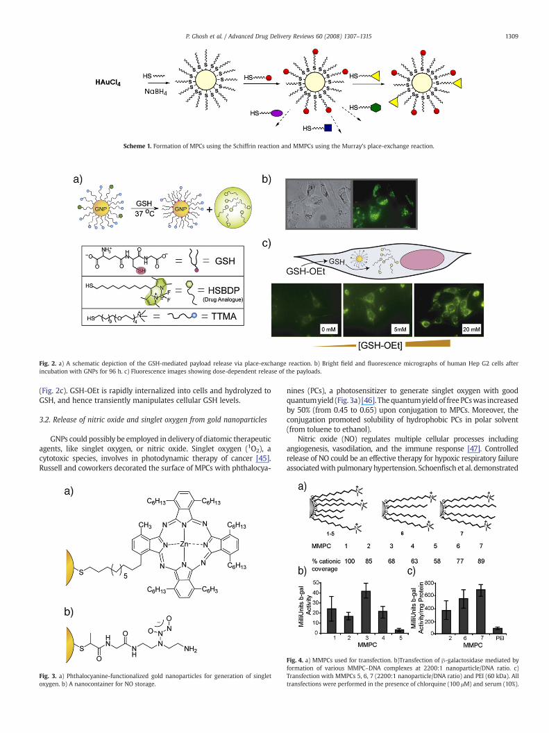
Scheme 1. Formation of MPCs using the Schiffrin reaction and MMPCs using the Murray's place-exchange reaction.
Fig. 2. a) A schematic depiction of the GSH-mediated payload release via place-exchange reaction. b) Bright field and fluorescence micrographs of human Hep G2 cells afterincubation with GNPs for 96 h. c) Fluorescence images showing dose-dependent release of the payloads.
1309P. Ghosh et al. / Advanced Drug Delivery Reviews 60 (2008) 1307–1315
(Fig. 2c). GSH-OEt is rapidly internalized into cells and hydrolyzed toGSH, and hence transiently manipulates cellular GSH levels.
3.2. Release of nitric oxide and singlet oxygen from gold nanoparticles
GNPs could possibly be employed in delivery of diatomic therapeuticagents, like singlet oxygen, or nitric oxide. Singlet oxygen (1O2), acytotoxic species, involves in photodynamic therapy of cancer [45].Russell and coworkers decorated the surface of MPCs with phthalocya-
Fig. 3. a) Phthalocyanine-functionalized gold nanoparticles for generation of singletoxygen. b) A nanocontainer for NO storage.
nines (PCs), a photosensitizer to generate singlet oxygen with goodquantumyield (Fig. 3a) [46]. Thequantumyield of freePCswas increasedby 50% (from 0.45 to 0.65) upon conjugation to MPCs. Moreover, theconjugation promoted solubility of hydrophobic PCs in polar solvent(from toluene to ethanol).
Nitric oxide (NO) regulates multiple cellular processes includingangiogenesis, vasodilation, and the immune response [47]. Controlledrelease of NO could be an effective therapy for hypoxic respiratory failureassociatedwith pulmonary hypertension. Schoenfisch et al. demonstrated
Fig. 4. a) MMPCs used for transfection. b)Transfection of β-galactosidase mediated byformation of various MMPC–DNA complexes at 2200:1 nanoparticle/DNA ratio. c)Transfection with MMPCs 5, 6, 7 (2200:1 nanoparticle/DNA ratio) and PEI (60 kDa). Alltransfections were performed in the presence of chlorquine (100 μM) and serum (10%).
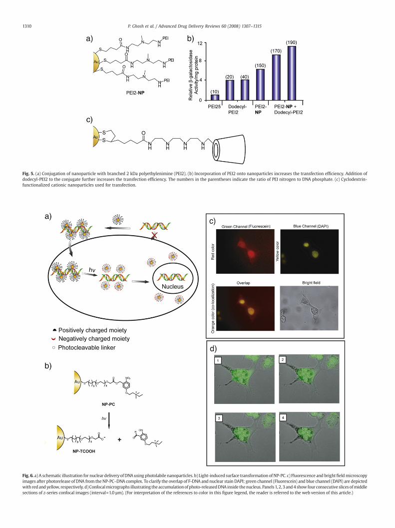
Fig. 5. (a) Conjugation of nanoparticle with branched 2 kDa polyethylenimine (PEI2). (b) Incorporation of PEI2 onto nanoparticles increases the transfection efficiency. Addition ofdodecyl-PEI2 to the conjugate further increases the transfection efficiency. The numbers in the parentheses indicate the ratio of PEI nitrogen to DNA phosphate. (c) Cyclodextrin-functionalized cationic nanoparticles used for transfection.
Fig. 6. a) A schematic illustration for nuclear deliveryof DNAusing photolabile nanoparticles. b) Light-induced surface transformation of NP-PC. c) Fluorescence and brightfieldmicroscopyimages after photorelease of DNA from the NP-PC–DNA complex. To clarify the overlap of F-DNA and nuclear stain DAPI; green channel (Fluorescein) and blue channel (DAPI) are depictedwith redandyellow, respectively. d)Confocalmicrographs illustrating theaccumulationofphoto-releasedDNA inside thenucleus. Panels1, 2, 3 and4 show four consecutive slices ofmiddlesections of z-series confocal images (interval=1.0 µm). (For interpretation of the references to color in this figure legend, the reader is referred to the web version of this article.)
1310 P. Ghosh et al. / Advanced Drug Delivery Reviews 60 (2008) 1307–1315

Fig. 7. a) Antisense DNA modified gold nanoparticle (ASNP). b) Before and c) after incubation with antisense DNA functionalized nanoparticles for EGFP-expressing C166 cellsReprinted with permission from Ref. [63].
1311P. Ghosh et al. / Advanced Drug Delivery Reviews 60 (2008) 1307–1315
that NO can be efficiently stored by covalent linking with polyamine-stabilized MPCs via formation of acid labile N-diazeniumdiolate (Fig. 3b)[10]. They showed effective release of NO from the water-solublenanocontainers in acidic condition (pH=3). pH-responsive materials areapplicable for drug delivery due to presence of mild acidic environmentinside inflammatory and tumor tissues (pH∼6.8), or cellular vesicles likeendosomes (pH ∼5.5–6) and lysosomes (pH ∼4.5–5.0) [48–50].
4. Gold nanoparticles for delivery of biomolecules
Gold nanoparticles are capable of delivering large biomolecules,without restricting themselves as carriers of only small moleculardrugs. Tunable size and functionality make them a useful scaffold forefficient recognition and delivery of biomolecules. They have shownthe success in delivery of peptides, proteins, or nucleic acids like DNAor RNA.
Fig. 8. Schematic representation of GSH-mediated disruption of nanoparticle–β-galprotein interactions.
Fig. 9. Photothermal effect of gold nanoparticles for releasing encapsulated materials. aPreparation of a microcapsule by layer-by-layer process, b) Encapsulation of therapeutiagents, c) Doping of GNPs in the capsule–shells, d) Rupture of the shell upon light irradiation
.
4.1. Gold nanoparticles for nucleic acids delivery via non-covalentconjugation
Gene therapy presents an ideal strategy for the treatment ofgenetic as well as acquired diseases [51]. Viruses provide a logicalvehicle for gene therapy, and have been shown to be highly efficient[52]. Viruses have nonetheless raised many safety concerns arisingfrom unpredictable cytotoxicity and immune responses [53]. Syn-thetic DNA delivery systems have overcome these limitations.However, current non-viral gene delivery systems suffer from lessefficiency. The vectors encounter numerous barriers between the siteof administration and localization in the cell nucleus [54]. Theeffective delivery vehicles need to provide efficient protection ofnucleic acid from degradation by nucleases, efficient cell entry, andrelease of the nucleic acid in functional form in the nucleus [55].
GNPs provide attractive candidates for gene delivery. They can bemade quite small to provide a high surface-to-volume ratio,maximizing the payload/carrier ratio. Perhaps equally important, themonolayer coverage of MPCs and MMPCs systems allow tuning of thecharge and hydrophobicity to maximize transfection efficiency while
)c.
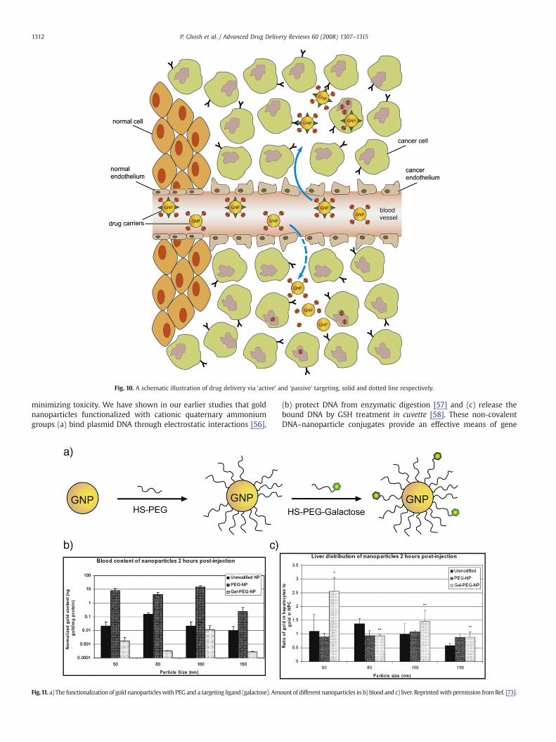
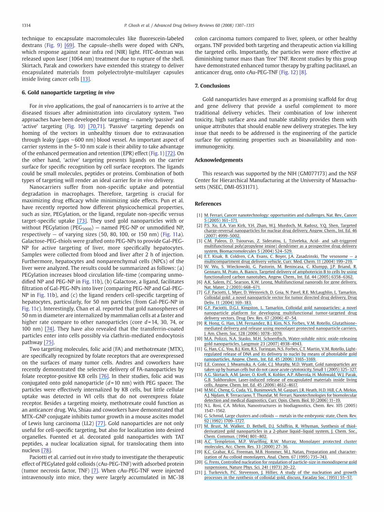
Fig. 10. A schematic illustration of drug delivery via ‘active’ and ‘passive’ targeting, solid and dotted line respectively.
1312 P. Ghosh et al. / Advanced Drug Delivery Reviews 60 (2008) 1307–1315
minimizing toxicity. We have shown in our earlier studies that goldnanoparticles functionalized with cationic quaternary ammoniumgroups (a) bind plasmid DNA through electrostatic interactions [56],
Fig.11. a) The functionalization of gold nanoparticleswith PEG and a targeting ligand (galactose). Am
(b) protect DNA from enzymatic digestion [57] and (c) release thebound DNA by GSH treatment in cuvette [58]. These non-covalentDNA–nanoparticle conjugates provide an effective means of gene
ount of different nanoparticles in b) blood and c) liver. Reprintedwith permission fromRef. [73].

Fig. 12. a) Change in tumor volume over days after treatment with TNF and CYT-6091. b)In vivo delivery of TNF–gold nanoparticle conjugate CYT-6091. Reprinted withpermission from Ref. [8].
1313P. Ghosh et al. / Advanced Drug Delivery Reviews 60 (2008) 1307–1315
delivery in mammalian 293T cells [59] (Fig. 4). Significantly,amphiphilic particles provided superior transfection vectors, indicat-ing that hydrophobicity of the nanoparticles improves the efficiency ofcellular uptake and/or the subsequent release of the DNA from theendosomal vesicle. This optimized nanoparticle was ∼8-fold moreefficient than polyethyleneimine (PEI).
Later on, Klibanov et al. functionalized gold nanoparticles withbranched polyethylenimine (PEI, 2 kDa) to provide hybrid AuNP-polymertransfection vectors (Fig. 5a) [60]. They determined that the transfectionefficiency intomonkey kidney (Cos-7) cells variedwith the PEI:goldmolarratio in the conjugates. At the optimized ratio, the conjugates were ∼12-fold more potent than the polymer itself (Fig. 5b). Similar to the abovestudies, their research indicated that increasing the hydrophobicity of thetransfection agent enhances cellular internalization and hence transfec-tion efficiency. Recently, Liu et al. fabricated effective binders of DNA byanchoring β-cyclodextrin on the periphery of oligo(ethylenediamino)-modified gold nanoparticles (OEA-CD-NP) (Fig. 5c) [61]. They havedemonstrated the ability of OEA-CD-NP to deliver plasmid DNA intobreast cancer cells (MCF-7).
Rotello et al. have recently reported photochemical uncaging of theloaded DNA on surface of the nanoparticles (Fig. 6a) [11]. A photolabilenanoparticle was designed to switch the nature of surface fromcationic to anionic upon irradiation. This particle was tailored with aphotocleavable o-nitrobenzyl ester linker and a quaternary ammo-
nium salt as endgroup (Fig. 6b). After complexation of the particlewith DNA, near-UV irradiation cleaves the nitrobenzyl linkage creatingan anionic carboxylate group and releasing the DNA. In vitro studiesusing a T7 RNA polymerase assay demonstrated that DNA transcrip-tion was restored upon UV irradiation. Cell culture studies indicatedthat the particle–DNA complexes were effectively taken up by cells.Release of FITC-labeled DNA upon irradiation was established byobservation of bright fluorescence inside the cells (Fig. 6c). Moreover,a high degree of nuclear localization was observed for the releasedDNA (Fig. 6d).
4.2. Gold nanoparticles for nucleic acid delivery by covalent linking
Nucleic acid strands could be easily modified with thiols (–SH) forgrafting onto nanoparticles. Nagasaki and coworkers conjugatedthiolated siRNA (SH-siRNA) with gold nanoparticles for cellulardelivery [62]. RNA-modified particles were coated with poly (ethyleneglycol)-block-poly(2-(N,N-dimethylamino)ethyl methacrylate) copo-lymer to enhance cellular internalization. These smart deliverysystems were effective for gene silencing in HuH-7 cells. Notably,RNAi activity has revolutionized the gene therapy strategy in twentyfirst century.
In general, polycationic materials are used for condensing DNA andtransporting it inside the cell. However, a breakthrough emerged in2006 when Mirkin et al. reported cellular internalization of oligonu-cleotide-modified nanoparticles (DNA-NPs), carrying large negativesurface potential (Fig. 7) [63]. These particles were stable againstenzymatic digestion. Incubation of cells with nanoparticles conju-gated with fluorophore-labeled DNA revealed efficient uptake of theparticles. In this system, antisense DNA hybridized with the particlecontrolled the protein expression. This was demonstrated using EGFP-expressing C166 with antisense DNA functionalized nanoparticles,providing a reduction of fluorescence intensity in cells. Very recently,they have found out the mechanism of cellular uptake of DNA-NP —
the endocytosis process is initiated by adsorption of numerous serumproteins onto particle's surface [64].
4.3. Functionalized gold nanoparticles for protein delivery
Gold nanoparticles can likewise be nanocarriers of peptides andproteins of interest. Rotello et al. have reported that cationic tetra-alkyl ammonium functionalized GNPs recognize the surface of ananionic protein through complementary electrostatic interaction andinhibit its activity (Fig. 8) [65]. The activity was recovered due torelease of free protein by treating the protein–particle complex withGSH, showing GNPs as potential protein transporters. In a recentstudy, Pokharkar, Sastry and coworkers have demonstrated functio-nalized gold nanoparticles as carriers of insulin [66]. The particleswere stabilized by chitosan, a non-toxic biopolymer. Chitosan-coatedparticles strongly adsorb insulin on their surface, and are effective fortransmucosal delivery of insulin.
5. Photothermal effect of gold nanoparticles in therapy
In addition to the surface chemistry of GNPs, their physicalproperties could be exploited for delivery applications [67]. GNPscause local heating when they are irradiated with light in the “waterwindow” (800 nm–1200 nm). El-Sayed et al. have recently reportedabout potential use of GNPs in photothermal destruction of tumors[68]. Citrate-stabilized GNPs (core d=30 nm) were coated with anti-EGFR (epidermal growth factor receptor) to target HSC3 cancer cells(human oral squamous cell carcinoma). The use of GNPs enhanced theefficacy of photothermal therapy by 20 times.
In another approach, optically responsive delivery systems havebeen designed by incorporation of Au nanospheres into the shells ofcapsules. Caruso et al. prepared microcapsules via layer-by-layer (LBL)
1314 P. Ghosh et al. / Advanced Drug Delivery Reviews 60 (2008) 1307–1315
technique to encapsulate macromolecules like fluorescein-labeleddextrans (Fig. 9) [69]. The capsule–shells were doped with GNPs,which response against near infra red (NIR) light. FITC-dextran wasreleased upon laser (1064 nm) treatment due to rupture of the shell.Skirtach, Parak and coworkers have extended this strategy to deliverencapsulated materials from polyelectrolyte-multilayer capsulesinside living cancer cells [13].
6. Gold nanoparticle targeting in vivo
For in vivo applications, the goal of nanocarriers is to arrive at thediseased tissues after administration into circulatory system. Twoapproaches have been developed for targeting— namely ‘passive’ and‘active’ targeting (Fig. 10) [70,71]. ‘Passive’ targeting depends onhoming of the vectors in unhealthy tissues due to extravasationthrough leaky (gaps ∼600 nm) blood vessel. An important aspect ofcarrier systems in the 5–10 nm scale is their ability to take advantageof the enhanced permeation and retention (EPR) effect (Fig. 1) [72]. Onthe other hand, ‘active’ targeting presents ligands on the carriersurface for specific recognition by cell surface receptors. The ligandscould be small molecules, peptides or proteins. Combination of bothtypes of targeting will render an ideal carrier for in vivo delivery.
Nanocarriers suffer from non-specific uptake and potentialdegradation in macrophages. Therefore, targeting is crucial formaximizing drug efficacy while minimizing side effects. Pun et al.have recently reported how different physicochemical properties,such as size, PEGylation, or the ligand, regulate non-specific versustarget-specific uptake [73]. They used gold nanoparticles with orwithout PEGylation (PEG5000) — named PEG-NP or unmodified NP,respectively — of varying sizes (50, 80, 100, or 150 nm) (Fig. 11a).Galactose-PEG-thiols were grafted onto PEG-NPs to provide Gal-PEG-NP for active targeting of liver, more specifically hepatocytes.Samples were collected from blood and liver after 2 h of injection.Furthermore, hepatocytes and nonparenchymal cells (NPCs) of theliver were analyzed. The results could be summarized as follows: (a)PEGylation increases blood circulation life-time (comparing unmo-dified NP and PEG-NP in Fig. 11b), (b) Galactose, a ligand, facilitatesfiltration of Gal-PEG-NPs into liver (comparing PEG-NP and Gal-PEG-NP in Fig. 11b), and (c) the ligand renders cell-specific targeting ofhepatocytes, particularly, for 50 nm particles (from Gal-PEG-NP inFig. 11c). Interestingly, Chan et al. reported that gold nanospheres of50 nm in diameter are internalized bymammalian cells at a faster andhigher rate compare to other nanoparticles (core d=14, 30, 74, or100 nm) [74]. They have also revealed that the transferrin-coatedparticles enter into cells possibly via clathrin-mediated endocytosispathway [75].
Two targeting molecules, folic acid (FA) and methotrexate (MTX),are specifically recognized by folate receptors that are overexpressedon the surfaces of many tumor cells. Andres and coworkers haverecently demonstrated the selective delivery of FA-nanoparticles byfolate receptor-positive KB cells [76]. In their studies, folic acid wasconjugated onto gold nanoparticle (d=10 nm) with PEG spacer. Theparticles were effectively internalized by KB cells, but little cellularuptake was detected in WI cells that do not overexpress folatereceptor. Besides a targeting moiety, methotrexate could function asan anticancer drug. Wu, Shiau and coworkers have demonstrated thatMTX–GNP conjugate inhibits tumor growth in a mouse ascites modelof Lewis lung carcinoma (LL2) [77]. Gold nanoparticles are not onlyuseful for cell-specific targeting, but also for localization into desiredorganelles. Fuented et al. decorated gold nanoparticles with TATpeptides, a nuclear localization signal, for translocating them intonucleus [78].
Paciotti et al. carried out in vivo study to investigate the therapeuticeffect of PEGylated gold colloids (cAu-PEG-TNF) with adsorbed protein(tumor necrosis factor, TNF) [7]. When cAu-PEG-TNF were injectedintravenously into mice, they were largely accumulated in MC-38
colon carcinoma tumors compared to liver, spleen, or other healthyorgans. TNF provided both targeting and therapeutic action via killingthe targeted cells. Importantly, the particles were more effective atdiminishing tumor mass than ‘free’ TNF. Recent studies by this grouphave demonstrated enhanced tumor therapy by grafting paclitaxel, ananticancer drug, onto cAu-PEG-TNF (Fig. 12) [8].
7. Conclusions
Gold nanoparticles have emerged as a promising scaffold for drugand gene delivery that provide a useful complement to moretraditional delivery vehicles. Their combination of low inherenttoxicity, high surface area and tunable stability provides them withunique attributes that should enable new delivery strategies. The keyissue that needs to be addressed is the engineering of the particlesurface for optimizing properties such as bioavailability and non-immunogenicity.
Acknowledgements
This research was supported by the NIH (GM077173) and the NSFCenter for Hierarchical Manufacturing at the University of Massachu-setts (NSEC, DMI-0531171).
References
[1] M. Ferrari, Cancer nanotechnology: opportunities and challenges, Nat. Rev., Cancer5 (2005) 161–171.
[2] P.S. Xu, E.A. Van Kirk, Y.H. Zhan, W.J. Murdoch, M. Radosz, Y.Q. Shen, Targetedcharge-reversal nanoparticles for nuclear drug delivery, Angew. Chem., Int. Ed. 46(2007) 4999–5002.
[3] C.M. Paleos, D. Tsiourvas, Z. Sideratou, L. Tziveleka, Acid- and salt-triggeredmultifunctional poly(propylene imine) dendrimer as a prospective drug deliverysystem, Biomacromolecules 5 (2004) 524–529.
[4] E.T. Kisak, B. Coldren, C.A. Evans, C. Boyer, J.A. Zasadzinski, The vesosome — amulticompartment drug delivery vehicle, Curr. Med. Chem. 11 (2004) 199–219.
[5] W. Wu, S. Wieckowski, G. Pastorin, M. Benincasa, C. Klumpp, J.P. Briand, R.Gennaro, M. Prato, A. Bianco, Targeted delivery of amphotericin B to cells by usingfunctionalized carbon nanotubes, Angew. Chem., Int. Ed. 44 (2005) 6358–6362.
[6] A.K. Salem, P.C. Searson, K.W. Leong, Multifunctional nanorods for gene delivery,Nat. Mater. 2 (2003) 668–671.
[7] G.F. Paciotti, L. Myer, D. Weinreich, D. Goia, N. Pavel, R.E. McLaughlin, L. Tamarkin,Colloidal gold: a novel nanoparticle vector for tumor directed drug delivery, DrugDeliv. 11 (2004) 169–183.
[8] G.F. Paciotti, D.G.I. Kingston, L. Tamarkin, Colloidal gold nanoparticles: a novelnanoparticle platform for developing multifunctional tumor-targeted drugdelivery vectors, Drug Dev. Res. 67 (2006) 47–54.
[9] R. Hong, G. Han, J.M. Fernandez, B.J. Kim, N.S. Forbes, V.M. Rotello, Glutathione-mediated delivery and release using monolayer protected nanoparticle carriers,J. Am. Chem. Soc. 128 (2006) 1078–1079.
[10] M.A. Polizzi, N.A. Stasko, M.H. Schoenfisch, Water-soluble nitric oxide-releasinggold nanoparticles, Langmuir 23 (2007) 4938–4943.
[11] G. Han, C.C. You, B.J. Kim, R.S. Turingan, N.S. Forbes, C.T. Martin, V.M. Rotello, Light-regulated release of DNA and its delivery to nuclei by means of photolabile goldnanoparticles, Angew. Chem., Int. Ed. 45 (2006) 3165–3169.
[12] E.E. Connor, J. Mwamuka, A. Gole, C.J. Murphy, M.D. Wyatt, Gold nanoparticles aretaken up by human cells but do not cause acute cytotoxicity, Small 1 (2005) 325–327.
[13] A.G. Skirtach, A.M. Javier, O. Kreft, K. Kohler, A.P. Alberola, H. Mohwald, W.J. Parak,G.B. Sukhorukov, Laser-induced release of encapsulated materials inside livingcells, Angew. Chem. Int. Ed. 45 (2006) 4612–4617.
[14] M.M.C. Cheng, G. Cuda, Y.L. Bunimovich, M. Gaspari, J.R. Heath, H.D. Hill, C.A. Mirkin,A.J. Nijdam, R. Terracciano, T. Thundat, M. Ferrari, Nanotechnologies for biomoleculardetection and medical diagnostics, Curr. Opin. Chem. Biol. 10 (2006) 11–19.
[15] N.L. Rosi, C.A. Mirkin, Nanostructures in biodiagnostics, Chem. Rev. 105 (2005)1547–1562.
[16] G. Schmid, Large clusters and colloids —metals in the embryonic state, Chem. Rev.92 (1992) 1709–1727.
[17] M. Brust, M. Walker, D. Bethell, D.J. Schiffrin, R. Whyman, Synthesis of thiol-derivatized gold nanoparticles in a 2-phase liquid–liquid system, J. Chem. Soc.,Chem. Commun. (1994) 801–802.
[18] A.C. Templeton, M.P. Wuelfing, R.W. Murray, Monolayer protected clustermolecules, Acc. Chem. Res. 33 (2000) 27–36.
[19] K.C. Grabar, R.G. Freeman, M.B. Hommer, M.J. Natan, Preparation and character-ization of Au colloid monolayers, Anal. Chem. 67 (1995) 735–743.
[20] G. Frens, Controlled nucleation for regulation of particle-size inmonodisperse goldsuspensions, Nature Phys. Sci. 241 (1973) 20–22.
[21] J. Turkevich, P.C. Stevenson, J. Hillier, A study of the nucleation and growthprocesses in the synthesis of colloidal gold, discuss, Faraday Soc. (1951) 55–57.
1315P. Ghosh et al. / Advanced Drug Delivery Reviews 60 (2008) 1307–1315
[22] J. Fan, S.W. Chen, Y. Gao, Coating gold nanoparticles with peptide molecules via apeptide elongation approach, Colloids Surf., B Biointerfaces 28 (2003) 199–207.
[23] H. Otsuka, Y. Nagasaki, K. Kataoka, PEGylated nanoparticles for biological andpharmaceutical applications, Adv. Drug Deliv. Rev. 55 (2003) 403–419.
[24] S. Takae, Y. Akiyama, H. Otsuka, T. Nakamura, Y. Nagasaki, K. Kataoka, Liganddensity effect on biorecognition by PEGylated gold nanoparticles: regulatedinteraction of RCA(120) lectin with lactose installed to the distal end of tetheredPEG strands on gold surface, Biomacromolecules 6 (2005) 818–824.
[25] H. Otsuka, Y. Akiyama, Y. Nagasaki, K. Kataoka, Quantitative and reversible lectin-induced association of gold nanoparticles modified with alpha-lactosyl-omega-mercapto-poly(ethylene glycol), J. Am. Chem. Soc. 123 (2001) 8226–8230.
[26] T. Ishii, H. Otsuka, K. Kataoka, Y. Nagasaki, Preparation of functionally PEGylatedgold nanoparticles with narrow distribution through autoreduction of auric cationby alpha-biotinyl-PEG-block-[poly(2-(N,N-dimethylamino)ethyl methacrylate)],Langmuir 20 (2004) 561–564.
[27] H. Khalil, D. Mahajan, M. Rafailovich, M. Gelfer, K. Pandya, Synthesis of zerovalentnanophase metal particles stabilized with poly(ethylene glycol), Langmuir 20(2004) 6896–6903.
[28] R.G. Shimmin, A.B. Schoch, P.V. Braun, Polymer size and concentration effects onthe size of gold nanoparticles capped by polymeric thiols, Langmuir 20 (2004)5613–5620.
[29] T.R. Tshikhudo, Z. Wang, M. Brust, Biocompatible gold nanoparticles, Mater. Sci.Technol. 20 (2004) 980–984.
[30] J.C. Olivier, R. Huertas, H.J. Lee, F. Calon, W.M. Pardridge, Synthesis of pegylatedimmunonanoparticles, Pharm. Res. 19 (2002) 1137–1143.
[31] M.J. Hostetler, J.E. Wingate, C.J. Zhong, J.E. Harris, R.W. Vachet, M.R. Clark, J.D.Londono, S.J. Green, J.J. Stokes, G.D. Wignall, G.L. Glish, M.D. Porter, N.D. Evans,R.W. Murray, Alkanethiolate gold cluster molecules with core diameters from1.5 to 5.2 nm: core and monolayer properties as a function of core size,Langmuir 14 (1998) 17–30.
[32] J.D. Gibson, B.P. Khanal, E.R. Zubarev, Paclitaxel-functionalized gold nanoparticles,J. Am. Chem. Soc. 129 (2007) 11653–11661.
[33] M.E. Anderson, Glutathione: an overview of biosynthesis and modulation, Chem.-Biol. Interact. 112 (1998) 1–14.
[34] H. Sies, Glutathione and its role in cellular functions, Free Radic. Biol. Med. 27(1999) 916–921.
[35] D.P. Jones, J.L. Carlson, V.C. Mody, J.Y. Cai, M.J. Lynn, P. Sternberg, Redox state ofglutathione in human plasma, Free Radic. Biol. Med. 28 (2000) 625–635.
[36] D.P. Jones, J.L. Carlson, P.S. Samiec, P. Sternberg, V.C. Mody, R.L. Reed, L.A.S. Brown,Glutathione measurement in human plasma evaluation of sample collection,storage and derivatization conditions for analysis of dansyl derivatives by HPLC,Clin. Chim. Acta 275 (1998) 175–184.
[37] G. Saito, J.A. Swanson, K.D. Lee, Drug delivery strategy utilizing conjugation viareversible disulfide linkages: role and site of cellular reducing activities, Adv. DrugDeliv. Rev. 55 (2003) 199–215.
[38] S.Y. Huang, P.Y. Shahriarm, J.H. Wang, I. Choudhury, M.J. Leibowitz, S. Stein, Apolyethylene glycol copolymer for carrying and releasing multiple copies ofcysteine-containing peptides, Bioconjug. Chem. 9 (1998) 612–617.
[39] N. Weerapreeyakul, R.G. Hollenbeck, P.J. Chikhale, Stability of bioreductive drugdelivery systems containing melphalan is influenced by conformational constraintand electronic properties of substituents, Bioorg. Med. Chem. Lett. 10 (2000)2391–2395.
[40] W.C. Shen, H.J.P. Ryser, L. Lamanna, Disulfide spacer between methotrexate andpoly(D-lysine)— a probe for exploring the reductive process in endocytosis, J. Biol.Chem. 260 (1985) 905–908.
[41] A. Bernkop-Schnurch, D. Guggi, Y. Pinter, Thiolated chitosans: development and invitro evaluation of a mucoadhesive, permeation enhancing oral drug deliverysystem, J. Control. Release 94 (2004) 177–186.
[42] S. Gunnarsdottir, M. Rucki, A.A. Elfarra, Novel glutathione-dependent thiopurineprodrugs: evidence for enhanced cytotoxicity in tumor cells and for decreasedbone marrow toxicity in mice, J. Pharm. Exp. Ther. 301 (2002) 77–86.
[43] K.E. Sapsford, L. Berti, I.L. Medintz, Materials for fluorescence resonance energytransfer analysis: beyond traditional donor–acceptor combinations, Angew.Chem., Int. Ed. 45 (2006) 4562–4588.
[44] E. Dulkeith, A.C.Morteani, T. Niedereichholz, T.A. Klar, J. Feldmann, S.A. Levi, F.C.J.M.van Veggel, D.N. Reinhoudt, M. Moller, D.I. Gittins, Fluorescence quenching of dyemolecules near gold nanoparticles: radiative and nonradiative effects, Phys. Rev.Lett. 89 (2002) 203002.
[45] K.R. Weishaupt, C.J. Gomer, T.J. Dougherty, Identification of singlet oxygen ascytotoxic agent in photo-inactivation of a murine tumor, Cancer Res. 36 (1976)2326–2329.
[46] D.C. Hone, P.I. Walker, R. Evans-Gowing, S. FitzGerald, A. Beeby, I. Chambrier, M.J.Cook, D.A. Russell, Generation of cytotoxic singlet oxygen via phthalocyanine-stabilized gold nanoparticles: a potential delivery vehicle for photodynamictherapy, Langmuir 18 (2002) 2985–2987.
[47] S. Mocellin, V. Bronte, D. Nitti, Nitric oxide, a double edged sword in cancerbiology: searching for therapeutic opportunities, Med. Res. Rev. 27 (2007)317–352.
[48] I. Mellman, R. Fuchs, A. Helenius, Acidification of the endocytic and exocyticpathways, Annu Rev Biochem 55 (1986) 663–700.
[49] K. Engin, D.B. Leeper, J.R. Cater, A.J. Thistlethwaite, L. Tupchong, J.D. Mcfarlane,Extracellular pH distribution in human tumors, Int. J. Hypertherm. 11 (1995)211–216.
[50] Q. Yang, S.H. Wang, P.W. Fan, L.F. Wang, Y. Di, K.F. Lin, F.S. Xiao, pH-responsivecarrier system based on carboxylic acid modified mesoporous silica andpolyelectrolyte for drug delivery, Chem. Mater. 17 (2005) 5999–6003.
[51] A.D. Miller, Human gene-therapy comes of age, Nature 357 (1992) 455–460.[52] P. Yeh, M. Perricaudet, Advances in adenoviral vectors: from genetic engineering to
their biology, Faseb J. 11 (1997) 615–623.[53] E. Check, Gene therapy: a tragic setback, Nature 420 (2002) 116–118.[54] C.M. Wiethoff, C.R. Middaugh, Barriers to nonviral gene delivery, J. Pharm. Sci. 92
(2003) 203–217.[55] M. Thomas, A.M. Klibanov, Non-viral gene therapy: polycation-mediated DNA
delivery, Appl. Microbiol. Biotechnol. 62 (2003) 27–34.[56] C.M. McIntosh, E.A. Esposito, A.K. Boal, J.M. Simard, C.T. Martin, V.M. Rotello,
Inhibition of DNA transcription using cationic mixed monolayer protected goldclusters, J. Am. Chem. Soc. 123 (2001) 7626–7629.
[57] G. Han, C.T. Martin, V.M. Rotello, Stability of gold nanoparticle-bound DNA towardbiological, physical, and chemical agents, Chem. Biol. Drug Des. 67 (2006) 78–82.
[58] G. Han, N.S. Chari, A. Verma, R. Hong, C.T. Martin, V.M. Rotello, Controlled recoveryof the transcription of nanoparticle-bound DNA by intracellular concentrations ofglutathione, Bioconjug. Chem. 16 (2005) 1356–1359.
[59] K.K. Sandhu, C.M. McIntosh, J.M. Simard, S.W. Smith, V.M. Rotello, Goldnanoparticle-mediated transfection of mammalian cells, Bioconjug. Chem. 13(2002) 3–6.
[60] M. Thomas, A.M. Klibanov, Conjugation to gold nanoparticles enhances poly-ethylenimine's transfer of plasmid DNA intomammalian cells, Proc. Natl. Acad. Sci.U. S. A. 100 (2003) 9138–9143.
[61] H. Wang, Y. Chen, X.Y. Li, Y. Liu, Synthesis of oligo(ethylenediamino)-beta-cyclodextrin modified gold nanoparticle as a DNA concentrator, Mol. Pharm. 4(2007) 189–198.
[62] M. Oishi, J. Nakaogami, T. Ishii, Y. Nagasaki, Smart PEGylated gold nanoparticles forthe cytoplasmic delivery of siRNA to induce enhanced gene silencing, Chem. Lett.35 (2006) 1046–1047.
[63] N.L. Rosi, D.A. Giljohann, C.S. Thaxton, A.K.R. Lytton-Jean, M.S. Han, C.A. Mirkin,Oligonucleotide-modified gold nanoparticles for intracellular gene regulation,Science 312 (2006) 1027–1030.
[64] D.A. Giljohann, D.S. Seferos, P.C. Patel, J.E. Millstone, N.L. Rosi, C.A. Mirkin,Oligonucleotide loading determines cellular uptake of DNA-modified goldnanoparticles, Nano Lett. 7 (2007) 3818–3821.
[65] A. Verma, J.M. Simard, J.W.E. Worrall, V.M. Rotello, Tunable reactivation ofnanoparticle-inhibited beta-galactosidase by glutathione at intracellular concen-trations, J. Am. Chem. Soc. 126 (2004) 13987–13991.
[66] D.R. Bhumkar, H.M. Joshi, M. Sastry, V.B. Pokharkar, Chitosan reduced goldnanoparticles as novel carriers for transmucosal delivery of insulin, Pharm. Res. 24(2007) 1415–1426.
[67] P.K. Jain, I.H. El-Sayed, M.A. El-Sayed, Au nanoparticles target cancer, Nano Today 2(2007) 18–29.
[68] X. Huang, W. Qian, I.H. El-Sayed, M.A. El-Sayed, The potential use of the enhancednonlinear properties of gold nanospheres in photothermal cancer therapy, LaserSurg. Med. 39 (2007) 747–753.
[69] A.S. Angelatos, B. Radt, F. Caruso, Light-responsive polyelectrolyte/gold nanopar-ticle microcapsules, J. Phys. Chem., B 109 (2005) 3071–3076.
[70] L. Brannon-Peppas, J.O. Blanchette, Nanoparticle and targeted systems for cancertherapy, Adv. Drug Deliver. Rev. 56 (2004) 1649–1659.
[71] I. Brigger, C. Dubernet, P. Couvreur, Nanoparticles in cancer therapy and diagnosis,Adv. Drug Deliv. Rev. 54 (2002) 631–651.
[72] D.F. Baban, L.W. Seymour, Control of tumour vascular permeability, Adv. DrugDeliv. Rev. 34 (1998) 109–119.
[73] J.M. Bergen, H.A. Von Recum, T.T. Goodman, A.P. Massey, S.H. Pun, Goldnanoparticles as a versatile platform for optimizing physicochemical parametersfor targeted drug delivery, Macromol. Biosci. 6 (2006) 506–516.
[74] B.D. Chithrani, A.A. Ghazani, W.C.W. Chan, Determining the size and shapedependence of gold nanoparticle uptake into mammalian cells, Nano. Lett. 6(2006) 662–668.
[75] B.D. Chithrani, W.C.W. Chan, Elucidating the mechanism of cellular uptake andremoval of protein-coated gold nanoparticles of different sizes and shapes, Nano.Lett. 7 (2007) 1542–1550.
[76] V. Dixit, J. Van den Bossche, D.M. Sherman, D.H. Thompson, R.P. Andres, Synthesisand grafting of thioctic acid–PEG–folate conjugates onto Au nanoparticles forselective targeting of folate receptor-positive tumor cells, Bioconjug. Chem. 17(2006) 603–609.
[77] Y.H. Chen, C.Y. Tsai, P.Y. Huang, M.Y. Chang, P.C. Cheng, C.H. Chou, D.H. Chen, C.R.Wang, A.L. Shiau, C.L. Wu, Methotrexate conjugated to gold nanoparticles inhibitstumor growth in a syngeneic lung tumor model, Mol. Pharm. 4 (2007) 713–722.
[78] J.M. de la Fuente, C.C. Berry, Tat peptide as an efficient molecule to translocate goldnanoparticles into the cell nucleus, Bioconjug. Chem. 16 (2005) 1176–1180.
Copyright © 2022 FDOKUMEN