Monodisperse PDLC composites generated by use of polyvinyl alcohol boric acid as matrix
Upload
independentCategory
view
1download
0
Monodisperse Protein Stabilized Gold Nanoparticles via a Simple Photochemical Process
Abdelghani Housni,† Marya Ahmed,† Shiyong Liu,‡ and Ravin Narain*,†
Department of Chemistry and Biochemistry, Biomolecular Sciences Program, Laurentian UniVersity, 935,Ramsey Lake Road, Sudbury, ON, Canada, and Department of Polymer Science and Engineering, UniVersity ofScience and Technology of China, Hefei, Anhui, China
ReceiVed: April 30, 2008; ReVised Manuscript ReceiVed: June 9, 2008
Protein stabilized, water soluble gold nanoparticles are essential for biomedicines and biotechnology. BovineSerum Albumin (BSA) is one of the most abundant proteins. It has a remarkable ability of binding andtransporting materials across cell membrane that makes it ideal for drug delivery and very useful forpharmaceutical industry. Herein, we explored the direct synthesis of BSA stabilized gold nanoparticles via asimple photochemical process. BSA stabilized gold nanoparticles are synthesized in one step, using Irgacure(I-2959) as photoinitiator. UV radiation facilitates the easy one step synthesis of protein stabilized goldnanoparticles without any denaturation of the protein, during the process. Polyacrylamide Gel (PAGE) showsthat there is no difference in the bands height and mobility of native BSA and UV irradiated BSA. PAGEresults are further confirmed by fluorescence spectroscopy of native and UV irradiated BSA. BSA/PEG(polyethylene glycol) mixed monolayer stabilized gold nanoparticles have also been prepared in one stepusing the photochemical process. Different ratios of PEG to BSA are used to evaluate the particle size andthe size distribution of gold nanoparticles. Transmission electron microscope (TEM) and dynamic light scattering(DLS) confirm the presence of nearly monodisperse mixed layer stabilized gold nanoparticles.
Introduction
Colloidal gold nanoparticles are a topic of great interestbecause of their unique optical and electronic properties.1–4
Colloidal water soluble gold nanoparticles have an absoluteimportance in biomedicines,5–8 biotechnology,9–11 electronics12
and catalysis.13 Controlled synthesis of water soluble goldnanoparticles is important for their use in many applications.Burst and Turkevich methods are two well-known widely usedmethods for the synthesis of gold nanoparticles.1–4 One phaseBurst method, also called “greener method” can successfullysynthesize water soluble gold nanoparticles in one step.4 Allthese methods produce the gold nanoparticles of well-definedsize ranges. Turkevich method deals with the size range of about20 nm in diameter.14 Burst method produces the particles ofabout 4-5 nm in diameter.15 Controlled synthesis of goldnanoparticles ranging from 1.5-5.2 nm in diameter has alsobeen reported.16 A recent study shows that cytotoxicity of goldnanoparticles, is highly dependent on their sizes.17 For thispurpose, another method is required to synthesize the goldnanoparticles of controlled and desirable size. Photochemicalmethod is an appropriate approach for this requirement. Scaianoet al. has reported the synthesis of unprotected water solublegold nanoparticles in one step, by photochemical method.18 Thesize of gold nanoparticles is highly dependent on the intensityof UV light and thus can be controlled. The next step is thesynthesis of ligand protected water soluble gold nanoparticlesin one step by photochemical method.
Stabilization of gold nanoparticles by the ligand of choice isnot only important for their long-term stability but it has alsoimportant applications in biomedicines and biotechnology.5–11
Until now gold nanoparticles have been stabilized by a variety
of biomolecules, like proteins, peptides, DNA and carbohydratemoieties.8,19,21 We have also reported the synthesis of biocom-patible water soluble gold nanoparticles, stabilized with glyco-polymers and poly(N-(isopropyl)acrylamide) (pNIPAM), byphotochemical method.22 The ease with which nearly mono-disperse glyco-functionalized gold nanoparticles were synthe-sized, provided an encouragement for the controlled synthesisof protein protected gold nanoparticles. Synthesis of proteinprotected gold nanoparticles is of particular interest to under-stand the protein’s specificity and binding capabilities. One ofthe most widely used proteins is Bovine Serum Albumin(BSA).23 Serum albumin is the most abundant protein in bloodplasma and it serves as a vehicle for intracellular transportation.8
Serum albumin is of great importance in pharmacology as theconjugation of drugs to albumin decreases their toxicity.24 Ithas also been reported that BSA conjugated nanoparticles showimproved stability against flocculation, increased quantum yieldand low toxicity. BSA can then be conjugated for the targetingpurpose.25 Typically bioconjugation of nanoparticles is a mul-tistep process. This requires the modification of nanoparticlesurface with a linker that can recognize the biomolecule andcan also protect the nanoparticle from uncontrolled growth andaggregation.26,27 Citrate capped, BSA conjugated gold nano-particles are one example of this process.26 Herein, we proposethe direct synthesis of nearly monodisperse BSA stabilized goldnanoparticles, without the need of a linker to stabilize the goldnanoparticles via a simple photochemical method, as shown inscheme 1.
Another approach is mixed monolayer stabilized gold nano-particles. Mixed monolayer gold nanoparticles are ideal for drugand gene delivery purposes. Peptide-protein mixed monolayerand peptide-PEG mixed monolayer gold nanoparticles havealready been reported.8,20 PEG is an ideal polymer to effectivelycreate the stealth barrier for hydrophobic gold nanoparticles.17,20
The cytotoxicity results of PEG stabilized gold nanoparticles
* To whom correspondence should be addressed. E-mail: [email protected]. Tel: +1705 675 1151 (2186). Fax: +1705 675 4844.
† Laurentian University.‡ University of Science and Technology.
J. Phys. Chem. C 2008, 112, 12282–1229012282
10.1021/jp803890a CCC: $40.75 2008 American Chemical SocietyPublished on Web 07/23/2008

prove the biocompatibility of this molecule. We also propose aone step synthesis of mixed monolayer stabilized gold nano-particles by the photochemical process, as shown in Scheme 2.PEG/BSA stabilized gold nanoparticles are synthesized by usingphotochemical initiator Irgacure-2959 in one step. We have alsoextended this simple photochemical process toward the synthesisof pNIPAM/BSA mixed monolayer coated gold nanoparticles.
Experimental Section
Materials. All chemicals were purchased from Sigma-Aldrichand were used without purification. pNIPAM was synthesizedaccording to reported method1 and the chain transfer agent,2-dodecylsulfanylthiocarbonylsulfanyl-2-methyl propionic acid(CTA), was obtained from Noveon Inc.
Methods
Dynamic Light Scattering (DLS). Dynamic light scatteringmeasurements were performed at room temperature using aViscotek DLS instrument having He-Ne laser at a wavelengthof 632 nm and pelltier temperature controller. BSA andpNIPAM stabilized gold nanoparticles solutions and BSA andPEG-SH stabilized gold nanoparticles solutions were filteredthrough Millipore membranes (0.45 µm pore size). The datawas recorded with OmniSize Software.
UV-Visible Spectroscopy. UV-visible absorption spectra(400-800 nm) were recorded on a Cary UV 100 spectropho-tometer from the aqueous solutions of BSA and pNIPAM-AuNPs and BSA and PEG-SH-AuNPs at room temperature.
Fluorescence Spectroscopy. Fluorescence measurementswere performed on an Olisrsm 1000 (Desa rapid-scanningmonochromator spectrophotometry system). The spectra wererecorded in the wavelength range of 310-500 nm uponexcitation at 295 nm, a 1.00 cm path length rectangular quartzcell was used for this study. Very dilute solutions of native BSAand irradiated BSA concentration of 76 µM were used in theexperiment.
Polyacrylamide Gel Electrophoresis (PAGE). PAGE wasdone on UV irradiated BSA and native BSA, of identicalconcentrations of 9.1 × 10⁻⁵ M.
Typical Synthesis of PNIPAM by RAFT Polymerization.N-Isopropylacrylamide (NIPAM) (2.0 g, 18 mmol, DPn ) 50)was dissolved in 5 mL of degassed dioxane. The requiredamount of RAFT agent CTA (58 mg, 0.2 mmol) and 4,4′-azobis(4-cyanovaleric acid) (12 mg, 43 µmol) were added andthe solution was purged with nitrogen. The polymerization wasthen carried out under nitrogen atmosphere in an oil bath at 65°C for 24 h. The viscous polymer solution was cooled to roomtemperature and diluted with deionized water. The polymersolution was dialyzed for 3 days using a dialysis membrane ofmolecular weight cut off (MWCO) 3400 and the water waschanged every 6 h. The polymer was obtained a flaky whitesolid after freeze-drying overnight.
Photochemical Synthesis of BSA and PEG-SH coated GoldNanoparticles (BSA and PEG-SH-AuNPs) in the Presenceof Irgacure-2959 (IRGC). BSA and PEG-SH coated Aunanoparticles were synthesized by photoirradiation using Irga-cure-2959 (IRGC) as a photoinitiator. The molar ratios ofHAuCl4, IRGC, BSA and PEG-SH in phosphate buffer were1:3:0.04:0.06 or 1:3:0.02:0.08. PEG-SH with molecular weight5 kDa (2.3 or 3.0 mg), 20 kDa (9.2 or 12.2 mg) or 30 kDa(13.7 or 18.3 mg) and BSA (20.4 mg or 10.2 mg, Mw ) 66400)were used. In a typical synthesis, required amount of BSA andPEG-SH were dissolved in phosphate buffer and solution wasadded to HAuCl4 (0.80 mg/mL) solution in phosphate buffer.The mixture was stirred using a magnetic stirrer at roomtemperature for 20 min. Irgacure-2959 (6.09 mM, 1 mL) wasdissolved in the mixture of phosphate buffer and MeOH (2:1,v/v). The initiator solution was then added to the HAuCl4 andPEG:BSA mixture and stirred for 20 min. The photoirradiationof the reaction mixture was carried out using sixteen 75 W UVlamps at wavelength a 300 or 253 nm in a Rayonet photoreactor(Southern N.E. Ultraviolet Co.) After 2 h, the reaction mixturewas taken off the photoreactor and stirred at room temperaturefor 20 h. The gold nanoparticles were purified by ultracentrifu-gation to remove any residual free polymer or free protein.
Photochemical Synthesis of BSA and PNIPAM coatedGold Nanoparticles (BSA and pNIPAM-AuNPs) in the
SCHEME 1: Photochemical Synthesis of BSA Stabilized Gold Nanoparticles
SCHEME 2: Photochemical Synthesis of Mixed Monolayer (BSA/PEG) Stabilized Gold Nanoparticles
Monodisperse Protein Stabilized Gold Nanoparticles J. Phys. Chem. C, Vol. 112, No. 32, 2008 12283

Presence of Irgacure-2959 (IRGC). The BSA and pNIPAMcoated Au nanoparticles were synthesized by photoirradiationusing Irgacure-2959 as photoinitiator. The molar ratios ofHAuCl4, IRGC, BSA and pNIPAM in phosphate buffer were1:3:0.04:0.06 or 1:3:0.02:0.08. The pNIPAM with molecularweight ∼5 kDa (2.3 or 3.0 mg) and BSA (20.4 mg or 10.2 mg,Mw ) 66 400) were used. A similar procedure as describedabove was carried out for the photochemical synthesis of theBSA and PNIPAM coated gold nanoparticles.
Critical Flocculation Concentration (CFC). The BSA-stabilized Gold nanoparticle were centrifuged at 30,000 rpmfor 90 min at room temperature. The modified particles werethen resuspended in Phosphate buffer solution. The centrifuga-tion step was repeated three times to ensure complete removalof free BSA. The approximate BSA-stabilized gold nanoparticleconcentration of 76 µM was used for Critical FlocculationConcentration (CFC) test. After each 5 min, 10, 100 and 200µL of 10% NaCl (1.7 M) solution was added to this solution.Then the CFC tests were done by UV-vis measurement in thewavelength range of 400-700 nm.
Results and Discussion
Serum Albumin serves as a depot and transport protein forthe endogenous and exogenous compounds. It can bind and trans-port long chain fatty acids with high affinity and promotes thesolubility of fatty acids in plasma. Albumin is also known tobind with billirubin and to decrease its toxicity.24 These functionsof albumin make it suitable for drug delivery process. One ofthe most studied albumins is Bovine Serum Albumin (BSA).The pronounced capability of BSA to associate noncovalentlywith small molecules is attributed to the presence of at leastsix binding domains on the surface of protein. Due to the largenumber of various ligands that can bind on the surface of protein,the binding capability of protein is regarded as nonspecific.24
BSA also tends to aggregate with itself in solution, indicatingits nonspecific binding. However, BSA is still of great interestin pharmacology due to the biocompatible and water solubleproperties, it can render to the drugs. Another factor that makesBSA an interesting vehicle for drug delivery is the presence ofnumerous functional groups on the surface of BSA that can befurther bioconjugated for the targeting purpose.
One way to improve the function of BSA as drug deliveryvehicle is by its bioconjugation to gold nanoparticles. Goldnanoparticles themselves have attracted a lot of attention inbiomedicine and biotechnology, because of their biocompat-ibility, optical properties and surface modification capabilities.1–4
Surface tailoring of gold nanoparticles with BSA can improvethe nonspecific binding of BSA.24 Also BSA conjugated goldnanoparticles can serve as a better drug delivery vehiclescompared to BSA alone, as they can be detected in solutiondue to the optical properties of gold nanoparticles. The surfacearea of gold can be modified for the targeting purpose and goldnanoparticles render some degree of monodispersity and sizecontrol to BSA, which are ideal characteristics for drug delivery.The synthesis of BSA conjugated gold nanoparticles has beena challenge. A number of studies report the synthesis of BSAstabilized nanoparticles, however the synthesis requires multiplesteps. The process requires an additional molecule called a‘linker’ to stabilize the nanoparticles. The linker also recognizesBSA and immobilizes it on the surface of nanoparticles.25–30
To the best of our knowledge, Sun et al. have reported the directsynthesis of BSA stabilized quantum dots,23 but there are noreports for the synthesis of BSA stabilized gold nanoparticles,where BSA itself controls the size of gold nanoparticles. BSA
is a diverse protein, with many lysine residues on the surfaceand has only one free thiol at position 34 from the cysteineresidue.19,24 The stabilization of gold nanoparticles by BSAthrough its thiol group is now documented.26,35We thereforeassume that the free thiol group on BSA is involved instabilizing gold nanoparticles.
We have synthesized BSA stabilized gold nanoparticles viaa photochemical process in one step, without the requirementof a linker for the stabilization of gold nanoparticles. Photo-chemistry is a novel approach for the synthesis of goldnanoparticles. Since its introduction by Scaiano et al. only afew manuscripts report the synthesis of nanoparticles by thismethod..18,22,31 The method requires a photochemical initiatorIrgacure (I-2959) to initiate the reduction of Au3+ to Au in thepresence of UV light. The intensity of UV light is crucial forthe size and shape of nanoparticles.18,31 As mentioned earlier,the shape, size and narrow size distribution of gold nanoparticlesare important factors to consider for their applications. We havetherefore focused on the constant intensity of UV light of awavelength of 300 nm, and used different ratios of initiator togold salt and gold salt to BSA to determine the optimumconditions for the synthesis of narrow size distribution goldnanoparticles.
The results of some of the experiments conducted usingdifferent ratios of initiator to AuCl4 are shown in Table 1. DLSdata is also compared to TEM data. The mass distribution fromDLS is also compared to the intensity distribution of DLS(Figure-2). Overall we have found that mass distribution by DLSis in close agreement with TEM data. Experiment AuNP-1 isconducted at room temperature in sunlight, TEM images inFigure 1 (A-C) show that gold nanoparticles obtained areoctahedron in shape. TEM also shows that gold nanoparticlesare in the size range of 50 nm. The DLS results shown in Table1are in close agreement with the TEM data. DLS mass distribu-tion confirms the presence of monodisperse nanoparticles ofabout 52 nm in diameter. The slight difference in TEM dataand DLS results is because TEM provides the size of core ofnanoparticles, while DLS also takes ligand shell into consid-eration. The results are in agreement with Scaiano et al. wherenearly monodisperse unprotected gold nanoparticles wereobtained by sunlight.18 However in our experiment the particlesare BSA protected. It should also be noted that both DLS andTEM data are obtained from samples containing residual freeBSA in solution. The results also show that intensity of radiationplays a dominant role in determining the shape of nanoparticles.Wang et al. has successfully synthesized flower shaped nano-particles by using UV radiation at wavelength of 253 nm.31 Toobtain spherical monodisperse BSA protected gold nanoparticleswithin minutes, we have focused on UV radiation at awavelength of 300 nm, and then different initiator to gold ratiosare evaluated to obtain monodisperse nanoparticles of controlledsizes. Table 1 shows that while performing the nanoparticles
TABLE 1: Particle Size and Size Distribution of GoldNanoparticles Synthesized in Sunlight or in a Photoreactorof Wavelength of 300 nm
samplesmolar ratio
[AuCl4]/[I]/[BSA]DLS size
(nm)size distr.
(DLS)TEM size
(nm)
AuNP-1a 1:0.625:0.008 52 0.18 50AuNP-2 1:0.625:0.008 5 0.71 4AuNP-3 1:1.25:0.008 8 0.18 5AuNP-4 1:2.5:0.008 5 0.09 4AuNP-5 1:3:0.44 4 0.15 4
a Experiment done in sunlight.
12284 J. Phys. Chem. C, Vol. 112, No. 32, 2008 Housni et al.

synthesis in a photoreactor, the size and shape of nanoparticlescan be controlled. The photoinitiator decomposes quantitatively
in the photoreactor which causes a rapid reduction of Au3+ toAu0 and subsequently enhance the nucleation process, thus
Figure 1. TEM images of BSA stabilized gold nanoparticles generated in sunlight (A-C) (AuNP-1) and in a photoreactor (sample AuNP-2)(D-F) (300 nm) using Irgacure as photoinitiator.
Monodisperse Protein Stabilized Gold Nanoparticles J. Phys. Chem. C, Vol. 112, No. 32, 2008 12285

providing smaller gold nanoparticles. DLS data show that thenanoparticles have an average diameter of 5 nm with broad sizedistribution (0.71) when the ratio of AuCl4 to initiator is 1:0.63(sample AuNPs-2). TEM micrographs of the BSA stabilizedgold nanoparticles (Figure 1 (D-F)) show a size range of 4-7nm in diameter which is in agreement with DLS size distribu-tion. DLS mass distribution is considered more reliable thanintensity distribution to measure the hydrodynamic radius ofthe particles as the mass distribution represents the sizes of thedominating species in solution.32,33 DLS intensity distributionis useful to determine any aggregation of the nanoparticles insolution. For a better control on the size distribution of thenanoparticles, subsequent experiments were conducted with theincrease in molar ratio of initiator to AuCl4. It was found thatincreasing the molar ratio of initiator to gold salt up to 3remarkably improves the size distribution of the nanoparticles.TEM images further confirm the presence of relatively mono-disperse BSA stabilized gold nanoparticles. Therefore, by settingthe ratio of initiator to AuCl4 of 3:1, monodisperse protectedgold nanoparticles can be prepared reproducibly. We have alsoinvestigated the influence of changing the concentration of BSAon the formation of the nanoparticles and their stability whilemaintaining the initiator to AuCl4 ratio to 3:1.
The results show that by increasing the concentration of BSAto AuCl4 from 0.02 to 0.04 molar ratio, the size and polydis-persity of gold nanoparticles decreases (Table 2). This is inaccordance with the results of Wang et al. that report the effectof the concentration of ligand on the size and size distributionof gold nanoparticles.15 Wang et al. reports the size control ofgold nanoparticles using polymeric ligands, the polydispersityobtained by different types of polymers is 0.2 or greater.15 Inthis experiment, we have obtained a relatively narrow sizedistribution of protected gold nanoparticles, using a protein thatis known for its aggregation capability. However, over time,we have observed some aggregation of the pure BSA stabilizedgold nanoparticles.
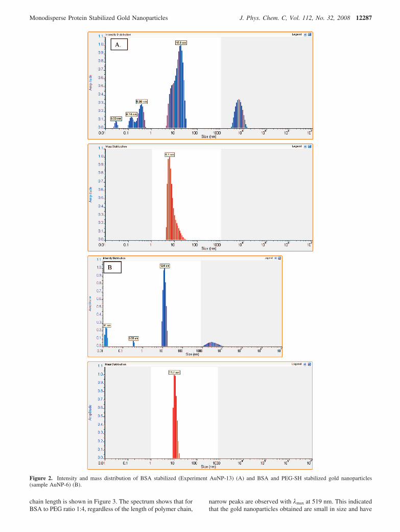
To further improve the size distribution and stability of proteinstabilized gold nanoparticles for longer period of time, mixedmonolayer protected gold nanoparticles were considered. BSAstabilized and mixed monolayer stabilized gold nanoparticlesin solution are shown (see Supporting Information S2). Thecomparison of size and polydispersity of BSA stabilized andBSA and PEG-SH stabilized gold nanoparticles is shown (Figure2). Therefore, we have first selected polyethylene glycol (MeO-PEG-SH) of varying molecular weights for the synthesis ofmixed monolayer gold nanoparticles. Polyethyleneglycol is abiocompatible polymer, which is used extensively to preventthe nonspecific adsorption of serum protein on the drug or genedelivery vehicle.20 Therefore, one potential advantage of includ-ing PEG in mixed monolayer is the prevention of nonspecific
adsorption of BSA stabilized gold nanoparticles on surfaces.We have studied the effect of different chain lengths of PEGpolymer on the size and polydispersity of gold nanoparticles.Feldheim et al. reports the mixed monolayer stabilized goldnanoparticles, where the mixed monolayer is composed of apeptide and PEG.8 However, the process of making mixedmonolayer stabilized gold nanoparticles requires a multistepreaction pathway and a broad size distribution of the nanopar-ticles is obtained. The molecular weights of PEG used in thisstudy are 0.9, 1.5 and 5 kDa. It has also been reported that 5kDa PEG are comparatively better in preventing the nonspecificadsorption of proteins on the nanoparticles. We have thereforeused higher molecular weight PEG polymers (5, 20 and 30 kDa)to enhance the stability of nanoparticles and to get a pronouncedeffect of polymer chain length on gold nanoparticles. We havealso studied the effect of varying the molar ratio of BSA toPEG on the stability of the gold nanoparticles. As shown inTable 2, increasing the molecular weight of PEG from 5 to 20kDa, improves drastically the DLS size distribution of theparticles for the PEG to BSA ratio 1:4. (Experiment AuNP-7and AuNP-9) The slight increase in particle size by DLS isassociated with the higher molecular weight of the PEGpolymer. However, when the molecular weight of PEG is 30kDa, there is a slight increase in the size distribution of thenanoparticles. Wang et al. has reported the similar results wherehydrophobic polymer stabilized gold nanoparticles are comparedwith PEG stabilized gold nanoparticles.15 The molecular weightof PEG used in their study is only 3 kDa and the size distributionreported is around 0.2. It has been concluded that hydrophobicpolymer chains are better stabilizing ligands for gold nanopar-ticles as they provide better colloidal stability.15 The sizes andsize distribution of nanoparticles stabilized with the ligandcontaining BSA to PEG ratio 2:3 are shown in Table 2. It isapparent from the DLS results that the above-mentioned mixedligand provides some control of the size distribution of the goldnanoparticles. Gold nanoparticles stabilized with the ligandcontaining BSA to PEG molar ratio 1:4 show that increasingpolymer chain length decreases the nanoparticle size distribution.However, with ligand containing BSA to PEG ratio 2:3, thesize distribution of nanoparticles increases. (Experiment AuNP-6, AuNP-8 and AuNP-10) This indicates that the stability ofthe BSA stabilized gold nanoparticles is dependent on densePEG brushes on the nanoparticles. DLS results indicate thatPEG-BSA stabilized gold nanoparticles are much more stableas compared to pure BSA stabilized gold nanoparticles (Figure2). Furthermore, BSA-PEG stabilized gold nanoparticles do notshow aggregation even after 3 weeks.
To further confirm that BSA to PEG ratio 1:4 is better instabilizing the gold nanoparticles, UV visible spectroscopy isdone. UV visible spectrum of nanoparticles with different PEG
TABLE 2: Effect of Polymer Concentration, Molecular Weight of Polymer and Molar Ratio of [AuCl4]/[BSA]/[polymer] onParticle Size and Size Distribution of Nanoparticles.a
samples polymer used BSA (µM) polymer (Mn - kg.mol-1) molar ratio [AuCl4]/[BSA]/[polymer] DLS size (nm) size distr. (DLS)
AuNP-6 PEG-SH 76.0 5 1:0.04:0.06 12 0.16AuNP-7 PEG-SH 38.0 5 1:0.02:0.08 8 0.27AuNP-8 PEG-SH 76.0 20 1:0.04:0.06 13 0.20AuNP-9 PEG-SH 38.0 20 1:0.02:0.08 9 0.17AuNP-10 PEG-SH 76.0 30 1:0.04:0.06 13 0.23AuNP-11 pNIPAM 76.0 ∼5 1:0.04:0.06 10 0.32AuNP-12 pNIPAM 38.0 ∼5 1:0.02:0.08 8 0.29AuNP-13 - 40.6 - 1:0.02:0 8 0.45AuNP-14 - 81.3 - 1:0.04:0 6 0.24
a Molar ratio of [AuCl4]/[I] is set to 1:3 and gold nanoparticles synthesized in a photoreactor at a wavelength of 300 nm.
12286 J. Phys. Chem. C, Vol. 112, No. 32, 2008 Housni et al.
chain length is shown in Figure 3. The spectrum shows that forBSA to PEG ratio 1:4, regardless of the length of polymer chain,
narrow peaks are observed with λmax at 519 nm. This indicatedthat the gold nanoparticles obtained are small in size and have
Figure 2. Intensity and mass distribution of BSA stabilized (Experiment AuNP-13) (A) and BSA and PEG-SH stabilized gold nanoparticles(sample AuNP-6) (B).
Monodisperse Protein Stabilized Gold Nanoparticles J. Phys. Chem. C, Vol. 112, No. 32, 2008 12287

a narrow size distribution. However for the BSA to PEG ratio2:3, the peaks are broader for all the PEG chain lengths, andλmax is at 514 nm, 509 and 515 nm respectively for 5, 20 and30 kDa PEG chain length. The broader peaks indicate theformation of aggregates due to larger size or instability of goldnanoparticles. DLS results, as shown in Table 2, confirm thatsize of nanoparticles having BSA to PEG ratio 2:3 are onlyslightly larger than the nanoparticles having BSA to PEG ratio1:4. The broader peaks in UV-visible spectrum of nanoparticles,stabilized with ligand containing BSA to PEG ratio 2:3 are dueto the aggregation of nanoparticles. The cause of this aggregationis related to the higher content of BSA in the ligand shell. Toprove this, we obtained UV-visible spectra of gold nanopar-ticles synthesized with increasing concentration of BSA instabilizing shell, while keeping PEG chain lengths constant. UVvisible spectra indicate that the increase in the concentration ofBSA in the mixed monolayer causes enormous changes inspectrum (see Figure 4). The increase in concentration of BSAis responsible for the aggregation of nanoparticles and hencethe broadening of peaks in UV-visible spectrum. We have alsoshown that this trend is independent of the polymer chain lengthused in the stabilizing shell. The increase in the concentrationof BSA in mixed monolayer regardless of the PEG chain lengthin ligand shell, causes aggregation apparent by the broad peakof the spectra (see Supporting Information S3).
Once mixed monolayer stabilized gold nanoparticles areobtained, we determined critical coagulation concentration ofour samples assess their stability in high salt conditions. It haspreviously been shown that addition of peptides to goldnanoparticles can decrease their stability in salt solutions.9,20
Figure 5 shows that with the increase in salt concentration inBSA stabilized gold nanoparticles, there is a slight red shift inUV spectrum, indicating the aggregation. However, it shouldbe noted that even after the addition of 1 M NaCl solution inour sample no visible color change was observed, and λmax
shifted only by 1 nm. Thus BSA stabilized nanoparticles arestable to high salt concentration. The stability of mixedmonolayer synthesized gold nanoparticles in high salt conditionis also studied. The mixed monolayer stabilized gold nanopar-ticles do not show a red shift in UV visible spectrum even whenthe salt concentration in solution exceeds to 1 M (see Figure 6and Supporting Information S4 and S5). This indicates thatmixed monolayer stabilized gold nanoparticles are betterprotected than BSA stabilized gold nanoparticles and are betterable to withstand with high salt content.
Another concern while using UV irradiated approach for thesynthesis of gold nanoparticles was the denaturation of proteinduring the process. UV radiation is one of the many factorsthat can denature the protein, and the physiological functioning
Figure 3. UV-visible spectra of mixed monolayer stabilized goldnanoparticles. The particles are synthesized using BSA and PEG-SH(5, 20 or 30 kDa), by varying the [BSA]/ [PEG] molar ratio: 1:4 (A)and 2:3 (B).
Figure 4. UV-visible spectra of mixed monolayer stabilized goldnanoparticles with increasing concentration of BSA.
Figure 5. Stability of BSA/AuNPs was assessed by measuring thecritical flocculation concentration using 1.7 M NaCl solution. Molarratio of [AuCl4]/[BSA] were 3:0.02 (AuNP-13).
12288 J. Phys. Chem. C, Vol. 112, No. 32, 2008 Housni et al.

of protein is disrupted. To address this issue, we performedpolyacrylamide gel electrophoresis (PAGE) on UV irradiatedand native BSA (see Supporting Information S6) PAGE resultsindicate that native BSA and UV irradiated BSA show the exactsame pattern. Both of them tend to dimerize and trimerize inbuffer solution, indicated by the slow moving bands in the gel.This is a well-known property of BSA that is not denatured.36
Thus our approach for the gold nanoparticles synthesis by UVradiation does not damage the overall structure of protein. Tofurther confirm our PAGE result, we performed fluorescencespectroscopy on native and UV irradiated BSA samples.Fluorescence spectroscopy is useful to obtain local informationabout the conformation of protein. BSA has intrinsic fluores-cence because of the presence of two tryptophan residue atposition 134 and 212. The more specific information about localstructure of this protein can be obtained by selectively excitingthe tryptophan residues.19,24 To study this we have performedfluorescence spectrum of native and UV irradiated BSA byexciting the tryptophan residue at 235 nm. As shown in Figure7, there is almost no change in the emission spectra of nativeand UV irradiated BSA, indicating that protein is physiologicallyactive.19,34
For the purpose of versatility, we have also synthesizedpNIPAM/BSA mixed monolayer stabilized gold nanoparticles.Table 2 shows nearly monodisperse particles obtained from the
experiments. pNIPAM is a water soluble polymer that exhibitsamphiphillic behavior near its lower critical solution temperature(LCST ) 32 °C). We have used a one step facile technique forthe synthesis of BSA-PEG or BSA-pNIPAM stabilized goldnanoparticles. In future experiments, binding behavior of BSAwith different exogenous or endogenous substances in thepresence and absence of gold nanoparticles will be studied. Thepresence of mixed monolayer on the binding behavior of BSAwill also be studied.
Conclusion
In conclusion, we have studied the synthesis of BSA stabilizedgold nanoparticles by the photochemical process. We havesuccessfully synthesized monolayer and mixed monolayerstabilized gold nanoparticles in one step. We have also studiedthe effect of concentration of BSA on the core size andpolydispersity of gold nanoparticles. DLS data and TEM imageshave confirmed the presence of relatively monodisperse goldnanoparticles. The polydispersity of mixed monolayer stabilizedgold nanoparticles is largely dependent on the concentration ofBSA in mixed monolayer. UV visible spectrum obtained formixed monolayer stabilized gold nanoparticles indicates ag-gregation with the increase in BSA content. The stability ofgold nanoparticles is determined by critical coagulation con-centration determination, which indicates the presence of stablenanoparticles in high salt conditions. Finally, PAGE andfluorescence spectrum have confirmed that BSA is not denaturedduring the photochemical process and it still possess its nativeconformation.
Acknowledgment. The authors thank Natural Sciences andEngineering Research Council of Canada for financial supportof this research work.
Supporting Information Available: TEM micrographs,digital photograph of protected gold nanoparticles, UV-visiblespectra of gold nanoparticles and PAGE experiment. Thismaterial is available free of charge via the Internet at http://pubs.acs.org.
References and Notes
(1) Dahl, A. J.; Maddux, S. L. B.; Hutchison, E. J. Chem. ReV. 2007,107, 2228.
(2) Ghosh, K. S.; Pal, T. Chem. ReV. 2007, 107, 4797.(3) Daniel, M-C.; Astruc, D. Chem. ReV. 2004, 104, 293.(4) Rosi, L. N.; Mirkin, A. C. Chem. ReV. 2005, 105, 1547.(5) Fuente, M. J.; Alcantara, D.; Penades, S. IEE Trans. Nanobiosci.
2007, 6, 275.(6) Ojeda, R.; Paz, L. J.; Barrientos, G. A.; Lomas, M.; Penades, S.
Carbohydr. Res. 2007, 342, 448.(7) Zhou, X.; Zhang, X.; Yu, X.; Zha, X.; Fu, Q.; Liu, B.; Wang, X.;
Chen, Y.; Chen, Y.; Shan, Y.; Jin, Y.; Wu, Y.; Liu, J.; Kong, W.; Shen, J.Biomaterials 2008, 29, 111.
(8) Ryan, A. J.; Overton, W. K.; Speight, E. M.; Oldenburg, N. C.;Loo, L.; Robarge, W.; Franzen, S.; Feldheim, L. D. Anal. Chem. 2007, 79,9150.
(9) Medley, C. D.; Smith, J. E.; Tang, Z.; Wu, Y.; Bamrungsap, S.;Tan, W. Anal. Chem. 2008, 80, 1067.
(10) Schofield, L. C.; Field, A. R.; Russell, A. D. Anal. Chem. 2007,79, 1356.
(11) Fuente, M. J.; Penades, S. Glycoconjugate J. 2004, 21, 149.(12) Labande, A.; Ruiz, J.; Astruc, D. J. Am. Chem. Soc. 2002, 124,
1782.(13) Liu, Z.; Hu, P. J. Am. Chem. Soc. 2002, 124, 14770.(14) Li, D.; He, Q.; Cui, Y.; Duan, L.; Li, J. Biochem. biophys. Res.
Commun. 2007, 355, 488.(15) Wang, Z.; Tan, B.; Hussain, I.; Schaeffer, N.; Wyatt, F. M.; Brust,
M.; Cooper, I. A. Langmuir. 2007, 23, 885.
Figure 6. Stability of BSA/PEG-SH AuNPs assessed by measuringthe critical flocculation concentration using 1.7 M NaCl solution (sampleAuNP-7).
Figure 7. Fluorescence emission spectra of 76 µM BSA before andafter UV-Irradiation (300 nm) at pH 7.41 upon excitation at 295 nm in0.1 M phosphate buffer solution.
Monodisperse Protein Stabilized Gold Nanoparticles J. Phys. Chem. C, Vol. 112, No. 32, 2008 12289

(16) Hostetler, J. M.; Wingate, E. J.; Zhong, C-J.; Harris, E. J.; Vachet,W. R.; Clark, R. M.; Londono, D. J.; Green, J. S.; Stokes, J. J.; Wignall,D. G.; Glish, L. G.; Porter, D. M.; Evans, D. N.; Murray, W. R. Langmuir.1998, 14, 17.
(17) Pan, Y.; Neuss, S.; Leifert, A.; Fischler, M.; Wen, F.; Simon, U.;Schmid, G.; Brandau, W.; Jahnen-Dechent, W. Small 2007, 3, 1941.
(18) Mcgilvray, L. K.; Decan, R. M.; Wang, D.; Scaiano, C. J. J. Am.Chem. Soc. 2006, 128, 15980.
(19) Shang, L.; Wang, Y.; Jiang, J.; Dong, S. Langmuir 2007, 23, 2714.(20) Liu, Y.; Shipton, K. M.; Ryan, J.; Kaufman, D. E.; Franzen, S.;
Feldheim, L. D. Anal. Chem. 2007, 79, 2221.(21) Bergen, M. J.; Recum, A. H.; Goodman, T. T.; Massey, P. A.; Pun,
H. S. Macromol. Biosci. 2006, 6, 506.(22) Narain, R.; Housni, A.; Gody, G.; Boullanger, P.; Charreyre, M.;
Delair, T. Langmuir 2007, 23, 12835.(23) Meziani, J. M.; Pathak, P.; Harruff, A. B.; Hurezeanu, R.; Sun,
Y-P. Langmuir 2005, 21, 2008.(24) Hansen, K. U. Pharmacol. ReV. 1981, 33, 17.(25) Derfus, M. A.; Chan, C. W.; Bhatia, N. S. Nano Lett. 2004, 4, 11.(26) Brewer, H. S; Glomm, R. W.; Johnson, C. M.; Kang, K. M.;
Franzen, S. Langmuir 2005, 21, 9303.
(27) Wangoo, N.; Bhasin, K. K.; Boro, R.; Suri, R. C. Anal. Chim. Acta2008, 610, 142.
(28) Raschke, G; Kowarik, S.; Franzl, T.; Sonnichsen, C.; Klar, A. T.;Feldmann, J Nano Lett. 2003, 3, 935.
(29) Xie, H.; Tkachenko, G. A.; Glomm, R. W.; Ryan, A. J.; Brennaman,K. M.; Papanikolas, M. J.; Franzen, S.; Feldheim, L. D. Anal. Chem. 2003,75, 5797.
(30) Wang, L.; Wang, L.; Zhu, C.; Wei, X.; Kan, X. Anal. Chim. Acta2002, 468, 35.
(31) Wang, L.; Wei, G.; Guo, C.; Sun, >L.; Sun, Y.; Song, Y.; Yang,T.; Li, Z. Colloids Surf., A. 2008, 312, 148.
(32) Yang, T.; Li, Z.; Wang, L.; Guo, C.; Sun, Y. Langmuir 2007, 23,10533.
(33) Philo, S. J. AAPS J. 2006, 8, E564.(34) Royer, A. C. Chem. ReV. 2006, 106, 1769.(35) Rangnekar, A.; Sarma, K. T.; Singh, K. A.; Deka, J.; Ramesh, A.;
Chattopadhyay, C. Langmuir 2007, 23, 5700.(36) Restani, P.; Ballabio, C.; Cattaneo, A.; Isoardi, P.; Terracciano,
L.; Fiocchi, A. Allergy 2004, 59, 21.
JP803890A
12290 J. Phys. Chem. C, Vol. 112, No. 32, 2008 Housni et al.
Copyright © 2022 FDOKUMEN