
Photochemical Control of Rheology - Repositório da ...
157
João Miguel Ribeiro Avó Mestre em Bioorgânica Photochemical Control of Rheology Dissertação para obtenção do Grau de Doutor em Química Orientador: Professor Doutor A. Jorge Parola, Professor Associado, FCT-UNL Co-orientador: Professor Doutor João C. Lima, Professor Associado, FCT-UNL Júri: Presidente: Professora Doutora Maria Luísa Dias de Carvalho de Sousa Leonardo Arguentes: Professor Doutor Francisco Galindo Honrubia Doutor Carlos Miguel Calisto Baleizão Vogais: Professor Doutor Carlos Alberto Mateus Afonso Professora Doutora Paula Cristina de Sério Branco Novembro de 2014
-
Upload
khangminh22 -
Category
Documents
-
view
0 -
download
0
Transcript of Photochemical Control of Rheology - Repositório da ...

João Miguel Ribeiro Avó
Mestre em Bioorgânica
Photochemical Control of Rheology
Dissertação para obtenção do Grau de Doutor em
Química
Orientador: Professor Doutor A. Jorge Parola, Professor
Associado, FCT-UNL
Co-orientador: Professor Doutor João C. Lima, Professor
Associado, FCT-UNL
Júri:
Presidente: Professora Doutora Maria Luísa Dias de Carvalho de Sousa Leonardo
Arguentes: Professor Doutor Francisco Galindo Honrubia
Doutor Carlos Miguel Calisto Baleizão
Vogais: Professor Doutor Carlos Alberto Mateus Afonso
Professora Doutora Paula Cristina de Sério Branco
Novembro de 2014

II

III
João Miguel Ribeiro Avó
Mestre em Bioorgânica
Photochemical Control of Rheology
Dissertação para obtenção do Grau de Doutor em
Química
Supervisor: Professor Doutor A. Jorge Parola, Professor
Associado, FCT-UNL
Co-supervisor: Professor Doutor João C. Lima, Professor
Associado, FCT-UNL

IV

V
“The respect of those you respect is worth more than the applause of
the multitude.”
— Arnold Glasow

VI

VII
Copyright
A Faculdade de Ciências e Tecnologia e a Universidade Nova de Lisboa têm o direito,
perpétuo e sem limites geográficos, de arquivar e publicar esta dissertação através de exemplares
impressos reproduzidos em papel ou de forma digital, ou por qualquer outro meio conhecido ou que
venha a ser inventado, e de divulgar através de repositórios científicos e de admitir a sua cópia e
distribuição com objectivos educacionais ou de investigação, não comerciais, desde que seja dado
crédito ao autor e editor.

VIII
IX
Agradecimentos
E eis que chega ao fim esta jornada. Talvez mais do que o conhecimento adquirido durante
estes 4+ anos, fica toda uma evolução psicológica que resulta do aglomerado de sucessos e fracassos,
da montanha russa emocional que acompanha um doutoramento. Assim, quero agradecer a todos os
que me acompanharam neste pedaço de vida.
Em primeiro lugar os meus orientadores. Ao Jorge Parola pela confiança que depositou em
mim e que me permitiu realizar uma investigação que me interessava, e que me obrigou a ir mais além
para resolver os inúmeros problemas que surgiram. Ao João Lima, que com o seu sempre presente
“tough-love” me proporcionou momentos de extrema miséria intelectual, mas que foram essenciais
para aperfeiçoar o meu espírito crítico e pensar mais além, características essenciais de um bom
cientista.
A todo o grupo de fotoquímica, por terem criado um bom (ainda que por vezes demasiado
barulhento) ambiente de trabalho, tão necessário para manter a criatividade. Em especial ao Artur e à
Ana Marta, que para além das discussões científicas, me deram um importante apoio nos maus
momentos.
À minha família, pelo apoio que me deram “fora das 4 linhas”. Ao meu Pai, que sempre foi o
meu porto seguro e me deu segurança para chegar até aqui e à Linda por toda a paciência para nos
aturar. À minha Mãe, que me fez ser como sou hoje e que estou certo que ficaria orgulhosa por ter
chegado até aqui.
E por fim, à Carina, o meu tudo. Foste a minha âncora emocional e sem ti teria certamente
desistido disto a dada altura. Obrigado por toda a confiança que tiveste em mim mesmo quando eu já
não tinha. Obrigado por tudo.

X

XI
Resumo
O trabalho de investigação elaborado no âmbito desta tese de Doutoramento teve como
objectivo a preparação de novos fluídos fotorreológicos. Para isso, foram seguidas duas estratégias
distintas. A primeira baseou-se na síntese de compostos tripodais funcionalizados com unidades
fotodimerizáveis derivadas de ácido cinâmico, coumarina e antraceno. Foram sintetizados dois
conjuntos de compostos variando as unidades centrais bem como os espaçadores, obtendo-se
moléculas de diferente peso molecular e solubilidade. Os compostos sintetizados foram caracterizados
quanto às suas propriedades fotoquímicas e todos exibiram reactividade quando irradiados com luz
ultravioleta. Em particular, ambos os compostos derivados de coumarina apresentaram a maior
reactividade quanto à fotopolimerização, resultando na formação de nanopartículas dendriméricas ou
no aumento da viscosidade de soluções orgânicas. A segunda estratégia focou-se no desenho
cuidadoso de líquidos iónicos fotossensíveis, baseado em resultados de diversos estudos quantitativos
de relação estrutura-propriedade. Desta forma, foram sintetizados líquidos iónicos com catiões
funcionalizados com unidades de ácido cinâmico ou coumarina. Aquando da sua irradiação, todos os
compostos apresentaram reactividade, resultando na alteração das suas propriedades físicas, tais como
o ponto de fusão ou viscosidade. Para além disso, foram desenvolvidos novos derivados de coumarina
com diferentes propriedades fotofísicas e fotoquímicas com possível aplicação na síntese de novos
líquidos iónicos fotossensíveis.
Palavras-Chave: Fotoquímica, Fotopolimerização, Fotorreologia, Líquidos Iónicos, Coumarinas

XII

XIII
Abstract
The main objective of the research work developed in the framework of this PhD thesis was
the preparation and development of novel photorheological fluids. This was pursued following two
distinct strategies. The first one focused on the synthesis of tripodal compounds functionalized with
photodimerizable moieties of cinnamic acid, coumarin and anthracene. Two sets of compounds were
prepared, varying the central unit as well as spacers resulting in molecules with different solubilities
and molecular weight. All compounds were characterized towards their photochemical properties and
all exhibited photoreactivity upon irradiation with ultra-violet light. In particular, both coumarin
derivatives exhibited the greatest photopolymerization reactivity, resulting in the formation of
dendrimeric nanoparticles or in the increase of viscosity of organic solutions. The second strategy was
focused on the careful design of photosensitive ionic liquids, based on the results of several
quantitative structure-property relationship studies. Thus, photosensitive ionic liquids were
synthesized bearing cinnamic acid or coumarin moieties in the organic cation. Upon irradiation, all
compounds exhibited reactivity, which resulted in changes in their physical properties, such as melting
point or viscosity. In addition, novel coumarin chromophores with different photophysical and
photochemical properties were developed. It is expected that these compounds may find application in
the preparation of new photosensitive ionic liquids.
Keywords: Photochemistry, Photopolymerization, Photorheology, Ionic Liquids, Coumarins

XIV

XV
Contents
I. Introduction ..................................................................................................................................... 3
Stimuli-responsive materials ............................................................................................................... 3
Stimuli-responsive rheological fluids .................................................................................................. 4
Photorheological fluids .................................................................................................................... 6
Thesis Overview ................................................................................................................................ 17
II. Photopolymerizable compounds for photochemical control of rheology...................................... 21
II.1 – Low molecular-weight photopolymerizable molecules ........................................................... 23
Conclusions ................................................................................................................................... 33
II.2 – High-molecular weight photopolymerizable compounds ........................................................ 34
Conclusions ................................................................................................................................... 48
III. Photoactive ionic liquids ........................................................................................................... 53
III.1 – Introduction ............................................................................................................................ 53
Ionic liquids in smart material systems ......................................................................................... 54
Viscosity of ionic liquids ............................................................................................................... 60
III.2 – Cinnamic acid based ionic liquids .......................................................................................... 62
III.3 – Coumarin based ionic liquids ................................................................................................. 80
III.3.1 – Novel coumarin chromophores for the preparation of photoresponsive ionic liquids .... 91
III.4 – Conclusions .......................................................................................................................... 104
IV. Conclusions and Future Perspectives ...................................................................................... 105
V. Experimental Part ........................................................................................................................ 106
Synthesis .......................................................................................................................................... 107
Procedures ................................................................................................................................... 107
Photochemistry ................................................................................................................................ 122
Dynamic Light Scattering ............................................................................................................... 123
Atomic Force Microscopy ............................................................................................................... 123
Viscometry ...................................................................................................................................... 123
Photophysics .................................................................................................................................... 123

XVI
Computational calculations ............................................................................................................. 124
VI. References ............................................................................................................................... 125

XVII
Figure index
Figure I.1 – Schematic representation of suspended particle alignment induced by an electric or
magnetic field (adapted from [5]) ........................................................................................................... 5
Figure I.2 – Microscopic photographs of paraffin-based ER fluid before (left) and after (right)
application of an electric field (adapted from [13]) ................................................................................. 6
Figure I.3 – Schematic representation of different micellar aggregate morphology upon increase in
concentration. .......................................................................................................................................... 8
Figure I.4 – Schematic representation of different effective head group sizes of anionic surfactant and
corresponding micellar aggregation morphologies (adapted from [29]) ................................................. 9
Figure I.5 – Schematic representation of isomerization induced changes in aggregation behaviour of
azobenzene-based cationic surfactant micelles (adapted from [42]). .................................................... 11
Figure I.6 – Light-induced gelation of aqueous laponite suspensions: initially (left) particles are
stabilized by pluronic surfactant and unconnect; upon UV irradiation (right), the particles assemble
into a network, causing the fluid to turn into a gel (adapted from []) ................................................... 12
Figure I.7 – Schematic representation of branched PEG polymer modified with 9-anthracene
carboxylic acid used in the formulation of a PR fluid (adapted from [62]). ......................................... 13
Figure I.8 – Schematic representation of light-induced crosslinking between polymer chains due to
anthracene dimerization used in the photochemical controlled release of bioactive molecules (adapted
from [66]). ............................................................................................................................................. 14
Figure I.9 – Schematic representation of light-induced crosslinking between polymer chains due to
coumarin dimerization used in the photochemical control of fluid viscosity (adapted from [68]). ...... 15
Figure I.10 – (a) Schematic representation of the photoresponse of cinnamate monomer 11 and dimer
12; (b) Photographic images representing the photorheological effect of acetone solutions of 11
(adapted from [76]). .............................................................................................................................. 16
Figure II.1 – Schematic representation of photocontrolled reversible polymerization of tripodal
molecules bearing photosensitive moieties. .......................................................................................... 21
Figure II. 2 – Representation of orbital symmetry of frontier orbitals of generic alkene (left) and
symmetry forbidden orbital interaction in a thermal [2+2] cycloaddition (right) ................................. 22
Figure II. 3 – Frontier orbitals of generic alkene in the excited state (left) and symmetry allowed
orbital interactions in a photochemical induced [2+2]-cycloaddition (right) ........................................ 22
Figure II. 4 – Symmetry allowed frontier orbital interactions between one coumarin molecule in the
ground state and one molecule in the excited state ............................................................................... 23
Figure II.5 – Molecular structure of tris(2-aminoethyl)amine 13 ........................................................ 23
XVIII
Figure II.6 – (a) UV-Vis spectral modifications of 1.1×10⁻⁵
M 16 in chloroform upon irradiation at 300
nm. Total irradiation time is 20 min; (b) Photochemical quantum yield dependence on concentration of
16. .......................................................................................................................................................... 25
Figure II. 7 – 1H NMR spectra of 16 in chloroform acquired before and after irradiation at 300 nm. . 26
Figure II. 8 – (a) UV-Vis spectral modifications of chloroform solution of 22 at 1.0×10⁻⁵
M upon
irradiation at 330 nm. Total irradiation time is 60 min; (b) Photochemical quantum yield variation with
increasing concentration of 22; (c) Precipitation-induced UV-Vis spectral modifications of a 2.0×10⁻³
M solution of 22 in chloroform upon irradiation at 330 nm. Total irradiation time is 40 min. ............. 29
Figure II. 9 – 1H NMR spectra of 22 acquired before and after irradiation at 330 nm in chloroform. . 30
Figure II. 10 – (a) Correlation curve variation upon irradiation at 330 nm of a solution of 22 at 8.3×10-
3 M in chloroform; (b) Time and concentration dependence of photoinduced growth of dendrimeric
nanoparticles of 22 in chloroform; (c) and (d) AFM images of a suspension of dendrimeric
nanoparticles of 22 obtained after 10 and 400 minutes of irradiation at 330 nm, respectively. ............ 32
Figure II. 11 – Schematic representation of the proposed photoinduced dendrimeric growth of
nanoparticles of 22 due to dimerization of coumarin moieties. ............................................................ 32
Figure II.12 – Molecular structures of backbones used for the preparation of photopolymerizable
tripodal compounds: tris(2-aminoethyl)amine and tris(tetraethyleneglycol)phloroglucinol 23. ........... 35
Figure II. 13 – (a) UV-Vis spectral modification of a solution of 1.4×10⁻⁵
M 31 in chloroform upon
irradiation at 300 nm. Total irradiation time is 20 min; (b) Photochemical quantum yield dependence
on concentration of 31. .......................................................................................................................... 37
Figure II. 14 – 1H NMR spectra of 31 in chloroform before and after irradiation at 300 nm. .............. 37
Figure II. 15 – (a) UV-Vis spectral modification of a chloroform deaerated solution of 36 at 3.0×10⁻⁵
M upon irradiation at 360 nm. Total irradiation time is 10 min; (b) Photochemical quantum yield
dependence on the concentration of 39 ................................................................................................. 39
Figure II. 16 – 1H NMR spectra of 36 in chloroform obtained before and after irradiation at 360nm.
Total irradiation time is 60 min. ............................................................................................................ 40
Figure II. 17 – Viscosity of binary mixtures of water and acetonitrile at different ratios, determined by
direct measurement with conventional viscometer (black dots) and calculated indirectly from DLS
data (red dots). ....................................................................................................................................... 41
Figure II.18 - (a) UV-Vis spectral modification of a solution of 39 at 1.1×10⁻⁵
M in chloroform upon
irradiation at 320 nm. Total irradiation time is 60 min; (b) Photochemical quantum yield dependence
on concentration of 39. .......................................................................................................................... 43
Figure II. 19 – 1H NMR spectra of 39 in chloroform obtained before and after irradiation. ................ 44
Figure II. 20 – Representation of the three main rheological behaviors exhibited by fluids ................ 45
Figure II. 21 – Schematic representation of the typical components of a rotational rheometer ............ 46
Figure II. 22 – Viscosity curve of a DMF solution of 39 at 10% (w/v) before (black dots) and after (red
dots) irradiation at 320 nm. Total irradiation time is 120 min. ............................................................. 47

XIX
Figure III.1 - Molecular structure of the first synthesized ionic liquids; a) ethanolammonium nitrate
40; b) ethylammonium nitrate 41 .......................................................................................................... 53
Figure III. 2 - Representative ionic liquid components ......................................................................... 54
Figure III. 3 - Molecular structure of ionic liquids used as electrolytes; (a) methylpropylpyrrolidinium
bis(trifluoromethyl-sulfonyl)imide 42; (b) polymerizable 2-(dimethylethylamino)ethylmethacrylate
bromide 43 ............................................................................................................................................. 55
Figure III. 4 – Ionogel based wearable pH sensor for the analysis of human sweat [141] .................... 56
Figure III. 5 – Reversible color switching between three oxidation states of pure
ethylmethylimidazolium vanadium (V) oxide IL upon electrolysis (adapted from [146]) ................... 57
Figure III. 6 – Increased selectivity of methylred based IL over conventional sodium salt from pH 1 to
7 (adapted from [147]) .......................................................................................................................... 57
Figure III. 7 – Molecular structures of azobenzene based photochromic ionic liquids (from [148] and
[149]) ..................................................................................................................................................... 58
Figure III. 8 – Cinnamate-based photoresponsive ionic liquids (from [154]) ....................................... 59
Figure III. 9 – Effect of cation alkyl chain length and counterion on hexafluorophosphate (red dots)
and bis(trifluoromethyl-sulfonyl)imide (black dots) alkylmethylimidazolium ILs (from [161]).......... 61
Figure III. 10 - Crystal structures of the compounds 5a (top) and 5b (bottom), showing the label
scheme for all non-H-atoms which are represented as thermal ellipsoids drawn at the 50% probability
............................................................................................................................................................... 66
Figure III. 11 - Representation of the diverse C−H•••O (yellow dashed lines), C−H•••N (blue dashed
lines) and C−H•••F (green dashed lines) weak hydrogen bonds found in the crystal structures of the
compounds 55 and 58: (a) highlight of the interactions between adjacent cationic and anionic species
in 55; (b) crystal packing of 55 viewed in the [1 0 0] direction of the unit cell; (c) crystal packing of 58
viewed in the [1 0 0] direction of the unit cell ...................................................................................... 67
Figure III. 12 - Spectral modifications of 55 at 5.4 x 10⁻⁵
M in acetonitrile upon irradiation at 300 nm
(a) and 240 nm (b). Under the used experimental setup, PSS was reached after 10 min irradiation at
300 nm while the recovery by irradiating at 240 nm required ca. 600 min. .......................................... 69
Figure III. 13 – 1H NMR spectrum of irradiated solution of ionic liquid 55 at the photostationary state
............................................................................................................................................................... 69
Figure III. 14 - 1H NMR spectrum of irradiated solution of ionic liquid 58 at the photostationary state
............................................................................................................................................................... 69
Figure III. 15 - 1H NMR spectrum of a solution of ionic liquid 55 after irradiation at 240 nm (55rec) . 70
Figure III. 16 – DSC thermograms of ionic liquids 55 (a) and 58 (b) before irradiation (i), at the PSS
(PSS) and after photochemical recovery (rec). .......................................................................................... 70
Figure III. 17 – 1H NMR spectrum of 55 in chloroform after irradiation at 300 nm in neat conditions.
Total irradiation time is 120 min. .......................................................................................................... 71

XX
Figure III. 18 - 1H NMR spectrum of 55 in chloroform after irradiation at 300 nm in neat conditions
under inert atmosphere. Total irradiation time is 120 min. ................................................................... 72
Figure III. 19 - Isomer fraction dependency of melting temperature depression of irradiated neat
samples (black dots) and acetonitrile solution (red dot) of 55. ............................................................ 73
Figure III. 20 - Spectral modifications of 60 at 1.2 x 10⁻⁵
M in acetonitrile upon irradiation at 300 nm
(a) and 240 nm (b). Under the used experimental setup, PSS was reached after 15 min irradiation at
300 nm while the recovery by irradiating at 240 nm required ca. 600 min ........................................... 75
Figure III. 21 – 1H NMR spectrum of 60 irradiated in chloroform at 300 nm. Total irradiation time is
120 min. ................................................................................................................................................. 75
Figure III. 22 – 1H NMR spectrum of 60 in chloroform after irradiated at 300 nm in neat conditions.
Total irradiation time is 120 min. .......................................................................................................... 76
Figure III. 23 - 1H NMR spectrum of 60 in chloroform after irradiated at 300 nm in neat conditions
under inert atmosphere. Total irradiation time is 120 min. ................................................................... 76
Figure III. 24 - Viscosity curve of pure 60 before (black dots) and after (red dots) irradiation at 300
nm. Total irradiation time is 120 min. ................................................................................................... 77
Figure III. 25 – Quadratic dependence of the scattered intensity in IL 60 before (black circles) and
after (red circles) irradiation compared with that obtained in the reference solvent (chloroform, blue
squares) .................................................................................................................................................. 79
Figure III. 26 – (a) Spectral modifications of 62 at 1.7x10⁻⁵
M in acetonitrile upon irradiation at 300
nm. Total irradiation time is 60 min. (b) Photochemical quantum yield dependence with concentration
of 62....................................................................................................................................................... 82
Figure III. 27 - 1H NMR spectrum of 62 in chloroform before and after irradiation at 330 nm. Total
irradiation time is 120 min. ................................................................................................................... 83
Figure III. 28 – 1H NMR spectra of 4-methyl-7-alkoxy coumarin derivative in chloroform before and
after photoinduced dimerization. ........................................................................................................... 83
Figure III. 29 - 1H NMR spectrum of 62 in chloroform after irradiation at 330 nm in neat conditions.
Total irradiation time is 120 min. .......................................................................................................... 85
Figure III. 30 - 1H NMR spectrum of 62 in chloroform after irradiation at 330 nm in neat conditions
under inert atmosphere. Total irradiation time is 120 min. ................................................................... 85
Figure III. 31 - DSC thermograms of ionic liquid 62 before (i) and after (PSS) irradiation at 330 nm. 86
Figure III. 32 - (a) Spectral modifications of 64 at 1.1x10⁻⁵
M in acetonitrile upon irradiation at 330
nm. Total irradiation time is 60 min. (b) Photochemical quantum yield dependence with concentration
of 64....................................................................................................................................................... 87
Figure III. 33 - 1H NMR spectrum of 64 in chloroform obtained after irradiation at 330 nm. Total
irradiation time is 120 min. ................................................................................................................... 87
Figure III. 34 - Viscosity curve of pure 64 before (black dots) and after (red dots) irradiation at 330
nm. Total irradiation time is 120 min .................................................................................................... 88

XXI
Figure III. 35 - Quadratic dependence of the scattered intensity in IL 64 before (black circles) and after
(red circles) irradiation compared with that obtained in the reference solvent (chloroform, blue
squares) .................................................................................................................................................. 90
Figure III. 36 – (a) Fluorescence lifetime decays of 3-vinyl coumarins 72b (black) and 72c (red) and
their respective 3-styryl counterparts 73b (green) and 73c (blue); Fluorescence lifetime d 3-styryl
coumarins 73b (green) and 73c (blue) and their respective nitro derivatives 73f (brown) and 73g
(orange). ................................................................................................................................................ 94
Figure III. 37 – UV-Vis spectral transformation of a solution of 72a at 2.3 mM in acetonitrile upon
irradiation at 313 nm. Total irradiation time is 30 min. ........................................................................ 94
Figure III. 38 - 1H NMR spectra (400 MHz, CDCl3) of a solution of coumarin 77 before (red) and after
(blue) irradiation for 60 min. λirrad = 275 nm ......................................................................................... 97
Figure III. 39 - 1H NMR spectra (400 MHz, CDCl3) of a solution of coumarin 80a irradiated at
different times: 0 min (red); 5 min (yellow); 10 min (green); 20 min (blue); 120 min (purple). λ irrad =
360 nm ................................................................................................................................................... 98
Figure III. 40 - 1H NMR spectra (400 MHz, CDCl3) of a solution of coumarin 80b irradiated at
different times: 0 min (red); 5 min (yellow); 10 min (green); 20 min (blue); 120 min (purple). λirrad =
370 nm ................................................................................................................................................... 99
Figure III. 41 - 1H NMR spectra (400 MHz, CDCl3) of a solution of coumarin 80c irradiated at
different times: 0 min (red); 5 min (yellow); 10 min (green); 20 min (blue); 120 min (purple). λ irrad =
333 nm ................................................................................................................................................... 99
Figure III. 42 - Frontier orbitals for compound 79 obtained from density functional theory (DFT)
calculations .......................................................................................................................................... 100
Figure III. 43- Frontier orbitals for compound 80a obtained from density functional theory (DFT)
calculations .......................................................................................................................................... 100
Figure III. 44 – UV-Vis spectral modification of coumarins 80a (a), 80b (b) and 80c (c) in acetonitrile
upon irradiation at the longest wavelength transition. ......................................................................... 102
Figure III. 45 - Calculated spectra for the Z and E isomers of 80a and the combined spectra of the
mixture of isomers with experimental molar fractions ........................................................................ 103
Figure III. 46 – Fluorescence variation of coumarin 80b adsorbed into hydroxyethylacrylamide/
poly(ethyleneglycol) ............................................................................................................................ 103

XXII

XXIII
Scheme Index
Scheme I.1 – Photochemical dimerization of 9-methyanthracene 1 ....................................................... 7
Scheme I.2 – Photochemical dimerization reaction of coumarin 4 ......................................................... 9
Scheme I.3 – General network of reactions of flavylium compounds in acidic and neutral aqueous
media, exemplified for 4’,7-dihydroxyflavylium 10. ............................................................................ 10
Scheme II. 1 - Synthetic pathway for the preparation of tripodal cinnamic acid derivative 16: i) thionyl
chloride, chloroform, 89%; ii) tris(2-aminoethyl)amine, K2CO3 or NEt3, dichloromethane, 91% or
34%, respectively .................................................................................................................................. 24
Scheme II.2 – Synthetic pathway for the preparation of tripodal anthracene derivative 19: i) thionyl
chloride, chloroform; ii) tris(2-aminoethyl)amine, K2CO3 or NEt3, dichloromethane .......................... 27
Scheme II. 3 - Synthetic pathway for the preparation of tripodal coumarin derivative 22: i) thionyl
chloride, chloroform, 90%; ii) tris(2-aminoethyl)amine, K2CO3 or NEt3, dichloromethane, 95%........ 27
Scheme II. 4 – Synthetic pathway for the preparation of tripodal cinnamate derivative 31: i) K2CO3,
acetone, 72%; ii) phloroglucinol, K2CO3, acetonitrile, 8.6% (for precursor 30). .................................. 36
Scheme II. 5 – Synthetic pathway for the preparation of anthracene precursor 34: i) NaNO2, NaOH,
water, 53%; ii) NaBH4, methanol, 24% ................................................................................................ 38
Scheme II. 6 – Synthetic pathway for the preparation of tripodal anthracene derivative 36: i) K2CO3,
acetone, 76%; ii) phloroglucinol, K2CO3, acetonitrile, 61%. ................................................................ 38
Scheme II. 7 – Synthetic pathway for the preparation of tripodal coumarin derivative 39: i) K2CO3,
acetone, 60%; ii) phloroglucinol, K2CO3, acetonitrile, 63%. ................................................................ 42
Scheme III. 1 – Photochemical reaction of generic spiropyran compounds ......................................... 56
Scheme III. 2 - Ring-closing and ring-opening of photosensitive diarylethene-containing ionic liquids
153 ......................................................................................................................................................... 59
Scheme III. 3 – Synthetic pathway for the preparation of cinnamate organic cations: i)
methanol/H2SO4, 94%; ii) 1,3-dibromopropane, K2CO3, acetone, 94%; iii) N-methylimidazole,
acetonitrile, 76%; iv) trimethylamine, acetonitrile, 66%....................................................................... 63
Scheme III. 4 - Synthetic pathway for the preparation of cinnamate photoresponsive ionic liquids: i)
1,3-dibromopropane, K2CO3, acetone, 94%; ii) 1,6-dibromohexane, K2CO3, acetone, 89%; iii) N-
methylimidazole, acetonitrile, 76%; iv) LiNTf2, dichloromethane, 70-76%. ....................................... 65
Scheme III. 5 – Synthetic pathway for the preparation of room temperature cinnamate ionic liquids: i)
triphenylphosphine, Br2, acetonitrile, 87%; ii) K2CO3, acetone, 91%; iii) N-methylimidazole,
acetonitrile, 91%. ................................................................................................................................... 74
Scheme III. 6 - Synthetic pathway for the preparation of coumarin photoresponsive ionic liquids: i)
1,3-dibromopropane, K2CO3, acetone, 92%; ii) 1,13-dibromotetraethyleneglycol, K2CO3, acetone,
86%; iii) N-methylimidazole, acetonitrile, 75-91%. ............................................................................. 81

XXIV
Scheme III. 7 –Alternative photochemical reactions taken by coumarin derivatives ........................... 84
Scheme III. 8 – Suggested alternative photochemical reactions taken by compounds 62 .................... 84
Scheme III. 9 – Synthetic pathway for the preparation of 3-styryl coumarins 73: i) Pd(OAc)2, DMF, 9-
98%........................................................................................................................................................ 92
Scheme III. 10 – Suggested photochemical reactions of hypothetical coumarin-based stilbene analog
74. .......................................................................................................................................................... 95
Scheme III. 11 – Synthetic pathway for the preparation of 5-styryl coumarins 80a-c: i) OXONE®,
HBr, dichloromethane, 91%; ii) [PdCl2(dppf)].CH2Cl2, CH2CHBF3K, NEt3, n-propanol, 92%; iii)
Pd(PPh3)4, AgOAc, Ar-I, DMF, 50-90%............................................................................................... 95

XXV
Table Index
Table III. 1 – Melting points of ionic cinnamate derivatives 53 and 54 ................................................ 64
Table III. 2 - Melting points of ionic cinnamate derivative 53 with different counterions ................... 64
Table III. 3 - Absorption Maxima (λmax), Photochemical Quantum Yields (ΦR), and Cis Isomer
Fraction before Irradiation and at the Photostationary State (PSS) of Ionic Liquids 55 and 58 in
Acetonitrile ............................................................................................................................................ 68
Table III. 4 - Melting (Tm) and Peak (Tp) Temperatures and Melting Enthalpies (ΔHm) of ILs 55 and
58 before Irradiation at 300 nm (i), after the PSS Has Been Reached (PSS) and after Recovery upon
Irradiation at 240 nm (rec) ...................................................................................................................... 71
Table III. 5 - Time dependent cis isomer ratio and melting point depression (ΔTm) of irradiated neat
samples of ionic liquids 55 and 58 ........................................................................................................ 73
Table III. 6 - Absorption Maxima (λmax), Photochemical Quantum Yields (ΦR), and Cis Isomer
Fraction before Irradiation and at the Photostationary State (PSS) of IL 60 ......................................... 75
Table III. 7 - βHRS, βJ=1 (dipolar) and βJ=3 (octupolar) values (in atomic units*, using Convention T)
and depolar-ization ratios, DR, and anisotropies, ρ, deduced from HRS measurements at 1064 nm for
compound 60, before and after irradiation ............................................................................................ 78
Table III. 8 - Melting (Tm) and Peak (Tp) Temperatures and Melting Enthalpies (ΔHm) of IL 62 before
(i) and after (PSS) irradiation at 330 nm .................................................................................................. 86
Table III. 9 - βHRS, βJ=1 (dipolar) and βJ=3 (octupolar) values (in atomic units*, using Convention T)
and depolar-ization ratios, DR, and anisotropies, ρ, deduced from HRS measurements at 1064 nm for
compound 64, before and after irradiation ............................................................................................ 89
Table III. 10 - Absorption and Emission Maxima, Fluorescence Quantum Yield and Average
Fluorescent Lifetime of Selected Coumarins in Acetonitrile Solutions ................................................ 93
Table III. 11 – Synthetic yields and Z/E ratio of coumarins 80a-c ........................................................ 96
Table III. 12 - Absorption and Emission Maxima, Molecular Absorptivity, Fluorescence Quantum
Yield, Average Fluorescent Lifetime and radiative and apparent nonradiative rate constants of selected
coumarins .............................................................................................................................................. 96
Table III. 13 - Fluorescence and Reaction Quantum Yield, Average Fluorescent Lifetime and
radiative, nonradiative and reactive rate constants of selected coumarins .......................................... 101

XXVI

XXVII
Abbreviation List
A Absorbance
AFM Atomic force microscopy
CA Cinnamic acid
CMC Critical micelle concentration
Compd Compound
Conc. Concentration
CTAB Cetyltrimethylammonium bromide
DFT Density functional theory
Dh Hydrodynamic diameter
DLS Dynamic light scattering
DMF Dimethylformamide
DSC Differential scanning calorimetry
Dt Diffusion coefficient
EDTA Ethylenediaminetetraacetate
ER Electrorheological
HOMO Highest occupied molecular orbital
HRS Hyper-Rayleigh scattering
Hz Hertz
IL Ionic liquid
k Boltzmann constant
knr Non-radiative rate constant
knr’ Apparent non-radiative rate constant
kr Radiative rate constant
LED Light-emitting diode
LMWGs Low-molecular weight gelator
LUMO Lowest unoccupied molecular orbital
M Molar
MALDI-MS Matrix-assisted laser desorption/ionization-mass spectrometry
MR Magnetorheological
NEt3 Triethylamine
nm Nanometer
NMR Nuclear magnetic ressonance
OMCA o-methylcinnamic acid
PCA p-Coumarin acid

XXVIII
PEG Polyethyleneglycol
ppm Parts per million
PR Photorheological
PSS Photostationary state
QSPR Quantitative structure-property relationship
SMAs Shape-memory alloys
SN Nucleophilic substitution
SOMO Single occupied molecular orbital
SRIL Stimuli-responsive ionic liquid
SRM Stimuli responsive materials
T Temperature
TDDFT Time-dependent density functional theory
TEG Tetraethylene glycol
Tf Triflyl
UV Ultra-violet
Vis Visible
Β Hyperpolarizability
ε Molar absorptivity
η Viscosity
λ Wavelength
λabs Wavelength of absorption maximum
λem Wavelength of emission maximum
τ Delay time
τm Average excited state lifetime
ΦF Fluorescence quantum yield
ΦR Photochemical quantum yield

1
Chapter I

2

3
I. Introduction
Stimuli-responsive materials
Throughout human history, technological development has been closely linked to the nature of
the materials used for creating tools and other devices. In fact, pre-historic periods were named after
these materials, i.e. Stone, Bronze and Iron Ages, evidencing their crucial role in human evolution. In
Modern Age, the discovery of the first synthetic polymer by Baekeland, in the first decade of the
twentieth century, marked the beginning of a technological period that lasts until today, the Age of
Polymers. Over the past one hundred years, polymers have grown in popularity and can be found
ubiquitously, progressively replacing wood and metals in a wide variety of applications. However, in
more recent years, the driving force for technological change in many respects has shifted towards
information technology. Instead of traditional structural functions, new materials are required to have
sensibility or actuation capabilities. The increasing need for highly autonomous systems and devices
that require less human control led to pursuit for materials that can intelligently interact with their
environment and structures that assess their own health. Such systems could have a tremendous impact
in advancing many fields including medicine, microelectronics, and robotics, among others. Therefore,
the increasing interest in materials that respond to external stimuli, such as fluctuations in temperature,
pressure or light intensity and change their physico-chemical properties accordingly led to the
beginning of the Smart Material Age.
Smart, or stimuli-responsive material (SRM) is currently defined as “a system or material
which has built-in or intrinsic sensors, actuators and control mechanisms whereby it is capable of
sensing a stimulus, responding to it in a predetermined manner and extent, in an appropriate time, and
reverting to its original state as soon as the stimulus is removed”.1 The first material to evidence a
smart behavior was potassium sodium tartarate, commonly known as Rochelle salt. This compound
displays both pyroelectric and piezoelectric effects, becoming electrically polarized upon an applied
temperature change or mechanical stress. This property led to its extensive use in gramophones,
microphones and earpieces. 2 However, the properties of Rochelle salt are only representative of a
small fraction of smart materials, which can produce a variety of responses to a wide range of stimuli.
Depending on the external stimuli they respond to, or the transformation that is generated, smart
materials can be divided in different classes. For example, pyroelectrics are materials that become
electrically polarized upon a temperature variation. 3 They are particularly used for infrared detection
in surveillance and targeting applications. The infrared radiation is absorbed and induces an increase in
temperature of the pyroelectric component, changing its polarization and, thus, generating an electric
signal. Analogously, piezoelectrics are another class of smart materials that exhibit electric
polarization with an external stimulus. In this case, this effect occurs when mechanical stress is

4
induced. 4 The converse effect can be exhibited, i.e., when an electric field is applied, a dimensional
change takes place. Since the discovery of this effect in the end of the 19th century, piezoelectrics
gathered great attention from the scientific community and became the most representative class of
smart materials. They find application in a wide variety of devices in fields ranging from automotive
industry and telecommunications to medical instruments or military weaponry. 5 Shape-memory alloys
(SMAs) are another class of smart materials that respond to external-stimuli with dimensional
changes. These materials, upon proper thermal and mechanical treatment, have the ability to remember
up to two shapes which they had previously occupied. When subjected to a mechanical load below a
certain temperature, SMAs can be plastically deformed beyond their elastic limit, but then are capable
of regaining their original shape if they are then heated above a certain temperature. SMAs are
employed as actuators in systems used in very different fields, from spacecraft and aircraft to
medicine, robotics or even household appliances. 5 There are also smart materials that generate visual
responses to external stimuli. For example, luminescent materials display the property of emitting
energy in the form of light after excitation by an external stimulus. This excitation can be achieved by
irradiation of light of different wavelength, heating or application of an electric field. Light-emitting
diodes (LED) are the most common type of luminescent materials, which are composed by an
electroluminescent material comprised between two electrodes. Instead of emission of light,
chromogenic materials respond essentially to the same stimuli by changing their color. Electrochromic
materials are the most common type of chromogenics, and the majority of them are composed by
metal oxides that undergo a redox reaction when an electric field is applied, although organic
compounds have recently been employed. Chromogenic and luminescent materials find use in sensor
devices, paints, smart windows, household appliances or automotive industry.6
Stimuli-responsive rheological fluids
One particular type of SRM is the class of rheological fluids. These materials are developed
and used in their liquid state and respond to external stimuli by varying properties such as viscosity
and viscoelasticity. This change is optimally fast and reversible and can be quite substantial, turning a
low viscosity fluid into an almost-solid substance. This phenomenon has a longstanding fundamental
and practical interest, with possible applications in clutches, brakes, vibration damping, valves and
other devices. The vast majority of smart rheological materials currently used are sensitive to
application of magnetic or electric fields. These fluids typically consist of a dispersed, polarizable
particulate phase suspended in a non-conductive carrier fluid (hydrocarbon or silicone oil).7 Upon
application of an electric or magnetic field, the dispersed particles become polarized and begin to
interact with each other, forming chain-like structures which increase fluid viscosity (Figure I.1).

5
Figure I.1 – Schematic representation of suspended particle alignment induced by an electric or magnetic field
(adapted from [5])
Such particulate suspensions are usually composed by metal oxides, aluminosilicates, silica or
polymers, for electrorheological fluids, or ferromagnetic and paramagnetic solid particles for
magnetorheological fluids. For example, Dürrschmidt and Hoffmann prepared an electrorheological
fluid composed of hydrophobically modified saponite particles suspended in n-hexadecane.8 Although
the fluid exhibited a significant electrorheological effect, sedimentation of clay particles occurred,
reducing its durability. This is a common problem with ER fluids and typically a stabilizer is
required.9 An usual strategy to overcome this drawback involves coating charged particles with
surfactants of opposite charge, significantly increasing ER fluid durability.10
In the case of magnetorheological fluids, coating agents are particularly important due to
magnetic nature and high density of particles used.11
Jang and co-workers evidenced this importance
in magnetorheological fluids composed of carbonyl iron microparticles suspended in mineral oil. By
using polyvinyl butyral as a coating agent, the sedimentation, abrasion and oxidation rate of metal
particles were reduced without compromising the magnetorheological effect.12
Instead of using coating
agents to prevent aggregation of solid particles, different formulations with high durability can be
prepared from liquid-liquid emulsions.13
Pan and McKinley were pioneers in developing emulsion
systems that exhibit electrorheological effect, by suspending conductive polychlorinated paraffin in
silicon oil.13
After application of an electric field, the paraffin droplets align forming microscopic
fiber-like structures (Figure I.2), increasing viscosity of the emulsion.

6
Figure I.2 – Microscopic photographs of paraffin-based ER fluid before (left) and after (right) application of an
electric field (adapted from [13])
Photorheological fluids
Photorheological (PR) fluids are systems that generate similar rheological changes to those of
ER and MR fluids, although in response to light stimuli. These changes usually occur at the molecular
level and therefore, the majority of PR fluids are single phase solutions where aggregation and
sedimentation do not pose durability problems.14
Besides, these systems present other advantages over
conventional rheological stimuli-responsive fluids. For instance, whereas ER and MR fluids must
often be comprised between two plates, PR fluids do not require a specific apparatus, allowing
switching by a light source at a single location, even physically removed from the fluid.15
In addition,
light presents the ability to be directed to a precise spot with micron-level resolution and allows
rheology control at the microscopic scale, which expands the applicability of stimuli-responsive
rheological materials as microvalves or flow sensors for microfluidic systems or as nanocarriers for
controlled drug delivery systems.16,17
Finally, due to the chemical nature of the rheological effect, PR
fluids tend to retain property changes after the stimulus is removed. However, this property often gives
rise to reversibility issues in these type of fluids, with the recovery to the initial state often being
slow.14,15
Depending on the system originating the rheological effect, PR fluids can essentially be
classified into four classes: surfactant-, nanoparticle-, polymer- or gelator-based PR fluids.
Surfactant-based photorheological fluids
One of the first occurrences of photorheological effect was reported by Wolff and co-workers
during the 1980s.18,19
Their studies concerned the selective production of the unstable head-to-head
7
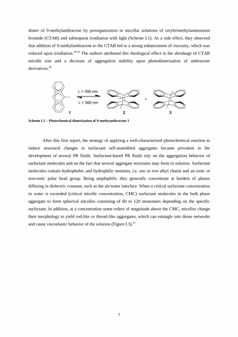
dimer of 9-methylanthracene by preorganization in micellar solutions of cetyltrimethylammonium
bromide (CTAB) and subsequent irradiation with light (Scheme I.1). As a side effect, they observed
that addition of 9-methylanthracene to the CTAB led to a strong enhancement of viscosity, which was
reduced upon irradiation.18,19
The authors attributed this rheological effect to the shrinkage of CTAB
micelle size and a decrease of aggregation stability upon photodimerization of anthracene
derivatives.20
Scheme I.1 – Photochemical dimerization of 9-methyanthracene 1
After this first report, the strategy of applying a well-characterized photochemical reaction to
induce structural changes in surfactant self-assembled aggregates became prevalent in the
development of several PR fluids. Surfactant-based PR fluids rely on the aggregation behavior of
surfactant molecules and on the fact that several aggregate structures may form in solution. Surfactant
molecules contain hydrophobic and hydrophilic moieties, i.e. one or two alkyl chains and an ionic or
non-ionic polar head group. Being amphiphilic they generally concentrate at borders of phases
differing in dielectric constant, such as the air/water interface. When a critical surfactant concentration
in water is exceeded (critical micelle concentration, CMC) surfactant molecules in the bulk phase
aggregate to form spherical micelles consisting of 60 to 120 monomers depending on the specific
surfactant. In addition, at a concentration some orders of magnitude above the CMC, micelles change
their morphology to yield rod-like or thread-like aggregates, which can entangle into dense networks
and cause viscoelastic behavior of the solution (Figure I.3).21

8
Figure I.3 – Schematic representation of different micellar aggregate morphology upon increase in concentration.
Furthermore, when present at small concentrations, certain aromatic solutes may affect the
aggregation state of surfactant molecules and increase viscosity by up to two orders of magnitude. For
instance, 2,2,2-trifluoro-1-(9-anthryl)-ethanol when solubilized in dilute CTAB solutions produced a
highly viscous fluid due to the formation of rod-like micelles.22
The same effect was obtained in
CTAB solutions containing 9-antracenecarboxylic acid and ionene polyelectrolytes.23
However, it was
shown that upon irradiation with UV light and concomitant dimerization of anthracene derivatives, the
viscosity was reduced significantly, which was attributed to the disruption of interactions between
aromatic solute and surfactant molecules and shrinkage in micelle size. Similar results have been
obtained for other anthracene analogs and different surfactants. Lehnberger and Wolff used
acridizinium bromide, which is known to undergo [4+4] photocycloaddition identically to anthracene,
in tetramethylammoniumhydrogen-2-dodecyl malonate (TMHM) aqueous solution obtaining a PR
fluid whose viscosity was greatly reduced after irradiation.24,25
Other photoresponsive molecules that undergo photochemical dimerization have shown to
have similar interaction with surfactant aggregates and gained importance for the development of
novel PR fluids. For instance, coumarins are a natural class of organic compounds and have been
extensively investigated mainly due to their outstanding optical properties, i.e., high extinction
coefficient and fluorescence quantum yield.26
However, coumarins are also known to yield dimers of
different geometries upon irradiation (Scheme I.2) and this reaction was explored as a way to induce
viscosity changes in surfactant solutions. Yu and Wolff were among the first to report the preparation
of PR fluids with coumarins as photosensitive moieties.27
Their systems were composed by 6-
alkylcoumarins and surfactant (CTAB or Triton X-100) solutions. Depending on the size of the alkyl
substituent, the observed photorheological effects were different. Short substituents yielded direct PR
effect, i.e. an increase in viscosity upon irradiation, whilst long alkyl substituents had an opposite
effect. Despite the relatively high number of reports on surfactant-based PR fluids, the mechanism of
action of the aromatic solutes was unclear until Raghavan and co-workers made a series of reports

9
where several cinnamic acid derivatives were employed in surfactant solutions to prepare PR fluids. In
their first report, o-methylcinnamic acid (OMCA) was dissolved in aqueous solutions of CTAB and
found to induce the formation of long entangled worm-like micelles, resulting in highly viscous
mixtures.28
Photoisomerization of the photosensitive molecule led to a drastic reduction in micellar
length, with consequent reduction of viscosity. Using the same cinnamic acid (CA) derivative, but
with a zwitterionic surfactant (erucyl dimethyl amidopropyl betaine, EDAB), the same group
formulated a system that displayed the opposite effect of the previous one, i.e., photoisomerization of
trans-OMCA to cis-OMCA resulted in the formation of long wormlike micelles and increased
viscosity.29
These experiments shed light on the mechanism behind light-induced variations of micelle
structures. The interactions between aromatic compounds and surfactant molecules give rise to
changes in effective sizes of head and tail groups, giving rise to two effective geometries, cone and
cylinder (Figure I.4). Micellar aggregates shape, and consequently size, are governed by the effective
shape of surfactant molecules: conic geometries give rise to rather spherical micelles, whereas
cylinder-shaped surfactant molecules aggregate as long, entangled wormlike micelles.
Scheme I.2 – Photochemical dimerization reaction of coumarin 4
Figure I.4 – Schematic representation of different effective head group sizes of anionic surfactant and corresponding
micellar aggregation morphologies (adapted from [29])
This same principle was applied in the development of nonaqueous fluids, using p-coumaric
acid (PCA) as photoresponsive molecule and lecithin as surfactant.30
Usually, this lipid forms short
reverse micelles in apolar organic solvents, but when PCA is added to the solution, micelles grow into

10
long chains, forming an organogel. Upon irradiation with UV light, PCA isomerized and interactions
with lecithin molecules were disrupted, leading to a free-flowing liquid with low viscoelasticity.
Although the majority of PR fluids displayed significant viscosity changes (factors of 1000 to
10000), reversibility is usually low due to much lower absorptivity of photoproducts over the entire
wavelength spectrum. To overcome this problem, Pereira et al. applied a different photoisomerizable
compound, trans-2,4,4’-trihydroxychalcone in CTAB and salicylic acid solutions.31
Chalcones are
aromatic ketones that are structurally similar to cinnamic acids, and therefore, also undergo
photoisomerization reactions. However, depending on substituents and conditions, irradiation of
chalcones gives rise to a multiequilibrium system involving several chemical species interconvertible
by light and pH (Scheme I.3).
Scheme I.3 – General network of reactions of flavylium compounds in acidic and neutral aqueous media, exemplified
for 4’,7-dihydroxyflavylium 10.
Because trans-chalcone species interacts with the head groups of surfactant molecules, this
equilibrium allowed switching from long wormlike to short spherical micelles, with a consequent 10-
fold decrease in viscosity. By controlling the pH values of the solution, flavylium cation was allowed
to hydrate and revert slowly to trans-chalcone species, rendering this PR fluid completely reversible.
The salicylic acid used in this work is known to intercalate with CTAB molecules and form worm-like
micelles yielding solutions with viscoelastic properties.32,33
For this reason, Sakai and co-workers used
it to elongate mixed micelles of CTAB and an azobenzene-modified cationic surfactant.34
Irradiation
with UV light led to a decrease in viscosity due to isomerization of azobenzene surfactant and
consequent micelle structure disruption. Further irradiation with visible light returned the system to its
original state. In effect, appending azobenzene structural units in the lipophilic tail of surfactants offers
the ability to control uniquely aggregation properties through irradiation with appropriate wavelengths
with high reversibility.35,36,37,38,39
The planar trans form of such surfactants is more hydrophobic than
the nonplanar cis (UV light) form, and hence the CMC, which typically correlates with the
hydrophobicity of the surfactant tails, is lower for the trans than the cis isomer of the surfactant.40,41

11
Therefore, several reports on PR fluids based on intrinsically photosensitive surfactants containing
azobenzene moieties were made.42
,43
Worth mentioning is a photocontrolable gel developed by Lee
and co-workers, where an azobenzene-based cationic surfactant was mixed with modified poly(acrylic
acid).42
In this system, irradiation of UV-light allowed switching of aggregation behavior of surfactant
micelles, which acted as crosslinkers for polymer chains resulting in the formation of a gel (Figure
I.5).
Figure I.5 – Schematic representation of isomerization induced changes in aggregation behaviour of azobenzene-
based cationic surfactant micelles (adapted from [42]).
Nanoparticle-based photorheological fluids
There are innumerous reports on the viscosity control of fluids by the addition of solid
nanoparticles. 5,8
-12 PR fluids based on the aggregation of nanoparticles rely on the same principles as
most MR and ER fluids, i.e., the rheological effect is caused by the self-assembly of anisotropic
nanoparticles into a complex network.5,8
-12 However, whereas the aggregation is caused by temporary
polarization of nanoparticles in MR or ER fluids, a different strategy is required when using light
stimuli. Therefore, nanoparticles with intrinsically anisotropic charge distribution are usually required
for the formulation of this type of PR fluids. Laponite is a synthetic hydrous sodium lithium
magnesium silicate clay composed of anisotropic disc-shaped nanoparticles whose surface is
negatively charged. However, the edge of the laponite particles are composed of hydrous oxide and
become positively charged in acidic medium.44,45
For these interesting properties, laponite has been

12
applied extensively in cosmetics, paints, foods and pharmaceuticals as a thickening agent.46
Recently,
it has been applied in the formulation of PR fluids in which nanoparticle aggregation and consequent
viscosity increase were controlled with UV-light irradiation.47,48
Besides laponite, these systems were
composed by Pluronic F-127, a nonionic surfactant known to stabilize laponite suspensions,49
and
diphenyliodonium-2-carboxylate monohydrate as a photoresponsive molecule. This latter compound
belongs to the class of photo-acid generators, a group of molecules that undergo photolysis and
generate a photoproduct with an acidic moiety.50,51,52
Therefore, irradiation of these PR fluids leads a
decrease in pH and, in turn, laponite particle edges become positively charged. This causes the
surfactant to desorb and form micelles in solution and laponite particles to assemble into a three-
dimensional network, resulting in a significant increase in fluid viscosity, as depicted in Figure I.6.47,48
Figure I.6 – Light-induced gelation of aqueous laponite suspensions: initially (left) particles are stabilized by pluronic
surfactant and unconnect; upon UV irradiation (right), the particles assemble into a network, causing the fluid to turn
into a gel (adapted from [47])
Unlike MR or ER fluids, these systems do not revert to the initial state after removal of the
stimulus, requiring an increase in pH to disassemble laponite nanoparticles and obtain a liquid
suspension. However, the aggregation of nanoparticles can be reversibly controlled with light stimuli
as evidenced by Yuan and co-workers in a report involving polyorganosiloxane nanoparticles
functionalized with p-nitrocinnamate.53
Upon irradiation, cinnamate moieties dimerize causing
aggregation of nanoparticles, which in turn results in increased viscosity.
Polymer-based photorheological fluids

13
One of the most widespread fluids evidencing photorheological effect in technological
applications are light-activated resins. These materials undergo photopolymerization when exposed to
UV or visible light, which results in a quick transition from liquid to solid state. Light-activated resins
are typically composed of reactive oligomers, reactive diluents and photoinitiators, which are
responsive for generating free radicals and starting polymerization. Their photochemical nature brings
advantages over traditional polymerization processes because no mixing is required allowing for
denser and clearer resins.54
Therefore, they find application in a wide variety of fields from electronics
to medicine.55
For example, Chen-Yang et al. developed a UV curable resin based on
photopolymerizable modified cyclotriphosphazenes for fire-retarding wood coatings.56
In dentistry,
UV curing resins are constantly evolving and play a crucial role replacing traditional metal amalgams
as restorative materials.57
One common characteristic among light-activated resins is the fact that the
photochemical polymerization is irreversible. Nonetheless, photopolymerization and other
photochemical reactions occurring in polymer systems can also be explored for the development of PR
fluids that give rise to reversible sol-gel transitions. For instance, Zheng and co-workers, synthesized a
branched polyethylene glycol (PEG) polymer modified with 9-anthracene carboxylic acid (Figure I.7)
that underwent reversible photo-cross-linking when exposed to alternating wavelengths of irradiation.
The system exhibited sol-to-gel transition and its properties such as viscosity, topography and
swellability were tunable with light.58
The same group applied the same strategy on an eight-branched
PEG-based hydrogel, modified with p-nitrocinnamic acid. This molecule exhibited high
photoreactivity,59,60,61
allowing the formulation of a PR fluid that underwent rapid photogelation and
photoscission.62
The system was further studied as a light-controlled drug delivery system for skin
regeneration applications.63
Figure I.7 – Schematic representation of branched PEG polymer modified with 9-anthracene carboxylic acid used in
the formulation of a PR fluid (adapted from [62]).

14
A similar system was obtained through photopolymerization of cinnamylidene acetyl grafted
onto PEG-based polymers. Irradiation with UV-light induced growth of polymer chains, which in turn
led to the formation of a gel.64
In a different report, six polyurethanes containing coumarin dimer
components in the polymer backbone were prepared.65
It was shown that upon irradiation with UV
light coumarin dimers undergo photocleavage of cyclobutane rings to yield dicoumarins, breaking
polymer backbone and reducing system viscosity. Re-polymerization was achieved after irradiation at
longer wavelengths due to photodimerization of coumarin moieties. In addition to appending
photodimerizable moieties in polymer backbone, it is also possible to incorporate them as side groups
to obtain photorheological fluids. This way, it is possible to control cross-linking between polymer
chains with light stimuli. This strategy was employed by several groups in the preparation of PR fluids
with varied composition and applicability. For example, Wells and co-workers synthesized a graftable
anthracene-based cross-linker for the modification of a potential variety of polymers.66
This molecule
was used on alginate and hyaluronic acid to obtain photogels whose properties were reversibly altered
with UV-light exposure. In addition, the modified hydrogels exhibited controlled release of bioactive
molecules through irradiation due to disruption of polymer backbone by photocrosslinking (Figure
I.8).66
Coumarins have also been grafted onto polymer backbones to induce photo-crosslinking and
change rheological properties. Bergmann and co-workers demonstrated this effect on a polymer films
that expand due to the formation of free-volume between polymer chains by photodimerization of
coumarins.67
A similar strategy was employed by Dai and Kim in the preparation of a β–cyclodextrin
polymer that increases viscosity upon irradiation.68
In this system, coumarin derivatives were allowed
to form complexes with polymer β–cyclodextrin residues and dimers formed by exposure to UV-light
acted as cross-linkers (Figure I.9).
Figure I.8 – Schematic representation of light-induced crosslinking between polymer chains due to anthracene
dimerization used in the photochemical controlled release of bioactive molecules (adapted from [66]).

15
Figure I.9 – Schematic representation of light-induced crosslinking between polymer chains due to coumarin
dimerization used in the photochemical control of fluid viscosity (adapted from [68]).
Crosslinking of polymer chains through dimerization of cinnamate derivatives has also been
reported. By functionalizing phospholipid polymers randomly with cinnamoyl groups, a
photoresponsive hydrogel was obtained, with potential applicability in drug delivery systems due to
their biocompatibility.69
p-Nitrocinnamate was also used for the preparation of a photosensitive
gelatin, yielding a smart hydrogel with great biocompatibility, whose swelling properties were
addressable with light.70
PR fluids based on low-molecular-weight gelators
Low-molecular-weight gelators (LMWGs) have been synthesized based on a wide variety of
compounds, from ionic amphiphiles to small biomolecules.71,72
These compounds form fibril structures
in solution due to intermolecular interactions, such as hydrogen bonding, which may entangle and lead
to solutions with high viscosity and elasticity, in a similar fashion to certain polymer solutions.
Therefore, in parallel to systems previously described, by introducing photoresponsive moieties into
LMWGs molecular structure, it is possible to control intermolecular interaction responsible for
gelation with light stimuli, and obtain fluids with photorheological effect.73
Oligopeptides and other amino-acid derivatives are commonly used as gelators due to a
combination of interactions such as van der Waals and ionic interactions, π-π stacking and hydrogen
bonding that leads to the formation of fibrils. Expectedly, several reports on PR fluids based on
LMWGs involve modification of this class of molecules. For instance, two units of phenylalanine
were connected by photoresponsive maleic–fumaric acid amides and showed sol-to-gel transition upon
UV irradiation. The effect was attributed to morphological transformation at the supramolecular level
due to isomerization of the gelator.74
In a different work, dialanine functionalized with
photoisomerizable spiropyran was applied to obtain a PR fluid with sol-to-gel transition. In this

16
system, non-planar spiropyran moieties prevent interactions among dipeptide LMWGs while the
isomerized merocyanine moieties have a strong tendency to form π–π stacking that can support
dipeptide interactions.75
Kuang et al. functionalized an oligopeptide with p-nitrocinnamate to obtain a dendron capable
of self-assemble into fibrous network in common organic solvents at low concentrations and form a
gel.76
Upon irradiation gel to sol transition occurred due to the photodimerization of cinnamate groups
which disrupted gel structure (Figure I.10)
Figure I.10 – (a) Schematic representation of the photoresponse of cinnamate monomer 11 and dimer 12; (b)
Photographic images representing the photorheological effect of acetone solutions of 11 (adapted from [76]).
In summary, photorheological fluids have been developed based on a variety of systems, from
surfactant solutions to nanoparticle suspensions. In general, these materials give rise to a
photorheological effect through the control of molecular aggregation phenomena through irradiation of
light. Surfactant-based fluids are currently the most widespread type of PR materials due to their
availability and simplicity to prepare, often giving rise to strong viscosity changes upon irradiation.
However, the ever-growing need for drug delivery systems in vivo, had led to shift in scientific interest
towards biocompatible polymers and gels as the basis for the development of novel PR fluids. These
systems, while providing strong PR effects, require long preparation processes due to the need for
polymer derivatization. Finally, nanoparticle-based PR fluids have shown to give rise to significant
viscosity changes, although lacking the reversibility exhibited by surfactant- or polymer-based
systems. Therefore, research in PR fluids is still required to develop systems that give rise to strong
viscosity changes with good reversibility, while being readily available and biocompatible.

17
Thesis Overview
The main objective of this thesis research is to develop new photorheological systems based
on a variety of photoresponsive moieties that undergo dimerization and/or isomerization reactions.
On Chapter II, the syntheses of tripodal photopolymerizable compounds bearing cinnamate,
anthracene and coumarin moieties are described. The photochemical properties are characterized and
their capability to undergo photopolymerization is evaluated. The resulting effect on their rheological
properties is also investigated.
On Chapter III, the design and synthesis of photoactive ionic liquids bearing cinnamate or
coumarin derivatives is described. These compounds are characterized towards physical properties
such as melting point and viscosity. In addition, their photochemical reactivity is investigated, and the
effect of irradiation of their physical properties is evaluated. In addition, the preparation and
photophysical and photochemical characterization of novel coumarin chromophores are also
described.
On Chapter IV, the methodology followed for the synthesis and characterization of the
compounds presented throughout the thesis is described.

18

19
Chapter II

20

21
II. Photopolymerizable compounds for photochemical
control of rheology
In this chapter, the preparation of photopolymerizable molecules is presented. These compounds
were designed as three-branched molecules bearing photodimerizable moieties as chain ends. Through
photochemical dimerization/cleavage reactions of these functional groups, it was anticipated that the
synthesized tripodal molecules would undergo light-controlled polymerization (Figure II.1) and allow
modulation of crosslinking density or molecular weight of the photoreactive polymer.
Figure II.1 – Schematic representation of photocontrolled reversible polymerization of tripodal molecules bearing
photosensitive moieties.
The formation of polymers by photoinitiated polymerization is the basis of a large variety of
commercial applications.77
Its use dates back ca. 4000 years to when Egyptians and Babylonians used
sunlight to photocrosslink linens during mummification and to waterproof papyrus boats via the
photopolymerization of an asphalt oil.78
Nowadays, most of these systems rely on free radical
polymerization reactions, and are usually composed of acrylate or methacrylate monomers and a free-
radical generating photoinitiator. These reactions begin with the formation of a free-radical species
through the absorption of light and result in a typical chain growth mechanism.79,80
Due to the high
speed of this process, photoinitiated free radical polymerization has been used extensively in medical
applications, microfluidics or materials engineering.79,81
However, although these systems are widely
applicable, some disadvantages concerning reversibility and rate control are often presented. Thus,
great scientific interest has recently been directed towards thermally reversible photopolymerizable
composites in order to expand the range of potential applications. In addition, significant effort to
study and control radical termination has been made in the past decades.82
,83
On the other hand,
photodimerization reactions have been used as an alternative to modulate polymerization reactions
with great control.84
Although significantly slower than free radical generation, photodimerization
often presents the advantage of allowing photochemical and thermal reversibility. In addition, they
offer the possibility to carry out photopolymerization in the presence of oxygen, which is known to
quench radical formation.85
Photodimerization reactions occur through [2+2] or [4+4] cycloaddition

22
mechanism. They belong to the group of pericyclic reactions which usually take place in a concerted
way. The most relevant reactions in this group are 4+2-cycloadditions, also known as Diels-Alder
reactions.86,87,88,89
These thermally initiated reactions are relevant in many biological systems for the
formation of six-membered ring structures90
, besides their widespread use in natural material
synthesis91
. On the contrary, [2+2]- and [4+4]-cycloadditions are mostly photochemically induced and
form a four-membered cyclobutane ring from two π-bonds or an eight-membered cyclooctane ring
from four π-bonds with the formation of new σ-bonds. The differences between thermal and
photochemical processes are justified by the molecular orbitals involved in these pericyclic reactions.
According to the Woodward-Hoffmann rules, the cycloaddition reactions take place between the
highest occupied molecular orbital (HOMO) of one molecule and the lowest unoccupied molecular
orbital (LUMO) of another molecule (Figure II. 2). Moreover, the formation of a new binding
interaction must be symmetry allowed, i.e., the lobes of the involved orbitals must have the same sign.
For example, in a [2+2]-cycloaddition, interaction between HOMO and LUMO is symmetry forbidden
and therefore the reaction cannot be thermally initiated.
Figure II. 2 – Representation of orbital symmetry of frontier orbitals of generic alkene (left) and symmetry forbidden
orbital interaction in a thermal [2+2] cycloaddition (right)
However, the opposite occurs if the reaction is photochemically induced. When an electron is
promoted from HOMO to the LUMO by absorption of a photon with the correct energy, two single
occupied molecular orbitals (SOMO) are formed (Figure II. 3). Thus, when a molecule in the excited
state approaches a molecule in the ground state, two binding interactions can be established between
the two SOMO of the excited molecule and the LUMO and HOMO of the ground state molecule,
resulting in the formation of two new σ-bonds. (Figure II. 3)
Figure II. 3 – Frontier orbitals of generic alkene in the excited state (left) and symmetry allowed orbital interactions in
a photochemical induced [2+2]-cycloaddition (right)
The change in molecular orbital symmetry can be demonstrated by computational studies
(Figure II. 4) on unsubstituted coumarin 4, which is one of the most applied photosensitive moieties in

23
a variety of smart materials due to its well characterized photodimerization reactions. In particular,
several photorheological fluids based on the photoinduced dimerization of such molecules has been
previously described. 27,65,67
Figure II. 4 – Symmetry allowed frontier orbital interactions between one coumarin molecule in the ground state and
one molecule in the excited state
II.1 – Low molecular-weight photopolymerizable molecules
The first set of tripodal photopolymerizable molecules described in this chapter were
synthesized using tris(2-aminoethyl)amine, 13 (Figure II.5), as triple-branched backbone, which was
functionalized with photoresponsive moieties through nucleophilic addition/elimination reaction with
acyl chlorides, obtained from the corresponding carboxylic acid derivatives as described in schemes
Scheme II. 1-3.
Figure II.5 – Molecular structure of tris(2-aminoethyl)amine 13
This synthetic strategy allowed for the preparation of molecules bearing three amide moieties,
which are important functions in the establishment of intermolecular hydrogen-bond type interactions,
necessary for the formation of several organogels. 92,93

24
Scheme II. 1 - Synthetic pathway for the preparation of tripodal cinnamic acid derivative 16: i) thionyl chloride,
chloroform, 89%; ii) tris(2-aminoethyl)amine, K2CO3 or NEt3, dichloromethane, 91% or 34%, respectively
Compound 16 was prepared through condensation of cinnamoyl chloride 15 and 13 in
dichloromethane. Initially, this reaction was carried out in the presence of triethylamine in order to
remove the hydrogen chloride byproduct and prevent ester hydrolysis. After formation of 16, removal
of the base catalyst was achieved through flash chromatography. However, when triethylamine was
replaced with potassium carbonate, the reaction yield improved significantly (34 to 91%). In addition,
the base catalyst could be removed by filtration, and chromatography purification was not necessary.
The photochemical properties of 16 were investigated in chloroform solution. The UV-Vis spectrum is
in agreement with the expected spectra for unsubstituted trans-cinnamic acid derivatives, with the
characteristic strong charge transfer πstyryl-π*carbonyl band at ca. 270 nm, the weak πstyryl-π*styryl band at
ca. 300 nm and an additional band at lower wavelengths (Figure II.6a).94
Upon irradiation at 300 nm,
spectral transformations were detected, which suggest that a photochemical process takes place. As
previously mentioned, cinnamic acid derivatives are known to undergo two main photochemical
reactions upon irradiation, trans-to-cis photoisomerization and [2+2] photocycloaddition. Both
reactions give rise to similar spectral transformations characterized by a decrease in absorbance around
the absorption maximum and an increase at shorter wavelengths. However, UV-Vis spectra obtained
for irradiated solutions of 16 show that the variations are not in accordance with isomerization nor
dimerization reactions. Whilst a decrease in absorbance at ca. 270 nm is detected, an increase at lower
wavelengths was not observed, suggesting that trans-cinnamate moieties in 16 do not undergo
dimerization or isomerization (Figure II.6a). Moreover, the absence of isosbestic points in these
spectra indicates that a photodegradation process may occur upon irradiation. In addition,
determination of the reaction quantum yield shows that the reaction rate is not affected by
concentration of 16 (Figure II.6b), thus, evidencing that a bimolecular process such as
photodimerization does not occur upon irradiation.

25
0
0.5
1
200 250 300
irradiationat 300 nm
Wavelength (nm)
A
(a)
0
0.1
0.2
10-5
0.0001 0.001 0.01
R
concentration (M)
(b)
Figure II.6 – (a) UV-Vis spectral modifications of 1.1×10⁻⁵ M 16 in chloroform upon irradiation at 300 nm. Total
irradiation time is 20 min; (b) Photochemical quantum yield dependence on concentration of 16.
To shed light on the nature of the photochemical process taken by compound 16, irradiated
solutions were analyzed by NMR spectroscopy. Both isomerization and dimerization photoproducts
could, in principle, be detected by the presence of characteristic peaks, such as doublet at ca. 6.0 ppm
with a coupling constant of ca. 12 Hz corresponding to the α-protons of the cis-cinnamate moieties or
multiplets with lower chemical shifts (3.5-4.5 ppm) corresponding to the cyclobutane protons arising
from photodimerization. However, the complexity of the 1H NMR spectrum obtained for irradiated
chloroform solutions of 16 suggests that multiple photoproducts are formed upon irradiation (Figure
II. 7). Whilst the peaks corresponding to unreacted trans-16 are still discernible, in particular the
doublets at 6.6 and 7.6 ppm corresponding to the α- and β- protons of the cinnamate moieties,
respectively, a large variety of peaks with undefined multiplicity are present in both the aliphatic and
aromatic regions of the spectrum. Moreover, the characteristic peaks of the expected photoproducts are
absent, which is in accordance with the spectral changes previously observed in the UV spectrum upon
irradiation (Figure II.6a). Thus, through spectroscopic analysis of irradiated solutions of 16, it was
evidenced that its cinnamate moieties do not undergo the expected photochemical reactions of trans-
to-cis irradiation or [2+2] cycloaddition. Instead, photodegradation into a variety of compounds seems
to be the apparent photochemical pathway. It is proposed that photo-oxidation of the tertiary amine
moiety is the main photodegradation pathway.95

26
Figure II. 7 – 1H NMR spectra of 16 in chloroform acquired before and after irradiation at 300 nm.
It was demonstrated that the selected cinnamate moiety was inadequate to induce the
anticipated photopolymerization of low molecular-weight tripodal molecules. Therefore, it was
replaced with an anthracene moiety in the design of a different tripodal compound. As previously
mentioned, anthracene derivatives undergo photochemical dimerization through [4+4] cycloaddition.
In addition, unlike cinnamic acid derivatives, the photodimerization quantum yield of anthracene
derivatives is usually rather high.96,97
This is due to the fact that the excited state lifetime of anthracene
derivatives is usually long lived (10-20 ns).98
For this reason, anthracenes have been widely used in
photorheological systems, as shown in the previous chapter.22,23,62,66
Thus, tripodal compound 19 was
designed, and its synthetic pathway followed the same strategy used for the preparation of compound
16 (Scheme II.2). Anthracene-9-carboxyl chloride, 18, was readily prepared from anthracene-9-
carboxylic acid 17 in the presence of thionyl chloride in chloroform solution. However, the
condensation reaction with 13 in the presence of triethylamine did not yield the envisioned tri-
anthracene derivative 19. In fact, NMR spectroscopic data collected after purification of the reaction
mixture evidenced that only the mono-anthracene derivative was prepared. It is expected that the large
size of the anthracene moiety gives rise to significant sterical hindrance, thus, inhibiting the
condensation reaction on the primary amine moieties of compound 13. The same results were obtained
when triethylamine base catalyst was replaced with potassium carbonate.

27
Scheme II.2 – Synthetic pathway for the preparation of tripodal anthracene derivative 19: i) thionyl chloride,
chloroform; ii) tris(2-aminoethyl)amine, K2CO3 or NEt3, dichloromethane
It was envisioned that the sterically hindered anthracene moiety could be replaced with a less
bulky chromophore that also undergoes photochemical dimerization. In the previous chapter, it was
demonstrated that coumarin moieties give rise to dimerization reactions in a variety of systems. In
particular, these reactions have been explored in the control of viscosity in photorheological fluids.
Although the photodimerization quantum yield for these compounds is generally lower than for
anthracene derivatives, it is still significantly higher than those exhibited by cinnamic acid moieties
due to the difference in their excited state lifetimes.99,100,101,102
Thus, coumarin-3-carboxylic acid 20
was selected as the photosensitive precursor allowing derivatization into a tripodal molecule with the
same synthetic strategy used for the preparation of 16. In this case, replacing the triethylamine base
catalyst with potassium carbonate also resulted in better reaction yields and a more simplified
purification process. Thus, compound 22 was obtained from coumarin-3-carboxylic acid in a two-step
reaction in good yield (Scheme II. 3).
Scheme II. 3 - Synthetic pathway for the preparation of tripodal coumarin derivative 22: i) thionyl chloride,
chloroform, 90%; ii) tris(2-aminoethyl)amine, K2CO3 or NEt3, dichloromethane, 95%
The photochemical properties of 22 were investigated in chloroform solution with UV-Vis
spectroscopy. The absorption spectrum 22 exhibits two bands without vibrational structure in the UV
region at approximately 300 and 350 nm, corresponding to the typical π-π* transitions associated to
the charge transfer from the benzenic cycle to the pyranone moiety of basic coumarin skeletons

28
(Figure II. 8a).103
These transitions are associated to an excitation from the HOMO and the HOMO-1
orbital, respectively, to the LUMO. Due to the presence of an electron-withdrawing amide moiety
group at position 3, the absorption maxima of 22 are shifted bathochromically (~30–40 nm) in
comparison with unsubstituted coumarin.104
In addition, the calculated molar absorptivity value ε of
38893 M-1
cm-1
is approximately three times higher than that of unsubstituted coumarin (ca. 9000-
12500 M-1
cm-1
, depending on solvent polarity), due to the presence of three independent
chromophores.104
In order to evaluate the photochemical properties of compound 22, irradiation at the longest
wavelength transition (330 nm) was carried out and the transformations were followed by UV-Vis
spectroscopy. Absorption spectra of chloroform solutions of 22 show that upon irradiation a
photochemical reaction takes place, indicated by the spectral transformations depicted in Figure II. 8a.
These changes are in agreement with the expected photodimerization reaction described for coumarin
derivatives. However, these results are insufficient to determine whether the photodimerization occurs
between two coumarin units from the same three-branched molecule or from different molecules.
Therefore, ΦR values were calculated for irradiated chloroform solutions at different concentrations of
22. It was evidenced that the photochemical reaction quantum yield increased with increasing
concentration, suggesting that dimerization process of 22 under the studied conditions is
intermolecular (Figure II. 8b). NMR spectra obtained for chloroform solutions of 22 before and after
irradiation are also in agreement with a photodimerization reaction pathway (Figure II. 9). This is
evidenced by the low intensity peaks that become visible in the 1H NMR spectrum after irradiation.
The peaks at low field correspond to the amide and aromatic protons of the coumarin dimers and
therefore, the chemical shifts are close to those of unreacted 22. At high field, the singlet at ca. 3.5
ppm corresponds to protons of the cyclobutane moiety formed upon dimerization. The fact that only
one signal corresponding to cyclobutane moieties is present may be indicative that only one isomer is
formed upon dimerization.
By UV-Vis spectroscopy, it was also observed that, at higher concentrations, irradiation led to
increased light-scattering due to the formation of small particles, evidenced by the absorbance spectra
depicted in Figure II. 8c.

29
0
0.5
1
250 300 350 400
A
Wavelength (nm)
irradiationat 330 nm
(a)
0
0.0125
0.025
10-5
0.0001 0.001 0.01
R
concentration (M)
(b)
0
0.5
250 300 350 400 450
A
Wavelength (nm)
irradiationat 330 nm
(c)
Figure II. 8 – (a) UV-Vis spectral modifications of chloroform solution of 22 at 1.0×10⁻⁵ M upon irradiation at 330 nm.
Total irradiation time is 60 min; (b) Photochemical quantum yield variation with increasing concentration of 22; (c)
Precipitation-induced UV-Vis spectral modifications of a 2.0×10⁻³ M solution of 22 in chloroform upon irradiation at
330 nm. Total irradiation time is 40 min.
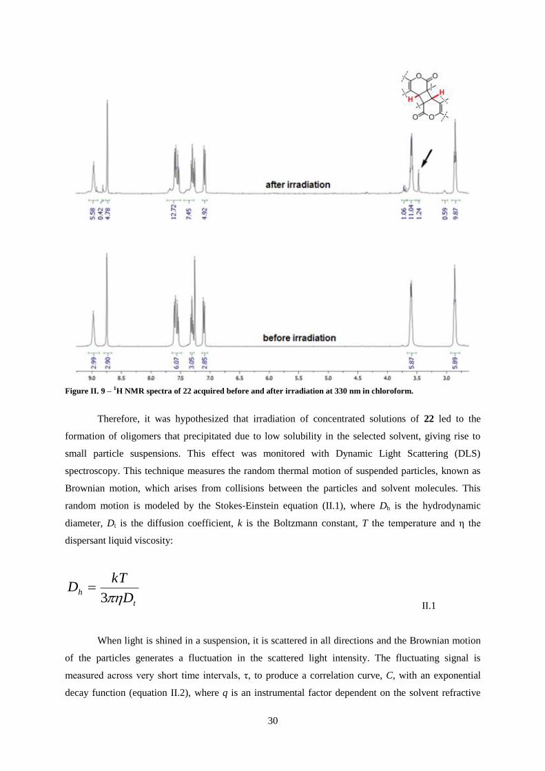
30
Figure II. 9 – 1H NMR spectra of 22 acquired before and after irradiation at 330 nm in chloroform.
Therefore, it was hypothesized that irradiation of concentrated solutions of 22 led to the
formation of oligomers that precipitated due to low solubility in the selected solvent, giving rise to
small particle suspensions. This effect was monitored with Dynamic Light Scattering (DLS)
spectroscopy. This technique measures the random thermal motion of suspended particles, known as
Brownian motion, which arises from collisions between the particles and solvent molecules. This
random motion is modeled by the Stokes-Einstein equation (II.1), where Dh is the hydrodynamic
diameter, Dt is the diffusion coefficient, k is the Boltzmann constant, T the temperature and η the
dispersant liquid viscosity:
t
hD
kTD
3
II.1
When light is shined in a suspension, it is scattered in all directions and the Brownian motion
of the particles generates a fluctuation in the scattered light intensity. The fluctuating signal is
measured across very short time intervals, τ, to produce a correlation curve, C, with an exponential
decay function (equation II.2), where q is an instrumental factor dependent on the solvent refractive

31
index, laser wavelength and scatter angle, and from which the diffusional coefficient, Dt, is extracted.
From equation II.1, particle size can be calculated.
)2exp( 2qDC t II.2
Figure II. 10a depicts the correlation curves obtained for chloroform solutions of 22 with
increasing irradiation time lengths. It is shown that irradiation leads to correlation curves with longer
decay times, which is caused by a reduction of diffusional motion of particles. From Stokes-Einstein
equation, it is demonstrated that this effect can be derived by either an increase in system viscosity, η,
or an increase in particle size, Dh. In order to determine whether the changes observed in DLS
spectroscopic measurements were caused by the growth of the detected nanoparticles, samples
irradiated for different time lengths were analyzed with atomic-force microscopy (AFM). This
technique belongs to the class of scanning probe microscopies, which can measure local properties
such as height, friction or conductivity with an appropriate probe. Atomic-force microscopes operate
measuring force between sample and a probe with a sharp, nanometric tip. Thus, topographical images
with high resolution at the nanoscopic scale can be obtained.105
Figure II. 10c-d depicts two AFM
images obtained for irradiated solutions of 22 collected at different irradiation time lengths. In both
samples, spherical shaped particles were detected. However, particle size varies significantly with
irradiation length. It is shown that after 10 minutes of irradiation, i.e., the time at which the first
particles are detected by DLS spectroscopy, particles with diameters of ca. 100 nm are observed
(Figure II. 10c), whereas longer irradiation yielded larger particles, with particles with ca. 400 nm
being observed after irradiating for 400 minutes (Figure II. 10d). Thus, it was evidenced through AFM
imaging that the reduction of diffusion coefficient Dt detected by DLS spectroscopy was caused by an
increase in particle size induced by irradiation rather than an increase in viscosity. With the Stokes-
Einstein equation II.1, it is possible to calculate the mean size of suspended particles and it is
evidenced that they show a virtually linearly dependence with irradiation time length, as depicted in
Figure II. 10b. Repeating the irradiation experiments at different concentrations of 22 shows that the
growth rate of these particles is significantly affected by concentration, which is in accordance with
the results obtained for ΦR measurements(Figure II. 8).

32
0
1
1 10 100 1000 104
C
(ms)
irradiation
at 330 nm
(a)
0
200
400
600
0 200 400 600
siz
e (
nm
)
irradiation time (min)
(b)10 mM5 mM1 mM
Figure II. 10 – (a) Correlation curve variation upon irradiation at 330 nm of a solution of 22 at 8.3×10⁻³ M in
chloroform; (b) Time and concentration dependence of photoinduced growth of dendrimeric nanoparticles of 22 in
chloroform; (c) and (d) AFM images of a suspension of dendrimeric nanoparticles of 22 obtained after 10 and 400
minutes of irradiation at 330 nm, respectively.
It was hypothesized that intermolecular photochemical dimerization of coumarin moieties of
22 results in photopolymerization into insoluble oligomers that grow dendrimerically with continuous
irradiation through photochemical dimerization of terminal coumarin moieties with solvated 22
monomers (Figure II. 11).
Figure II. 11 – Schematic representation of the proposed photoinduced dendrimeric growth of nanoparticles of 22 due
to dimerization of coumarin moieties.
(c) (d)

33
Conclusions
In conclusion, photopolymerizable compounds based on coumarin and cinnamic acid were
designed and prepared from readily available photosensitive precursors in good yields. Their
photochemistry in organic solution was characterized and it was evidenced that intermolecular
photodimerization reaction occurred upon irradiation in the case of coumarin derivatives.
Photochemical quantum yields were determined and the obtained values were low, resulting in low
conversion levels after long irradiation periods. Therefore, no photorheological effect was determined.
In addition, it was demonstrated that coumarin derivative gave rise to insoluble aggregates of
nanometric size upon irradiation, which were analyzed by DLS spectroscopy and atomic force
microscopy. It was evidenced that photopolymerization of 22 lead to the formation of oligomers that
grow dendrimerically into spherical nanoparticles during irradiation at absorption maximum. In order
to increase the quantum yield for the photopolymerization of three-branched compounds, synthesis of
an anthracene derivative was attempted. Due to high sterical hindrance by anthracene moieties, tri-
functionalized compound was not obtained. Therefore, it is necessary to review the design of such
compounds in order to increase the solubility of the resulting photoproducts and also the
photochemical quantum yield.

34

35
II.2 – High-molecular weight photopolymerizable compounds
In this chapter, the preparation of high molecular-weight photopolymerizable compounds is
presented. Similarly to the compounds described in the previous chapter, these molecules were
designed as three-branched structures bearing photodimerizable units. However, in order to increase
solubility and prevent precipitation of the final photoproducts, tris(2-aminoethyl)amine 13 was
replaced with tris(tetraethyleneglycol)phloroglucinol 23 (Figure II.12). In addition, different
photosensitive derivatives were used in order to increase chemical reactivity and prevent the formation
of byproducts. The followed synthetic pathways are summarized in Scheme II. 4-7.
Figure II.12 – Molecular structures of backbones used for the preparation of photopolymerizable tripodal
compounds: tris(2-aminoethyl)amine and tris(tetraethyleneglycol)phloroglucinol 23.
Compound 31 was prepared in four steps from tetraethyleneglycol, TEG, and p-coumaric acid.
TEG was derivatized into 1,13-dibromotetraethyleneglycol 24 in the presence of triphenylphosphine
and bromine in acetonitrile providing two leaving groups suitable for SN-type reactions. p-Coumaric
acid was derivatized into its methyl ester 27, through an acid-catalyzed esterification in methanol in
order to allow the phenolic moiety to be used as a nucleophile in SN-type reaction with 24 to yield
cinnamate-tetraethyleneglycol-bromide conjugate 28. This compound was reacted with phloroglucinol
to obtain the desired final product 31. The final step yield was considerably low (<10%), which was
attributed to the low reactivity of bromide as a leaving group. Thus, compound 24 was replaced with
1,13-ditosyltetraethylene glycol 25 due to the higher reactivity of tosylate as a leaving group.
However, this strategy failed to improve the overall reaction yield. It was observed that the polarity of
conjugate 29 and its precursor 25 was very similar, rendering impossible the purification through
common chromatographic techniques. Therefore, the process was repeated replacing 25 with 1,13-
diiodotetraethylene glycol 26. In principle, iodide has reactivity lower than tosylate but higher than
bromide, which should lead to better reaction yields in the synthesis of 31. However, although this
strategy allowed resolution of reaction mixture in the preparation of 30, final step yield was not
significantly improved (8.6%).

36
Scheme II. 4 – Synthetic pathway for the preparation of tripodal cinnamate derivative 31: i) K2CO3, acetone, 72%; ii)
phloroglucinol, K2CO3, acetonitrile, 8.6% (for precursor 30).
The photochemical properties of 31 were investigated in solution, and its ability to undergo
photopolymerization was tested. The obtained UV spectrum and molar absorptivity ε values are in
agreement with the expected results for p-coumaric acid derivatives.84
The band centered at ca. 300
nm corresponds to π-π* transitions of the cinnamate moieties and is bathochromic shifted, when
compared to the spectra of native cinnamate esters, due to the substituent at para position of the
coumarate moiety (Figure II. 13). Therefore, the weak band related with πstyryl-π*styryl could not be
detected. In addition, the strong absorption band below 250 nm corresponds to electronic transitions in
benzene rings of both coumarate and phloroglucinol moieties. Upon irradiation at 300 nm, changes in
the UV spectrum of 31 were detected. In fact, there is a decrease in absorbance in the low energy
band, with a minor hypsochromic shift in absorption maximum and a concomitant increase in
absorbance at lower wavelengths (Figure II. 13). These spectral transformation are consistent with
trans-to-cis isomerization and [2+2]-cycloaddition that can occur in coumarate moieties. The
photochemical quantum yield ΦR was determined at different concentrations in order to determine the
nature of the process. Figure II. 13b shows that varying concentration did not yield changes in ΦR,
suggesting that an intramolecular reaction takes place upon irradiation. These results could arise from
both trans-to-cis isomerization or [2+2]-cycloaddition between two coumarate moieties in the same
molecule. Therefore, 1H NMR spectra of irradiated solution were collected to shed light on the
photochemical reactivity of 31. The spectrum depicted in Figure II. 14, obtained after photostationary
state was achieved, evidences that only photoisomerization takes place upon irradiation. This
conclusion is supported by the appearance of a doublet at 5.8 ppm with coupling constant of 12 Hz,
corresponding to the α-protons of cis-coumarate moieties and the lack of new signals at 3.5-4.5 ppm,
which would indicate the presence of cyclobutane moieties resulting from photodimerization.

37
0
0.5
1
1.5
250 300 350
irradiationat 300 nm
A
Wavelength (nm)
(a)
0
0.05
0.1
0.15
10-5
0.0001 0.001 0.01
R
concentration (M)
(b)
Figure II. 13 – (a) UV-Vis spectral modification of a solution of 1.4×10⁻⁵ M 31 in chloroform upon irradiation at 300
nm. Total irradiation time is 20 min; (b) Photochemical quantum yield dependence on concentration of 31.
Figure II. 14 – 1H NMR spectra of 31 in chloroform before and after irradiation at 300 nm.
These results show that cinnamate derivatives were not adequate to give rise to
photopolymerization in molecules with tripodal backbones through photoinduced [2+2]-cycloaddition.
The fact that cinnamate derivatives may undergo an additional photochemical process, i.e.,
photoisomerization, in addition to the typically low excited state lifetime of these moieties lead to low
dimerization quantum yields and justify why it did not occur upon irradiation of 31.

38
Following the same strategy described previously for low molecular weight tripodal
compounds (chapter II.1), the cinnamate moieties were replaced with anthracene derivatives. Due to
the higher excited state lifetime, and, consequently the higher photodimerization quantum yield of
anthracenes it was envisioned that the synthesized compound would undergo photopolymerization
upon irradiation and allow the anticipated photochemical control of viscosity of its organic solutions.
Therefore, the synthesis of a tripodal anthracene derivative was attempted. 2-hydroxy-anthracene 34
was selected as the photoresponsive moiety and prepared from 2-amino-anthraquinone 32 in a two-
step reaction (Scheme II. 5) with an overall low reaction yield for the synthesis of 34 (13%).
Scheme II. 5 – Synthetic pathway for the preparation of anthracene precursor 34: i) NaNO2, NaOH, water, 53%; ii)
NaBH4, methanol, 24%
Nonetheless, compound 34 was used as a nucleophile in the synthesis of compound 35, which
was reacted with phloroglucinol in the presence of potassium carbonate to obtain 36 in good yield
(Scheme II. 6).
Scheme II. 6 – Synthetic pathway for the preparation of tripodal anthracene derivative 36: i) K2CO3, acetone, 76%; ii)
phloroglucinol, K2CO3, acetonitrile, 61%.
The photochemical properties of 36 were investigated by UV-Vis spectroscopy. The obtained
UV spectrum is in agreement with the expected results for anthracene derivatives, with a strong
absorption band centered at 350 nm with several maxima, corresponding to vibrational sub-levels.106
In addition, the absorption bands are bathochromically shifted when compared to native anthracene
derivative, due to the electron donating effects of the alkoxy moiety at position 2. Upon irradiation of
chloroform solutions of 36 at 360 nm, transformations in the UV spectrum were observed (Figure II.
15a). The decrease in absorbance at absorption maxima is consistent with the anticipated

39
photodimerization reaction of anthracene derivatives. In addition, determination of ΦR values in
solutions of 36 showed that the reaction photochemical reaction rate increases on the concentration of
36, which suggests that intermolecular [4+4]-cycloaddition occurs upon irradiation (Figure II. 15b).
To corroborate this hypothesis, 1H NMR spectrum of a solution of 36 was collected before and after
irradiation at 360 nm (Figure II. 16).
0
1
2
300 400
irradiation
at 360 nm
A
Wavelength (nm)
(a)
0
0.1
0.2
0.3
0.4
10-6
10-5
0.0001 0.001
R
concentration (M)
(b)
Figure II. 15 – (a) UV-Vis spectral modification of a chloroform deaerated solution of 36 at 3.0×10⁻⁵ M upon
irradiation at 360 nm. Total irradiation time is 10 min; (b) Photochemical quantum yield dependence on the
concentration of 39
It was observed that, at the photostationary state, the proton peaks in the obtained 1H NMR
spectrum were rather broad with low resolution, whereas the peak on the spectrum acquired before
irradiation were narrow with well-defined multiplicity. Nonetheless, proton peaks were detected
between 6.2 and 7.0 ppm, which are consistent with the formation of benzenic moieties in anthracene
dimers. It was hypothesized that the detected peak broadening was caused by polymerization of 36,
since polymer NMR spectra are typically defined by broad, poorly defined peak areas and resolution,
due to chain entanglement.107,108

40
Figure II. 16 – 1H NMR spectra of 36 in chloroform obtained before and after irradiation at 360nm. Total irradiation
time is 60 min.
In order to evaluate the rheological effect of the photochemical polymerization of 36 in
organic solutions, viscosity measurements were attempted. Although the low overall reaction yield did
not allow the preparation of significant amounts of 36, necessary for direct viscosity measurements in
conventional rheometers, it was envisioned that indirect viscosity determination could be possible
using dynamic light scattering spectroscopy. It was demonstrated in chapter II.1 that the Stokes-
Einstein equation (equation II.1), relates the diffusion coefficient of a suspended particle with the
viscosity of the solvent. Thus, using a suspension of particles with a known size, it is possible, in
principle, to determine fluid viscosity from the variations of the correlation curve obtained in DLS
experiments (equation II.5).
thDD
kT
3 II.5

41
In order to validate this method, silica nanoparticles with diameters of 100 nm were suspended
in binary mixtures of water and acetonitrile, and the corresponding correlation curves were obtained.
The calculated viscosity values for the mixture correlate well with the results obtained by direct
viscometry (Figure II. 17).
0
0.5
1
0 0.25 0.5 0.75 1
water
(
mP
a.s
)
Viscometer
DLS
Figure II. 17 – Viscosity of binary mixtures of water and acetonitrile at different ratios, determined by direct
measurement with conventional viscometer (black dots) and calculated indirectly from DLS data (red dots).
However, when this method was applied for the determination of viscosity values of solutions
of 36, before and after irradiation, no variations were detected. These results can be justified by the
difference between microscopic and macroscopic viscosity. The first is related with the immediate
environment surrounding the solute, and has a direct effect on diffusion processes. The macroscopic
viscosity is related to the resistance to flow and, therefore, is what is measured in conventional
viscometers. In simple liquids, the macroscopic and microscopic viscosities are the same, but differ
significantly in systems such as macromolecule solutions, gels and polymer films. This is due to the
fact that long polymer chains form crosslinks between different parts of solution, which does not affect
solute diffusion but significantly increases flow resistance. For example, this difference has been
demonstrated in aqueous solutions of sucrose and poly(vinyl pyrrolidone), in which a change in
polymer concentration from 0 to 20% led to a viscosity several orders of magnitude higher, while the
sucrose diffusion coefficient changed only by a factor of four.109
In order to demonstrate by conventional viscometric techniques that the photopolymerization
of polyethyleneglycol-based tripodal derivatives would give rise to an increase in viscosity upon
irradiation, it was necessary to improve the synthetic method and prepare larger amounts of the desired
compound. In chapter II.1 it was demonstrated that coumarin moieties are adequate to induce

42
photopolymerization of tripodal compounds, albeit with a low quantum yield. Thus, the anthracene
moiety was replaced with commercially available 4-methyl-7-hydroxycoumarin 37 for the preparation
of tripodal analog 39. This strategy led to a significant improvement of the overall reaction yield
allowing the preparation of larger amounts of tripodal coumarin derivative. Furthermore, due to the
difference in polarity between coumarin and coumarate moieties, preparation and purification of
tosylate derivative 38 was possible, which in turn led to a significant improvement in final step
reaction yield, due to the higher reactivity of tosylate as a leaving group (Scheme II. 7).
Scheme II. 7 – Synthetic pathway for the preparation of tripodal coumarin derivative 39: i) K2CO3, acetone, 60%; ii)
phloroglucinol, K2CO3, acetonitrile, 63%.
The photochemical properties of 39 were investigated in solution, and its ability to undergo
photopolymerization was tested. The obtained UV spectrum and molar absorptivity, ε, values are in
agreement with the expected results for coumarin derivatives, exhibiting two bands without vibrational
structure at approximately 220 and 320 nm, corresponding to π-π* transitions from HOMO-1 and
HOMO to LUMO, respectively (Figure II.18a).104
In addition, the distance between the two absorption
bands is decreased and the ratio of their intensities is changed, when compared to the spectrum of 22.
This is due to the electron-donating nature of the alkoxy substituent in position 7, opposed to the
electron-withdrawing effect of the amide substituent in position 3 of 22. Upon irradiation of
chloroform solutions of 39 at 320 nm, transformations in the UV spectrum were observed (Figure
II.18a). The decrease in absorbance at absorption maximum and a slight increase at shorter
wavelengths are consistent with previous results for tripodal coumarin derivative 39, which undergoes
photodimerization upon irradiation. In addition, calculations of ΦR show that the reaction rate
increases with increasing concentration of 39 suggesting that an intermolecular process takes place
upon irradiation (Figure II.18b).

43
0
0.5
1
250 300 350
irradiationat 320 nm
A
Wavelength (nm)
(a)
0
0.025
0.05
10-5
0.0001 0.001 0.01
R
concentration (M)
(b)
Figure II.18 - (a) UV-Vis spectral modification of a solution of 39 at 1.1×10⁻⁵ M in chloroform upon irradiation at 320
nm. Total irradiation time is 60 min; (b) Photochemical quantum yield dependence on concentration of 39.
Irradiated solutions were analyzed through NMR spectroscopy to confirm that 39 undergoes
photochemical dimerization. 1H NMR spectra, depicted in Figure II. 19, acquired before and after
irradiation, are consistent with the formation of coumarin dimers. This conclusion is supported by the
reduction of singlet intensity at 6.10 ppm, corresponding to the proton at position 3 and the appearance
of a singlet at 3.45 ppm and doublets at 6.60 and 7.20 ppm corresponding to the cyclobutane and
benzenic rings formed upon dimerization, respectively. Furthermore, the broadening of the signals at
low chemical shift is consistent with the formation of polymers.107,108

44
Figure II. 19 – 1H NMR spectra of 39 in chloroform obtained before and after irradiation.
It was also observed that the irradiated solutions remained clear even at higher concentrations
of 39 as opposed to what was detected for coumarin derivative 22, demonstrating that the introduction
of polyether linkers led to higher solubility of the photoproducts. The reversibility of the
photochemistry of 39 was also evaluated. However, it was observed that irradiation at shorter
wavelengths (< 250 nm) did not result in spectral transformations, followed by UV-Vis or NMR
spectroscopy.
With the confirmation that 39 undergoes intermolecular photodimerization, the effect of this
reaction on the rheological properties of organic solutions was evaluated through determination of the
corresponding viscosity curves, before and after irradiation.
The viscosity curve of a fluid is usually the first and most important rheological measurement
as it demonstrates the interdependency of the shear rate applied to it and its viscosity. It is used to
determine the physical and mechanical properties of a variety of materials, and provides valuable
information required to define their flow behavior and applicability (Figure II. 20). For instance, in
lubricant industry it is desired that a fluid retains its viscosity in a wide range of shear rate, which is
known as Newtonian behavior. However, most fluids do not exhibit Newtonian behavior and have

45
viscosity dependent on the applied shear. In cosmetic and paint industries materials are often shear-
thinning fluids, i.e., their viscosity decreases with increasing shear rate, which is useful to spread
evenly when pressure is applied and remain in place when shear forces are removed after application.
On the other hand, for some fluids, known as shear-thickening fluids, the viscosity increases with
increased shearing.
Figure II. 20 – Representation of the three main rheological behaviors exhibited by fluids
This type of measurements is often conducted on rotational rheometers which are based on the
principle that the torque required to turn an object in a fluid is a function of its viscosity. The
rheometer is a constant speed motor with a torque detection system which is typically a suspended
torsion bar. When the drive system turns, the sample resistance, i.e., its viscosity, gives rise to a twist
in the torsion bar which is used to calculate the torque. In this case, a cone and plate rheometer was
used. This equipment consists of a shallow cone (angles in the order of 1º) which is placed with a
known gap into a liquid standing on a horizontal plate (Figure II. 21). Typically, the plate is rotated
while the force on the cone, dependent on the liquid viscosity, is measured.

46
Figure II. 21 – Schematic representation of the typical components of a rotational rheometer
In order to avoid concentration variations during rheological experiments, which could, in
principle, occur with volatile solvents due to evaporation in an open system, chloroform was replaced
with dimethylformamide (DMF). It was confirmed that this solvent did not significantly affect the
photopolymerization reaction of 39 and the same photostationary state was achieved, although with a
lower ΦR (from 0.035 to 0.010 at 10-3
M). This effect was attributed to DMF viscosity (0.920 mPa s at
20 °C) being considerably higher than chloroform viscosity (0.563 mPa·s at 20 °C), which, in
principle, affects diffusion of molecules in solution. The rheometry results obtained for 39 in DMF
solution are depicted in Figure II. 22.

47
0
0.1
0.2
0.3
0.1 1 10 100 1000
(
Pa
.s)
Shear rate (s-1
)
initial
irradiated
Figure II. 22 – Viscosity curve of a DMF solution of 39 at 10% (w/v) before (black dots) and after (red dots)
irradiation at 320 nm. Total irradiation time is 120 min.
It is observed that before and after irradiation, the organic solution of 39 exhibits shear-
thinning behavior, which is often associated with polymer solutions.110
However, the zero-shear
viscosity is significantly different in both states. Non-irradiated solutions have a zero-shear viscosity
of ca. 0.03 Pa.s-1
, which is approximately 30 times higher than DMF viscosity, evidencing the
thickening effect of ethylene glycol-based polymers.111
Upon irradiation, the solution viscosity
increases significantly to ca. 0.27 Pa.s-1
. This effect is expectable due to the increase in molecular
weight of the solute resulting from the photopolymerization of 39.112
In fact, the relation between
solution viscosity, η, and the molecular weight of the polymer solute, M, is described by the Mark-
Houwink-Sakurada equation (II.3) where ηs is the solvent viscosity, c the polymer concentration and k’
is the proportionality constant, specific for each solvent-polymer system.
cMks
'
II.3
Since polymer concentration is constant before and after irradiation and if k’ remains constant,
it can be assumed that photopolymerization of 39 led to a ca. 9-fold increase in molecular weight of
the dissolved monomers, since the final viscosity, ηf, is approximately 9 times higher than the initial
viscosity, ηi:

48
fi
i
s
i
fi
i
s
i
f
s
i
i
s
i
MM
cMkn
n
cMkcMk
cMkn
n
cMkn
n
cMkn
n
9
'
')'(9
'
'9
'
II.4
It is possible to do this approximation since the intrinsic viscosity of a polymer solution does
not depend on conversion. In fact, this method is frequently used for the in-line monitoring of average
molecular weight in polymerization reactions.113,114,115,116,117,118
From the viscosity curves depicted in
Figure II. 22, it is also demonstrated that as the shear rate increases, the solution viscosity tends to a
value very close to that of pure DMF, which can be explained by the reduction of entanglement
between polyether chains as well as other secondary intermolecular interactions when high shear is
applied.113,116
Viscosity measurements demonstrate that photopolymerization of tripodal coumarin derivative
39 gave rise to changes in the rheological properties of DMF solutions, essentially on their viscosity,
due to the increase of molecular weight of the solute.
Conclusions
The results obtained for high-molecular weight compounds 31, 36 and 39 show that the careful
design and selection of photoresponsive moieties led to a significant improvement in rheological effect
over their low-molecular weight counterparts. It was evidenced that compounds bearing anthracene
and coumarin moieties underwent photopolymerization upon irradiation, unlike the cinnamic acid
derivative, which exhibited only a photoisomerization process. In addition, the selected tetraethylene
glycol spacer led to a marked increase in photoproduct solubility over the ethylene spacer of
compound 22. Therefore, it was possible to photopolymerize coumarin derivative 39 in organic
solution, which led to a 9-fold viscosity increase upon irradiation. However, due to the low viscosity
of the solution prior to irradiation, the overall rheological effect did not yield significant results. In
order to render this type of compounds more applicable as photorheological systems, it would be
necessary to prepare solutions with much higher viscosity prior to irradiation, which could be achieved
by increasing the length of the polyethylene glycol spacers.

49
Chapter III

50

51
Parts of this Chapter have been published:
J. Gordo, J. Avó, A. J. Parola, J. C. Lima, A. Pereira, P. S. Branco, Org. Lett. 2011, 13, 5112
J. Avó, S. Martins, A. J. Parola, J. C. Lima, P. S. Branco, J. P. P. Ramalho, A. Pereira, Chem. Plus
Chem. 2013, 78, 789
J. Avó, L. Cunha-Silva, J. C. Lima, A. J. Parola, Org. Lett. 2014, 16, 2582

52

53
III. Photoactive ionic liquids
III.1 – Introduction
During the past forty years, ionic liquids (ILs) attracted considerable attention and their
research became one of the most rapidly developing areas of modern physical chemistry, technologies
and engineering. This class of compounds is defined as salts whose melting temperature is below 100
°C. Therefore, they are composed predominantly of ions and ionic pairs which lead to interesting
properties such as negligible vapor pressure at room temperatures, high ionic conductivity, wide liquid
temperature range or high thermal stability. Due to their unique characteristics, ILs have been used in
a variety of applications, for instance, in electrochemical processes, purification and storage of gases,
highly conductive materials, lubricants and propellants or extraction and synthetic solvents.119
The
beginning of the history of ionic liquids is subject of controversy, although it is most commonly dated
back to 1888, when Gabriel and Weiner reported the synthesis of ethanolammonium nitrate (Figure
III.1a), with a melting point of 52-54 °C.120
However, the first account of synthesis of an ionic liquid is
often attributed to Walden, for his preparation of the first room temperature ionic liquid (m.p. 12 °C),
ethylammonium nitrate (Figure III.1b).121
Figure III.1 - Molecular structure of the first synthesized ionic liquids; a) ethanolammonium nitrate 40; b)
ethylammonium nitrate 41
After these initial reports, the interest in this class of salts increased, especially with the
discovery of chloroaluminates, formed by combining quaternary heterocyclic cations with aluminum
chloride. These materials exhibited a great deal of potential for use in a variety of areas, specifically as
electrolytes in battery applications, but all suffered from extreme sensitivity to moisture or had high
acidity/basicity.122
In the early 1990s, a major breakthrough was accomplished by Wilkes, with the
preparation of moisture stable ionic liquids, achieved by the replacement of the aluminum chloride
with other anions, such as tetrafluoroborate or haxafluoroborate.119
Furthermore, it was evidenced
that, by varying the size and nature of the cation and/or anion, the properties of ILs can be tailored to
meet the requirements of specific applications to create an almost infinite set of “designer solvents”.123
Thus, a large variety of anions and cations (Figure III. 2) have been used and the number of available
ILs and the range of possible applications have grown exponentially.124

54
Figure III. 2 - Representative ionic liquid components
The first commercial use of task-specific ionic liquids was developed by BASF on the
production of alkoxyphenylphosphines, using N-methylimidazole to scavenge acid in a process named
BASIL (biphasic acid scavenging utilizing ionic liquids). The reaction results in the formation of N-
methylimidazolium which separates from the pure product to be recycled.125
Expectedly, the scientific
and technological interest in ionic liquids for task-specific applications increased significantly and
many reports followed. Presently, most of these ionic liquids are employed as catalysts for organic
reactions, components in electronic devices, stabilizers in nanomaterials, solvents for analytical and
purification methods or lubricants.119
For example, butylmethylimidazolium hydrogenosulfate has
been used to overcome problems often associated with cellulose hydrolysis, acting as both solvent and
catalyst for the process, resulting in lower overall cost and higher purity of products.124
A large variety
of ionic liquids based on tetralkylammonium and alkyl imidazolium halides were employed as
stabilizing media in metal nanoparticle suspensions.126,127
As a result of their high concentration in
weak coordinating anions and high viscosity, these ILs prevent particle sedimentation and oxidation,
without compromising their activity. Ionic liquids have also shown great potential for application in
separation techniques, due to tunable properties such as thermal stability, viscosity and extractability
for organic and inorganic ions. In particular, special attention has been devoted to application of ILs as
matrixes in MALDI-MS, for the quantification of biomolecules and synthetic polymers.128,129
Lubricant industry also turned attention towards ILs applying them both in neat state or as additives to
existing base oils, owing to their good adsorbability to metal surfaces and good high pressure
performance.130
In addition, conventional lubricants may also be modified with ionic moieties to create
ionic liquid lubricants with higher durability and stability, lower toxicity, flammability and
volatility.131
Ionic liquids in smart material systems
With new frontiers in materials science opened by the unique properties of ionic liquids,
attention was drawn to the application of these compounds in smart systems which undergo a change
in their molecular configuration in response to an externally applied stimulus. With high ionic
conductivity, large electrochemical window, excellent thermal and chemical stability and low vapor
pressure, ionic liquids exhibit the properties required for a good electrolyte. Therefore, this class of
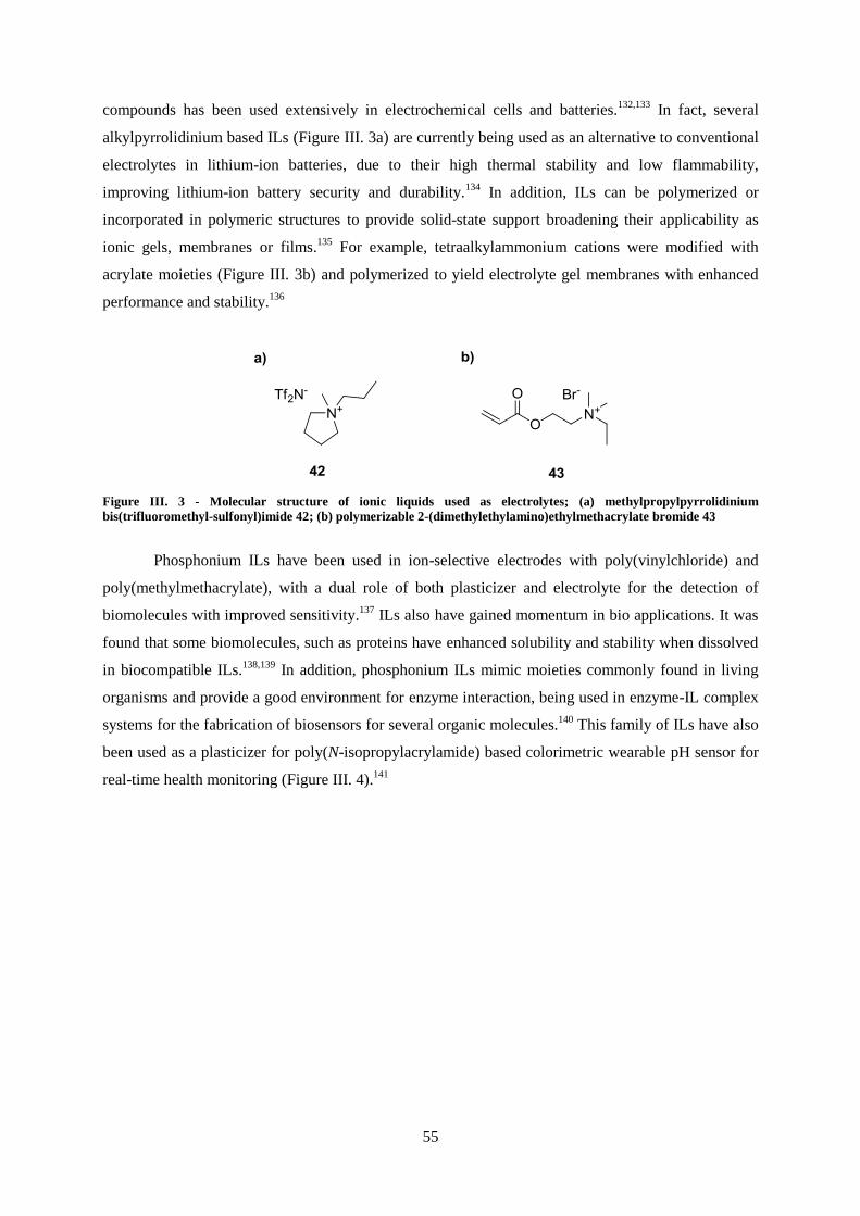
55
compounds has been used extensively in electrochemical cells and batteries.132,133
In fact, several
alkylpyrrolidinium based ILs (Figure III. 3a) are currently being used as an alternative to conventional
electrolytes in lithium-ion batteries, due to their high thermal stability and low flammability,
improving lithium-ion battery security and durability.134
In addition, ILs can be polymerized or
incorporated in polymeric structures to provide solid-state support broadening their applicability as
ionic gels, membranes or films.135
For example, tetraalkylammonium cations were modified with
acrylate moieties (Figure III. 3b) and polymerized to yield electrolyte gel membranes with enhanced
performance and stability.136
Figure III. 3 - Molecular structure of ionic liquids used as electrolytes; (a) methylpropylpyrrolidinium
bis(trifluoromethyl-sulfonyl)imide 42; (b) polymerizable 2-(dimethylethylamino)ethylmethacrylate bromide 43
Phosphonium ILs have been used in ion-selective electrodes with poly(vinylchloride) and
poly(methylmethacrylate), with a dual role of both plasticizer and electrolyte for the detection of
biomolecules with improved sensitivity.137
ILs also have gained momentum in bio applications. It was
found that some biomolecules, such as proteins have enhanced solubility and stability when dissolved
in biocompatible ILs.138,139
In addition, phosphonium ILs mimic moieties commonly found in living
organisms and provide a good environment for enzyme interaction, being used in enzyme-IL complex
systems for the fabrication of biosensors for several organic molecules.140
This family of ILs have also
been used as a plasticizer for poly(N-isopropylacrylamide) based colorimetric wearable pH sensor for
real-time health monitoring (Figure III. 4).141

56
Figure III. 4 – Ionogel based wearable pH sensor for the analysis of human sweat [141]
A system with similar composition was coupled with the photochemical properties of
spiropyrans to create a photo-controlled valve for microfluidics systems.142
Due to the mechanism of
the photochemical reaction of these compounds (Scheme III. 1), which involves the formation of a
zwitterionic merocyanine 45, it was possible to control the photochemical reaction kinetics and
consequently the rate of contraction of the valves through the application of a phosphonium IL.143
Scheme III. 1 – Photochemical reaction of generic spiropyran compounds
The high applicability of ionic liquids in smart material formulation led to an explosion of
interest of developing new methods for the synthesis of intrinsically stimuli-responsive ionic liquids
(SRILs), and many reports followed.119
Combining the excellent properties of conventional ILs for
electrochemical applications with the redox properties of organic and inorganic compounds,
electroresponsive ionic liquids became a representative class of SRILs. One of the first reports of this
type of ILs was made by Kavanagh and co-workers in which phosphonium ILs were derivatized with
viologen moieties in the organic cation to yield electrochromic ILs with fast switching kinetics144
.
Alternatively, similar electrochromic responses can be achieved with metal complexes. Branco et al.
prepared ionic liquids sensitive to both electric and magnetic fields, by using
ethylenediaminetetraacetate (EDTA) metal complexes of iron(III), cobalt(III) and chromium(III)
which were combined with several organic cations.145
The resulting compounds display many
advantages over conventional systems for electrochromic cells and present a potential alternative to

57
common magnetic fluids used in magnetic resonance imaging, with tunable magnetic properties and
reduced toxicity. Other electrochromic ionic liquids were synthesized by the same group taking
advantage of the redox properties of vanadium, using vanadium (V) oxide as the counterion in
imidazolium, tetraalkylammonium and tetraalkylphosphonium ILs (Figure III. 5).146
Figure III. 5 – Reversible color switching between three oxidation states of pure ethylmethylimidazolium vanadium
(V) oxide IL upon electrolysis (adapted from [146])
Additional chromogenic ionic liquids, which were sensitive to different stimuli, have been
synthesized for a variety of applications. For instance, Zhang et al. developed a pH-sensitive ionic
liquid combining a naturally anionic pH indicator, methyl red, with imidazolium and pyrrolidinium
cations.147
The resulting ILs exhibited increased pH responsiveness in comparison with the precursor
methyl red sodium salt due to increased solubility in water, resulting in a selective response in a wider
pH range (Figure III. 6).
Figure III. 6 – Increased selectivity of methylred based IL over conventional sodium salt from pH 1 to 7 (adapted
from [147])
The same chromophore, i.e., an anionic azobenzene, was used by Branco et al. for the
synthesis of a photoresponsive IL (Figure III. 7a). By reacting methyl orange sodium salt with a

58
variety of organic cations, several photochromic ionic liquids were obtained. The compounds retained
the typical photochemical behavior of azobenzenes, i.e., trans-cis isomerization with concomitant
change of color, although with slower kinetics due to their rather high viscosity.148
The same
photosensitive unit was also used to create a photoresponsive ILs with conductivity addressable with
visible light.149
However, in this report the azobenzene moiety was covalently bound to an
imidazolium cation (Figure III. 7b), resulting in faster isomerization kinetics. In organic solution, ILs
exhibited photoinduced variation in conductivity attributed to changes in aggregation state of ionic
clusters due to photoisomerization in the organic cation.
Figure III. 7 – Molecular structures of azobenzene based photochromic ionic liquids (from [148] and [149])
In addition to azobenzenes, other photoresponsive moieties have also been used for the
derivatization of cations and anions in ionic liquid synthesis. For example, 1,2-diphenylethene, which
is known to undergo reversible trans-cis isomerization upon light irradiation, has been functionalized
with imidazolium cations for the preparation of several photoresponsive ILs.150
The resulting
compounds were photoresponsive, with melting points and fluorescence quantum yields significantly
reduced upon trans-to-cis isomerization, a behavior similar to unsubstituted 1,2-diphenylethene.151
,152
Moreover, Yam et al. have synthetized photochromic ionic liquids based on the photochemistry of
other 1,2-diarylethene moieties.153
However, instead of trans-cis isomerization, these ionic liquids
exhibited reversible photocyclization (Scheme III. 2). The compounds were complexed with transition
metals and their photochemical properties were exploited for the switching of catalytic or
enantioselective activities.

59
Scheme III. 2 - Ring-closing and ring-opening of photosensitive diarylethene-containing ionic liquids 153
In parallel with the trend followed in the field of photorheological materials (chapter I),
attention is shifting towards applying naturally occurring photoresponsive molecules in the preparation
of photoresponsive ionic liquids. For instance, Salum and co-workers have used cinnamic acid
derivatives for the preparation of photoresponsive ionic liquids.154
These molecules occur naturally in
a number of plants and are extracted from plant-derived products, such as foods, cosmetics or
medicines.155,156
Besides, the carboxylic acid moiety makes them widely applicable as counter-ions for
organic cations.154
Thus, several ILs were prepared combining commercially available trans-cinnamic
acids and organic bases (Figure III. 8). Irradiation of the obtained compounds in organic solution
allowed the isolation of cis-cinnamate ILs with high purity, providing a highly efficient method for
preparation and isolation of cis-cinnamic acids.
Figure III. 8 – Cinnamate-based photoresponsive ionic liquids (from [154])

60
Viscosity of ionic liquids
Ionic liquids are increasingly gaining relevance as “green” alternatives for traditional organic
solvents, mainly due to their extremely low vapor pressure and volatility, significantly reducing
solvent loss, environment pollution and safety hazard.119
Thus, ILs have been subject of a great
number of studies on the determination of thermodynamic and thermophysical properties in the past
decade, especially their viscosity. The knowledge of the viscosity of ILs is of major importance from
an engineering point of view as it plays a major role in stirring, mixing and pumping operations. 157
Additionally, it also affects other transport properties such as diffusion. Therefore, the viscosity values
of a particular IL will greatly influence in its applicability. While highly viscous ILs do not make for
good solvents in chemical processing, due to negative effects on handling and power requirements or
reduction of heat and/or heat transfer in reaction and extraction processes, they are favored in
applications such as stationary phases for gas-liquid chromatography.158
Also, fixed viscosity ranges
are a requirement for the application of ILs as lubricants.159
As a result, a fundamental understanding
of the relationship between viscosity and ionic structure is necessary for the rational design and
synthesis of ILs with desired viscosity value.157
Several approaches were used to determine the
correlation between structure of ionic liquids and their viscosity, from quantitative structure-property
relationship (QSPR) correlations to qualitative analysis based on direct observation, which evidence
that viscosity is governed by structural characteristics of ionic pairs, such as cation size, length of
substituted group, flexible ether oxygen on alkyl chain, and anion shape.160,161
Dzyuba and Bartsch
studied a series of alkylimidazolium hexafluorophosphates and bis(trifluoromethyl-sulfonyl)imides
regarding the influence of the alkyl chain size on density, surface tension and viscosity.160
It was
shown that the increase in the number of carbons of the alkyl group leads to a general increase of
viscosity, which was attributed to van der Waals interactions (Figure III. 9). Furthermore,
hexafluorophosphate containing ILs exhibited higher viscosity values than their bis(trifluoromethyl-
sulfonyl)imide analogs, which was attributed to the higher symmetry of the anion (less conformational
degrees of freedom). In fact, this was later demonstrated in a systematic QSPR study on imidazolium
based ILs, in which highly symmetric or almost spherical anions gave rise to the highest viscosity
values (Figure III. 9).161

61
0
25
50
75
100
250
300
350
400
450
500
1 2 3 4 5 6 7 8 9
vis
cosity / c
P
n
Figure III. 9 – Effect of cation alkyl chain length and counterion on hexafluorophosphate (red dots) and
bis(trifluoromethyl-sulfonyl)imide (black dots) alkylmethylimidazolium ILs (from [161])
In a study involving ILs prepared from a variety of cations and anions, Seki et al.
demonstrated that the viscosity of ILs is affected by molecular shape rather than the molecular weight
of their components.162
In particular, molecular structural changes on the cation species had a stronger
effect on the physical properties of ILs than that of anion species, with main influence from
conjugation (bond saturation, chainlike or cyclic) and conformation (planar or non-planar). Cations
with higher positive charge delocalization, e.g. aromatic vs saturated or phosphorous- vs nitrogen-
containing cationic center, lead to less viscous ILs due to weaker cation-anion interaction.163
In
addition, hydrogen bond interaction through characteristic moieties between cation and anion can also
dramatically affect IL viscosity as demonstrated by computational studies.164
It was also demonstrated
that the formation of ionic clusters in ILs, e.g. due to addition of salts, results in increased viscosity.165
These studies shed light on the relationship between molecular structure and macro-viscosity of ILs,
allowing for the rational design and synthesis of ionic liquids with desired viscosity.166
Furthermore, it
was evidenced that conformational changes on cation and anion molecular structure affect
dramatically IL physical properties. In this light, several photosensitive ionic liquids were designed
and synthesized through functionalization of organic cations with photoresponsive moieties.

62

63
III.2 – Cinnamic acid based ionic liquids
For the design of photoresponsive ionic liquids based on cinnamic acid photochemistry, it was
envisioned that the photosensitive moiety should be appended on the cationic component. This can be
justified by several QSPR studies evidencing that structural changes occurring in IL cations have
stronger effect on physical properties than those occurring on anions.160-166
Moreover, although
carboxylic acids are often used as anion precursors in IL synthesis,154,167
they often give rise to
unstable ILs due to thermal decomposition of carboxylate moieties via decarboxylation or SN2-type
nucleophilic substitution with cations.168
Therefore, p-coumaric acid 51 was selected as cinnamic acid
derivative due to its phenolic moiety, which offers the possibility to append a variety of substituents
through SN-type nucleophilic substitution reactions. In this light, methyl p-coumarate 27 was
synthesized through acid catalyzed esterification and further derivatized with 1,3-dibromopropane to
yield compound 52. Trimethylamine and N-methylimidazole were reacted to give rise to two ionic
organic compounds, 53 and 54 (Scheme III. 3), and their melting points were determined (Table III.
1). As expected, compound 53 with a methylimidazolium cation had a lower melting point than its
trimethylammonium counterpart 54, due to lower symmetry and higher charge delocalization.163
Therefore, N-methylimidazole was selected as cation precursor for the synthesis of photoresponsive
ionic liquids.
Scheme III. 3 – Synthetic pathway for the preparation of cinnamate organic cations: i) methanol/H2SO4, 94%; ii) 1,3-
dibromopropane, K2CO3, acetone, 94%; iii) N-methylimidazole, acetonitrile, 76%; iv) trimethylamine, acetonitrile,
66%

64
Table III. 1 – Melting points of ionic cinnamate derivatives 53 and 54
Compound Cation Anion Melting point
53
143-145 °C
54
211-213 °C
QSPR studies referred previously show that anion symmetry and charge delocalization have a
marked effect on IL melting points.161-166
Thus, rational anion metathesis was performed for compound
53 with several counterions in order to reduce the melting point to below 100 °C. The results are
summarized in Table III. 2.
Table III. 2 - Melting points of ionic cinnamate derivative 53 with different counterions
Cation Anion Melting point
143-145 °C
121-123 °C
115-117 °C
90-92 °C
80-82 °C
It is evidenced that an increase in charge delocalization (due to conjugated aromatic rings or
electron-withdrawing CF3 moieties) leads to a decrease in melting temperature. Additionally, a
reduction of anion symmetry has a similar effect, except for the case of
bis(trifluoromethanesulfonyl)imide (Tf2N-), which gave rise to the lowest melting temperature of all
anions. This result can be justified by the fact that this anion may give rise to conformational isomers,
increasing the number of degrees of freedom and enhancing the melting point reduction effect.169,170,171
From this perspective, the synthetic strategy formerly described was followed with an additional anion
metathesis step (Scheme III. 4), to yield ionic liquids 55 and 58 with melting points below 100 °C.

65
Scheme III. 4 - Synthetic pathway for the preparation of cinnamate photoresponsive ionic liquids: i) 1,3-
dibromopropane, K2CO3, acetone, 94%; ii) 1,6-dibromohexane, K2CO3, acetone, 89%; iii) N-methylimidazole,
acetonitrile, 76%; iv) LiNTf2, dichloromethane, 70-76%.
In order to confirm the formation of the obtained ionic liquids 55 and 58, X-ray diffraction
analysis was performed. Both compounds were dissolved in a mixture of CHCl3 and MeOH 1:1 and
the solution was allowed to be in contact with a slow diffusion of Et2O leading to the formation of
colorless single-crystals. The crystal structures (Figure III. 10) of the two compounds were determined
in the triclinic space group P−1 with both asymmetric units revealing only one respective organic
cation and one Tf2N− anion. The organic cation in compound 55 reveals considerable planarity with
the phenyl and the imidazolium rings practically in the same plane (the dihedral angle between the
average planes of these two aromatic groups is ca. 3.563°), while the structural arrangement of the
organic cation in 58 is significantly distinct, showing the imidazolium ring almost perpendicular to the
phenyl group (the dihedral angle between the planes is ca. 79.341°). This structural difference is
certainly related with the size of the alkylic chain between the cinnamate moieties and the imidazolium
groups, as well as with the numerous C−H···X (X = F, N or O) intermolecular interactions involving
adjacent Tf2N−
anions.

66
Figure III. 10 - Crystal structures of the compounds 5a (top) and 5b (bottom), showing the label scheme for all non-H-
atoms which are represented as thermal ellipsoids drawn at the 50% probability
Furthermore, the crystal packing arrangement in both 55 and 58 is strongly influenced by an
extensive network of C−H···O weak hydrogen bonds involving neighboring organic cations, as well as
C−H···F, C−H···N and C−H···O hydrogen bonding between the organic moieties and the inorganic
Tf2N− anions (Figure III. 11). In compound 55 the adjacent organic cations interact via the
C4−H4···O2, C13−H13B···O2 and C17−H17A···O3 with H···O distances of 2.547, 2.553 and 2.480 Å,
respectively (Figure III. 11a). Furthermore, the inorganic anions establish various C−H···O
C7−H7···O4 (2.661 Å), C14−H14···O5 (2.597 Å), C14−H14···O6 (2.440 Å), C16−H16···O7 (2.519 Å) and
C17−H17B···O6 (2.536 Å) and C−H···F C11−H11B···F2 (2.657 Å), C13−H13A···F2 (2.566 Å) and
C17−H17C···F3 (2.379 Å), as well as C15−H15···N3 (2.474 Å) weak hydrogen bound interactions with
contiguous organic molecules leading to a 3D supramolecular structure (Figure III. 11b). The same
type of interactions is observed in the crystal packing of 58 ultimately originating a 3D supramolecular
network: C11−H11B···O2 (2.691 Å) interact neighboring organic cations, while the weak hydrogen
bound interactions between the inorganic and the organic species are also C−H···O C16−H16A···O7
(2.616 Å), C17−H17···O5 (2.573 Å), C17−H17···O7 (2.256 Å), C20−H20A···O5 (2.707 Å) and
C20−H20B···O4 (2.590 Å), C−H···F C14−H14B···F5 (2.509 Å) and C18−H18···F2 (2.541 Å) and
C19−H19···N3 (2.628 Å) (Figure III. 11c).

67
Figure III. 11 - Representation of the diverse C−H•••O (yellow dashed lines), C−H•••N (blue dashed lines) and
C−H•••F (green dashed lines) weak hydrogen bonds found in the crystal structures of the compounds 55 and 58: (a)
highlight of the interactions between adjacent cationic and anionic species in 55; (b) crystal packing of 55 viewed in
the [1 0 0] direction of the unit cell; (c) crystal packing of 58 viewed in the [1 0 0] direction of the unit cell
Irradiation of the obtained ionic liquids should, in principle, affect the intricate network of
intermolecular interactions in crystal packing, due to photochemical reactions of isomerization and/or
dimerization, and ultimately induce changes in their physical properties. Thus, the photochemical
behavior of ionic liquids 55 and 58 was investigated. In acetonitrile solution, both compounds
underwent photochemical reactions when irradiated at their absorption maxima (Figure III. 12a). The
decrease in absorbance around 300 nm and a slight increase in absorbance at lower wavelengths are
consistent with the expected photoreactivity of cinnamic acid derivatives, leading to species with blue-
shifted maxima. In order to determine whether the photochemical reaction consists of
photodimerization or photoisomerization, irradiations were carried out for acetonitrile solutions over a
wide range of concentration and photochemical quantum yields (ΦR) were calculated (results
summarized in Table III. 3). For both compounds, ΦR values did not vary within the selected
concentration range, which led to the conclusion that an intramolecular reaction occurs upon
irradiation, since the reaction rate does not depend on the concentration of the ionic liquid.
Furthermore, the existence of an isosbestic point at 265 nm indicates that no secondary reactions occur
during irradiation.172

68
Table III. 3 - Absorption Maxima (λmax), Photochemical Quantum Yields (ΦR), and Cis Isomer Fraction before
Irradiation and at the Photostationary State (PSS) of Ionic Liquids 55 and 58 in Acetonitrile
compound λabs (nm) conc. (M) ΦR initial cis fraction PSS cis fraction
55 304
10-2
10-3
10-4
10-5
0.12
0.11
0.11
0.12
0.0 0.47
58 304
10-2
10-3
10-4
10-5
0.11
0.10
0.12
0.11
0.0 0.48
To corroborate these results, 1H NMR spectra were obtained for the irradiated solutions of
both ionic liquids and it was evidenced that photoisomerization was the only photochemical reaction
taking place upon irradiation and photostationary states (PSS) were composed only of trans and cis
isomers in similar proportions (Figure III. 13 and 14). This conclusion is supported by the doublet at
5.84 ppm, corresponding to the β-proton of (Z)-isomer of species 55 and 58. Also, the intensities of
(E)-Hβ and (Z)-Hβ peaks add to the same value of HA peaks (imidazolium ring protons, with chemical
shifts of 8.73 ppm), indicating that (E) and (Z) isomers are the only species bearing an imidazolium
cation. In addition, the occurrence of photodecarboxylation or photodimerization would give rise to
additional doublets at 5.00-5.50 ppm (styrene moiety) or multiplets at 3.50-4.50 ppm (cyclobutane
moiety), respectively, which were not detected. For both compounds, further conversion from trans to
cis isomer was not possible, due to the similar absorbance of both isomers at the excitation
wavelength.173
The reversibility of the system was evaluated both thermally and photochemically,
followed by UV-Vis and 1H NMR spectroscopy. Irradiated solutions were allowed to stand overnight
at 50 °C and thermal recovery was not detected, as expected for room temperatures.174
The
photochemical reversibility was evaluated by irradiation at 240 nm. The spectral modifications
observed by UV-Vis show full recovery of the initial spectrum (Figure III. 12b). The degree of
reversibility from isomer mixture to pure trans-55 was further corroborated by NMR data (Figure III.
15). Full conversion to the trans-isomer was achieved after long irradiation times due to the low
absorption at selected wavelength and lower cis-to-trans isomerization quantum yield (0.015).

69
Figure III. 12 - Spectral modifications of 55 at 5.4 x 10⁻⁵ M in acetonitrile upon irradiation at 300 nm (a) and 240 nm
(b). Under the used experimental setup, PSS was reached after 10 min irradiation at 300 nm while the recovery by
irradiating at 240 nm required ca. 600 min.
Figure III. 13 – 1H NMR spectrum of irradiated solution of ionic liquid 55 at the photostationary state
Figure III. 14 - 1H NMR spectrum of irradiated solution of ionic liquid 58 at the photostationary state

70
Figure III. 15 - 1H NMR spectrum of a solution of ionic liquid 55 after irradiation at 240 nm (55rec)
Therefore, the occurrence of photodimerization of cinnamate moieties was excluded for this
concentration range, in accordance with previously described results for cinnamate ILs.154
The melting point (Tm) and enthalpies of melting (ΔHm) of the ionic liquids were determined by
differential scanning calorimetry (DSC). The obtained curves are depicted in Figure III. 16 and the
corresponding values are summarized in Table III. 4. Both 55 and 58 showed significantly lower
melting points (onset temperatures) and ΔHm after irradiation (photostationary state). While pure trans-
55 (55i) isomer had an initial melting point of 83 ⁰C, its photostationary state (55PSS) composed by
both cis and trans isomers melted at 55 ⁰C. Moreover, its lower enthalpy of melting and larger
difference between onset (Tm) and peak (Tp) temperatures indicates that the structural order is reduced
after irradiation, due to the presence of both isomers.175,176
For ionic liquid 58 the changes in physical
properties were even more significant.
60 80 100
Temperature (ºC)
Hea
t flow
(e
xo
up
)
55i
55rec
55PSS
(a)
20 40 60 80
Temperature (ºC)
He
at
flo
w (
exo
up
)
58i
58rec
58PSS
(b)
Figure III. 16 – DSC thermograms of ionic liquids 55 (a) and 58 (b) before irradiation (i), at the PSS (PSS) and after
photochemical recovery (rec).

71
Table III. 4 - Melting (Tm) and Peak (Tp) Temperatures and Melting Enthalpies (ΔHm) of ILs 55 and 58 before
Irradiation at 300 nm (i), after the PSS Has Been Reached (PSS) and after Recovery upon Irradiation at 240 nm (rec)
compound 55 58
55i 55PSS 55rec 58i 58PSS 58rec
Tm (°C) 83.4 54.9 82.2 53.4 51.8
Tp (°C) 86.0 66.1 85.4 55.8 n.d. 55.1
ΔHm (Jg-1) 63.8 20.9 63.3 62.3 62.2
Upon irradiation at 240 nm to achieve complete recovery of trans isomers (55rec and 58rec),
both Tm and ΔHm were measured by DSC. The values obtained are very close to those of non-
irradiated samples (55, 58), showing that chemical and physical transformations are reversibly
adjustable. After characterization of the photochemical behavior of ionic liquids 55 and 58 in
solution, their photochemistry and resulting physical properties modifications were investigated in
neat conditions. Both compounds were irradiated at 300 nm at their melting temperature as thin films,
and the photochemical transformations were followed by 1H NMR spectroscopy. NMR data show that,
although 55 and 58 also undergo trans-to-cis isomerization upon irradiation under these conditions,
other photochemical reactions take place with significant quantum yields, as indicated by the presence
of several proton peaks with high chemical shifts (Figure III. 17). Photostationary states were
achieved after one hour of irradiation and were composed of a mixture of trans-55 (ca. 45%), cis-55
(ca. 30%) and other new compounds (ca. 25%).
Figure III. 17 – 1H NMR spectrum of 55 in chloroform after irradiation at 300 nm in neat conditions. Total
irradiation time is 120 min.
Although the complexity of the obtained NMR spectra does not allow the identification of the
new species, it is estimated that photo-oxidative processes occurring in cinnamate moieties may give
rise to different imidazolium salts. To test this hypothesis, ionic liquids 55 and 58 were melted,

72
deaerated in vacuo and irradiated under inert atmosphere. NMR data show that under these conditions,
both compounds undergo photochemical trans-to-cis isomerization upon irradiation, while other
photochemical processes are significantly reduced (from ca. 25% to <3%) (Figure III. 18). These
experiments had to be carried out in closed quartz tubes to achieve inert atmosphere, and thus, ionic
liquid samples had low surface-to-volume ratio which in turn led to heterogeneous mixtures of isomers
during irradiation. Therefore, photochemical quantum yields could not be determined and
photostationary states were not achieved. However, melting points were still lowered after partial
trans-to-cis isomerization of compounds 55 and 58 (Table III. 5). Moreover, it appears that melting
temperature depression (ΔTm) is directly proportional to the fraction of cis-isomer in the irradiated
sample (Figure III. 19) and extrapolates to the values of irradiated solution samples with equal
fractions of both isomers. These results suggest that irradiation of thin films of 55 and 58 under inert
atmosphere would yield similar ΔTm values to those obtained for irradiated solutions, if the same
isomerization rate was achieved. The reversibility in neat conditions was also investigated. Samples
were irradiated in solution in order to achieve 1:1 trans-cis isomer ratio of the photostationary state,
dried in vacuo and allowed to stand at 90 °C overnight and no changes in 1H NMR spectra were
observed. Equally, irradiation of thin films of 55 and 58 did not yield any changes. These results can
be justified by the low absorptivity at the selected wavelength and low quantum yield of cis-to-trans
isomerization. Moreover, photoisomerization kinetics can be significantly reduced in highly viscous
media, which is the case of these molten ILs. 177
,178
Figure III. 18 - 1H NMR spectrum of 55 in chloroform after irradiation at 300 nm in neat conditions under inert
atmosphere. Total irradiation time is 120 min.

73
Figure III. 19 - Isomer fraction dependency of melting temperature depression of irradiated neat samples (black dots)
and acetonitrile solution (red dot) of 55.
Table III. 5 - Time dependent cis isomer ratio and melting point depression (ΔTm) of irradiated neat samples of ionic
liquids 55 and 58
compound irradiation time
(hours)
cis
isomer fraction ΔTm (°C)
55
1 0.10 7
3 0.16 11
6 0.20 14
58 4 0.10 7
After these results, it was anticipated that a room temperature ionic liquid bearing cinnamate
moieties would exhibit light-controllable rheological properties, such as viscosity. As demonstrated by
numerous QSPR studies on the structure of ionic liquids, flexible ether oxygens on alkyl chains of
organic cations affect cation-anion interaction by increasing the number of conformational degrees of
freedom, thus, influencing properties such as viscosity and melting points.162
In this light, an
imidazolium ionic liquid bearing cinnamate moiety and a tetraethyleneglycol spacer was synthesized
(Scheme III. 5). Following a similar strategy to that previously described, a room temperature ionic
liquid, 60, was obtained by replacing the alkane chain spacer with tetraethylene glycol moiety. DSC
data showed that compound 60 remains liquid at temperatures as low as -42 °C, evidencing the notable
effect of the polyether chain on the melting point. Thus, anion metathesis was not performed.

74
Scheme III. 5 – Synthetic pathway for the preparation of room temperature cinnamate ionic liquids: i)
triphenylphosphine, Br2, acetonitrile, 87%; ii) K2CO3, acetone, 91%; iii) N-methylimidazole, acetonitrile, 91%.
In a similar approach to previously described ionic liquids 55 and 58, the photochemical
behavior of IL 60 was studied both in solution and neat conditions. Upon irradiation at absorption
maxima, photochemical processes took place as indicated by a decrease in absorbance around 300 nm
and a slight increase in absorbance at lower wavelengths, which are consistent with the
photoisomerization processes occurring in analogs 55 and 58 (Figure III. 20a). Quantum yield (ΦR)
calculation also show that the photochemical reaction rate is not affected by concentration, indicating
that it has an intramolecular nature, in parallel with the behavior of compounds 55 and 58 (Table III.
6). These data were corroborated by 1H NMR spectra of irradiated samples (Figure III. 21), evidencing
that only trans-to-cis photoisomerization takes places upon irradiation. This conclusion is supported
by the doublet at 5.84 ppm, corresponding to the β-proton of (Z)-isomer of 60 and by the two singlets
around 10.2 ppm, which indicate that (E) and (Z) isomers are the only species bearing an imidazolium
cation. In addition, species resulting from photodecarboxylation or photodimerization reactions would
give rise to additional doublets at 5.00-5.50 ppm (styrene moiety) or multiplets at 3.50-4.50 ppm
(cyclobutane moiety), respectively, which were not detected. The obtained photostationary state was
composed by both isomers in equal proportions, comparable to the behavior of previously studied ILs.
Furthermore, the system also showed to be fully reversible when irradiated at lower wavelengths
(Figure III. 20b).

75
Figure III. 20 - Spectral modifications of 60 at 1.2 x 10⁻⁵ M in acetonitrile upon irradiation at 300 nm (a) and 240 nm
(b). Under the used experimental setup, PSS was reached after 15 min irradiation at 300 nm while the recovery by
irradiating at 240 nm required ca. 600 min
Figure III. 21 – 1H NMR spectrum of 60 irradiated in chloroform at 300 nm. Total irradiation time is 120 min.
Table III. 6 - Absorption Maxima (λmax), Photochemical Quantum Yields (ΦR), and Cis Isomer Fraction before
Irradiation and at the Photostationary State (PSS) of IL 60
compound λabs (nm) conc. (M) ΦR initial cis fraction PSS cis fraction
60 305
10-3
10-4
10-5
10-6
0.11
0.10
0.12
0.11
0.0 0.49
Compound 60 has also shown to undergo degradation when irradiated in neat conditions, as
evidenced by 1H NMR data depicted in Figure III. 22. The irradiated sample gave rise to a complex
spectrum with a high number of peaks around 10.0 ppm corresponding to different imidazolium
0
0.1
0.2
0.3
200 250 300 350 400
Wavelength (nm)
irradiation
at 300 nm
(a)
A
0
0.1
0.2
0.3
200 250 300 350 400
Wavelength (nm)
A
irradiation
at 240 nm
(b)

76
bearing species, as well as several new signals at 7.8, 6.6 and 4.5-4.0 ppm (Figure III. 22). Irradiation
of 60 in controlled non-oxidative atmosphere did not cause degradation, and the obtained 1H NMR
spectrum was similar to that obtained for irradiation in acetonitrile solution. However, in this case the
composition of the photostationary state was different than those of compounds 55 and 58, with a
higher fraction of cis isomer (Figure III. 23).
Figure III. 22 – 1H NMR spectrum of 60 in chloroform after irradiated at 300 nm in neat conditions. Total irradiation
time is 120 min.
Figure III. 23 - 1H NMR spectrum of 60 in chloroform after irradiated at 300 nm in neat conditions under inert
atmosphere. Total irradiation time is 120 min.
The effect of these transformations on the physical properties of 60 was investigated. In this
case, changes in viscosity rather than melting point were tested. Several QSPR studies have shown
that the factors that govern melting point in ILs also affect viscosity, such as cation size, shape and
symmetry or cation-anion interactions.161-166
It was evidenced that trans-to-cis isomerization induced
significant melting point reduction in ILs 55 and 58, probably due to disruption in van der Waals and
hydrogen bond interaction established between the organic cations and Tf2N- anions. It is then
expectable that the same photochemical reaction would lead to a decrease in IL viscosity after
irradiation. Therefore, the viscosity of neat samples of 60 was measured before and after irradiation at

77
300 nm, and the results are depicted in Figure III. 24. The same experimental apparatus used for the
characterization of the rheological properties of compound 39, described in the previous chapter, was
employed to evaluate the photorheological effect displayed by IL 60. The flow curve associated with
the non-irradiated state exhibits a Newtonian plateau of the shear rate at low shear rates followed by a
shear thinning regime at high shear rates. This flow behavior is characteristic of thixotropic fluids
which are usually composed of molecules that have a longer range structure that can be altered when a
certain level of mechanical shearing is reached. Moreover, it has been demonstrated that ILs with
nanostructures composed of aggregates due to intermolecular hydrogen bond networks form liquid
crystalline phases and exhibit this rheological behavior. It is then expected that, upon a certain shear
level is achieved, intermolecular interactions between cation chains are disrupted, breaking IL
nanostructure and consequently decreasing viscosity. 179,180,181,182
The rather long transition between
Newtonian and shear-thinning behaviors can be explained by the diverse intermolecular interaction
types that can form in 60. Upon irradiation into the photostationary state, these rheological properties
are significantly changed. The zero-shear viscosity is lower in the irradiated form which can be
explained by a weakening in IL nanostructure due to an increase in disorder arising from the mixture
of cis and trans isomers. In addition, the Newtonian region of the flow curve extends to higher shear
rates, indicating that weaker intermolecular interactions are established in the photostationary state of
60.
10
100
0.01 0.1 1 10 100 1000 104
Vis
cosity (
Pa.s
)
Shear rate (s-1
)
Figure III. 24 - Viscosity curve of pure 60 before (black dots) and after (red dots) irradiation at 300 nm. Total
irradiation time is 120 min.
This behavior follows the same trend of melting point depression exhibited by 55 and 58 upon
irradiation. It is then proposed that trans-to-cis isomerizations weakens cation-anion and cation-cation
supramolecular interactions leading to a decrease in viscosity, probably due to a reduction in cation

78
planarity. In order to shed light on the effect of isomerization on intermolecular interactions on the
liquid state, Hyper-Rayleigh scattering (HRS) spectroscopy was conducted on neat samples of 60,
before and after irradiation. HRS is a spontaneous process in which a system illuminated with photons
with a given frequency ω scatters photons of frequency 2ω. HRS spectroscopy allows the
determination of the system hyperpolarizability (βHRS), i.e., the ability to scatter light at the second
harmonic frequency, which is a function of size, shape and nature of scatters.183,184
The HRS
hyperpolarizability, as well as its dipolar (βJ=1) and octupolar (βJ=3) tensorial components, associated
depolarization (DR) and anisotropy (ρ) ratios for compound 60 before and after irradiation are
summarized in Table III.7, and the associated variation of HRS response is shown in Figure III. 25.
Table III. 7 - βHRS, βJ=1 (dipolar) and βJ=3 (octupolar) values (in atomic units*, using Convention T) and depolar-
ization ratios, DR, and anisotropies, ρ, deduced from HRS measurements at 1064 nm for compound 60, before and
after irradiation
compound βHRS DR ρ βJ=1 βJ=3
60 506 5.1 0.80 950 760
60irrad 433 5.1 0.80 830 670
* 1 atomic unit of β = 3.62x10-42 m4 V-1 = 8.641x10⁻³³ esu.
Before irradiation, the amplitude of βHRS (503 at. units) is considerably high, c.a. ten to twenty
times stronger than that reported for other imidazolium based ILs.185
In principle, no dynamical
enhancement of that hyperpolarizability is expected since the absorption band at 300 nm is far from
the harmonic response at 532 nm. The depolarization ratio (DR) is approximately 5, which is typical
of a two-state model (HOMO-LUMO) where the hyperpolarizability has a strong 1-D character.186
In
addition, the multipolar analysis of the HRS measurements in 60 clearly reveals a dominant dipolar
character (βJ=1>βJ=3) of its hyperpolarizability in contrast again with imidazolium (and even
pyridinium, pyrrolidinium or alkylammonium) based ILs where the octupolar character strongly
dominates the second order nonlinear optical response. These results indicate that such HRS response
excludes a simple molecular cluster in favor of a nanocluster whose organization is probably very
close to a crystal packing with a dominant dipolar response, as demonstrated for analogs 55 and 58. 187
After irradiation (60irrad), the HRS parameters are quite identical in proportion except that their
respective amplitudes are 12% lower which is in agreement with the weak loss of viscosity observed
for the irradiated sample, and correlates well with the melting point depression exhibited by 55 and 58
due to weakening of intermolecular interactions.

79
0
500
1000
1500
2000
2500
3000
3500
0 1 2 3 4 5 6 7
HCCl3JAD50 AJAD50 AI
0
500
1000
1500
2000
2500
3000
3500
HR
S I
nte
nsity
(arb
. u
nit)
Incident power (arb. unit)
chloroform
60
60i
Figure III. 25 – Quadratic dependence of the scattered intensity in IL 60 before (black circles) and after (red circles)
irradiation compared with that obtained in the reference solvent (chloroform, blue squares)

80

81
III.3 – Coumarin based ionic liquids
In previously described systems (chapter II), coumarin derivatives have shown to undergo
photochemical dimerization upon irradiation which was explored to induce photocontrolled
polymerization-scission of branched compounds. Thus, it was envisioned that organic cations of ILs
bearing coumarin moieties could undergo photodimerization, and the subsequent changes in physical
properties should be more significant than those resulting from trans-to-cis isomerization of cinnamate
moieties. In effect, photodimerization would yield dicationic dimers, whose high symmetry and long
alkylic chain lengths have shown to give rise to strong supramolecular interactions leading to highly
viscous ILs.163,164
For the design of photoresponsive ionic liquids based on coumarin photochemistry, the same
synthetic strategy used for the preparation of ILs 55, 58 and 60 was employed. Instead of p-coumaric
acid 51, 4-methyl-7-hydroxy-coumarin 31 was used as photosensitive moiety and linked to N-
methylimidazolium cation through a alkylic chain and a polyether spacer (Scheme III. 6)
Scheme III. 6 - Synthetic pathway for the preparation of coumarin photoresponsive ionic liquids: i) 1,3-
dibromopropane, K2CO3, acetone, 92%; ii) 1,13-dibromotetraethyleneglycol, K2CO3, acetone, 86%; iii) N-
methylimidazole, acetonitrile, 75-91%.
DSC data confirmed that both compounds melted below 100 °C and therefore no anion
metathesis was performed. Similarly to IL 60, the tetraethylene glycol moiety in the molecular
structure of compound 64 had a marked effect on its melting temperature and it remained liquid up to -
37 °C.
The photochemical behavior of compound 62 was initially investigated in solution. Figure III.
26a depict the spectral transformations of acetonitrile solutions of 62 upon irradiation at absorption
maxima. It is evidenced that the compound undergoes photochemical transformations when irradiated,
with a decrease in absorption around ca. 330 nm and a slight increase at lower wavelengths. These
results are consistent with the expected photodimerization reaction of coumarin moieties and are
comparable to the spectral transformations exhibited by compound 39 (chapter II.2). However,
although the determined ΦR values were similar to those calculated for 39, there was no variation with

82
increasing concentration, suggesting that an intramolecular photochemical reaction took place upon
irradiation (Figure III. 26b).
0
0.25
0.5
200 250 300 350 400
irradiationat 330 nm
A
Wavelength (nm)
(a)
0
0.02
0.04
10-5
0.0001 0.001 0.01
R
concentration (M)
(b)
Figure III. 26 – (a) Spectral modifications of 62 at 1.7x10⁻⁵ M in acetonitrile upon irradiation at 300 nm. Total
irradiation time is 60 min. (b) Photochemical quantum yield dependence with concentration of 62.
Thus, 1H NMR spectra were acquired for solutions of 62 before and after irradiation. The
results, depicted in Figure III. 27, show that after irradiation only two imidazolium containing species
are present in solution, indicated by the two singlets at ca. 10.3 and 10.8 ppm, which correspond to the
C2-H proton of imidazolium moieties. However, when compared to the 1H NMR spectra of 4-methyl-
7-hydroxy-coumarin before and after dimerization (Figure III. 28), it is evidenced that 62 does not
undergo [2+2]-cycloaddition. Although there is a reduction in peak intensity at 6.0 ppm,
corresponding to proton C3-H of coumarin moiety, there is no concomitant increase in peak intensity
at 3.5 ppm corresponding to the cyclobutane counterpart. Instead, a singlet at a slightly lower chemical
shift at 5.8 ppm is detected, suggesting that C=C double bond remains after irradiation. Furthermore, a
singlet at ca. 1.3 ppm corresponding to the methyl protons of cyclobutane ring is not detected. Thus, it
is anticipated that the photochemistry of coumarin moiety in 62 does not follow a conventional
pathway.

83
Figure III. 27 - 1H NMR spectrum of 62 in chloroform before and after irradiation at 330 nm. Total irradiation time is
120 min.
Figure III. 28 – 1H NMR spectra of 4-methyl-7-alkoxy coumarin derivative in chloroform before and after
photoinduced dimerization.

84
It has been demonstrated that unsubstituted coumarin in gaseous state and isolated in
cryogenic matrixes media can give rise to photolytical reactions upon irradiation (Scheme III. 7).188,189
Ring-contraction into the Dewar isomer 65 and subsequent decarboxylation to yield
benzocyclobutadiene 66 has been detected in cryogenic argon. In principle, this photochemical
pathway (Scheme III. 8, pathway A) would yield a photoproduct with a 1H NMR spectrum consistent
with the one obtained after irradiation of 62. It has also been shown that coumarin derivatives can give
rise to Norrish Type I reactions, i.e., homolytical photocleavage of lactone moiety.188
In gaseous state,
this reaction leads to decarbonylation into benzofuran 68, whilst in solid media results in the formation
of aldehyde-ketene 69. However, both of these compounds would give rise to proton peaks with
higher chemical shift, and thus, are not consistent with the obtained results.189
Furthermore, lactone
ring opening is known to occur in the ground state in the presence of mild and strong nucleophiles.190
Since the irradiation was not carried out in inert atmosphere, it is hypothesized that hydration into
cinnamic acid derivative 71 could take place (Scheme III. 8, Pathway B).
Scheme III. 7 –Alternative photochemical reactions taken by coumarin derivatives
Scheme III. 8 – Suggested alternative photochemical reactions taken by compounds 62

85
It was also observed that the photochemistry of 62 does not change in neat conditions. Upon
irradiation of a melted sample a very similar 1H NMR spectrum was obtained (Figure III. 29),
demonstrating that the photochemical reaction does not depend on the solvent.
Figure III. 29 - 1H NMR spectrum of 62 in chloroform after irradiation at 330 nm in neat conditions. Total irradiation
time is 120 min.
Thus, in order to shed light on the mechanism of the photochemical reaction of 62, irradiation
of neat anhydrous samples was carried out under inert atmosphere. In principle, in the absence of
water and other nucleophiles, 62 cannot undergo ring-opening of lactone moiety, whereas
decarboxylation may still occur. The 1H NMR spectrum acquired after irradiation under these
conditions show that, indeed, 62 did not undergo any photochemical transformation, suggesting that
the previously detected reaction occurred through ring-opening and concomitant hydration
mechanism, resulting in the formation of cinnamic acid 71 (Figure III. 30). It is expected that the cis-
isomer is primarily formed, due to the decrease in absorption observed in UV-Vis spectra (Figure III.
26a).
Figure III. 30 - 1H NMR spectrum of 62 in chloroform after irradiation at 330 nm in neat conditions under inert
atmosphere. Total irradiation time is 120 min.

86
Similarly to what was conducted for cinnamate ionic liquids 55 and 58, the effect the
photochemical reaction of 62 on its melting point was also investigated by differential scanning
calorimetry. The thermograms obtained before and after irradiation (Figure III. 31), show that the
melting point (onset temperature, Tm) is lower in the irradiated sample. Furthermore, the melting
enthalpy (ΔHm) is also significantly reduced, suggesting that, in parallel to what was observed for
compounds 55 and 58, that the structural order is reduced after irradiation, due to the presence of
molecules of both 62 and photoproduct 71.
50 60 70 80 90
Temperature (ºC)
Heat flo
w (
exo u
p)
62i
62PSS
Figure III. 31 - DSC thermograms of ionic liquid 62 before (i) and after (PSS) irradiation at 330 nm.
Table III. 8 - Melting (Tm) and Peak (Tp) Temperatures and Melting Enthalpies (ΔHm) of IL 62 before (i) and after
(PSS) irradiation at 330 nm
compound 62i 62PSS
Tm (°C) 68.7 64.9
Tp (°C) 71.4 67.1
ΔHm (Jg-1) 59.8 40.9
The photochemistry of 64 was also studied both in solution and neat conditions and followed
by UV-Vis and NMR spectroscopy. It was observed that irradiation gave rise to spectral changes
similar to those exhibited by compound 62 (Figure III. 32a). Although the photochemical quantum
yield did not vary with concentration of 64, its values were lower than those calculated for analog 62
(Figure III. 32b). 1H NMR spectra obtained after irradiation (Figure III. 33) demonstrate that a similar

87
photochemical reaction occurs upon irradiation of 64. Therefore, it is shown that, in parallel to what
was observed for cinnamate-based ionic liquids, the tetraethylene glycol spacer had no significant
effect on the photochemical properties of the synthesized ionic liquids.
0
0.15
0.3
200 250 300 350 400
irradiationat 330 nm
A
Wavelength (nm)
(a)
0
0.015
0.03
10-5
0.0001 0.001 0.01
R
concentration (M)
(b)
Figure III. 32 - (a) Spectral modifications of 64 at 1.1x10⁻⁵ M in acetonitrile upon irradiation at 330 nm. Total
irradiation time is 60 min. (b) Photochemical quantum yield dependence with concentration of 64.
Figure III. 33 - 1H NMR spectrum of 64 in chloroform obtained after irradiation at 330 nm. Total irradiation time is
120 min.
The effect of the photochemical reaction on the physical properties of 64 was followed by
viscosity measurements. Several QSPR studies have shown that the nature of intermolecular
interactions established between cations and anions have a marked effect on IL viscosity. Thus,
although 64 does not undergo the expected photodimerization reaction with concomitant increase in
cation size and symmetry, it is anticipated that ring-opening of the lactone moiety gives rise to a

88
carboxylic acid function, which can give rise to hydrogen bond type interaction and increase
viscosity.164
Therefore, the viscosity of neat samples of 64 was measured before and after irradiation at
330 nm, and the results are depicted in Figure III. 34. The same experimental apparatus used for the
characterization of the rheological properties of compound 60, described in chapter III.1, was
employed to evaluate the photorheological effect displayed by IL 64. The flow curve associated with
the non-irradiated state exhibits a Newtonian behavior with a constant viscosity at the selected shear
rate range. This behavior is significantly different from what was observed for cinnamate analog 60,
suggesting that no liquid crystalline phases are formed in the presence of the coumarin moiety.
However, upon irradiation, the rheological properties of 64 are markedly changed.
The zero-shear viscosity is significantly higher in the irradiated form (from ca. 7 to 90 Pa.s)
which can be explained by a strengthening of intermolecular interactions arising from the formation of
carboxylic acid moieties. In addition, instead of displaying a Newtonian behavior, a shear thinning
effect is observed at high shear rates, suggesting that formation of liquid crystalline phases occur at the
irradiated state.
0
50
100
1 10 100 1000
(
Pa
.s)
Shear rate (s-1
)
initial
irradiated
Figure III. 34 - Viscosity curve of pure 64 before (black dots) and after (red dots) irradiation at 330 nm. Total
irradiation time is 120 min
In order to shed light on the effect of the photochemical reaction on intermolecular
interactions on the liquid state, Hyper-Rayleigh scattering (HRS) spectroscopy was conducted on neat
samples of 64, before and after irradiation. Analogously to what was carried out for compound 60, the
hyperpolarizability, as well as its dipolar (βJ=1) and octupolar (βJ=3) tensorial components, associated
depolarization (DR) and anisotropy (ρ) ratios were determined for 64 before and after irradiation and

89
the results are summarized in Table III.9 and the associated variation of HRS response is shown in
Figure III. 35.
Table III. 9 - βHRS, βJ=1 (dipolar) and βJ=3 (octupolar) values (in atomic units*, using Convention T) and depolar-
ization ratios, DR, and anisotropies, ρ, deduced from HRS measurements at 1064 nm for compound 64, before
and after irradiation
compound βHRS DR ρ βJ=1 βJ=3
64 227 4.8 0.86 420 360
64irrad 423 5.1 0.80 790 640
* 1 atomic unit of β = 3.62 x 10-42 m
4 V
-1 = 8.641 x 10⁻³³ esu.
In parallel to what was observed for compound 60, the amplitude of βHRS (227 at. units)
determined for 64 is considerably high when compared to other imidazolium ionic liquids, which, in
combination with the values determined for DR, ρ, βJ=1 and βJ=3, indicates that the supramolecular
structure of this PRIL is organized as a nanocluster with a dominant dipolar response. However, the
HRS response is weaker than that of the cinnamate analog, suggesting that the size of the nanocluster
is smaller. These results are in good agreement with the observed values for the zero-shear viscosity of
64, which is significantly lower than those determined for cinnamate IL 60.
After irradiation, the HRS response of 64 increases significantly, indicating that the ionic
nanocluster grows upon the photochemical reaction of the coumarin moiety. These results correlate
well with those obtained in rheological experiments, where a significant increase in viscosity was
observed upon irradiation. These results further support the formation of photoproduct 71, whose
carboxylic acid moiety should lead to the formation of strong intermolecular hydrogen-bonding type
interactions and, consequently, in large ionic clusters.

90
0
200
400
600
800
1000
1200
1400
0 1 2 3 4 5 6 7
HCCl3JAD95AJAD95AI
HR
S Inte
nsity
(arb
. unit)
Incident power (arb. unit)
chloroform
64
64i
Figure III. 35 - Quadratic dependence of the scattered intensity in IL 64 before (black circles) and after (red circles)
irradiation compared with that obtained in the reference solvent (chloroform, blue squares)

91
III.3.1 – Novel coumarin chromophores for the preparation of photoresponsive
ionic liquids
Coumarin-based ionic liquids 62 and 64 exhibited photosensitivity which was applied to
induce transformations at the molecular level, leading to changes in physical properties. These
transformations were attributed to photochemical ring opening of the lactone moiety of the coumarin
chromophore, since photochemical [2+2]-cycloaddition did not occur. Similarly, compounds 55, 58
and 60 did not undergo photodimerization, and only trans-to-cis isomerization resulted upon
irradiation, which also resulted in modification of physical properties. However, compound 39, with
the same coumarin moiety as ILs 62 and 64 exhibited the anticipated photochemical [2+2]-
cycloaddition. Thus, it was hypothesized that dimerization of coumarin derivatives could not be
achieved in electrically charged molecules, such as ionic liquids. Nevertheless, coumarins are versatile
chromophores with outstanding photophysical properties that find application in a wide variety of
systems such as organic light-emitting diodes, optical brighteners and fluorescent
sensors.191,192,193,194,195,196,197
In addition, they are a common motif in a variety of naturally occurring
compounds and display diverse pharmacological activity, namely as anticoagulant, antimicrobial,
antibacterial, anticancer, anti-HIV, and antioxidant. 198,199,200,201
Therefore, it was envisioned that new
coumarin-based chromophores could be developed and functionalized with imidazolium cations to
give rise to novel photoresponsive ionic liquids. Due to the high fluorescence quantum yield (ΦF) of
coumarin dyes, this strategy can, in principle, be employed to prepare fluorescent ionic liquids,
expanding their applicability to luminescent materials. This class of compounds has gathered much
attention in recent years, and efforts are made towards the development of ionic liquids with ever
increasing fluorescence quantum yields, in particular in the visible region.202
In this chapter, the preparation of novel coumarin chromophores with extended π-electron
system conjugation is described, and their photophysical and photochemical properties are studied.
The preparation of 3-aryl coumarin moieties was previously reported using Heck coupling reactions
between coumarin and aryl iodides in good yields. Following the same synthetic strategy a series of 3-
styryl coumarins were prepared from their 3-vinyl analogs (Scheme III. 9) and their photophysical and
photochemical properties were studied.

92
Scheme III. 9 – Synthetic pathway for the preparation of 3-styryl coumarins 73: i) Pd(OAc)2, DMF, 9-98%.
The obtained 3-substituted coumarins were characterized towards their photophysical
properties, such as UV-Vis absorption and emission spectra, fluorescence quantum yields (ΦF) and
excited state lifetimes (τm) (Table III. 10). It was showed that the nature of the substituent at position 7
and the extension of the conjugation of π-electron system have a marked effect on the absorption
spectra of coumarins 72-73. Introduction of electron-donating moieties gives rise to significant
bathochromic shifts in the absorption maxima. This phenomenon is particularly evident for
diethylamine substituent in compounds 72c, 73c and 73g that display maximum absorption at
wavelengths 70-90 nm higher than the unsubstituted analogs. Conversely, introduction of electron-
withdrawing groups results in the coumarin core did not yield significant changes in the UV spectrum
when compared to the parent compound. These results are explained by the fact that absorption bands
in coumarin derivatives are related to the charge transfer from the benzenic cycle to the pyranone
moiety. Inclusion of the aryl moiety in the vinyl substituent at position 3 also resulted in significant
bathochromic shifts in the UV spectra of coumarins, due to the extension of the conjugated π-electron
system. This effect was particularly pronounced when the aryl moiety contained a nitro group in para
position due to its electron-withdrawing character. The emission spectra of the obtained compounds
were also acquired and the fluorescence quantum yield (ΦF) and the fluorescence decay times were
determined. All coumarins follow a bi-exponential decay law, which reveals the existence of two
states. These states could be a locally excited state (Franck-Condon state, longer lived) and a charge
transfer state (shorter lived), in accordance with the known behavior of a large diversity of coumarin
derivatives.203

93
Table III. 10 - Absorption and Emission Maxima, Fluorescence Quantum Yield and Average Fluorescent Lifetime of
Selected Coumarins in Acetonitrile Solutions
compound R1 R
2 Ar
abs
nm
cm-1
M-1
em
nm F
m
ns
72a H H - 326 23535 413 0.08 0.39
72b H OMe - 333 27607 425 0.99 2.84
72c H NEt2 - 400 33212 470 0.98 2.84
72d H OCOPh - 328 25044 420 0.23 3.28
72e H OCOCH2CH=CH2 - 320 23143 430 0.19 2.81
72f -OCH2O- - 363 14504 448 0.65 3.68
73a H H H 351 27618 440 0.61 2.48
73b H OMe H 366 27860 464 0.71 2.75
73c H NEt2 H 421 24682 477 0.85 2.25
73d -OCH2O- H 369 18408 469 0.53 2.95
73e H H NO2 371 40311 539 0.04 0.08
73f H OMe NO2 392 46130 610 0.24 1.23
73g H NEt2 NO2 459 57040 >800 n.d. n.d.
Except for the unsubstituted compounds 72a and 73a all of the 3-styryl coumarins exhibit a lower
fluorescence quantum yield than their 3-vinyl counterparts. These results are corroborated by the
fluorescence decay times determined for these compounds, with the 3-styryl derivatives having
slightly shorter average decay times than the 3-vinyl derivatives (Figure III. 36a). These data suggest
that the introduction of an extra phenyl group in the system opens a new nonradiative deactivation
pathway. Trans-to-cis isomerization is a reasonable hypothesis, and it is supported by previous
reports.204
The unpredictably low fluorescence quantum yield of 72a, which cannot undergo cis-trans
isomerization, can be explained by its high photoreactivity (ΦR≈0.20 at λirr=313 nm). This behavior is
not exhibited by any of the other 3-vinyl coumarin derivatives, suggesting that the addition of an
electron donating or withdrawing group at position 7, as well as the presence of a styryl group in
position 3, deactivates this photoreaction. The shape of the absorption bands obtained for irradiated
solutions of 72a resembles the UV spectrum of unsubstituted coumarin, suggesting that the
photochemical reaction occurs on the vinyl moiety at position 3 (Figure III. 37).

94
0.01
0.1
1
0 5 10 15
no
rmaliz
ed
co
unts
(ns)
(a)
0.0001
0.001
0.01
0.1
1
0 5 10 15
no
rmaliz
ed
co
un
ts
(ns)
(b)
Figure III. 36 – (a) Fluorescence lifetime decays of 3-vinyl coumarins 72b (black) and 72c (red) and their respective 3-
styryl counterparts 73b (green) and 73c (blue); Fluorescence lifetime d 3-styryl coumarins 73b (green) and 73c (blue)
and their respective nitro derivatives 73f (brown) and 73g (orange).
The introduction of a p-nitro group in the 3-styryl moiety leads to considerable changes in the
fluorescence properties of coumarin derivatives (73e-g), evidenced by a decrease in both ΦF and τm. In
the case of coumarin 73g, ΦF and τm are specially low and short, respectively, and the emission
maximum occurs above 800 nm (above the instrumental resolution), due to the charge transfer
coupling between the electron donating NEt2 group and electron withdrawing NO2 group.205,206
0
0.5
1
250 300 350 400
A
Wavelength / nm
irradiation
at 313 nm
Figure III. 37 – UV-Vis spectral transformation of a solution of 72a at 2.3 mM in acetonitrile upon irradiation at 313
nm. Total irradiation time is 30 min.
It was demonstrated that extension of the π-electron system conjugation in coumarins, through
addition of vinyl or styryl moieties led to compounds with interesting photophysical properties with
absorption and emission shifted to longer wavelengths when compared with parent coumarin. In
addition, insertion of electron donating groups in the coumarin core led to compounds with very high
fluorescence quantum yields, particularly in the case of 3-vinyl derivatives. However, the majority of

95
these compounds did not exhibit the anticipated photochemical activity required for the development
of novel photoresponsive ionic liquids. Nonetheless, it was shown that Heck coupling reactions are
convenient for the preparation of new coumarin derivatives. It was envisioned that appending the
styryl moiety in the benzenic ring of the coumarin core could yield compounds with photochemical
behavior similar to stilbene derivatives, which are known to undergo isomerization and/or oxidative
cyclization upon irradiation (Scheme III. 10).
Scheme III. 10 – Suggested photochemical reactions of hypothetical coumarin-based stilbene analog 74.
Previous reports showed that bromination of coumarin occurred in position 5 when a hydroxyl
or alkoxy substituent was present in position 6. Therefore, 5-bromo-6-methoxycoumarin 78, and
subsequently 5-vinyl-6-methoxy coumarin 79, were obtained from 6-methoxy coumarin 77 through
reaction with OXONE/HBr and Suzuki coupling reactions. The extension of the -electron system to
5-styryl coumarins 80a-c was achieved by Heck palladium coupling reactions in moderate to high
yields (Scheme III. 11 and Table III. 11). As expected, the yield decreases when electron donating
groups are present at the iodoaryl substrate which is a consequence of a less favorable palladium
insertion step (80b vs 80a). On the other hand, the yield increases with the presence of electron
withdrawing groups (80c vs. 80a). As opposed to what was observed in the synthesis of 3-styryl
coumarins, this reaction was less stereoselective and the 5-styrylcoumarin derivatives 80a-c were
obtained as a mixture of both E and Z isomers (Table III. 11).
Scheme III. 11 – Synthetic pathway for the preparation of 5-styryl coumarins 80a-c: i) OXONE®, HBr,
dichloromethane, 91%; ii) [PdCl2(dppf)].CH2Cl2, CH2CHBF3K, NEt3, n-propanol, 92%; iii) Pd(PPh3)4, AgOAc, Ar-I,
DMF, 50-90%.

96
Table III. 11 – Synthetic yields and Z/E ratio of coumarins 80a-c
The photophysics of coumarins 77, 79 and 80a-c was studied regarding their absorption and
emission properties, as well as fluorescence quantum yield, ΦF, excited state decay lifetimes, τ, and
radiative and apparent non-radiative rate constants, kr and knr’ (Table III. 12).
Table III. 12 - Absorption and Emission Maxima, Molecular Absorptivity, Fluorescence Quantum Yield, Average
Fluorescent Lifetime and radiative and apparent nonradiative rate constants of selected coumarins
compound R abs
nm
ε
cm-1 M-1
em
nm ΦF
τm
ns
τ1 ns (amp)
τ2 ns (amp)
kr
ns-1
knr’
ns-1
77 - 275
340
12890
4974 430 0.02 0.3
0.2 (0.89)
2.5 (0.11) 0.06 3.06
79 - 287
350
13619
3128 462 0.03 0.8
0.1 (0.36)
1.1 (0.64) 0.04 1.21
80a H
269
308
360
19321
21204
6248
512 0.12 3.2 0.8 (0.08)
3.5 (0.92) 0.04 0.27
80b OMe
279
317
370
22619
21689
8363
576 0.30 3.9 0.1 (0.08)
4.2 (0.92) 0.08 0.18
80c NO2 333 22268 475 <0.01 n.d.* n.d.* n.d.* n.d.*
*not determined due to the very low fluorescence quantum yield
Similarly to what was observed for compounds 73a-g, the extension of the conjugation of the
-electron system led to bathochromic shifts both in absorption and emission bands of 5-styryl
coumarins 80a-c when compared to compounds 77 or 79. In addition, the fluorescence lifetime
follows a biexponential decay suggesting that the same excited states are formed upon absorption of
light. However, introduction of styryl moieties in position 5 increases both fluorescence quantum
yields and excited state lifetimes, essentially affecting the long-lived excited state of these coumarins,
a behavior contrary to that exhibited by 3-styryl analogs, where more extended conjugation led to a
decrease of both photophysical parameter values. In order to shed light on the effect of styryl
substituents in the emissive behavior of compounds 80a-c, the radiative (kr) and apparent non-radiative
rate constants (knr’) were calculated for all compounds (equations III.1-2).
Compound R Yield (%) Z/E ratio
80a H 80 32/68
80b OMe 50 33/67
80c NO2 90 17/83

97
r
m
nr
nrr
m kkkk
11
'
' III.1
mrF k III.2
Whilst kr values are similar for all studied coumarins, knr’ values are significantly larger for
coumarins 77 and 79, suggesting that introduction of the 5-styryl moiety affects essentially the non-
radiative deactivation process. This could be explained by the anticipated photochemical reactions for
these coumarin derivatives. Therefore, the photochemistry of all coumarins was investigated through
1H NMR spectroscopy. Spectra of irradiated solutions of compounds 77, 79 and 80a-c revealed that
with the exception of 79, all the coumarins undergo photochemical reactions upon irradiation at
appropriate wavelengths. However, there were two distinct photochemical reactions occurring in these
coumarins. Compound 77 underwent photodimerization with a quantum yield of 0.32 (Figure III. 38),
which is corroborated by the presence of new multiplets at 3.80 ppm the doublets at 6.60, 6.85 and
7.10 ppm corresponding to the cyclobutane moiety and benzenic ring of coumarin dimers,
respectively.
Figure III. 38 - 1H NMR spectra (400 MHz, CDCl3) of a solution of coumarin 77 before (red) and after (blue)
irradiation for 60 min. λirrad = 275 nm

98
On the other hand, 5-styryl derivatives 80a-c exhibited E-Z isomerization around the exocyclic
double bond Cα-Cβ with relatively high quantum yields ΦP (from 0.44 to 0.75) (Figure III. 39-41). For
compound 80a, this is evidenced by the intensity increase in doublets at 6.6 and 7.6 ppm,
corresponding to the exocyclic double bond protons of the Z isomer. Similar results were obtained for
compounds 80b and 80c, with doublets with increased intensity detected at 6.3 and 7.3 ppm and at 6.2
and 7.8 ppm, respectively in their 1H NMR spectra.
Figure III. 39 - 1H NMR spectra (400 MHz, CDCl3) of a solution of coumarin 80a irradiated at different times: 0 min
(red); 5 min (yellow); 10 min (green); 20 min (blue); 120 min (purple). λirrad = 360 nm

99
Figure III. 40 - 1H NMR spectra (400 MHz, CDCl3) of a solution of coumarin 80b irradiated at different times: 0 min
(red); 5 min (yellow); 10 min (green); 20 min (blue); 120 min (purple). λirrad = 370 nm
Figure III. 41 - 1H NMR spectra (400 MHz, CDCl3) of a solution of coumarin 80c irradiated at different times: 0 min
(red); 5 min (yellow); 10 min (green); 20 min (blue); 120 min (purple). λirrad = 333 nm
Unlike compound 77 or the 5-styryl derivatives, 5-vinyl coumarin 79 did not evidence
photochemical reactivity and changes in 1H NMR spectra were not detected. However, a relatively

100
high knr’ was determined for this compound, suggesting that it undergoes a significant deactivation
process. It was hypothesized that this radiationless deactivation pathway could be similar to the 5-
styryl derivatives, going through a rotation around the exocyclic double bond Cα-Cβ upon irradiation.
This hypothesis is compatible with the observed absence of changes in NMR spectra since in
compound 79 there are no substituents at the β-position. Furthermore, computational studies show that
the exocyclic double bond length of styrene increases in the first excited state, reducing its double
bond character and facilitating the rotation.207,208
Therefore, it is estimated that an analogous process
can occur in 5-vinyl coumarin and to test this theory, similar computational calculations were made for
compounds 79 and 80a.
Figure III. 42 - Frontier orbitals for compound 79 obtained from density functional theory (DFT) calculations
Figure III. 43- Frontier orbitals for compound 80a obtained from density functional theory (DFT) calculations
Frontier molecular orbitals for these compounds are shown in Figure III. 42-43. For coumarins
79 and 80a the S1 state is mainly due to the HOMO-LUMO transition, both for the E and Z isomers.
Excitation of the most abundant E isomer to form its first excited state leads to a change in molecular
orbital symmetry around Cα-Cβ. Thus an electron excitation from the HOMO to the LUMO leads to a
reduction of electronic density around the exocyclic C=C double bond and therefore a decrease of the

101
rotational barrier in the excited state, for both 5-vinyl coumarin 79 and 5-styryl coumarin 80a. These
results suggest that the main deactivation pathway of 79 could be similar to that of 5-styryl coumarins,
i.e., rotation around C=C double bond, and thus, the photodimerization pathway does not occur in
coumarins with unsaturated moieties at position 5.
The fact that knr’ value is significantly larger for 79 when compared to 5-styryl-coumarins
could arise from the lack of substituents at Cβ, that may slow down rotation around C=C double bond.
With ΦP values determined for coumarins 77 and 80a-b, it is possible to calculate reaction rate
constant kp (equation III.3) and non-radiative rate constant knr (equation III.4).
m
Ppk
III.3
pnrnr kkk ' III.4
The relatively large kp values calculated for 80a-b indicate that photoisomerization is the main
non-radiative pathway of the deactivation of the excited state for 5-styryl coumarins, and justify the
low fluorescence of these derivatives (Table III. 13). The difference in the isomerization quantum
yields between coumarins 80a-c can be justified by the nature of the substituent at the para position of
the styryl group. In principle, electron-donating groups such as the methoxy group should increase the
double bond character of the exocyclic double bond rendering the isomerization less effective, whilst
electron-withdrawing groups such as the nitro moiety should have the opposite effect.
Table III. 13 - Fluorescence and Reaction Quantum Yield, Average Fluorescent Lifetime and radiative, nonradiative
and reactive rate constants of selected coumarins
compd R ΦF τm
ns
kr
ns-1
knr
ns-1
PSS
isomer
ratio
ΦP
kp
ns-1
75 - 0.02 0.3 0.06 3.06 - 0.32 1.00
77 - 0.03 0.8 0.04 1.21 - - -
80a H 0.12 3.2 0.04 0.27 55/45 0.59 0.18
80b OMe 0.30 3.9 0.08 0.18 45/55 0.44 0.11
80c NO2 <0.01 n/d* n/d* n/d* 49/51 0.75 n/d*
*not determined due to the very low fluorescence quantum yield
In order to evaluate the applicability of these 5-styryl coumarin derivatives as photochromic
dyes, E-Z isomerization was also followed by UV-Vis spectroscopy. Table III. 13 shows the initial and
photostationary state mole fractions of both isomers, as well as photoreaction quantum yield, ΦP,
(dimerization for 79, and isomerization for 80a-c) and respective rate constants. Spectral changes

102
observed upon irradiation of solutions of 80a-c in methanol are depicted in Figure III. 44. Because all
the compounds were isolated as isomer mixtures, irradiation was carried out at the lowest energy
transition, in order to irradiate preferentially the most abundant E isomer. The decrease in absorbance
at the absorption maximum of 308 nm, concomitant with an increase in absorbance at the lower
wavelength part of the spectrum is consistent with E-Z photoisomerization, with the Z-isomer being
less stable due to sterical hindrance that forces the aromatic ring out-of-plane and prevents more
extended conjugation.
0
0.35
0.7
200 300 400 500
A
Wavelength (nm)
irradiation
at 350 nm
(a)
0
0.35
0.7
200 300 400 500
A
Wavelength (nm)
irradiation
at 370 nm
(b)
0
0.5
1
200 300 400 500
A
Wavelength (nm)
irradiation
at 380 nm
(c)
Figure III. 44 – UV-Vis spectral modification of coumarins 80a (a), 80b (b) and 80c (c) in acetonitrile upon irradiation
at the longest wavelength transition.
The isomer ratio at the photostationary state was determined by NMR spectroscopy to be ca.
50% for 80a-c (Table III. 13). Further conversion was not possible probably due to the rather high
similarity in the absorbance spectra of both isomers. This was demonstrated by TDDFT calculations
for lowest-lying excited singlet states for compound 80a. Figure III. 45 depicts an overlay of the
calculated electronic spectra of both E and Z isomers, as well as a mixture of the E and Z spectra with
the experimentally (NMR) determined percentages of 0.68 and 0.32 respectively (Table III. 11). It is
demonstrated that the observed spectral changes upon irradiation of compound 80a (Figure III. 46a)
are consistent with a decrease in the amount of E isomer which is the species that absorbs
preferentially in the 300 nm region and an increase of Z isomer which is the isomer that absorbs more
strongly at longer wavelengths. The determined maxima at 266, 300 and 335 nm are consistent with
the maxima observed experimentally at 269, 308 and 360 nm shown in Table III. 12. Furthermore, the
predicted spectra for pure trans and pure cis isomers are show absorption bands at similar wavelength
values, justifying the isomer proportion at the photostationary state.

103
Figure III. 45 - Calculated spectra for the Z and E isomers of 80a and the combined spectra of the mixture of isomers
with experimental molar fractions
From experimental NMR and UV-Vis spectroscopy data, the difference in molar absorptivities
of isomers were determined and the low values further support these results (ε=1143 cm-1
M-1
for
80a (317 nm); ε=703 cm-1
M-1
for 80b (317 nm); ε=1502 cm-1
M-1
for 80c (333 nm)).
The photoswitching of 5-styryl coumarin derivatives was also tested in plastic media. After adsorbtion
in thin N-hydroxyethylacrylamide/poly (ethyleneglycol)dimethacrylate copolymer film, coumarin 80b
was irradiated and its photoreaction followed by fluorescence spectroscopy. The results are depicted in
Figure III. 46b.
0
0.5
1
400 450 500 550 600 650
I (a
.u.)
Wavelength (nm)
irradiation
at 350 nm
(a)
Figure III. 46 – Fluorescence variation of coumarin 80b adsorbed into hydroxyethylacrylamide/ poly(ethyleneglycol)
dimethacrylate copolymer film upon irradiation at 350 nm; (a) followed by fluorescence spectroscopy; (b) photograph
under UV light.
(b)

104
From the studies conducted in coumarin derivatives with unsaturated moieties in positions 3 or
5, it is demonstrated that extension of the π-electron system leads to chromophores with different
photophysical and photochemical properties. Whereas derivatization with vinyl and styryl moieties at
position 3 affects mainly the absorption and emission properties of coumarins, resulting in highly
fluorescent compounds that absorb and emit light at longer wavelengths, derivatization at position 5
leads to compounds with different photochemistry from parent compounds. Instead of the typical
photodimerization reaction of coumarins, these compounds exhibited a photochemical reaction similar
to stilbene derivatives, undergoing photoisomerization around the exocyclic double bond. Although
the photochromism evidenced by these compound is negligible, due to the similarity of absorption
spectra of both isomers, their emissive properties are distinct and photoswitching from an emissive to
a non-emissive state is possible. Thus, 5-styryl coumarin derivatives are interesting chromophores for
the development of photoactive ionic liquids. Not only it was previously demonstrated that trans-to-cis
isomerization may give rise to changes in the physical properties of ILs, the photoswitching of
fluorescent properties may extend the applicability of this class of compounds.
III.4 – Conclusions
Through functionalization of organic imidazolium cations with photoresponsive moieties, a new
class of stimuli responsive ionic liquids was obtained. By varying the chemical nature of the spacer
between the imidazolium moiety and the chromophore, it was possible to tailor the melting point of
the prepared ionic liquids. From UV-Vis and NMR spectroscopy data it was demonstrated that
cinnamic acid based ionic liquids undergo the anticipated reversible trans-to-cis isomerization upon
irradiation. Crystallography and Hyper-Rayleigh scattering spectroscopy data showed that this
reaction led to a disruption of intermolecular interactions established in the ionic network, which
resulted in a decrease in melting point or viscosity. Coumarin based analogs did not give rise to the
anticipated photodimerization reaction, which should yield the opposite effect due to the formation of
larger and more symmetrical cations. Instead, it was demonstrated that these compounds undergo
irreversible lactone ring opening upon irradiation. However, due to the formation of carboxylic acid
moieties, the strength of intermolecular interactions in the ionic network increased, resulting in an
increase in viscosity. In addition, two families of styryl coumarins were prepared and it was
demonstrated that their photochemical and photophysical properties are greatly influenced by the
position and nature of the appended substituents. In particular, 5-styryl coumarins exhibited trans-to-
cis isomerization around the exocyclic double bond upon irradiation with good yields, which expands
their applicability and lead to novel photoresponsive ionic liquids with interesting properties.

105
IV. Conclusions and Future Perspectives
Stimuli-responsive rheological fluids have gained considerable scientific and technological
interest over the past decades. They find application in a variety of systems, especially in the domain
of clutches, brakes, vibration damping and valves, where magnetic or electric fields are the applied
stimuli of choice. However, the development of rheological fluids that respond to irradiation of light
offer a third paradigm for rheology modulation and overcomes several issues inherent to magneto- or
electrorheological fluids, such as stability and durability problems or the need for complex system
assemblies. In addition, photorheological fluids present the advantage of allowing the control of
rheology at the microscopic and nanoscale, expanding the applicability of these materials as
microvalves for microfluidic systems or photocontrolled carriers for drug delivery. Nonetheless,
photorheological fluids still present several drawbacks that need to be addressed and are subject of
great attention in scientific research. For example, from the photochemical point of view, these
materials are often based on isomerization and dimerization reactions of photoresponsive molecules,
which have inherently slow and inefficient or incomplete reversibility. In addition, being usually
aqueous or organic solutions, PR fluids can only be applied in pressure and temperature conditions
that common solvents can withstand, limiting their application. Thus, the development of novel
photorheological fluids presents many challenges and is of great fundamental interest. Under this light,
the systems described in this thesis, in particular those based on photoresponsive ionic liquids, present
a breakthrough in photorheological fluid research, allowing for the preparation of materials with
expanded applicability, due to the high thermal stability and low vapor pressure this class of
compounds. In addition, being single component systems, these IL-based PR fluids exhibit the same
changes in both microviscosity and macroviscosity, which does not occur in polymer/surfactant based
fluids. Albeit these developments, the reversibility is still a factor that requires further attention. One
possible strategy to overcome this drawback consists of replacing the naturally occurring
photoresponsive moieties of cinnamic acid and coumarin with synthetic derivatives that present
different photochemical properties. For instance, the anthracene based compounds evidenced
improved photochemical quantum yields over the coumarin counterparts, and are known to exhibit
reversible photochemistry. On the other hand, the novel coumarin chromophores with extended
conjugation evidenced enhanced photophysical and photochemical properties and can also be
exploited for the development of novel PR fluids with greater reversibility and stability, especially if
used for the functionalization of organic cation or anions.
It can be concluded that new pathways for the development of photorheological fluid have been
opened with this work. However, in parallel with many scientific findings, new strategies pose new
problems, and therefore, there is still a lot of effort to be made in this area.

106

107
V. Experimental Part
Synthesis
Acetone, acetonitrile, dichloromethane and chloroform were dried by standing 48 hours in 3Å
molecular sieves under Argon atmosphere.209
Triethylamine, N-methylimidazole and tris(2-
aminoethyl)amine were distilled under reduced pressure and stored in 3Å molecular sieves for 24
hours before use. All other chemicals were of analytical grade and used as obtained from the supplier.
1H and
13C NMR spectra were recorded at 400 MHz and 100MHz respectively using a Brüker
AMX400 in CDCl3 referenced to the solvent for both proton and carbon spectra. Elemental analyses
(EA) were performed using a Thermofinnigan Flash EA 112 Series.
Procedures
The numbering of the molecular structures of the synthesized compounds did not follow IUPAC rules.
Instead, moieties within each compound were numbered separately in order to clarify NMR signal
attribution.
Tris(2-(cinnamamide)ethyl)amine (16)
Cinnamic acid 14 (2.0 g, 13.5 mmol) was dissolved in dry chloroform under inert atmosphere and
thionyl chloride (3.2 g, 27 mmol) was added dropwise while stirring. The reaction mixture was
refluxed for 12 hours and diluted with dichloromethane. The resulting solution was washed with water
until gas evolution was not detected. The organic phase was washed with NaHCO3 1%, dried (MgSO4)
and evaporated in vacuo to yield 2.0 g (89 %) of cinnamoyl chloride 15 as pale brown solid, which
was used in subsequent reactions without purification.
Cinnamoyl chloride 15 (2.0 g, 12 mmol) and dry tris(2-aminoethyl)amine 13 (513 mg, 3.5 mmol) were
dissolved in dry dichloromethane and dry potassium carbonate (1.8 g, 13 mmol) was added. The

108
resulting mixture was refluxed while stirring for 12 hours. After cooling down to room temperature,
the white solid was filtered off. The filtrate was diluted with 150 mL dichloromethane, washed with
saturated NaHCO3 and brine and dried with anhydrous MgSO4. The organic phase was concentrated
by rotatory evaporation and petroleum ether was added. The resulting precipitate was filtered off and
washed with cold diethyl ether, to yield 1.7 g (91%) of compound 16 as a pale yellow solid. For
analytical purposes, compound 16 was recrystallized with diethyl ether from chloroform. 1H RMN
(CDCl3) δ (ppm): 7.53 (d, J = 15.6 Hz, 3H, H3’), 7.14-7.20 (m, 9H, H5’, H7’, H9’), 6.99 (t, J = 7.6 Hz,
9H, H4, H6’, H8’), 6.60 (d, J = 15.6 Hz, 3H, H2’), 3.47 (m, 6H, H3), 2.66 (m, 6H, H4); 13
C NMR
(CDCl3) δ (ppm): 161.9, 140.9, 134.6, 129.4, 128.6, 127.8, 120.6, 53.2, 37.5; EA calculated for
C33H36N4O3: C 73.85; H 6.76; N 10.44; found: C 73.93; H 6.84; N 10.32.
Tris(2-(coumarin-3-amide)ethyl)amine (22)
To a stirred solution of 2 g (10.5 mmol) of coumarin-3-carboxylic acid (20) in dry chloroform, 2.5 g
(21 mmol) of thionyl chloride were added dropwise for 5 min. The resulting mixture was refluxed for
12 hours. After cooling down to room temperature, dichloromethane (150 mL) was added and the
solution was washed with water (20 mL) until gas evolution was not detected. The organic phase was
then washed with NaHCO3 1% and dried with anhydrous MgSO4. The solvent was removed by
rotatory evaporation to yield 2 g (90%) of acyl chloride derivative 21 as a pale yellow solid, which
was used in subsequent reactions without purification.
Acyl chloride derivative 21 (2 g, 9.5 mmol) and dry tris(2-aminoethyl)amine 13 (0.44g, 3 mmol) were
dissolved in dry dichloromethane and dry potassium carbonate was added (1.38g, 10 mmol). The
resulting mixture was refluxed while stirring for 12 hours. After cooling down to room temperature,
the white solid was filtered off. The filtrate was diluted with 150 mL dichloromethane, washed with
saturated NaHCO3 and brine and dried with anhydrous MgSO4. The organic phase was concentrated
by rotatory evaporation and petroleum ether was added. The resulting precipitate was filtered off and

109
washed with cold diethyl ether, to yield 1.9 g (95%) of compound 22 as a pale yellow solid. For
analytical purposes, compound 22 was purified from chloroform with diethyl ether.
1H NMR (CDCl3) δ (ppm): 8.97 (m, 3H, H4), 8.75 (s, 3H, H4’), 7.61-7.54 (m, 6H, H5’, H7’), 7.31 (t, J =
7.46 Hz, 3H, H6’), 7.10 (d, J = 8.32 Hz, 3H, H8’), 3.62-3.57 (m, 6H, H3), 2.86 (t, J = 5.56 Hz, 3H, H2);
13C NMR (CDCl3) δ (ppm): 161.7, 161.0, 154.3, 147.5, 133.4, 129.7, 124.9, 119.0, 118.7, 116.4, 53.2,
38.2. EA calculated for C36H30N4O9: C 65.25; H 4.56; N 8.46; found: C 64.93; H 4.28; N 8.32.
1,11-dibromo-3,6,9-trioxaundecane (24)
To a suspension of 15.72 g (62 mmol) of triphenylphosphine in dry acetonitrile at 0º C, 9.6 g (62
mmol) of bromine were added dropwise under inert atmosphere during 30 minutes. To the resulting
mixture, a solution of 6.0 g (31 mmol) of tetraethylene glycol (59) in acetonitrile was added dropwise.
The reaction was allowed to warm up to room temperature and stirred for 48 hours. The white
precipitate was removed by filtration and the solvent was removed by rotatory evaporation, resulting
in an orange-colored residue, which was extracted several times with hexane. The combined extracts
were concentrated in vacuo and 8.52 g (87%) of colorless oil were obtained. For analytical purposes,
further purification was achieved by silica gel flash column chromatography (dichloromethane as
eluent). 1H NMR (CDCl3) δ (ppm): 3.82 (t, J = 6.34 Hz, 4H, H2, H10), 3.68 (s, 8H, H4, H5, H7, H8),
3.48 (t, J = 6.34 Hz, 4H, H1, H11); 13
C NMR (CDCl3) δ (ppm): 71.2, 70.7, 70.5, 30.4; EA calculated for
C8H16Br2O3·0.1 CH2Cl2: C 29.61; H 4.97; found: C 29.84; H 4.68
1-11-di-O-tosyl-3,6,9-trioxaundecane (25)
A solution of 10 g (51.5 mmol) of tetraethylene glycol (59) and 19.6 g (103 mmol) of p-
toluenesulfonyl chloride in dichloromethane was cooled to 0 °C with an ice bath. Potassium hydroxide
(23 g, 412 mmol) was carefully added in small portions so that the mixture was kept below 5 °C. The
reaction was left stirring for 5 hours at 0 °C and dichloromethane and water were added. The organic

110
layer was separated and the water layer was extracted with dichloromethane. The combined organic
extracts were washed with water and dried with anhydrous magnesium sulfate. The solvent was
removed in vacuo to yield 25.1 g (97 %) of a colorless oil, requiring no further purification. 1H NMR
(CDCl3) δ (ppm): 7.77 (d, J = 7.9 Hz, 4H, H2’, H6’), 7.32 (d, J = 7.9 Hz, 4H, H3’, H5’), 4.13 (t, J = 4.4
Hz, 4H, H1, H11), 3.66 (t, J = 4.4 Hz, 4H, H2, H10), 3.54 (s, 8H, H4-8), 2.42 (s, 6H, H7’); 13
C NMR
(CDCl3) δ (ppm): 144.9, 133.0, 129.9, 128.0, 70.7, 70.5, 69.3, 68.7, 21.6; EA calculated for
C22H30O9S2: C 52.57; H 6.02; S 12.76; found: C 52.71; H 6.07; S 11.44.
1,11-diiodo-3,6,9-trioxaundecane (26)
To a solution of 10.0 g (20 mmol) of 59 in acetone, sodium iodide 12.0 g (80 mmol) was added and
the resulting suspension was refluxed for 8 hours. The white solid was filtered off and the solvent was
removed by rotatory evaporation. The residue was redissolved in dichloromethane, washed with brine
and dried over potassium sulfate. Removal of the solvent in vacuo afforded 7.7 g (94%) of Di-iodo
TEG as a pale orange liquid. For analytical purposes, further purification was achieved by silica gel
flash column chromatography (hexanes/ethyl acetate eluent). 1H NMR (CDCl3) δ (ppm): 3.74 (t, J =
7.0 Hz, 4H, H2, H10), 3.65 (s, 8H, H4-8), 3.25 (t, J = 7.0 Hz, 4H, H1, H11); 13
C NMR (CDCl3) δ (ppm):
72.0, 70.7, 70.2, 3.0. EA calculated for C8H16I2O3: C 23.21; H 3.90; found: C 23.50; H 3.88.
(E)-Methyl p-(hydroxy)cinnamate (27)
To a solution of 5 g (30 mmol) of p-hydroxy cinnamic acid (51) in methanol (50 mL), a drop of conc.
sulfuric acid was added. The resulting mixture was refluxed while stirring for 5 hours. After cooling
down to room temperature, dichloromethane (100 mL) and water (100 mL) were added. The organic
phase was collected and the aqueous phase washed twice with dichloromethane (100 mL). The
combined organic extracts were washed with NaHCO3 1%, brine and dried with anhydrous MgSO4.
The solvent was removed by rotatory evaporation to yield 5.0 g (94%) of white solid, requiring no
further purification. 1H NMR (CDCl3) δ (ppm): 7.64 (d, J = 15.9 Hz, 1H, H3), 7.43 (d, J = 7.9 Hz, 2H,

111
H5, H9), 6.85 (d, J = 8.0 Hz, 2H, H6, H8), 6.30 (d, J = 15.9 Hz, 1H, H2), 5.69 (s, 1H, H11), 3.80 (s, 3H,
H10). 13
C NMR (CDCl3) δ (ppm): 168.3, 158.0, 144.9, 130.0, 127.0, 115.9, 115.0, 51.8 . EA calculated
for C10H10O3: C 67.41; H 5.66; found: C 67.66; H 5.37. m.p. 135-137 °C
(E)-Methyl p-(11-bromo-3,6,9-trioxaundecanoxy)cinnamate (28)
To a solution of 1.0 g (5.6 mmol) of (E)-methyl p-(hydroxy)cinnamate (27) in dry acetone, 5.4 g (16.9
mmol) of 1,11-dibromo-3,6,9-trioxaundecane (24) were added. To the resulting mixture, 1.55 g (11.2
mmol) of potassium carbonate were added in one portion and the reaction was refluxed overnight
while stirring. The white solid was removed by filtration and the solvent was removed by rotatory
evaporation. The residue was purified by silica gel flash column chromatography (hexanes/ethyl
acetate eluent) and 2.1 g (91%) of dark yellow oil were obtained. After dissolution in chloroform and
filtration through a pad of activated charcoal a pale yellow oil was obtained. 1H NMR (CDCl3) δ
(ppm): 7.64 (d, J = 16.0 Hz, 1H, H3), 7.46 (d, J = 8.6 Hz, 2H, H5, H9), 6.92 (d, J = 8.7 Hz, 2H, H6, H8),
6.31 (d, J = 16.0 Hz, 1H, H2), 4.16 (t, J = 4.7 Hz, 2H, H1’), 3.87 (t, J = 4.8 Hz, 2H, H2’), 3.79-3.82 (m,
5H, H10, H10’), 3.73 (m, 2H, H4’), 3.69 (m, 2H, H5’), 3.67 (s, 4H, H7’, H8’), 3.46 (t, J = 6.3 Hz, 2H,
H11’); 13
C NMR (CDCl3) δ (ppm): 167.8, 160.6, 144.5, 129.7, 127.3, 115.4, 115.0, 71.2, 70.9, 70.7,
70.6, 70.5, 67.6, 51.6, 30.4, 30.3; EA calculated for C18H25BrO6·0.35 CHCl3: C 48.01; H 5.57; found:
C 48.09; H 5.57
(E)-Methyl p-(11-iodo-3,6,9-trioxaundecanoxy)cinnamate (30)
1,11-diiodo-3,6,9-trioxaundecane (26) (5.0 g, 12 mmol) and (E)-Methyl p-(hydroxy)cinnamate (27)
(1.07 g, 6 mmol) were dissolved in dry acetonitrile and potassium carbonate (1.24 g, 9 mmol) was
added. The reaction was stirred and refluxed overnight under inert atmosphere. The white solid was

112
removed by filtration and the filtrate was concentrated in vacuo. The residue was redissolved in
dichloromethane and washed with saturated sodium bicarbonate, brine, and dried over MgSO4.
Removal of the solvent afforded a brown colored liquid which was purified by silica gel flash column
chromatography (hexanes/ethyl acetate eluent), to yield 2.0 g (72%) of Iodo-TEG-Cinnamate as pale
yellow oil. 1H NMR (CDCl3) δ (ppm): 7.64 (d, J = 16.0 Hz, 1H, H3), 7.46 (d, J = 8.8 Hz, 2H, H5, H9),
6.92 (d, J = 8.8 Hz, 2H, H6, H8), 6.31 (d, J = 16.0 Hz, 1H, H2), 4.16 (t, J = 4.8 Hz, 2H, H1’), 3.87 (t, J =
4.8 Hz, 2H, H2’), 3.79 (s, 3H, H10), 3.66-3.76 (m, 10H, H4’-10’), 3.25 (t, J = 7.0 Hz, 2H, H11’); 13
C NMR
(CDCl3) δ (ppm): 167.6, 160.4, 144.6, 129.9, 127.1, 115.2, 115.0, 71.2, 70.9, 70.8, 70.7, 70.5, 67.6,
51.6, 30.4, 3.0; EA calculated for C18H25IO6: C 46.56; H 5.43; found: C 46.20; H 5.23.
1,3,5-tris(11-(p-(3-methoxy-3-oxoprop-1-enyl)phenoxy)3,6,9-trioxaundecanoxy)benzene (31)
To a solution of 2.0 g (4.3 mmol) of (E)-Methyl p-(11-iodo-3,6,9-trioxaundecanoxy)cinnamate (30) in
dry acetonitrile, 155 mg (1.23 mmol) of sublimed phloroglucinol and 690 mg (5.0 mmol) of dry
potassium carbonate were added. The reaction mixture was stirred and refluxed overnight under inert
atmosphere. The white solid was filtered off and the solvent was removed by rotatory evaporation.
The oily residue was purified by silica gel flash column chromatography (hexanes/ethyl acetate eluent)
and 120 mg (8.6 %) of pale yellow oil were obtained. 1H RMN (CDCl3) δ (ppm): 7.63 (d, J = 16.7,
3H, H3), 7.44 (d, J = 8.9 Hz, 6H, H5, H9), 6.89 (d, J = 8.9 Hz, 6H, H6, H8), 6.29 (d, J = 16.9 Hz, 3H,
H2), 6.08 (s, 3H, H1’’), 4.11-4.14 (m, 6H, H1’), 4.03 (t, J = 4.37 Hz, 6H, H2’), 3.85 (t, J = 5.1 Hz, 6H,
H11’), 3.78-3.81 (m, 15H, H10, H10’), 3.67-3.72 (m, 24H, H4’-8’); 13
C NMR (CDCl3) δ (ppm): 167.8,
161.7, 160.6, 144.5, 129.7, 127.3, 115.4, 115.0, 71.2, 70.9, 70.7, 70.6, 70.5, 67.6, 51.6, 30.4, 30.3. EA
calculated for C60H78O21·0.2 AcOEt : C 63.34; H 6.96; found: C 63.66; H 6.97.
2-hydroxyanthracene (34)

113
To a solution of 2-aminoanthraquinone (32) (1.0 g, 4.5 mmol) in 96% H2SO4 (13 mL) at 0ºC, sodium
nitrite (380 mg, 5.5 mmol) was added while stirring. The solution was stirred at room temperature for
3 hours and the reaction mixture was poured into ice. The resulting slurry was heated to reflux for 30
minutes, during which a dark yellow precipitate was formed. The mixture was cooled to room
temperature and the solid was filtered off and washed with cold water. After recrystallization with
glacial acetic acid, 520 mg (53%) of 33 were collected. 33 was dissolved in methanol and sodium
borohydride (200 mg, 5.3 mmol) was carefully added portionwise to the solution while stirring at 0
°C. The reaction mixture was stirred at room temperature overnight and water was added. The mixture
was extracted with dichloromethane and the combined organic extracts were washed with water and
brine. After drying with anhydrous MgSO4 the solvent was removed by rotatory evaporation and the
crude product was purified by flash chromatography (dichloromethane as eluent), resulting in 120 mg
(24%) of 34 a pale yellow solid. 1H NMR (CD3CN) δ (ppm): 8.37 (1H, d, J = 2.0 Hz, H1), 8.25 (1H, s,
H10), 8.17 (1H, s, H9), 7.97 (m, 3H, H5, H8, H4), 7.43 (1H, m, H6), 7.27 (1H, m, H7), 7.18 (1H, dd, J1 =
8.9 Hz, J2 = 2.2 Hz, H3).
2-(11-O-tosyl-3,6,9-trioxaundecanoxy)-anthracene (35)
To a solution of 34 in acetone (100 mg, 0.5 mmol), potassium carbonate (138 mg, 1 mmol) and 1-11-
di-O-tosyl-3,6,9-trioxaundecane (25) (502 mg, 1 mmol) were added. The mixture was stirred and
heated to reflux for 16 h. After cooling down to room temperature, the solid was removed by filtration
and the solvent was evaporated. The crude product was purified by column chromatography with
silica gel (ethyl acetate/petroleum ether eluent) to yield 200 mg (76%) of 35 as a pale yellow oil. 1H
NMR (CDCl3) δ (ppm): 8.77 (s, 3H, H1), 8.31 (s, 3H, H10), 8.21 (s, 3H, H9), 7.85-7.95 (m, 9H, H5, H8,
H4), 7.77 (d, J = 7.9 Hz, 4H, H2’, H6'’), 7.35-7.43 (m, 6H, H6, H7), 7.30 (d, J = 7.9 Hz, 4H, H3’’, H5’’),
7.15 (m, 3H, H3), 4.47 (t, J = 4.86 Hz, 6H, H1’), 4.25 (t, J = 4.76 Hz, 6H, H2’), 3.92 (t, J = 4.98 Hz, 6H,
H11’), 3.80 (t, J = 4.98 Hz, 6H, H10’), 3.68-3.76 (m, 24H, H4’-8’), 2.42 (s, 6H, H7’).
1,3,5-tris(11-(anthracene-9-oxy)-3,6,9-trioxaundecanoxy)benzene (36)

114
To a solution of 200 mg (0.38 mmol) of 35 in dry acetonitrile, 14 mg (0.11 mmol) of sublimed
phloroglucinol and 70 mg (0.5 mmol) of dry potassium carbonate were added. The reaction mixture
was stirred and refluxed overnight under inert atmosphere. The white solid was filtered off and the
solvent was removed by rotatory evaporation. The oily residue was purified by silica gel flash column
chromatography (hexanes/ethyl acetate eluent) and 80 mg (61 %) of dark yellow amorphous solid
were obtained. 1H NMR (CDCl3) δ (ppm): 8.78 (s, 3H, H1), 8.30 (s, 3H, H10), 8.21 (s, 3H, H9), 7.84-
7.95 (m, 9H, H5, H8, H4), 7.37-7.43 (m, 6H, H6, H7), 7.15-7.18 (m, 6H, H3, H1’’), 4.47 (t, J = 4.86 Hz,
6H, H1’), 4.25 (t, J = 4.76 Hz, 6H, H2’), 3.92 (t, J = 4.98 Hz, 6H, H11’), 3.80 (t, J = 4.98 Hz, 6H, H10’),
3.68-3.76 (m, 24H, H4’-8’); 13
C NMR (CDCl3) δ (ppm): 161.7, 146.0, 133.6, 131.9, 128.9, 128.3, 128.0,
127.0, 126.0, 125.7, 125.0, 123.0 120.9, 120.8, 103.4, 71.2, 70.9, 70.7, 70.6, 70.5, 67.6, 30.4, 30.3
4-methyl-7-(11-O-tosyl-3,6,9-trioxaundecanoxy)-coumarin (38)
To a solution of 10.0 g (20 mmol) of 1-11-di-O-tosyl-3,6,9-trioxaundecane (25) and 3.5 g (20 mmol)
of 4-methyl-7-hydroxy-coumarin (31) in acetonitrile, potassium carbonate (3.45 g, 25 mmol) was
added. The mixture was refluxed overnight while stirring and the white solid was removed by
filtration. The solvent was removed by rotatory evaporation and the residue was purified by silica gel
flash column chromatography (hexanes/ethyl acetate eluent) to yield 6.1 g (60 %) of white solid. 1H
NMR (CDCl3) δ (ppm): 7.78 (d, J = 8.3 Hz, 2H, H2’’, H6’’), 7.49 (d, J = 8.8 Hz, 1H, H5), 7.33 (d, J =
8.2 Hz, 2H, H3’’, H5’’), 6.88 (dd, J1 = 2.4 Hz, J2 = 8.8 Hz, 1H, H6), 6.82 (d, J = 2.4 Hz, 1H, H8), 4.12-
4.21 (m, 4H, H1’, H11’), 3.88 (t, J = 4.8 Hz, 2H, H10’), 3.64-3.72 (m, 6H, H2’, H4’, H8’), 3.59 (s, 4H, H5’,
H7’), 2.43 (s, 3H, H7’’), 2.39 (s, 3H, H11); 13
C NMR (CDCl3) δ (ppm): 161.9, 161.3, 155.2, 152.6,
144.8, 133.0, 129.8, 128.0, 125.6, 113.7, 112.7, 112.0, 101.6, 70.9, 70.8, 70.7, 70.6, 69.4, 69.2, 68.7,
68.0, 21.6, 18.7; EA calculated for C25H30O9S: C 59.28; H 5.97; S 6.33; found: C 59.01, H 5.81, S
5.84.

115
1,3,5-tris(11-(4-methyl-coumarin-7-oxy)-3,6,9-trioxaundecanoxy)benzene (39)
To a solution of 2.0 g (3.95 mmol) of 38 in dry acetonitrile, 142 mg (1.13 mmol) of sublimed
phloroglucinol and 620 mg (4.5 mmol) of dry potassium carbonate were added. The reaction mixture
was stirred and refluxed overnight under inert atmosphere. The white solid was filtered off and the
solvent was removed by rotatory evaporation. The oily residue was purified by silica gel flash column
chromatography (hexanes/ethyl acetate eluent) and 820 mg (63 %) of pale yellow oil were obtained.
1H NMR (CDCl3) δ (ppm): 7.46 (d, J = 8.8 Hz, 3H, H5), 6.87 (dd, J1 = 2.4 Hz, J2 = 8.8 Hz, 3H, H6),
6.80 (d, J = 2.4 Hz, 3H, H8), 6.11 (d, J = 0.8 Hz, 3H, H3), 6.06 (s, 3H, H1’’), 4.16 (t, J = 4.5 Hz, 6H,
H1’), 4.03 (t, J = 4.5 Hz, 6H, H2’), 3.87 (t, J = 4.9 Hz, 6H, H11’), 3.80 (t, J = 4.9 Hz, 6H, H10’), 3.68-
3.73 (m, 24H, H4’-8’), 2.37 (d, J = 0.8 Hz, 9H, H11). 13
C NMR (CDCl3) δ (ppm): 161.9, 161.6, 161.3,
155.2, 152.6, 125.6, 113.7, 112.7, 112.0, 101.6, 93.0, 70.9, 70.8, 68.0, 21.6, 18.7; EA calculated for
C60H72O21·0.18 C4H8O2: C 63.82; H 6.43; found: C 63.51; H 6.52.
(E)-Methyl p-(3-bromopropoxy)cinnamate (52)
To a solution of 2.5 g (14 mmol) of (E)-methyl p-(hydroxy)cinnamate (27) in dry acetone (50 mL), 8.4
g (42 mmol) of 1,3-dibromopropane, and 4 g (30 mmol) of K2CO3 were added. The resulting mixture
was refluxed while stirring for 16 h under inert atmosphere. After cooling down to room temperature,
the white solid was filtered off and the solvent was removed by rotatory evaporation. The residue was
washed with hexanes and filtered to yield 4 g (94%) of a white crystalline solid, requiring no further
purification. 1H NMR (CDCl3) δ (ppm): 7.65 (d, J = 16.0 Hz, 1H, H3), 7.47 (d, J = 8.6 Hz, 2H, H5,

116
H9), 6.91 (d, J = 8.6 Hz, 2H, H6, H8), 6.31 (d, J = 16.0 Hz, 1H, H2), 4.14 (t, J = 5.7 Hz, 2H, H1’), 3.79
(s, 3H, H10), 3.60 (t, J = 6.3 Hz, 2H, H3’), 2.36 – 2.30 (m, 2H, H2’). 13
C NMR (CDCl3) δ (ppm): 167.7,
160.5, 144.4, 129.8, 127.4, 115.4, 114.9, 65.4, 51.6, 32.2, 29.8. EA calculated for C13H15BrO3: C
52.19; H 5.05; found: C 52.14; H 4.88. m.p.72-74 °C
(E)-3-(3-(p-(3-methoxy-3-oxoprop-1-enyl)phenoxy)propyl)-1-N-methyl-imidazol-3-ium bromide (53)
To a solution of 1 g (3.5 mmol) of (E)-Methyl p-(3-bromopropoxy)cinnamate (52) in dry acetonitrile,
0.287 g (3.5 mmol) of 1-methylimidazole was added. The resulting mixture was refluxed while
stirring for 16h under inert atmosphere. The solvent was then removed by rotatory evaporation and the
residue was washed with diethyl ether to yield 1.01 g (76%) of a pale yellow solid. For analytical
purposes further purification was achieved by recrystallization with chloroform and ether. 1
H NMR
(CDCl3) δ (ppm): 10.60 (s, 1H, H2’’), 7.61 (d, J = 15.8 Hz, 1H, H3), 7.45 (d, J = 6.2 Hz, 2H, H5, H9),
7.33 (s, 1H, H4’’) 7.30 (s, 1H, H5’’), 6.86 (d, J = 6.4 Hz, 2H, H6, H8), 6.30 (d, J = 15.8 Hz, 1H, H2), 4.62
(m, 2H, H3’), 4.10 (m, 2H, H1’), 4.06 (s, 3H, H6’’), 3.78 (s, 3H, H10), 2.50 (s, 2H, H2’). 13
C NMR
(CDCl3) δ (ppm): 167.6, 159.8, 144.2, 138.2, 129.8, 127.7, 123.0, 122.4, 115.8, 114.8, 64.2, 51.6,
47.4, 36.8, 29.8. EA calculated for C17H21BrN2O3: C 53.55; H 5.55; N 7.35; found: C 53.87; H 5.40; N
7.25. m.p. = 142-145 ᵒC
(E)-N-(3-(p-(3-methoxy-3-oxoprop-1-enyl)phenoxy)propyl)-trimethyl-amine bromide (54)
To a solution of 1 g (3.5 mmol) of (E)-Methyl p-(3-bromopropoxy)cinnamate (52) in acetonitrile, an
ethanolic solution of triethylamine 31% (1.4 mL, 7.0 mmol) was added. The resulting mixture was
stirred at room temperature for 48 hours. The solvent was then removed by rotatory evaporation and
the residue was washed with diethyl ether to yield 824 mg (66%) of a pale yellow solid. For analytical
purposes further purification was achieved by recrystallization with chloroform and ether. 1
H NMR
(CDCl3) δ (ppm): 7.61 (d, J = 15.8 Hz, 1H, H3), 7.45 (d, J = 6.2 Hz, 2H, H5, H9), 6.86 (d, J = 6.4 Hz,
2H, H6, H8), 6.30 (d, J = 15.8 Hz, 1H, H2), 4.72 (m, 2H, H3’), 4.10 (m, 2H, H1’), 3.78 (s, 3H, H10), 3.21

117
(s, 9H, H1’’) 2.50 (s, 2H, H2’). 13
C NMR (CDCl3) δ (ppm): 167.6, 159.8, 144.2, 129.8, 127.7, 115.8,
114.8, 64.2, 51.6, 42.3, 38.8, 29.8. EA calculated for C16H24BrNO3: C 53.64; H 6.75; N 3.91; found: C
53.81; H 6.70; N 3.98. m.p. = 211-213 ᵒC
(E)-3-(3-(p-(3-methoxy-3-oxoprop-1-enyl)phenoxy)propyl)-1-N-methyl-imidazol-3-ium
bis(trifluoromethylsulfonyl)amide (55).
To a solution of 0.5 g (1.31 mmol) of 53 in dichloromethane (10 mL), 0.76 g (2.62 mmol) of lithium
bis(trifluoromethylsulfonyl)amide was added. The suspension was stirred for 16h and then water (25
mL) and dichloromethane (50 mL) were added. The organic extract was dried by filtration through
anhydrous MgSO4 and the solvent was removed by rotatory evaporation to yield a pale yellow viscous
liquid. This residue was dissolved in dichloromethane (10 mL) and filtered through activated charcoal
(50 mg). The colorless solution was dried under vacuum and 0.53 g (70%) of colorless oil were
recovered. This oil solidified after standing 10h protected from light at room temperature. No bromide
ion was detected by the silver (I) nitrate titration test. Further purification was achieved by
recrystallization from chloroform/methanol with ether. 1
H NMR (CDCl3) δ (ppm): 8.85 (s, 1H, H2’’),
7.62 (d, J = 16.0, 1H, H3), 7.46 (d, J = 8.4 Hz, 2H, H5, H9), 7.28 (s, 1H, H4’’), 7.24 (s, 1H, H5’’), 6.85
(d, J = 8.5 Hz, 2H, H6, H8), 6.31 (d, J = 16.0 Hz, 1H, H2), 4.44 (t, J = 7.0 Hz, 2H, H3’), 4.05 (t, J = 5.6
Hz, 2H, H1’), 3.93 (s, 3H, H6’’), 3.79 (s, 3H, H10), 2.46 – 2.27 (m, 2H, H2’). 13
C NMR (CDCl3) δ (ppm):
167.69, 159.74, 144.21, 136.46, 129.84, 127.76, 123.55, 122.66, 115.77, 114.68, 63.88, 51.65, 47.36,
36.43, 29.50. EA calculated for C19H21F6N3O7S2: C 39.24; H 3.64; N 7.23; S 11.03; found: C 39.18; H
3.72; N 7.38; S 11.09 m.p. 80-82 ºC
(E)-methyl p-(6-bromohexyloxy)cinnamate (56).
To a solution of 2.5 g (14 mmol) of (E)-methyl p-(hydroxy)cinnamate (2) in dry acetone (50 mL), 10.2
g (42 mmol) of 1,6-dibromohexane, and 4 g (30 mmol) of K2CO3 were added. The resulting mixture

118
was refluxed while stirring for 16 h under inert atmosphere. After cooling down to room temperature,
the white solid was filtered off and the solvent was removed by rotatory evaporation. The residue was
washed with hexanes and filtered to yield 4.2 g (89%) of a white crystalline solid, requiring no further
purification. 1H NMR (CDCl3) δ (ppm): 7.65 (d, J = 16.0 Hz, 1H, H3), 7.46 (d, J = 8.4 Hz, 2H, H5,
H9), 6.89 (d, J = 8.5 Hz, 2H, H6, H8), 6.30 (d, J = 15.9 Hz, 1H, H2), 3.99 (t, J = 6.2 Hz, 2H, H1’), 3.79
(s, 3H, H10), 3.43 (t, J = 6.7 Hz, 2H, H6’), 1.90 (m, 2H, H2’), 1.81 (m, 2H, H5’), 1.54 (m, 4H, H3’, H4’).
13C NMR (CDCl3) δ (ppm): 167.80, 160.9, 144.6, 129.7, 127.0, 115.2, 114.8, 67.9, 51.6, 33.8, 32.7,
29.0, 27.9, 25.3 . EA calculated for C16H21BrO3: C 56.32; H 6.20; found: C 56.31; H 6.12. m.p. 74-76
ᵒC
(E)-3-(6-(p-(3-methoxy-3-oxoprop-1-enyl)phenoxy)hexyl)-1-N-methyl-imidazol-3-ium bromide (57).
To a solution of 1 g (2.91 mmol) of (E)-methyl p-(6-bromohexyloxy)cinnamate (56) in dry
acetonitrile, 0.238 g (2.91 mmol) of 1-methylimidazole was added. The resulting mixture was refluxed
while stirring for 16h under inert atmosphere. The solvent was then removed by rotatory evaporation
and the remaining residue was washed with diethyl ether to yield 0.99 g (79%) of a pale yellow solid.
For analytical purposes further purification was achieved by recrystallization with chloroform and
ether. 1H NMR (CDCl3) δ (ppm): 10.49 (s, 1H, H2’’), 7.62 (d, J = 16.0 Hz, 1H, H3), 7.44 (d, J = 8.78
Hz, 2H, H5, H9), 7.39 (s, 1H, H4’’), 7.34 (s, 1H, H5’’), 6.86 (d, J = 8.77 Hz, 2H, H6, H8), 6.28 (d, J =
16.0 Hz, 1H, H2), 4.34 (t, J = 7.4 Hz, 2H, H6’), 4.09 (s, 3H, H6’’), 3.96 (t, J = 6.3 Hz, 2H, H1’), 3.77 (s,
3H, H10), 2.01 – 1.90 (m, 2H, H5’), 1.81 – 1.74 (m, 2H, H2’), 1.54-1.49 (m, 2H, H4’), 1.46-1.40 (m, 2H,
H3’). 13
C NMR (101 MHz, CDCl3) δ 167.80, 160.81, 144.53, 140.07, 136.78, 129.75, 127.02, 123.02,
121.54, 115.21, 114.83, 67.67, 51.59, 50.10, 36.78, 30.20, 28.83, 25.92, 25.45.EA calculated for
C20H27Br2N2O3·0.14 CHCl3: C 54.97; H 6.22; N 6.37; found: C 54.80; H 6.37; N 6.41. m.p. = 153-155
ᵒC
(E)-3-(6-(p-(3-methoxy-3-oxoprop-1-enyl)phenoxy)hexyl)-1-N-methyl-imidazol-3-ium
bis(trifluoromethylsulfonyl)amide (58).

119
To a solution of 0.5 g (1.18 mmol) of 57 in dichloromethane (10 mL), 0.68 g (2.36 mmol) of lithium
bis(trifluoromethylsulfonyl)amide was added. The suspension was stirred for 16h and then water (25
mL) and dichloromethane (50 mL) were added. The organic extract was dried by filtration through
anhydrous MgSO4 and the solvent was removed by rotatory evaporation to yield a pale yellow viscous
liquid. This residue was dissolved in dichloromethane (10 mL) and filtered through activated charcoal
(50 mg). The colorless solution was dried under vacuum and 0.56 g (76%) of colorless oil were
recovered. This oil solidified after standing 10h protected from light at room temperature. No bromide
ion was detected by the silver (I) nitrate titration test. Further purification was achieved by
recrystallization from chloroform/methanol with ether. 1H NMR (CDCl3) δ (ppm): 8.76 (s, 1H, H2’’),
7.62 (d, J = 16.0 Hz, 1H, H3), 7.44 (d, J = 8.7 Hz, 1H, H5, H9), 7.27 (s, 2H), 6.86 (d, J = 8.8 Hz, 1H,
H6, H8), 6.29 (d, J = 16.0 Hz, 1H, H2), 4.19 (t, J = 7.5 Hz, 1H, H6’’), 3.96 (t, J = 6.3 Hz, 1H, H1’), 3.92
(s, 1H, H4’’), 3.78 (s, 1H, H5’’), 1.93-1.86 (m, 2H, H5’), 1.81-1.74 (m, 2H, H2’), 1.55-1.48 (m, 2H, H4’),
1.43-1.36 (m, 2H, H3’); 13
C NMR (CDCl3) δ (ppm): 167.8, 160.9, 144.5, 136.2, 129.7, 127.0, 123.6,
122.2, 121.4, 119.0, 115.2, 114.8, 67.6, 51.6, 50.1, 36.4, 30.0, 28.7, 25.8, 25.3. EA calculated for
C22H27F6N3O7S2: C 42.37; H 4.36; N 6.74; S 10.28; found: C 42.40; H 4.62; N 6.94; S 10.20 m.p. 52-
54 ºC
(E)-3-(11-(p-(3-methoxy-3-oxoprop-1-enyl)phenoxy)3,6,9-trioxaundecanoxy)-1-N-methyl-imidazol-3-
ium bromide (60)
To a solution of 1.0 g (2.4 mmol) of E)-Methyl p-(11-bromo-3,6,9-trioxaundecanoxy)cinnamate (28)
in acetonitrile, 197 mg (2.4 mmol) of N-methylimidazole were added dropwise. The reaction was
refluxed overnight and allowed to cool down to room temperature. The solvent was removed by
rotatory evaporation and the residue was washed with boiling diethyl ether, yielding 1.1 g (91%) of a
pale yellow oil. For analytical purposes further purification was achieved by filtration in chloroform
solution through a pad of activated charcoal and removal of solvent in vacuo. 1H NMR (CDCl3) δ

120
(ppm): 10.37 (s, 1H, H2’’), 7.61-7.65 (m, 2H, H3, H4’’), 7.47 (d, J = 8.7 Hz, 2H, H5, H9), 7.13 (t, J = 1.7
Hz, 1H, H5’’) 6.88 (d, J = 8.7 Hz, 2H, H6, H8), 6.31 (d, J = 16.4 Hz, 1H, H2), 4.58 (t, J = 4.5 Hz, 2H,
H11’), 4.14 (t, J = 4.4 Hz, 2H, H1’), 3.95 (s, 3H, H6’’), 3.85-3.90 (m, 4H, H2’, H10’), 3.79 (s, 3H, H10),
3.71-3.73 (m, 2H, H8’), 3.60-3.67 (m, 6H, H4’, H5’, H7’); 13
C NMR (CDCl3) δ (ppm): 167.5, 160.3,
144.1, 137.3, 129.7, 127.2, 123.4, 123.2, 122.7, 115.4, 114.8, 70.6, 70.3, 70.2, 70.1, 69.4, 68.8, 67.5,
51.5, 49.6, 36.4. EA calculated for C22H31BrN2O6·0.2 CHCl3: C 50.96; H 6.01, N 5.35; found: C
50.71, H 5.96, N 5.43
4-methyl-7-(3-bromopropoxy)-coumarin (61)
To a solution of 2.0 g (11.3 mmol) of 4-methyl-7-hydroxycoumarin (31) in dry acetone (50 mL), 6.8 g
(34 mmol) of 1,3-dibromopropane, and 3.2 g (23 mmol) of K2CO3 were added. The resulting mixture
was refluxed while stirring for 16 h under inert atmosphere. After cooling down to room temperature,
the white solid was filtered off and the solvent was removed by rotatory evaporation. The residue was
washed with hexanes and filtered to yield 3.08 g (92%) of a white crystalline solid, requiring no
further purification. 1H NMR (CDCl3) δ (ppm): 7.48 (d, J = 8.6 Hz, 1H, H5), 6.85 (d, J = 8.6 Hz, 1H,
H6), 6.80 (s, 1H, H8), 6.12 (s, 1H, H3), 4.16 (t, J = 5.6 Hz, 2H, H1’), 3.60 (t, J = 6.3 Hz, 2H, H3’), 2.38
(s, 3H, H11), 2.33-2.36 (m, 2H, H2’); 13
C NMR (CDCl3) δ (ppm): 161.6, 161.1, 155.2, 152.4, 125.6,
113.7, 112.3, 112.0, 101.5, 65.8, 31.9, 29.5, 18.6; EA calculated for C13H13BrO3: C 52.55; H 4.41;
found: C 52.81; H 4.33. m.p.78-80 °C
3-(3-(4-methyl-7-coumarinoxy)propyl)-1-N-methylimidazol-3-ium bromide (62)
To a solution of 1.0 g (3.4 mmol) of 4-methyl-7-(3-bromopropoxy)-coumarin (61) in dry acetonitrile,
0.277 g (3.4 mmol) of N-methylimidazole was added. The resulting mixture was refluxed while
stirring for 16h under inert atmosphere. The solvent was then removed by rotatory evaporation and the

121
residue was washed with diethyl ether to yield 0.99 g (75%) of a pale yellow solid. For analytical
purposes further purification was achieved by recrystallization with chloroform and ether. 1
H NMR
(CDCl3) δ (ppm): 10.62 (s, 1H, H2’’), 7.50 (d, J = 8.6 Hz, 1H, H5), 7.38 (s, 1H, H4’’), 7.29 (s, 1H, H5’’),
6.88 (dd, J1 = 2.2 Hz, J2 = 8.6 Hz, 1H, H6), 6.75 (d, J = 2.2 Hz, 1H, H8), 6.12 (s, 1H, H3), 4.67 (t, J =
7.0 Hz, 2H, H3’), 4.18 (t, J = 5.8 Hz, 2H, H1’), 4.08 (s, 3H, H6’’), 2.54 (m, 2H, H2’), 2.39 (s, 3H, H11).
13C NMR (CDCl3) δ (ppm): 161.5, 161.1, 154.8, 153.0, 137.1, 125.8, 123.4, 122.4, 113.8, 112.1,
111.7, 101.6, 65.7, 47.0, 36.4, 29.5, 18.5 . EA calculated for C17H19BrN2O3: C 53.84; H 5.05; N 7.39;
found: C 53.60; H 5.00; N 7.45. m.p. = 142-145 ᵒC
4-methyl-7-(11-bromo-3,6,9-trioxaundecanoxy)coumarin (63)
4-methyl-7-hydroxy coumarin (31) (1.0 g, 5.7 mmol) and 1,11-dibromo-3,6,9-trioxaundecane (24)
(5.5 g, 17.1 mmol) were dissolved in dry acetonitrile. Potassium carbonate (1.6 g, 11.4 mmol) was
added in one portion and the reaction mixture was heated to reflux overnight while stirring. The white
solid was removed by filtration and the solvent was removed by rotatory evaporation. The residue was
purified by silica gel flash column chromatography (hexanes/ethyl acetate eluent) to yield 2.0 g (86%)
of pale yellow oil. 1H NMR (CDCl3) δ (ppm): 7.48 (d, J = 8.8 Hz, 1H, H5), 6.88 (dd, J1 = 2.2 Hz, J2 =
8.8 Hz, 1H, H6), 6.82 (d, J = 2.2 Hz, 1H, H8), 6.12 (s, 1H, H3), 4.18 (t, J = 4.7 Hz, 2H, H1’), 3.89 (t, J =
4.6 Hz, 2H, H2’), 3.80 (t, J = 6.3 Hz, 2H, H10’), 3.67-3.73 (m, 8H, H4’-8’), 3.46 (t, J = 6.2 Hz, 2H, H11’),
2.39 (s, 3H, H11); 13
C NMR (CDCl3) δ (ppm): 161.6, 161.1, 155.2, 152.4, 125.6, 113.7, 112.3, 112.0,
101.5, 71.2, 70.7, 70.5, 30.4, 18.6; EA calculated for C18H23BrO6: C 52.06; H 5.58; found: C 52.13; H
5.50
3-(11-(4-methyl-7-coumarinoxy)-3,6,9-trioxaundecanoxy)-1-N-methylimidazol-3-ium bromide (64)

122
Compound 63 (1.0 g, 2.4 mmol) and N-methylimidazole (197 mg, 2.4 mmol) were dissolved in dry
acetonitrile. The reaction was refluxed overnight and allowed to cool down to room temperature. The
solvent was removed by rotatory evaporation and the residue was washed with boiling diethyl ether,
yielding 1.1 g (91%) of pale yellow oil. For analytical purposes further purification was achieved by
filtration in chloroform solution through a pad of activated charcoal and removal of solvent in vacuo.
1H NMR (CDCl3) δ (ppm): 10.17 (s, 1H, H2’’), 7.67 (s, 1H, H4’’), 7.50 (d, J = 8.8 Hz, 1H, H5), 7.37 (s,
1H, H5’’), 6.83 (dd, J1 = 2.4 Hz, J2 = 8.8 Hz, 1H, H6), 6.78 (d, J = 2.4 Hz, 1H, H8), 6.10 (s, 1H, H3),
4.57 (t, J = 4.4 Hz, 2H, H11’), 4.18 (t, J = 4.4 Hz, 2H, H1’), 4.01 (s, 3H, H6’’), 3.85-3.88 (m, 4H, H2’,
H10’), 3.70-3.72 (m, 2H, H8’), 3.60-3.65 (m, 6H, H4’-7’), 2.38 (s, 3H, H11); 13
C NMR (CDCl3) δ (ppm):
161.6, 161.1, 155.2, 152.4, 137.3, 125.6, 123.4, 123.2, 122.7, 113.7, 112.3, 112.0, 101.5, 70.6, 70.3,
70.2, 70.1, 69.4, 68.8, 67.5, 18.6
EA calculated for C22H29BrN2O6: C 53.13; H 5.88; N 5.63; found: C 53.25; H 5.82; N 5.85.
Coumarins 72, 73 and 77-80 were synthesized as described in chapter III.2.1 and obtained from Prof.
Dr. Paula de Sério Branco, FCT-UNL and Prof. Dr. António Pereira, University of Évora.
Photochemistry
All solvents used for spectroscopic characterization were of analytical grade and used as obtained
from the supplier. UV-Vis absorption spectra were acquired using a Cary Varian 100
spectrophotometer using 1 nm slits. Fluorescence spectra were obtained using a Horiba Jobin Yvon
SPEX 212 I Fluorolog spectrofluorimeter using 1 nm slits and 1 s integration time. All irradiations of
photoresponsive compound solutions were carried out in quartz 1 cm path length cells in a 200 W
Xe/Hg Arc lamp coupled with a ThorLabs UG11 Colored Glass UV-Passing Filter, 275 - 375 nm for
concentrated solutions (A>1) or in Horiba Jobin Yvon spectrofluorimeter (A≤1) for dilute solutions.
Photochemical quantum yields were determined from NMR data collected for irradiated 3 mL
deuterated chloroform solutions of the selected compound. Aliquots of 0.5 mL were taken at each time
interval.
Actinometry of the lamps used for irradiation was carried out using the conventional ferrioxalate
actinometer.
Neat ionic liquid samples were irradiated in the liquid state in thin films spread over 1 cm quartz plates
(under open atmosphere) or in 0.2 cm wide quartz tubes (under inert atmosphere). For compounds 53,
58 and 62 the temperature was kept above the melting point during irradiation. For compounds 60 and
64 the irradiation was conducted at room temperature.

123
Dynamic Light Scattering
DLS spectroscopy experiments were conducted on laser light scattering instrument (SZ-100
nanopartica, Horiba) at a 90º scattering angle on quartz cuvettes at 25 ºC. All tests were run six times
during 30 seconds and the average values were presented. Particle size was calculated by fitting the
correlation curves using solver mathematical software.
Atomic Force Microscopy
AFM images were taken by the vibrating mode in air on an AFM Workshop TT-AFM instrument. For
the imaging of nanoparticles of compound 22 the samples were prepared by depositing 20 uL of an
irradiated solution at 1 mg.mL-1
onto a freshly cleaved mica lamella which was dried overnight under
reduced pressure.
Viscometry
Rheological measurements were performed in a TA Instruments AR500 stress-controlled rheometer,
using a standard cone and plate geometry. For compound 39 a cone with diameter of 40 mm, angle of
2º, and a gap of 57 μm was used. The viscosity curves were recorded for a single 2 mL solution of 39
in DMF at 10% (w/v) was measured at 20 °C before and after irradiation. For ionic liquid neat samples
of compounds 60 and 64, a cone with diameter of 20 mm, angle of 1º, and a gap of 57 μm was used.
The viscosity curves of both samples were recorded prior and after to irradiation at 20 °C.
Photophysics
For fluorescence experiments coumarin solutions were prepared in analytical grade acetonitrile or
methanol and respective spectra were recorded directly. Previously the coumarin samples were
purified by high performance liquid chromatography using a Macherey-Nagel Nucleosil 300-7 C-18
column with isocratic methanol as eluent. Purified fractions were concentrated in vacuo and
absorbance was adjusted below 0.1, to avoid re-absorption of emitted light.210
Fluorescence
measurements were performed on a SPEX 212 I Fluorolog Spectrofluorimeter. All emission spectra
were collected with 3 nm slit bandwidth for excitation and emission, with correction files.
Fluorescence quantum yields of compounds 72, 73, 77, 79 and 80 were calculated using a Jobin-Yvon
integrating sphere for SPEX 212 I Fluorolog Spectrofluorimeter or by comparison with a solution of
diphenylhexatriene in deaerated cyclohexanol as standard.211

124
Time-resolved fluorescence measurements were carried out by the single photon counting technique,
using picosecond laser excitation. The fluorescence was selected by a monochromator (Jobin-Yvon,
with a grating of 100 lines mm-1
(20 nm bandwidth)) and detected by a microchannel plate
photomultiplier (Hamamatsu 2809U-01). Fluorescence decays were deconvoluted from the excitation
pulse using Striker’s Sand program, which uses the improved modulating functions method that is
then refined using a non-linear least-square analysis.212
Computational calculations
Computational studies were performed by Prof. J.P. Prates Ramalho, University of Évora.

125
VI. References
1 J.M. Jani, M. Leary, A. Subic, M.A. Gibson, Mater. Des. 2014, 56, 1078-1113
2 Newnham, R.E.; Cross, L. Eric (November 2005). "Ferroelectricity: The Foundation of a Field from Form to
Function". MRS Bulletin 30: pp. 845–846 3 Webster, John G (1999). The measurement, instrumentation, and sensors handbook. pp. 32–113. ISBN 978-0-
8493-8347-2. 4 Holler, F. James; Skoog, Douglas A; Crouch, Stanley R (2007). "Chapter 1". Principles of Instrumental
Analysis (6th ed.). Cengage Learning. p. 9 5 R. Lane, B. Craig, AMPTIAC Quat., 2003, 7, 9-14
6 C.M. Lampert Mater. Today, 2004, 7, 28-35
7 Handbook of Materials Selection, Edited by Myer Kutz ISBN 0-471-35924-6 2002 John Wiley & Sons, Inc.,
New York - James A. Harvey pp 401-418 8 T. Dürrschmidt, H. Hoffmann, Coll. Surf. A, 1999, 156, 257-269
9 J.W. Goodwin, G.M. Markham, B. Vincent, J. Phys. Chem. 1997, 101, 1961-1967
10 T. Hao, Adv. Mater., 2001, 13, 1847-1857
11 M. Kciuk, R. Turczyn, J. Achiv. Mat. Manuf. Eng., 2006, 18, 127-130
12 I. B. Jang, H. B. Kim, J. Y. Lee, J. L. You, and H. J. Choi, J. Appl. Phys., 2005, 97, 10Q912
13 X.D. Pan, G.H. McKinley, J. Colloid Interface Sci., 1997, 195, 101-113
14 A.M. Ketner, R. Kumar, T.S. Davies, P.W. Elder, S.R. Raghavan, J. Am. Chem. Soc., 2007, 129, 1553-1559
15 K. Sun, R. Kumar, D.E. Falvey, S.R. Raghavan, J. Am. Chem. Soc., 2009, 131, 7135-7141
16 C.B. Gong, M.H.W. Lam, H.X. Yu, Adv. Funct. Mater., 2006, 16, 1759–1767
17 Y. Li, X.R. Jia, M. Gao, H. He, G.C. Kuang, Y. Wei, J. Polym. Sci. A, 2010, 148, 551–557.
18 T.Wolff, N. Müller, J. Photochem. 1983, 23, 131-140
19 N. Müller, T. Wolff, G. von Bünau, J. Photochem. 1984, 24, 37-43
20 T. Wolff, C.S. Emming, T.A. Suck, G. von Bünau, J. Phys. Chem., 1989, 93, 4894-4898
21 M. Doi, S.F. Edwards, J. Chem. Sot. Faraday Trans. 2, 1978, 78 , 560-918.
22 T. Wolff, K.J. Kerperin, J. Colloid Interface Sci., 1993, 157, 185-195.
23 J. Frömmel, T. Wolff, J. Colloid Interface Sci., 1998, 201, 86-92.
24 H. Ihmels, J. Luo, J. Photochem. Photobiol. A, 2008, 200, 3-9.
25 C. Lehnberger, T. Wolff, J. Colloid Interface Sci., 1999, 213, 187-192.
26 N.A. Kuznetsova, O.L. Kaliya, Usp. Khimii., 1992, 61, 1243.
27 X. Yu, T. Wolff, Langmuir, 2003, 19, 9672-9679.
28 A.M. Ketner, R. Kumar, T.S. Davies, P.W. Elder, S.R. Raghavan, J. Am. Chem. Soc., 2007, 129, 1553-1559.
29 R. Kumar, S.R. Raghavan, Soft Matter, 2009, 5, 797-803.
30 R. Kumar, A.M. Ketner, S.R. Raghavan, Langmuir, 2010, 26, 5405-5411.
31 M. Pereira, C.R. Leal, A.J. Parola, U.M. Schevens, Langmuir, 2010, 26, 16715-16721.
32 T. Shikata, H. Hirata, T. Kotaka, Langmuir 1987, 3, 1081–1086
33 T. Shikata, H. Hirata, T. Kotaka, Langmuir 1989, 5, 398-405
34 H. Sakai, Y. Orihara, H. Kodashima, A. Matumura, T. Ohkubo, K. Tsuchiya, M. Abe, J. Am. Chem. Soc.
2005, 127, 13454-13455. 35
T. Hayashita, T. Kurosawas, T. Miyata, K. Tanaka, M. Igawa, Colloid Polym. Sci. 1994, 272, 1611. 36
Y.J. Shin, L.N. Abbott, Langmuir 1999, 15, 4404. 37
J. Eastoe, M.S. Dominguez, P. Wyatt, A. Beeby, R.K. Heenan, Langmuir 2002, 18, 7837. 38
J. Eastoe, M. Sanchez-Dominguez, H. Cumber, G. Burnett, P. Wyatt, R.K. Heenan, Langmuir 2003, 19, 6579. 39
T. Shang, K.A. Smith, T.A. Hatton, Langmuir 2003, 19, 10764. 40
R. Nagarajan, C.C. Wang, Langmuir 1995, 11, 4673. 41
R. Nagarajan, Langmuir 2002, 18, 31. 42
C.T. Lee, Jr., K.A. Smith, T.A. Hatton, Macromolecules, 2004, 37, 5397-5405. 43
R.K. Ahmad, D. Faure, P. Goddard, R. Oda, D.M. Bassani, Org. Biomol. Chem., 2009, 7, 3173-3178. 44
M. Kroon, W.L. Vos, G.H. Wegdam, Phys. Rev. E, 1998, 57, 1962–1970. 45
Van Olphen, H. 1977 An introduction to clay colloid chemistry. New York, NY: Wiley 46
J. Zebrowski, V. Prasad, W. Zhang, L.M. Walker, D.A. Weitz., Colloids Surf. A, 2003, 213, 189-197. 47
K. Sun, R. Kumar, D.E. Falvey, S.R. Raghavan, J. Am. Chem. Soc., 2009, 7135-7141. 48
K.A. Juggernauth, A.E. Gros, N.A.K. Meznarich, B.J. Love, Soft Matter, 2011, 7, 10108-10115. 49
A. Nelson, T. Cosgrove, Langmuir, 2005, 21, 9176–9182. 50
E. Reichmanis, F.M. Houlihan, O. Nalamasu, T.X. Neenan, Chem. Mater., 1991, 3, 394–407. 51
J.L. Dektar, N.P. Hacker, J. Org. Chem., 1990, 55, 639–647.

126
52 H.Y. Gu, W.Q. Zhang, K.S. Feng, D.C. Neckers, J. Org. Chem., 2000, 65, 3484–3488.
53 X. uan, . Schnell, S. uth, W. Sch rtl, Langmuir, 2008, 24, 5299-5305.
54 T. Frecska, J. Rad. Curing, 1987, 14, 26.
55 C. Carlini, L. Angiolini, D. Caretti, E. Corelli, J. Appl. Polym. Sci., 1997, 64, 2237-2246.
56 Y.W. Chen-Yang, J.R. Chuang, Y.C. Yang, C.Y. Li, Y.S. Chiu, J. Appl. Polym. Sci., 1998, 69, 115-122.
57 N.B. Cramer, J.W. Stansbury, C.N.Bowman, J. Dent. Res., 2011, 90, 402-416.
58 Y. Zheng, M. Mimic, S.V. Mello, M. Mabrouki, F.M. Andreopoulos, V. Konka, S.M. Pham, R.M. Lebanc,
Macromolecules, 2002, 35, 5228-5234. 59
L. M. Minsk, J. G. Smith, W. P. Van Deusen, J. F. Wright, J. Appl. Polym. Sci. 1959, 11, 302 60
E. M. Robertson, W. P. Van Deusen, L. M. Minsk, J. Appl. Polym. Sci. 1959, 11, 308. 61
F. D. Lewis, S. L. Quillen, P. D. Hale, J. D. Oxman, J. Am. Chem. Soc. 1988, 110, 1261. 62
Y. Zheng, F.M. Andreopoulos, M. Mimic, Q. Huo, S.M. Pham, R.M. Leblanc, Adv. Func. Mater. 2001, 11,
37-40. 63
F.M. Andreopoulos, I. Persaud, Biomaterials, 2006, 27, 2468-2476. 64
F.M. Andreopoulos, C.R. Deible, M.T. Stauffer, S.G. Weber, W.R. Wagner, E.J. Beckman, A.J. Russel, J. Am.
Chem. Soc. 1996, 118, 6235-6240. 65
Y. Chen, K.H. Chen, J. Polym Sci. A, 1997, 35, 613-624. 66
L.A. Wells, M.A. Brook, H. Sheardown, Macromol. Biosci. 2011, 11, 988-998. 67
G. Bergmann, P. O. Jackson, J. H. C. Hogg, T. Stirner, . O’Neill, W. L. Duffy S. . Kelly, G. F. Clark,
Appl. Phys. Lett., 2005, 87, 061914. 68
J. Dai, J.C. Kim, Korean J. Chem. Eng., 2012, 29, 323-328. 69
J. Yamaguchi, J. Watanabe, M. Takai, K. Ishihara, J. Appl. Polym. Sci., 2007, 104, 44-50. 70
K.M. Gattás-Asfura, E. Weisman, F.M. Andreopoulos, M. Mimic, B. Muller, S. Sirpal, S.M. Pham, R.M.
Leblanc, Biomacromolecules, 2005, 6, 1503-1509. 71
L. A. Estroff, A.D. Hamilton, Chem. Rev., 2004, 104, 1201-1217. 72
M. Loos, B.L. Feringa, J.H. van Esch, Eur. J. Org. Chem. 2005, 3615-3631. 73
I. Tomatsu, K. Peng, A. Kros, Adv. Drug Deliv. Rev., 2011, 63, 1257-1266. 74
L. Frkanec, M. Jocic, J. Makarevic, K. Wolsperger, M. Zinic, J. Am. Chem. Soc., 2002, 124, 9716-9717. 75
Z.J. Qiu, H.T. Yu, J.B. Li, Y. Wang, Y. Zhang, Chem. Commun., 2009, 3342–3344. 76
G.C. Kuang, Y. Ji, X.R. Jia, Y. Li, E.Q. Chen, Z.X. Zhang, Y. Wei, Tetrahedron, 2009, 65, 3496-3501. 77
Pfeiffer, R. in Photopolymerization Fundamentals and applications; ACS: Washington D.C., 1997, vol 673 78
P.G. Bahn, Nature 1992, 356, 109 79
E. Reichmanis, J. Crivello, Chem. Mater. 2014, 26, 533-548. 80
R. Phillips, J. Photopolym., 1984, 25, 79-82. 81
B. Baroli, J. Chem. Technol. Biotechnol., 2006, 81, 491–499. 82
K. Matyjaszewski, J. Spanswick, Mater. Today, 2005, 8, 26-33. 83
M.C. Davies, J.V. Dawkins, D.J. Hourston, Polymer, 2005, 1739-1753. 84
W.L. Dilling, Chem. Rev., 1983, 83, 1-47. 85
V.A. Bhanu, K. Kishore, Chem. Rev., 1991, 91, 99-117. 86
O. Diels, K. Alder, Ber. Dtsch. Chem. Ges., 1929, 62, 554-562. 87
O. Diels, K. Alder, Ber. Dtsch. Chem. Ges., 1929, 62, 2081-2087. 88
O. Diels, K. Alder, Liebigs Ann. Chem., 1928, 460, 98-122. 89
O. Diels, K. Alder, Liebigs Ann. Chem., 1929, 470, 62-103. 90
S. Laschat, Angew. Chem., Int. Ed., 1996, 35, 289-291. 91
S. Kotha, M. Meshram, A. Tiwari, Chem. Soc. Rev., 2009, 38, 2065-2092. 92
K. Hanabusa, J. Tange, Y. Taguchi, T. Koyama, H. Shirai, J. Chem. Soc. Commun.,1993, 390. 93
F. Fages, F. Vögtle, . Žinic, Top. Curr. Chem. 2005, 256, 77-131 94
S. Wawzonek, S.C. Wang, P. Lyons, J. Org. Chem., 1950, 15, 593-599. 95
J. Santamaria, R. Ouchabane, J. Rigaudy, Tetrahedron Lett., 1989, 30, 3977-3980. 96
A. Nakamura, Y. Inoue , J. Am. Chem. Soc. 2003, 125, 966-972. 97
H. Dürr, H. Bouas-Laurent, Photochromism: Molecules and Systems, Gulf Professional Publishing,
12/03/2003 98
R. Ghazy, S.A. Zim, M. Shaheen, F. El-Mekawey, Opt. Laser Technol., 2004, 36, 463-469. 99
G. Jones II, W.R. Jackson, C. Choi, J. Phys. Chem. 1985, 89, 294-300. 100
S. Karthikeyan, V. Ramamurthy, J. Org. Chem. 2007, 72, 452-458. 101
D. Shimada, R. Kusaka, Y. Inokuchi, M. Ehara, T. Ebata, Phys. Chem. Chem. Phys., 2012, 14, 8999-9005. 102
T. Hoyer, W. Tuszynski, C. Lineau, Chem. Phys. Lett., 2007, 443, 107-112. 103
J. Donovalova, M. Cigan, H. Stankovicova, J. Gaspar, M. Danko, A. Gaplovsky, P. Hrdlovic, Molecules,
2002, 17, 3259-3276.

127
104 R.H. Abu-Eittah, B.A.H. El-Tawil, Can. J. Chem., 1985, 63, 1173-1179.
105 F.J. Giessibl, Rev. Mod. Phys., 2003, 75, 949-1034.
106 R.N. Jones, Chem. Rev., 1947, 41, 353-371.
107 K.J. Liu, W. Burlant, J. Polym. Sci. A., 1967, 5, 1407-1413.
108 P. Klemarczyk, Polymer, 2001, 42, 2837-2848.
109 Y. Nishijima, G. Oster, J. Polym. Sci. 1956, 19, 337.
110 V.N. Kalashnikov, J. Rheol, 1994, 38, 1385-1403.
111 S. Thomas, W. Yang, Advances in Polymer Processing: From Macro- To Nano- Scales. 2009, Elsevier
112 M.R. Kasaai, Carbohydr. Polym. 2007, 68, 477-488.
113 M.P. Vega, E.L. Lima, J.C. Pinto, Polymer, 2001, 42, 3909-3914.
114 A. Penlidis, D.C.H. Chien, J. Macromol. Sci. Rev. Macromol. Chem. Phys., 1990, 30, 1-42.
115 O. Kammona, E.G. Chatzi, C. Kiparissides, J. Macromol. Sci. Rev. Macromol. Chem. Phys., 1999, 39, 57-
134. 116
S.R. Ponnuswam, S.L. Saha, J. Appl. Polym. Sci., 1986, 32, 3239. 117
S.R. Ponnuswam, S.L. Saha, C. Kiparissides, Ind. Engng. Chem. Res., 1987, 26, 229. 118
M. Zisenis, Eur. Polym. J., 1997, 33, 773-780. 119
S.T. Handy, Applications of Ionic Liquids in Science and Technology, 2011, Intech 120
S. Gabriel, J. Weiner, Chem. Ber., 1888, 21, 2669-2679. 121
Paul Walden (1914), Bull. Acad. Sci. St. Petersburg, pages 405-422. 122
H.L. Chum, V.R. Koch, L.L. Miller, R.A. Osteryoung, J. Am. Chem. Soc., 1975, 97, 3264-3265. 123
M. Freemantle, Chem. Eng. News, 1998, 76, 32. 124
H. Olivier-Bourbigou, L. Magna, D. Morvan, Appl. Catal. A., 2010, 373, 1-56. 125
M. Freemantle, Chem. Eng. News, 2003, 81, 9. 126
L.S. Ott, R.G. Finke, Coord. Chem. Rev., 2007, 251, 1075. 127
E. Redel, R. Thomann, C. Janiak, Chem. Commun., 2008, 1789. 128
A. Berthod, J.A. Crank, K.I. Rundlett, D.W. Armstrong, Rapid Commun. Mass Spectrom., 2009, 23, 3409-
3422. 129
M. Zabet-Moghaddam, E. Heinzle, A. Tholey, Rapid Commun. Mass Spectrom., 2004, 18, 141-148. 130
A.E. Somers, P.C. Howlett, D.R. MacFarlane, M. Forsyth, Lubricants, 2013, 1, 3-21. 131
H. Kondo, Tribology Lett., 2008, 31, 211-218. 132
D. Monti, E. Jonsson, M.R. Palacin, P. Johansson, J. Pow. Sour. 2014, 245, 630-636. 133
A. Deshpande, L. Kariyawasam, P. Dutta, S. Banerjee, J. Phys. Chem. C, 2013, 117, 25343-25351. 134
G.B. Appetecchi, G.T. Kim, M. Montanino, F. Alessandrini, S. Passerini, ECS Trans., 2008, 11, 119-129. 135
M.A. Susan, T. Kaneko, A. Nosa, M. Watanabe, J. Am. Chem. Soc. 2005, 127, 4976–4983. 136
M. Li, B. Yang, L. Wang, Y. Zhang, Z. Zhang, F. Fang, Z. Zhang, J. Membr. Sci., 2013, 447, 222-227. 137
D.V. Chernyshov, N.V. Shuedene, E.R. Antipova, I.V. Pletnev, Anal. Chim. Acta, 2008, 621, 178–184. 138
K. Fujita, D.R. MacFarlane, M. Forsyth, Chem. Commun. 2005, 4804-4806. 139
S.N. Baker, T.M McCleskey, S. Pandey, G.A. Baker, Chem. Commun. 2004, 940-941. 140
J. Zhang, J. Lei, Y. Liu, J. Zhao, H. Ju, Biosens. Bioelectron. 2009, 24, 1858-1863. 141
F. Benito-Lopez, S. Coyle, R. Byrne, C- O’Toole, C. Barry, D. Diamond, Body Sensor Networks, 2010, 291-
296. 142
F. Benito-Lopez, R. Byrne, A.M. Raduta, N.E. Vrana, G. McGuiness, D. Diamond, Lab Chip, 2010, 10, 195–
201. 143
R. Byrne, S. Coleman, K.J. Fraser, A. Raduta, D.R. MacFarlane, D. Diamond, Phys. Chem.
Chem. Phys. 2009, 11, 7286–7291. 144
A. Kavanagh, K.J. Fraser, R. Byrne, D. Diamond, Appl. Mater. Interfaces, 2013, 5, 55−62. 145
A. Branco, L.C. Branco, F. Pina, Chem. Commun., 2011, 47, 2300–2302. 146
A. Branco, J. Belchior. L.C. Branco, F. Pina, RSC Adv., 2013, 3, 25627-25630. 147
Q. Zhang, S. Zhang, S. Liu, X. Ma, L. Lu, Y. Deng, Analyst, 2011, 136, 1302–1304. 148
L.C. Branco, F. Pina, Chem. Commun., 2009, 6204-6206. 149
S. Zhang, S. Liu, Q. Zhang, Y. Deng, Chem. Commun., 2011, 47, 6641–6643. 150
H. Tamura, Y. Shinohara, T. Arai, Chem. Lett., 2010, 39, 240-241. 151
N. DiCesare, J. R. Lakowicz, J. Phys. Chem. A 2001, 105, 6834-6840. 152
A. Kvaran, Á.E. Konradsson, C. Evans, J.K.F. Geirsson, J. Mol. Str. 2000, 553, 79-90. 153
V.W.W. Yam, J.K.W. Lee, C.C. Ko, N. Zhu, J. Am. Chem. Soc., 2009, 131, 912-913. 154
M.L. Salum, C.J. Robles, R. Erra-Balsells, Org. Lett., 2010, 12, 4808-4811. 155
S. Caccamese, R. Azzolina, Chromatographia, 1979, 12, 545. 156
W.S. Wong, D. Guo, X.L. Wang, Z.Q. Yin, B. Xia, N. Li, Plant Physiol. Biochem. 2005, 43, 929. 157
J. Jacquemin, P. Husson, A.A.H. Padua, V. Majer, Green Chem., 2006, 8, 172-180.

128
158 J.L. Anderson, D.W. Armstrong, Anal Chem. 2003, 75, 4851-4858.
159 Z. Mu, W. Liu, S. Zhang, F. Zhou, Chem. Lett. 2004, 33, 524-525.
160 S.V. Dzyuba, R.A. Bartsch, Chem. Phys. Chem., 2002, 3, 161-166.
161 B.K. Chen, M.J. Lianga, T.Y. Wu, H.P. Wang, Fluid Phase Equil. 2013, 350, 37-42.
162 S. Seki, T. Kobayashi, Y. Kobayashi, K. Takei, H. Miyashiro, K. Hayamizu, S. Tsuzuki, T. Mitsugi, Y.
Umebayashi, J. Mol. Liquids, 2010, 152, 9-13. 163
Z.B. Zhou, H. Matsumoto, K. Tatsumi, Chem. Eur. J. 2006, 12, 2196-2212. 164
G.R. Yu, S.J. Zhang, G.H. Zhou, X.M. Liu, X.C. Chen AIChE J. 2007, 53, 3210-3221. 165
M.L.P. Le, F. Alloin, P. Strobel, J.C. Lepretre, C.P. del Valle, P. Judeinstein, J. Phys. Chem. B, 2010, 114,
894-903 166
G. Yu, L. Wen, D. Zhao, C. Asumana, X. Chen, J. Mol. Liquids, 2013, 184, 51-59. 167
R.L. Gardas, Rile Ge, P. Goodrich, C. Hardacre, A. Hussain, D.W. Rooney, J. Chem. Eng. Data 2010,
55, 1505–1515 168
M.T. Clough, K. Geyer, P.A. Hunt, J. Mertes, T. Weltonm Phys. Chem .Chem. Phys., 2013, 15, 20480-20495. 169
M. Herstedt, W.A. Henderson, M. Smirnov, L. Ducasse, L. Servant, D. Talaga, J.C. Lassegues, J. Mol. Struc.
2006, 783, 145-156. 170
P. Johansson, S.P. Gejji, J. Tegenfeldt, J. Lindgren, Electrochim. Acta, 1998, 43, 1375. 171
M. Herstedt, M. Smirnov, P. Johansson, M. Chami, J. Grondin, L. Servant, J.C. Lassegues, J. Raman
Spectrosc., 2005, 36, 762. 172
S.E. Braslavsky, Pure Appl. Chem., 2007, 79, 293-465. 173
G. S. Hu, J.M. Jia, Y.S. Chung, J.H. Lee, D.J. Yun, W.S. Chung. G.H. Yi, T. H. Kim, D. H. Kim, Mol. Biol.
Rep. 2011, 38, 3741-3750. 174
M.B. Hocking, Can. J. Chem., 1969, 47, 4567-4576. 175
J. Ahmed, J.X. Zhang, Z. Song, S.K. Varshney, J. Therm. Anal. Calorim., 2009, 95, 957-964. 176
K. Inoue, S. Hoshino, J. Polym. Sci., Part B: Polym. Phys., 1984, 22, 1969-1977. 177
F. Serra, E.M. Terentjev, Macromol. 2008, 41, 981-986. 178
D.Hadzi, J. Chem Soc 1956, 2143 179 H.J. Castej n, T.J. Wynn, Z.M. Marcin, J. Phys. Chem. B, 2014, 118, 3661-3668.
180 J.A. Smith, G.B. Webber, G.G. Warr, R. Atkin, J. Phys. Chem. B, 2013, 117, 13930-13935.
181 S.N. Butler, F. Müller-Plathe, J. Mol. Liquids, 2013, 192, 114-117.
182 G.L. Burrell, N.F. Dunlop, F. Separovic, Soft Matter, 2010, 6, 2080-2086.
183 T. Verbiest, L. Derhaeg, E. Kelderman, J.F.J. Engbersen, W. Verboom, D.N. Reinhoudt, K. Clays, A.
Persoons, In: Organic Materials for non-linear optics III. Royal Society of Chemistry, Cambridge, 326-331. 184
P. Kaatz, D.P. Shelton, Mol. Phys. 1996, 88, 683-691. 185
V. Rodriguez, J. Raman Spectrosc. 2012, 43, 627-636. 186
F. Castet, E. Bogdan, A. Plaquet, L. Ducasse, B. Champagne, V. Rodriguez, J. Chem. Phys. 2012, 136,
024506 187
V. Rodriguez, J. Grondin, F. Adamietz, Y. Danten, J. Phys. Chem. B, 2010, 114, 15057–15065. 188
N. Kus, S. Breda, I. Reva, E. Tasal, C. Ogretir, R. Fausto, Photochem. Photobiol., 2007, 83, 1237–1253. 189
T. Yatsuhashi, N. Nakashima, J. Phys. Chem. A, 2000, 104, 1095-1099. 190
N.N Lopéz-Castillo, A.D. Rojas-Rodriguez, B.M. Porta, M.J. Cruz-Gomez, Adv. Chem. Eng. Sci. 2013, 3,
195-201 191
a) R.S. Koefo, K.R. Mann, Inorg. Chem. 1989, 28, 2285; b) A.R. Jagtap, V.S. Satam, R.N. Rajule, V.R.
Kanetkar, Dyes Pigm. 2009, 82, 84. 192
a) C.H. Chang, H.C. Cheng, Y.J. Lu, K.C. Tien, H.W. Lin, C.L. Lin, C.J. Yang; C.C Wu, Org. Electron.
2010, 11, 247; b) T.Z. Yu, P. Zhang, Y.L. Zhao, H. Zhang, J. Meng, D.W. Fan, Org. Electron. 2009, 10, 653. 193
a) A. Dorlars, C.W. Schellhammer, J. Schroeder, Angew. Chem., Int. Ed. Engl. 1975, 14, 665; b) S.S Keskin,
N. Aslan, N. Bayrakceken, Spectrochim. Acta, Part A 2009, 72, 254; c) J. Kido, Y. Iizumi, Appl. Phys. Lett.
1998, 73, 2721. 194
a) C.R. Moylan, J. Phys. Chem., 1994, 98, 13513; b) A. Painelli, F. Terenziani, Synth. Met. 2001, 124, 171. 195
J.H. Kim, H.J. Kim, S.H. Kim, J.H. Lee, J.H. Do, H.J. Kim, Tetrahedron Lett. 2009, 50, 5958 196
R.L. Sheng, P.F. Wang, Y.H. Gao, Y. Wu, W.M. Liu, J.J. Ma, H.P. Li, S.K. Wu, Org. Lett., 2008, 10, 5015 197
W.Y. Lin, L. Yuan, X.W. Cao, W. Tan, Y.M. Feng, Eur. J. Org. Chem. 2008, 4981 198
J.R.S. Hoult, M. Paya, Gen. Pharmacol., 1996, 27, 713. 199
I. Kostova, Curr. Med. Chem., 2005, 5, 29. 200
R. Dayam, R. Gundla, L.Q. Al-Mawsawi, N. Neamati, Med. Res. Rev., 2008, 28, 118. 201
P.T. Thuong, T.M. Hung, T.M. Ngoc, D.T. Ha, B.S. Min, S.J. Kwack, T.S. Kang, J.S. Choi, K. Bae,
Phytother. Res. 2010, 24, 101. 202
X.W. Chen, J.W. Liu, J.H. Wang, J. Phys. Chem. B, 2011, 115, 1524
129
203 C.Ranjitha, K.K. Vijayana, V.K. Praveena, N.S.S. Kumar, Spectrochimica Acta Part A, 2010, 75, 1610–1616;
M. Kaholek, P. Hrdlovic, J. Photochem. Photobiol. A, 1997, 108, 283-288. 204
J.N. Moorthy, P. Venkatakrishnan, G. Savitha, R.G. Weiss, Photochem. Photobiol. Sci. 2006, 5, 903 205
M. Suresh, A. Das, Tetrahedron Lett. 2009, 50, 5808. 206
B. Valeur, I. Leray, Coord. Chem. Rev., 2000, 205, 3. 207
J. Del Nero , C. P. De Melo, Int. J. Quantum Chem., 2003, 95, 213-218. 208
J.M. Smith , J.L. Knee, Laser Chem., 1994, 14, 131-141. 209
D.B.G. Williams, M. Lawton, J. Org. Chem. 2010, 75, 8351-8354. 210
I.L. Arbela, J. Photochem. 1980, 14, 97-105. 211
A.T.R. Williams, S.A. Winfield , J.N. Miller, Analyst, 1983, 108, 1067-1071. 212
G. Striker, V. Subramaniam, C.A.M. Seidel, A. Volkmer, J. Phys. Chem. 1999, 103, 8612-8617