Spectroscopic investigation of interaction between bovine gamma globulin and gold nanoparticles
8
© 2015 IOP Publishing Ltd 1. Introduction Nanoparticles have attracted significant attention by the researchers because of their unique properties [1–5] and wide applications in the diverse fields [6–13]. Particularly in medical applications, use of nanoparticles especially in drug delivery as well as in vaccines finds great importance [14, 15]. To successfully exploit several fascinating properties, like small size and large surface to volume ratio etc, of nanoparticles in biological processes, interaction of nanoparticles with different biomolecules such as lipids [16], nucleic acids [17] and proteins [18–20] have been studied by several groups. Considering potential effects of nanoparticles to combat against various diseases, it is very important to study their interaction with the carrier proteins of our body. As albumins are the major constituents of the transportation system, studies on albumin proteins in conjunction gold nanoparticles (AuNPs) have been published in recent years [21–24]. After albumins, globulins are the major component of serum proteins, which constitute about 36% of plasma proteins. Among them gamma globulins play important role as circulating protein as well as in inflammatory response. It is observed from literature review that a little effort has been reported on interaction among AuNPs and gamma globulins. In the present study, we have selected Bovine gamma globulins (BGG) for interaction with AuNPs. As it belongs to an important part of blood constituents. Therefore, being an important part of blood constitu- ents, gamma globulin interaction with AuNPs needs to be understood in details. BGG-AuNPs bio-conjugates have been made and characterized using UV-Vis, Zeta sizer and Fourier transform infrared spectroscopy. Fluorescence spectroscopy was performed to under- stand the interaction of BGG-AuNPs and extent of their interaction was evaluated quantitatively. 2. Experimental Materials: Hydrogen tetrachloroaurate (III) hydrate (HAuCl 4 .3H 2 O), tri-sodium citrate (Na 3 C 6 H 5 O 7 ), bovine gamma globulin (BGG), phosphate buffer solution (PBS) tablets. All the chemicals were used as received without any further purification. Deionized (DI) water used in the preparation had a resistivity of 18.2 MΩ obtained from Lab Pure Andel BIO-AGE. 2.1. Preparation of gold nanoparticles AuNPs with an average size of 17 nm were prepared and stabilized using tri-sodium citrate. 1 mM solution of auric acid (HAuCl 4 . 3H 2 O), in 50 ml DI water was boiled for 10 min. To this boiling solution, 30 mM tri-sodium citrate (in 5 ml DI water) was added instantly, colour of Spectroscopic investigation of interaction between bovine gamma globulin and gold nanoparticles Ankita Sharma 1 , Kalyan S Ghosh 2 , Bhanu P Singh 3 and Arvind K Gathania 1,4 1 Department of Physics, National Institute of Technology, Hamirpur 177 005, India 2 Department of Chemistry, National Institute of Technology, Hamirpur 177 005, India 3 Department of Physics, Indian Institute of Technology, Bombay Powai, Mumbai 400076, India E-mail: [email protected] Keywords: gamma globulin, gold nanoparticle, bio-conjugates, fluorescence spectroscopy Abstract The interaction of Citrate-capped gold nanoparticles (AuNPs) with Bovine gamma globulin (BGG) was studied using Fourier transform infrared (FTIR), UV-Visible and Fluorescence spectroscopy. FTIR has confirmed the conjugation of AuNPs and BGG. Fluorescence quenching of tryptophan has confirmed the strong interaction between BGG and AuNPs. UV-Visible and Fluorescence spectroscopy have investigated the extent of interaction by determining the binding constants. Binding constants evaluated from UV-Visible and Fluorescence data are in good agreement with each other. An independent class of binding site on BGG for AuNPs has been predicted, where AuNPs interact with a highly solvent accessible tryptophan residue. PAPER 4 Author to whom any correspondence should be addressed. RECEIVED 13 September 2014 REVISED 10 December 2014 ACCEPTED FOR PUBLICATION 7 January 2015 PUBLISHED 26 February 2015 Methods Appl. Fluoresc. 3 (2015) 025002 doi:10.1088/2050-6120/3/2/025002
Transcript of Spectroscopic investigation of interaction between bovine gamma globulin and gold nanoparticles
© 2015 IOP Publishing Ltd
1. Introduction
Nanoparticles have attracted significant attention by the researchers because of their unique properties [1–5] and wide applications in the diverse fields [6–13]. Particularly in medical applications, use of nanoparticles especially in drug delivery as well as in vaccines finds great importance [14, 15]. To successfully exploit several fascinating properties, like small size and large surface to volume ratio etc, of nanoparticles in biological processes, interaction of nanoparticles with different biomolecules such as lipids [16], nucleic acids [17] and proteins [18–20] have been studied by several groups. Considering potential effects of nanoparticles to combat against various diseases, it is very important to study their interaction with the carrier proteins of our body. As albumins are the major constituents of the transportation system, studies on albumin proteins in conjunction gold nanoparticles (AuNPs) have been published in recent years [21–24]. After albumins, globulins are the major component of serum proteins, which constitute about 36% of plasma proteins. Among them gamma globulins play important role as circulating protein as well as in inflammatory response. It is observed from literature review that a little effort has been reported on interaction among AuNPs and gamma globulins.
In the present study, we have selected Bovine gamma globulins (BGG) for interaction with AuNPs. As it belongs to an important part of blood constituents. Therefore, being an important part of blood constitu-ents, gamma globulin interaction with AuNPs needs to be understood in details. BGG-AuNPs bio-conjugates have been made and characterized using UV-Vis, Zeta sizer and Fourier transform infrared spectroscopy. Fluorescence spectroscopy was performed to under-stand the interaction of BGG-AuNPs and extent of their interaction was evaluated quantitatively.
2. Experimental
Materials: Hydrogen tetrachloroaurate (III) hydrate (HAuCl
4.3H
2O), tri-sodium citrate (Na
3C
6H
5O
7),
bovine gamma globulin (BGG), phosphate buffer solution (PBS) tablets. All the chemicals were used as received without any further purification. Deionized (DI) water used in the preparation had a resistivity of 18.2 MΩ obtained from Lab Pure Andel BIO-AGE.
2.1. Preparation of gold nanoparticlesAuNPs with an average size of 17 nm were prepared and stabilized using tri-sodium citrate. 1 mM solution of auric acid (HAuCl
4. 3H
2O), in 50 ml DI water was boiled
for 10 min. To this boiling solution, 30 mM tri-sodium citrate (in 5 ml DI water) was added instantly, colour of
A Sharma et al
Printed in the UK
025002
mAf
© 2015 IOP Publishing Ltd
2015
3
methods Appl. fluoresc.
mAf
2050-6120
10.1088/2050-6120/3/2/025002
2
00
00
methods and Applications in fluorescence
IOP
26
February
2015
Spectroscopic investigation of interaction between bovine gamma globulin and gold nanoparticles
Ankita Sharma1, Kalyan S Ghosh2, Bhanu P Singh3 and Arvind K Gathania1,4
1 Department of Physics, National Institute of Technology, Hamirpur 177 005, India2 Department of Chemistry, National Institute of Technology, Hamirpur 177 005, India3 Department of Physics, Indian Institute of Technology, Bombay Powai, Mumbai 400076, India
E-mail: [email protected]
Keywords: gamma globulin, gold nanoparticle, bio-conjugates, fluorescence spectroscopy
AbstractThe interaction of Citrate-capped gold nanoparticles (AuNPs) with Bovine gamma globulin (BGG) was studied using Fourier transform infrared (FTIR), UV-Visible and Fluorescence spectroscopy. FTIR has confirmed the conjugation of AuNPs and BGG. Fluorescence quenching of tryptophan has confirmed the strong interaction between BGG and AuNPs. UV-Visible and Fluorescence spectroscopy have investigated the extent of interaction by determining the binding constants. Binding constants evaluated from UV-Visible and Fluorescence data are in good agreement with each other. An independent class of binding site on BGG for AuNPs has been predicted, where AuNPs interact with a highly solvent accessible tryptophan residue.
PaPer
4 Author to whom any correspondence should be addressed.
received 13 September 2014
revised
10 December 2014
accePted for Publication
7 January 2015
Published 26 February 2015
Methods Appl. Fluoresc. 3 (2015) 025002 doi:10.1088/2050-6120/3/2/025002
2
A Sharma et al
the solution turned faint blue. The two solutions were boiled with constant stirring for 10 min. The colour of solution turned wine red in the end, which indicated the reduction of gold (III) and formation of nanoparticles. Heat was removed and the solution was stirred for the next 10 min. Eventually a wine red coloured solution of AuNPs was obtained. The concentration of the AuNPs is approximately ~0.52 nM L−1.
2.2. Preparation of AuNP-BGG bio-conjugatesThe AuNPs-BGG bio-conjugates were prepared by mixing the solution of BGG and AuNPs in PBS (0.1 mol L−1, pH 7.2). A series of bio-conjugates were prepared by varying the concentration of AuNPs (from 0 to 0.025 nM) keeping the BGG concentration constant.
AuNPs were characterized by High resolution Transmission electron microscopy (TEM, JEOL 2100F). All the samples were characterized with Fourier Transform Infra-red spectroscopy (FTIR, PerkinElmer Spectrum 65 spectrometer), Zeta Sizer (Malvern Zeta-sizer Nano ZS), Ultraviolet-Visible spectroscopy (UV-Vis, PerkinElmer, LAMBDA 750). The change in con-formation of proteins was evaluated with Fluorescence spectroscopy (Cary Eclipse Fluorescence spectrometer, Agilent Technologies).
3. Results and discussion
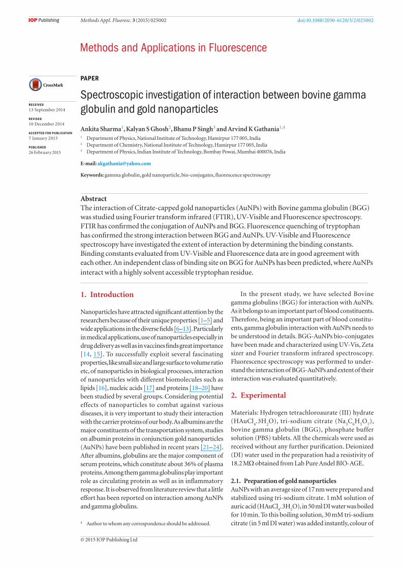
3.1. Transmission electron microscopyTEM image and selected area electron diffraction (SAED) pattern of AuNPs are shown in figures 1(a) and (b) respectively. TEM image reveals that the AuNPs formed are in nano-range with almost spherical shape and narrow size distribution. Histogram was obtained from the sample of near 175 particles, as shown in inset of figure 1(a) along with log normal distribution. Average particle size has been estimated to be ~17 nm with a standard deviation of 5.47 nm and positive skewness 0.0564. The SAED pattern confirms the crystallinity of the AuNPs. The corresponding HRTEM
image of the AuNPs is shown in inset of figure 1(b). The interplanar distance of the fringes is measured to be about 0.118 nm, which corresponds to the distance between the (2 2 2) planes of the gold crystal lattice.
3.2. Zeta potentialZeta potential of the samples was measured to be −29.2 mV for pure AuNPs and −4.54 mV for bio-conjugates. It reflects that there is change in the surroundings of AuNPs on interacting with BGG and it is possible that the surface of the AuNPs is carrying BGG molecules either through physical or chemical interactions.
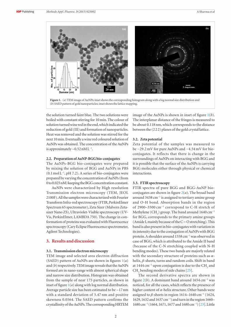
3.3. FTIR spectroscopyFTIR spectra of pure BGG and BGG-AuNP bio-conjugates are shown in figure 2(a). The broad band around 3438 cm−1 is assigned to tertiary amine group and O-H bond. Absorption bands in the region of 2900–3500 cm−1 correspond to C-H stretch of Methylene (CH
2) group. The band around 1640 cm−1
for BGG, corresponds to the primary amine groups (Amide I, mainly because of the C = O stretching). This band is also present in bio-conjugates with variation in its intensity due to the conjugation of AuNPs with BGG protein. A shoulder around 1558 cm−1 was observed in case of BGG, which is attributed to the Amide II band (because of the C-N stretching coupled with N-H bending modes). These two bands are majorly related with the secondary structure of proteins such as α-helix, β-sheets, turns and random coils. Shift in band at 1444 cm−1 upon conjugation is due to the CH
2 and
CH3 bending modes of side chains [25].
The second derivative spectra are shown in figure 2(b). A dominant band around 1654 cm−1 was noticed, for all the cases, which reflects the presence of higher content of α-helix structure. Other bands were assigned to β-sheets in region 1614–1640 cm−1 (1624, 1629, 1632 and 1637 cm−1) and turn in the region 1660–1680 cm−1 (1664, 1671, 1677 and 1680 cm−1) [23]. Little
Figure 1. (a) TEM image of AuNPs; inset shows the corresponding histogram along with a log normal size distribution and (b) SAED pattern of gold nanoparticles; inset shows the lattice mapping.
Methods Appl. Fluoresc. 3 (2015) 025002
3
A Sharma et al
shift in bands associated with β structures were noticed in bio-conjugates as compared to those for pure BGG. Intensity was also increased implying an increase in β-structure content. Another major shift was noticed for band corresponding to antiparallel β-sheet structure from 1693 cm−1 for BGG to 1697 cm−1 for bio-conju-gates. All these shifts indicate that there is a change in the secondary structure of BGG upon interaction with AuNPs. The band around 1648 cm−1 is related to ran-dom coil or disordered structure. The intensity of this band decreased in case of bio-conjugates, suggesting a decrease in random structure.
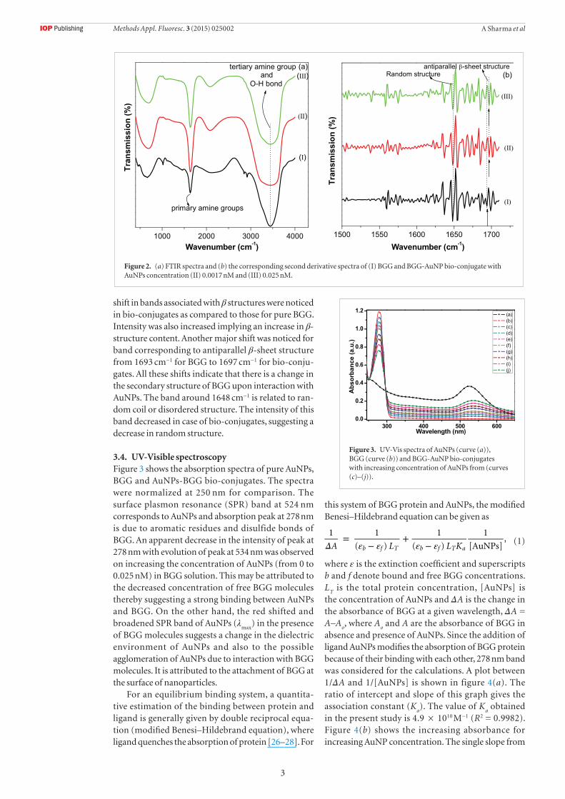
3.4. UV-Visible spectroscopyFigure 3 shows the absorption spectra of pure AuNPs, BGG and AuNPs-BGG bio-conjugates. The spectra were normalized at 250 nm for comparison. The surface plasmon resonance (SPR) band at 524 nm corresponds to AuNPs and absorption peak at 278 nm is due to aromatic residues and disulfide bonds of BGG. An apparent decrease in the intensity of peak at 278 nm with evolution of peak at 534 nm was observed on increasing the concentration of AuNPs (from 0 to 0.025 nM) in BGG solution. This may be attributed to the decreased concentration of free BGG molecules thereby suggesting a strong binding between AuNPs and BGG. On the other hand, the red shifted and broadened SPR band of AuNPs (λ
max) in the presence
of BGG molecules suggests a change in the dielectric environment of AuNPs and also to the possible agglomeration of AuNPs due to interaction with BGG molecules. It is attributed to the attachment of BGG at the surface of nanoparticles.
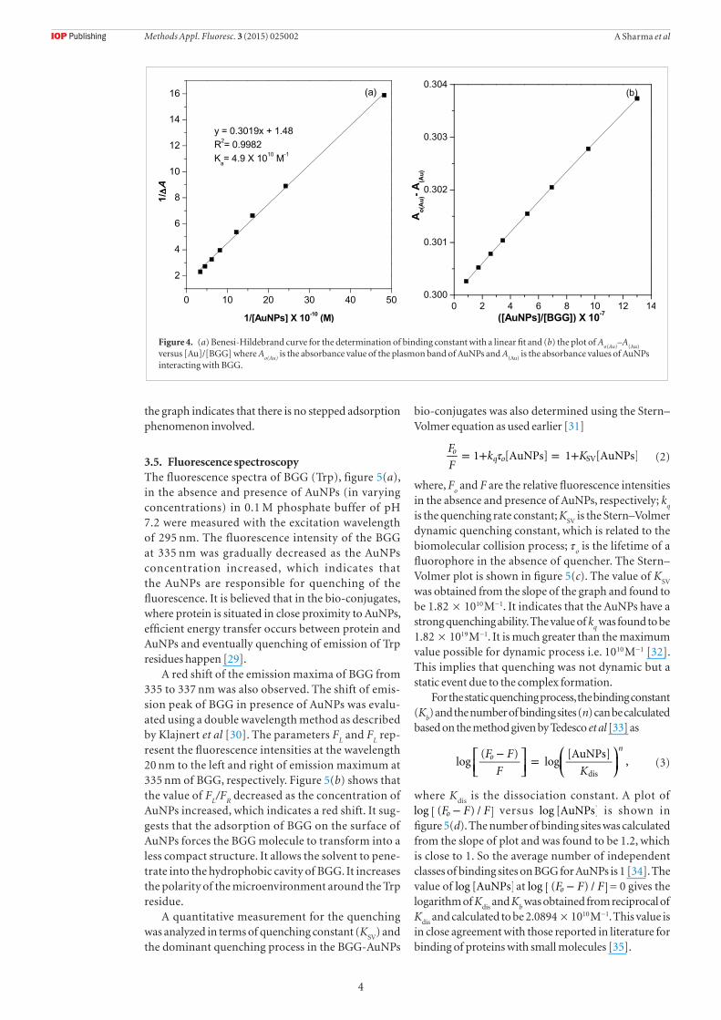
For an equilibrium binding system, a quantita-tive estimation of the binding between protein and ligand is generally given by double reciprocal equa-tion (modified Benesi–Hildebrand equation), where ligand quenches the absorption of protein [26–28]. For
this system of BGG protein and AuNPs, the modified Benesi–Hildebrand equation can be given as
Δ ε ε ε ε=
−+
−A L L K
1 1
( )
1
( )
1
[AuNPs],
b f T b f T a(1)
where ε is the extinction coefficient and superscripts b and f denote bound and free BGG concentrations. L
T is the total protein concentration, [AuNPs] is
the concentration of AuNPs and ΔA is the change in the absorbance of BGG at a given wavelength, ΔA = A–A
o, where A
o and A are the absorbance of BGG in
absence and presence of AuNPs. Since the addition of ligand AuNPs modifies the absorption of BGG protein because of their binding with each other, 278 nm band was considered for the calculations. A plot between 1/ΔA and 1/[AuNPs] is shown in figure 4(a). The ratio of intercept and slope of this graph gives the association constant (K
a). The value of K
a obtained
in the present study is 4.9 × 10¹⁰ M−1 (R2 = 0.9982). Figure 4(b) shows the increasing absorbance for increasing AuNP concentration. The single slope from
Figure 2. (a) FTIR spectra and (b) the corresponding second derivative spectra of (I) BGG and BGG-AuNP bio-conjugate with AuNPs concentration (II) 0.0017 nM and (III) 0.025 nM.
Figure 3. UV-Vis spectra of AuNPs (curve (a)), BGG (curve (b)) and BGG-AuNP bio-conjugates with increasing concentration of AuNPs from (curves (c)–(j)).
Methods Appl. Fluoresc. 3 (2015) 025002
4
A Sharma et al
the graph indicates that there is no stepped adsorption phenomenon involved.
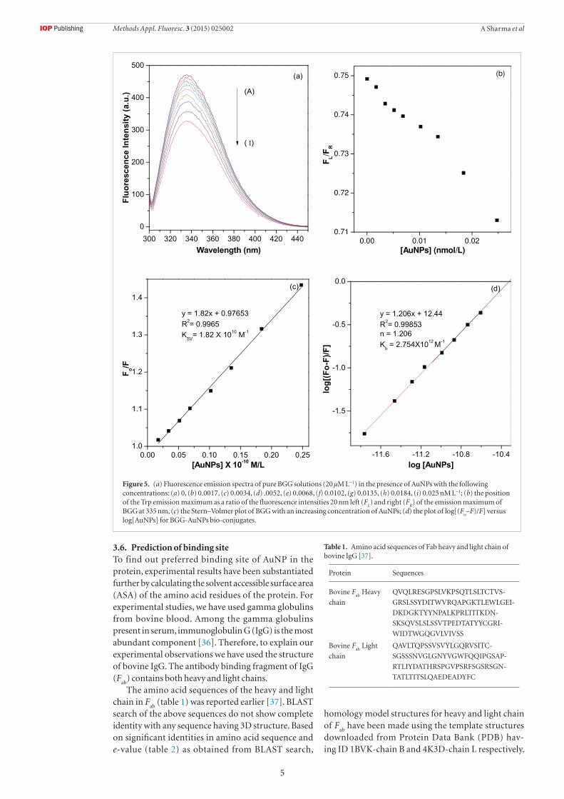
3.5. Fluorescence spectroscopyThe fluorescence spectra of BGG (Trp), figure 5(a), in the absence and presence of AuNPs (in varying concentrations) in 0.1 M phosphate buffer of pH 7.2 were measured with the excitation wavelength of 295 nm. The fluorescence intensity of the BGG at 335 nm was gradually decreased as the AuNPs concentration increased, which indicates that the AuNPs are responsible for quenching of the fluorescence. It is believed that in the bio-conjugates, where protein is situated in close proximity to AuNPs, efficient energy transfer occurs between protein and AuNPs and eventually quenching of emission of Trp residues happen [29].
A red shift of the emission maxima of BGG from 335 to 337 nm was also observed. The shift of emis-sion peak of BGG in presence of AuNPs was evalu-ated using a double wavelength method as described by Klajnert et al [30]. The parameters F
L and F
L rep-
resent the fluorescence intensities at the wavelength 20 nm to the left and right of emission maximum at 335 nm of BGG, respectively. Figure 5(b) shows that the value of F
L/F
R decreased as the concentration of
AuNPs increased, which indicates a red shift. It sug-gests that the adsorption of BGG on the surface of AuNPs forces the BGG molecule to transform into a less compact structure. It allows the solvent to pene-trate into the hydrophobic cavity of BGG. It increases the polarity of the microenvironment around the Trp residue.
A quantitative measurement for the quenching was analyzed in terms of quenching constant (K
SV) and
the dominant quenching process in the BGG-AuNPs
bio-conjugates was also determined using the Stern–Volmer equation as used earlier [31]
τ= + = +F
Fk K1 [AuNPs] 1 [AuNPs]o
q o SV (2)
where, Fo and F are the relative fluorescence intensities
in the absence and presence of AuNPs, respectively; kq
is the quenching rate constant; KSV
is the Stern–Volmer dynamic quenching constant, which is related to the biomolecular collision process; τ
o is the lifetime of a
fluorophore in the absence of quencher. The Stern–Volmer plot is shown in figure 5(c). The value of K
SV
was obtained from the slope of the graph and found to be 1.82 × 10¹⁰ M−1. It indicates that the AuNPs have a strong quenching ability. The value of k
q was found to be
1.82 × 10¹⁹ M−1. It is much greater than the maximum value possible for dynamic process i.e. 1010 M−1 [32]. This implies that quenching was not dynamic but a static event due to the complex formation.
For the static quenching process, the binding constant (K
b) and the number of binding sites (n) can be calculated
based on the method given by Tedesco et al [33] as
⎡⎣⎢
⎤⎦⎥
⎛⎝⎜
⎞⎠⎟− =F F
F Klog
( )log
[AuNPs],o
n
dis(3)
where Kdis
is the dissociation constant. A plot of −F F Flog [ ( ) / ]o versus log [AuNPs] is shown in
figure 5(d). The number of binding sites was calculated from the slope of plot and was found to be 1.2, which is close to 1. So the average number of independent classes of binding sites on BGG for AuNPs is 1 [34]. The value of log [AuNPs] at −F F Flog [ ( ) / ]o = 0 gives the logarithm of K
dis and K
b was obtained from reciprocal of
Kdis
and calculated to be 2.0894 × 10¹⁰ M−1. This value is in close agreement with those reported in literature for binding of proteins with small molecules [35].
Figure 4. (a) Benesi-Hildebrand curve for the determination of binding constant with a linear fit and (b) the plot of Ao(Au)
–A(Au)
versus [Au]/[BGG] where A
o(Au) is the absorbance value of the plasmon band of AuNPs and A
(Au) is the absorbance values of AuNPs
interacting with BGG.
Methods Appl. Fluoresc. 3 (2015) 025002
5
A Sharma et al
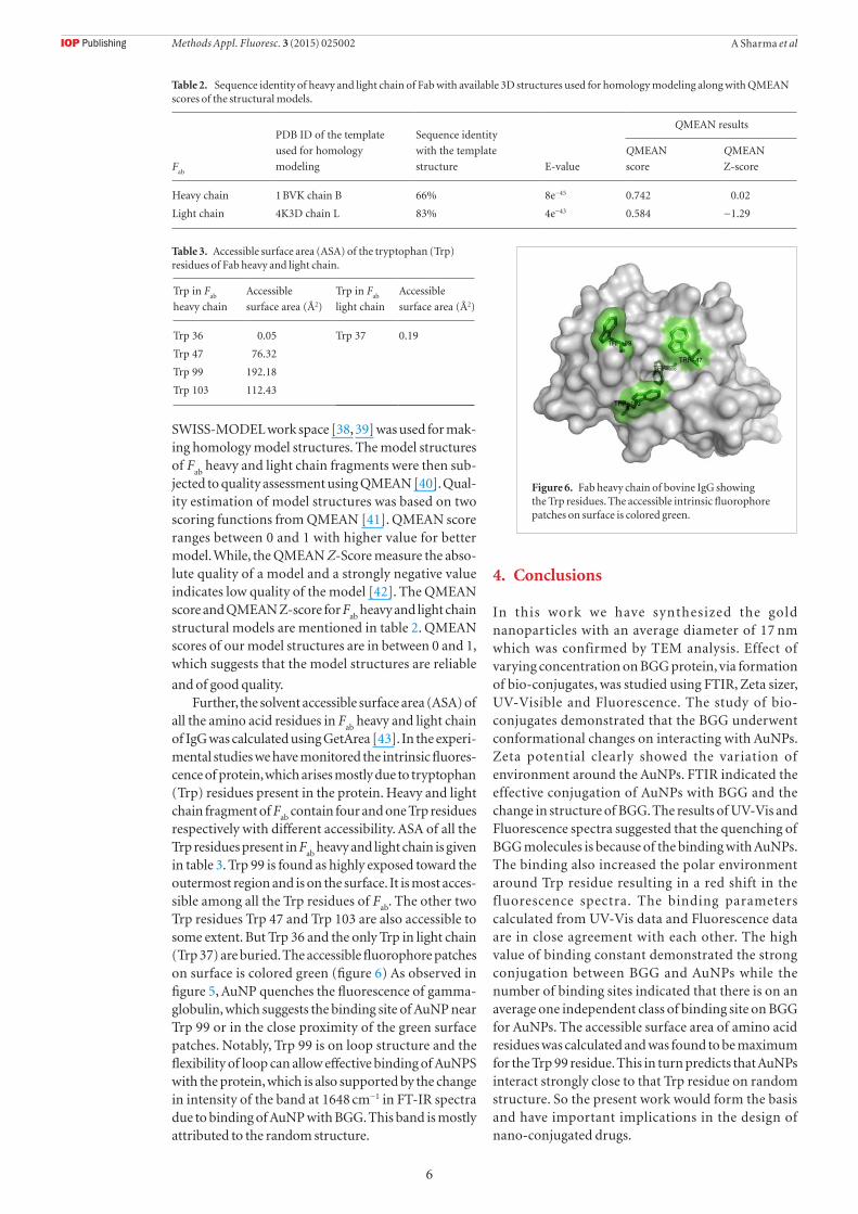
3.6. Prediction of binding siteTo find out preferred binding site of AuNP in the protein, experimental results have been substantiated further by calculating the solvent accessible surface area (ASA) of the amino acid residues of the protein. For experimental studies, we have used gamma globulins from bovine blood. Among the gamma globulins present in serum, immunoglobulin G (IgG) is the most abundant component [36]. Therefore, to explain our experimental observations we have used the structure of bovine IgG. The antibody binding fragment of IgG (F
ab) contains both heavy and light chains.The amino acid sequences of the heavy and light
chain in Fab
(table 1) was reported earlier [37]. BLAST search of the above sequences do not show complete identity with any sequence having 3D structure. Based on significant identities in amino acid sequence and e-value (table 2) as obtained from BLAST search,
homology model structures for heavy and light chain of F
ab have been made using the template structures
downloaded from Protein Data Bank (PDB) hav-ing ID 1BVK-chain B and 4K3D-chain L respectively.
Figure 5. (a) Fluorescence emission spectra of pure BGG solutions (20 µM L−1) in the presence of AuNPs with the following concentrations: (a) 0, (b) 0.0017, (c) 0.0034, (d) .0052, (e) 0.0068, (f) 0.0102, (g) 0.0135, (h) 0.0184, (i) 0.025 nM L−1; (b) the position of the Trp emission maximum as a ratio of the fluorescence intensities 20 nm left (F
L) and right (F
R) of the emission maximum of
BGG at 335 nm, (c) the Stern–Volmer plot of BGG with an increasing concentration of AuNPs; (d) the plot of log[(Fo–F)/F] versus
log[AuNPs] for BGG-AuNPs bio-conjugates.
Table 1. Amino acid sequences of Fab heavy and light chain of bovine IgG [37].
Protein Sequences
Bovine Fab
Heavy
chain
QVQLRESGPSLVKPSQTLSLTCTVS-
GRSLSSYDITWVRQAPGKTLEWLGEI-
DKDGKTYYNPALKPRLTITKDN-
SKSQVSLSLSSVTPEDTATYYCGRI-
WIDTWGQGVLVIVSS
Bovine Fab
Light
chain
QAVLTQPSSVSVYLGQRVSITC-
SGSSSNVGLGNYVGWFQQIPGSAP-
RTLIYDATHRSPGVPSRFSGSRSGN-
TATLTITSLQAEDEADYFC
Methods Appl. Fluoresc. 3 (2015) 025002
6
A Sharma et al
SWISS-MODEL work space [38, 39] was used for mak-ing homology model structures. The model structures of F
ab heavy and light chain fragments were then sub-
jected to quality assessment using QMEAN [40]. Qual-ity estimation of model structures was based on two scoring functions from QMEAN [41]. QMEAN score ranges between 0 and 1 with higher value for better model. While, the QMEAN Z-Score measure the abso-lute quality of a model and a strongly negative value indicates low quality of the model [42]. The QMEAN score and QMEAN Z-score for F
ab heavy and light chain
structural models are mentioned in table 2. QMEAN scores of our model structures are in between 0 and 1, which suggests that the model structures are reliable
and of good quality.Further, the solvent accessible surface area (ASA) of
all the amino acid residues in Fab
heavy and light chain of IgG was calculated using GetArea [43]. In the experi-mental studies we have monitored the intrinsic fluores-cence of protein, which arises mostly due to tryptophan (Trp) residues present in the protein. Heavy and light chain fragment of F
ab contain four and one Trp residues
respectively with different accessibility. ASA of all the Trp residues present in F
ab heavy and light chain is given
in table 3. Trp 99 is found as highly exposed toward the outermost region and is on the surface. It is most acces-sible among all the Trp residues of F
ab. The other two
Trp residues Trp 47 and Trp 103 are also accessible to some extent. But Trp 36 and the only Trp in light chain (Trp 37) are buried. The accessible fluorophore patches on surface is colored green (figure 6) As observed in figure 5, AuNP quenches the fluorescence of gamma-globulin, which suggests the binding site of AuNP near Trp 99 or in the close proximity of the green surface patches. Notably, Trp 99 is on loop structure and the flexibility of loop can allow effective binding of AuNPS with the protein, which is also supported by the change in intensity of the band at 1648 cm−1 in FT-IR spectra due to binding of AuNP with BGG. This band is mostly attributed to the random structure.
4. Conclusions
In this work we have synthesized the gold nanoparticles with an average diameter of 17 nm which was confirmed by TEM analysis. Effect of varying concentration on BGG protein, via formation of bio-conjugates, was studied using FTIR, Zeta sizer, UV-Visible and Fluorescence. The study of bio-conjugates demonstrated that the BGG underwent conformational changes on interacting with AuNPs. Zeta potential clearly showed the variation of environment around the AuNPs. FTIR indicated the effective conjugation of AuNPs with BGG and the change in structure of BGG. The results of UV-Vis and Fluorescence spectra suggested that the quenching of BGG molecules is because of the binding with AuNPs. The binding also increased the polar environment around Trp residue resulting in a red shift in the fluorescence spectra. The binding parameters calculated from UV-Vis data and Fluorescence data are in close agreement with each other. The high value of binding constant demonstrated the strong conjugation between BGG and AuNPs while the number of binding sites indicated that there is on an average one independent class of binding site on BGG for AuNPs. The accessible surface area of amino acid residues was calculated and was found to be maximum for the Trp 99 residue. This in turn predicts that AuNPs interact strongly close to that Trp residue on random structure. So the present work would form the basis and have important implications in the design of nano-conjugated drugs.
Table 2. Sequence identity of heavy and light chain of Fab with available 3D structures used for homology modeling along with QMEAN scores of the structural models.
Fab
PDB ID of the template
used for homology
modeling
Sequence identity
with the template
structure E-value
QMEAN results
QMEAN
score
QMEAN
Z-score
Heavy chain 1 BVK chain B 66% 8e−45 0.742 0.02
Light chain 4K3D chain L 83% 4e−43 0.584 −1.29
Table 3. Accessible surface area (ASA) of the tryptophan (Trp) residues of Fab heavy and light chain.
Trp in Fab
heavy chain
Accessible
surface area (Å2)
Trp in Fab
light chain
Accessible
surface area (Å2)
Trp 36 0.05 Trp 37 0.19
Trp 47 76.32
Trp 99 192.18
Trp 103 112.43
Figure 6. Fab heavy chain of bovine IgG showing the Trp residues. The accessible intrinsic fluorophore patches on surface is colored green.
Methods Appl. Fluoresc. 3 (2015) 025002
7
A Sharma et al
Acknowledgment
One of the authors (Ankita Sharma) is thankful to the National Institute of Technology, Hamirpur (HP), India, for providing financial assistance and the Advanced Instrumentation Research Facility, Jawaharlal Nehru University, New Delhi, India, for their TEM facility. The authors are also grateful to Professor B S Kaith, Department of Chemistry, National Institute of Technology, Jalandhar, India, for the Fluorescence facility.
References
[1] El-Brolossy T A, Abdallah T, Mohamed M B, Abdallah S, Easawi K and Negm S 2008 Shape and size dependence of the surface plasmon resonance of gold nanoparticles studied by photoacoustic technique Eur. Phys. J. Spec. Top. 153 361–4
[2] Hao E, Schatz G C and Hupp J T 2004 Synthesis and optical properties of anisotropic metal nanoparticles J. Fluoresc. 14 331–41
[3] Sharma A, Singh B P and Gathania A K 2014 Synthesis and characterization of dodecanethiol-stabilized gold nanoparticles Indian J. Pure Appl. Phys. 52 93–100
[4] Sharma A, Dhiman N, Singh B P and Gathani A K 2014 Green synthesis of gold nanoparticles using extracts of artocarpus lakoocha fruit and its leaves and eriobotrya japonica leaves Mater. Res. Express 1 25042
[5] Thakur S and Gathania A K 2014 Effect of calcination on the optical properties of YVO4: Eu 3 Nano-phosphors Int. J. Lumin. Appl. 4 53–5
[6] Sapsford K E, Algar W R, Berti L, Gemmill K B, Casey B J, Oh E, Stewart M H and Medintz I L 2013 Functionalizing nanoparticles with biological molecules: developing chemistries that facilitate nanotechnology Chem. Rev. 113 1904–2074
[7] Sau T K, Rogach A L, Jackel F, Klar T A and Feldmann J 2010 Properties and applications of colloidal nonspherical noble metal nanoparticles Adv. Mater. 22 1805–25
[8] Somani P R, Somani S P and Umeno M 2008 Application of metal nanoparticles decorated carbon nanotubes in photovoltaics Appl. Phys. Lett. 93 033315
[9] Zhang Y-J, Huang R, Zhu X-F, Wang L-Z and Wu C-X 2012 Synthesis, properties and optical applications of noble metal nanoparticle-biomolecule conjugates Chin. Sci. Bull. 57 238–46
[10] Dhiman N, Singh B P and Gathania A K 2012 Synthesis and characterization of dye-doped TiO
2-SiO
2 core-shell composite
microspheres J. Nanophoton. 6 063511 [11] Gathania A K, Dhiman N, Sharma A and Singh B P 2011
Morphology and optical study of dye-doped TiO2-SiO
2 thin
films Smart Nano-Micro Mater. Devices (VIC AUS Hawthorn, 05–07 Decmber 2011) vol 8204 82043N
[12] Dhiman N, Singh B P and Gathania A K 2012 Fabrication and optical study of dye doped TiO
2-SiO
2 multilayer colloidal
structures Colloids Surf. A: Physicochem. Eng. Asp. 409 69–73 [13] Sharma A, Dhiman N, Singh B P and Gathania A K 2014
Spectroscopic investigations on metallo-dielectric gold@silica composites J. Mol. Struct. 1074 522–6
[14] Doria G, Conde J, Veigas B, Giestas L, Almeida C, Assuncao M, Rosa J and Baptista P V 2012 Noble metal nanoparticles for biosensing applications Sensors 12 1657–87
[15] Arvizo R R, Bhattacharyya S, Kudgus R A, Giri K, Bhattacharya R and Mukherjee P 2012 Intrinsic therapeutic applications of noble metal nanoparticles: past, present and future Chem. Soc. Rev. 41 2943–70
[16] Tatur S, Maccarini M, Barker R, Nelson A and Fragneto G 2013 Effect of functionalized gold nanoparticles on floating lipid bilayers Langmuir 29 6606–14
[17] Goodman C M, Chari N S, Han G, Hong R, Ghosh P and Rotello V M 2006 DNA-binding by functionalized gold nanoparticles: mechanism and structural requirements Chem. Biol. Drug Des. 67 297–304
[18] Ojha K, Chowdhury P K and Ganguli A K 2012 Fluorescence and CD studies of protein denaturation in the presence of sub-picomolar gold nanoparticles Indian J. Chem. 51 1561–6
[19] Chandra G, Ghosh K S, Dasgupta S and Roy A 2010 Evidence of conformational changes in lysozyme molecule on silver colloids Int. J. Biol. Macromolecules 47 361–5
[20] Lacerda S H, Park J J, Meuse C, Pristinski D, Becker M L, Karin A and Douglas J F 2010 Interaction of gold nanoparticles with human blood proteins ACS Nano 4 365–79
[21] Pramanik S, Banerjee P, Sarkar A and Bhattacharya S C 2008 Size-dependent interaction of gold nanoparticles with transport protein: a spectroscopic study J. Lumin. 128 1969–74
[22] Joshi P, Chakraborty S, Dey S, Shanker V, Ansari Z A, Singh S P and Chakrabarti P 2011 Binding of chloroquine gold nanoparticles with bovine serum albumin J. Colloid Interface Sci. 355 402–9
[23] Shang L, Wang Y, Jiang J and Dong S 2007 pH-dependent protein conformational changes in albumin:gold nanoparticle bioconjugates: a spectroscopic study Langmuir 23 2714–21
[24] Naveenraj S, Anandan S, Kathiravan A, Renganathan R and Ashokkumar M 2010 The interaction of sonochemically synthesized gold nanoparticles with serum albumins J. Pharm. Biomed. Anal. 53 804–10
[25] Susi H and Byler D M 1983 Protein structure by fourier transform infrared spectroscopy second derivative spectra Biochem. Biophys. Res. Commun. 115 391–7
[26] Benesi H A and Hildebrand J H 1949 A spectrophotometric investigation of the interaction of iodine with aromatic hydrocarbons J. Am. Chem. Soc. 71 2703–7
[27] Sahoo B K, Ghosh K S and Dasgupta S 2008 Molecular interactions of isoxazolcurcumin with human serum albumin: spectroscopic and molecular modeling studies Biopolymers 91 108–19
[28] Maiti T K, Ghosh K S, Samanta A and Dasgupta S 2008 The interaction of silibinin with human serum albumin: a spectroscopic investigation J. Photochem. Photobiol. A: Chem. 194 297–307
[29] Delfino I and Cannistraro S 2009 Optical investigation of the electron transfer protein azurin–gold nanoparticle system Biophys. Chem. 139 1–7
[30] Klajnert B, Stanislawska L, Bryszewska M and Palecz B 2003 Interactions between PAMAM dendrimers and bovine serum albumin BBA-Proteins Proteomics 1648 115–26
[31] Sahoo B K, Ghosh K S and Dasgupta S 2009 An investigation of the molecular interactions of Diacetylcurcumin with ribonuclease a Protein Pept. Lett. 16 1485–95
[32] Yang T, Li Z, Wang L, Guo C and Sun Y 2008 Synthesis, characterization and self-assembly of protein lysozyme mono-layer-stabilized gold nanoaperticles Appl. Phys. Lett. 92 133104
[33] Tedesco A C, Oliveira D M, Lacava Z G M, Azevedo R B, Lima E C D, Gansau C, Buske N and Morais P C J 2003 Determination of binding constant Kb of biocompatible ferrite-based magnetic fluids to serum albumin J. Appl. Phys. 93 6704
[34] Shi X J, Li D, Xie J, Wang S, Wu Z Q and Chen H 2012 Spectroscopic investigation of the interactions between gold nanoparticles and bovine serum albumin Chin. Sci. Bull. 57 1109–15
[35] Ao L-M, Gao F, Pan B-F, Cui D-X and Gu H-C 2006 Interaction between gold nanoparticles and bovine serum albumin or sheep antirabbit immunoglobulin G Chin. J. Chem. 24 253–6
[36] Junqueira L C and Carneiro J 2003 Basic Histology (New York: McGraw-Hill)
[37] O’Brien P M, Aitken R, O’Neil B W and Campo M S 1999 Generation of native bovine mAbs by phage display Proc. Natl Acad. Sci. USA 96 640–5
Methods Appl. Fluoresc. 3 (2015) 025002
8
A Sharma et al
[38] Arnold K, Bordoli L, Kopp J and Schwede T 2006 The swiss-model workspace: a web- based environment for protein structure homology modeling Bioinformatics 22 195–201
[39] Kiefer F, Arnold K, Künzli M, Bordoli L and Schwede T 2009 The swiss-model repository and associated resources Nucleic Acids Res. 37 D387–92
[40] Benkert P, Künzli M and Schwede T 2009 QMEAN server for protein model quality estimation Nucleic Acids Res. 37 W510–4
[41] Benkert P, Tosatto S C E and Schomburg D 2008 QMEAN: a comprehensive scoring function for model quality assessment Proteins 71 261–77
[42] Benkert P, Biasini M and Schwede T 2011 Toward the estimation of the absolute quality of individual protein structure models Bioinformatics 27 343–50
[43] Fraczkiewicz R and Braun W 1998 Exact and efficient analytical calculation of the accessible surface areas and their gradients for macromolecules J. Comput. Chem. 19 319–33
Methods Appl. Fluoresc. 3 (2015) 025002