


BACTERIOLOGICAL STUDIES ON CHRONIC MASTITIS IN BOVINE
155
PDF created with pdfFactory trial version www.pdffactory.com
Transcript of BACTERIOLOGICAL STUDIES ON CHRONIC MASTITIS IN BOVINE

الرحمـن الرحیمهللا بسم ا
والبحرأقلـم من شجرةفي االرض ولو أنمــا ﴿
إن اهللا هللامانفدت كلمت أبحر ه من بعدة سبعةیمد
﴾عزيز حكيم
العظیمهللا صدق ا
)٢٧(سورة لقمان االیة
PDF created with pdfFactory trial version www.pdffactory.com

ACKNOWLEDGEMENT
I am greatly indebted to gracious Allah for helping me to carry
out this work.
I would like to express my sincere gratitude and deep
appreciation to Prof. Dr. Mahmoud Essam Hatem Prof. and Head
of Microbiology Department, Faculty of Veterinary Medicine, Cairo
University for his kind supervision, faithful guidance, valuable
advice throughout the course of the present work and for the time
that he has given up to complete the present study.
I would like to introduce my great appreciation to Major
General Dr. Fathy Gaber Awad the consultant of chief stuff of
logistics authority for veterinary services for his kind supervision,
continuous and Valuable encouragement and supplying the facilities
required for my study.
I would like to express my sincere thanks to Brigadier General
Dr. Amin Abd El Fattah Badr Director of The Military Institute of
Health and Epidemiology for his kind help, valuable advice
throughout the course of present study continuous and valuable
encouragement.
PDF created with pdfFactory trial version www.pdffactory.com

List of Contents
Ⅰ. Introduction. ------------------------------------------------ 1
Ⅱ. Review of Literature. ------------------------------------- 4
Ⅱ.1. Etiology of bovine chronic mastitis.----------------------- 4
Ⅱ.2. Pathogenises of chronic Mastitis.-------------------------- 14
Ⅱ.3. Diagnosis of chronic mastitis.------------------------------ 21
Ⅱ.4. Antibiotic sensitivity of different bacterial species causing chronic mastitis 23
Ⅱ.5. Use of antibiotics and vaccines in case of chronic mastitis. 30
Ⅲ. Materials and Methods. ----------------------------------- 34
Ⅲ.1. Material.------------------------------------------------------- 34
Ⅲ.1.1 Animals.------------------------------------------------------- 34
Ⅲ.1.2. Milk samples.------------------------------------------------- 34
Ⅲ.1.3. Materials used for sample collection.--------------------- 35
Ⅲ.1.4. Materials used for direct microscopic examination.------------ 35
Ⅲ.1.5. Media.--------------------------------------------------------- 35
Ⅲ.1.6. Reagents used for biochemical identification.----------- 42
Ⅲ.1.7. Stains.---------------------------------------------------------- 46
Ⅲ.1.8. Materials used for Agar disc diffusion method.---------- 47
Ⅲ.2. Methods.------------------------------------------------------- 50
PDF created with pdfFactory trial version www.pdffactory.com

Ⅲ.2.1. Collection of milk samples.--------------------------------- 50
Ⅲ.2.2. Methods of bacteriological examination.----------------- 50
Ⅲ.2.3. Methods of antibiotic susceptibility testing.------------- 59
Ⅳ. Results. ------------------------------------------------------ 61
V. Discussion.--------------------------------------------------- 84
Ⅵ. Summary.---------------------------------------------------- 100
Ⅶ. Recommendations ------------------------------------------ 103
Ⅷ. Abstract ------------------------------------------------------ 104
Ⅸ References. ---------------------------------------------------
105 X Arabic Summary-------------------------------------------- 127
PDF created with pdfFactory trial version www.pdffactory.com

List of Tables
Table Title Page
1 Concentration of antibacterial discs used in Agar Disc
Diffusion Method (ADD) for antibacterial susceptibility. 49
2 Differentiation between Staphylococcus aureus,
Staphylococcus epidermidis, and Staphylococcus
saprophyticus 54
3 Identification of the isolates of Arcanobacterium
pyogenes. 55
4 Identification of the isolates of
Bacillus sp. 56
5 Differentiation between Escherichia coli and
Klebsiella pneumoniae : 57
6 Identification of the isolates of Pseudomonas
aeruginosa 58
7 Interpretation zones in Agar Disc Diffusion Method
(ADD) for antibacterial Susceptibility. 60
8 The mean of the affected chronic quarters per one cow
in each of the different test locations. 61
9 The prevalence of the isolated organisms in relation to
the total number of examined samples. 62
PDF created with pdfFactory trial version www.pdffactory.com

Table Title Page
10 The prevalence of each isolated organism to the total
number of isolated pathogens. 63
11 The incidence of each of isolated pathogen in different
experiment locations in relation to the total bacterial
isolates. 65
12 The distribution of chronic mastitis according to the
affected quarter involvement of the examined animals. 66
13 The distribution of chronic mastitis according to the
affected quarter involvement for each pathogen. 66
14 Distribution of chronic mastitis within the four udder
quarters according to quarter involvement of the
examined animals in case of affected animals that had no
bacterial growth. 67
15 Antibiogram of Staphylococcus aureus recovered from
cows with chronic bovine mastitis to antimicrobial
agents. 69
16 Antibiogram of Staphylococcus epidermidis recovered
from cows with chronic bovine mastitis to antimicrobial
agents. 71
PDF created with pdfFactory trial version www.pdffactory.com

Table Title Page
17 Antibiogram of Staphylococcus saprophyticus recovered
from cows with chronic bovine mastitis to antimicrobial
agents 73
18 Antibiogram of Streptococcus sp. recovered from cows
with chronic bovine to antimicrobial agents. 75
19 Antibiogram of Bacillus cereus recovered from cows
with chronic bovine mastitis to antimicrobial agents. 77
20 Antibiogram of Escherichia coli recovered from cows
with chronic bovine mastitis to antimicrobial agents. 79
21 Antibiogram of Klebsiella pneumoniae recovered from
cows with chronic bovine mastitis to antimicrobial
agents. 81
PDF created with pdfFactory trial version www.pdffactory.com

List of Graphics
Graph Title Page
1 The prevalence of each isolated organism to the total
number of isolated pathogens. 64 2 Susceptibility pattern of Staphylococcus aureus
recovered from cows with chronic bovine mastitis
against different antibacterials. 70 3 Susceptibility pattern of Staphylococcus epidermidis
recovered from cows with chronic bovine mastitis
against different antibacterials 72 4 Graphic (4): Susceptibility pattern of Staphylococcus
saprophyticus recovered from cows with chronic
bovine mastitis against different antibacterials 74 5 Susceptibility pattern of Streptococcus sp. recovered
from cows with chronic bovine mastitis to different
antibacterials. 76 6 Susceptibility pattern of Bacillus cereus recovered
from cows with chronic bovine mastitis to different
antibacterials. 78 7 Susceptibility pattern of Escherichia coli recovered
from cows with chronic bovine mastitis to different
antibacterials. 80
PDF created with pdfFactory trial version www.pdffactory.com

Graph Title Page
8 Susceptibility pattern of Klebsiella pneumoniae
recovered from cows with chronic bovine mastitis
to different antibacterials. 82
PDF created with pdfFactory trial version www.pdffactory.com

List of photographs
photo Title Page
1 Citrate test. 39
2 Methyl Red test 42
3 Voges Proskaur test 43
4 Indole test 44
5 Novobiocin sensitivity test 54
6 Antibiotic sensitivity test to 18 antibacterial agents 68
PDF created with pdfFactory trial version www.pdffactory.com

LIST OF ABBREVIATIONS
API Analytical Profile Index A. pyogenes Arcanobacterium pyogenes A. pyogenes Actinomyces pyogenes
BMEC Bovine mammary epithelial cells BMSC Bulk milk somatic cell count CNS Coagulase-negative Staphylococcus CMT California mastitis tests CP5 Capsular polysaccharide type 5 CLSI Clinical and Laboratory Standards Institute DCT Dry-cow antibiotic therapy
FEMS Federation of European Microbiological Societies IMI Intramammary infection. IP Interpretation
ID Identification IMVIC test Indole, Methyl red, Vogas Proskaur and Citrate tests
MIC Minimum inhibitory concentration PIA Polysaccharide intercellular adhesion SCC Somatic cell count TSI Triple sugar iron
TSST Toxic shock syndrome toxin TSST-1 Toxic shock syndrome toxin-1
VP Voges Proskaur test vs. Versus
PDF created with pdfFactory trial version www.pdffactory.com

INTRODUCTION
1
I. INTRODUCTION
Bovine mastitis is considered to be one of the most important
destructive diseases producing abnormal milk and monetary loses to
dairy industry (Schalm et al., 1971). The economic losses of
mastitis are due to reduced milk production, discarded milk during
treatment, early cow replacement cost, drugs, veterinary service and
labour (Harmon, 1995). Information regarding the prevalence of
mastitis pathogens and costs associated with different bacterial
infection is of interest to the dairy industry (Wilson et al., 1997).
In Egypt mastitis causes great damage through threatening the
dairy animal health. The greatest losses of mastitis are mostly due to
subclinical form in which there are no detectable abnormalities of
the milk. However, the causative microorganism/s act as invisible
potential source for spreading the infection (EL. Kholly et al., 1994).
Most surveys in different countries have shown that up to 50% of
lactating animals, at any time, may suffer from chronic latent
mastitis after suffering from subclinical mastitis (Grunert and
Weight, 1979).
Most infections are caused by various species of streptococci,
staphylococci, and Gram-negative rods, specially lactose-
fermenting organisms of enteric origin, commonly termed
PDF created with pdfFactory trial version www.pdffactory.com

INTRODUCTION
2
coliforms. From an epidemiologic standpoint, the source of infection
may be regarded as contagious or environmental. Except for
Mycoplasma Sp. which may spread from cow to cow through
aerosol transmission and invade the udder subsequent to bacteremia,
contagious pathogens are spread during milking by milker’s hands
or the liners of the milking unit. Species that utilize this mode of
transmission include Staphylococcus aureus, Streptococcus
agalactiae, and Corynebacterium bovis. Most other species are
opportunistic invaders from the cow’s environment, although some
other streptococci and staphylococci may also have a contagious
component. Gangrenous mastitis can also occur, particularly when
subclinical, chronic infections of Staphylococcus aureus, become
severe at times of immunosuppression (MERK, 1998).
Many genera and species of pathogenic bacteria are
incriminated as causal agents of severe mastitis in dairy Friesian
cows .Teat dipping and dry cow therapy have reduced the mastitis
caused by the Staphylococcus aureus and Streptococcus
agalactiae, but failed to prevent mastitis caused by other types of
mastitis causing bacteria (Oz et al., 1985) .
PDF created with pdfFactory trial version www.pdffactory.com

INTRODUCTION
3
The aim of the present study was to:
(A)- Isolation and identification of causative bacterial agent/s of
chronic mastitis in dairy Friesian cows at different dairy farms.
(B)- Antibiogram for the bacteria isolated aiming to find the most
suitable treatment for each case.
PDF created with pdfFactory trial version www.pdffactory.com

LITERATURE
4
Π. LITERATURE
Π. 1. Etiology of bovine chronic mastitis:
Jasper and Dellinger (1975) studied the prevalence of
Escherichia coli infection regarding to the association with the
other infections. The study showed that Escherichia coli
infections were mostly related to the mammary glands that were
not infected with other pathogens. In addition, they reported high
susceptibility of the mammary gland to Escherichia coli
infection particularly after treatment of streptococcal and
staphylococcal infections.
Hill and Shears (1979) examined 28 mastitic cows with
Escherichia coli for persistence and recurrence through 120
days. The study showed that only 5 cases of the 28 had long
recurrent infection.
Inui et al.(1979) applied bacteriological and pathological
studies on the mammary gland of 17 cows with chronic mastitis.
The isolated pathogens were: Staphylococcus aureus (5 cases );
Staphylococcus aureus + Staphylococcus epidermidis (one
case); Staphylococcus aureus, Corynebacterium pyogenes and
Staphylococcus epidermidis (one case); Corynebacterium
PDF created with pdfFactory trial version www.pdffactory.com

LITERATURE
5
pyogenes + Staphylococcus epidermidis (one case);
Corynebacterium pyogenes + Corynebacterium bovis (one case);
Candida krusei, K. pneumoniae, Pseudomonas aeruginosa,
haemolytic streptococci, and Corynebacterium pyogenes each
were isolated from one case; Escherichia coli + Nocardia
asteroides each from 2 cases.
Jain (1979) reported that Streptococcus agalactiae was the
predominant causative microorganism of bovine chronic mastitis before
the antibiotics era.
Smith and Hagstad (1985) studied results of examination of 418
milk samples collected during one year from a herd of 69 cows with
chronic staphylococcal mastitis. The study showed that coagulase-
negative staphylococci and Staphylococcus epidermidis isolates
detected decreased with advancing age, whilst Staphylococcus aureus
infections increased with age. Prevalence of Staphylococcus aureus
infections increased throughout lactation.
Garg and Kapoor (1986) identified pure isolates of Rhodococcus
equi from two cows each with chronic mastitis in one quarter. The two
Rhodococcus equi isolates were of B type colony, did not haemolyse
10% sheep blood agar and produced 'equi factor', catalase and urease.
PDF created with pdfFactory trial version www.pdffactory.com

LITERATURE
6
Wilson et al. (1991) recorded the incidence of different pathogens
in 49 quarters with chronic mastitis. Isolated agents were:
Staphylococcus aureus (18.4%); Staphylococcus sp. (7.3%); no
growth (20.2%). Environmental pathogens streptococci other than
Streptococcus agalactiae, Escherichia coli, Klebsiella sp., Enterobacter sp. and Citrobacter sp.(22.0%). Other pathogens
(Serratia sp., Bacillus sp., diphtheroids, Corynebacterium sp. and
Actinomyces pyogenes), Pseudomonas sp. and Nocardia sp.),
represented 11.9%. Mixed pathogens (2 agents isolated) were 12.8%
and contaminated samples constituted 7.3%.
Samborski et al. (1992) determined the prevalences of pathogenic
bacteria in 114 quarter mastitic milk samples of cows (76 samples were
obtained from subclinical mastitic quarter and 38 samples from clinical
chronic mastitic quarters). Pathogens isolated were Streptococcus
agalactiae (44.7%), Streptococcus uberis (8.8%), Streptococcus
dysgalactiae (5.3%), Staphylococcus aureus (24.5%), Staphylococcus
epidermidis (15.8%) and Escherichia coli (0.9%).
Nemeth et al. (1994) examined and compared the biochemical
properties and certain potential virulence factors of Escherichia coli
isolates that were obtained from bovine mastitis and bovine feces. The
study included 50 isolates of Escherichia coli taken from bovine
mastitic milk, 50 from feces of mastitic cows and 50 from feces of
PDF created with pdfFactory trial version www.pdffactory.com

LITERATURE
7
healthy cows. The study revealed that none of the properties that were
investigated constituted potential virulence factors or markers for the
ability to induce mastitis. They concluded that mastitic Escherichia coli
are simply opportunistic pathogens.
Erer et al. (1996) examined 883 cows at abattoir before slaughter
using California mastitis reagent. The study showed mastitis in 118
cows (13.4%) with 232 mastitic quarters of which 163 chronic mastitic
quarters were identified histologically after slaughter. 184 isolates were
obtained: Staphylococcus aureus from 87 (47.3%), Actinomyces
pyogenes from 30 (16.3%) , Escherichia coli from 15 (8.2%) ,
Candida albicans from 12 (6.5%) , Staphylococcus epidermidis from 8
(4.3%) Klebsiella pneumoniae from 7 (3.8%) , Bacillus subtilis from 4
(2.2%) , Streptococcus dysgalactiae from 4 (2.2%) , Flavobacterium
from 3 (1.6%) , Bacillus cereus from 2 (1.1%) and Proteus mirabilis
from 1 (0.5%).
Gonzalez (1996) identified Bacillus cereus as an uncommon
cause of mastitis. Pure cultures of Bacillus cereus were obtained from
samples taken from first lactation cow with chronic mastitis in two
quarters with frequent flare-ups. Although, few colonies of Bacillus sp.
which were grown on blood agar plates represented contamination.
However, Bacillus sp. was considered as a cause of intramammary
PDF created with pdfFactory trial version www.pdffactory.com

LITERATURE
8
infection in a cow if culture of sequential milk samples was pure with
concomitant high SCC with clinical signs of udder disease.
Owens et al. (1997) compared the results of success of antibiotic
therapy with results of antimicrobial susceptibility testing for bovine
mastitis bacterial pathogens to evaluate the predictability of use of the in
vitro antibacterial susceptibility tests. The study showed that the in vitro
testing was considered to be a predictor of therapy outcome in case of
mastitis caused by Staphylococcus sp., newly acquired Staphylococcus
aureus, Streptococcus uberis, Streptococcus dysgalactiae, and
Streptococcus agalactiae, but was not considered to be a useful
predictor of efficacy for chronic mastitis caused by Staphylococcus
aureus.
Costa et al. (1998) determined the prevalence of environmental
mastitis in dairy herds and identified the main environmental pathogens
of 20,310 quarters of 5,216 cows. The isolated environmental bacteria
were Streptococcus uberis (21.1%), Enterobacteriaceae (8.3%) and
Nocardia sp. (6.6%).
Böhmer et al.(1999) identified the causative organisms of chronic
mastitis in 100 lactating German Black Pied cows. The study showed
that chronic mastitis was a major problem in 14% of cows.
Streptococcus dysgalactiae (3cows), Streptococcus uberis (13),
PDF created with pdfFactory trial version www.pdffactory.com

LITERATURE
9
Enterococcus sp. (1), Staphylococcus aureus (4), and yeasts (4), while
27 cows showed unclear Causes.
Twardona et al. (1999) reported the incidence of chronic mastitis
in 71 Holstein Friesian cows aged 2.8-8 years. Chronic mastitis was
identified in 35 quarters. Staphylococcus epidermidis, Streptococcus
agalactiae and Staphylococcus aureus were isolated from 34.9, 28.1
and 29.1% quarters respectively.
Larsen et al. (2000) examined 625 isolates of Staphylococcus
aureus from bovine mastitis in 9 dairy herds for their diversity and
compared the results with that of isolates from other sites by phage- and
ribotyping. The study included isolates obtained from bovine skin lesions,
asymptomatic human carriers, milking personnel and non-farm-related
human carriers. They aimed to investigate whether colonization of
milkers with Staphylococcus aureus could be a source of infection for
bovine mastitis. The study revealed that certain types predominated in
one or several herds during the study period, whereas the presence of
other types was of a more sporadic nature. Within the individual herds,
there was a close relation between ribo- and phage types of
Staphylococcus aureus isolated from bovine mastitis and bovine skin
lesions. Isolates from milking personnel, however, were not identical to
any of the predominant intramammary strains. These results supported
PDF created with pdfFactory trial version www.pdffactory.com

LITERATURE
10
the hypothesis that the human reservoir of Staphylococcus aureus does
not play any major role as a source of bovine mastitis.
Pitkala et al. (2004) estimated the prevelance of bovine mastitis,
distribution of mastitis pathogens and in vitro antibacterial susceptibility
of different mastitis pathogens in total of 21,661 quarter milk samples
from 3,282 dairy cows. They compared results with a previous survey
done in 1995 and reported decrease in the prevalence of mastitis from
38% to 31% in their study (of bacterial and non bacterial causes). At the
same time they reported a significant increase in the prevelance of
mastitis due to bacterial causes from 21.0% to 33.5% as result to
increase of the prevalence of Corynebacterium bovis mastitis. With a
decrease in Staphylococcus aureus prevalence, the coagulase-negative
staphylococcal mastitis remained the most common bacteria isolated
constituting almost 50% of the reported cases. Staphylococci
showed high penicillin resistance (52.1% for Staphylococcus aureus and
32.0 for coagulase-negative staphylococci).
Armenteros et al. (2002) applied clinical and bacteriological
examinations to determine the clinical and bacteriological status of
3,069 animals. The study revealed 274 mastitic quarters of which
18.02% were chronic mastitis and 3.7% were with atrophy, 3.02%
showed clinical mastitis and 45.1% had subclinical mastitis. The
prevalences of different pathogens were : Staphylococcus aureus
PDF created with pdfFactory trial version www.pdffactory.com

LITERATURE
11
30.5%, Corynebacterium bovis 9.2% and Streptococcus agalactiae
8.3%.
Beytut et al. (2002) performed bacteriological and clinical
examinations for 950 cows before slaughter. They were examined
pathologically after slaughter to determine the incidence of chronic
mastitis and the prevalences of causative bacterial causes. The
study showed that 105 cows (11.05%) with 116 mastitic quarters of
which 47 quarters were with chronic mastitis. The prevalences of
different pathogens were : Staphylococcus aureus (43.75%),
Corynebacterium pyogenes (Arcanobacterium pyogenes) (19.79%),
Streptococcus agalactiae (9.37%), Staphylococcus epidermidis
(8.33%), Escherichia coli (7.29%), Streptococcus dysgalactiae
(4.16%), Bacillus subtilis (3.15%) and Bacillus cereus (2.08%).
Benites et al. (2003) evaluated the microbiological status of
various structures in the mammary glands from naturally infected
dairy cows following slaughter. When all samples were
considered, coagulase-negative Staphylococcus sp. were the
most prevalent (35.7%) followed by coagulase-positive
Staphylococcus (12.2%), Corynebacterium bovis (2.4%),
Prototheca sp. (1.9%), and Streptococcus dysgalactiae (1.5%).
PDF created with pdfFactory trial version www.pdffactory.com

LITERATURE
12
Makovec and Ruegg (2003) examined 83,650 milk samples
submitted for microbiological examination in 2001. Samples
identified as contaminated constituted 9.5%. Samples coded as no
growth were 49.7%. Staphylococcus aureus (9.7%), Streptococcus
agalactiae (3.0%). Coagulase-negative Staphylococcus sp. were
17.5%, environmental Streptococcus sp. were 20.1%, and
Escherichia coli constituted 6.7% of all isolates.
Malinowski et al. (2003) isolated the etiological agents of bovine
mastitis in 3,888 quarter milk samples collected from 972 selected cows
of total 3,288 lactating cows. Cases examined were either with high
bulk milk somatic cell count (more than 400,000 cells) or had received
treatment many times through previous 3-6 months. Chronic mastitis
was noted in 8.1% of the quarters. The prevalences of different
pathogens were: Staphylococcus aureus (34.5%), CAMP-negative
streptococci (21.1%), coagulase-negative staphylococci (15%) and
Streptococcus agalactiae (4.8%).
Zadoks et al. (2003) examined Streptococcus uberis isolates from
infections (n = 84) detected in 70 quarters of 46 cows. Samples were
tested by random amplified polymorphic DNA (RAPD) fingerprinting.
The result showed multiple quarters of a cow were mostly infected by
one strain . The majority of all infections were subclinical, and
PDF created with pdfFactory trial version www.pdffactory.com

LITERATURE
13
infections attributed to predominant strains were more chronic than
infections attributed to other strains.
Zadoks et al. (2005) identified the specific sources of mastitis-
causing Streptococcus uberis strains by investigation of the relation
between the environmental and fecal strains of Streptococcus uberis
and that which were obtained from mastitis samples. Their study
revealed that the environmental and fecal shedding of strains of
Streptococcus uberis play a role in the maintenance of Streptococcus
uberis infection.
Taponen et al. (2006) studied persistence of subclinical and clinical
mastitis caused by coagulase-negative Staphylococcus sp. in cows either
treated with antimicrobials or left untreated and identified the most
common coagulase-negative Staphylococcus sp. causing bovine mastitis
in 133 quarters. The bacteriological diagnosis was based on biochemical
(API) testing. Staphylococcus simulans (43.6%) followed by
Staphylococcus chromogenes (23.3%) were the most common species
isolated from the milk samples. The study revealed that the severity and
persistence of intramammary infection were unaffected by coagulase-
negative Staphylococcus sp. which were involved.
PDF created with pdfFactory trial version www.pdffactory.com

LITERATURE
14
Π. 2 . Pathogenises of chronic Mastitis:
Thorn and Nilson (1962) recorded that Pseudomonas
aeruginosa in mastitic milk samples were shed intermittently in very
small numbers. The organism may disappear spontaneously from
lactation to a next while the Pseudomonas aeruginosa infection still
persists. They recommended that milk samples should be incubated
overnight before culturing on blood agar medium to detect the
organism. The study recorded that the organism showed high resistance
to be overcome by the antibiotic intramammary infusion.
Liu and Mercer (1963) confirmed that the Pseudomonas
aeruginosa may show persistent mastitis in bovine. They recorded that
the Pseudomonas aeruginosa mastitis is characterized by the
intermittent flare-ups with few days or several weeks’ intervals.
Schalm et al. (1964) reported the effect of the inflammatory
reaction on the bovine mastitis causing Escherichia coli in the
mammary gland tissues. The study revealed that the infection induced
an inflammatory reaction that in turn reduced the Escherichia coli
population in the mammary tissue. In a feed-back mechanism, when the
inflammatory reaction subsided the organism began to increase again
giving the infection intermittent manner.
PDF created with pdfFactory trial version www.pdffactory.com

LITERATURE
15
Schalm and Lasminis (1968) studied the recurrent infection in
case of Pseudomonas aeruginosa bovine mastitis. They reported the
intermittent shedding of the Pseudomonas aeruginosa during the
course of infection. The study revealed that in case of confirmed
infection the Pseudomonas aeruginosa isolation in mastitic milk
samples may failed for several times before detection even in case of
milk sample incubation.
Buddle et al. (1987) studied the prevalence of the reinfection of
bovine mammary glands after dry-cow antibiotic therapy (DCT). They
examined quarters for the presence of new or reactivated Staphylococcus
aureus or streptococcal infections. They studied the susceptibility to
reinfection in the following year in relation to the number of quarters
infected prior to DCT. The high susceptibility was recorded in cows with
3–4 quarters that were infected prior to DCT. In contrast, the cows with
1–2 infected quarters showed very low susceptibility to reinfection or
new infection.
Doymaz et al. (1988) evaluated the antigenic effect of chronic
Staphylococcus aureus infection on the humoral immune response of
the bovine mammary gland. They compared results of the infected
quarters with that of the non-infected. They detected no significant
differences that suggested that the antigenic effect of chronic
Staphylococcus aureus infection on the humoral immune response of
PDF created with pdfFactory trial version www.pdffactory.com
LITERATURE
16
the bovine mammary gland is minimal. The results concluded that the
suboptimal stimulation of the mammary immune system may result in
persistent Staphylococcus aureus infection
Matsunaga et al. (1993) examined 58 Staphylococcus aureus
isolates from mastitic milk of cows with chronic, acute and peracute
mastitis to determine and compare their production of virulence
associated factors. Factors studied were toxic shock syndrome toxin-1
(TSST-1), staphylococcal enterotoxins, alpha-haemolysin beta-
haemolysin, delta-haemolysin, DNase, egg-yolk factor, clumping factor
and protein A. These factors were produced at a relatively high
frequency by isolates from chronic mastitis unlike isolates of acute and
peracute mastitis.
Myllys et al. (1997) characterized Staphylococcus aureus isolates
(N=40) from 20 quarters by random amplified polymorphic DNA-PCR
(RAPD-PCR), ribotyping, biotyping. The isolates were taken at the
acute phase of infection then 3 weeks after cessation of therapy. They
compared the paired isolates. The results suggested that the chronic
nature of Staphylococcus aureus infections was due to the persistence
of the original infective strain.
Döpfer et al. (2000) tested seven strains of Escherichia coli
isolated from clinical cases of bovine mastitis and one Salmonella
PDF created with pdfFactory trial version www.pdffactory.com

LITERATURE
17
typhimurium as control strain, for their ability to adhere to and invade
bovine mammary epithelial cells in vitro. Four of them were with
chronic intramammary infections with recurrent episodes of clinical
mastitis and three strains were isolated from single cases of clinical
mastitis. The four strains isolated from chronic recurrent cases invaded
twice as frequently as and three times faster than the strains isolated
from single cases of clinical mastitis
Hensen et al. (2000) investigated the expression of capsular
polysaccharide type 5 (CP5) in situ in both the early and chronic stages
of experimental Staphylococcus aureus mastitis by immunochemical
staining of tissue sections with specific antibodies against CP5. They
recorded that in chronic infection, CP5 - positive Staphylococcus
aureus were located deeply in the interstitial tissue the thing that
probably help the bacteria to withstand the host defense mechanisms.
Tollersrud et al. (2000) examined eighty-six isolates of
Staphylococcus aureus obtained from 81 different cows from dairy
herds with an equal representation of the acute, chronic and subclinical
cases of mastitis. The isolates were characterized biochemically and
with respect to serotype, multilocus enzyme electrophoresis genotypes,
antibiotic sensitivity, and production of enterotoxins A through D and
toxic shock syndrome toxin-1 (TSST-1). No correlation was found
between the factors studied and the clinical classification of mastitis.
PDF created with pdfFactory trial version www.pdffactory.com

LITERATURE
18
Döpfer et al. (2001) examined two strains of Escherichia coli
isolated from the milk of two different cows suffering from persistent
mastitis for their in vitro adhesion to and invasion of three primary
mammary epithelial cell culture. Two of cell cultures derived from
mastitic mammary biopsies of two infected cows while the third was
obtained from non infected quarter of the second cow. They compared
the results with that of the Staphylococcus aureus, Streptococcus
dysgalactiae and Streptococcus uberis. The study indicated that
Escherichia coli invaded the cells less efficiently than Staphylococcus
aureus, about as efficiently as Streptococcus dysgalactiae and more
efficiently than Streptococcus uberis.
Brouillette et al. (2003) used in vitro assays and a mouse model of
mastitis in case of Staphylococcus aureus to demonstrate the
intracellular component of the infection. They as well assessed and
compared the capacities of the parental and fibronectin-binding protein
deficient strains to bind and to invade epithelial cells to determine the
role of fibronectin-binding proteins in the processes of colonization and
internalization. Their study demonstrated that Staphylococcus aureus
was able to cause an intracellular infection and that the elimination of
one adhesion protein delayed, but did not prevent, the infection.
PDF created with pdfFactory trial version www.pdffactory.com

LITERATURE
19
Vasudevan et al. (2003) examined in vitro ability for biofilm
formation and the presence of the ica gene loci responsible for the
biofilm translation in 35 isolates of Staphylococcus aureus from bovine
mastitis. Although only 24 of the 35 Staphylococcus aureus isolates
produced biofilm in vitro, all the 35 isolates were found to possess the
ica locus genes. The presence of the ica locus genes in all
Staphylococcus aureus isolates of bovine mastitis confirms its potential
role as a virulence factor in the pathogenesis of bovine mastitis.
Strandberg et al. (2005) used the quantitative real-time PCR to
study quantitatively the innate immune responses induced by the in
vitro stimulation of bovine primary mammary epithelial cells. They
determined the innate immune responses against each of Gram-negative
lipopolysaccharide and Gram-positive lipoteichoic acid bacterial cell
wall components. The results showed limited cytokine response to
Gram-positive lipoteichoic acid contrary to that of the Gram-negative
lipopolysaccharide. These results may explain why mastitis caused by
Gram-positive bacteria has greater potential for chronic intra-mammary
infection than Gram-negative infection.
Dogan et al. (2006) compared the ability of Escherichia coli strains
associated with persistent bovine mastitis with that associated with
transient mastitis to adhere to, invade, survive and replicate within
cultured mammary epithelial cells. They examined three strains of E. coli
PDF created with pdfFactory trial version www.pdffactory.com

LITERATURE
20
taken from cases of transient bovine mastitis, and three strains associated
with persistent bovine mastitis. They found that E. coli strains associated
with persistent bovine mastitis were better able to invade and replicate
within cultured mammary epithelial cells than transient strains.
Tamilselvam et al. (2006) defined the ability of Streptococcus
uberis isolates as important bovine chronic mastitis pathogens to persist
intracellularly. Studies were on time-dependent internalization and
survival of S. uberis strains when cultured in bovine mammary
epithelial cells (MAC-T). S. uberis strains and a Staphylococcus
aureus strain used as positive control were cultured. All isolates taken
from cows with clinical mastitis. Results showed that S. uberis could
survive intracellularly up to 120 h without apparent loss of host cells
viability. S. aureus internalized more efficiently and cell death was
observed after 72 h of incubation. Intracellular persistence of S. uberis
may be associated with the spread of the infection to deeper tissues and
development of persistent intramammary infection.
PDF created with pdfFactory trial version www.pdffactory.com

LITERATURE
21
Π. 3 . Diagnosis of chronic mastitis :
Krzywoszynski (1977) performed comparison between results of
bacterial examination of milk samples of cows with chronic mastitis
collected at the first milk jets and residual milk obtained by injection of
40 IU oxytocin after milking. The study showed that Staphylococcus
aureus was isolated from 56.3 and 39.3% of residual and first milk
samples from the same quarters. Study Indicated that residual milk
samples is of greatest value in detecting staphylococcal mastitis.
Hanselmann (1978) carried out bacteriological and cytological
examinations on 672 quarter milk samples from 120 cows. The
frequency of pathogens in chronic mastitis was Streptococcus
agalactiae (15%), other streptococci (7%), Staphylococcus (45%),
Micrococcaceae (31%), others 2%. The study showed that inapparent
infections that excrete pathogens represented 26% of infected quarters.
Barto et al. (1984) determined the prevalence of different
streptococcal clinical mastitis in 309 cows by bacteriological and
cytological investigations. Results showed that chronic mastitis
were 18.9%.
Zecconi et al. (1993) in a Streptococcus agalactiae eradication
programme on 56,130 lactating cows documented that the somatic cell
counts on bulk milk had no close correlation with mastitis prevalence.
PDF created with pdfFactory trial version www.pdffactory.com
LITERATURE
22
Gronlund et al. (2005) evaluated the use of acute phase proteins:
Haptoglobin and serum amyloid A (SAA) as detectors for chronic sub-
clinical mastitis. Their study was performed on 41 cows with composite
somatic cell count (CSCC) above 300,000 cells /mL, and 11 healthy
cows with CSCC below 80,000 cells / mL at least during two months
prior to sampling. Though study revealed that haptoglobin and SAA
concentrations below the detection limit (≥ 0.3 mg/L and ≥ 0.9 mg/L
respectively), were considered as good indicators of healthy udder
quarters, substantial variation in their concentrations in milk was
observed in udder quarters with chronic sub-clinical mastitis.
PDF created with pdfFactory trial version www.pdffactory.com

LITERATURE
23
Π. 4 . Antibiotic sensitivity of different bacterial species
causing chronic mastitis :
Sogaard (1982) applied in vitro antibiotic sensitivity tests on 131
isolates of Escherichia coli recovered from acute and chronic bovine
mastitis by determination of minimum inhibition concentration (MIC)
with eight antibiotics. In milk ampicillin showed the highest bactericidal
activity followed by polymyxin B and colistin. All strains examined
were sensitive to gentamicin.
Bertoldini et al. (1985) tested the susceptibility of 250 isolates of
Staphylococcus aureus and 250 isolates of Streptococcus agalactiae
isolated from chronic bovine mastitis to penicillin, cloxacillin,
ampicillin, tetracycline, oxytetracyciline, thiamphenicol, amiosidine,
erythromycin, neomycin, kanamycin, dihydrostreptomycin, and
rifamycin. Amoxicillin and erythromycin were the most active
antibiotics against penicillin-susceptible strains of Staphylococcus
aureus.
Sikorski (1986) determined the percentage of chronic mastitis
cases that resulted from bovine acute mastitis for 100 cows with 110
mastitic quarters after antibiotic therapy with colistin and ampicillin.
Original pathogens of acute mastitis were Coliforms or E. coli 38.2%,
staphylococci and streptococci each in 15.5%, micrococci and
PDF created with pdfFactory trial version www.pdffactory.com

LITERATURE
24
pseudomonas each in 1.8%, Corynebacterium pyogenes in 2.7%,
yeasts in 0.9% and mixed infections in 0.9%. 22.7% of samples were
negative. The study revealed that acute mastitis had changed into
chronic mastitis in 3.7% of the cases after therapy. He found that 5%
of Escherichia coli or coliforms were resistant to colistin, 62% to
ampicillin, while 25% of staphylococci were resistant to ampicillin.
Binde and Gjul (1988) studied the prevalence of Staphylococcus
aureus in milk samples of chronic mastitic cows in two separate
regions and determined the percentage of penicillin resistance strains in
each case. Penicillin resistant isolates of Staphylococcus aureus were
5.8% and 9.0% in acute mastitis cases. They reported an increase in
penicillin resistant isolates of Staphylococcus aureus in case of chronic
mastitis reaching 12.5% and 22.6%.
Orm et al. (1989) recorded results of laboratory examination of
bovine milk samples for determination of the causative agents of
chronic and subclinical mastitis. The commonest cause of chronic
mastitis were Streptococcus sp. , Streptococcus dysgalactiae and
Streptococcus uberis were predominant. Nearly all streptococci were
sensitive to penicillin.
Saikia et al. (1989) applied in vitro antibiotic sensitivity test on 35
milk samples taken from 35 cows with clinical chronic mastitis. Cases
PDF created with pdfFactory trial version www.pdffactory.com

LITERATURE
25
showed little response to antibiotic therapy. The high sensitivity was
detected with gentamicin (88%) and ampicillin (80%). The least
sensitivity was with oxytetracyciline (40%), penicillin (44%) and
tetracycline (44%) and nitrofurantoin (20%). Each of oxytetracycilline,
penicillin and tetracycline were used previously in conjunction with
streptomycin in treatment of these cows.
Hartmann (1990) performed an antibiogram on 1215 isolates
from cows with acute or chronic mastitis. Pathogens included 304
DNase-positive isolates of Staphylococci, 304 DNase-negative isolates
of Staphylococci, 304 CAMP-negative isolates of Streptococci and 303
Strains of Enterobacteriaceae. The used antibiotics were penicillin,
cloxacillin, neomycin and gentamicin. In addition they used the
combinations of: penicillin+ neomycin, cloxacillin+ gentamicin and
nafcillin+ penicillin+ dihydrostreptomycin. The highest sensitivity of
staphylococci and Enterobacteriaceae was with gentamicin. The
combination of cloxacillin+ gentamicin was fully effective in the Gram-
positive spectrum (against streptococci and staphylococci), as in the
Gram -negative spectrum (against E. coli).
Murdough et al. (1996) determine the alterations over 6-week
period in viability after freezing of isolates of nine bacterial species
Staphylococcus aureus, Staphylococcus hyicus, Staphylococcus
chromogenes, Staphylococcus xylosus, Streptococcus agalactiae,
PDF created with pdfFactory trial version www.pdffactory.com

LITERATURE
26
Streptococcus dysgalactiae, Streptococcus uberis, Corynebacterium
bovis, and Escherichia coli. Minimal inhibition concentration (MIC)
values of 200 isolates of Escherichia coli recovered from clinical
bovine mastitis were determined for ampicillin, cephalexin, ceftazidime,
dihydrostreptomycin, gentamicin, tetracycline, trimethoprim-
sulfadiazine, and ciprofloxacin by an agar dilution method. The freezing
of quarter milk samples for 6 week did not affect viability of any of
these pathogens.
Fang (1996) evaluated the extended effect of certain antibiotics in
case of mastitis-causing Staphylococcus aureus and Escherichia coli.
Fluorometric technique based on the bacterial phosphatase activity was
used. The tested antibiotics were: ampicillin, enrofloxacin, gentamicin
and tetracycline. They recorded no post antibacterial effect of ampicillin
on either bacterial species. Enrofloxacin, gentamicin and tetracycline
exhibited post antibacterial effect of varying effects. Gentamicin and
tetracycline exhibited longer post antibacterial effect than enrofloxacin
on Staphylococcus aureus (4.2–4.8 h vs. 1.4 h). However, longer post
antibacterial effects on Escherichia coli were seen with enrofloxacin
(2.3 h) and gentamicin (1.7 h) than with tetracycline (0.7 h).
Raimundo et al. (1999) performed PCR-coagulase gene typing of
151 Staphylococcus aureus isolates from seven farms. They
distinguished only sex PCR types. PCR type 1 was the predominant
PDF created with pdfFactory trial version www.pdffactory.com

LITERATURE
27
type in five of the seven farms. The study revealed that most isolates
were resistant to penicillin with the exception of only 41 isolates
obtained from one farm.
Costa et al. (2000) determined in vitro susceptibility pattern of
Staphylococcus sp. isolated from mammary parenchyma of slaughtered
dairy cows to 12 different anti-microbials using Kirby and Bauer
standardized diffusion method. A total of 45 isolates of Staphylococcus
sp. [33 coagulase-negative Staphylococcus and 12 Staphylococcus
aureus] were used. 84.44% and 86.66% of the 45 isolates were resistant
to ampicillin and penicillin, respectively. The highest sensitivity was to
cephalothin (84.44%), gentamicin (80%) and to sulphazotrin (77.77%).
Coagulase-negative Staphylococci generally showed higher resistance
(P < 0.05) than Staphylococcus aureus.
DeOliveira et al. (2000) examined 811 strains of Staphylococcus
aureus from cases of chronic mastitis for their minimum inhibitory
concentrations MIC with 16 antibacterials and antibacterial combinations.
The antimicrobial agents tested were penicillin, ampicillin, oxacillin,
cephalothin, ceftiofur, amoxicillin + clavulanate, penicillin + novobiocin,
enrofloxacin, premafloxacin, erythromycin, clindamycin, lincomycin,
pirlimycin, neomycin, lincomycin + neomycin, and sulfamethazine. The
study showed that wide use of different mastitis control programs
decreased the incidence of resistance in Staphylococcus aureus strains.
PDF created with pdfFactory trial version www.pdffactory.com

LITERATURE
28
Hussain et al. (2002) recorded the antibiotic sensitivity pattern of
Staphylococcus aureus isolates of 21 acute/chronic mastitis samples
taken from 18 cows. Results showed susceptibility to gentamicin and
ciprofloxacin. Moderate susceptibility (75%) was detected with
ofloxacin and cloxacillin. Least sensitivity was reported with
norfloxacin, sparfloxacin and clarithromycin. Complete resistance was
seen with ampicillin, amoxicillin and penicillin.
Lehtolainen et al. (2003) determined the Minimal inhibition
concentration (MIC) values of 200 Escherichia coli isolates from
clinical bovine mastitis by agar dilution method. In vitro antimicrobial
susceptibility of the Escherichia coli isolates was high; only 27%
showed resistance to one or more tested antimicrobial agents. No
gentamicin; ceftazidime; or ciprofloxacin-resistant isolates were
detected. Eleven percent of all isolates were resistant to two or more
antimicrobial agents. Tetracycline was most often associated with
multiresistant patterns. Antimicrobial resistance appeared to pose no
problem in Escherichia coli isolated from mastitic milk of both
countries probably due to the controlled use of antimicrobial agents.
Vintov et al. (2003-a) assessed the antimicrobial resistance of 292
Staphylococcus aureus isolates. The isolates were collected through the
period from 1950 (86 isolates), 1992 (107 isolates), and 2000 (99
isolates). The tested antimicrobial agents were : bacitracin , ceftiofur ,
PDF created with pdfFactory trial version www.pdffactory.com
LITERATURE
29
chloramphenicol , ciprofloxacin , erythromycin, florfenicol , gentamicin
, kanamycin , oxacillin , penicillin , spectinomycin , streptomycin ,
sulphamethoxazole , quinupristin+ dalfopristin , avilamycin ,
trimethoprim , vancomycin , trimethoprim+ sulphamethoxazole ,
tetracycline , tiamulin , and virginiamycin. The prevalences of
antimicrobial resistances remained low through the three periods.
Vintov et al. (2003-b) examined 815 isolates of Staphylococcus
aureus to investigate the diversity of phage types related to their
penicillin resistance. The isolates were tested for their susceptibilities to
different antimicrobial agents. The used antimicrobial agents were:
avilamycin, bacitracin, ceftiofur, chloramphenicol, ciprofloxacin,
erythromycin, florfenicol, gentamicin, kanamycin, oxacillin, penicillin,
spectinomycin, streptomycin, sulphamethoxazole, quinupristin+
dalfopristin, tetracycline, tiamulin, trimethoprim, trimethoprim+
sulphamethoxazol, vancomycin and virginiamycin. The study showed
that a large number of phage types of Staphylococcus aureus can cause
bovine mastitis, but that some types predominate. Penicillin resistance is
widespread among Staphylococcus aureus from bovine mastitis, but
resistance to other antimicrobial agents is limited. An association
between certain phage groups and penicillin resistance was observed
that could suggest that the use of penicillin in the bovine environment
has selected for specific types of Staphylococcus aureus with a high
frequency of resistance.
PDF created with pdfFactory trial version www.pdffactory.com

LITERATURE
30
Π.5. Use of antibiotics and vaccines in case of chronic
mastitis :
Verheijden et al. (1984) tested the sensitivity of 18 isolates of
Staphylococcus sp. using of minimum inhibition concentration (MIC)
method to penicillin-G, cloxacillin and spiramycin. The study together
with the data concerning clinical efficacy trials showed the effectiveness
of spiramycin for treatment of chronic mastitis caused by penicillin-G
resistant Staphylococcus aureus.
Sanchez et al. (1988) concluded that bovine mastitis due to
Staphylococcus aureus may become chronic and show high antibiotic
resistance due to its ability to survive within the mammary gland
macrophages and polymorphonuclear neutrophils. They evaluated the
effectiveness of antibiotics against Staphylococcus aureus surviving
within the bovine mammary gland macrophage. Their study showed an
excellent activity of rifampicin for treatment of the intracellular
Staphylococcus aureus. They explained that this result was due to
potentiation of the rifampicin activity in the intracellular acidic
compartment of the phagolysosome.
Rachid et al. (2000) studied the influence of sub-inhibitory
concentrations of antibiotics on the expression of the ica operon gene
which lead to the synthesis of a polysaccharide intercellular adhesion
PDF created with pdfFactory trial version www.pdffactory.com

LITERATURE
31
(PIA) biofilm in Streptococcus epidermidis. Their study revealed that
subinhibitory concentrations of tetracycline enhanced ica expression.
They recorded no effect on ica expression in case of chloramphenicol,
penicillin, oxacillin, clindamycin, gentamicin and ofloxacin.
Erythromycin showed weak effect.
Diarra et al. (2003) investigated the effects of penicillin G alone or
in combination with bovine lactoferrin on the susceptibility of
Staphylococcus aureus to be phagocytosed by the bovine
polymorphonuclear leukocytes. They used the mouse mastitis model.
Their study clarified that the susceptibility of Staphylococcus aureus to
phagocytosis was decreased in the presence of penicillin G alone. At the
same time lactoferrin alone did not affect phagocytosis. Results
indicated that lactoferrin acts with penicillin G to enhance the
phagocytosis of Staphylococcus aureus by bovine polymorphonuclear
leukocytes and to decrease the invasion of mammary epithelial cells by
this important mastitis pathogen.
Brouillette et al (2004) assessed the ability of Staphylococcus
aureus small-colony variants SCV (isogenic hemB mutant) which have
been implicated in chronic and persistent bovine mastitis, to colonize
mouse mammary glands and persist under antibiotic treatment. They as
well compared the results with that of classical Staphylococcus aureus
Newbould. The study showed that the hemB mutant was as susceptible
as Staphylococcus aureus Newbould to cephapirin, ciprofloxacin,
PDF created with pdfFactory trial version www.pdffactory.com

LITERATURE
32
lysostaphin, oxacillin, rifampicin and vancomycin in vitro. At the same
time, although the hemB mutant has a reduced ability to colonize
mammary glands, the SCV phenotypes were over a 100 times more
persistent when antibiotic was administrated in vivo.
Malouin et al. (2005) assessed the susceptibility of intracellular
Staphylococcus aureus against antimicrobial compounds to be used for
the in vivo treatment of bovine mastitis. They used the hemB deletion
mutant of Staphylococcus aureus Newbould as prototype for small
colony variants intracellular Staphylococcus aureus. Nine compounds
were tested for their minimum inhibitory concentrations (MIC). The
study identified the lead compounds to be useful for treating persistent,
intracellular infections.
Talbot and Lacasse (2005) reported the progress in mastitis
vaccine use. Their study recorded some reports which described
prototype vaccines against Streptococcus uberis, Streptococcus
agalactiae, Escherichia coli and Staphylococcus aureus. Yet, these
vaccines were not in common use especially in case chronic forms of
mastitis. They suggested that these were related to the intracellular
forms of bacteria which were protected from some immune
mechanisms and are often not a reachable target for these vaccines.
Their study revealed a recent strategy by use DNA expression vector
plasmids as vaccines which express virulence-associated antigens in
vivo.
PDF created with pdfFactory trial version www.pdffactory.com

LITERATURE
33
Melchior et al. (2006) studied the epidemiological data concerning
the bacteriological cure following antimicrobials treatment that vary
between 0% and 80% of the recurrent mastitis without any evidence of
a significant loss of activity of the antibiotics used for bovine mastitis.
They concluded that one of the most convincing hypotheses that
explain therapy resistance is the ability of many staphylococci, as well
as other microorganisms, to grow in biofilms in infected tissues, thus
innate resistance is created to almost all therapeutic agents. They as well
reported evidence suggests that antibiotics are not only less effective
against bacterial biofilms, but also may stimulate the biofilm formation.
Smith et al. (2006) evaluated the efficacy of combination of both
vaccination and antibiotic treatment with intramammary pirlimycin in
treatment of Staphylococcus aureus chronic mastitis. The study was
applied on 50 chronically mastitic cows. 20 cows were received 3 doses
of a polyvalent Staphylococcus aureus bacterin and intramammary
pirlimycin treatment. The rest were not received any treatment.
Elimination of Staphylococcus aureus infection in the treated group was
40% while in the non-treated was 9%. Results revealed that combination
of both vaccination and antibiotic treatment can be successful in
eliminating some cases of chronic intramammary Staphylococcus
aureus infections in dairy cattle.
PDF created with pdfFactory trial version www.pdffactory.com

MATERIALS
AND
METHODS
PDF created with pdfFactory trial version www.pdffactory.com

MATERIALS AND METHODS
34
Ⅲ. MATERIALS AND METHODS
Ⅲ.1.Materials:
Ⅲ.1.1. Animals: Seventy-nine machine milked Friesian cows from five dairy
herds located in Nobareia area (Alexandria), Sues, Tal El Kabeer
(Sharkia), Assuit and El-Fayiom during a period of sex months
(February to July, 2006) Animals had chronic mastitis and
showed developing fibrosis. They were treated for mastitis for at
least 2-3 times before they were assigned by their herds’
administrations as unresponsive for treatment. Antibacterials
were banded for all cows for at least one week before collection
of samples. Cows were of different ages (3-5 years), milking
seasons and lactation stages. They showed different patterns of
affected quarter sites.
Ⅲ.1.2. Milk samples: A number of 150 quarter cow milk samples were collected
from 79 cows. Samples were marked for their affected quarter as
follows: anterior right quarter (AR), anterior left quarter (AL),
posterior right quarter (PR) and posterior left quarter (PL). The
samples were labeled with the local farm number of each cow,
the affected quarter and date of sampling. The samples were
collected from the examined animals and immediately transferred
PDF created with pdfFactory trial version www.pdffactory.com

MATERIALS AND METHODS
35
in ice tanks to the laboratory within a few hours from sampling
where they were subjected to bacteriological examination. All
samples were taken from quarters which showed clinical signs of
chronic mastitis (fibrosis, firmness, abnormal milk secretions)
and were previously drug-dosed for at least 2-3 times before they
were diagnosed as chronically unresponsive mastitic cases.
Ⅲ.1.3.Materials used for sample collection: - Sterile 50 ml capacity screw capped cups with tight cover.
- Ethyl alcohol (70%).
- Ice box for transportation of milk samples.
Ⅲ.1.4.Materials used for direct microscopic examination: - Clean glass slides.
- Xylol.
- Absolute ethyl alcohol.
- Clean glass covers
- Stains (Ziehl-Neelsen stain – Gram’s stain –Loeffler’s
methylene blue).
Ⅲ.1.5. Media:
Ⅲ.1.5.1.Media used for isolation, identification and
preservation of bacteria through the course of the study:
PDF created with pdfFactory trial version www.pdffactory.com

MATERIALS AND METHODS
36
Ⅲ.1.51.1.Blood agar base medium (Oxoid ,1990) :
Nutrient agar plus 5% defibrinated sheep blood freshly
collected apparently healthy animals. The medium was used as
an enriched medium for the isolation of the pathogenic bacteria
as well as the determination of the type of haemolysis.
Peptone 10 g Meat extract 10 g Sodium Chloride 5 g Agar agar 20 g Defibrinated sheep blood 70 mL Distilled water 1 liter pH 7.2 - 7.6 Sterilized by autoclaving 121 oC for 15 minutes and cooled to 50oC before aseptic addition of sterile blood.
Ⅲ .1.5.1.2.MacConkey`s bile salt lactose agar medium
(Cheesbrough,(1984):
It was used as a selective medium for isolation of Enterobacteriaceae
(Cruickshank et al., 1975).
Peptone 20.0 g Lactose 10 g Bile salt 1.5 g Crystal violet 0.001 g Neutral red 10 g Sodium Chloride 5 g Agar agar 15.0 g Distilled water 1 liter pH 7.2 - 7.6 Sterilized by autoclaving 121 oC for 15 minutes
PDF created with pdfFactory trial version www.pdffactory.com

MATERIALS AND METHODS
37
Ⅲ.1.5.1.3 Mannitol salt agar medium (Oxoid, 1990):
It was used for the selective isolation of the staphylococci.
Beef extract 1.0 g Peptone 10.0 g Mannitol 10.0 g Sodium chloride 75.0 g Phenol red 0.025 g Agar agar 15.0 g Distilled water 1 liter pH 7.3 - 7.7 Sterilized by autoclaving 121 oC for 15 minutes
Ⅲ.1.5.1. 4. Nutrient agar medium (Oxoid, 1990):
It was used for isolation of the causative organisms aiming
to study the colonial characteristics. It may be used as agar plate
or slant (slope) agar tube to obtain single typical pure colony.
Peptone 10 g Meat extract 10 g Sodium Chloride 5 g Agar agar 20 g Distilled water 1 liter pH 7.2 - 7.6 Sterilized by autoclaving 121 oC for 15 minutes
Ⅲ .1.5.1.5. Semi solid Nutrient agar (soft agar) medium
(0.4%) (Cruickshank et al. (1975):
It was used for preservation of the purified isolates and for
detection of motility 0. 4% agar is added.
PDF created with pdfFactory trial version www.pdffactory.com

MATERIALS AND METHODS
38
Peptone 10 g Meat extract 10 g Sodium Chloride 5 g Agar agar 4 g Distilled water 1 liter pH 7.2 - 7.6 Sterilized by autoclaving 121 oC for 15 minutes
Ⅲ1.5.2. Media used for biochemical identification:
Ⅲ.1.5.2.1. Peptone water 1% (Oxoid, 1990):
It was used as the base for the sugar fermentation media
tests and for the detection of indole production by the use of
Kovac`s reagent.
Peptone 10.0 g Sodium chloride 5.0 g Distilled water 1.0 liter pH 7.2 ± 0.2 Sterilized by autoclaving 121 oC for 15 minutes
Ⅲ.1.5.2.2. Glucose phosphate broth (Oxoid, 1990):
It was used in methyl red (MR) and Voges Proskaur (VP)
tests in which the examined isolate was inoculated to be tested by
the specific reagent of each test. It is composed of:
Peptone 7.0 g Glucose 5.0 g Dipotassium hydrogen phosphate 5.0 g Distilled water 1.0 liter pH 7.4 ± 7.6 Sterilized by autoclaving 121 oC for 15 minutes
PDF created with pdfFactory trial version www.pdffactory.com

MATERIALS AND METHODS
39
Ⅲ.1.5.2.3. Simmon’s citrate medium (Oxoid, 1990):
It was used for detection of citrate utilization by the
microorganism. It was prepared in slant tube. The suspected
isolate was inoculated on the medium and incubated for 7 days at
37oC. The development of blue coloration indicates citrate
utilization.
Ammonium hydrogen phosphate 0.2 g Magnesium sulphate 0.2 g Sodium chloride 5.0 g Sodium citrate 2.0 g Sodium ammonium phosphate 0.8 g Agar agar 15.0 g Bromo thymol blue 0.08 g Distilled water 1.0 liter ph 6.9 Sterilized by autoclaving 121 oC for 15 minutes
Photo (1) Citrate test. - Bluish color indicate citrate utilization in left tube (positive reaction) - Green color indicates negative result.
PDF created with pdfFactory trial version www.pdffactory.com

MATERIALS AND METHODS
40
Ⅲ.1.5.2.4. Sugar fermentation medium (Oxoid, 1990):
It was used for determination of the ability of the isolated
organism to ferment different sugars (produce acid) and the production
of gas. An inverted Durham’s tube is placed in the tube to collect the
produced gas. It contains 1% solution of tested sugar (glucose, lactose,
sucrose, Mannitol, dulcitol, inositol, arabinose, raffinose, trehalose,
sorbitol or inulin). The inoculum of examined isolate is inoculated in
the tube and incubated for 48 hours at 37oC.
Peptone 10.0 g
Beef extract 1.0 g
Sodium chloride 5.0 g
Phenol red 0.018 g
Sugar (tested sugar) 5.0-10.0 g
Distilled water 1.0 liter
pH 6.9 - 7.3
Sterilized by autoclaving 121 oC for 15 minutes
PDF created with pdfFactory trial version www.pdffactory.com

MATERIALS AND METHODS
41
Ⅲ.1.5.2.5. Triple sugar iron agar medium “TSI”(Oxoid, 1990):
It was used for the detection of hydrogen sulphide. The
inoculum of suspected isolate is streaked on the medium slant
and stabbed the bottom. The slant is incubated for 48 hours at
37oC. The appearance of yellow color indicates acid formation.
And the black color resulted from hydrogen sulphide that
indicates positive reaction.
Beef extract 3.0 g Yeast extract 3.0 g Peptone 20.0 g Glucose 1.0 g Lactose 10.0 g Sucrose 10.0 g Ferrous sulphate 0.2 g Sodium chloride 5.0 g Sodium thiosulphate 0.3 g Sodium ammonium phosphate 0.8 g Agar agar 20.0 g Distilled water 1.0 liter Phenol red (0.2 % solution) 12.0 ml pH 7.2 ± 0.2 Sterilized by autoclaving 121 oC for 15 minutes
PDF created with pdfFactory trial version www.pdffactory.com

MATERIALS AND METHODS
42
Ⅲ.1.6. Reagents used for biochemical identification: The used reagents were proposed according to Cruickshank
et al. (1975) and Finegold and Martin (1982) as follow:
Ⅲ.1.6.1. Methyl red reagent (Finegold and Martin, 1982):
It was added to 5 ml of glucose phosphate broth with the
inoculated isolate after incubation for 48 hours at 37oC. The
appearance of red coloration indicates high hydrogen ion
(positive reaction) while yellow coloration indicates negative
result as seen in Photo (2).
Methyl red 0.1 g Ethanol (absolute) 300.0 ml Distilled water 1 liter
Photo (2) Methyl Red test. - Yellow color indicates negative result tube (Left). - Strong red color indicates positive reaction (Right).
PDF created with pdfFactory trial version www.pdffactory.com

MATERIALS AND METHODS
43
Ⅲ.1.6.2. Voges Proskaur (Finegold and Martin, 1982):
It was prepared by addition of 0.6 ml alcoholic to 1 ml of
40% potassium hydrogen solution. The reagent was added to 5
ml of glucose phosphate broth with the inoculated isolate after
incubation for 48 hours at 37oC. The tube was thoroughly shaken
and examined after 15 minutes and one hour. The appearance of
strong red coloration indicates (positive reaction).
alpha-naphthol 0.6 ml 40% potassium hydrogen containing 0.3% creatine 0.2 ml Absolute alcohol 1 liter
Photo (3) Voges Proskaur test. - Clear yellow color of peptone water before addition of alpha-naphthol tube (3). - Yellow color indicates negative result (tube (4). - Strong pinkish red color at the surface of the medium indicates (positive reaction)
tube (1) and (2).
PDF created with pdfFactory trial version www.pdffactory.com

MATERIALS AND METHODS
44
Ⅲ.1.6.3. Kovac’s reagent for indole test (Finegold and Martin, 1982):
0.5 ml of the reagent was trickled down the side of a tube
containing 1% peptone water with the inoculated isolate after
incubation for 48 hours at 37oC. The tube was thoroughly shaken
and examined after few minutes. The appearance of rosy colored
ring indicates (positive reaction).
Para dimethyl mine benzaldehyde 0.1 g Iso amyl alcohol 95.0 ml Hydrochloric acid 50.0 ml
photo (4 ) Indole test Pinkish red colored ring at the surface of the medium indicates (positive reaction) .
PDF created with pdfFactory trial version www.pdffactory.com

MATERIALS AND METHODS
45
Ⅲ.1.6.4.: Hydrogen peroxide solution 3% for catalase test
(Finegold and Martin, 1982):
1 ml of 3% hydrogen peroxide (30 volume) was added to the isolate
inoculum of 24-hours agar slant culture on a clean slide. Gas bubbles
production indicates the presence of catalase enzyme (produced by both
Gram-positive cocci and Gram-positive bacilli) which is considered
positive reaction. Absence of gas babbles indicates negative results.
Ⅲ.1.6.5. Rabbit plasma for coagulase test (Cruickshank et
al., 1975):
It was used for the detection of the coagulase enzyme
production which agglutinates the citrated rabbit blood plasma.
The reagent was prepared from citrated anticoagulated rabbit’s
blood plasma. The tested inoculum was inoculated to 0.5 ml of
the diluted (1:4) citrated plasma in a sterile agglutination tube.
Then the tube was incubated at 37oC to be examined for 4, 8, 12,
18 and 24 hours for the production of clot.
Ⅲ.1.6.6. Oxidase test discs (Himedia):
It was used for the detection of presence of cytochrome
oxidase enzyme in the examined isolate. The used discs were
containing 1% tetramethyl-p-phenylnedimine dihydrochloride
solution. The discs were streaked with the examined inoculum
PDF created with pdfFactory trial version www.pdffactory.com

MATERIALS AND METHODS
46
using glass rod. Reduction of the color of the reagent on the disc
to a deep purple blue color within 60 seconds indicates positive
test. Negative result was judged if the change in color occurs
after 60 seconds or if no change was recorded .
Ⅲ.1.7. Stains:
Ⅲ.1.7.1. Ziehl – Neelsen stain (Cheesbrough, (1984):
For exclusion of tuberculous mastitis all samples were stained.
- Carbol fuchsin (filtered).
- Acid alcohol (1% Hcl in absolute ethanol).
- Methylene blue.
Ⅲ.1.7.2. Gram's stain (Cheesbrough, (1984):
Films were prepared from the mastitic milk as well as
suspected colonies on different media then they were heat-fixed
and stained with Gram stain.
-Crystal violet
-Lugols iodine.
- 90% ethyl alcohol.
- Diluted carbol fuchsin.
PDF created with pdfFactory trial version www.pdffactory.com

MATERIALS AND METHODS
47
Ⅲ.1.7.3. Loeffler's methylene blue (Cheesbrough, (1984):
It was used for the examination of milk samples obtained
from chronic mastitic cows that were suspected to be infected
with streptococcus agalactiae.
- Methylene blue. - Ethanol (absolute). - Potassium hydroxide.
Ⅲ.1.8. Materials used for agar disc diffusion method
(determinations of antibiogram of the isolates):
Ⅲ.1.8.1. Media used for antibiotic sensitivity:
Ⅲ.1.8.1.1. Mueller Hinton agar (Finegold and Martin, 1982):
This medium was used for disk diffusion test. It produce
large and clear zone of inhibition when sensitive organism meet
active antibiotic.
Beef infusion 300.0 g
Bio-case 17.5 g
Starch 1.5 g
Agar 17 g
Distilled water 1 liter
ph 7.4
Sterilized by autoclaving 121 oC for 15 minutes
PDF created with pdfFactory trial version www.pdffactory.com

MATERIALS AND METHODS
48
Ⅲ.1.8.1.2 Nutrient broth (Finegold and Martin, 1982):
It was used in standardization of the inoculum dilution of
bacterial isolates before the inoculation on the Mueller Hinton
agar plates.
Peptone 10 g Meat extract 10 g Sodium Chloride 5 g Distilled water 1 liter ph 7.4 Sterilized by autoclaving 121 oC for 15 minutes
Ⅲ.1.8.2. McFarland 0.5 nephlometer turbidity tube:
The standard McFarland 0.5 nephlometer tube [0.05ml of
1% barium chloride hydrate (Bacl2-2H2O) to 9.95 ml of 1%
sulfuric acid] was used to standardize the dilution of the
inoculum in the Muller Hinton broth according to Finegold and
Martin (1982).
Ⅲ.1.8.3. Antibiotic discs:
Eighteen antibiotic discs supplied from Oxoid were used in
this investigation to detect the susceptibility of the obtained
isolates according to the method described by Finegold and
Martin (1982) The content of each disc is given in Table (1):
PDF created with pdfFactory trial version www.pdffactory.com

MATERIALS AND METHODS
49
Table (1): Concentration of antibacterial discs used in Agar Disc
Diffusion Method (ADD) for antibacterial susceptibility.
Conc. µg disc Symbol CompanyAntibacterial Agent NO
10 AML Oxoid Amoxicillin 1 30 AMC Oxoid Amoxicillin & clavulanic acid 2 10 AMP Oxoid Ampicillin 3 30 FOX Oxoid Cefoxitin 4 30 C L Oxoid Cephalexin 5 30 C Oxoid Chloramphenicol 6 30 CTX Oxoid Cefotaxim 7 5 CIP Oxoid Ciprofloxacin 8 30 DO Oxoid Doxycycline 9 15 E Oxoid Erythromycin 10 10 CN Oxoid Gentamicin 11 30 K Oxoid Kanamycin 12 30 N Oxoid Neomycin 13 10 NOR Oxoid Norfloxacin 14 5 RA Oxoid Rifampin 15 10 S Oxoid Streptomycin 16 5 TE Oxoid Tetracycline 17
10 units P Oxoid Penicillin----------------------- 18
Adapted from CLSI (Clinical and Laboratory Standards
Institute, 2005).
PDF created with pdfFactory trial version www.pdffactory.com

MATERIALS AND METHODS
50
Щ .2.Methods :
Щ.2.1.Collection of milk samples (Blood and Handerson,
1986) : All samples were obtained from clinically chronic mastitic
cows that showed a degree of developing fibrosis. The cow ID,
quarter ID and the date of collection were recorded. All udder s,
teats and hands of the milkers were washed perfectly with soap
and water. Just before collection of samples, 70% ethyl alcohol
was used to disinfect teats, teat orifices. The first two or three jets
of milk from each quarter were discarded. The next 20-25 mL
were collected separately in a clean sterile pre-labeled screw-
capped cups. Each quarter sample was labeled to show the local
farm-cow ID number, quarter ID number and a study serial
number. Samples were kept chilled to 4°C in ice container and
were delivered to the laboratory within few hours.
Щ .2.2. Methods of bacteriological examination
(Quinn et al. , 2002):
Щ .2.2.1. Direct microscopical examination:
Щ .2. 2.1.1. Defattening of milk samples: Milk samples were centrifuged at 3000 r.p.m. for 20
minutes. Smears were prepared from the deposit of samples.
The heat fixed milk smears were defattined by xylol for 2
PDF created with pdfFactory trial version www.pdffactory.com

MATERIALS AND METHODS
51
minutes then fixed by ethyl alcohol 95% followed by watching
with water, films were stained using different stains.
Щ .2.2.1.2. Staining:
Щ.2.2.1.2.1. Ziehl – Neelsen stain:
It was used for exclusion of tuberculous mastitis. The
staining was according to Cheesbrough (1984). All milk
samples stained and examined were T.B. microorganisms free.
Щ.2.2.1.2.2. Loeffler’s methylene blue:
All milk samples obtained from chronic mastitic cows were
stained with methylene blue according to Cheesbrough (1984).
Щ.2.2.2. Cultivation on ordinary and selective media:
Bacteriological culturing was carried out as follow:
1- Milk samples were incubated for 18-24 hours at 37oC before
culturing on the ordinary and selective media (Seadawy,
2004).
2- Using sterile platinum loop, approximately 0.01–0.03 ml of
each milk sample were streaked onto each of nutrient agar
medium, blood agar and MacConkey agar media.
3- The plates were incubated at 37 °C, and examined after 24 and
48 h. When slow growing or unusual bacteria were
PDF created with pdfFactory trial version www.pdffactory.com

MATERIALS AND METHODS
52
suspected, longer incubation periods were used. If growth
did not appear within 3 days, plates were considered
negative.
4- Plates of solid media were examined macroscopically for
growth, colonial morphology and any change of the media
such as haemolysis and change in color of the medium or
colonies.
5- Single pure culture of each bacterium was obtained by sub -
culturing from single typical colony and transfer onto an
agar slant.
6- Inoculated slants were incubated at 37°C for 24 hours.
7- Single pure colony was picked up and stained with Gram’s
stain, examined microscopically under oil immersion lens.
8- Stained films were examined and their characteristics were
recorded (Gram’s stain reaction; the aggregation, shape and
arrangement of the isolated organism.
9- Another identical colony was picked up and stabbed onto semi
solid agar for preservation.
Щ .2. 2.3 Gram's stain:
Films were prepared from mastitic milk as well as from
suspected colonies on different media, heat fixed and stained
with Gram stain according to Cheesbrough (1984).
PDF created with pdfFactory trial version www.pdffactory.com
MATERIALS AND METHODS
53
Щ.2. 2. 4. Methods of biochemical identification of:
Pure cultures of the isolates were identified biochemically
according to Koneman et al. (1992) and Quinn et al. (2002) as
follow:
Щ.2. 2. 4.1. Identification of Gram positive cocci:
They were identified by their characteristic morphology,
mannitole salt tolerance, cell aggregation, type of haemolysis,
catalase production, coagulase production, and novobiocin
sensitivity into staphylococci and streptococci. Furthermore
staphylococci were differentiated into Staphylococcus aureus,
Staphylococcus epidermidis, and Staphylococcus saprophyticus
while the streptococci were classified into hemolytic streptococci
and non hemolytic streptococci as follow:
Щ .2. 2. 4.1. 1. Identification of Staphylococcus sp:
The suspected colonies were picked up and prepared in pure
culture for biochemical and cultural characteristics according to
Collee et al. (1996) and Quinn et al. (2002) by use of the following
tests: catalase tests, coagulase tests using tube method, haemolysis
pattern, pigmentation, Mannitole and maltose fermentation test, salt
tolerance using mannitole salt agar, novobiocin sensitivity as shown
in photo (5). Tests and characteristics used for Differentiation
PDF created with pdfFactory trial version www.pdffactory.com

MATERIALS AND METHODS
54
between the isolated mastitis Staphylococcus pathogens are seen in
Table (2):
Table (2); Differentiation between Staphylococcus aureus,
Staphylococcus epidermidis, and Staphylococcus saprophyticus
tests Staphylococcus aureus
Staphylococcus epidermidis
Staphylococcus saprophyticus
Coagulase test (tube test) + - - Haemolysis + -W -
Colony pigmentation +W - d Mannitol fermentation test + - -
Mannitol salt agar W - d Novobiocin sensitivity S S R
D: 11%-89% of the colonies give positive S: sensitive R: resistant
+W: positive to weak reaction -W: weak to negative reaction W: weak reaction.
+: over 90% of strains are positive - : over 90% of strains are negative
Photo (5) Novobiocin sensitivity test to differentiate between Staphylococcus epidermidis (sensitive) (at the right) and Staphylococcus saprophyticus (resistant) (at the left)
PDF created with pdfFactory trial version www.pdffactory.com

MATERIALS AND METHODS
55
Щ.2. 2. 4.1.2. Identification of Streptococcus sp.:
Streptococcus sp. were identified into hemolytic streptococci
and non hemolytic streptococci according to Collee et al. (1996)
and Quinn et al. (2002) by their characteristics morphology and
catalase negative reaction. Then they were subjected for further
identification by their haemolysis pattern on blood agar, CAMP
test and sugar fermentation test .
Щ.2. 2. 4.2. Identification of Arcanobacterium pyogenes:
Isolates of Arcanobacterium pyogenes were identified
according to their cultural and morphological examination.
Then they were confirmed by the catalase tests, glucose,
mannitol, lactose and oxidase test according to Quinn et al.
(2002) as shown in Table(3):
Table (3): identification of the isolates of Arcanobacterium pyogenes:
Features and tests Arcanobacterium pyogenes Shape Gram positive Pleomorphic (cocci,
coccoid, club ,rod ) Cell aggregations Chinese litter arrangement Spore formation Non spore forming Haemolysis on blood agar β- haemolysis Catalase - Glucose + Mannitol + Lactose + Oxidase test -
PDF created with pdfFactory trial version www.pdffactory.com

MATERIALS AND METHODS
56
Щ .2. 2.4.3. Identification of Gram positive bacilli (Bacillus
sp.):
Isolates of Gram positive bacilli (Bacillus sp.) were
identified according to Quinn et al. (2002) by their colonial
characteristics, Gram’s stain, endospore formation, catalase test
and haemolysis pattern, as shown in Table (4):
Table ( 4 ): identification of the isolates of (Bacillus sp.):
Features and tests Bacillus sp.
Shape Gram positive bacilli
Spore formation +
Haemolysis on blood agar β- haemolysis
Catalase test +
PDF created with pdfFactory trial version www.pdffactory.com

MATERIALS AND METHODS
57
Щ .2. 2. 4.4. Identification of Enterobacteriaceae:
Isolates of Gram negative bacilli (Escherichia coli and
Klebsiella pneumoniae) were identified according to Quinn et
al.(2002) by their colonial growth on the MacConkeys bile salt
lactose agar medium, Gram’s stain, oxidase test, sugar fermentation
test, and the IMViC test which included indole test, methyl red
test, Voges Proskaur test and citrate utilization test as shown in
Table (5):
Table(5): Differentiation between Escherichia coli and Klebsiella
pneumoniae :
Features and tests Escherichia coli Klebsiella pneumoniae
Shape Gram negative bacilli Gram negative bacilli
Spore formation non spore former non spore former
Haemolysis on blood agar Some strains show haemolysis -
Catalase test + +
Oxidase test - -
Indole test, + -
Methyl red test + -
Voges Proskaur test - +
Citrate utilization test - +
PDF created with pdfFactory trial version www.pdffactory.com

MATERIALS AND METHODS
58
Щ .2. 2. 4.5. Identification of Pseudomonas aeruginosa:
Isolates of Pseudomonas aeruginosa were identified
according to Kerig and Holt (1984) and Quinn et al. (2002) by
use of catalase test, oxidase test, pigment production, sugar
fermentation of glucose, sucrose and lactose, haemolysis on
blood agar and growth on MacConkey agar as shown in the
following Table (6):
Table ( 6 ): identification of the isolates of Pseudomonas
aeruginosa:
tests Pseudomonas aeruginosa
Shape Gram negative bacilli
Spore formation non spore former
Catalase test +
Oxidase test +
Glucose fermentation test +
Lactose fermentation test -
Sucrose fermentation test -
Diffuse pigment production +
Colony odor grape like odor
Growth on the MacConkey’s agar +
PDF created with pdfFactory trial version www.pdffactory.com

MATERIALS AND METHODS
59
Щ .2.3. Methods of antibiotic susceptibility testing:
For antibiotic susceptibility testing, pure isolates were
suspended in nutrient broth and the suspension was adjusted to a
turbidity equivalent to 0.5 McFarland standards. The antibiotic
susceptibility test was carried out by the agar disc diffusion
technique (ADD) according to Bauer et al. (1966) and Quinn et
al. (2002).
Sterile cotton swab was dipped into the bacterial suspension of
each isolate. Then it was streaked on a Muller-Hinton agar plate.
Eighteen selected antibacterial discs were placed on the inoculated plate
for each isolate. The plates were inverted and incubated at 35°C - 37°C
for 16-24 hours. The isolates were categorized as susceptible,
intermediate and resistant based on the interpretive criteria developed by
CLSI (2005).
The tested antibiotics were amoxicillin, amoxicillin &
clavulanic acid, ampicillin, cefoxitin, cephalexin, chloramphenicol,
cefotaxim, ciprofloxacin, doxycycline, erythromycin, gentamicin,
kanamycin, neomycin, norfloxacin, rifampin, streptomycin,
tetracycline and penicillin.
The concentration of each antibiotic and interpretative diameter was reported in Table (7).
PDF created with pdfFactory trial version www.pdffactory.com

MATERIALS AND METHODS
60
Table (7): Interpretative zone diameters of the antibiotic discs
used in Agar Disc Diffusion Method (ADD) for antibacterial
Susceptibility.
Interpretation Antibacterial Agent NO I P
S ≥ I R ≤ ≥20 19 ≤18 Amoxicillin 1
≥18 14-17 ≤13 Amoxicillin & clavulanic acid 2
≥17 14-16 ≤13 Ampicillin 3
≥18 15-17 ≤14 Cefoxitin 4
≥18 15-17 ≤14 Cephalexin 5
≥18 13-17 ≤12 chloramphenicol 6
≥23 15-22 ≤14 Cefotaxim 7
≥21 16-20 ≤15 Ciprofloxacin 8
≥16 13-15 ≤12 Doxycycline 9
>23 14-22 ≤13 Erythromycin 10
>15 13-14 ≤12 Gentamicin 11
>17 14-15 ≤13 Kanamycin 12
≥17 13-16 ≤12 Neomycin 13
≥17 13-16 ≤12 Norfloxacin 14
≥20 17-19 ≤16 Rifampin 15
≥15 12-14 ≤11 Streptomycin 16
≥19 15-18 ≤14 Tetracycline 17
≥29
≥22
12-21
≤28
≤11
Penicillin-------------------------
Organisms other than Staph-
18
IP: interpretation S: sensitive R: resistant I: intermediate Adapted from CLSI (Clinical and Laboratory Standards Institute, 2005).
PDF created with pdfFactory trial version www.pdffactory.com
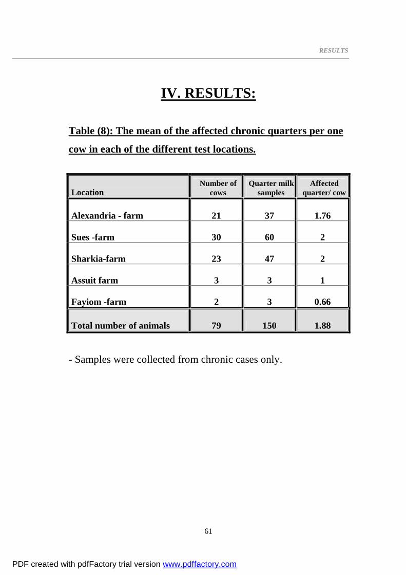
RESULTS
61
IV. RESULTS:
Table (8): The mean of the affected chronic quarters per one
cow in each of the different test locations.
Location Number of
cows Quarter milk
samples Affected
quarter/ cow
Alexandria - farm 21 37 1.76
Sues -farm 30 60 2
Sharkia-farm 23 47 2
Assuit farm 3 3 1
Fayiom -farm 2 3 0.66
Total number of animals 79 150 1.88 - Samples were collected from chronic cases only.
PDF created with pdfFactory trial version www.pdffactory.com

RESULTS
62
Table (10): The prevalence of the isolated organisms in
relation to the total number of examined samples:
Isolated microorganism Number of isolates
% of the isolate to the total number of samples tested
1- Staphylococcus aureus 23 15.3%
2- Staphylococcus epidermidis 25 16.7%
3- Staphylococcus saprophyticus 19 12.7%
4- Streptococcus sp. 3 2 %
5- Bacillus cereus 24 16 %
6- Arcanobacterium pyogenes 1 0.7%
7- Escherichia coli 25 16.7%
8- Klebsiella pneumoniae 2 1.3 %
9- Pseudomonas aeruginosa 1 0.7 %
Number of isolates to total number of samples = 123 82%
10- Samples with no bacterial growth 27 18%
Total number of Quarter milk samples 150 100.10%
%: was calculated according to the total number of the examined
quarter milk samples.
All milk samples stained and examined were T.B. microorganisms
free.
PDF created with pdfFactory trial version www.pdffactory.com

RESULTS
63
Table (10): The prevalence of each isolated organism to the
total number of isolated pathogens.
Isolated microorganism Number of isolates
% of the isolate to the total number of isolated pathogens
1- Staphylococcus aureus 23 18.7%
2- Staphylococcus epidermidis 25 20.3%
3- Staphylococcus saprophyticus 19 15.4%
4- Streptococcus sp. 3 2.4%
5- Bacillus cereus 24 19.5%
6- Arcanobacterium pyogenes 1 0.8%
7- Escherichia coli 25 20.3%
8- Klebsiella pneumoniae 2 1.6%
9- Pseudomonas aeruginosa 1 0.8%
Total number of bacterial isolates 123 100 %
% : was calculated according to the total number of the
recovered bacterial isolates.
PDF created with pdfFactory trial version www.pdffactory.com

RESULTS
64
Graphic(1): The prevalence of each isolated organism to the
total number of isolated pathogens .
Stap
hylo
cocc
us a
ureu
s
Stap
hylo
cocc
us e
pide
rmid
is
Stap
hylo
cocc
us sa
prop
hytic
us
Stre
ptoc
occu
s sp
.
Bac
illu
s ce
reus
Arca
noba
cter
ium
pyo
gene
s
Esc
heri
chia
col
i
Kle
bsie
lla
pne
umon
iae
Pseu
dom
onas
aer
ugin
osa
0.00%
5.00%
10.00%
15.00%
20.00%
25.00%
1 2 3 4 5 6 7 8 9
Series1
PDF created with pdfFactory trial version www.pdffactory.com

RESULTS
65
Table (11): The incidence of each of isolated pathogen in
different experiment locations in relation to the total
bacterial isolates.
Isolated
microorganism
Nobareia- farm (Alexandria) Suis -farm Sharkia-farm Assuit Fayiom -farm
No % No % No % No % No %
Staphylococcus aureus 7 20 7 15.56 7 18.9 1 33.3 1 33.3
Staphylococcus epidermidis 8 22.85 8 17.78 8 21.6 1 33.3 0 0
Staphylococcus saprophyticus 6 17.15 6 13.33 7 18.9 0 0 0 0
Streptococcus sp. 1 2.86 2 4.44 0 0 0 0 0 0
Bacillus cereus 4 11.4 12 26.67 7 18.9 0 0 1 33.3
Arcanobacterium pyogenes 1 2.86 0 0 0 0 0 0 0 0
Escherichia coli 6 17.15 10 22.22 8 21.6 1 3..33 0 0
Pseudomonas aeruginosa 1 2.86 0 0 0 0 0 0 0 0
Klebsiella pneumoniae 1 2.86 0 0 0 0 0 0 1 33.3
Total number of bacterial isolates 35 100 45 100 37 100 3 100 3 100
% was calculated according to the total number of isolated
pathogens to total isolates in each farm.
PDF created with pdfFactory trial version www.pdffactory.com
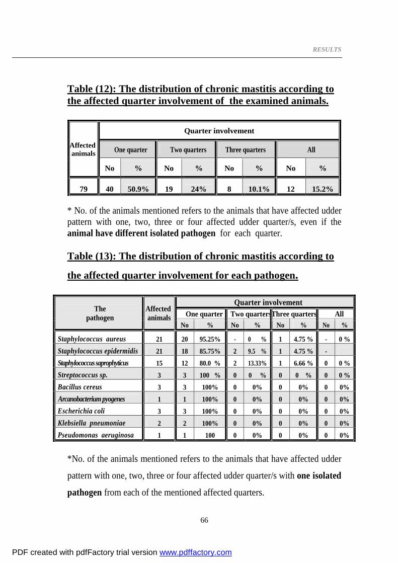
RESULTS
66
Table (12): The distribution of chronic mastitis according to the affected quarter involvement of the examined animals.
Affected animals
Quarter involvement
One quarter Two quarters Three quarters All
No % No % No % No %
79 40 50.9% 19 24% 8 10.1% 12 15.2% * No. of the animals mentioned refers to the animals that have affected udder pattern with one, two, three or four affected udder quarter/s, even if the animal have different isolated pathogen for each quarter. Table (13): The distribution of chronic mastitis according to
the affected quarter involvement for each pathogen.
The pathogen
Affected animals
Quarter involvement One quarter Two quartersThree quarters All
No % No % No % No %
Staphylococcus aureus 21 20 95.25% - 0 % 1 4.75 % - 0 % Staphylococcus epidermidis 21 18 85.75% 2 9.5 % 1 4.75 % - Staphylococcus saprophyticus 15 12 80.0 % 2 13.33% 1 6.66 % 0 0 % Streptococcus sp. 3 3 100 % 0 0 % 0 0 % 0 0 % Bacillus cereus 3 3 100% 0 0% 0 0% 0 0% Arcanobacterium pyogenes 1 1 100% 0 0% 0 0% 0 0% Escherichia coli 3 3 100% 0 0% 0 0% 0 0% Klebsiella pneumoniae 2 2 100% 0 0% 0 0% 0 0% Pseudomonas aeruginosa 1 1 100 0 0% 0 0% 0 0%
*No. of the animals mentioned refers to the animals that have affected udder
pattern with one, two, three or four affected udder quarter/s with one isolated
pathogen from each of the mentioned affected quarters.
PDF created with pdfFactory trial version www.pdffactory.com

RESULTS
67
Table (14): Distribution of chronic mastitis within the four
udder quarters according to quarter involvement of the
examined animals in case of affected animals that had no
bacterial growth .
Affected animals
Quarter involvement
One quarter Two quarters Three quarters All
No % No % No % No %
22 18 81.8% 4 18.2% 0 0% 0 0%
* No. of the animals mentioned refers to the animals that have affected udder
pattern with one, two, three or four affected udder quarter/s without isolation
of bacterial pathogen, even if the animal has additional affected quarter/s
with isolated pathogen.
PDF created with pdfFactory trial version www.pdffactory.com

RESULTS
68
Antibiogram of the recovered pathogens was performed by agar disc diffusion method (shown in photo 6).
Photo (6): Shows the inhibition zones of different antibiotic
discs
- Results with eighteen antibiotics are shown as the following tables (15-21).
PDF created with pdfFactory trial version www.pdffactory.com

RESULTS
69
- Table (15): Antibiogram of Staphylococcus aureus (23 isolates) recovered from cows with chronic bovine mastitis to antimicrobial agents.
No Antibacterial Agent
Conc. µg disc Symbol
I P
S I R
No % No % No %
1 Ampicillin 10 AMP 18 75 1 9 4 16
2 Amoxicillin & Clavulanic acid 30 AMC 16 69 2 8 5 21
3 Amoxicillin 10 AML 15 65 1 4 7 30
4 Penicillin 10 units P 5 22 0 0 18 78
5 Gentamicin 10 CN 21 92 1 4 1 4
6 Kanamycin 30 K 20 87 0 0 3 13
7 Streptomycin 10 S 19 82.7 0 0 4 17.3
8 Neomycin 30 N 18 78 4 17.3 1 4.3
9 Cephalexin 30 C L 15 65 3 13 5 21
10 Cefoxitin 30 FOX 14 60 4 17 5 21
11 Cefotaxime 30 CTX 12 52.2 8 34.7 3 13
12 Ciprofloxacin 5 CIP 23 100 0 0 0 0
13 Norfloxacin 10 NOR 22 96 1 4 0 0
14 Doxycycline 30 DO 17 74 2 9 4 17.3
15 Tetracycline 5 TE 7 30 5 21.7 11 47.8
16 Chloramphenicol 30 C 19 82.6 2 8.6 2 8.6
17 Rifampin 5 RA 18 89 4 17 1 4.3
18 Erythromycin 15 E 13 57 6 26 4 17.3
IP: interpretation S: sensitive R: resistant I: intermediate
PDF created with pdfFactory trial version www.pdffactory.com

Results
70
Graphic (2) : Susceptibility pattern of Staphylococcus aureus recovered from cows with chronic bovine mastitis against different antibacterials.
Am
picillin
Am
oxicillin & C
lavulanic acid
Am
oxicillin
Penicillin
Gentam
icin
Kanam
ycin
Streptomycin
Neom
ycin
Cephalexin
Cefoxitin
Cefotaxim
e
Ciprofloxacin
Norfloxacin
Doxycycline
Tetracycline
Chloram
phenicol
Rifam
pin
Erythromycin
0
20
40
60
80
100
120
1 2 3 4 5 6 7 8 9 10 11 12 13 14 15 16 17 18
Series1
PDF created with pdfFactory trial version www.pdffactory.com

RESULTS
71
Table (16): Antibiogram of Staphylococcus epidermidis (25 isolates) recovered from cows with chronic bovine mastitis to antimicrobial agents.
No Antibacterial Agent
Conc. µg disc Symbol
I P S I R
No % No % No %
1 Amoxicillin & Clavulanic acid 30 AMC 6 24 5 20 14 56
2 Ampicillin 10 AMP 5 20 5 20 15 60
3 Amoxicillin 10 AML 3 12 2 8 20 80
4 Penicillin 10 units P 2 8 0 0 23 92
5 Gentamicin 10 CN 22 88 3 12 0 0
6 Streptomycin 10 S 20 80 3 12 2 8
7 Kanamycin 30 K 18 72 6 24 1 4
8 Neomycin 30 N 17 68 5 20 3 12
9 Cefoxitin 30 FOX 11 44 4 16 10 40
10 Cephalexin 30 C L 9 36 4 16 12 48
11 Cefotaxime 30 CTX 8 32 5 20 12 48
12 Norfloxacin 10 NOR 23 92 2 8 0 0
13 Ciprofloxacin 5 CIP 22 88 2 8 1 4
14 Doxycycline 30 DO 16 64 7 28 2 8
15 Tetracycline 5 TE 14 56 5 20 6 24
16 Chloramphenicol 30 C 16 64 3 12 6 24
17 Rifampin 5 RA 9 36 3 12 13 52
18 Erythromycin 15 E 4 16 11 44 10 40
IP: interpretation S: sensitive R: resistant I: intermediate
PDF created with pdfFactory trial version www.pdffactory.com
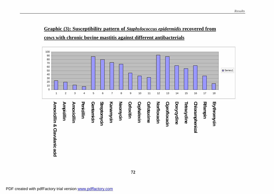
Results
72
Graphic (3): Susceptibility pattern of Staphylococcus epidermidis recovered from
cows with chronic bovine mastitis against different antibacterials
Am
oxicillin & C
lavulanic acid
Am
picillin
Am
oxicillin
Penicillin
Gentam
icin
Streptomycin
Kanam
ycin
Neom
ycin
Cefoxitin
Cephalexin
Cefotaxim
e
Norfloxacin
Ciprofloxacin
Doxycycline
Tetracycline
Chloram
phenicol
Rifam
pin
Erythromycin
0102030405060708090
100
1 2 3 4 5 6 7 8 9 10 11 12 13 14 15 16 17 18
Series1
PDF created with pdfFactory trial version www.pdffactory.com

RESULTS
73
Table (17): Antibiogram of Staphylococcus saprophyticus ( 19 isolates ) recovered from cows with chronic bovine mastitis to antimicrobial agents.
No Antibacterial Agent
Conc. µg disc Symbol
I P S I R
No % No % No %
1 Amoxicillin & Clavulanic acid 30 AMC 13 68 1 4 5 26
2 Ampicillin 10 AMP 10 52 1 5 8 42
3 Amoxicillin 10 AML 8 42.2 1 5.3 10 52.6
4 Penicillin 10 units P 1 5.3 0 0 18 94.7
5 Gentamicin 10 CN 18 95 1 5 0 0
6 Kanamycin 30 K 14 74 4 21 1 5
7 Neomycin 30 N 14 74 4 21 1 5
8 Streptomycin 10 S 13 68 3 16 3 16
9 Cefoxitin 30 FOX 9 47 4 21 6 31
10 Cephalexin 30 C L 9 47 1 5 9 47
11 Cefotaxime 30 CTX 6 31.5 3 15.7 10 52.6
12 Norfloxacin 10 NOR 16 84 1 5 2 10
13 Ciprofloxacin 5 CIP 14 74 4 21 1 5
14 Doxycycline 30 DO 16 84 3 16 0 0
15 Tetracycline 5 TE 8 42 5 26.3 6 31.5
16 Chloramphenicol 30 C 12 63 4 21 3 15.7
17 Erythromycin 15 E 8 42 6 32 5 26
18 Rifampin 5 RA 6 32 5 26.3 8 42
IP: interpretation S: sensitive R: resistant I: intermediate
PDF created with pdfFactory trial version www.pdffactory.com

Results
74
Graphic (4): Susceptibility pattern of Staphylococcus saprophyticus recovered
from cows with chronic bovine mastitis against different antibacterials
Am
oxicillin & C
lavulanic acid
Am
picillin
Am
oxicillin
Penicillin
Gentam
icin
Kanam
ycin
Neom
ycin
Streptomycin
Cefoxitin
Cephalexin
Cefotaxim
e
Norfloxacin
Ciprofloxacin
Doxycycline
Tetracycline
Chloram
phenicol
Erythromycin
Rifam
pin
0102030405060708090
100
1 2 3 4 5 6 7 8 9 10 11 12 13 14 15 16 17 18
Series1
PDF created with pdfFactory trial version www.pdffactory.com

RESULTS
75
Table (18): Antibiogram of 3 isolates Streptococcus sp. recovered from cows with chronic bovine mastitis to antimicrobial agents.
No Antibacterial Agent
Conc. µg disc Symbol
I P S I R
No % No % No %
1 Ampicillin 10 AMP 2 66.6 1 33.3 0 0
2 Amoxicillin & Clavulanic acid 30 AMC 1 33.3 1 33.3 1 33.3
3 Penicillin 10 units P 1 33.3 1 33.3 1 33.3
4 Amoxicillin 10 AML 1 33.3 0 0 2 66.6
5 Gentamicin 10 CN 3 100 0 0 0 0
6 Kanamycin 30 K 3 100 0 0 0 0
7 Streptomycin 10 S 3 100 0 0 0 0
8 Neomycin 30 N 2 66.6 1 33.3 0 0
9 Cefoxitin 30 1 2 66.6 0 0 1 33.3
10 Cephalexin 30 C L 2 66.6 0 0 1 33.3
11 Cefotaxime 30 CTX 2 66.6 0 0 1 33.3
12 Ciprofloxacin 5 CIP 3 100 0 0 0 0
13 Norfloxacin 10 NOR 2 66.6 1 33.3 0 0
14 Doxycycline 30 DO 3 100 0 0 0 0
15 Tetracycline 5 TE 3 100 0 0 0 0
16 Erythromycin 15 E 2 66.6 1 33.3 0 0
17 Chloramphenicol 30 C 2 66.6 1 33.3 0 0
18 Rifampin 5 RA 2 66.6 1 33.3 0 0
IP: interpretation S: sensitive R: resistant I: intermediate
PDF created with pdfFactory trial version www.pdffactory.com

Results
76
Graphic(5): Susceptibility pattern of Streptococcus sp. recovered from cows
with chronic bovine mastitis to different antibacterials.
h85 A
mpicillin
Am
oxicillin & Clavulanic acid
Penicillin
Am
oxicillin
Gentam
icin
Kanam
ycin
Streptomycin
Neom
ycin
Cefoxitin
Cephalexin
Cefotaxim
e
Ciprofloxacin
Norfloxacin
Doxycycline
Tetracycline
Erythromycin
Chloram
phenicol
Rifam
pin
0
20
40
60
80
100
120
1 2 3 4 5 6 7 8 9 10 11 12 13 14 15 16 17 18
Series1
PDF created with pdfFactory trial version www.pdffactory.com

RESULTS
77
Table (19): Antibiogram of Bacillus cereus (24 isolates) recovered from cows with chronic bovine mastitis to antimicrobial agents.
No Antibacterial Agent
Conc. µg disc Symbol
I P S I R
No % No % No %
1 Amoxicillin & Clavulanic acid 30 AMC 13 54 2 8 9 37.5
2 Amoxicillin 10 AML 10 42 1 4 13 54
3 Ampicillin 10 AMP 9 38 4 16 11 45
4 Penicillin 10 units P 2 8.3 0 0 22 91.7
5 Gentamicin 10 CN 22 92 1 4 1 4
6 Streptomycin 10 S 21 88 1 4 2 8
7 Neomycin 30 N 20 83 3 12.6 1 4.3
8 Kanamycin 30 K 14 58 7 29 3 12.5
9 Cefoxitin 30 FOX 16 66 4 16 4 16
10 Cephalexin 30 C L 15 62.5 3 12.5 6 25
11 Cefotaxime 30 CTX 7 29.2 9 37.5 8 33.3
12 Norfloxacin 10 NOR 23 96 9 0 1 4
13 Ciprofloxacin 5 CIP 22 91.7 2 8.3 0 0
14 Doxycycline 30 DO 21 88 1 4 2 8
15 Tetracycline 5 TE 13 54 6 25 5 20.8
16 Chloramphenicol 30 C 15 62.5 5 20.8 4 16.6
17 Rifampin 5 RA 13 54 3 12.5 8 33
18 Erythromycin 15 E 12 48 6 24 7 28
IP: interpretation S: sensitive R: resistant I: intermediate
PDF created with pdfFactory trial version www.pdffactory.com

Results
78
Graphic(6): Susceptibility pattern of Bacillus cereus recovered from cows
with chronic bovine mastitis todifferent antibacterials.
Am
oxicillin & Clavulanic acid
Am
oxicillin
Am
picillin
Penicillin
Gentam
icin
Streptomycin
Neom
ycin
Kanam
ycin
Cefoxitin
Cephalexin
Cefotaxim
e
Norfloxacin
Ciprofloxacin
Doxycycline
Tetracycline
Chloram
phenicol
Rifam
pin
Erythromycin
5442 38
8.3
92 88 83
5866 62.5
29.2
96 91.7 88
5462.5
5448
0
20
40
60
80
100
120
1 2 3 4 5 6 7 8 9 10 11 12 13 14 15 16 17 18
Series1
PDF created with pdfFactory trial version www.pdffactory.com

RESULTS
79
Table (20) Antibiogram of Escherichia coli (25 isolates)
recovered from cows with chronic bovine mastitis to
antimicrobial agents.
No Antibacterial Agent
Conc. µg disc Symbol
I P S I R
No % No % No %
1 Amoxicillin & Clavulanic acid 30 AMC 7 28 3 12 15 60
2 Amoxicillin 10 AML 3 12 0 0 22 88
3 Ampicillin 10 AMP 0 0 4 16 21 84
4 Penicillin 10 units P 0 0 0 0 25 100
5 Gentamicin 10 CN 21 84 4 16 0 0
6 Kanamycin 30 K 16 64 5 20 4 16
7 Neomycin 30 N 14 56 5 20 6 24
8 Streptomycin 10 S 6 24 5 20 14 56
9 Cefoxitin 30 FOX 7 28 8 32 10 40
10 Cephalexin 30 C L 7 28 5 20 13 52
11 Cefotaxime 30 CTX 6 24 8 32 11 44
12 Doxycycline 30 DO 15 60 3 12 7 28
13 Tetracycline 5 TE 11 48 0 0 13 52
14 Norfloxacin 10 NOR 21 84 1 4 3 12
15 Ciprofloxacin 5 CIP 20 20 3 12 2 8
16 Chloramphenicol 30 C 13 52 1 4 11 44
17 Rifampin 5 RA 5 20 5 20 15 60
18 Erythromycin 15 E 0 0 8 32 17 68
IP: interpretation S: sensitive R: resistant I: intermediate
PDF created with pdfFactory trial version www.pdffactory.com

Results
80
Graphic (7): Susceptibility pattern of Escherichia coli recovered from cows with
chronic bovine mastitis to different antibacterials.
Am
oxicillin & Clavulanic acid
Am
oxicillin
Am
picillin
Penicillin
Gentam
icin
Kanam
ycin
Neom
ycin
Streptomycin
Cefoxitin
Cephalexin
Cefotaxim
e
Doxycycline
Tetracycline
Norfloxacin
Ciprofloxacin
Chloram
phenicol
Rifam
pin
Erythromycin
0102030405060708090
1 2 3 4 5 6 7 8 9 10 11 12 13 14 15 16 17 18
PDF created with pdfFactory trial version www.pdffactory.com

RESULTS
81
Table (21): Antibiogram of two isolates Klebsiella
pneumoniae recovered from cows with chronic bovine
mastitis to antimicrobial agents
NO Antibacterial Agent
Conc. µg disc Symbol
I P
S I R
No % No % No %
1 Amoxicillin 10 AML 0 0 0 0 2 100
2 Amoxicillin & Clavulanic acid 30 AMC 1 50 0 0 1 50
3 Ampicillin 10 AMP 0 0 1 50 1 50
4 Penicillin 10 units P 0 0 0 0 2 100
5 Gentamicin 10 CN 2 100 0 0 0 0
6 Kanamycin 30 K 0 0 1 50 1 50
7 Neomycin 30 N 0 0 1 50 1 50
8 Streptomycin 10 S 0 0 1 50 1 50
9 Cephalexin 30 C L 0 0 1 50 1 50
10 Cefotaxime 30 CTX 0 0 1 50 1 50
11 Cefoxitin 30 FOX 0 0 1 50 1 50
12 Ciprofloxacin 5 CIP 2 100 0 0 0 0
13 Norfloxacin 10 NOR 1 50 1 50 0 0
14 Doxycycline 30 DO 0 0 1 50 1 50
15 Tetracycline 5 TE 0 0 0 0 2 100
16 Chloramphenicol 30 C 1 50 1 50 0 0
17 Erythromycin 15 E 0 0 0 0 2 100
18 Rifampin 5 RA 0 0 0 0 2 100
IP: interpretation S: sensitive R: resistant I: intermediate
PDF created with pdfFactory trial version www.pdffactory.com

Results
82
Graphic (8): Susceptibility pattern of Klebsiella pneumoniae recovered from cows with
chronic bovine mastitis to different antibacterials.
Am
oxicillin
Am
oxicillin & Clavulanic acid
Am
picillin
Penicillin
Gentam
icin
Kanam
ycin
Neom
ycin
Streptomycin
Cephalexin
Cefotaxim
e
Cefoxitin
Ciprofloxacin
Norfloxacin
Doxycycline
Tetracycline
Chloram
phenicol
Erythromycin
Rifam
pin
0
20
40
60
80
100
120
1 2 3 4 5 6 7 8 9 10 11 12 13 14 15 16 17 18
PDF created with pdfFactory trial version www.pdffactory.com

RESULTS
83
Antibiogram of Arcanobacterium pyogenes recovered from
cows with chronic bovine mastitis to antimicrobial agents:
The recovered isolate was sensitive to gentamicin,
kanamycin, neomycin, cefotaxime, ciprofloxacin and rifampin.
Resistance to amoxicillin, amoxicillin & clavulanic acid, ampicillin,
cephalexin, cefoxitin, doxycycline, chloramphenicol and
erythromycin was recorded.
Antibiogram of Pseudomonas aeruginosa recovered from
cows with chronic bovine mastitis to antimicrobial agents:
The recovered isolate was Resistant to all used eighteen
antibiotics.
PDF created with pdfFactory trial version www.pdffactory.com

DISCUSSION
84
V. DISCUSSION
Mastitis is a worldwide disease causing high economic
losses in dairy industry. Losses in milk yield, increased
replacement heifer costs, loss of antibiotic contaminated milk,
drug and veterinary costs, extra labour and loss of cows with
high genetic value from the herd are among the aforementioned
losses (Gulliemette et al., 1996).
Three degrees of mastitis are commonly recognized; acute,
chronic (the clinical mastitis forms) and subclinical mastitis.
Chronic mastitis is less severe than acute mastitis and is seen in
most dairy herds (Labib, 1994)
Bovine mastitis often becomes chronic, and it is important
to identify quickly the new clinical cases in order to control
infection in the herd (Yousef, 2005).
The current study was planned to investigate the bacterial
etiology of chronic mastitis in bovine. One hundred fifty samples
were obtained from clinical cases of seventy-nine chronic
mastitic cows. From the data of Table (8), incidence of the
affected chronic quarters per each cow was 1.88 quarter/cow.
The higher incidence was recorded in Sues–farm and Sharkia-
PDF created with pdfFactory trial version www.pdffactory.com

DISCUSSION
85
farm (2 quarter/cow), while the lower incidence was recorded in
Fayiom- farm (0.66 quarter/cow).
From the data given in table (9), it is evident that out of
150 samples collected from chronic mastitic cows, 123 (82%)
bacterial isolates were recovered. The ratio of the total bacterial
isolates was nearly similar to that reported for all forms of
clinical mastitis by Odongo and Ambanim (1989) and Sargeant et
al. (1998) who recorded rates of 83.9% and 82.3%. On the other
hand, Wilson et al.(1991) and Benites et al. (2003) recorded
lower incidences (79.8% and 67% respectively).
The possible reasons for the failure to isolate pathogens
from mastitic quarters include spontaneous bacteriological
recovery with a persistent state of chronically changed mammary
tissues, too few pathogens to be detected, pathogens inhibited by
antibiotic residues, pathogens killed after the samples was taken
and before culturing (Miltenburg et al., 1996)
Out of these 123 isolates, 95 (77.2%) were Gram positive
bacteria and 28 (22.8%) isolates were Gram negative organisms.
Several factors have been reported to influence the
prevalences of pathogens. A correlation of Gram-positive bacteria
PDF created with pdfFactory trial version www.pdffactory.com

DISCUSSION
86
with the chronic udder inflammation was suggested, while Gram-
negative bacteria were associated with acute catarrhal
inflammation of alveoli (Inui et al., 1979). Other factors
including the definition of the culture-positive result and the
procedure used for freezing of the samples. Freezing has a
significant effect on the survival of some pathogenic organisms
causing mastitis. With the longer period of freezing there was a
significant decrease in of Escherichia coli and Arcanobacterium
pyogenes positive samples, while an increase in number of
sample positive for coagulase-negative staphylococci. On the
other hand, freezing had no effect on the Staphylococcus aureus
and Streptococci (Schukken et al., 1989). During the present
investigation all of the examined samples were kept frozen as
recommended by Murdough et al. (1996) because immediate
culturing of samples was not possible.
The predominant Gram positive bacteria were Staphylococcus
sp. [67 isolates (54.5%)]. The study result was higher than that
reported by Hanselmann (1978) and Wilson et al. (1991) (45%
and 7.3% respectively).
The coagulase negative staphylococci were predominating
[44 isolates (35.8%)]. The recorded ratio is nearly similar to that
reported by Benites et al. (2003) who reported 35.7%. This ratio
PDF created with pdfFactory trial version www.pdffactory.com

DISCUSSION
87
was higher than that recorded by Lafi et al. (1994), Miltenburg et
al. (1996), Kudinha and Simango (2002), Malinowski et al.(2003)
and who recorded 16%, 4.2%, 22.9% and 15% respectively.
Results in table (10) showed that out of these isolates,
coagulase negative Staphylococcus epidermidis constituted
20.3% of the total isolates. These result was nearly similar with
that reported for all forms of clinical mastitis by Mahbub-E-
Elahi et al.(1996) who identified 18.0% of the isolates as
Staphylococcus epidermidis. On the other hand Twardona et
al.(1999) recorded higher incidence (34.9%) while lower incidences
were recorded by Inui et al. (1979) and Beytut et al. (2002) who
identified 11.8%, 8.33% of the isolates, respectively as
Staphylococcus epidermidis.
The coagulase positive Staphylococcus aureus represented
18.7% of the total isolates. The recorded ratio was nearly similar
to that recorded by Wilson et al. (1991) and Kudinha and Simango
(2002) who reported 18.4% and 17.1%, respectively. On the other
hand, higher incidences were recorded by Erer et al. (1996),
Twardona et al. (1999) and Beytut et al. (2002) who reported 47.3%,
29.1% and 43.75% respectively. Lower incidences were recorded
by Benites et al.(2003) and Lafi et al. (1994) who identified
PDF created with pdfFactory trial version www.pdffactory.com

DISCUSSION
88
12.2% and 14% of the isolated pathogens as Staphylococcus
aureus.
Β-haemolytic streptococci and B. cereus represented 2%
and 16% respectively. On the other hand, Inui et al. (1979)
identified higher ratio of isolates (5.9%) as Β-haemolytic
streptococci, while Erer et al. (1996), Mahbub-E-Elahi et
al.(1996) and Beytut et al. (2002) identified lower incidences of
the pathogens as Gram positive Bacillus cereus (1.1%, 2.08%
and 4.6% of the total isolates respectively).
One isolate of Arcanobacterium pyogenes was recovered
with a prevalence of 0.8% that was exceeded by the higher
incidences which were recorded by Inui et al.(1979), Erer et al.
(1996) and Beytut et al.(2002), who identified 16.3%, 16.3% and
19.79% of mastitic isolates, respectively as Arcanobacterium
pyogenes
Gram negative bacteria were also recovered. The
predominant Gram negative Escherichia coli were 25isolates
(16.7%). The recorded prevalence was higher than that reported
by Jha et al.(1994) and Miltenburg et al. (1996) who identified
16.8% and 16.7% of the isolates recovered from cases of clinical
mastitis (two forms) respectively as Escherichia coli . Too lower
PDF created with pdfFactory trial version www.pdffactory.com

DISCUSSION
89
incidences were recorded Samborski et al. (1992) and by Beytut et
al. (2002) who identified 0.9% and 7.29% of the isolated
pathogens, respectively as Escherichia coli.
Two isolates of Klebsiella pneumoniae (1.3%) were
recovered. Higher incidence was recorded by Inui et al. (1979) and
Erer et al. (1996) who identified 5.9% and 3.8% of the isolated
mastitic pathogens, respectively as Klebsiella pneumoniae.
Prevalence of Escherichia coli and Klebsiella sp. on
whole-herd surveys may be evaluated in lower ratios. These may
be due to the relatively short duration of intramammary
infections caused by Gram-negative bacteria (Sears et al., 1993)
compared with agents that usually persist longer in the mammary
gland. Wilson et al.(1997) suggested that Cram-negative bacteria
remain a less common cause of bovine mastitis regarding to the
Gram-positive pathogens.
One isolate of Pseudomonas aeruginosa (0.7%) was
recovered. On the other hand Wilson et al. (1997) recorded
lower incidence (0.1%) while the result recorded by Inui et al,
(1979) was higher (5.9%).
PDF created with pdfFactory trial version www.pdffactory.com

DISCUSSION
90
Results in Table (11) showed that in case of Alexandria-
farm, Staphylococcus epidermidis was the higher prevalent
organism (22.85%) followed by Staphylococcus aureus (20%)
while the Streptococcus sp., Arcanobacterium pyogenes,
Pseudomonas aeruginosa and Klebsiella pneumoniae were the
least (2.86% for each).
Bacillus cereus was isolated in the higher ratio in Sues-
farm (26.67%) followed by Escherichia coli (22.22%) while the
lower ratio was reported for Streptococcus sp. (4.44%).
In case of the Sharkia-farm Staphylococcus epidermidis
together with Escherichia coli constituted the higher ratios of the
isolated organisms (21.6% for each).
In Assuit, the isolated pathogens were Staphylococcus
aureus, Staphylococcus epidermidis and Escherichia coli. They
were equally isolated (33.3% for each).
Staphylococcus aureus, Klebsiella pneumoniae and
Bacillus cereus were equally isolated (33.3%) in Fayiom –farm.
Distribution of chronic mastitis according to the affected
quarter involvement was shown in Table (12). Animals were
categorized according to number of affected quarters (whether
PDF created with pdfFactory trial version www.pdffactory.com

DISCUSSION
91
the animal was infected with one pathogen or more). Results
revealed that animals with one mastitic quarter were the greatest
group (50.6%). The recorded ratio was much higher than that
reported by Mahbub-E-Elahi et al. (1996) which was 34% and
based on data of the clinical mastitis (two forms of the clinical
mastitis). The second group was those which had two affected
quarters (22.8%). That ratio was nearly similar to that obtained
by Mahbub-E-Elahi et al. (1996) who recorded 24.67%. The
third group was the affected whole udder (15.2%) followed by
those of three mastitic quarters (10.2%).
Data in Table (12) represent the distribution of chronic
mastitis in the affected quarters for each pathogen. 95.25% of the
Staphylococcus aureus infections were found in only one
affected quarter per cow. On the same time, neither two quarter
infections nor, four-udder infections with Staphylococcus
aureus were reported at the same cow.
Each of 85.75% Staphylococcus epidermidis isolates and
80% Staphylococcus saprophyticus isolates were isolated in one
quarter infections and they were not isolated from the all udder
four quarters of any cow.
PDF created with pdfFactory trial version www.pdffactory.com

DISCUSSION
92
Although twelve cases of four-quarter infections seen in
table (12) were found with multiple pathogens at the same cow,
none of them were infected with one pathogen in its four quarters
as seen in Table (13).
The data in Table (15) represent the applied antibiogram
study on 23 isolates of Staphylococcus aureus The results of the
sensitivity tests with the tested members of β-lactam group of
antibiotics revealed that the higher incidence of sensitivity was
with ampicillin (78.2% of the isolates were sensitive) followed
by the combination of amoxicillin and clavulanic acid (69%)
while the lower incidence was recorded in case of penicillin
(22%). The result was nearly agreed with that recorded by Binde
and Gjul (1988) while Winter and Duetz (1998) recorded higher
incidences.
In case of tested elements of the aminoglycosides group, the
higher incidence was with gentamicin (92%) followed by
kanamycin (87%) while the lower incidence was with neomycin
(78%).
In case of the tested members of the cephalosporins group,
Staphylococcus aureus represented the higher incidence of
PDF created with pdfFactory trial version www.pdffactory.com

DISCUSSION
93
sensitivity with cephalexin (65%). followed by cefoxitin (60%)
The lower incidence was (52.2%) in case of cefotaxime.
On the other hand, all the isolates of Staphylococcus aureus
were sensitive for ciprofloxacin and it was 96% in case of
norfloxacin while the incidence of the sensitive isolates was 74%
for doxycycline, it was 30% for tetracycline. Higher incidences
were reported for rifampin (89%) and chloramphenicol (82.6%)
while it was 57% for Erythromycin.
The data in Table (16) represent the applied antibiogram
study on 25 isolates of Staphylococcus epidermidis. The results
of the sensitivity tests with the tested members of β-lactam group
of antibiotics revealed that the higher incidence of sensitivity was
with combination of amoxicillin and clavulanic acid (24% of the
isolates were sensitive) followed by ampicillin (20%) while the
lower incidence was recorded with penicillin (8 %).
In case of tested elements of the aminoglycosides group, the
higher incidence was with gentamicin (88%) followed with
streptomycin (80%) while the lower incidence was with
neomycin (68%).
PDF created with pdfFactory trial version www.pdffactory.com

DISCUSSION
94
In case of the tested members of the cephalosporins group,
Staphylococcus epidermidis represented the higher incidence of
sensitivity with cefoxitin (44%), followed by cephalexin (36%).
The lower incidence was (32%) in case of cefotaxime.
On the other hand, 92% the isolates of Staphylococcus
epidermidis were sensitive for norfloxacin and it was 88% in
case of ciprofloxacin while the incidence of the sensitive isolates
was 64% for doxycycline, it was 56% for tetracycline.
The incidence with chloramphenicol, rifampin and
erythromycin were 64%, 36% and 16% respectively.
The data in Table (17) represent the applied antibiogram
study on 19 isolates of Staphylococcus saprophyticus. The
results of the sensitivity tests with the tested members of β-
lactam group of antibiotics revealed that the higher incidence of
sensitivity was with combination of amoxicillin and clavulanic
acid (68% of the isolates were sensitive) followed by ampicillin
(52%) while the lower incidence was recorded with penicillin
(5.3 %).
PDF created with pdfFactory trial version www.pdffactory.com

DISCUSSION
95
For aminoglycosides group, the higher incidence was with
gentamicin (95%) followed with kanamycin (74%) while the
lower incidence was with streptomycin (68%).
With the tested members of cephalosporins group, 47% of
the isolates of Staphylococcus saprophyticus were sensitive in
case of cefoxitin and cephalexin. Lower incidence (31.5%) was
recorded in case of cefotaxime.
On the other hand, 84% the isolates of Staphylococcus
saprophyticus were sensitive for norfloxacin and it was 74% in
case of ciprofloxacin while the incidence of the sensitive isolates
was 84% for doxycycline, it was 42% for tetracycline.
The incidence with chloramphenicol, erythromycin and
rifampin were 63%, 42% and 32% respectively.
The data in Table (18) represent the applied antibiogram
study on 3 isolates of Streptococcus sp. The results of the
sensitivity tests with the tested members of β-lactam group of
antibiotics revealed that the higher incidence of sensitivity was
with ampicillin (66.6% of the isolates were sensitive) followed
by the other three tested members (33.3% for each).
PDF created with pdfFactory trial version www.pdffactory.com

DISCUSSION
96
For aminoglycosides group, all isolates of Streptococcus
sp. were sensitive (100%) for kanamycin, streptomycin and
gentamicin.
With the tested members of cephalosporins group, the
incidence the sensitive isolates of Streptococcus sp was 66.6%
for each of cefoxitin, cephalexin and cefotaxime.
On the other hand, all of the isolates of Streptococcus sp
were sensitive for ciprofloxacin and it was 66.6% in case of
norfloxacin. All of the isolates were sensitive to doxycycline and
tetracycline.
The incidence with chloramphenicol, erythromycin and
rifampin were 66.6% for each.
The data in Table (19) represent the applied antibiogram
study on 24 isolates of Bacillus cereus The results of the
sensitivity tests with the tested members of β-lactam group of
antibiotics revealed that the higher incidence of sensitivity was
with the combination of amoxicillin and clavulanic acid (54% of
the isolates were sensitive) followed by amoxicillin (42%) while
the lower incidence was recorded with penicillin (8.3 %).
PDF created with pdfFactory trial version www.pdffactory.com

DISCUSSION
97
For aminoglycosides group, the higher incidence was with
gentamicin (92%) followed with streptomycin (88%) while the
lower incidence was with kanamycin (58%).
With the tested members of cephalosporins group, Bacillus
cereus represented the higher incidence of sensitivity with
cefoxitin (66%) followed by cephalexin (62.5%). The lower
incidence was (29.2.5%) in case of cefotaxime.
On the other hand, 96% the isolates of Bacillus cereus
were sensitive for norfloxacin and it was 91.7% in case of
ciprofloxacin.
The incidence of the sensitive isolates was 88% for
doxycycline and 54% for tetracycline.
The incidence with chloramphenicol, rifampin and
erythromycin were 62.5%, 54% and 48% respectively.
Only one isolate of Arcanobacterium pyogenes was identified
and was resistant for the tested members of β-lactam group,
streptomycin, cephalexin, norfloxacin, tetracycline, doxycycline,
chloramphenicol and erythromycin.
PDF created with pdfFactory trial version www.pdffactory.com
DISCUSSION
98
Contrarily, it was sensitive for gentamicin, kanamycin,
neomycin, cefotaxime, ciprofloxacin and rifampin.
The data in Table (20) represent the applied antibiogram
study on 25 isolates of Escherichia coli. In case of β-lactam
group of antibiotics, the higher incidence of sensitive isolates
was with combination of amoxicillin and clavulanic acid (28 %
of the isolates were sensitive) followed by amoxicillin (12%)
while no isolates were sensitive for ampicillin or penicillin. The
present result nearly agreed with Sogaard (1982) and Sikorski
(1986) who reported that all isolates were sensitive.
For aminoglycosides group, the higher incidence was with
gentamicin (84%) followed with kanamycin (64%) while the
lower incidence was with streptomycin (24%).
With the tested members of cephalosporins group, 28% of
the isolates of Escherichia coli were sensitive in case of
cefoxitin and cephalexin while the incidence was (24.5%) in case
of cefotaxime.
On the other hand, 84% the isolates of Escherichia coli
were sensitive for norfloxacin and it was 20% in case of
ciprofloxacin while the incidence of the sensitive isolates was
PDF created with pdfFactory trial version www.pdffactory.com
DISCUSSION
99
60% for doxycycline. The recorded percentage in case
tetracycline was 48%.
The incidence with chloramphenicol and rifampin were
52%, 20% respectively. No isolates were sensitive to
erythromycin.
From the data given in Table (21), only two isolates of
Klebsiella pneumoniae were picked and were sensitive (100%)
for gentamicin and ciprofloxacin. One isolate (50%) was
sensitive for the combination of amoxicillin and clavulanic acid,
norfloxacin and chloramphenicol. For other tested antibiotics, all
isolates were resistant.
Only one isolate of Pseudomonas aeruginosa was picked
and showed resistance for all used antibiotics.
PDF created with pdfFactory trial version www.pdffactory.com

SUMMARY
100
VI. SUMMERY
Mastitis is still implicated as one of the major disease
problems in dairy animals. From the economical point of view,
chronic mastitis results in eradication of the cow that became
unfit to be kept as milk producing cow cconstituting great losses
to dairy producers. Due to the little number researches about
the causes of the chronic mastitis, the present study was
performed.
The present study was planned to discuss the following:
1- To detect the prevalence of the most important bacterial
pathogens isolated from the clinically chronic mastitic
cows.
2- Purification as well as morphological, cultural and
biochemical identification of the recovered isolates.
3- An antibiogram study on the recovered isolates was
undertaken to explore the sensitivity patterns of the
recovered isolates to eighteen antibiotics.
The study revealed that the bacterial pathogens were
isolated from (82%) of chronically mastitic quarters. The Gram
positive bacteria constituted 77.2% of the isolates while the
Gram negative organisms were 22.8%. On one hand
Staphylococcus sp. were the predominant of the Gram positive
PDF created with pdfFactory trial version www.pdffactory.com

SUMMARY
101
bacteria (54.5%). On the other hand Escherichia coli was the
predominant of the Gram negative isolates (20.3%).
Staphylococcus epidermidis and Escherichia coli were the
predominant organism (20.3% each) followed by Bacillus
cereus (19.5%), Staphylococcus aureus (18.7%) and
Staphylococcus saprophyticus (15.4%). Both of Streptococcus
sp., Arcanobacterium pyogenes, Klebsiella pneumoniae and
Pseudomonas aeruginosa were recovered in low prevalences
(2.4%, 0.8%, 1.6% and 0.8% respectively).
The coagulase negative Staphylococcus sp. represented the
widely spreading organism in-between quarters in a same one
cow (infected two quarters in the same one cow represented
22.9% of the isolates while in case of infected three quarter was
11.4 %. Staphylococcus aureus was the next spreading
organism (infection in all quarters by 4.75 % of the isolates).
Sensitivity test of eighteen antibiotics showed that the
norfloxacin and gentamicin were the most effective antibiotic in
case of Staphylococcus aureus, Staphylococcus epidermidis,
Staphylococcus saprophyticus, Bacillus cereus and
Escherichia coli.
PDF created with pdfFactory trial version www.pdffactory.com

SUMMARY
102
In addition, ciprofloxacin was effective in case of
Staphylococcus aureusو Staphylococcus epidermidis and
Bacillus cereus, doxycycline for Staphylococcus saprophyticus
and kanamycin for Escherichia coli.
Ciprofloxacin and gentamicin were effective in case of
Klebsiella pneumoniae.
The Pseudomonas aeruginosa was resistant to all of the
tested antibiotics.
PDF created with pdfFactory trial version www.pdffactory.com

Recommendations
PDF created with pdfFactory trial version www.pdffactory.com

RECOMMENDATIONS
103
VII. RECOMMENDATIONS
- The wide spreading contagious pathogens (Staphylococcus
aureus and Coagulase negative staphylococcus) comparing
to the environmental pathogens (Escherichia coli,
Klebsiella pneumoniae and Pseudomonas aeruginosa)
indicate the need to follow rigorous program of teat dipping
and dry cow antibiotic therapy (intramammary antibiotic
treatment at drying off).
- Improvement of the used dipping programs during the
milking process to combat the wide spreading of the
contagious pathogens isolated in case of chronic mastitis by
periodical estimation of the validity of used agents and the
continuous change of used materials
- Heifers can be given intramammary antibiotic treatment
during gestation.
- Giving attention to the clinical use of some antibiotics to
which most of the chronic mastitis-causing pathogens
showed sensitivity (norfloxacin, gentamicin, ciprofloxacin
and doxycycline).
PDF created with pdfFactory trial version www.pdffactory.com
ABSTRACT
104
VIII. ABSTRACT
Examination of 150 chronically mastitic quarter milk samples collected
from 79 machine milked Friesian cows from five dairy herds. Animals had
chronic mastitis and showed developing fibrosis and were assigned as chronic
mastitic cases. The bacterial pathogens were isolated from 82% of the samples.
The Gram positive bacteria constituted 77.2% of the isolates while the Gram
negative organisms were 22.8%. Staphylococcus sp. represented 54.5% of the
total isolates. Staphylococcus epidermidis (20.3%), Staphylococcus aureus
(18.7%), Staphylococcus saprophyticus (15.4%), Escherichia coli (20.3%),
Streptococcus sp. (2.4%), Arcanobacterium pyogenes (0.8%), Klebsiella
pneumoniae (1.6%) and Pseudomonas aeruginosa (0.8%) were isolated..
coagulase negative Staphylococcus sp represented the most spreading organisms
in-between quarters in a same one cow followed by Staphylococcus aureus. An
antibiogram of the isolates revealed that norfloxacin, gentamicin, ciprofloxacin,
and doxycycline were the most effective antibiotics in case of Staphylococcus
aureus, Staphylococcus epidermidis, Bacillus cereus, Staphylococcus
saprophyticus and Escherichia coli. The Pseudomonas aeruginosa was resistant
to all of the tested antibiotics.
PDF created with pdfFactory trial version www.pdffactory.com

REFERANCES
105
IX. REFERENCES
Armenteros, M.; Peña, J.; Pulido, J. L. and Linares, E. (2002):
Characterization of the situation in dairy herds affected by
bovine mastitis.
Revista de Salud Animal 24 (2): 99-105. Cited from http://
www.ncbi. nlm. nih.gov/pubmed
Barto, J.; Czokoly, P. and Hlinka, D. (1984): Classification of
bovine mastitis caused by group L streptococci in an
infected herd.
Veterinárhacekstvi, 34 (9): 395-396.
Bauer, A. W.; Kirby W. M. M.; Sherris, J. C. and Turk, M.
(1966): Antibiotic susceptibility testing by standardized
single disc method.
American Journal of Clinical Pathology. 45 (493-496).
Benites, N. R.; Melville, P. A. and Costa E. O. (2003): Evaluation
of the microbiological status of milk and various structures
in mammary glands from naturally infected dairy cows.
Tropical Animal Health Production 35 (4): 301-307.
PDF created with pdfFactory trial version www.pdffactory.com

REFERANCES
106
Bertoldini, G.; Burnner, F. and Redaellin, G. (1985): Further
studies on the antibiotic sensitivity of streptococci and
staphylococci associated with bovine mastitis.
Archivio Veterinario Italiano 36 (4): 49-101. Cited from
http://ezproxy.htu.se/menu
Beytut, E.; Aydn, F.; Özcan, K. and Genc, O. (2002):
Pathological and bacteriological investigations on bovine
mastitis in Kars Region and its surrounds.
Kafkas Üniversitesi Veteriner Fakültesi Dergisi 8 (2):111-
122.
Binde, M. and Gjul, G. G. (1988): Penicillin resistance of S.
aureus strains isolated from bovine mammary glands with
acute and chronic mastitis.
Norsk Veterinaertidsskrift 100 (5): 345-348.
Blood, D. C. and Handerson, J. A. (1986): veterinary medicine.
3rd Edition. Baillierc, Tindall and Gassel, London
Böhmer, N. and Schneider, P. (1999): The isopathic-homeopathic
mastitis therapy: practical example.
Ganzheitliche Tiermedizin 13 (2): 80-82.
PDF created with pdfFactory trial version www.pdffactory.com

REFERANCES
107
Brouillette, E.; Grondin, G.; Shkreta, L.; Lacasse, P. and
Talbot, B. G. (2003): In vivo and in vitro demonstration
that Staphylococcus aureus is an intracellular pathogen in
the presence or absence of fibronectin-binding proteins.
Microbial Pathogenesis 35 (4):159-168.
Brouillette, E.; Martinez, A.; Boyll, B. J.; Alien, N. E. and
Malouin, F. (2004): Persistence of a Staphylococcus
aureus small-colony variant under antibiotic pressure in
vivo.
FEMS Immunology and Medical Microbiology 41 (1): 35-
41.
Buddle, B. M.; Herceg, M.; Ralston, M. J. and Pulford, H. D.
(1987): Reinfection of bovine mammary glands following
dry-cow antibiotic therapy.
Veterinary Microbiology 15 (3): 191-199.
Cheesbrough, Monica (1984): Medical laboratory manual for
tropical countries.
2 nd Edition. Tropical health technology and Butterworth.
CLSI (2005): Clinical and Laboratory Standards Institute
Performance Standards Antimicrobial susceptibility Testing.
PDF created with pdfFactory trial version www.pdffactory.com

REFERANCES
108
Collee, J. G.; Fraser, A. G.; Marmion, B. P. and Simmons, A.
(1996): Practical Medical Microbiology.
14th Edition. Churchill, LivingStone, New York.
Costa, E. O.; Benites, N. R.; Guerra, J. L. and Melville, P. A.
(2000): Antimicrobial susceptibility of Staphylococcus spp.
isolated from mammary parenchymas of slaughtered dairy
cows.
J. Vet. Med, Infectious Disease Vet Public Health 47(2): 99-
103.
Costa, E. O.; Ribeiro, A. R.; Watanabe, E. T. and Melville, P.
A. (1998): Infectious bovine mastitis caused by
environmental organisms.
Zentralbl Veterinarmed B., 45(2): 65-71. Cited from
http://ezproxy.htu.se/menu.
Cruickshank, R.; Dugid, J. p.; Mormion, B. P. and Swain, R.
H. A. (1975): The practice of medical microbiology.
12th Edition. Churchill Livingstone, London, U.K...
DeOliveira A. P.; Watts, J. L.; Salmon, S. A. and Aarestrup
FM.(2000):Antimicrobial susceptibility of Staphylococcus
aureus isolated from bovine mastitis in Europe and the
United States.
Journal of Dairy Science 83 (4): 855- 862.
PDF created with pdfFactory trial version www.pdffactory.com

REFERANCES
109
Diarra, M. S.; Petitclerc, D.; Deschênes, É.; Lessard, N.
Grondin, G.; Talbot, B. G. and Lacasse, P. (2003):
Lactoferrin against Staphylococcus aureus Mastitis:
Lactoferrin alone or in combination with penicillin G on
bovine polymorphonuclear function and mammary
epithelial cells colonization by Staphylococcus aureus
Veterinary Immunology and Immunopathology 95 (1-
2):33-42.
Dogan, B.; Klaessig, S.; Rishniw, M.; Almeida, R. A.; Oliver, S.
P.; Simpson, K. and Y.H. Schukken (2006): Adherent
and invasive Escherichia coli are associated with persistent
bovine mastitis.
Journal of Veterinary Microbiology 116 (4): 270-282.
Dopfer, D.; Almeida, R. A.; Lam, T. J.; Nederbragt, H. ;
Oliver, S. P. and Gaastra, W. (200٠): Adhesion and
invasion of Escherichia coli from single and recurrent
clinical cases of bovine mastitis in vitro.
Veterinary Microbiology 74 (4): 331-343.
Döpfer, D.; Nederbragt, H.; Almeida, R. A. and Gaastra, W. (2001):
Studies about the mechanism of internalization by mammary epithelial
cells of Escherichia coli isolated from persistent bovine mastitis.
Veterinary Microbiology 80 (3): 285-296.
PDF created with pdfFactory trial version www.pdffactory.com

REFERANCES
110
Doymaz, M. Z.; Sordillo, L. M.; Oliver, S. P. and Guidry, A. J.
(1988): Effects of staphylococcus aureus mastitis on
bovine mammary gland plasma cell populations and
immunoglobulin concentrations in milk.
Veterinary Immunology and Immunopathology 20 (1): 87-
93.
El Kholy, A. M.; Hosein, H. I. and Thabet, A. El-R(1994):
Chemical and cytobacteriologicals studies for the detection
of clinical mastitis.
Assuit Vet. Med. J., 30 (60): 154-164
Erer, H.; Ates, M.; Kran, M. M.; Ciftci, M. K. and Kaya, O.
(1996): Pathological and bacteriological studies on bovine
mastitis.
Veteriner Bilimleri Dergisi, 12 (123-133).
Fang, W. (1996): A novel fluorometric method for evaluation of
the post antibiotic effect of antibacterial drugs on mastitis-
causing Staphylococcus aureus and Escherichia coli •
Journal of Microbiological Methods 26 (1-2): 151-159
Finegold, S. D.; and Martin W. J. (1982): Diagnostic
Microbiology.
6th Edition. the Mosby company, USA
PDF created with pdfFactory trial version www.pdffactory.com

REFERANCES
111
Garg, D. N. and Kapoor, P. K. (1986): Isolation and characterization of
Rhodococcus (Corynebacterium) equi from cows with mastitis.
Indian Journal of Comparative Microbiology Immunology and
Infectious Diseases 7 (23): 91-95.
Gonzalez, R. N. (1996): Prototheca, Yeast, and Bacillus as a Cause of
Mastitis.
National Mastitis Council Annual Meeting Proceedings (1996) p.
82.
Gronlund, U. ; Hallen, S. C. and Persson, W. K. ; (2005): Haptoglobin
and serum amyloid A in milk from dairy cows with chronic sub-
clinical mastitis.
Vet. Res. 36 (2): 191-198.
Grunert, E. and Weight, U. (1979): Enterkrnkheiten aus uiatrik,
kurzgefabte D Arstelling. Verlag. und H hapter, 3. Uberarbeitete
und erweiterte und erweiterte Auflage. Abstract in English
language was Cited from http://ezproxy.htu.se/menu.
Gulliemette, J. M.; Bouchard, E. And Bigraspoulin, M. (1996): Mastitis
and its control. Increases in somatic cell count.
Producteur de lait Quebecois 16 (6):24-27. Abstract in English
language was cited from http://ezproxy.htu.se/menu.
PDF created with pdfFactory trial version www.pdffactory.com

REFERANCES
112
Hanselmann, R. (1978): Epidemiology of chronic mastitis.
Clinical and microbiological studies on the course of the
disease on a herd basis.
Zur Enzootologie der chronischen Mastitis Klinische und
mikrobiologische Verlaufsuntersuchungen auf Betriebsbasis.,
1978, p. 77pp., 32 ref.
Harmon, V. R. (1995): Mastitis and milk quality
Milk Quality edited by F. Harding.
Hartmann, H. (1990): Rates of resistance of mastitis pathogens
from cows in Switzerland.
Schweiz Arch Tierheilkd 132 (6): 325-9. Abstract in
English Cited from http://www.ncbi.nlm.nih.gov/pubmed
Hensen, S.; Pavicic, M.;, Lohuis J.; DeHoog J. and Poutrel,
B.(2000): Location of Staphylococcus aureus within the
experimentally infected bovine udder and the expression of
capsular polysaccharide type 5 in situ.
Journal of Dairy Science 83 (9):1966-1975.
Hill, A. W. and Shears, A. L. (1979): Recurrent coliform mastitis
in dairy cow.
Vet. Rec., 105: 299-301.
PDF created with pdfFactory trial version www.pdffactory.com

REFERANCES
113
Hussain, S. A.; Willayat, M. M.; Munshi, Z. H. and Asifa, N.
(2002): Isolation and antibiogram of coagulase positive
Staphylococci from bovine mastitic milk.
Indian Journal of Comparative Microbiology Immunology
and Infectious Diseases 23 (2): 191-192.
Inui, S.; Kume, T. Hiramune, T. and Murase, N. (1979):
Pathological survey of bovine mastitis.
Bulletin of the National Institute of Animal Health (78): 25-
38.
Jain, N. C. (1979): Common factors infectious mastitis
Journal of Dairy Science 62 (1): 128-134.
Jha, V. C.; Thakur, R. P. and Yadav, J. N.(1994): Bacterial
species isolated from clinical bovine mastitis and their
antibiotic sensitivity patterns
Veterinary Review (Kahamandu) 9 (1): 21-23
Kerig, N. R. and Holt, J. G. (1984): Bergey’s Manual of Systemic
Bacteriology.
8th Edition.Williams and Willkins, Baltimore, London.
PDF created with pdfFactory trial version www.pdffactory.com

REFERANCES
114
Koneman, E. W.; Allen, S.D.; Janda, W. M. Schreckenberger,
P. C. and Winn, W. C. (1992): Introduction to diagnostic
Microbiology.
4th Edition. J. B. Lippincott Company Philadelphia, USA.
Krzywoszynski, W. (1977): Value of residual milk in the
diagnosis of chronic mastitis in cows.
Polskie Archiwum Weterynaryjne 20 (1): 105-120.
Kudinha, T. and Simango, C. (2002): Prevalence of coagulase-
negative staphylococci in bovine mastitis in Zimbabwe.
Journal of the South African Veterinary Association 73 (2):
62-65.
Labib, Sahar, R. M. (1994): Bacteriological studies on Recurrent
Mastitis in Friezian cows.
Thesis of M. V. Sc., Cairo University, Faculty of Veterinary
Medicine, Department of microbiology.
Lafi, S. Q.; Al-Rawashdeh, O. F; Ereifej, K. I. and Hailat, N. Q.
(1994): Incidence of clinical mastitis and prevalence of
subclinical udder infections in Jordanian dairy cattle.
Preventive Veterinary Medicine 18 (2): 89-98.
PDF created with pdfFactory trial version www.pdffactory.com

REFERANCES
115
Larsen, H. D.; Sloth, K. H.; Elsberg, C.; Enevoldsen, C.;
Pedersen, L. H.; Eriksen, N. H. R.; Aarestrup, F. M.
and Jensen, N. E.(2000): The dynamics of Staphylococcus
aureus intramammary infection in nine Danish dairy herds.
Veterinary Microbiology 71 (1-2): 89-101.
Lehtolainen, T.; Shwimmer, A.; Shpigel, N.; Honkanen-
Buzalski, T. and Pyorala, S.(2003): n vitro antimicrobial
susceptibility of Escherichia coli isolates from clinical bovine
mastitis in Finland and Israel.
Journal of Dairy Science 86 (12): 3927- 3932.
Liu, P. V. and Mercer, C. B. (1963): Virulence of Pseudomonas
aeruginosa.
Journal Hygiene 61:485.
Mahbub-E-Elahi, A. T. M.; Rahman, M. A.; Rahman, M. M.;
Rahman, M. M.; Rahman, M. M. and Prodhan, M. A.
M. (1996): Isolation and identification of bacteria from
different quarters of mastitis affected dairy cows in
Bangladesh.
Bangladesh Veterinary Journal 30 (1/2): 63-65. Cited from
Veterinary Bulletin 68 (4): 329 "Abstract 2213”.
PDF created with pdfFactory trial version www.pdffactory.com

REFERANCES
116
Makovec, J. A .and Ruegg, P. L. (2003): Results of milk samples
submitted for microbiological examination in Wisconsin from
1994 to 2001.
Journal of dairy science, 86 (11): 3466-3472.
Malinowski, E.; Klossowska, A.; Kaczmarowski, M.; Kotowski, K.;
Nadolny, M. and Kuzama, K. (2003): Health status of mammary
glands and etiological agents of mastitis in herds with a high
somatic cell count.
Medycyna Weterynaryjna, 59 (2): 128-132.
Malouin, F.; Brouillette, E.; Martinez, A.; Boyll, B. J.; Toth, J. L.;
Gage, J. L. and Allen, N. E. (2005): Identification of
antimicrobial compounds active against intracellular
Staphylococcus aureus.
FEMS Immunology and Medical Microbiology 45 (2, 1): 245-252.
Matsunaga, T.; Kamata, S. I.; Kakiichi, N. and Uchida, K. (1993):
Characteristics of Staphylococcus aureus isolated from peracute,
acute and chronic bovine mastitis.
Journal of Veterinary Medical Science, 55 (2): 297-300.
Melchior, M. B.; Vaarkamp, H. and Fink-Gremmels, H. (2006):
Biofilms: A role in recurrent mastitis infections?
The Veterinary Journal 171 (3): 398-407.
PDF created with pdfFactory trial version www.pdffactory.com
REFERANCES
117
Merck (2006): The Merck Veterinary Manual Cited from the site:
http://www.merckvetmanual.com/mvm/index.jsp
Service of Merck & Co., Inc.,
Miltenburg, J. D.; de Lange, D.; Crauwels, A. P. P.; Bongers, J. H.;
Tielen, M. J. M.; Schukken, Y. H. and Elbers, A. R. W (1996):
Incidence of clinical mastitis in a random sample of dairy herds in
the southern Netherlands.
Veterinary Record 139 (9): 204-207.
Murdough, P. A.; Deitz, K. E. and Pankey, J. W. (1996): Effects of
freezing on the viability of nine pathogens from quarters with
subclinical mastitis.
Journal of Dairy Science, 79 (2): 334 – 336.
Myllys, V.; Ridell, J.; Bjorkroth, J.; Biese, I. and Pyorala, S. (1997):
Persistence in bovine mastitis of Staphylococcus aureus clones as
assessed by random amplified polymorphic DNA analysis,
ribotyping and biotyping.
Vet. Microbiol, 57 (2-3): (245-51).
Nemeth, J.; Muckle, C. A. and Gyles, C. L. (1994): In vitro comparison
of bovine mastitis and fecal Escherichia coli isolates.
Veterinary Microbiology 40 (3-4): 231-238.
PDF created with pdfFactory trial version www.pdffactory.com

REFERANCES
118
Odongo, M. O. and Ambanim A. I. A. (1989): Microorganisms
isolated from bovine milk samples submitted to the
veterinary diagnostic laboratory.
Bulletin of animal health and production in Africa 37
(2): 195-196.
Orm, S.; Olesen, L. and Madsen, K.K. (1989): Antibiotic
resistance of pathogenic bacterial strains in the udder.
Dansk Veterinaertidsskrift 72 (12): 668-673. Cited from
Owens, W.; Ray, C.; Watts, J. and Yancey, R. (1997):
Comparison of success of antibiotic therapy during
lactation and results of antimicrobial susceptibility tests for
bovine mastitis.
J. Dairy Sci. 80 (2):313-317.
Oxoid manual, (1990): the oxoid manual.
6th Edition. Unipath ltd., united kingdom.
Oz, H. H.; Fransworth, r. j. and Larson, V. L. (1985):
Environmental Mastitis.
Vet. Bull. 55 (11): 829-841.
PDF created with pdfFactory trial version www.pdffactory.com

REFERANCES
119
Pitkala, A.; Haveri, M.; Pyorala, S.; Myllys, V. and Honkanen-
Buzalski, T. (2001): Bovine mastitis in Finland 2001 prevalence,
distribution of bacteria, and antimicrobial resistance.
J Dairy Sci. 87 (8): 2433-2441
Quinn, P. J.; Markey, B. K.; Carter, M. E.; Donnelly, W. J. C.;
Leonard, F. C. and Maguire, D. (2002): Veterinary
Microbiology and Microbial Diseases.
Blackwell Scientific Publication, Oxford, London.
Rachid, S.; Ohlsen, K. ; Witte, W. ; Hacker, J. ; and Ziebuhr, W.
(2000) : Effect of Subinhibitory Antibiotic Concentrations on
Polysaccharide Intercellular Adhesion Expression in Biofilm-
Forming Staphylococcus epidermidis
Antimicrobial Agents and Chemotherapy 44 (12): 3357-3363.
Raimundo, O.; Deighton, M.; Capstick, J.; and Gerraty, N. (1999):
Molecular typing of Staphylococcus aureus of bovine origin by
polymorphisms of the coagulase gene.
Vet. Microbiol 66 (4): 275-284.
Saikia, G. K.; Barman, N. N.; Boro, B. R. and Das, J. (1989):
Bacteriological investigation on chronic mastitis on cattle
farm.
PDF created with pdfFactory trial version www.pdffactory.com

REFERANCES
120
Indian Journal of Comparative Microbiology Immunology and
Infectious Diseases 10 (3):153-155.
Samborski, Z.; Twardona, J.; Bielas, W.; Fronczek, T. and
Kaniowska, F. N. (1992): Aureomycin and Syntarpen in
the treatment of subclinical and clinical chronic mastitis in
cows. Medycyna Weterynaryjna, 48 (2): 76-78.
Sanchez, M. ; Ford, C. and Yancey, R. J. (1988): Evaluation of
antibiotic effectiveness against Staphylococcus aureus
surviving within the bovine mammary gland macrophage.
J. Antimicrob Chemother. 21 (6): 773-786.
Sargeant, J. M.; Scott, H. M.; Leslie, K. E.; Ireland, M. J. and
Bashiri, A. (1998): Clinical mastitis in dairy cattle in
Ontario: Frequency of occurrence and bacteriological
isolates.
Canadian Veterinary Journal 39 (l): 33-38. Cited from
Veterinary Bulletin 68 (5): 533 "Abstract 3090”.
Schalm, O. W.; Carroll, E. J. and Jain, N. C. (1971): bovine
mastitis.
Lea and Fibiger, Philadelphia.
Cited in J. Dairy Research ( 1981), 48:167
PDF created with pdfFactory trial version www.pdffactory.com

REFERANCES
121
Schalm, O. W. ; Lasmanis, J. (1968): The leukocytes origin and
function in mastitis.
Amr. J. Vet. Res. Ass., 135: 1688
Schalm, O. W.; Lasmanis, J. and Carroll, E. J. (1964):
Pathogenises of coli form mastitis in cattle.
Amr. J. Vet. Res., 25: 75
Schukken, Y. H.; Smith, J. A. H.; Grommers, F. J.; Van de
Geer, D. and Brand, A. (1989): Intramammary infections
and risk factors for clinical mastitis in herds with low somatic
cell counts in bulk milk.
Vet. Rec., 125: 393-396.
Seadawy, Hala, M. A. (2004): Bacteriological and mycological
studies on subclinical mastitis in bovines
M. V. Sc. Thesis, Department of microbiology, Faculty of
Veterinary Medicine, Cairo University
Sears, P. M.; Gonzalez, R. N.; Wilson, D. J.; and Han, H. R.
(1993): Procedures for mastitis diagnosis and control.
Veterinary Clinics of North America 9 (3): 449.
PDF created with pdfFactory trial version www.pdffactory.com

REFERANCES
122
Sikorski, J. (1986): Treatment of acute bovine mastitis by a
combination of colistin and ampicillin.
Zur Therapie der akuten mastitis des Rindes mit einem
Kombinationspräparat von Colistin und Ampicillin., p.
131pp., 130 ref.
Smith, R. E. and Hagstad, H. V. (1985): Infection of the bovine
udder with coagulase-negative staphylococci.
Kieler Milchwirtschaftliche Forschungsberichte 37 (4):
611-614. Cited from http://www.ncbi.nlm.nih.gov/pubmed.
Smith, G.; Lyman, R. and Anderson, K. (2006): Efficacy of
vaccination and antimicrobial treatment to eliminate
chronic intramammary Staphylococcus aureus infections in
dairy cattle.
J. Am. Vet. Med Assoc. 228 (3): 422-425.
Sogaard, H. (1982): In-vitro antibiotic susceptibility of E. coli
isolated from acute and chronic bovine mastitis with
reference to clinical efficacy.
Nord Vet Med. 34 (7-9): 248-54.
PDF created with pdfFactory trial version www.pdffactory.com

REFERANCES
123
Strandberg, Y.; Gray, C.; Vuocolo, T.; Donaldson, L.;
Broadway, M. and Tellam, R. (2005):
Lipopolysaccharide and lipoteichoic acid induce different
innate immune responses in bovine mammary epithelial
cells.
Cytokine 31 (1): 72-86.
Talbot, B. G. and Lacasse, P. (2005): Progress in the
development of mastitis vaccines.
Livestock Production Science 98 (1-2): 101-113.
Tamilselvam, B.; Almeida, R.; Dunlap, J. R.; and Oliver, S.
P. (2006): Streptococcus uberis internalizes and persists
in bovine mammary epithelial cells
Microbial Pathogenesis 40 (6): 279-285.
Taponen, S.; Simojoki, H.; Maarit, H.; Helle D.; Larsen and
Helsinki, S. P. (2006): Clinical characteristics and
persistence of bovine mastitis caused by different species of
coagulase-negative staphylococci identified with API or
AFLP.
Veterinary Microbiology 115(1-3): 199-207.
PDF created with pdfFactory trial version www.pdffactory.com

REFERANCES
124
Thorne, H. and Nilson, P. O. (1962): Pavisande avpeudomonas
aeruginosa imiolkprove.
Nord. Vet. Med., 14: 547.
Tollersrud, T.; Kenny, K.; Caugant, D. A. and Lund, A. (2000):
Characterization of isolates of Staphylococcus aureus from
acute, chronic and subclinical mastitis in cows in Norway.
APMIS 108 (9): 565-72.
Twardona, J.; Mordak, R.; Dejneka, G.; Dobrzynaska, Z. and
Dzieciol, M. (1999): Efficacy of Cloxa-coli (Virbac)
formulation in the treatment of subclinical and chronic
mastitis in lactating dairy cows.
Zdot overycie Weterynaryjne 74 (8): 402-403.
Vasudevan, P.; Nair, M. K. M.; Annamalai, T. and
Venkitanarayanan, K. S. (2003): Phenotypic and genotypic
characterization of bovine mastitis isolates of
Staphylococcus aureus for biofilm formation
Veterinary Microbiology 92 (1-2): 179-185.
Verheijden, J. H.; Vecht, U.; Van Leeuwen, W. and Van Miert
A. S. (1984): Spiramycin and antibacterial therapy of
mastitis.
PDF created with pdfFactory trial version www.pdffactory.com
REFERANCES
125
Tijdschr Diergeneeskd 109 (13): 554-556. Cited from http://
www. ncbi. nlm.nih.gov/pubmed
Vintov, J.; Aarestrup, F. M.; Zinn, C. E. and Olsen, J. E. (2003-
a): Association between phage types and antimicrobial
resistance among bovine Staphylococcus aureus from 10
countries •
Veterinary Microbiology 95 (1-2) 133-147.
Vintov J.; Aarestrup, F. M.; Zinn, C. E. and Olsen, J. E. (2003-
b): Phage types and antimicrobial resistance among Danish
bovine Staphylococcus aureus isolates since the 1950s.
Veterinary Microbiology 97 (1-2): 63-72.
Wilson, D. J.; Herer, P. S. and Sears, P. M. (1991): N-acetyl-
beta--D-glucosaminidase, etiologic agent, and duration of
clinical signs for sequential episodes of chronic clinical
mastitis in dairy cows.
Journal of Dairy Science 74 (5): 1539-1543.
Wilson, D. J.; Gonzalez, R. N. and Das, H. H. (1997): Bovine
mastitis pathogens in New York and Pennsylvania:
Prevalence and effect on somatic cell count and milk
production.
Journal of Dairy Science 80 (10): 2592 – 2598.
PDF created with pdfFactory trial version www.pdffactory.com

REFERANCES
126
Winter, P. and Duetz, A. (1998): In vitro susceptibility of bovine
mastitis pathogens.
Veterinary bulletin 69 (2): 126.
Yousef, Ashgan M. (2005): Molecular typing of major pathogens
from bovine mastitis.
Thesis of D. V. Sc., Cairo University, Faculty of Veterinary
Medicine, Department of microbiology (2005).
Zadoks, R. ; Gillespie, B.; Barkema, H.; Sampimon, O.; Oliver
S. and Schukken Y. (2003): Clinical, epidemiological and
molecular characteristics of Streptococcus uberis infections
in dairy herds.
Epidemiol. Infect. 130 (2):335-349.
Zadoks, R. N.; Tikofsky, L. L. and Boor, K. J. (2005):
Ribotyping of Streptococcus uberis from a dairy's
environment, bovine feces and milk.
Veterinary Microbiology 109 (3-4):257-265.
Zecconi, A.; Vicenzoni, G.; Facchin, E. and Ruffo, G. (1993):
Eradication of Streptococcus agalactiae mastitis.
Cytological, therapeutic and production aspects.
Obiettivie Documenti Veterinari, 14 (3): 43- 48.
PDF created with pdfFactory trial version www.pdffactory.com

ARABIC
SUMMARY
PDF created with pdfFactory trial version www.pdffactory.com

امللخص العريب ------------------------------------------------------------------------------------------------------------------------------------------------------------------------------------------------------------------------------------------------------------------------------------------------------------------------
1
الملخص العربـــي
التي تصـيب األبقـار من أهم األمراض االضرع واحد إلتهابيعتبر
وخاصـة الضـرع إلتهاب يؤدي النظر االقتصاديةة من وجهو. الحلوبة
يـتم كنتيجـة لـذلك وإدرار لـبن البقرة لقيمتها كحيوان فقدلمنه المزمن
لمنتجي فادحةديه مما يتسبب في خسائر اقتصاالقطيع الحيوان من استبعاد
تيار هذا الموضوع لقلة األبحاث في مصر وعالميا علـي إخوقد تم األلبان
.الضرع المزمن في االبقار إلتهابمسببات
:التاليةقد تم في هذه الدراسة دراسة النقاط و
فـي الضـرع المـزمن إلتهـاب لمرض البكتيريةتحديد المسببات - ١
إلتهـاب بمصـابه أرباعمن الحصول عليها تم البان لعينات االبقار
. مزمن ضرع
تنقيه وتصنيف العترات المعزولة طبقـا للصـفات المورفولوجيـه - ٢
.والمزرعيه والبيوكيميائيه
مـن الحـاالت المزمنـه لحساسيه للعترات المعزوله اختبارإجراء إ - ٣
.بكتيريثماني عشر مضاد ل تهالتحديد حساسي
عيناتمن % ٨٢ن مالمسببات البكتيريه عزل أظهرت الدراسه ولقد
وضـحت أوقـد .التي تم فحصهاالضرع المزمن إلتهابالمصابه ب رباعاأل
األكثـر الجرام هي البكتيريا ةصبغل ةموجبال ةن المسببات البكتيريأ نتائجال
وقـد تم عزلهـا البكتيريا التيجمالي إمن % ٧٧.٢ذ شكلت نسبه اانتشار
شكلت قدو. %)٢٢.٨( هالباقي الجرام النسبه ةصبغل ةسالبالشكلت البكتيريا
PDF created with pdfFactory trial version www.pdffactory.com

امللخص العريب ------------------------------------------------------------------------------------------------------------------------------------------------------------------------------------------------------------------------------------------------------------------------------------------------------------------------
2
ـ ستافيلـوكوكساإلبكتيريا ةصـبغ ل ةالنسبه الغالبه من البكتيريـا الموجب
النسبه الغالبه مـن اإليشيريشيا كواليبكتيريا كانت و % ٥٤ ةبنسبالجرام
%).٢٠.٣(الجرام ةصبغلالبكتيريا السالبه
يدس مربيدياإلستافـيلوكوكس إد شكل كل من ميكروب فق وتحديدا
حيث لهعلي نسبه من الميكروبات المعزوأ اإليشيريشيا كواليميكروب و
ةبنسب سيريسباسيلس الميكروب جاءثم % ٢٠.٣ شكل كل منهم نسبه
ميكروبثم % ١٨.٧بنسبه سووريأ اإلستافيلوكوكسميكروب ثم % ١٩.٥
.%١٥.٤بنسبه ستافيلوكوكس سابروفيتيكس إلا
تيه بنسب صغيره وهي ميكـروب اآلالميكروبات كل منتم عزل ولقد
ــتريبتوكوكس ــدممالاالســ ــيل للــ ــروب %) ٢.٤( ســ وميكــ
ـ %) ٠.٨(ركانوباكتربيوجينزاأل %) ١.٦(بسيال نيمـوني ـوميكروب الكلي
%).٠.٨( السودوموناس ايروجينوزاوميكروب
جيوالزالكوآ ستافيلوكوكس السالبه إلختبارااإل اتميكروب تكان ولقد
فـي البقـره الضرع أرباعنتشار بين نسبه في اإل االعلي اتالميكروب يه
بقـار أها مـن تم عزله منه لمن العترات المعزو% ٢٢.٩ن أالواحده حيث
من % ١١.٤ تكانما نيب رباعثنين من األإبنفس الميكروب في همصاب
ميكـروب وكـان .الضرع أرباعثالثه إلتهاب بقار مصابه بأمن العترات
عترات تم المن % ٤.٧٥(نتشار التالي في اإل وه اإلستافيلوكوكس أوريوس
. )رباعضرع في جميع األ إلتهابببقار مصابه أعزلها من
PDF created with pdfFactory trial version www.pdffactory.com

امللخص العريب ------------------------------------------------------------------------------------------------------------------------------------------------------------------------------------------------------------------------------------------------------------------------------------------------------------------------
3
ولقد تم اجراء اختبار الحساسيه للعترات المعزوله بإسـتخدام ثمـاني
تـأثيرا كثر هما األكان الجنتامايسين والنورفلوكساسين احيوي اعشر مضاد
كوكس أوريـوس و اإلستافــيلوكوكس اإلسـتافيلو اتميكروبمع كل من
ــابروفيتيكس ــتافيلوكوكس س ــدس واإلس ــيريس وإبيديرمي ــيلس س الباس
ـ ةرجفقد تم تسجيل د باالضافه لما سبقو. اإليشيريشيا كواليو مـن ةعالي
واإلسـتافيلوكوكس أوريـوس اتميكروبمع سيبروفلوكساسين للالحساسيه
كـذلك بالنسـبة والباسـيلس سـيريس واإلستافـيلوكوكس إبيديرميـدس
الكانامايسين و ،كروب اإلستافيلوكوكس سابروفيتيكسميمع لدوكسيسيكليينل
كما كـان ميكـروب الكليبسـيال نيمـوني اإليشيريشيا كواليميكروب مع
. حساسا للسيبروفلوكساسين والجنتامايسين
لكـل ةمقاومالمعزول يروجينوزا أميكروب السودوموناس ظهرأ وقد
.خدمهستالم ةويالمضادات الحي
PDF created with pdfFactory trial version www.pdffactory.com

ص العريب لخستامل ------------------------------------------------------------------------------------------------------------------------------------------------------------------------------------------------------------------------------------------------------------------------------------------------------------------------
4
لص العربـــيستخالم
٧٩عينة من أرباع ابقار حلوبه مصابه بإلتهاب ضرع مزمن تم جمعها من ١٥٠تم فحص
أرباع االبقار المصابه كانت تظهـر عليهـا . بقرة فريزيان ضمن خمسة قطعان لألبقار الحلوبه
ة من وقد تم عزل المسببات البكتيري. عالمات التليف وتم تشخيصها كحاالت إلتهاب ضرع مزمن
وكانت نسبة المسببات البكتيرية الموجبة لصبغة الجرام هي . من العينات التي تم فحصها% ٨٢
بكتيريا وشكلت %). ٢٢.٨(البكتيريا السالبه لصبغة الجرام و إجمالي البكتيريا المعزولهمن % ٧٧.٢
وكوكس يـدس ولقد تم عزل كل من ميكروب اإلستافـيلوكوكس إبيديرم%. ٥٤نسبة اإلستافيـل
وميكـروب اإلسـتافيلوكوكس أوريـوس %) ١٩.٥(و ميكروب الباسيلس سيريس %) ٢٠.٣(
و ميكروب االسـتريبتوكوكس %) ١٥.٤(و ميكروب اإلستافيلوكوكس سابروفيتيكس %) ١٨.٧(
و وميكروب اإليشيريشيا كوالي %) ٠.٨(و ميكروب األركانوباكتر بيوجينز%) ٢.٤(المسيل للدم
و ميكـروب السـودوموناس ايروجينـوزا %) ١.٦(بسيال نيمـوني وميكروب الكلي%) ٢٠.٣(
)٠.٨.(%
ولقد كان ميكروبات ااإلستافيلوكوكس السالبه إلختبار الكوآجيوالز هـي الميكروبـات
. االعلي نسبه في اإلنتشار بين األرباع في البقره الواحده ثم ميكروب اإلستافيلوكوكس أوريوس
وقد اظهرتنتائج اختبار الحساسيه للعترات المعزوله بإستخدام ثماني عشر مضاد حيوي ان
النورفلوكساسين والجنتامايسين و السيبروفلوكساسين والدوكسيسيكليين االكثر تأثيرا مع كل من
ميكـروب ميكروب اإلستافــيلوكوكس إبيديرميـدس و ميكروب اإلستافيلوكوكس أوريوس و
. س و ميكروب اإلستافيلوكوكس سابروفيتيكس و وميكروب اإليشيريشيا كـوالي الباسيلس سيري
بينما اظهر ميكروب السودوموناس ايروجينوزا المعزول مقاومـه لكـل المضـادات الحيويـه
. المستخدمه
PDF created with pdfFactory trial version www.pdffactory.com

نبذه عن الباحث-----------------------------------------------------------------------------------------------------------------------------------------------------------------------------------------------------------------------------------------------------------------------------------------------------------------------
-
ــ ــوزارة الدـف ــ ــ ــ ـــــ ــ ـاعــ ــ ــ
ــھ العسكریة االكادیمیھ الطبیــمعھد الصحھ والوبائیـ ـاتـ ـ
ــأك: االسم • ــ .رم محمد نبیھ السیدــ م ١٩٦٤ر عام ــاكتوب ١٨: تاریخ المیالد •ــالعباسی: محل المیالد • ــ ــالق –ھ ــ ــ ــ اھرةــــمصری :الجنسیھ • ــ ــ ھــــالعلوم الطبیھ البیط بكالوریوس: التأھیل • .هجامعھ القاھر/ریھ من كلیھ الطب البیطريــ
ــام دبلوم ــ ــ ــ ــ ــ ــ ــ ــ ــكلی /ان حیوالراض ـ . ةجامعھ القاھر /ھ الطب البیطري ـ
ــالمیكروبیولوجی ماجیستیر: الدرجة • ــ ــ ـــ ــ الطبیھ اـــة عن التھاب بكتیری اتدراس: عنوان الرسالھ • ــ ــ ــ ــالضرع المزمن في الماشی ولوجـی ــ ــ ــ ــ .ھـ
ــتحت اش محمود عصام حاتم/ االستاذ الدكتور :رافــ
ـاـاستاذ ورئیس قسم المیكروبیولوجی .امعھ القاھـرةـكلیھ الطب البیطري ج
ــفتحي ج/ دكتور واءـل ــ ابر عوضــ
مستشار رئیس ھیئھ االمداد والتموین ــوزاره الدف/ للخدمات البیطریھ ــ .ــاعـ
ـاـتامین عبد الف/ دكتور میدــع ح بدرـ
ــ ــمدیر معھد الصحھ والوقائی ــ ــ ـات ـــھ العسكری ــ ـــاالكادیمیھ الطبیـ ــ ــــ ــ ة ـ
PDF created with pdfFactory trial version www.pdffactory.com

الرحمـن الرحیمهللا بسم ا
والبح رأقل ـم م ن ش جرة ف ي االرض ول و أنمـ ـا ﴿
إن اهللا هللامانف دت كلم ت أبح ر ه م ن بع دة س بعة یم د
﴾عزيز حكيم
العظیمهللا صدق ا
)٢٧(سورة لقمان االیة
PDF created with pdfFactory trial version www.pdffactory.com