Reticuloruminal Disturbances in the Bovine - Open Access ...
10
PEER REVIEWED Reticuloruminal Disturbances in the Bovine Jennifer M. lvany, DVM; D. Michael Rings, DVM, MS, Diplomate ACVIM; David E. Anderson, DVM, MS, Diplomate ACVS Department of Veterinary Clinical Sciences The Ohio State University 601 Vernon L Tharp Street Columbus, OH 43210-1089 Abstract The forestomach of the ruminant animal is a highly effective energy-producing factory which can utilize a wide variety of feedstuffs. Many complex interactions occur within the forestomachs to digest, mix, and ab- sorb nutrients from feed. Slight alterations in forestom- ach balance can lead to severe alterations in function, such as hypomotility, hypermotility, bloat, acidosis, or putrefaction. It is essential to understand and be able to treat and prevent forestomach disease in cattle. Some of the most common diseases are reviewed here, pos- sible treatments given, and the use of motility-enhanc- ing drugs is briefly discussed. Resume Les pre estomacs du ruminant constituent une usine efficace de production d'energie pouvant utiliser plusieurs types d'aliments. Plusieurs interactions com- plexes ont lieu dans les pre estomacs afin de digerer, melanger et absorber les nutriments des aliments. Des changements mineurs dans l'equilibre des pre estomacs peuvent produire des modifications importantes de fonctionnement incluant l'hypomotilite, l'hypermotilite, le ballonnement, l'acidose ou la putrefaction. II est essentiel de bien comprendre et d'etre en mesure de traiter et de prevenir les maladies des pre estomacs du betail. Quelques unes des maladies les plus courantes sont revues de meme que les traitements possibles. L'interet d'utiliser des medicaments qui favorisent la motilite est discute brievement. Introduction Ruminants have the ability to effectively utilize otherwise unusable forages for food. This is possible 56 because of a highly-developed forestomach. Within this viscus, billions of bacteria and protozoa provide the ru- minant animal with the ability to digest and utilize coarse forages and other feedstuffs. This internal eco- system is in delicate balance, however, and alterations of the environment in the forestomach can cause severe systemic disease. Management systems designed to increase production often induce these imbalances. Normal Anatomy The forestomach is comprised of the reticulum, rumen and omasum, while the abomasum is the true glandular stomach. The adult bovine rumen is capable of holding up to 100-175L of semi-fluid ingesta. The abomasum is capable of holding only 10-15L of fluid normally, but expands to two or three times that size with stasis or outflow obstruction (eg. LDA, RDA, abo- masal volvulus). 23 Ruminal contents are normally strati- fied with fluid and very small particles ventrally, a long-fiber mat floating above the fluid and small par- ticle layer, and a small free gas cap on top. 23 , 52 The rumen essentially occupies the entire left side of the cow's abdomen, extending from the diaphragm to the pelvic inlet. The size depends on the amount and consistency of the ingesta. The caudal sacs of the rumen are usually palpable per rectum. The omasum and reticulum are located in the cranial abdominal cavity, and are not palpable per rectum. The reticu- lum lies below the esophageal opening into the rumi- nal cardia, while the omasum is in the cranial right abdominal quadrant, medial to the liver. In adults, the abomasum is usually located just to the right of midline on the ventral abdominal floor, though its lo- cation varies. The abomasum is rarely palpable per rectum, except in cases of abomasal pathology caus- ing distension and caudodorsal displacement. In THE BOVINE PRACTITIONER-VOL. 36, NO. 1
-
Upload
khangminh22 -
Category
Documents
-
view
0 -
download
0
Transcript of Reticuloruminal Disturbances in the Bovine - Open Access ...

PEER REVIEWED
Reticuloruminal Disturbances in the Bovine
Jennifer M. lvany, DVM; D. Michael Rings, DVM, MS, Diplomate ACVIM; David E. Anderson, DVM, MS, Diplomate ACVS Department of Veterinary Clinical Sciences The Ohio State University 601 Vernon L Tharp Street Columbus, OH 43210-1089
Abstract
The forestomach of the ruminant animal is a highly effective energy-producing factory which can utilize a wide variety of feedstuffs. Many complex interactions occur within the forestomachs to digest, mix, and absorb nutrients from feed. Slight alterations in forestomach balance can lead to severe alterations in function, such as hypomotility, hypermotility, bloat, acidosis, or putrefaction. It is essential to understand and be able to treat and prevent forestomach disease in cattle. Some of the most common diseases are reviewed here, possible treatments given, and the use of motility-enhancing drugs is briefly discussed.
Resume
Les pre estomacs du ruminant constituent une usine efficace de production d'energie pouvant utiliser plusieurs types d'aliments. Plusieurs interactions complexes ont lieu dans les pre estomacs afin de digerer, melanger et absorber les nutriments des aliments. Des changements mineurs dans l'equilibre des pre estomacs peuvent produire des modifications importantes de fonctionnement incluant l'hypomotilite, l'hypermotilite, le ballonnement, l'acidose ou la putrefaction. II est essentiel de bien comprendre et d'etre en mesure de traiter et de prevenir les maladies des pre estomacs du betail. Quelques unes des maladies les plus courantes sont revues de meme que les traitements possibles. L'interet d'utiliser des medicaments qui favorisent la motilite est discute brievement.
Introduction
Ruminants have the ability to effectively utilize otherwise unusable forages for food. This is possible
56
because of a highly-developed forestomach. Within this viscus, billions of bacteria and protozoa provide the ruminant animal with the ability to digest and utilize coarse forages and other feedstuffs. This internal ecosystem is in delicate balance, however, and alterations of the environment in the forestomach can cause severe systemic disease. Management systems designed to increase production often induce these imbalances.
Normal Anatomy
The forestomach is comprised of the reticulum, rumen and omasum, while the abomasum is the true glandular stomach. The adult bovine rumen is capable of holding up to 100-175L of semi-fluid ingesta. The abomasum is capable of holding only 10-15L of fluid normally, but expands to two or three times that size with stasis or outflow obstruction (eg. LDA, RDA, abomasal volvulus).23 Ruminal contents are normally stratified with fluid and very small particles ventrally, a long-fiber mat floating above the fluid and small particle layer, and a small free gas cap on top. 23,52
The rumen essentially occupies the entire left side of the cow's abdomen, extending from the diaphragm to the pelvic inlet. The size depends on the amount and consistency of the ingesta. The caudal sacs of the rumen are usually palpable per rectum. The omasum and reticulum are located in the cranial abdominal cavity, and are not palpable per rectum. The reticulum lies below the esophageal opening into the ruminal cardia, while the omasum is in the cranial right abdominal quadrant, medial to the liver. In adults, the abomasum is usually located just to the right of midline on the ventral abdominal floor, though its location varies. The abomasum is rarely palpable per rectum, except in cases of abomasal pathology causing distension and caudodorsal displacement. In
THE BOVINE PRACTITIONER-VOL. 36, NO. 1

preruminants, the abomasum is larger than the rumen and occupies most of the ventral abdomen. 23
Normal Motility
The rumen normally contracts 0.5-2 times per minute. There are two contraction cycles which function independently.6•7•9·26•47•54 The primary cycle is responsible for mixing of ingesta and initiating its passage into the omasum. This cycle begins at the reticulum and moves caudally along the dorsal and ventral sacs of the rumen. At the height of this contraction, the omasal orifice opens and ingesta passes into the omasum. This contraction cycle occurs once per minute. The secondary cycle is responsible for passage of gas from the rumen to the esophagus for eructation. It begins in the caudal blind sacs and pushes the rumen gas cap cranially to the cardia, where it can pass into the esophagus. The rate of the secondary contractions is controlled by the gas or fluid pressure in the dorsal rumen sac. Increased pressure stimulates tension receptors in the medial wall of the dorsal sac, activating the dorsal vagus nerve. One secondary cycle follows each primary cycle. Receptors around the cardia prevent it from opening to release the gas if it is covered by fluid or ingesta. 6•7•26•47 Thus, cattle lying in lateral recumbency are more prone to bloat because of failure to eructate.
Motor impulses originate in the gastric centers of the medulla oblongata, unlike impulses controlling the intrinsic movement of the intestine.5·9·17 They are transmitted by the vagus nerves. There is no spontaneous activity or inherent rhythm to these impulses, they are generated solely by input from sensory nerves in the forestomachs detecting distension, acidity, pressure and tactile stimuli from forage particles. 17•22,30,31
Splanchnic innervation also exists, and will inhibit ruminal motility if these nerves are activated by distension, manipulation, or adrenal secretion of cortisol.17·26 Lack of forestomach movement results from lack. of stimulatory impulses, increased inhibitory impulses, direct suppression of the gastric center, or vagal nerve damage. The strength of the contractions is also determined by the amount and type of nerve impulses going to the gastric center. 5·26·30 High levels of VFA will inhibit ruminal and abomasal motility through activation of sensory receptors .9•17•48
Excitatory inputs to the forestomach are initiated from low tension receptors in the reticulum, acid receptors in the abomasum and receptors in the mouth. Inhibitory stimuli include high tension in the wall of the forestomach, high tension in the abomasum, hypocalcemia, high concentration ofVFAin the rumen, pain anywhere in the body and certain drugs or toxins. 17,30,31,33
Normal forestomach motility is also dependent on the ability of the forestorriach muscles to contract, which
FEBRUARY, 2002
can be inhibited by hypocalcemia.17,32 Normal motility also requires an intact vagus nerve to transmit the impulse to the forestomach. The left and right branches of each vagus nerve divide in the thorax, and join in the abdomen to become the dorsal and ventral branches. The ventral vagus nerve innervates the reticulum, omasum and abomasum, while the dorsal vagus innervates the rumen and some parts of the other stomachs. 9·22,23,31
Rumination results in the regurgitation of large feed particles for remastication and swallowing and consumes 6 to 9 hours of an average cow's day.5 Without rumination, the rumen would be lacking in fluids and buffers from saliva and would not be able to efficiently digest coarse fibrous feedstuffs. Rumination is initiated by epithelial receptors in the reticulum and distal esophageal region.5•6•7•17•22•30,31,47 These receptors are activated by abrasion of feed particles or by increased VFA in the rumen fluid. At least one vagus nerve is required for rumination. Rumination can be inhibited voluntarily by higher centers in the brain.
The esophageal groove reflex is a specialized motility pattern of the forestomach which allows milk to bypass the reticulorumen in calves. The proximal end of the groove is in the cardia region of the rumen, continuing as a shallow muscle-lined canal to the more ventral reticulo-omasal orifice.23 Motility (closure) of the esophageal groov.:e is stimulated by the ingestion of milk into the oral cavity and proximal esophagus. 4,10,11,49 Stimuli from oral receptors pass along the dorsal vagus to the medulla where the motor reflex is initiated. 7•47 The reflex can be initiated by the act of suckling alone after it is established, and is not stimulated by esophageal feeding of calves. 3·4•10,11,28 This reflex is gradually lost after milk feeding ceases. It can be pharmacologically inhibited by dopamine and abomasal distension, 7 and can be stimulated by metoclopramide, vasopressin, or oral solutions of 10% copper sulfate, sodium bicarbonate, or sodium chloride.3·4·7·10·11·44·46·49 Induced closure of the esophageal groove lasts only 15 seconds to 2 minutes.
In general, ruminants cannot vomit as can many monogastric animals.17 If true vomiting does occur, it is likely due to forestomach disease, particularly vagal indigestion or similar problems which result in overfilling of the rumen. Vomiting can also occur with lower esophageal sphincter disease, such as lymphosarcoma or papillomatosis, due to the interference with proper esophageal function. Ruminants routinely regurgitate ingesta in the process of ruminating, but this does not result in expulsion of ingesta from the mouth and is truly a reverse peristalsis, rather than a vomiting reflex. They may regurgitate around an orogastric tube, e'.Jpecially when there is some reticuloruminal disease process. Ruminants do, however, experience "internal vomiting" in which fluid is refluxed from the aboma-
57
sum into the rumen. This is usually due to obstructive disease of the abomasum or intestines, and can cause an increase in ruminal chloride levels.
Normal Physiology
The reticulorumen contains many species of bacteria and protozoa which coexist with each other and with the host animal. The growth and existence of these organisms depends on many factors, including diet composition and consistency, rumen motility, consumption of water and saliva, systemic disease or imbalances, and the existence of other organisms in the stomach. All rumen bacteria are anaerobes. 48,52 Different groups of bacteria ferment the feedstuffs taken in by the animal, further ferment the products of primary fermentation, and recycle or dispose of end products of the above two groups. The composition of the bacterial population depends on the type of diet consumed by the animal.
Fiber, carbohydrate and protein all affect the number of bacteria required for fermentation and digestion. Large amounts of rapidly fermentable carbohydrates alter the rumen ecosystem in favor of bacteria which thrive at low pH. If nutrients are in short supply, bacteria will die off, leading to a fermentation deficit. 48,52 If passage rates increase, the number of bacteria which digest cellulose and tough fiber will decrease because they have insufficient time to digest feedstuffs before they pass out of the rumen.48·52 Bacteria from the rumen also provide protein for the animal as they are digested with the ingesta.
Adaptation of bacterial po·pulations to feeding changes takes at least a week. 38·39·48,52 Sudden changes
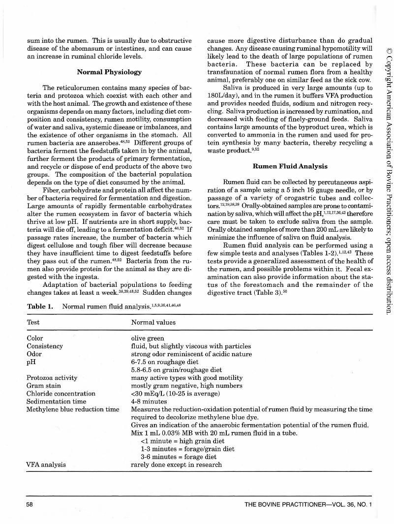
Table 1. Normal rumen fluid analysis. 1·5·9·36,41•46,48
Test Normal values
olive green
cause more digestive disturbance than do gradual changes. Any disease causing ruminal hypomotility will likely lead to the death of large populations of rumen bacteria. These bacteria can be replaced by transfaunation of normal rumen flora from a healthy animal, preferably one on similar feed as the sick cow.
Saliva is produced in very large amounts (up to 180Uday), and in the rumen it buffers VFA production and provides needed fluids, sodium and nitrogen recycling. Saliva production is increased by rumination, and decreased with feeding of finely-ground feeds. Saliva contains large amounts of the byproduct urea, which is converted to ammonia in the rumen and used for protein synthesis by many bacteria, thereby recycling a waste product. 9·52
Rumen Fluid Analysis
Rumen fluid can be collected by percutaneous aspiration of a sample using a 5 inch 16 gauge needle, or by passage of a variety of orogastric tubes and collectors.12·19·38·39 Orally-obtained samples are prone to contamination by saliva, which will affect the pH, 1·12•17•36,43 therefore care must be taken to exclude saliva from the sample. Orally obtained samples of more than 200 mL are likely to minimize the influence of saliva on fluid analysis.
Rumen fluid analysis can be performed using a few simple tests and analyses (Tables 1-2).1·12,43 These tests provide a generalized assessment of the health of the rumen, and possible problems within it. Fecal examination can also provide information about the status of the forestomach and the remainder of the digestive tract (Table 3).50
Color Consistency Odor pH
fluid, but slightly viscous with particles strong odor reminiscent of acidic nature 6-7 .5 on roughage diet
Protozoa activity Gram stain Chloride concentration Sedimentation time Methylene blue reduction time
VFA analysis
58
5.8-6.5 on grain/roughage diet many active types with good motility mostly gram negative, high numbers <30 mEq/L (10-25 is average) 4-8 minutes Measures the reduction-oxidation potential of rumen fluid by measuring the time required to decolorize methylene blue dye. Gives an indication of the anaerobic fermentation potential of the rumen fluid. Mix 1 mL 0.03% MB with 20 mL rumen fluid in a tube.
<1 minute= high grain diet 1-3 minutes= forage/grain diet 3-6 minutes = forage diet
rarely done except in research
THE BOVINE PRACTITIONER-VOL. 36, NO. 1

Table 2. Rumen fluid abnormalities.
Test
Color
Consistency Odor
pH
Protozoa activity Gram stain Chloride concentration Sedimentation time
Methylene blue reduction time
Table 3. Fecal appearance. 47
Very firm or shiny mucus-covered
Very large particle size
Very small particle size
Large amounts of whole grains
Foul odor
Foamy or pasty fluid consistency
Indigestion
Abnormal findings
Grey = acidosis Black or very dark = putrefaction, prolonged stasis Grey and clotted = clotted milk ingesta from calves, putrefaction Yellowish = high grain feeding Foamy = bloat or vagal indigestion Acidic = rumen acidosis Foul = putrefaction Ammonia = urea poisoning <5.5 = probable high grain feeding or acidosis >7.5 = probable salivary contamination * abomasal reflux will not reduce the pH below normal low range Reduced= rumen acidosis, indigestion, anorexia, hypomotility, etc. Many gram positive bacteria= lactic acidosis >30 mEq/L = abomasal reflux <4 minutes= inactive rumen microflora (acidosis, indigestion, anorexia) >8 minutes = overactive rumen microflora (frothy bloat, vagal indigestion) > 10 minutes = indigestible diets, prolonged anorexia, rumen acidosis, protozoa! death
slow (long) forestomach transit time dehydration normal in bulls and animals on dry hay diets rapid (short) forestomach transit time vagal indigestion, traumatic reticulitis tooth problems . slow (long) forestomach transit time vagal indigestion abomasal displacement rumen acidosis rapid (short) transit time overfeeding of grain (possible acidosis) abnormal fermentation bad feed, acidosis or rumen putrefaction digested blood products or endothelium abnormal fermentation rumen acidosis (usually smell acidic)
Simple indigestion in the ruminant is usually caused by feed changes or consumption of abnormal feeds, resulting in an altered microbial population in the rumen. Clinical signs include anorexia for one or two days, followed by diarrhea and a rapid return to normal without treatment.5•17•52 These animals are often examined and treated by veterinarians · for a variety of diseases, but
would recover without therapy. Characteristically, rumen motility is reduced in strength and frequency, the rumen remains full, and there are few changes in the animal's rumen or blood parameters. The animal is rarely depressed or dehydrated. The rapid spontaneous recovery is due to the acclimation of rumen microflora to the n.0w diet, and elimination of toxins or inhibitory substances from the rumen. Transfaunation may be helpful to reestablish the normal microflora in the rumen. 17
FEBRUARY, 2002 59

Rumen Tympany (Free-Gas Bloat)
Free-gas bloat occurs because the animal cannot relieve the gas pressure in the rumen. It is an indication of disease, not a disease itself. 20,51 Free-gas bloat develops due to abnormal fermentation within the rumen, physical obstruction of the cardia or esophagus, lack of rumen motility, or fluid covering the cardia. Bloat does not occur due to excess gas production. Bloat is easily diagnosed by observation as the left paralumbar fossa becomes markedly distended when viewed from the rear. The animal may exhibit colic, and often shows signs of respiratory difficulty if distension is severe.
Intraluminal physical obstruction of the esophagus (choke) is easily diagnosed by palpation, passage of a stomach tube or endoscopy. Extraluminal obstruction may be more difficult to diagnose. Common extraluminal obstructions include enlarged lymph nodes, tumors and strictures. Extraluminal obstructions generally produce bloat which is chronic in nature, while intraluminal obstructions are more acute. Intraluminal obstructions are often large foreign bodies such as potatoes, beets, turnips, hedge apples, etc. Accumulations of dry grain or pellets consumed rapidly may also cause obstruction.
Hypomotility of the rumen can also cause bloat because the rumen is unable to contract sufficiently to produce a good secondary contraction cycle.6•7•9,20,31,33,52
Hypomotility may result from hypocalcemia, systemic disease or chemical inhibition. Bloat itself causes hypomotility due to stimulation of stretch receptors in the wall, which may allow bloat to become more severe by reducing eructation.9•20,47 Obstruction of the cardia occurs in lateral recumbency, and in conditions which cause fluid accumulation in the rumen, such as grain overload or Type II vagal indigestion.
Treatment for free-gas bloat is to relieve the inciting cause. If due to an obstruction, the object should be removed by pulling it out, pushing it down into the rumen, rumenotomy to remove it through the rumen, or (as last resort) surgical esophagotomy.15
•20
,51 If the bloat
is digestive in nature, the primary problem must be identified and resolved or the bloat will recur. Passage of a stomach tube may provide immediate relief. Bloat therapies such as mineral oil, poloxalene, and other surfactants are not useful for treatment of free-gas bloat as there is no problem with surface tension of the ingesta. Transfaunation is helpful if the problei;n is digestive in nature, as it reestablishes the normal rumen microflora. Repeated transfaunations may be necessary. Chronic-bloating animals, such as those with vagal indigestion, may require temporary or permanent fistulation of the rumen to prevent chronic gas distension. This procedure prevents motility-reducing tension in the rumen wall from developing, thereby improving rumen motility until a normal rumen environment has
60
been re-established. Chronic-bloating animals are good candidates for early slaughter ifno improvement is seen after three weeks of treatment.
Rumen Tympany (Frothy Bloat)
Frothy bloat is a specific type of ruminal distension caused by the formation of a stable foam which cannot be eructated. The foam is not a true foam, but a complex structure involving ingesta, fluid and gas. The fluid viscosity is increased by soluble proteins from the diet, preventing coalescing of the gas bubbles in the fluid. The frothy ingesta is sensed by the receptors near the cardia to be fluid or ingesta, rather than gas, therefore eructation is inhibited. 20,51 Frothy bloat results from the feeding oflegumes or winter wheat pasture, or from feeding excess concentrates in the diet.
Individual cattle may be physiologically predisposed to development of frothy bloat,24
•51 such as those with large
rumen volumes and certain salivary proteins. Susceptible cattle have no mucin in their saliva to dissolve the slime. Cattle grazing young, rapidly growing plants are more inclined to bloat because of the high water content, which lessens saliva production. Bloat-inducing plants have easily-chewed leaves and stems, which are rapidly fermented by bacteria in the rumen. Certain varieties of forages have thicker stems and leaves and are therefore bloat-resistant. Grain bloat occurs because a mucoprotein slime develops during the digestion of large quantities of grain. This slime is stable at the low rumen pH induced by grain feeding, and stabilizes the bubbles in the foam. Feeding of both high grain and high-quality alfalfa provides the highest risk for frothy bloat. This diet is commonly fed to dairy cattle and growing steers.
Diagnosis of frothy bloat involves passage of a stomach tube into a bloated animal and finding very little or no free gas, demonstration of stable foam in the tube, and continued bloat of the animal with the tube in place.
Frothy bloat is treated with surface-acting agents to break down the bubbles in the foam to allow eructation. Such agents include poloxalene (44mg/kg PO), mineral oil and detergents. Preventative treatments include alteration of the ration composition, daily feeding of poloxalene or oil in the ration, and ionophores fed daily in the ration or given as a rumen bolus. Ionophores reduce the incidence of frothy bloat by reducing the number of protozoa in the rumen. 24,25,51
Acidosis (Acute)
Acute rumen acidosis (lactic acidosis, grain overload, rumen overload, acid indigestion) is a true emergency in food animal practice, often resulting in death within 24 hours if not promptly and aggressively treated. This disease results from consumption of excess readily
THE BOVINE PRACTITIONER-VOL. 36, NO. 1

fermentable carbohydrates (concentrates), resulting in rapid production of lactate in the rumen. 8,13,16,45 This condition can be due to consumption oflarge quantities of concentrate without adaptation, or accidental overfeeding or access to concentrate feeds. Animals in groups are more likely to overconsume due to competition. Particle size and surface area dramatically affect the rate of fermentation. Carbohydrate feeds most commonly involved are grains, but other fermentable carbohydrates have been incriminated (fruit, potatoes, beets, other root crops, bakery byproducts).
Consumption of large amounts -of carbohydrate feeds reduces the buffering capacity of the rumen by reducing the amount of saliva produced. Once in the rumen, the carbohydrates are rapidly fermented to produce large quantities of VFA and lactate. 13 This fermentation is principally due to Streptococcus bovis, which grows particularly fast when there are readilyfermentable carbohydrates. S. bovis produces lactate as its end product of fermentation. If a moderate amount of carbohydrate is consumed, the S. bovis population will decrease once the carbohydrate is fermented. The production of lactate will then decline, leading to resolution of the problem as the pH rises. 17,27,45
If a large amount of carbohydrate is consumed, acidosis results and the rumen pH may drop from a normal of pH 6 to 7.5 down to a pH of 5 to 5.5. Reduced pH and increased osmolarity kill more of the normal bacteria in the rumen, including lactate-using bacteria like Megasphera elsdenii and Selenomonas ruminantium. Lactobacilli overgrow, leading to the production of more lactate and unusable end products. At the lower rumen pH, even the S. bovis which started the whole process are killed by the high concentration of lactate. The low rumen pH also affects the metabolism of those bacteria still alive in the rumen, resulting in impaired conversion oflactate to propionate. Low pH increases rumen amylase activity, producing more glucose from starches in the diet. This increased glucose concentration prevents the lactate-using bacteria from converting lactate to acetate. Both of these changes in metabolism result in increased concentrations of lactate in the rumen. 13,27,45,48,52
Systemic effects of rumen acidosis are varied. There is increased absorption of VFA from the rumen as the concentration of undissociated VFA increases. VFA may also be metabolized by the rumen epithelium, releasing lactate and ketones into the circulation. 17 Increased lactate, VFA and ketones in the systemic circulation lead to acidosis and potential liver damage. 2 The high levels of VFA reduce rumen motility, which actually protects the animal from further fermentation products. 8•27 Lactate also increases the rumen osmolarity, causing fluid to be drawn from the systemic circulation. This leads to systemic dehydration despite a huge
FEBRUARY, 2002
amount of fluid in the rumen. 13 Dehydration contributes to systemic acidosis. The absorption oflactate and VFA is not sufficient to cause systemic acidosis. 17 The death of large numbers of bacteria in the rumen may cause systemic endotoxemia;33 endotoxin is absorbed readily through the damaged ruminal mucosa.
Treatment of acute acidosis requires aggressive treatment. If the owner does not want to pursue treatment and the animal can still stand, immediate slaughter is recommended. Animals which are depressed, recumbent, blind, or have an extremely distended abdomen have a grave prognosis. The preferred treatment for acute acidosis is rumenotomy with removal of all grain and most rumen content followed by transfaunation with rumen content from a normal animal. Warm water lavage of the rumen is recommended.
If the owner is unwilling to spend the money for a rumenotomy, a large bore stomach tube (Kingman tube) can be used to lavage the rumen with warm water to remove as much ingesta as possible, and to administer magnesium hydroxide into the rumen (lg/kg BW). Intravenous fluids and sodium bicarbonate are necessary to normalize hydration and blood pH. Systemic antibiotics are recommended to protect against rumenitis-induced bacteremia and to prevent liver abscess formation. Anti-inflammatories should also be used to reduce the incidence of rumenitjs and laminitis, and to provide support against shock and endotoxemia. If the animal survives, further treatment is similar to that recommended for bloat or indigestion, including repeated transfaunation. Rumen acidosis may lead to destruction of the rumen epithelium causing acute problems such as perforation and peritonitis, allow colonization by mycotic organisms leading to chronic rumenitis, or lead to long-term problems such as liver abscesses and caudal vena caval thrombosis. 17•27 Laminitis may result from rumen acidosis, presumably due to the release of histamine and other systemic toxins. 13
Prevention requires careful feed management or the feeding of ionophores to alter VFA production and reduce lactate concentrations. 35 Ionophores are not currently approved for use in lactating dairy cattle in the US, though they are approved in other countries (New Zealand, Canada).
Acidosis (Chronic)
Chronic rumen acidosis is usually a nutritional problem caused by long-term feeding ofrations with high levels of concentrate and insufficient fiber. Acidosis is one of the most important nutritional problems in the dairy industry today because it causes reduced milk production, increased incidence of subclinical laminitis, production of off-flavored milk, and chronic damage to the rumen.17•27•37,38 High grain feeding causes selection
61
for lactate-producing bacteria and a reduction in cellulolytic bacteria. These changes lead to rapid fermentation of high carbohydrate feeds. Lactate does not accumulate in the rumen because there are lactate-using bacteria to metabolize it. The rumen pH remains below 6 most of the time, but blood pH is normal. High VFA levels may result from feeding silages high in VFA or from the alteration in rumen bacterial metabolism which occurs at low pH (increased butyrate and propionate and reduced acetate). Butyrate and propionate stimulate ruminal epithelial proliferation and parakeratosis, a condition which clumps the ruminal papillae and reduces surface area for absorption. , Ulcers may develop in the rumen wall, allowing bacteria access to the bloodstream where they may colonize the liver or distant body sites. The low rumen pH also reduces the population of protozoa and the number of species ofbacteria. Thus, the cattle are at higher risk of problems if there are feed changes. Laminitis may result from chronic sub-clinical rumen acidosis. 13
Incidentally, acetate is the VFA most used for the production oflactose, which is the major sugar constituent of milk. Milk production is reduced when chronic acidosis occurs, not only due to the reduction in appetite which accompanies the acidosis, but also due to reduced secretion of lactose. 9,52
Treatment of chronic acidosis requires alteration of the ration to reduce the amount of concentrate feeding, or to increase the long fiber proportion. Improvement will take several weeks as the rumen microflora must adapt to the new diet, and the rumen papillae must regenerate. Some cows will not improve despite the changes, and systemic problems may be permanent (such as liver abscesses). Prevention involves feeding management and, where permitted by law, the use of ionophores to prevent rumen acidosis. 35
Vagal Indigestion
Vagal indigestion is a syndrome rather than a unique disease in itself. Symptoms include rumen distension, alteration of rumen motility, delayed passage of ingesta, and sometimes bradycardia.14,4o,41,42 The cow will usually become anorexic. Bloat may occur, but is usually mild. Hypermotility indicates an intact dorsal vagus nerve, while complete rumen stasis indicates damage to the ventral vagus. 7•29 Eventually, with extreme distension, the rumen takes on an "L-shape" in the abdomen, which leads to the classic "papple" shape to the abdomen when viewed from behind. There are numerous classification systems for the types of vagal indigestion. They describe in different ways the outflow problems with the reticulorumen and the omasum and abomasum. The reader is referred to reviews of vagal indigestion as described in textbooks for these classification systems.14,17,53
62
Removal of rumen content using a large bore stomach tube or by rumenotomy may provide short-term relief for the animal, but without resolution of the inciting cause the distension will return. A thorough search should be made for reticuloruminal outflow or abomasal outflow obstructions, as well as interference with or inflammation of the vagus nerves in the thorax or abdomen. Many times, no inciting cause can be found, and therapy is unsuccessful. Treatment with antibiotics, anti-inflammatories, and sometimes a temporary rumen fistula may lead to partial resolution. Motility-enhancing drugs have been used, but few are truly effective in clinical use.
Reticuloruminal Putrefaction ("Rumen Drinkers")
Esophageal groove closure in calves allows the passage of milk from the esophagus to the omasum, bypassing the reticulorumen.4•10•11 Failure of the closure reflex, especially in older calves, allows accumulation of milk in the reticulorumen. A similar problem may result from repeated tube feedings which do not stimulate groove closure and allow milk replacer to enter the reticulorumen. 3•
4•28 Milk in the reticulorumen then fer
ments, rather than forming a curd as it would in the abomasum. Fermentation of the milk in the reticulorumen leads to digestive upset, fluid rumen distension and diarrhea. 3,4,18
Diagnosis of this problem is by signalment and rumen fluid examination. Treatment involves weaning from milk feeding and transfaunation with fresh rumen fluid to establish a normal rumen microflora population.
Forestomach Motility Enhancing Drugs
A number of therapeutic agents have been used in the attempt to modify the motility of the reticulorumen for correction of motility disorders. They include metoclopramide, bethanechol, butorphanol, carbamylcholine, diazepam, morphine and neostigmine. None have been particularly useful in clinical settings, and all are expensive to use in adult cattle.
Neostigmine is an inhibitor of acetylcholinesterase which requires normal vagus activity to be present, therefore it is oflittle use in animals with vagal insufficiency. 5 N eostigmine may improve the strength of the primary contractions without altering the speed of contractions, so it may be useful in animals with reduced strength of rumen contractions.5
•44 Carbamylcholine and
bethanechol are muscarinic agonists which have activity on the reticulorumen. Carbamylcholine causes uncoordinated contractions of the forestomach, leading to non-propulsive mixing of the forestomach contents.5 It also requires an intact vagus nerve to exert an effect on
THE BOVINE PRACTITIONER-VOL. 36, NO. 1
the reticulorumen. Bethanechol increases muscular tone without affecting rate of contraction or inducing disorganized contractions. 44 At low doses, bethanechol has been shown to increase rumen tone, but not alter the rate of contraction, therefore repeated low doses may be useful for treatment of rumen hypomotility. 44 High doses of bethanechol are inhibitory to rumen motility.
Metoclopramide decreases intraruminal pressure during contractions without changing the rate of contraction. 21 An increase in the ratio of secondary to primary rumen contractions has been recorded in cattle given metoclopramide, indicating possible usefulness in promoting eructation, but perhaps less useful for promoting primary contractions.44 Serotonin increased motility and tone in early studies before the widespread availability of serotonin supplements, and no further work has been performed to evaluate its usefulness. 44 Naloxone reportedly will stimulate rumen motility, but was being evaluated mainly as an antagonist for other drugs. 44
Butorphanol has been shown to completely inhibit reticuloruminal contractions in a dose-dependent manner.21 Xylazine can reduce the contractility of the reticulorumen by reducing the frequency of contraction and the force of contractions.44 Morphine can also inhibit reticuloruminal motility and, at low doses, induce hyperexcitability. 44 Reduction of esophageal motility has been noted with morphine administration, which may reduce the ability to regurgitate or eructate. Diazepam, when administered to cattle at low doses, increases feed intake in the hour following administration, but does not increase total feed intake. 6•34 Atropine uniformly reduces rumen motility and tone, which is likely the cause of atropine-induced bloat and colic in ruminants. 44
It appears that there is no effective drug regimen which is reliably effective in promoting reticuloruminal motility in cattle. Further investigation of such drugs may prove to be useful in the treatment of valuable animals which do not respond to surgical or other medical therapy.
Conclusions
There are many causes of reticuloruminal disturbances in the bovine. A thorough understanding of the anatomy and physiology of the forestomach of the ruminant animal is necessary to understand the complexity of the disease processes that may disrupt normal function. A thorough history, complete physical examination and rumen fluid analysis are important tools to diagnose reticuloruminal disorders. Treatment is based upon the diagnosis; proper nutritional management is an important component of the prevention and treatment plan for most disorders. Currently available motility enhancing drugs are of limited value.
FEBRUARY, 2002
References
1. Alonso AN: Diagnostic analysis of rumen fluid. Vet Clin North Am Food Anim Pract 1:363-376, 1979. 2. Bide RW: Excess rumen product anions in cattle I: blood clearance rates and reduced liver function from sub lethal doses of volatile fatty acids, lactate and succinate. Can J Comp Med 47:222-229, 1983. 3. Breukink HJ, Wensin TH, Weeren-Keverling-Buisman A, et al: Consequences of failure of the reticular groove reflex in calves fed milk replacer. Vet Q 10:126-135, 1988. 4. Chapman HW, Butler DG, Newell M: The route of liquids administered to calves by esophageal feeder. Can J Vet Res 50:84-87, 1986. 5. Constable PD: The ruminant forestomach, in: Howard JL, Smith RA(eds): Current Veterinary Therapy 4. Food Animal Practice. Philadelphia, WB Saunders Co, 1999, pp 502-507. 6. Constable PD, Hoffsis GF, Rings DM: The reticulorumen: normal and abnormal motor function. Part I. Primary contraction cycle. Compend Contin Educ Pract Vet 12:1008-1015, 1990. 7. Constable PD, Hoffsis GF, Rings DM: The reticulorumen: normal and abnormal motor function. Part II. Secondary contraction cycles, rumination, and esophageal groove closure. Compend Contin Educ Pract Vet 12:1169-1174, 1990. 8. Crichlow EC, Leek BF: Ruminal lactic acidosis: relationship of forestomach motility to nondissociated volatile fatty acid levels. Am J Vet Res 47:1015-1018, 1986. 9. Cunningham, JG. Textbook of Veterinary Physiology, ed 2. Philadelphia, WB Saunders Co, 1997, pp 272-287, 331-356. 10. Dirksen GU, Garry FB: Diseases of the forestomachs in calves -Part I. Compend Cont Educ Pract Vet 9:F140-F146, 1987. 11. Dirksen GU, Garry FB: Diseases of the forestomachs in calves -Part IL Compend Cont Educ Pract Vet 9:Fl 73-Fl 79, 1987. 12. Dirksen G, Smith MC: Acquisition and analysis of bovine rumen fluid. Bovine Pract 22:108-116, 1987. 13. Dunlop RH: Pathogenesis of ruminant lactic acidosis. Adu Vet Sci Comp Med 16:259-302, 1972. 14. Ferrante PL, Whitlock RH: Chronic (vagus) indigestion in cattle. Compend Cont Educ Pract Vet 3:S231-S237, 1981. 15. Garry F: Managing bloat in cattle. Vet Med 85:643-650, 1990. 16. Garry FB: Diagnosing and treating indigestion caused by fermentative disorders. Vet Med 85:660-670, 1990. 17. Garry FB: Indigestion in ruminants. In: Smith BP: Large Animal Internal Medicine, ed 2. St Louis, Mosby Press, 1996, pp 824-858. 18. Garry FB: Rumen putrefaction (esophageal groove dysfunction, rumen drinkers). In: Howard JL, Smith RA, eds. Current Veterinary Therapy 4. Food Animal Practice. Philadelphia, WB Saunders Co, 1999, pp 510-512. 19. Geishauser T: A probe for collection of ruminal fluid in juvenile cattle and cows. Bovine Pract 28:113-116, 1994. 20. Guard C: Bloat or ruminal tympany. In: Smith BP: Large Animal Internal Medicine, ed 2. St Louis, Mosby Press, 1996, pp 865-868. 21. Guard C, Schwark W, Kelton D, et al: Effects ofmetoclopramide, clenbuterol and butorphanol on ruminoreticular motility of calves. Cornell Vet 78:89-98, 1988. 22. Habel RE: A study of the innervation of the ruminant stomach. Cornell Vet 46:555-628, 1956. 23. Habel RE: Ruminant digestive system, in R Getty (ed): Sisson and Grossman's The Anatomy of the Domestic Animals, ed 5. Philadelphia, WB Saunders Co, 1975, pp 885-890. 24. Howarth R, Cheng K-J, Majak W, et al: Ruminant bloat, in Milligan LP, Grovum WL, Dobson A (eds): Control of digestion and metabolism in ruminants. Englewood Cliffs, Prentice Hall, 1986, pp 516-527. 25. Katz MP, Nagaraja TG, Fina LR: Ruminal changes in monensinand lasalocid-fed cattle grazing on bloat-provocative alfalfa pasture. J Anim Sci 63:1246-1257, 1986. 26. Kay R: Rumen function and physiology. Vet Rec 113:6-9, 1983.
63

27. Kersting KW, Thompson JR: Lactic acidosis (rumen overload, rumen acidosis, grain overload, engorgement toxemia, rumenitis). In: Howard JL, Smith RA, eds. Current Veterinary Therapy 4. Food Animal Practice. Philadelphia, WB Saunders Co, 1999, pp 507-510. 28. Lateur-Rowet HJ, Breukink HJ: The failure of the oesophageal groove reflex, when fluids are given with an oesophageal feeder to newborn and young calves. Vet Q 5:68-74, 1983. 29. Leek BF: Vagus indigestion in cattle. Vet Rec 82:498-499, 1968. 30. Leek BF, Harding RH: Sensory nervous receptors in the ruminant stomach and the reflex control of reticuloruminal motility, in McDonald IW, Warner ACI (eds): Digestion and metabolism in the ruminant. Armadale, NSW, New England Publishing Unit, 1975, pp 60-76. 31. Leek BF: Reticuloruminal function and dysfunction. Vet Rec 84:238-243, 1969. 32. Lirette A, Kelly JM, Milligan LP, et al: Effects of psychological stress, acute cold stress and diet on forestomach contractions in cattle. Can J Anim Sci 68:399-407, 1988. 33. Lohuis JACM, Verheijden JHM, Burvenich C, et al: Pathophysiological effects of endotoxin in ruminants. Vet Q 10:109-116, 1988. 34. McLaughlin CL, Krabill LF, GC Scott, et al: Chemical stimulants of feeding animals. Fed Proc 35:579, 1976. 35. Nagaraja TG, Avery TB, Galitzer SJ, et al: Prevention of lactic acidosis in cattle by lasalocid or monensin. J Anim Sci 53:206-216, 1981. 36. Nichols RE, Penn KE: Simple methods for the detection of unfavorable changes in the ruminal ingesta. · J Am Vet Med Assoc 133 :27 5-277, 1958. 37. Nordlund KY, Garrett EF: Rumenocentesis: a technique for collecting rumen fluid for the diagnosis of subacute rumen acidosis in dairy herds. Bovine Pract 28:109-112, 1994. 38. Nordlund KV: Questions and answers regarding rumenocentesis and the diagnosis of herd-based subacute rumen acidosis. Bovine Pract. 28:75-81, 1996. 39. Rebhun WC, Fubini SL, Miller TK: Vagus indigestion in cattle: Clinical features, causes, treatments, and long-term follow-up of 112 cases. Compend Cont Educ Pract Vet 10:387-391, 1988. 40. Rebhun WC: Rumen collapse in cattle. Cornell Vet 77:244-250, 1987.
Abstract
41. Rebhun WC: Vagus indigestion in cattle. J Am Vet Med Assoc 176:506-510, 1980. 42. Rehage J, Kaske M, Stockhofe-Zurwieden N, et al: Evaluation of the pathogenesis of vagus indigestion in cows with traumatic reticuloperitonitis. J Am Vet Med Assoc 207:1607-1611, 1995. 43. Rings DM, Rings MB: Rumen fluid analysis. Agri Pract 14:26-29, 1993. 44. Ruckebush Y: Pharmacology of reticulo-ruminal motor function. J Vet Pharm Ther 6:245-272, 1983. 45. Russel JB, Hino T: Regulation of lactate production in Streptococcus bovis: a spiraling effect that contributes to rumen acidosis. J Dairy Sci 68:1712-1721, 1985. 46. Scholz H: Utilization of the reticular groove contraction in adult cattle - A therapeutical alternative for the practitioner? Bou Pract 23:148-152, 1988. 47. Sellers AF, Stevens CE: Motor functions of the ruminant forestomach. Physiol Rev 46:634-659, 1966. 48. Slyter LL: Influence of acidosis on rumen function. J Anim Sci 43:910-929, 1976. 49. Smith BL, Reynolds GW, Embling PP: Zinc solutions and closure of the reticular groove in sheep. New Zealand J ExpAgric 5:261-263, 1977. 50. Stober M, Serrano HS: Gross findings in bovine feces. Vet Med Rev 4:361-379, 1974. 51. Streeter RN, Prado ME: Bloat, or rumen tympany, in Howard JL, Smith RA (eds): Current Veterinary Therapy 4. Food Animal Practice. Philadelphia, WB Saunders Co, 1999, pp 512-514. 52. Van Soest PJ: Nutritional ecology of the ruminant, ed 2. Corvallis, 0 & B Books, 1994, pp 230-280. 53. Whitlock RH: Vagal indigestion: in Howard JL, Smith RA (eds): Current Veterinary Therapy 4. Food Animal Practice. Philadelphia, WB Saunders Co, 1999, pp 517-522. 54. Wyburn RS: The mixing and propulsion of the stomach contents ofruminants, in Ruckebush Y, Thivend P (eds): Digestive physiology and metabolism in ruminants. Lancaster, MTB Press, 1980, pp 35-51.
Effectiveness and Kinetic Behaviour of Tilmicosin in the Treatment of Respiratory Infections in Sheep F. Naccari, F. Giofre, M. Pellegrino, M. Calo, P. Licata, S. Carli Veterinary Record (2001) 148:773-776
Nineteen sheep which were anorexic, pyrexic, coughing, dyspnoeic and had a nasal discharge and symptomatic thoracic sounds on ausculation, received a single subcutaneous dose of 10 mg/kg bodyweight of tilmicosin. The clinical signs were eliminated within four to six days. The kinetic profiles of the drug after a
64
single subcutaneous injection were compared in five healthy sheep and five infected sheep. More of the drug was absorbed by the infected animals and its concentration remained higher for significantly longer. The drug was well tolerated and no local or systemic side effects were observed.
THE BOVINE PRACTITIONER-VOL. 36, NO. 1

ScourGuard 3®(K)/C has always provided broad-spectrum protection
against the leading causes of scours. And now, ScourGuard 3(K)/C
also delivers a stronger punch against rotavirus-a calf-killer.
In fact, a new challenge study demonstrates that ScourGuard
significantly reduces rotavirus shedding that can spread disease
and kill calves in your operation.1 So ask your veterinarian or
animal health supplier about improved ScourGuard 3(K)/C today.
Multiple strains of rotavirus protection
Demonstrated to reduce calf loss due to rotavirus1
Dem·onstrated to reduce viral (rotavirus) shedding1
Tissue-friendly2
Field experience More than 12years
1. Data on file, Pfizer Animal Health, study #2934H-60-00-012. 2. Data on file, Pfizer Animal Health, study #2134H·60-00-075. ScourGuard 3 is a registered trademark and Beef Friendly is a trademark of Pfizer Inc © 2001 Pfizer Inc. SGD1001023 3005
--
----.i~)_--_.-: ___ -._._. ---
. _ ·• ·:./ ~nimal Health • __ .,.:< _;- ·· · · www. pfiz<::• rbecf.corn - ... ~~:.-.:-.::...-=:.......... -
~...; _;.,r ~ ,.. -