Sleep Spindles Predict Stress-Related Increases in Sleep Disturbances
9
HUMAN NEUROSCIENCE ORIGINAL RESEARCH ARTICLE published: 10 February 2015 doi: 10.3389/fnhum.2015.00068 Sleep spindles predict stress-related increases in sleep disturbances ThienThanh Dang-Vu 1,2,3,4,5,6 *, Ali Salimi 1,2,3,4 , Soufiane Boucetta 1,2,3,4 , Kerstin Wenzel 6 , Jordan O’Byrne 1,2,3,4 , Marie Brandewinder 7 , Christian Berthomier 7 and Jean-Philippe Gouin 3,5,6 1 Department of Exercise Science, Concordia University, Montréal, QC, Canada 2 Center for Studies in Behavioral Neurobiology, Concordia University, Montréal, QC, Canada 3 PERFORM Center, Concordia University, Montréal, QC, Canada 4 Centre de Recherche de l’Institut Universitaire de Gériatrie de Montréal, Montréal, QC, Canada 5 Center for Clinical Research in Health, Concordia University, Montréal, QC, Canada 6 Department of Psychology, Concordia University, Montréal, QC, Canada 7 Physip SA, Paris, France Edited by: Christian O’Reilly, McGill University, Canada Reviewed by: Gary N. Garcia-Molina, Philips Research North America, USA Julio Fernandez-Mendoza, Penn State Milton S. Hershey Medical Center, USA *Correspondence: Thien Thanh Dang-Vu, Department of Exercise Science, Center for Studies in Behavioral Neurobiology, PERFORM Center, Concordia University, 7141 Sherbrooke Street West, SP 165-27, Montreal, QC H4B 1R6, Canada e-mail: [email protected] Background and Aim: Predisposing factors place certain individuals at higher risk for insomnia, especially in the presence of precipitating conditions such as stressful life events. Sleep spindles have been shown to play an important role in the preservation of sleep con- tinuity. Lower spindle density might thus constitute an objective predisposing factor for sleep reactivity to stress.The aim of this study was therefore to evaluate the relationship between baseline sleep spindle density and the prospective change in insomnia symptoms in response to a standardized academic stressor. Methods: Twelve healthy students had a polysomnography recording during a period of lower stress at the beginning of the academic semester, along with an assessment of insomnia complaints using the insomnia severity index (ISI). They completed a second ISI assessment at the end of the semester, a period coinciding with the week prior to final examinations and thus higher stress. Spindle density, amplitude, duration, and frequency, as well as sigma power were computed from C4–O2 electroencephalography derivation during stages N2–N3 of non-rapid-eye-movement (NREM) sleep, across the whole night and for each NREM sleep period.To test for the relationship between spindle density and changes in insomnia symptoms in response to academic stress, spindle measurements at baseline were correlated with changes in ISI across the academic semester. Results: Spindle density (as well as spindle amplitude and sigma power), particularly during the first NREM sleep period, negatively correlated with changes in ISI (p < 0.05). Conclusion: Lower spindle activity, especially at the beginning of the night, prospectively predicted larger increases in insomnia symptoms in response to stress.This result indicates that individual differences in sleep spindle activity contribute to the differential vulnerability to sleep disturbances in the face of precipitating factors. Keywords: spindles, sleep, insomnia, stress, EEG INTRODUCTION The natural history of insomnia is hypothesized to involve three categories of factors: predisposing factors placing certain individuals at higher risk for insomnia complaints, precipitat- ing factors triggering the onset of insomnia, and perpetuating factors maintaining the insomnia over time (Spielman, 1986). The characterization of predisposing and precipitating factors is of prime importance not only to understand the pathophys- iology of insomnia but also to implement optimal preventive sleep interventions. In terms of predisposing factors, longitu- dinal studies have shown that the rate of new onset insomnia is higher among individuals with depressive or anxiety symp- toms, a family history of insomnia, high arousability predisposi- tion, poor general health condition, and pain syndrome (LeBlanc et al., 2009; Harvey et al., 2014). On the other hand, precipi- tating factors have been shown to vary with age; in particular, for younger individuals (<30 years old), stress-related factors at work/school constitute the most frequent precipitating events trig- gering insomnia onset (Bastien et al., 2004). Given one’s profile of predisposing factors, individuals are not equally vulnerable to the development of sleep disturbances in the face of com- mon precipitating factors such as stressful events (Drake et al., 2004). Beyond medical and psychological history, there has been no investigation of the inter-individual variations in sleep architec- ture – and sleep oscillations – as predisposing factors for the insomnia symptoms. Among the different components of sleep architecture, sleep spindles have been the subjects of intense Frontiers in Human Neuroscience www.frontiersin.org February 2015 |Volume 9 | Article 68 | 1
-
Upload
independent -
Category
Documents
-
view
1 -
download
0
Transcript of Sleep Spindles Predict Stress-Related Increases in Sleep Disturbances

HUMAN NEUROSCIENCEORIGINAL RESEARCH ARTICLE
published: 10 February 2015doi: 10.3389/fnhum.2015.00068
Sleep spindles predict stress-related increases in sleepdisturbancesThienThanh Dang-Vu1,2,3,4,5,6*, Ali Salimi 1,2,3,4, Soufiane Boucetta1,2,3,4, Kerstin Wenzel 6, Jordan O’Byrne1,2,3,4,Marie Brandewinder 7, Christian Berthomier 7 and Jean-Philippe Gouin3,5,6
1 Department of Exercise Science, Concordia University, Montréal, QC, Canada2 Center for Studies in Behavioral Neurobiology, Concordia University, Montréal, QC, Canada3 PERFORM Center, Concordia University, Montréal, QC, Canada4 Centre de Recherche de l’Institut Universitaire de Gériatrie de Montréal, Montréal, QC, Canada5 Center for Clinical Research in Health, Concordia University, Montréal, QC, Canada6 Department of Psychology, Concordia University, Montréal, QC, Canada7 Physip SA, Paris, France
Edited by:Christian O’Reilly, McGill University,Canada
Reviewed by:Gary N. Garcia-Molina, PhilipsResearch North America, USAJulio Fernandez-Mendoza, Penn StateMilton S. Hershey Medical Center,USA
*Correspondence:Thien Thanh Dang-Vu, Department ofExercise Science, Center for Studiesin Behavioral Neurobiology,PERFORM Center, ConcordiaUniversity, 7141 Sherbrooke StreetWest, SP 165-27, Montreal, QC H4B1R6, Canadae-mail: [email protected]
Background and Aim: Predisposing factors place certain individuals at higher risk forinsomnia, especially in the presence of precipitating conditions such as stressful life events.Sleep spindles have been shown to play an important role in the preservation of sleep con-tinuity. Lower spindle density might thus constitute an objective predisposing factor forsleep reactivity to stress. The aim of this study was therefore to evaluate the relationshipbetween baseline sleep spindle density and the prospective change in insomnia symptomsin response to a standardized academic stressor.
Methods: Twelve healthy students had a polysomnography recording during a period oflower stress at the beginning of the academic semester, along with an assessment ofinsomnia complaints using the insomnia severity index (ISI). They completed a second ISIassessment at the end of the semester, a period coinciding with the week prior to finalexaminations and thus higher stress. Spindle density, amplitude, duration, and frequency,as well as sigma power were computed from C4–O2 electroencephalography derivationduring stages N2–N3 of non-rapid-eye-movement (NREM) sleep, across the whole nightand for each NREM sleep period. To test for the relationship between spindle density andchanges in insomnia symptoms in response to academic stress, spindle measurements atbaseline were correlated with changes in ISI across the academic semester.
Results: Spindle density (as well as spindle amplitude and sigma power), particularly duringthe first NREM sleep period, negatively correlated with changes in ISI (p < 0.05).
Conclusion: Lower spindle activity, especially at the beginning of the night, prospectivelypredicted larger increases in insomnia symptoms in response to stress.This result indicatesthat individual differences in sleep spindle activity contribute to the differential vulnerabilityto sleep disturbances in the face of precipitating factors.
Keywords: spindles, sleep, insomnia, stress, EEG
INTRODUCTIONThe natural history of insomnia is hypothesized to involvethree categories of factors: predisposing factors placing certainindividuals at higher risk for insomnia complaints, precipitat-ing factors triggering the onset of insomnia, and perpetuatingfactors maintaining the insomnia over time (Spielman, 1986).The characterization of predisposing and precipitating factorsis of prime importance not only to understand the pathophys-iology of insomnia but also to implement optimal preventivesleep interventions. In terms of predisposing factors, longitu-dinal studies have shown that the rate of new onset insomniais higher among individuals with depressive or anxiety symp-toms, a family history of insomnia, high arousability predisposi-tion, poor general health condition, and pain syndrome (LeBlanc
et al., 2009; Harvey et al., 2014). On the other hand, precipi-tating factors have been shown to vary with age; in particular,for younger individuals (<30 years old), stress-related factors atwork/school constitute the most frequent precipitating events trig-gering insomnia onset (Bastien et al., 2004). Given one’s profileof predisposing factors, individuals are not equally vulnerableto the development of sleep disturbances in the face of com-mon precipitating factors such as stressful events (Drake et al.,2004).
Beyond medical and psychological history, there has been noinvestigation of the inter-individual variations in sleep architec-ture – and sleep oscillations – as predisposing factors for theinsomnia symptoms. Among the different components of sleeparchitecture, sleep spindles have been the subjects of intense
Frontiers in Human Neuroscience www.frontiersin.org February 2015 | Volume 9 | Article 68 | 1
Dang-Vu et al. Spindles and stress-related insomnia
research over the past decade (De Gennaro and Ferrara,2003; Fogeland Smith, 2011). Spindles are defined as waxing-and-waningelectroencephalography (EEG) waves oscillating at a frequency of11–16 Hz and predominant over central EEG derivations; spin-dles characterize stage N2 of non-rapid-eye-movement (NREM)sleep but can also be found during stage N3 (De Gennaro et al.,2000; Iber et al., 2007). Animal and human studies converge todemonstrate that sleep spindles are generated through the inter-play between specific populations of thalamic (particularly thala-mic reticular) and cortical neurons (Steriade and McCarley, 2005;Schabus et al., 2007). While the density of sleep spindles variesconsiderably between individuals, it has been shown that spin-dle density remains remarkably stable within a same individualacross different nights, thus constituting an individual trait (Gail-lard and Blois, 1981; De Gennaro et al., 2005). Spindles have beenshown correlated with measures of intellectual ability as well aswith the overnight retention of various types of memory traces,suggesting an important role for spindles in brain plasticity andsleep-related memory consolidation (Gais et al., 2002; Schabuset al., 2004, 2006; Morin et al., 2008; Fogel and Smith, 2011).EEG and functional neuroimaging studies have also demonstratedthat the cortical transmission of external – particularly acoustic –stimulation during sleep is drastically diminished during sleepspindles (Cote et al., 2000; Dang-Vu et al., 2011). These findingsindicate that spindles isolate the cortex from the environmentduring sleep, contributing to the preservation of sleep stability.It was then inferred that spindle density, as a trait, might con-stitute a biomarker of sleep stability in the face of noise. Thiswas confirmed by a study showing that individuals with lowerspindle density were more vulnerable to sleep disruption fromsounds presented throughout the night (Dang-Vu et al., 2010).Because spindles constitute an index of sleep stability, individualswith reduced spindle density might be more vulnerable to developinsomnia complaints, particularly when confronted to triggeringfactors such as stress. To the best of our knowledge, no studyhas investigated the role of sleep spindle as predisposing factor toinsomnia onset.
The aim of this study was to prospectively assess whetherspindle density would predict the worsening of sleep distur-bances in response to a standardized stressor. We chose to fol-low a population of undergraduate university students duringa period of increasing academic stress. In this context, assess-ing students at the beginning of the semester, corresponding toa lower stress period, and reevaluating them during a follow-up in the week preceding the final examinations, a period ofhigher stress, provides a unique opportunity to examine indi-vidual differences in the evolution of insomnia symptoms inresponse to a standardized stressor. The validity of this modelis supported by data showing an increase of sleep disturbancesin response to increased academic stress (Jernelov et al., 2009;Lund et al., 2010). Here, we hypothesized that a lower spindledensity at baseline during the low stress period would prospec-tively predict larger increases in sleep complaints from the lowto the high stress periods, therefore defining a neurophysiologi-cal vulnerability factor predisposing to the increase of insomniasymptoms.
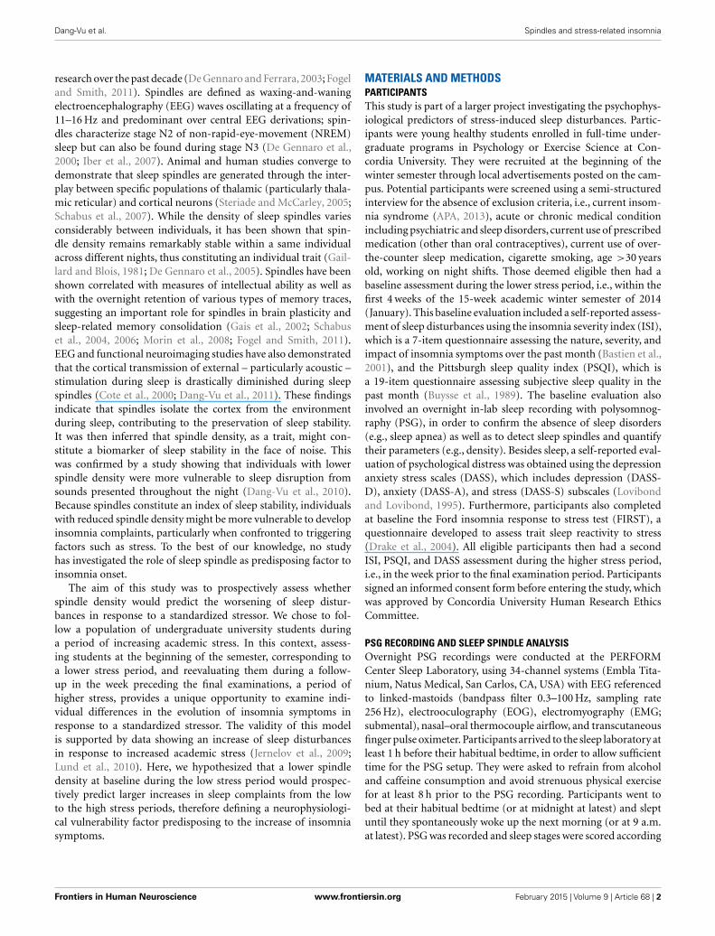
MATERIALS AND METHODSPARTICIPANTSThis study is part of a larger project investigating the psychophys-iological predictors of stress-induced sleep disturbances. Partic-ipants were young healthy students enrolled in full-time under-graduate programs in Psychology or Exercise Science at Con-cordia University. They were recruited at the beginning of thewinter semester through local advertisements posted on the cam-pus. Potential participants were screened using a semi-structuredinterview for the absence of exclusion criteria, i.e., current insom-nia syndrome (APA, 2013), acute or chronic medical conditionincluding psychiatric and sleep disorders, current use of prescribedmedication (other than oral contraceptives), current use of over-the-counter sleep medication, cigarette smoking, age >30 yearsold, working on night shifts. Those deemed eligible then had abaseline assessment during the lower stress period, i.e., within thefirst 4 weeks of the 15-week academic winter semester of 2014(January). This baseline evaluation included a self-reported assess-ment of sleep disturbances using the insomnia severity index (ISI),which is a 7-item questionnaire assessing the nature, severity, andimpact of insomnia symptoms over the past month (Bastien et al.,2001), and the Pittsburgh sleep quality index (PSQI), which isa 19-item questionnaire assessing subjective sleep quality in thepast month (Buysse et al., 1989). The baseline evaluation alsoinvolved an overnight in-lab sleep recording with polysomnog-raphy (PSG), in order to confirm the absence of sleep disorders(e.g., sleep apnea) as well as to detect sleep spindles and quantifytheir parameters (e.g., density). Besides sleep, a self-reported eval-uation of psychological distress was obtained using the depressionanxiety stress scales (DASS), which includes depression (DASS-D), anxiety (DASS-A), and stress (DASS-S) subscales (Lovibondand Lovibond, 1995). Furthermore, participants also completedat baseline the Ford insomnia response to stress test (FIRST), aquestionnaire developed to assess trait sleep reactivity to stress(Drake et al., 2004). All eligible participants then had a secondISI, PSQI, and DASS assessment during the higher stress period,i.e., in the week prior to the final examination period. Participantssigned an informed consent form before entering the study, whichwas approved by Concordia University Human Research EthicsCommittee.
PSG RECORDING AND SLEEP SPINDLE ANALYSISOvernight PSG recordings were conducted at the PERFORMCenter Sleep Laboratory, using 34-channel systems (Embla Tita-nium, Natus Medical, San Carlos, CA, USA) with EEG referencedto linked-mastoids (bandpass filter 0.3–100 Hz, sampling rate256 Hz), electrooculography (EOG), electromyography (EMG;submental), nasal–oral thermocouple airflow, and transcutaneousfinger pulse oximeter. Participants arrived to the sleep laboratory atleast 1 h before their habitual bedtime, in order to allow sufficienttime for the PSG setup. They were asked to refrain from alcoholand caffeine consumption and avoid strenuous physical exercisefor at least 8 h prior to the PSG recording. Participants went tobed at their habitual bedtime (or at midnight at latest) and sleptuntil they spontaneously woke up the next morning (or at 9 a.m.at latest). PSG was recorded and sleep stages were scored according
Frontiers in Human Neuroscience www.frontiersin.org February 2015 | Volume 9 | Article 68 | 2

Dang-Vu et al. Spindles and stress-related insomnia
to standard criteria (Iber et al., 2007). Sleep apnea syndrome wasdefined by an apnea–hypopnea index >5/h (exclusion criterion).Sleep spindles were automatically detected during stages N2 andN3 of NREM sleep on EEG C4–O2 derivation. This derivationwas chosen given the well-described central predominance of sleepspindles (De Gennaro et al., 2000). The spindle detection method(Aseega software, Physip, Paris, France) used data-driven crite-ria in order to cope with both inter-subject and inter-recordingcondition variabilities (Berthomier et al., 2007). It was based onan iterative approach. The first iteration aimed at determiningrecording-specific thresholds, based on EEG power ratios in delta,alpha, and sigma bands. The second iteration provided precisetemporal localization of the events. The final iteration enabledthe validation of detected events based on frequency and durationcriteria (>0.5 s). Iteration 1 and 3 dealt with raw EEG data, whileiteration 2 was applied on the EEG filtered in the spindle (sigma)frequency range using frequency bands adapted to each individ-ual based on his/her global spectral profile (median values for lowand high bands were 11.9 and 15.9 Hz, respectively). The densityof spindles was computed as the average number of detected spin-dles per 30 s EEG epoch for each subject. In addition to spindledensity, other spindle parameters were also computed for eachparticipant in order to comprehensively evaluate spindle activ-ity: average maximum spindle amplitude (in microvolts), averagespindle duration (in seconds), and average spindle frequency (inhertz). After cleaning of the main EEG artifacts, the EEG powerin the adapted sigma frequency range (in squared microvolts, per30 s epoch) was also computed using Hanning window. Spindleparameters and sigma power were first calculated considering theN2–N3 NREM sleep of the entire night. In addition, in order totake into account the variation of spindle activity across successiveNREM sleep periods (De Gennaro et al., 2000), spindle parame-ters, and sigma power were also calculated for each NREM sleepperiod. These periods were defined according to standard criteria:a NREM sleep period was defined by a period of at least 15 minof NREM sleep followed by at least 5 min of REM sleep, and thefirst four NREM sleep periods were considered given that there areusually four NREM sleep periods during overnight sleep in adults(Feinberg and Floyd, 1979). For completeness, spindle parametersand sigma power were also calculated during stage N2 only, for theentire night as well as for each NREM sleep period, and the datafrom these additional analyses are presented in the supplementarymaterial given that they showed results quite similar to those fromN2–N3 combined.
STATISTICAL ANALYSISChanges in self-reported sleep quality and stress over time werefirst evaluated using dependent t -tests. The evolution of spin-dle parameters across NREM sleep periods was then examinedusing one-way repeated measures analysis of variance (ANOVA)with Bonferroni post hoc tests. In order to test our main hypoth-esis, correlations between spindle density (during N2–N3 forthe whole night and for each NREM sleep period) and changesin self-reported sleep quality (score during high stress periodminus score during low stress period) were calculated using Pear-son product-moment correlation. Secondary analyses likewisetested the Pearson correlations between other spindle parameters
(spindle amplitude, duration, frequency) or sigma power and sleepquality changes. FIRST scores were also correlated with sleep qual-ity changes to evaluate the predictive potential of this self-reportedmeasure of sleep reactivity to stress in the context of academicstress. All analyses were considered significant at a p value <0.05,and were conducted with SPSS Statistics 22.0 (IBM, New York,NY, USA).
RESULTSOut of 22 potential participants, 12 were confirmed eligible andpresented at least 4 NREM sleep periods during their PSG record-ing. The majority of them were female students (10/12). Sevenof the participants were psychology majors; the remainders wereexercise science majors. Characteristics of the participants, includ-ing age, PSG parameters, PSQI, and ISI values are presented inTable 1. There was a significant increase in total ISI score from thelow to the high stress periods (t = 2.23, p= 0.047), as illustrated inFigure 1, but no significant change in any of the seven individualitems of the ISI. There was no significant change in PSQI (t = 0.44,p= 0.67). The total DASS score significantly increased (t = 2.9;
Table 1 | Characteristics of participants (N = 12, 10 females).
Parameters M (SD) Range
Age 21.08 (2.43) 17–25
PSG total sleep time (min) 417.64 (62.81) 326–527.5
PSG sleep efficiency (%) 85.86 (8.81) 66.3–96.8
PSG stage N1 (% of TST) 13.01 (8.15) 3.8–30.9
PSG stage N2 (% of TST) 51.58 (6.30) 42.5–61.4
PSG stage N3 (% of TST) 18.36 (5.10) 11.8–27.5
PSG stage REM (% of TST) 17.33 (2.39) 14.1–21.3
NREM period 1 (min) 91.13 (48.10) 44–176
NREM period 2 (min) 83.83 (40.50) 30.5–175.5
NREM period 3 (min) 87.17 (37.83) 43.5–169.5
NREM period 4 (min) 61.04 (28.08) 22–123.5
Apnea-hypopnea index (nb/h) 0.31 (0.39) 0–1
Arousal index (nb/h) 8.11 (3.05) 3.3–12.5
ISI low stress 5.75 (5.82) 0–15
ISI high stress 7.75 (6.05) 0–20
PSQI low stress 5.17 (3.16) 1–10
PSQI high stress 5.42 (3.85) 0–14
DASS-D low stress 3.08 (4.56) 0–17
DASS-D high stress 4.83 (4.76) 0–17
DASS-A low stress 3 (4.24) 0–13
DASS-A high stress 4.17 (4.43) 0–15
DASS-S low stress 5.17 (4.17) 0–16
DASS-S high stress 7.92 (5.3) 0–17
DASS-total low stress 11.25 (12.39) 0–46
DASS-total high stress 16.92 (13.73) 0–49
FIRST 21.83 (7.60) 11–32
DASS, depression anxiety stress scales (D, depression subscale; A, anxiety
subscale; S, stress subscale); FIRST, Ford insomnia response to stress test;
ISI, insomnia severity index; NREM period 1–4, first to fourth NREM sleep
period; PSG, polysomnography; PSQI, Pittsburgh sleep quality index; TST, total
sleep time.
Frontiers in Human Neuroscience www.frontiersin.org February 2015 | Volume 9 | Article 68 | 3

Dang-Vu et al. Spindles and stress-related insomnia
p= 0.014), including increases in depression [DASS-D: t = 2.55;p= 0.027)] and perceived stress (DASS-S: t = 3.48; p= 0.005)subscales, but not anxiety (DASS-A: t = 1.7; p= 0.12). There wasa significant change in spindle density across NREM sleep periods(F= 4.58, p= 0.033); post hoc tests showed that spindle densityduring the first NREM sleep period was significantly lower thanduring each of the three following NREM sleep periods (p= 0.031,0.025, and 0.026, respectively) (Figure 2A). Spindle duration also
FIGURE 1 | Self-reported sleep quality, as assessed by the insomniaseverity index (ISI) during low and high stress periods. This graphdepicts the evolution of ISI total score for each individual (n= 12) from thebeginning (low stress period) to the end of the semester (high stressperiod). Each individual is represented by a different colored line. There wasa significant increase in ISI from the low to the high stress period (p < 0.05).
demonstrated a significant change across NREM sleep periods(F= 11.62, p= 0.002), with post hoc tests revealing a significantlylower spindle duration during NREM sleep period 1 compared toeach of the next periods (p= 0.002) (Figure 2C). There was no sig-nificant change in spindle amplitude (F= 0.432, p= 0.735), spin-dle frequency (F= 1.664, p= 0.243), and sigma power (F= 0.869,p= 0.492) across NREM sleep periods (Figure 2).
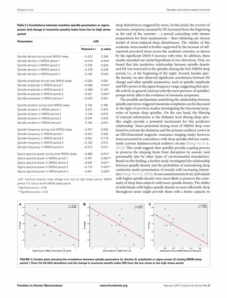
Given that PSQI did not show significant increase in responseto academic stress, bivariate correlations were performed betweenspindle parameters or sigma power and ISI change only (Table 2).When examining spindle density, there was a significant negativecorrelation between spindle density during the first NREM sleepperiod and ISI change (Figure 3A), i.e., lower spindle density at thebeginning of the night was associated with higher increases in sleepcomplaints in response to academic stress. The correlation was notsignificant for spindle density during the whole night or duringany other NREM sleep period. When looking at the other spin-dle parameters, duration, and frequency did not correlate with ISIchange. Spindle amplitude, however, negatively correlated with ISIchange, when considering either the first (Figure 3B) or the thirdNREM sleep period. Finally, there was a significant negative cor-relation between sigma EEG power, for the whole night as well asfor each NREM sleep period, and ISI change. This correlation withsigma power was the most significant during the first NREM sleepperiod (Figure 3C). FIRST score at baseline was not correlatedwith ISI change (r =−0.10; p= 0.75).
DISCUSSIONTaken together these results indicate that spindle activity consti-tutes a predisposing factor for the future aggravation of insomniacomplaints in response to stress. They confirm our hypothesis thatlower spindle density is associated with a higher vulnerability to
FIGURE 2 | Evolution of spindle parameters and sigma power acrossthe four NREM sleep periods: (A) spindle density; (B) spindlemaximum amplitude; (C) spindle duration; (D) spindle frequency;(E) EEG spectral power in the sigma frequency band. All values wereextracted from C4–O2 EEG derivation. The dots represent the mean value,
and the bars show the standard error of the mean. There was a significantincrease of spindle density and duration across NREM sleep periods(one-way repeated measures ANOVA, p < 0.05), but there was nosignificant change of spindle amplitude, frequency, and sigma poweracross periods.
Frontiers in Human Neuroscience www.frontiersin.org February 2015 | Volume 9 | Article 68 | 4
Dang-Vu et al. Spindles and stress-related insomnia
Table 2 | Correlations between baseline spindle parameters or sigma
power and change in insomnia severity index from low to high stress
period.
Parameters ∆ISI
Pearson’s r p value
Spindle density during total NREM sleep −0.322 0.308
Spindle density in NREM period 1 −0.578 0.049*
Spindle density in NREM period 2 −0.156 0.628
Spindle density in NREM period 3 −0.378 0.226
Spindle density in NREM period 4 −0.192 0.549
Spindle amplitude during total NREM sleep −0.502 0.097
Spindle amplitude in NREM period 1 −0.588 0.044*
Spindle amplitude in NREM period 2 −0.486 0.109
Spindle amplitude in NREM period 3 −0.591 0.043*
Spindle amplitude in NREM period 4 −0.545 0.067
Spindle duration during total NREM sleep 0.104 0.748
Spindle duration in NREM period 1 0.010 0.975
Spindle duration in NREM period 2 0.136 0.672
Spindle duration in NREM period 3 −0.074 0.819
Spindle duration in NREM period 4 0.164 0.610
Spindle frequency during total NREM sleep −0.167 0.603
Spindle frequency in NREM period 1 −0.257 0.420
Spindle frequency in NREM period 2 −0.091 0.779
Spindle frequency in NREM period 3 −0.182 0.572
Spindle frequency in NREM period 4 −0.210 0.512
Sigma spectral power during total NREM sleep −0.685 0.014*
Sigma spectral power in NREM period 1 −0.761 0.004**
Sigma spectral power in NREM period 2 −0.597 0.041*
Sigma spectral power in NREM period 3 −0.710 0.010**
Sigma spectral power in NREM period 4 −0.631 0.028*
∆ISI, insomnia severity index change from low to high stress period; NREM
period 1–4, first to fourth NREM sleep period.
*Significance at p < 0.05.
**Significance at p < 0.01.
sleep disturbances triggered by stress. In this study, the severity ofinsomnia symptoms assessed by ISI increased from the beginningto the end of the semester – a period coinciding with intensepreparations for final examinations – thus validating our chosenmodel of stress-induced sleep disturbances. The validity of thisacademic stress model is further supported by the increase of self-reported perceived stress across the academic semester, as shownby the significant DASS-S increase with time. In addition, theseresults extended our initial hypothesis in two directions. First, wefound that this predictive relationship between spindle densityand ISI was restricted to the spindles during the first NREM sleepperiod, i.e., at the beginning of the night. Second, besides spin-dle density, we also observed significant correlations between ISIchange and other spindle parameters, such as spindle amplitudeand EEG power in the sigma frequency range, suggesting that spin-dle activity in general (and not only the mere presence of spindles)prospectively affects the evolution of insomnia symptoms.
The possible mechanisms underlying the relationship betweenspindle and stress-triggered insomnia complaints can be discussedin the light of previous studies investigating the functional prop-erties of human sleep spindles. On the one hand, the filteringof external information at the thalamic level during sleep spin-dles might provide a potential mechanism for this predictiverelationship. Tones presented during most of NREM sleep werefound to activate the thalamus and the primary auditory cortex inan EEG/functional magnetic resonance imaging study; however,tones presented in coincidence with sleep spindles did not consis-tently activate thalamocortical auditory circuits (Dang-Vu et al.,2011). This result suggests that spindles provide a gating processto preserve the sleeping brain from disruption by sounds (andpresumably also by other types of environmental stimulation).Based on this finding, a further study investigated the relationshipbetween spindle density and the probability of maintaining sleepcontinuity under presentation of sounds with increasing intensi-ties (Dang-Vu et al., 2010). At any sound intensity level, individualswith higher spindle density were more likely to preserve the conti-nuity of sleep than subjects with lower spindle density. The abilityof individuals with higher spindle density to more efficiently sleepthroughout noise might provide them with a better capacity to
FIGURE 3 | Scatter plots showing the correlations between spindle parameters (A, density; B, amplitude) or sigma power (C) during NREM sleepperiod 1 (from C4–O2 EEG derivation) and the change in insomnia severity index (ISI) from the low stress to the high stress period.
Frontiers in Human Neuroscience www.frontiersin.org February 2015 | Volume 9 | Article 68 | 5

Dang-Vu et al. Spindles and stress-related insomnia
resist sleep disturbances in response to stress. Exposure to acutestress is indeed known to enhance sensitivity to noise (Hassonet al., 2013), and is thus likely to increase vulnerability to soundsduring sleep. Through the gating mechanisms associated withspindles, individuals with higher spindle density might be in abetter position to counter the deleterious consequences of stresson noise sensitivity during sleep, leading to a lower propensity toinsomnia complaints in response to stress. Future studies investi-gating sleep spindles in relation to acoustic stimulation in periodsof high stress are needed to support this interpretation.
Spindles have also been shown associated with a variety of cog-nitive measures. Higher number of spindles and higher EEG sigmapower have been shown positively correlated with better percep-tual and analytical skills measured by the performance intellectualquotient (Fogel et al., 2007), as well as with higher score at theRaven progressive matrices test reflecting general cognitive abil-ities (Bodizs et al., 2005; Schabus et al., 2006). These findingssuggest that spindles constitute a neurophysiological biomarker ofintellectual capacities. Beyond these correlations with broad scoresof cognitive abilities, previous studies also found that sleep spin-dle activity increased following procedural memory tasks such asmotor sequence learning (Barakat et al., 2011) and mirror tracingtask (Tamaki et al., 2008), as well as following declarative learn-ing tasks (word pairs) (Gais et al., 2002; Schabus et al., 2004),and these increases correlated with overnight improvements ofperformance. Therefore, an alternative explanation for the rela-tionship found in the present study is that individuals with higherspindle density or activity, given their higher cognitive abilities,might be more capable of efficiently learning their course materialsand thus managing academic stress, which might ultimately makethem less susceptible to sleep disturbances in such context. Bothmechanisms – sleep-protective and cognitive – are not mutuallyexclusive and might act synergistically to confer individuals withhigher spindle activity the ability to maintain sleep stability in theface of stress.
In this study, spindle density and other spindle parametersduring stages N2–N3 were computed separately for each NREMsleep period, in addition to their quantification throughout thewhole night. Indeed, spindle parameters do not remain constantthroughout the night (De Gennaro et al., 2000). We observed asignificant increase of spindle density and spindle duration overthe course of the night (particularly between NREM sleep period1 and each of the following NREM sleep periods). These observa-tions are in line with previous studies showing increases of spindledensity and duration across successive NREM sleep periods (DeGennaro et al., 2000; Martin et al., 2013). Interestingly, the modu-lation of spindle parameters throughout the night had an impacton the predictive relationships with sleep complaints, since spin-dle density only in the first NREM sleep period was significantlycorrelated with ISI change. This result suggests that sleep spindlesduring the early phase of the night have a predominant influenceon the perception of changes in insomnia complaints. The rea-sons for such effects of spindles during early night remain unclear.Interestingly, a differential effect of spindle activity as a functionof NREM sleep period was also observed in a study comparingspindle density between teenagers with major depressive disorder,teenagers at risk for depression and healthy controls (Lopez et al.,
2010). In contrast to our results, depressed and at-risk teenagershad lower spindle density than controls during the third and fourthNREM sleep period, but not at the beginning of the night. Thesevarious results suggest a differential clinical significance for spindleactivity depending on the corresponding NREM sleep period.
Our findings also demonstrated that, besides spindle density,other parameters reflecting spindle activity (spindle amplitude,sigma power) correlated with change of sleep quality in responseto stress. The correlations with spindle amplitude and EEG sigmapower suggest that spindle activity (i.e., intensity) – reflectingthe degree of thalamocortical synchronization – also modulatesthe perception of stress-induced sleep disturbances. Other stud-ies investigating the functional properties of sleep spindles alsoresorted to measures of spindle intensity: EEG sigma power cor-related with scores of intellectual abilities (Fogel et al., 2007), andspindle amplitude modulated the reactivation during sleep spin-dles of brain regions involved in the encoding of a declarativelearning task (Bergmann et al., 2012). In our study, correlationbetween sigma power and stress-induced sleep complaints wassignificant for all NREM sleep periods, while it was limited to thefirst NREM sleep period for spindle density (as well as the third forspindle amplitude). Such discrepancies between the effects of spin-dle density compared to those of sigma power were also observedin previous studies (Gais et al., 2002), which further underlinesthat EEG spectral power in the sigma frequency range cannotbe fully equated to the detection of sleep spindles as discreteevents. Indeed, sigma power also captures activities that do notmeet the standard criteria for full-blown sleep spindles, and thusconstitutes a more sensitive (but less specific) method for spin-dle quantification. Taken together these effects of spindle density,amplitude, and sigma power suggest that, although the contri-bution of spindles during early night seems more predominant,spindle activity throughout the whole night affects the perceptionof stress-induced changes in sleep quality.
If lower spindle density (and activity) constitutes a predisposingfactor for the surge of insomnia complaints, it could be expectedthat chronic insomniacs would tend to demonstrate lower spindlemeasures compared to good sleepers. A previous study analyzedthe differences in spindle density between chronic primary insom-niacs and good sleepers, by performing a visual detection of sleepspindles on C3 derivation (Bastien et al., 2009). Surprisingly, nosignificant difference in spindle density was found between groups.Further studies are needed to replicate this result, and to extendit to other (more sensitive) modalities of spindle detection as wellas to other measures of spindle activity such as spindle amplitudeand sigma power. If confirmed, the absence of change in spindleactivity in chronic insomniacs as a group might reflect the largeheterogeneity in the clinical presentation and sleep characteris-tics even within primary insomniacs. For instance, the presenceof objective sleep disturbances, as defined by PSG decreases intotal sleep time and sleep efficiency, is not observed consistentlyacross chronic insomniacs (Vgontzas et al., 1994). However, thepresence of objective short sleep duration defines a subgroup ofinsomniacs with a distinct clinical profile exemplified by a higherrisk for hypertension, diabetes, cognitive impairment, and mor-tality (Vgontzas et al., 2013). Likewise, it is possible that there isa subgroup of insomniacs characterized by a higher vulnerability
Frontiers in Human Neuroscience www.frontiersin.org February 2015 | Volume 9 | Article 68 | 6

Dang-Vu et al. Spindles and stress-related insomnia
to environmental disturbances due to a lesser amount of sleep-protective factors such as sleep spindles. In contrast, anothersubgroup might instead be characterized by less objective sleepdisruption and a larger contribution of cognitive-emotional fac-tors such as dysfunctional beliefs about sleep and higher levels ofanxiety and worry. Considering the chronic insomnia populationas a single group might dilute the alterations of sleep microarchi-tecture that possibly affect a subpopulation of insomniacs only.Future studies should further explore the quantification of sleep-protective mechanisms in chronic insomniacs and subgroups ofinsomniacs.
While the current study was primarily focused on the neuro-physiological predictors of sleep disturbances through the assess-ment of spindle activity, it should be reminded that psychologicaland medical factors also play an important role in the incidence ofinsomnia complaints. For instance, mental health problems, mal-adaptive personality traits, a positive family history of insomnia,and an objectively shorter sleep duration on PSG were associatedwith a higher risk of evolution of poor sleep toward chronic insom-nia (Fernandez-Mendoza et al., 2012). As for insomnia complaintsin response to stress, specific questionnaires of vulnerability tostress-induced insomnia have been developed, such as the FIRST(Drake et al., 2004). Surprisingly, the FIRST score did not predictchanges in ISI from low to high stress periods in our analysis. Thismight be explained by the high correlation between the FIRSTscore and ISI at baseline in this sample (rFIRST-ISI= 0.86), leavinglittle room to predict change over time. Nevertheless, it has beenpreviously shown that individuals with higher score on the FIRST(Drake et al., 2004) are more vulnerable to the first night effect(i.e., worse sleep quality during the first night of sleep recordingin lab) and to the sleep-disrupting effects of caffeine (Drake et al.,2004, 2006), and demonstrate higher risk of developing persistentinsomnia over time (Drake et al., 2014; Jarrin et al., 2014). Inter-estingly, the differential vulnerability to stress-induced insomniamay emerge from differences in hyperarousal predisposition, giventhe association between FIRST scores and indices of cognitive-emotional hyperarousal (Fernandez-Mendoza et al., 2010), whichhas recently been demonstrated to be (at least partially) heri-table (Fernandez-Mendoza et al., 2014). Because sleep spindlesmodulate sleep stability in response to environmental stimulation(Dang-Vu et al., 2010, 2011), lower spindle activity – which hasbeen found in the present study to predispose to higher increaseof sleep disturbances – might be considered as a trait predispos-ing to a state of neurobiological hyperarousal in which individualsare more vulnerable to externally driven sleep disruption. There-fore, these various findings on the vulnerability to stress-inducedinsomnia can be integrated within the framework of the hyper-arousal model for insomnia viewed from a psychophysiologicalperspective (Riemann et al., 2010).
There are several limitations to the current study. First, largersamples are needed to confirm these findings. Due to the lim-ited number of participants, correction for multiple comparisonswas not applied in the present data set, and thus our findingsneed replication. Second, only undergraduate university studentswere included in the present study due to the need of a naturallyoccurring stressor encompassing well-defined periods of lowerand higher stress, as provided by the model of academic stress.
Future studies should extend these findings to other populationsand other types of stressors, including chronic stressors that mayimpact the persistence of insomnia complaints over time. Third,assessment of sleep quality and insomnia complaints was eval-uated through self-reported questionnaires only: ISI and PSQI.The absence of significant PSQI change across the semester in ourstudy might indicate that the impact of academic stress on sleeppredominantly affects insomnia complaints rather than generalsleep quality. Furthermore, the nature of the stressor (academicstress) precluded the repetition of objective sleep measurementswith PSG, given the difficulty of having participants coming atthe sleep laboratory during busy periods of final examinations.The use of more practical objective measures of sleep such asactigraphy measurements might constitute an interesting comple-ment in future studies, in order to obtain not only objective butalso prolonged assessments of sleep over several days or weeks.Finally, we restricted our analyses to spindle activity over centralderivations (C4), given the centroparietal predominance of sleepspindle activity (De Gennaro et al., 2000). In order to avoid addi-tional comparisons in our limited sample, distinction between fastand slow spindles was not performed in this present analysis. Ourresults, however, suggest that the frequency of spindles did notaffect the change in insomnia symptoms, given the absence ofcorrelation between spindle frequency and ISI change (Table 2).Future studies on larger samples could further evaluate the roleof spindle frequency on sleep quality changes by analyzing therole of slow and fast spindles separately. The study of other EEGoscillations during sleep might also be of interest given previ-ous results indicating the contribution of brain oscillations inother frequency bands, such as alpha rhythms (McKinney et al.,2011) and slow wave activity (Dang-Vu et al., 2011; Schabus et al.,2012), to the preservation of sleep continuity in the face of externalstimulation.
CONCLUSIONOur study provides the first evidence for the contribution of sleepneurophysiological activity to the prospective increase of sleepdisturbances in response to a standardized stressor in a sampleof young healthy volunteers. In line with previous findings indi-cating that sleep spindle constitutes a biomarker of sleep stability,our results suggest that spindle activity also represents a predis-posing factor modulating the vulnerability to sleep disruption inconditions of stress. These results have implications for the under-standing of the neural mechanisms underlying the evolution ofsleep disturbances and particularly insomnia. They might alsohave clinical implications, by providing a biomarker for the iden-tification of individuals at risk for future sleep disruption. Finally,our findings emphasize the potential importance of future thera-peutic interventions aimed at enhancing sleep spindle activity inorder to preserve sleep quality.
AUTHOR CONTRIBUTIONSTDV and JPG designed the study. AS, SB, KW, and JOB acquiredthe data. TDV, AS, SB, MB, CB, and JG analyzed the data. TDV,AS, SB, and JPG interpreted the results. TDV wrote the manu-script. AS prepared the tables and figures. All the authors revised
Frontiers in Human Neuroscience www.frontiersin.org February 2015 | Volume 9 | Article 68 | 7
Dang-Vu et al. Spindles and stress-related insomnia
and commented the manuscript, gave their final approval of themanuscript, and agree to be accountable for all aspects of the work.
ACKNOWLEDGMENTSThis research was conducted in the absence of any commercialor financial relationships that could be construed as a poten-tial conflict of interest. The authors thank Ms. Ruby Bedi, Ms.Marilia Bedendi, Ms. Neressa Noel, and the Clinique SommeilSanté for their contribution to the setup and scoring of sleeprecordings. Dr. TDV receives research support from the Cana-dian Institutes of Health Research (CIHR), the Natural Sciencesand Engineering Research Council of Canada (NSERC), the Fondsde Recherche du Québec – Santé (FRQ-S), the Canada Founda-tion for Innovation (CFI), the Sleep Research Society Foundation(SRSF), the Fonds Québécois de Recherche sur le Vieillissement(RQRV), the Institut Universitaire de Gériatrie de Montréal, andConcordia University. Dr. JPG receives research support from theCanada Research Chair program, the CFI, the CIHR, and the SocialSciences and Humanities Research Council of Canada (SSHRC).Mr. JOB receives scholarship support from the CIHR.
SUPPLEMENTARY MATERIALThe Supplementary Material for this article can be found online athttp://www.frontiersin.org/Journal/10.3389/fnhum.2015.00068/abstract
REFERENCESAPA. (2013). Diagnostic and Statistical Manual of Mental Disorders, 5th Edn. Arling-
ton, VA: American Psychiatric Publishing.Barakat, M., Doyon, J., Debas, K., Vandewalle, G., Morin, A., Poirier, G., et al. (2011).
Fast and slow spindle involvement in the consolidation of a new motor sequence.Behav. Brain Res. 217, 117–121. doi:10.1016/j.bbr.2010.10.019
Bastien, C. H., St-Jean, G., Turcotte, I., Morin, C. M., Lavallee, M., and Carrier, J.(2009). Sleep spindles in chronic psychophysiological insomnia. J. Psychosom.Res. 66, 59–65. doi:10.1016/j.jpsychores.2008.05.013
Bastien, C. H., Vallieres, A., and Morin, C. M. (2001). Validation of the insom-nia severity index as an outcome measure for insomnia research. Sleep Med. 2,297–307. doi:10.1016/S1389-9457(00)00065-4
Bastien, C. H., Vallieres, A., and Morin, C. M. (2004). Precipitating factors of insom-nia. Behav. Sleep Med. 2, 50–62. doi:10.1207/s15402010bsm0201_5
Bergmann, T. O., Molle, M., Diedrichs, J., Born, J., and Siebner, H. R. (2012). Sleepspindle-related reactivation of category-specific cortical regions after learningface-scene associations. Neuroimage 59, 2733–2742. doi:10.1016/j.neuroimage.2011.10.036
Berthomier, C., Drouot, X., Herman-Stoica, M., Berthomier, P., Prado, J., Bokar-Thire, D., et al. (2007). Automatic analysis of single-channel sleep EEG: valida-tion in healthy individuals. Sleep 30, 1587–1595.
Bodizs, R., Kis, T., Lazar, A. S., Havran, L., Rigo, P., Clemens, Z., et al. (2005). Pre-diction of general mental ability based on neural oscillation measures of sleep. J.Sleep Res. 14, 285–292. doi:10.1111/j.1365-2869.2005.00472.x
Buysse, D. J., Reynolds, C. F. III, Monk, T. H., Berman, S. R., and Kupfer, D. J. (1989).The Pittsburgh sleep quality index: a new instrument for psychiatric practice andresearch. Psychiatry Res. 28, 193–213. doi:10.1016/0165-1781(89)90047-4
Cote, K. A., Epps, T. M., and Campbell, K. B. (2000). The role of the spindle inhuman information processing of high-intensity stimuli during sleep. J. SleepRes. 9, 19–26. doi:10.1046/j.1365-2869.2000.00188.x
Dang-Vu, T. T., Bonjean, M., Schabus, M., Boly, M., Darsaud, A., Desseilles, M., et al.(2011). Interplay between spontaneous and induced brain activity during humannon-rapid eye movement sleep. Proc. Natl. Acad. Sci. U.S.A. 108, 15438–15443.doi:10.1073/pnas.1112503108
Dang-Vu, T. T., McKinney, S. M., Buxton, O. M., Solet, J. M., and Ellenbogen, J. M.(2010). Spontaneous brain rhythms predict sleep stability in the face of noise.Curr. Biol. 20, R626–R627. doi:10.1016/j.cub.2010.06.032
De Gennaro, L., and Ferrara, M. (2003). Sleep spindles: an overview. Sleep Med. Rev.7, 423–440. doi:10.1053/smrv.2002.0252
De Gennaro, L., Ferrara, M., and Bertini, M. (2000). Topographical distribution ofspindles: variations between and within NREM sleep cycles. Sleep Res. Online 3,155–160. doi:10.1046/j.1365-2869.2000.00193.x
De Gennaro, L., Ferrara, M., Vecchio, F., Curcio, G., and Bertini, M. (2005). Anelectroencephalographic fingerprint of human sleep. Neuroimage 26, 114–122.doi:10.1016/j.neuroimage.2005.01.020
Drake, C., Richardson, G., Roehrs, T., Scofield, H., and Roth, T. (2004). Vulnerabilityto stress-related sleep disturbance and hyperarousal. Sleep 27, 285–291.
Drake, C. L., Jefferson, C., Roehrs, T., and Roth, T. (2006). Stress-related sleep dis-turbance and polysomnographic response to caffeine. Sleep Med. 7, 567–572.doi:10.1016/j.sleep.2006.03.019
Drake, C. L., Pillai, V., and Roth, T. (2014). Stress and sleep reactivity: a prospectiveinvestigation of the stress-diathesis model of insomnia. Sleep 37, 1295–1304.doi:10.5665/sleep.3916
Feinberg, I., and Floyd, T. C. (1979). Systematic trends across the night inhuman sleep cycles. Psychophysiology 16, 283–291. doi:10.1111/j.1469-8986.1979.tb02991.x
Fernandez-Mendoza, J., Shaffer, M. L., Olavarrieta-Bernardino, S., Vgontzas, A. N.,Calhoun, S. L., Bixler, E. O., et al. (2014). Cognitive-emotional hyperarousal inthe offspring of parents vulnerable to insomnia: a nuclear family study. J. SleepRes. 23, 489–498. doi:10.1111/jsr.12168
Fernandez-Mendoza, J., Vela-Bueno, A., Vgontzas, A. N., Ramos-Platon, M. J.,Olavarrieta-Bernardino, S., Bixler, E. O., et al. (2010). Cognitive-emotionalhyperarousal as a premorbid characteristic of individuals vulnerable to insom-nia. Psychosom. Med. 72, 397–403. doi:10.1097/PSY.0b013e3181d75319
Fernandez-Mendoza, J., Vgontzas, A. N., Bixler, E. O., Singareddy, R., Shaffer, M.L., Calhoun, S. L., et al. (2012). Clinical and polysomnographic predictors ofthe natural history of poor sleep in the general population. Sleep 35, 689–697.doi:10.5665/sleep.1832
Fogel, S. M., Nader, R., Cote, K. A., and Smith, C. T. (2007). Sleepspindles and learning potential. Behav. Neurosci. 121, 1–10. doi:10.1037/0735-7044.121.1.1
Fogel, S. M., and Smith, C. T. (2011). The function of the sleep spindle: a phys-iological index of intelligence and a mechanism for sleep-dependent memoryconsolidation. Neurosci. Biobehav. Rev. 35, 1154–1165. doi:10.1016/j.neubiorev.2010.12.003
Gaillard, J. M., and Blois, R. (1981). Spindle density in sleep of normal subjects.Sleep 4, 385–391.
Gais, S., Molle, M., Helms, K., and Born, J. (2002). Learning-dependent increases insleep spindle density. J. Neurosci. 22, 6830–6834.
Harvey, C. J., Gehrman, P., and Espie, C. A. (2014). Who is predisposed to insomnia:a review of familial aggregation, stress-reactivity, personality and coping style.Sleep Med. Rev. 18, 237–247. doi:10.1016/j.smrv.2013.11.004
Hasson, D., Theorell, T., Bergquist, J., and Canlon, B. (2013). Acute stress induceshyperacusis in women with high levels of emotional exhaustion. PLoS ONE8:e52945. doi:10.1371/journal.pone.0052945
Iber, C., Ancoli-Israel, S., Chesson, A. L., and Quan, S. F. (2007). The AASM Manualfor the Scoring of Sleep and Associated Events. Westchester, NY: American Academyof Sleep Medicine.
Jarrin, D. C., Chen, I. Y., Ivers, H., and Morin, C. M. (2014). The role of vul-nerability in stress-related insomnia, social support and coping styles on inci-dence and persistence of insomnia. J. Sleep Res. 23, 681–688. doi:10.1111/jsr.12172
Jernelov, S., Hoglund, C. O., Axelsson, J., Axen, J., Gronneberg, R., Grunewald, J.,et al. (2009). Effects of examination stress on psychological responses, sleep andallergic symptoms in atopic and non-atopic students. Int. J. Behav. Med. 16,305–310. doi:10.1007/s12529-008-9020-6
LeBlanc, M., Merette, C., Savard, J., Ivers, H., Baillargeon, L., and Morin, C. M.(2009). Incidence and risk factors of insomnia in a population-based sample.Sleep 32, 1027–1037.
Lopez, J., Hoffmann, R., and Armitage, R. (2010). Reduced sleep spindle activity inearly-onset and elevated risk for depression. J. Am. Acad. Child Adolesc. Psychiatry49, 934–943. doi:10.1016/j.jaac.2010.05.014
Lovibond, P. F., and Lovibond, S. H. (1995). The structure of negative emo-tional states: comparison of the depression anxiety stress scales (DASS) withthe beck depression and anxiety inventories. Behav. Res. Ther. 33, 335–343.doi:10.1016/0005-7967(94)00075-U
Frontiers in Human Neuroscience www.frontiersin.org February 2015 | Volume 9 | Article 68 | 8

Dang-Vu et al. Spindles and stress-related insomnia
Lund, H. G., Reider, B. D., Whiting, A. B., and Prichard, J. R. (2010). Sleep patternsand predictors of disturbed sleep in a large population of college students. J.Adolesc. Health 46, 124–132. doi:10.1016/j.jadohealth.2009.06.016
Martin, N., Lafortune, M., Godbout, J., Barakat, M., Robillard, R., Poirier, G., et al.(2013). Topography of age-related changes in sleep spindles. Neurobiol. Aging34, 468–476. doi:10.1016/j.neurobiolaging.2012.05.020
McKinney, S. M., Dang-Vu, T. T., Buxton, O. M., Solet, J. M., and Ellenbogen, J.M. (2011). Covert waking brain activity reveals instantaneous sleep depth. PLoSONE 6:e17351. doi:10.1371/journal.pone.0017351
Morin, A., Doyon, J., Dostie, V., Barakat, M., Hadj Tahar, A., Korman, M., et al.(2008). Motor sequence learning increases sleep spindles and fast frequencies inpost-training sleep. Sleep 31, 1149–1156.
Riemann, D., Spiegelhalder, K., Feige, B., Voderholzer, U., Berger, M., Perlis, M., et al.(2010). The hyperarousal model of insomnia: a review of the concept and itsevidence. Sleep Med. Rev. 14, 19–31. doi:10.1016/j.smrv.2009.04.002
Schabus, M., Dang-Vu, T. T., Albouy, G., Balteau, E., Boly, M., Carrier, J., et al.(2007). Hemodynamic cerebral correlates of sleep spindles during human non-rapid eye movement sleep. Proc. Natl. Acad. Sci. U.S.A. 104, 13164–13169.doi:10.1073/pnas.0703084104
Schabus, M., Dang-Vu, T. T., Heib, D. P., Boly, M., Desseilles, M., Vandewalle,G., et al. (2012). The fate of incoming stimuli during NREM sleep is deter-mined by spindles and the phase of the slow oscillation. Front. Neurol. 3:40.doi:10.3389/fneur.2012.00040
Schabus, M., Gruber, G., Parapatics, S., Sauter, C., Klosch, G., Anderer, P., et al.(2004). Sleep spindles and their significance for declarative memory consolida-tion. Sleep 27, 1479–1485.
Schabus, M., Hodlmoser, K., Gruber, G., Sauter, C., Anderer, P., Klosch, G.,et al. (2006). Sleep spindle-related activity in the human EEG and its rela-tion to general cognitive and learning abilities. Eur. J. Neurosci. 23, 1738–1746.doi:10.1111/j.1460-9568.2006.04694.x
Spielman, A. J. (1986). Assessment of insomnia. Clin. Psychol. Rev. 6, 11–25.doi:10.1016/0272-7358(86)90015-2
Steriade, M., and McCarley, R. W. (2005). Brain Control of Wakefulness and Sleep.New York, NY: Springer.
Tamaki, M., Matsuoka, T., Nittono, H., and Hori, T. (2008). Fast sleep spindle (13-15 Hz) activity correlates with sleep-dependent improvement in visuomotorperformance. Sleep 31, 204–211.
Vgontzas, A. N., Bixler, E. O., Kales, A., Manfredi, R. L., and Tyson, K. (1994). Validityand clinical utility of sleep laboratory criteria for insomnia. Int. J. Neurosci. 77,11–21. doi:10.3109/00207459408986015
Vgontzas, A. N., Fernandez-Mendoza, J., Liao, D., and Bixler, E. O. (2013).Insomnia with objective short sleep duration: the most biologically severephenotype of the disorder. Sleep Med. Rev. 17, 241–254. doi:10.1016/j.smrv.2012.09.005
Conflict of Interest Statement: The authors declare that the research was conductedin the absence of any commercial or financial relationships that could be construedas a potential conflict of interest.
Received: 28 November 2014; accepted: 27 January 2015; published online: 10 February2015.Citation: Dang-Vu TT, Salimi A, Boucetta S, Wenzel K, O’Byrne J, Brandewinder M,Berthomier C and Gouin J-P (2015) Sleep spindles predict stress-related increases insleep disturbances. Front. Hum. Neurosci. 9:68. doi: 10.3389/fnhum.2015.00068This article was submitted to the journal Frontiers in Human Neuroscience.Copyright © 2015 Dang-Vu, Salimi, Boucetta, Wenzel, O’Byrne, Brandewinder,Berthomier and Gouin. This is an open-access article distributed under the termsof the Creative Commons Attribution License (CC BY). The use, distribution or repro-duction in other forums is permitted, provided the original author(s) or licensor arecredited and that the original publication in this journal is cited, in accordance withaccepted academic practice. No use, distribution or reproduction is permitted whichdoes not comply with these terms.
Frontiers in Human Neuroscience www.frontiersin.org February 2015 | Volume 9 | Article 68 | 9