Curcumin derivatives: Molecular basis of their anti-cancer activity

Journal of Colloid and Interface Science 360 (2011) 39–51
Contents lists available at ScienceDirect
Journal of Colloid and Interface Science
www.elsevier .com/locate / jc is
Curcumin-loaded biocompatible thermoresponsive polymeric nanoparticles forcancer drug delivery
N. Sanoj Rejinold a, M. Muthunarayanan a, V.V. Divyarani a, P.R. Sreerekha a, K.P. Chennazhi a, S.V. Nair a,H. Tamura b, R. Jayakumar a,⇑a Amrita Centre for Nanosciences and Molecular Medicine, Amrita Institute of Medical Sciences and Research Centre, Amrita Vishwa Vidyapeetham University, Kochi 682 041, Indiab Faculty of Chemistry, Materials and Bioengineering, Kansai University, Osaka-564-8680, Japan
a r t i c l e i n f o
Article history:Received 14 December 2010Accepted 2 April 2011Available online 14 April 2011
Keywords:ChitosanThermoresponsive nanoparticlesLCSTCurcuminSpecific toxicityCancer drug delivery
0021-9797/$ - see front matter � 2011 Elsevier Inc. Adoi:10.1016/j.jcis.2011.04.006
⇑ Corresponding author. Fax: +91 484 2802020.E-mail addresses: [email protected],
(R. Jayakumar).
a b s t r a c t
This study aims at the formulation of curcumin with biodegradable thermoresponsive chitosan-g-poly(N-vinylcaprolactam) nanoparticles (TRC-NPs) for cancer drug delivery. The spherical curcumin-loadednanoparticles of size 220 nm were characterized, and the biological properties were studied using flowcytometry and cytotoxicity by MTT assay. The in vitro drug release was higher at above LCST comparedto that at below LCST. TRC-NPs in the concentration range of 100–1000 lg/mL were non-toxic to an arrayof cell lines. The cellular localization of the curcumin-loaded TRC-NPs was confirmed from green fluores-cence inside the cells. The time-dependent curcumin uptake by the cells was quantified by UV spectro-photometer. Curcumin-loaded TRC-NPs showed specific toxicity to cancer cells at above their LCST. Flowcytometric analysis showed increased apoptosis on PC3 compared to L929 by curcumin-loaded TRC-NPs.These results indicate that novel curcumin-loaded TRC-NPs could be a promising candidate for cancerdrug delivery.
� 2011 Elsevier Inc. All rights reserved.
1. Introduction
The limitations of current therapy provide a compelling ratio-nale for the development of alternative modalities for the targeteddelivery of therapeutics for the treatment of solid tumors [1]. Anexciting potential solution in cancer treatments is to encapsulatethe drug in a biocompatible material that can be injected intothe blood stream with the intention of delivering drug to a tumorsite in response to an external thermal source like radio frequency(RF) generator, which is the source that provides radio waves toheat and kill cancer cells [2]. Chitosan is a well-known biopolymerhaving many applications in tissue engineering [3–6], wound heal-ing [7], drug delivery [8–11], and also in gene delivery [12,13]. Inrecent years, much interest has been given on stimuli-sensitivepolymeric systems that show a phase transition in response toexternal stimulus such as temperature, pH, specific ion, and elec-tric field [14]. Among all intelligent polymers studied, tempera-ture- and pH-responsive polymeric systems have drawn muchmore attention, because these are the important environmentalfactors in the body, and some disease states manifest themselvesby a change in temperature and/ or pH. In recent years, several re-search groups have reported the preparation of pH- and tempera-
ll rights reserved.
ture-sensitive polymers based on the poly (N-isopropylacrylamide)(PNIPAAm) for biomedical applications [15]. Poly (N-vinyl capro-lactam) (PNVCL) is another well-studied polymer that shows agood response toward temperature. The actual interest in PNVCLis connected with its thermoresponsive nature, complexation abil-ity, and biocompatibility. The utility of these polymers as microdrug delivery carriers has been well explored. Presently, thereare no studies concerning the nanoformulation of chitosan-g-PNVCL with curcumin loading for cancer therapy.
Curcumin, a naturally occurring polyphenolic phytoconstituent,possesses anticancer, antioxidant, anti-inflammatory, hyperlipi-demic, antibacterial, wound healing, and hepatoprotective activi-ties [16–18]. The therapeutic efficacy of curcumin is limited dueto its poor oral bioavailability [19], which has been attributed toits poor aqueous solubility and extensive first-pass metabolism.Various attempts have been made through encapsulation in poly-meric nanoparticles, but no work has been reported in thermore-sponsive polymeric nanomaterials. Highly stable formulations arerequired to take full advantage of the EPR effect in treating solid tu-mors [20], in maximizing the duration of exposure, and for thermaltargeting using hyperthermia to direct the drug-encapsulated TRC-NPs to target cancer cells where the drug can then be releasedlocally. Since curcumin is specific to treat cancer cells, more effica-cies would be achieved by utilizing the LCST of carrier system.Nanoscale drug delivery vehicles formulated from biocompatiblechitosan and biodegradable thermoresponsive polymers constitute
40 N. Sanoj Rejinold et al. / Journal of Colloid and Interface Science 360 (2011) 39–51
an evolving approach to drug delivery and tumor targeting [21,22].Biodegradable thermoresponsive drug carriers are being purposelyengineered and constructed with nanometer dimensions. Such ap-proaches made it possible to develop smart materials like thermo-responsive drug delivery vehicles.
Here, we are explaining the formulation of the highly stablethermoresponsive nanoconstructs of curcumin using chitosan-g-PNVCL and stabilization chemistries, their characterization,in vitro cell uptake studies and toxicity in detail. In addition, effectsof this formulation on cell viability and apoptosis of both cancerand normal cells were investigated.
2. Materials and methods
2.1. Materials
Chitosan (Viscosity average molecular weight 20 kDa, Degree ofN-deacetylation (75–80%) was purchased from Koyochitosan Com-pany, Japan, and used as received. N-vinylcaprolactam (NVCL) waspurchased from Sigma Aldrich and recrystalized from n hexane be-fore use. Azo bis isobutyronitrile (AIBN), 1-ethyl-3-(3-dimethyl-aminopropyl) carbodiimide (EDC) and N-hydroxy succinimidde(NHS), penta sodium tripoly phosphate (TPP) were purchased fromSigma Aldrich and used without further purification. Isopropylalcohol and 3-mercaptopropionic acid (MPA) were supplied by Al-drich and used as supplied. Curcumin was purchased from Merk,Cochin, India. Acid dextrose citrate, sodium bicarbonate, and so-dium chloride were purchased from Merk, Cochin for blood com-patibility studies.
2.2. Synthesis of blank thermoresponsive chitosan-g-poly (N-vinylcaprolactam) nanoparticles (TRC-NPs)
The chitosan-g-poly (N-vinylcaprolactam) was prepared accord-ing to the reported method with modifications [14]. Chitosan-grafted polymer (1:9 ratio polymer, LCST 38 �C) was dissolved inacetic acid (1% solution), and the resulting solution was cross-linked with (sodium tripoly phosphate) TPP (Sigma Aldrich) in1:1 ratio. The resulting nanoformulation was centrifuged at30,000 rpm for 45 min and re-suspended and pelletized severaltimes in water until the pH became 7.4 (see Fig. 1).
2.3. Synthesis of Curcumin-loaded TRC-NPs
Curcumin-loaded TRC-NPs were prepared by a simple ioniccross-linking method by TPP via an aqueous chemistry route.Briefly, curcumin (5 mg in 1 ml ethanol) and polymer (50 mg in10 ml 1% acetic acid) were stirred for 5 min at low rpm. Further,the whole system was mixed with 100 ll solution of TPP and stir-red for 20 min at low rpm. The nanoparticle suspension was thencentrifuged at 21,000 rpm for about 30 min, and the residue wasre-suspended in water till the pH become 7.4. The loading chemis-try has been shown in Fig. 2.
2.4. Characterizations
FT-IR spectra of chitosan, N-vinylcaprolactam (NVCL), PNVCL-COOH, thermoresponsive chitosan-grafted poly (N-vinyl caprolac-tam) (chitosan-g-PNVCL), TRC-NPs, curcumin, curcumin-loadedTRC-NPs were carried out using KBr tablets (1% w/w of productin KBr) with a resolution of 4 cm�1 and 100 scans per sample ona Perkin Elmer Spectrum RX1 apparatus. Thermal studies weredone by (SII TG/DTA 6200 EXSTAR). The thermal stability and ther-mal decomposition of prepared systems were investigated usingTG/DTA. The temperature scans started from 25 to 500 �C at a rate
of 10 �C/min. The patterns of pure curcumin, TRC-NPs, and curcu-min-loaded TRC-NPs were obtained using the X-ray diffractometer(PANalytical X’Pert PRO) with Cu source of radiation. Measure-ments were taken at voltage of 40 kV and 25 mA. The scanned an-gle was set from 3� 6 2h P 40�, and the scan rate was 2�min�1. Theparticle size was measured by Dynamic light scattering (DLS-ZP /Particle Sizer Nicomp™ 380 ZLS) taking the average of 3 measure-ments. The surface morphology of nanoparticles was analyzed bySEM (JEOLJSM-6490LA) and AFM (JEOL JSPM-5200). The LCSTparameter of the systems was analyzed by UV spectrophotometer(Pharma spec) system starting from a temperature range of 0–45 �C and an average of three values were taken as the LCST ofthe synthesized materials.
2.5. Loading efficiency
The percentage of drug incorporated during nanoparticle prep-aration was determined by centrifuging the drug-loaded nanopar-ticles at 20,000 rpm for 30 min and separated the supernatant. Thesupernatant was assayed by UV spectrophotometer (UV-1700Pharma Spec) at 428 nm by dissolving in ethanol. The calculatedloading efficiency was 95% using the following formula:
Loading efficiency ¼ ðTotal amount of curcumin-Free curcuminÞTotal amount of curcumin
� 100
2.6. In vitro quantification of curcumin
For in vitro quantification of curcumin, a standard solution ofcurcumin in ethanol was prepared by dissolving 5 mg of curcuminin 100 ml ethanol solution. A serial dilution from 0.2 to 2 ml wastaken and diluted up to 25 ml and assayed the system at 428 nmusing (UV-1700 Pharma Spec) UV spectrophotometer. The datawere plotted to get a straight line for the quantification of un-known drug in the nanoparticle.
2.7. In vitro drug release
A known amount of lyophilized TRC-NPs (50 mg) encapsulatingcurcumin was dispersed in 10 ml phosphate buffer, pH 7.4, and thesolution was divided into 30 epppendorf tubes (500 ll each). Thetubes were kept in a thermostable water bath set at two differenttemperatures as 25 �C (below LCST) and 40 �C (above LCST). Freecurcumin is completely insoluble in water; therefore, at predeter-mined time intervals, the solution was centrifuged at 5000 rpm for7 min to separate the released curcumin (which will be in a pelletform) from the loaded TRC-NPs. The released curcumin was redis-solved in 3 ml ethanol to assay spectrophotometrically at 428 nm.The concentration of released drug was then calculated using stan-dard curve of curcumin in ethanol. The percentage of curcumin re-leased was determined from the following equation
Release ð%Þ ¼ Released curcumin from TRC� NPs
Total amount of curcumin in TRC� NPs� 100
2.8. Cell culture
MCF 7 (Human Breast cancer cell line, NCCS Pune), KB (oral can-cer cell line, NCCS Pune), and L929 (mouse fibroblast cell line, NCCSPune) were maintained in Minimum Essential Medium (MEM)supplemented with 10% fetal bovine serum (FBS). PC3 (prostatecancer cell line, NCCS Pune) was maintained in Dulbecco’s modi-fied Eagles Medium (DMEM-F12) supplemented with 10% fetal bo-vine serum (FBS). The cells were incubated in CO2 incubator with

Fig. 1. Possible mechanisms for the formulation of TRC-NPs (A) TPP cross-linking with protonated amide groups and (B) TPP cross-linking reaction with the protonatedresidual amine groups from chitosan.
N. Sanoj Rejinold et al. / Journal of Colloid and Interface Science 360 (2011) 39–51 41
5% CO2 at 37 �C. After attaining confluency, the cells were detachedfrom the flask with Trypsin–EDTA. The cell suspension was centri-fuged at 3000 rpm for 3 min and then re-suspended in the growthmedium for further studies. A separate study with nanoparticles atbelow (35 �C) and above (38 �C) LCST has been given in Supple-mentary section.
2.9. In vitro cell uptake studies by UV spectrophotometer
For in vitro drug quantification of curcumin-loaded TRC-NPswith PC3 and L929 cell lines, the cells were seeded on 24-wellplates with a seeding density of 50,000 cells/well. After attainingconfluency, wells were carefully washed with PBS buffer. Then,the particles at a concentration of 1 mg/ml were added along withthe media in triplicate to the wells and incubated at different timeintervals of 1, 3, 6, 9, 12, and 24 h, respectively, and processed forquantification of curcumin by UV analysis at 428 nm. The process-
ing involves pelletization of curcumin-loaded TRC-NPs with cells at1500 rpm for 5 min, drying and lysis of the pellet in methanol fol-lowed by probe sonication at higher amplitude for 5 min so thatcurcumin is extracted into the methanol fraction. The lysate wascentrifuged at 10,000 rpm for 5 min and absorption spectra ofsupernatant containing metabolic curcumin were recorded.
2.10. In vitro cell uptake studies by fluorescence microscopy
Acid-etched cover slips kept in 24-well plates were loaded withL929 and PC3 cells with a seeding density of 5000 cells per coverslip and incubated for 24 h for the cells to attach well. After the24-h incubation, the media was removed and the wells were care-fully washed with PBS buffer. Then, the particles at a concentrationof 1 mg/ml were added along with the media in triplicate to thewells and incubated for 4 h and processed for fluorescent micros-copy. The processing involved washing the cover slips with PBS

Fig. 2. Loading chemistry for curcumin on TRC-NPs. Inset (A) stable nanosuspension of curcumin-loaded TRC-NPs and (B) its nanopowder.
42 N. Sanoj Rejinold et al. / Journal of Colloid and Interface Science 360 (2011) 39–51
and thereafter fixing the cells in 3.7% paraformaldehyde (PFA) fol-lowed by a final PBS wash. The cover slips were air-dried andmounted onto glass slides with DPX as the mountant medium.The slides were then viewed under the fluorescence microscope(Olympus-BX-51).
2.11. Cellular uptake of curcumin-loaded nanoparticles by flowcytometry
Mouse fibroblast cell line (L929) and prostate cancer cell line(PC3) obtained from NCCS, Pune, were used for this study. Cellsin the log phase were seeded in a 24-well plate at a density of50,000 cells/cm2. Three different concentrations of the nanoparti-cles (0.2 mg/ml, 0.6 mg/ml, and1 mg/ml) were prepared by dilu-tion with the media. After attaining 90% confluency, the cellswere washed with PBS buffer, and different concentrations of thenanoparticles (100 ll) were added and incubated at 37 �C for24 h. Intracellular curcumin fluorescence was analyzed flow cyto-metrically after excitation with a 488-nm argon laser using FACSAria II (Beckton and Dickinson, Sanjose, CA). Fluorescence emissionabove 530 nm from 10,000 cells was collected, amplified, andscaled to generate single parameter histogram.
2.12. Cytotoxicity studies
For cytotoxicity experiments, L929 (mouse fibroblast cell line,NCCS Pune), MCF7 (Breast cancer cell line, NCCS Pune), PC3 (pros-tate cancer cell line, NCCS Pune), and KB (oral cancer cell line, NCCSPune) were seeded on a 96-well plate with a density of10,000 cells/cm2. MTT [3-(4,5-Dimethylthiazole-2-yl)-2,5-diphe-nyl tetrazolium] assay was used to evaluate cytotoxicity of the pre-pared nanoparticles, and this is a colorimetric test based on theselective ability of viable cells to reduce the tetrazolium compo-nent of MTT into purple-colored formazan crystals. Five differentconcentrations of the nanoparticles (0.2, 0.4, 0.6, 0.8, and 1 mg/ml) were prepared by dilution with the media. After attaining90% confluency, the cells were washed with PBS buffer, and differ-ent concentrations of the nanoparticles (100 ll) were added and
incubated. Cells in media alone devoid of nanoparticles acted asnegative control and wells treated with Triton X-100 as positivecontrol for a period of 24 h. Five milligrams of MTT (Sigma) wasdissolved in 1 ml of PBS and filter-sterilized. About10 ll of theMTT solution was further diluted to 100 ll with 90 ll of serum-free phenol red free medium. The cells were incubated with100 ll of the above solution for 4 h to form formazan crystals bymitochondrial dehydrogenases. About 100 ll of the solubilizationsolution (10% Triton X-100, 0.1 N HCl and isopropanol) was addedin each well and incubated at room temperature for 1 h to dissolvethe formazan crystals. The optical density of the solution was mea-sured at a wavelength of 570 nm using a Beckmann Coulter Elisaplate reader (BioTek Power Wave XS). Triplicate samples were ana-lyzed for each experiment.
2.13. Apoptosis assay by flow cytometry
2.13.1. Annexin V-FITC/PI stainingPhosphatidylserine (PS) translocation from the inner to the out-
er layer of plasma membrane is one of the important earliest apop-totic features. The PS exposure in both L929 and PC3 cells wasdetected using an Annexin V-FITC/PI Vybrant apoptosis assay kit(Molecular probes, Eugene, OR). After attaining 90% confluency,the cells were treated with three different concentrations of curcu-min nanoparticles as described previously. After being exposed tocurcumin nanoparticles for 24 h, cells were harvested by trypsin-ization and washed with ice-cold PBS for 5 min at 500 g at 4 �C.The supernatant was discarded and the pellet was re-suspendedin ice-cold 1X Annexin binding buffer (5 � 105–5 � 106 cells/ml).Five microliters of Annexin V–FITC solution and 1 ll of PI(100 lg/ml) were added to 100 ll of the cell suspensions. The sam-ples were mixed gently and incubated at room temperature for15 min in the dark. After incubation, 400 ll of ice-cold 1� bindingbuffer was added and mixed gently and analyzed by flow cytome-try. Cells in media alone devoid of nanoparticles (negative control)and cells treated with bare polymeric nanomaterials were alsoanalyzed in the same way. Triplicate samples were analyzed foreach experiment.
N. Sanoj Rejinold et al. / Journal of Colloid and Interface Science 360 (2011) 39–51 43
2.13.2. Mitochondrial membrane potential (DWm) assayJC-1 (5,50,6,60-tetrachloro-1,10,3,30-tetraethylbenzimidazolcar-
bocyanine iodide) is a lipophilic fluorochrome that is used to eval-uate the status of the DWm. Polarized mitochondria in healthy livecells form JC-1 aggregates which shows a red spectral shift, andthis is measured flow cytometrically to assess the DWm. Both JC-1 monomers and aggregates exhibit a green fluorescence that isalso exploited for the flow cytometric analysis of mitochondrialmembrane potential. Mitochondrial membrane potential was ana-lyzed flow cytometrically by mitochondrial membrane potentialdetection kit (BD Biosciences, San Diego, CA) according to manu-facturer’s protocol. Briefly, Cells at log phase were seeded in a24-well plate at a density of 50,000 cells/cm2. After reaching 90%confluency, the cells were washed with PBS buffer and challengedwith different concentrations of the bare and curcumin-loadedTRC-NPs for 24 h. At the end of incubation, cells were trypsinizedand washed with PBS buffer at 400g for 5 min at room tempera-ture. Cells were re-suspended in 0.5 ml of freshly prepared JC-1working solution and incubated in a CO2 incubator at 38 �C for15 min. After the incubation period, washed the cells two timeswith assay buffer, without forming clumps, and re-suspended in0.5 ml assay buffer. Cells were analyzed by FACS using green andred channel.
2.14. Hemolysis assay
Blood compatibility was evaluated by hemolysis assay. Freshhuman blood was used in this study. About 1.5 ml acid citrate dex-trose (ACD) was added to 10 ml fresh blood. To 1 ml of the bloodsample, 100 ll of samples of concentration ranging from 0.001 to0.01 mg/ml were added. The whole samples were incubated for2 h with shaking in an incubator chamber at 37 �C. The sampleswere spin down at 4500 rpm for 10 min to obtain the plasma (Plas-ma will be red in color if hemolysis happened). The plasma werecollected [(100 ll plasma + 1 ml Na2CO3 (0.01%)]. The OD valueswere read at 450, 380, and 415 nm. The plasma hemoglobin canbe found out according to the following equation
Plasma Hb ¼ ð2A415Þ � ½A380 þ A450� � 70:25f g
The obtained sample values were compared with that of(blood + saline) and with that of (blood + 1% triton) solution asnegative and positive controls. Each concentration was evaluatedin triplicate.
2.15. Statistics
Statistical analysis of the data was performed via one-way anal-ysis of variance (ANOVA) using origin software; a value of p < 0.05was considered significant (n = 3).
3. Results and discussion
3.1. FTIR studies
FTIR analysis was done to confirm the polymerization of NVCLand other prepared systems. Characteristic peaks of PNVCL-COOHare located at 1631 m�1 of amide I band and 1480 cm�1 of C–Nstretching vibration, while those of NVCL monomer at 1658 cm�1
(C=C) and 3000–3100 cm�1 (CH= and CH2=) disappeared (Fig 3A.d and c). Furthermore, there is a broad band at 3450 cm�1, whichassures the presence of carboxyl groups in synthesized polymer.The IR spectra of chitosan and chitosan-grafted PNVCL-COOH areshown in Fig. 3A. a and b, respectively. As seen, the spectrum ofgraft copolymer showed not only the characteristic stretchingvibration of hydroxyl, aliphatic C–H, and acetylated amino groups
from chitosan at 3450, 2922, and 1658 cm�1, respectively, but alsothe characteristic absorption bands of the amide I band at1631 cm�1 and C–N stretching at 2857 cm�1. The newly formedamide bond was confirmed at 1560 cm�1 [23]. The degree of sub-stitution (DS) of PNVCL-COOH onto chitosan was estimated to be�47%.
In the nanoformulations, characteristic peak shift was observeddue to the potential interaction of protonated amine and/or amidegroups and negatively charged TPP cross-linking agent. The twopossible mechanisms could be (1) TPP interaction with the proton-ated amide and/or (2) with the protonated amine from the residualchitosan in the grafted polymer (Fig. 1B). The amine peak at1630 cm�1 as shifted to 1629 cm�1 due to the interaction withthe tripoly phosphate, and with curcumin loading the peak againshifted to 1627 cm�1. Similarly, there is an amide peak shift from1560 cm�1 to 1555 cm�1, which is a good evidence for the possibil-ity of TPP-protonated amide interaction as explained before. Thewavenumber shift from higher frequency region to lowerfrequency region could be attributed to the fact that as TTPcross-linking reaction occurs the bond length would be increased,which ultimately results in the reduction in stretching frequency(stretching frequency and wavenumber are proportional to eachother, c = c/k or c = c�k�1 (where k�1 is the wavenumber) and there-by wavenumber shift as described above [24].
The curcumin loading and its nanoformulation were confirmedby FTIR analysis by a peak shift in Fig. 3B, b. After curcumin load-ing, the amine peak of grafted polymer was shifted from 1629 to1627 cm�1 and the amide peak was merged at the same peak.Similarly, the peaks at 3450 cm�1 seems sharper because of thepresence of more OH (un bound) groups from the curcumin. Thepeaks at 2857–2924 cm�1 were also sharper which could be attrib-uted to the presence of more number of C–H groups from thenanopolymeric formulations. The amide peak at 1555 cm�1 is van-ished; this throws light on the fact that curcumin-TRC-NPs amideinteraction in the nanoformulation. Moreover, the peaks at 980and 1087 cm�1 (Fig. 3B. b) confirm the presence of chitosan sac-charine residues in nanoformulations [25].
3.2. Lower critical solution temperature analysis of the synthesizedmaterials
The determined LCST of the systems were given in Table 1 andthe LCST transition for the grafted polymer in different ratios werealso included (Table 1). As seen in Fig. 4A, the chitosan-g-PNVCLshowed critical solution temperature of 38 �C and below this tem-perature the system was completely soluble in the water and astemperature increases the density of the particles increased(Fig. 4A. c) which could be attributed to reduced hydrogen bondinginteraction of the polymer with the water molecules and resultedin hydrogel formation. The most important observation that wecould find out during the analysis was the tunable nature of theLCST, which is purely based on the polymer concentration as de-picted in Table 1. The more hydrophilic nature of the grafted poly-mer would result in higher LCST.
In the present study, the phase transition behaviors of PNVCL-COOH, chitosan-g-PNVCL, and TRC-NPs in aqueous solutions wereinvestigated by measuring the optical transmittance at 480 nmover the temperature range 20–50 �C. There was no phase transi-tion observed for chitosan in the investigated temperature range.Pure PNVCL-COOH and chitosan-g-PNVCL copolymer exhibitedphase transition behavior as shown in Fig. 4B. For pure PNVCL-COOH, a LCST at 32 �C was clearly observed. The optical transmit-tance was almost 100% below 32 �C and it dropped immediately tozero once the temperature was raised above 32 �C. Moreover, theobserved LCST of PNVCL-COOH was found to be independent ofpH. The reason for the LCST characteristic in thermoresponsive

Fig. 3. (A) FTIR spectrum of (a) chitosan (b) chitosan-g-PNVCL (c) PNVCL-COOH (d) Monomer (NVCL) and (B) FTIR spectrum of (a) curcumin (b) curcumin-loaded TRC-NPs and(c) Bare TRC-NPs.
Table 1Variation in LCST with respect to polymer concentrations.
Material Determined LCST
PNVCL-COOH 32 �CChitosan-g-PNVCL (1:9) 38 �CChitosan-g-PNVCL (2:8) 41 �CChitosan-g-PNVCL (3:7) 42 �CChitosan-g-PNVCL (4:6) 45 �C
Fig. 4. (A) LCST transitions of chitosan-g-PNVCL at different temperatures (a) at25 �C, (b) at 38 �C (LCST) and (c) 42 �C and (B) LCST analysis of synthesized materialsin aqueous solution.
44 N. Sanoj Rejinold et al. / Journal of Colloid and Interface Science 360 (2011) 39–51
polymers has been well explained in the literature. In the case ofPNVCL-COOH, aggregation above the LCST is due to the disruptionof hydrogen bonding with water and the increasing hydrophobic
interaction among caprolactam groups. For chitosan-g-PNVCLco-polymer, its aqueous solution showed a temperature-dependenttransmittance change due to the introduction of thermosensitivePNVCL graft chains, and the LCST value was determined to be38 �C. The fact that the phase transition temperature of chitosan-g-PNVCL was the same as the LCST of pure PNVCL–COOH indicatesthat aggregation of the grafted PNVCL phase was not affected bythe chitosan component.
3.3. Nature of drug in nanoparticles (XRD studies)
To understand the nature of the synthesized nanoparticle, XRDpattern of pure curcumin, curcumin-loaded nanoparticles, andblank nanoparticles was studied. The characteristic peaks of curcu-min exhibited as shown in Supplementary figure S1C and can beinferred to traits of a high crystalline structure. There were nocharacteristic peaks of curcumin observed when entrapped intonanoparticles, possibly due to the formation of an amorphous com-plex with the intermolecular interaction occurring within the ma-trix. A similar phenomenon has been observed in literatureproviding evidence that the crystalline structure of drugs was con-verted to an amorphous state [26]. The more amorphous the sys-tem, the better would be the release percentage [27]. TG/DTAalso confirmed the same character to curcumin. The curcumin-loaded TRC-NPs showed broader spectrum in the XRD studies, sim-ilar to blank TRC-NPs. The high amorphous nature could be ac-counted to the two mechanisms as described in the previoussection.
3.4. Size and morphology of the nanoparticles
Fig. 5A and B shows the DLS spectra of bare TRC-NPs and curcu-min-loaded TRC-NPs, and Fig. 5C and D shows SEM images for thesame, and Fig. 5E shows AFM images of curcumin-loaded TRC-NPs.SEM and AFM analysis confirms that TRC-NPs are nearly sphericalin shape with an average diameter of 150 nm, whereas the curcu-min-loaded TRC-NPs showed a size range of 180–220 nm (Fig. 5D).From this analysis, it has been concluded that the increase in par-ticle size could be due to the loading of curcumin on TRC-NPs.
3.5. In vitro drug release
The drug loading efficiency of the TRC-NPs was determinedwith different drug concentrations as described in Table 2. Thedrug loading efficiency was found to �95% for all concentrations.

Fig. 5. Size and morphology of nanoparticles. DLS analysis of (A) bare TRC-NPs; (B)curcumin-loaded TRC-NPs; SEM image of (C) bare TRC-NPs; (D) curcumin-loaded TRC-NPsand (E) AFM image of curcumin-loaded (TRC-NPs).
N. Sanoj Rejinold et al. / Journal of Colloid and Interface Science 360 (2011) 39–51 45
In vitro drug release was done at two different temperatures (aboveand below LCST) to evaluate the thermoresponsive nature of thenanoparticle in PBS solution (pH 7.4) for 3 days. Fig. 6 shows therelease profile for the curcumin from the TRC-NPs at above and be-low the LCST of the carrier system. After three-day experiment,40% curcumin has been released at above LCST of the system, whileat below LCST, only a 5% drug release is observed which again con-firm the drug release mechanism is based on the LCST of the poly-meric carrier system. The 5% drug release could be due to thepresence of unbound drug onto the surface of the TRC-NPs. Thereare different factors involved in the complex formation of TRC-NPs with curcumin that include the van der Waals interactionbetween the hydrophobic moiety of the drug molecules and thePNVCL side chains and the hydrogen bonding between the polarfunctional groups of drug molecules and the hydroxyl groups ofchitosan-g-PNVCL.
There are three primary mechanisms by which the release ofdrug molecules can be controlled: erosion, diffusion, and swellingfollowed by diffusion [28]. In TRC-NPs, it was observed that drugrelease is quite fast and higher at above LCST in the initial periodcompared to that at below LCST. About 10–20% of the drug is re-leased in about 10 h at above LCST. Then, a much slower and al-most constant release rate is observed. It seems that releasefollows a swelling-controlled release mechanism, especially in thisinitial period of release at the above LCST. After this initial period,in which the swelling equilibrium is achieved, the release is mostprobably followed by a diffusion-controlled mechanism. In the ini-tial period, hydration followed by polymer chain relaxation takesplace by the penetration of water into the matrix. During this pro-cess, high release rates occur due to the presence of the drug at andnear the surface. After forming the gel structure, the release contin-
Table 2Loading efficiency (LE) with different drug concentrations.
Polymer concentration (mg) Drug concentration (mg) LE (%)
50 5 93.8 ± 1.350 3 94.2 ± 2.350 2 94.5 ± 3.3
ues with the diffusion mechanism through the matrix by a muchslower release rate. However, the drug release is very slow, almostless than 5% below LCST which confirms the above-said mecha-nism. At above LCST, the TRC-NPs would lose its hydrogen bondinginteraction with curcumin hence the drug–carrier interactionwould be less and carrier–carrier molecules interaction would behigh. In the case of below LCST, the hydrogen bonding would bestrong enough to hold the drug molecules on it. The release rateabove and below LCST at acidic and basic pH has been analyzedto investigate the effect of pH on the drug release rate of curcuminfrom the carrier molecules. As shown in Fig. 6, the release wasburst even within 3 h, indicating the highly protonated moleculesreleasing the curcumin from the carrier molecules. More interest-ingly, the release rate was very low at basic pH at above and belowLCST. At the basic pH, the carrier molecules would not be able toget protonated to release the drug molecules from it, delayingthe release in a very slower manner. However, the temperaturehas no effect on the curcumin release at the basic pH as observedin Fig. 6, at the same time, release was quite fast at acidic pH evenat the lower LCST, suggesting the protonation possibilities of thecarrier molecules.
3.6. In vitro cell uptake studies by UV spectrophotometer
In vitro drug quantification was carried out by treating the PC3and L929 cells with curcumin-loaded nanoparticles (1 mg/ml) inmethanol to lyse the cells so that the uptaken drug would be quan-tified spectrophotometrically at 428 nm. Fig. 7 shows the drug up-take by UV spectrophotometer. The cancer cells have taken upmore curcumin-loaded TRC-NPs compared to the normal cells.From the UV data, it is clear that OD is more on the cancer cells.The results indicated that the uptake increased with increasingtime in both cells. Similarly, we analyzed the same experimentswith different concentration of curcumin-loaded TRC-NPs also,which proved that uptake is purely dependent on the concentra-tion as well. The extracted curcumin was quantified using the UVspectrophotometer for better understanding of the entrappeddrugs inside the cells (see Supplementary figure S4). The preferen-tial uptake of curcumin by cancer cells is an interesting result in

Fig. 6. Drug release profile for the curcumin from the TRC-NPs at above (40 �C) and below LCST (25 �C) at different pHs.
Fig. 7. Absorption spectra of methanol lysates of L929 and PC3 cells treated withcurcumin-loaded TRC-NPs (1 mg/ml) at different time intervals. The bare TRC-NPshaven’t shown any absorbance at 428 nm. The bare curcumin (1 mg/ml inmethanol) was taken as control.
46 N. Sanoj Rejinold et al. / Journal of Colloid and Interface Science 360 (2011) 39–51
our experiment which we confirmed by the FACS-based uptakeanalysis.
3.7. Fluorescence microscopy
Cellular uptake studies of curcumin-loaded TRC-NPs were doneby visualizing the intrinsic fluorescence of curcumin using fluores-cence microscopy. Fig. 8 displays the microscopic images of fluo-rescence study. Images of control cells without any drug andcells incubated with empty TRC-NPs did not show any fluores-
cence. Because of the internalization of both free and encapsulatedcurcumin, however, images of the cells showed green fluorescence.
3.8. Cellular uptake of curcumin-loaded nanoparticles by flowcytometry
Flow cytometric histograms presented in Fig. 9 show curcuminaccumulation on both the cell lines at different concentrations.Curcumin is a fluorescent compound which can be analyzed byflow cytometry. Both cell lines showed increase in curcumin up-take in a concentration-dependent manner, and slightly higher up-take was noticed in PC3 compared to L929. The mean fluorescenceintensity graphs of TRC-NPs on both cell lines have also shownhere (Supplementary figure S5).
3.9. Cytotoxicity studies
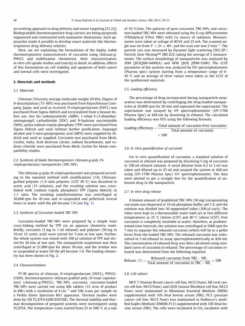
MTT assay was done for six different concentrations viz; 0.1–1 mg/ml. Triton X-100 was taken as the positive control for cyto-toxicity. Here, in our study, there is no significant cytotoxicity inany of the concentrations of the bare TRC-NPs studied (Fig. 10);similarly, we also compared the level of toxicity of nanoparticleswith the negative control, which are the cells grown in its respec-tive media. It is evident from Fig. 10A that compared to the nega-tive control, almost 98% cells are viable in all the six concentrationsof TRC-NPs. These results indicated that the prepared nanoparticlecarriers are non-toxic to L929, KB, PC3, and MCF7 cells. The sameexperiments we analyzed with curcumin-loaded TRC-NPs in thesame concentration range (0.1–1 mg/ml) (Fig. 10C) after incuba-tion at 38 �C. Moreover, bare curcumin at same concentrationsdid not show any kind of toxicity (Fig. 10B), probably due to thehydrophobic nature make it difficult to get dissolved in the media,thereby cells could not take the curcumin inside it. From ourexperimental observations, it is evident that the curcumin-loadedTRC-NPs did not show any toxicity onto L929 cells, while specifictoxicity has been observed onto PC3, KB, and MCF7 cells .Ourresults agreed the previous studies by Kunwar [29]. To confirmthe drug release that is only due to the LCST of the TRC-NPs, weconducted the same experiments at 35 �C (below LCST), showingno toxicity at any concentrations as in the Supplementary figureS3. For the better clarification of the same, we have taken the opti-cal micrographs of the treated and untreated curcumin-loadedTRC-NPs with L929 and PC3 cells, showing no morphologicalchange at below LCST, and it has been observed that the morphol-ogy changed once it treated above LCST (Supplementary figure S2),

Fig. 8. Fluorescent images showing cellular uptake of curcumin-loaded TRC-NPs on L929 and PC3 (A) displays the fluorescence of cells treated with bare curcumin (B)curcumin-loaded TRC-NPs and (C) bright field images. (D) Control cells (L929 and PC3) without curcumin and cells incubated with empty TRC-NPs did not show anyfluorescence.
Fig. 9. Showing flow cytometric histograms for curcumin-loaded TRC-NPs accumulation on L929 (red) and PC3 (green) cells at different concentrations. (A) Control cells, (B)low, (C) medium and (D) high concentrations. (For interpretation of the references to color in this figure legend, the reader is referred to the web version of this article.)
N. Sanoj Rejinold et al. / Journal of Colloid and Interface Science 360 (2011) 39–51 47
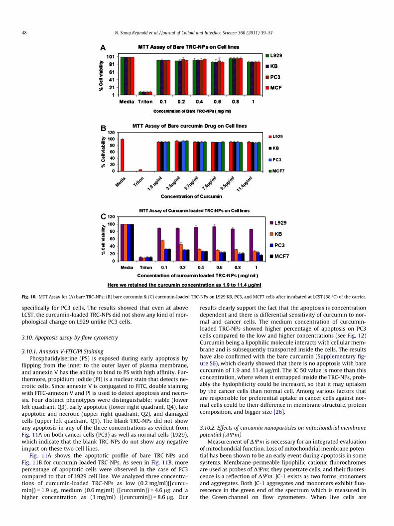
Fig. 10. MTT Assay for (A) bare TRC-NPs; (B) bare curcumin & (C) curcumin-loaded TRC-NPs on L929 KB, PC3, and MCF7 cells after incubated at LCST (38 �C) of the carrier.
48 N. Sanoj Rejinold et al. / Journal of Colloid and Interface Science 360 (2011) 39–51
specifically for PC3 cells. The results showed that even at aboveLCST, the curcumin-loaded TRC-NPs did not show any kind of mor-phological change on L929 unlike PC3 cells.
3.10. Apoptosis assay by flow cytometry
3.10.1. Annexin V-FITC/PI StainingPhosphatidylserine (PS) is exposed during early apoptosis by
flipping from the inner to the outer layer of plasma membrane,and annexin V has the ability to bind to PS with high affinity. Fur-thermore, propidium iodide (PI) is a nuclear stain that detects ne-crotic cells. Since annexin V is conjugated to FITC, double stainingwith FITC-annexin V and PI is used to detect apoptosis and necro-sis. Four distinct phenotypes were distinguishable: viable (lowerleft quadrant, Q3), early apoptotic (lower right quadrant, Q4), lateapoptotic and necrotic (upper right quadrant, Q2), and damagedcells (upper left quadrant, Q1). The blank TRC-NPs did not showany apoptosis in any of the three concentrations as evident fromFig. 11A on both cancer cells (PC3) as well as normal cells (L929),which indicate that the blank TRC-NPs do not show any negativeimpact on these two cell lines.
Fig. 11A shows the apoptotic profile of bare TRC-NPs andFig. 11B for curcumin-loaded TRC-NPs. As seen in Fig. 11B, morepercentage of apoptotic cells were observed in the case of PC3compared to that of L929 cell line. We analyzed three concentra-tions of curcumin-loaded TRC-NPs as low (0.2 mg/ml){[curcu-min]} = 1.9 lg, medium (0.6 mg/ml) {[curcumin]} = 4.6 lg and ahigher concentration as (1 mg/ml) {[curcumin]} = 8.6 lg. Our
results clearly support the fact that the apoptosis is concentrationdependent and there is differential sensitivity of curcumin to nor-mal and cancer cells. The medium concentration of curcumin-loaded TRC-NPs showed higher percentage of apoptosis on PC3cells compared to the low and higher concentrations (see Fig. 12)Curcumin being a lipophilic molecule interacts with cellular mem-brane and is subsequently transported inside the cells. The resultshave also confirmed with the bare curcumin (Supplementary fig-ure S6), which clearly showed that there is no apoptosis with barecurcumin of 1.9 and 11.4 lg/ml. The IC 50 value is more than thisconcentration, where when it entrapped inside the TRC-NPs, prob-ably the hydophilicity could be increased, so that it may uptakenby the cancer cells than normal cell. Among various factors thatare responsible for preferential uptake in cancer cells against nor-mal cells could be their difference in membrane structure, proteincomposition, and bigger size [26].
3.10.2. Effects of curcumin nanoparticles on mitochondrial membranepotential (DWm)
Measurement of DWm is necessary for an integrated evaluationof mitochondrial function. Loss of mitochondrial membrane poten-tial has been shown to be an early event during apoptosis in somesystems. Membrane-permeable lipophilic cationic fluorochromesare used as probes of DWm; they penetrate cells, and their fluores-cence is a reflection of DWm. JC-1 exists as two forms, monomersand aggregates. Both JC-1 aggregates and monomers exhibit fluo-rescence in the green end of the spectrum which is measured inthe Green channel on flow cytometers. When live cells are

Fig. 11. (A) FACS-based apoptosis profile for the bare TRC-NPs on L929 and PC3 cells at different concentrations (low, medium, and high concentrations) and (B) FACS-basedapoptosis profile for the curcumin-loaded TRC-NPs on L929 and PC3 cells at three different concentrations (low, medium, and high concentrations).
Fig. 12. Showing apoptosis profile of bare and curcumin-loaded TRC-NPs after 48-hincubation on L929 and PC3 cells, showing effective dosage of medium concentra-tion of curcumin-loaded TRC-NPs on PC3 cells.
N. Sanoj Rejinold et al. / Journal of Colloid and Interface Science 360 (2011) 39–51 49
incubated with JC-1, JC-1 penetrates the plasma membrane of cellsas monomers. Uptake of JC-1 into mitochondria is driven by theDWm. The DWm of normal, healthy mitochondria is polarized,and JC-1 is rapidly taken up by such mitochondria. This uptake in-creases the concentration gradient of JC-1, leading to the formationof JC-1 aggregates (known as J-aggregates) within the mitochon-
dria. JC-1 aggregates show a red spectral shift resulting in higherlevels of red fluorescence emission which is measured in the Redchannel of the flow cytometer. Here, we analyzed the state of theDWm after treatment with curcumin-loaded nanoparticles at dif-ferent concentrations by measuring the relative differences in thered fluorescence of the cationic dye JC-1 between bare TRC-NPsand curcumin-loaded TRC-NPs treated PC3 and L929 cells.Fig. 13A shows the DWm profile of both the cell lines treated withbare TRC-NPs, and Fig. 13B shows curcumin-loaded TRC-NPs for24 h. Our data indicate that exposure to curcumin-loaded TRC-NPs results in a dose-dependent decline in the DWm in cancer cellscompared to the normal cells, which indicates that the apoptosisinduced by the curcumin nanoparticle is cell sensitive and mito-chondrial-mediated. We have compared the data with the barecurcumin which did not show any type of decline on both PC3and L929 cells (see Supplementary figure S7).
3.11. Blood compatibility studies of the bare and curcumin-loadedTRC-NPs
The blood compatibility was a significative index for biomateri-als because the materials might be exposed in blood environmentand damaged the erythrocytes in certain degree or caused the
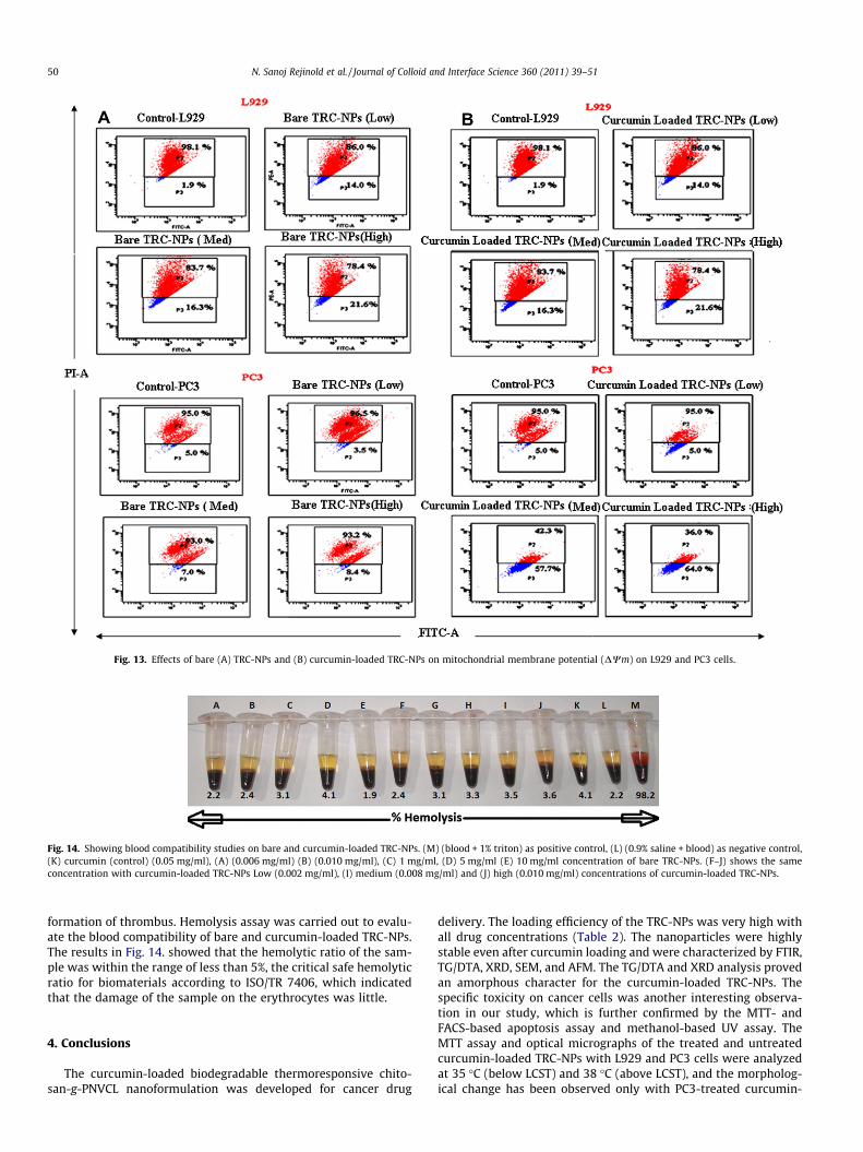
Fig. 13. Effects of bare (A) TRC-NPs and (B) curcumin-loaded TRC-NPs on mitochondrial membrane potential (DWm) on L929 and PC3 cells.
Fig. 14. Showing blood compatibility studies on bare and curcumin-loaded TRC-NPs. (M) (blood + 1% triton) as positive control, (L) (0.9% saline + blood) as negative control,(K) curcumin (control) (0.05 mg/ml), (A) (0.006 mg/ml) (B) (0.010 mg/ml), (C) 1 mg/ml, (D) 5 mg/ml (E) 10 mg/ml concentration of bare TRC-NPs. (F–J) shows the sameconcentration with curcumin-loaded TRC-NPs Low (0.002 mg/ml), (I) medium (0.008 mg/ml) and (J) high (0.010 mg/ml) concentrations of curcumin-loaded TRC-NPs.
50 N. Sanoj Rejinold et al. / Journal of Colloid and Interface Science 360 (2011) 39–51
formation of thrombus. Hemolysis assay was carried out to evalu-ate the blood compatibility of bare and curcumin-loaded TRC-NPs.The results in Fig. 14. showed that the hemolytic ratio of the sam-ple was within the range of less than 5%, the critical safe hemolyticratio for biomaterials according to ISO/TR 7406, which indicatedthat the damage of the sample on the erythrocytes was little.
4. Conclusions
The curcumin-loaded biodegradable thermoresponsive chito-san-g-PNVCL nanoformulation was developed for cancer drug
delivery. The loading efficiency of the TRC-NPs was very high withall drug concentrations (Table 2). The nanoparticles were highlystable even after curcumin loading and were characterized by FTIR,TG/DTA, XRD, SEM, and AFM. The TG/DTA and XRD analysis provedan amorphous character for the curcumin-loaded TRC-NPs. Thespecific toxicity on cancer cells was another interesting observa-tion in our study, which is further confirmed by the MTT- andFACS-based apoptosis assay and methanol-based UV assay. TheMTT assay and optical micrographs of the treated and untreatedcurcumin-loaded TRC-NPs with L929 and PC3 cells were analyzedat 35 �C (below LCST) and 38 �C (above LCST), and the morpholog-ical change has been observed only with PC3-treated curcumin-

N. Sanoj Rejinold et al. / Journal of Colloid and Interface Science 360 (2011) 39–51 51
loaded TRC-NPs, at 38 �C (see Supplementary figure S2), whichshows that the release mechanism is only through the LCST ofthe carrier. The hemolysis assay showed that bare and curcumin-loaded TRC-NPs are non-hemolytic at the concentration range of0.002–10 mg/ml. Apoptosis assays showed that the cell death isconcentration dependent and there is differential sensitivity tothe normal and cancer cells. JC-1 mitochondrial membrane poten-tial analysis confirmed that the apoptosis is mitochondrial-medi-ated. These results showed that curcumin-loaded TRC-NPs couldbe more beneficial for cancer treatment by a proper combinationtherapy with hyperthermia.
Acknowledgments
The Department of Biotechnology (DBT), Government of India,supported this work, under a grant of the Nanoscience and Nano-technology Initiative program (Ref. No. BT/PR10850/NNT/28/127/2008). This work was also partially supported by Department ofscience and technology (DST) under a center grant of the Nanosci-ence and Nanotechnology Initiative program monitored by Dr.C.N.R. Rao. One of the authors, N. Sanoj Rejinold would like toacknowledge Council of Scientific and Industrial Research (CSIR),Govt of India, for their financial support through a Senior ResearchFellowship (SRF) (Award no: 9/963(0017)2K11-EMR-1).
Appendix A. Supplementary material
Supplementary data associated with this article can be found inthe online version at doi:10.1016/j.jcis.2011.04.006.
References
[1] D.E. Meyer, B.C. Shin, G. A Kong, M.W. Dewhirst, A.J. Chilkoti, ControlledRelease 74 (2001) 13.
[2] L. Jun, W. Bochu, L. Peng, Med. Hypotheses 71 (2008) 249.
[3] J.P. Kashappa, Hyun. Drug. Dev. Res. 64 (2005) 114.[4] Y. Maeda, R. Jayakumar, H. Nagahama, T. Furuike, H. Tamura, Int. J. Biol.
Macromol. 42 (2008) 463.[5] H. Nagahama, T. Kashiki, N. Nwe, R. Jayakumar, T. Furuike, H. Tamura,
Carbohydr. Polym. 73 (2008) 456–463.[6] M. Peter, P.T. Sudheesh Kumar, N.S. Binulal, S.V. Nair, H. Tamura, R. Jayakumar,
Carbohydr. Polym. 78 (2009) 926.[7] P.T. Sudheesh Kumar, S. Abhilash, K. Manzoor, S.V. Nair, H. Tamura, R.
Jayakumar, Carbohydr. Polym. 80 (2010) 761.[8] E.M. Manjusha, C.M. Jithin, K. Manzoor, S.V. Nair, H. Tamura, R. Jayakumar,
Carbohydr. Polym. 80 (2010) 442.[9] N. Sanoj Rejinold, M. Muthunarayanan, K.P. Chennazhi, S.V. Nair, R. Jayakumar,
Int. J. Biol. Macromol. 48 (2011) 98–105.[10] N. Sanoj Rejinold, K.P. Chennazhi, H. Tamura, S.V. Nair, R. Jayakumar, Int. J. Biol.
Macromol. 83 (2011) 776–786.[11] N. Sanoj Rejinold, M. Muthunarayanan, K.P. Chennazhi, H. Tamura, S.V. Nair, R.
Jayakumar, Int. J. Biol. Macromol. 84 (2011) 407–416.[12] B. Gerrit, Adv. Drug Delivery Rev. 2 (2001) 45–150.[13] R.A.A. Muzzarelli, Carbohydr. Polym. 8 (1988) 1–21.[14] M. Prabaharan, J.G. Jamison, S.A. Douglas, G. Shaoqin, Macromol. Biosci. 8
(2008) 843–851.[15] A. Hatefi, B.J. Amsden, Controlled Release 80 (2002) 9.[16] K. Amith, B. Atanu, P. Ruchi, K.I. Priyadarshini, Biochim. Biophys. Acta 1760
(2006) 1513.[17] K. Anil, D. Samrita, P. Atish, Behav. Brain Res. 205 (2009) 384.[18] A. Bharat, K. Anushree, C.A. Bharti, Anticancer Res. 23 (2003) 363.[19] T.K. Biji, R.H. Scofield, Trends Pharmacol. Sci. 307 (2009) 334.[20] D.C. Drummond, C.O. Noble, Z. Guo, M.E. Hayes, C.C. Ingram, B.S. Gabriel, B.
Hann, B. Liu, J.W. Park, K. Hong, C.C. Benz, J.D. Marks, D.B. Kirpotin, J.Controlled Release 141 (2010) 13.
[21] R. Jayakumar, D. Menon, K. Manzoor, S.V. Nair, H. Tamura, Carbohydr. Polym.82 (2010) 227.
[22] M.P. Kyung, W.B. Jin, K.J. Yoon, W.S. Jung, D. Ki, Colloids Surf., B 6 (2008) 31.[23] B.S. Laura, Z. Yanjie, A. Vladislav, X.C. Litosh, C. Younhee, S.C. Paul, J. Am. Chem.
Soc. 131 (2009) 26.[24] N. Sanoj Rejinold, M. Muthunarayanan, N. Deepa, K.P. Chennazhi, S.V. Nair, R.
Jayakumar, Int. J. Biol. Macromol. 47 (2010) 37–43.[25] R.B. Devika, B.P. Varsha, AAPS Pharm. Sci. Technol. 7 (2006) E1–E6.[26] W. Abdelwahed, G. Degobert, S. Stainmesse, H. Fessi, Adv. Drug Del. Rev. 58
(2006) 1688.[27] V.T. Furqanmaulvi, T.G. Thakkar Soni, M.C. Gohe, T.R. Gandhi, J. Pharm. Sci. Res.
4 (2009) 1–14.[28] N.A. Peppas, Pharm. Acta Helv. 60 (1985) 110.[29] A. Kunwar, A. Barik, B. Mishra, K. Rathinasamy, R. Pandy, K.I. Priyadarshini,
Biochim. Biophys. Acta 1780 (2008) 673.
Copyright © 2022 FDOKUMEN