Curcumin derivatives: Molecular basis of their anti-cancer activity
11
Curcumin derivatives: Molecular basis of their anti-cancer activity Valentina Basile a , Erika Ferrari b , Sandra Lazzari b , Silvia Belluti a , Francesca Pignedoli b , Carol Imbriano a, * a Dipartimento di Biologia Animale, Universita ` di Modena e Reggio Emilia, via Campi 213/D, 41100 Modena, Italy b Dipartimento di Chimica, Universita ` di Modena e Reggio Emilia, via Campi 183, 41100 Modena, Italy 1. Introduction The rapid dynamics of spindle microtubules are essential to proper spindle function. Microtubules are favorite target of naturally occurring toxic molecules, most of which are produced by a large number of plants (reviewed in [1]). In fact, many microtubule-targeting agents (MITs) have been discovered by screening natural products and are currently in clinical trials. Curcumin, a common flavoring agent in the spice turmeric, perturbs microtubule assembly dynamics through tubulin binding, which results in its conformational changes [2]. Significant depolymerization of interphase microtubules and mitotic spindle microtubules have been observed after Curcumin incubation of HeLa, MCF-7, HT-29 and HCT-15 cells, thus leading to a mitotic arrest [2–4]. Curcumin exhibits cancer growth inhibition both in vitro and in vivo [5,6]: it suppresses cell proliferation in a variety of cancer cell lines and it inhibits tumorigenesis [7–17]. Multiple mechanisms of action are likely responsible for Curcumin various effects on cancer cells: G1/S arrest and apoptosis induction have been observed other than the mitotic block in different tumor cell lines [18–20]. Experimental evidence reveals a multitude of molecular targets, including transcription factors, cell cycle proteins, enzymes, cell surface adhesion protein and cytokines [21,22]. Although Curcumin has an evident anti-cancer activity, relative poor stability has been highlighted as one of the major problems in therapeutic applications. Stability in cell culture medium contain- ing 10% FBS or in human blood is greater than in phosphate buffer, but 50% of the molecule has still decomposed after 8 h [23,24]. To enhance metabolic stability and antiproliferative activity against human cancer cells, various Curcumin analogs have been synthesized, among which bis-DemethoxyCurcumin (bDMC) and diAcetylCurcumin (DAC)(Fig. 1) (reviewed in [25]). Although bDMC differs from its lead-compound in the chemical structure only with regard to methoxy substitution, it exhibits significantly different antioxidant, antitumor, and anti-inflammatory activities. bDMC shows higher cytotoxicity against human ovarian cancer cell line, increased antimutagenic, anticarcinogenic and antioxidant activity (reviewed in [25]). The inhibition of cancer cell invasion by bDMC highlights its enhanced antimetastasis potency [26]. The synthetic derivative DAC shows higher antibacterial activity against multiresistant bacteria [27] and a stronger nitric oxide (NO) and O 2 anion scavenging activity [28,29], although it has Biochemical Pharmacology 78 (2009) 1305–1315 ARTICLE INFO Article history: Received 28 May 2009 Accepted 26 June 2009 Keywords: Natural compounds Curcumin analogs Cell cycle regulation p53 DNA-damaging agents ABSTRACT Curcumin, a phenolic compound from the plant Curcuma longa L., has shown a wide-spectrum of chemopreventive, antioxidant and antitumor properties. Although its promising chemotherapeutic activity, preclinical and clinical studies highlight Curcumin limited therapeutic application due to its instability in physiological conditions. To improve its stability and activity, many derivatives have been synthesized and studied, among which bis-DemethoxyCurcumin (bDMC) and diAcetylCurcumin (DAC). In this report, we show that both bDMC and DAC are more stable than Curcumin in physiological medium. To explore the mechanism of their chemotherapeutic effect, we studied their role in proliferation in the HCT116 human colon cancer cells. We correlated kinetic stability and cellular uptake data to their biological effects. Both bDMC and DAC impair correct spindles formation and induce a p53- and p21 CIP1/WAF1 -independent mitotic arrest, which is more stable and long-lasting for bDMC. A subsequent p53/p21 CIP1/WAF1 -dependent inhibition of G1 to S transition is triggered by Curcumin and DAC as a consequence of the mitotic slippage, preventing post-mitotic cells from re-entering the cell cycle. Conversely, the G1/S arrest induced by bDMC is a direct effect of the drug and concomitant to the mitotic block. Finally, we demonstrate that bDMC induces rapid DNA double-strand breaks, moving for its possible development in anti-cancer clinical applications. ß 2009 Elsevier Inc. All rights reserved. Abbreviations: bDMC, bis-DemethoxyCurcumin; DAC, diAcetylCurcumin; MIT, microtubule-targeting agents; BrdU, bromodeoxyuridine; PI, propidium iodide; DMSO, dimethylsulfoxide; DSB, double-strand break; FACS, Fluorescence Activated Cell Sorter; ChIP, Chromatin immunoprecipitation; ATM, Ataxia Telangiectasia Mutated; ATR, ATM and Rad3-related. * Corresponding author. Tel.: +39 059 2055542; fax: +39 059 2055548. E-mail addresses: [email protected], [email protected] (C. Imbriano). Contents lists available at ScienceDirect Biochemical Pharmacology journal homepage: www.elsevier.com/locate/biochempharm 0006-2952/$ – see front matter ß 2009 Elsevier Inc. All rights reserved. doi:10.1016/j.bcp.2009.06.105
-
Upload
independent -
Category
Documents
-
view
4 -
download
0
Transcript of Curcumin derivatives: Molecular basis of their anti-cancer activity
Biochemical Pharmacology 78 (2009) 1305–1315
Curcumin derivatives: Molecular basis of their anti-cancer activity
Valentina Basile a, Erika Ferrari b, Sandra Lazzari b, Silvia Belluti a, Francesca Pignedoli b, Carol Imbriano a,*a Dipartimento di Biologia Animale, Universita di Modena e Reggio Emilia, via Campi 213/D, 41100 Modena, Italyb Dipartimento di Chimica, Universita di Modena e Reggio Emilia, via Campi 183, 41100 Modena, Italy
A R T I C L E I N F O
Article history:
Received 28 May 2009
Accepted 26 June 2009
Keywords:
Natural compounds
Curcumin analogs
Cell cycle regulation
p53
DNA-damaging agents
A B S T R A C T
Curcumin, a phenolic compound from the plant Curcuma longa L., has shown a wide-spectrum of
chemopreventive, antioxidant and antitumor properties. Although its promising chemotherapeutic
activity, preclinical and clinical studies highlight Curcumin limited therapeutic application due to its
instability in physiological conditions. To improve its stability and activity, many derivatives have been
synthesized and studied, among which bis-DemethoxyCurcumin (bDMC) and diAcetylCurcumin (DAC).
In this report, we show that both bDMC and DAC are more stable than Curcumin in physiological
medium. To explore the mechanism of their chemotherapeutic effect, we studied their role in
proliferation in the HCT116 human colon cancer cells. We correlated kinetic stability and cellular uptake
data to their biological effects. Both bDMC and DAC impair correct spindles formation and induce a p53-
and p21CIP1/WAF1-independent mitotic arrest, which is more stable and long-lasting for bDMC. A
subsequent p53/p21CIP1/WAF1-dependent inhibition of G1 to S transition is triggered by Curcumin and
DAC as a consequence of the mitotic slippage, preventing post-mitotic cells from re-entering the cell
cycle. Conversely, the G1/S arrest induced by bDMC is a direct effect of the drug and concomitant to the
mitotic block. Finally, we demonstrate that bDMC induces rapid DNA double-strand breaks, moving for
its possible development in anti-cancer clinical applications.
� 2009 Elsevier Inc. All rights reserved.
Contents lists available at ScienceDirect
Biochemical Pharmacology
journal homepage: www.e lsev ier .com/ locate /b iochempharm
1. Introduction
The rapid dynamics of spindle microtubules are essential toproper spindle function. Microtubules are favorite target ofnaturally occurring toxic molecules, most of which are producedby a large number of plants (reviewed in [1]). In fact, manymicrotubule-targeting agents (MITs) have been discovered byscreening natural products and are currently in clinical trials.Curcumin, a common flavoring agent in the spice turmeric,perturbs microtubule assembly dynamics through tubulin binding,which results in its conformational changes [2]. Significantdepolymerization of interphase microtubules and mitotic spindlemicrotubules have been observed after Curcumin incubation ofHeLa, MCF-7, HT-29 and HCT-15 cells, thus leading to a mitoticarrest [2–4].
Curcumin exhibits cancer growth inhibition both in vitro and in
vivo [5,6]: it suppresses cell proliferation in a variety of cancer cell
Abbreviations: bDMC, bis-DemethoxyCurcumin; DAC, diAcetylCurcumin; MIT,
microtubule-targeting agents; BrdU, bromodeoxyuridine; PI, propidium iodide;
DMSO, dimethylsulfoxide; DSB, double-strand break; FACS, Fluorescence Activated
Cell Sorter; ChIP, Chromatin immunoprecipitation; ATM, Ataxia Telangiectasia
Mutated; ATR, ATM and Rad3-related.
* Corresponding author. Tel.: +39 059 2055542; fax: +39 059 2055548.
E-mail addresses: [email protected], [email protected]
(C. Imbriano).
0006-2952/$ – see front matter � 2009 Elsevier Inc. All rights reserved.
doi:10.1016/j.bcp.2009.06.105
lines and it inhibits tumorigenesis [7–17]. Multiple mechanisms ofaction are likely responsible for Curcumin various effects on cancercells: G1/S arrest and apoptosis induction have been observedother than the mitotic block in different tumor cell lines [18–20].Experimental evidence reveals a multitude of molecular targets,including transcription factors, cell cycle proteins, enzymes, cellsurface adhesion protein and cytokines [21,22].
Although Curcumin has an evident anti-cancer activity, relativepoor stability has been highlighted as one of the major problems intherapeutic applications. Stability in cell culture medium contain-ing 10% FBS or in human blood is greater than in phosphate buffer,but 50% of the molecule has still decomposed after 8 h [23,24].
To enhance metabolic stability and antiproliferative activityagainst human cancer cells, various Curcumin analogs have beensynthesized, among which bis-DemethoxyCurcumin (bDMC) anddiAcetylCurcumin (DAC)(Fig. 1) (reviewed in [25]). AlthoughbDMC differs from its lead-compound in the chemical structureonly with regard to methoxy substitution, it exhibits significantlydifferent antioxidant, antitumor, and anti-inflammatory activities.bDMC shows higher cytotoxicity against human ovarian cancer cellline, increased antimutagenic, anticarcinogenic and antioxidantactivity (reviewed in [25]). The inhibition of cancer cell invasion bybDMC highlights its enhanced antimetastasis potency [26]. Thesynthetic derivative DAC shows higher antibacterial activityagainst multiresistant bacteria [27] and a stronger nitric oxide(NO) and O2
�� anion scavenging activity [28,29], although it has

Fig. 1. Structures of Curcumin, bDMC and DAC.
V. Basile et al. / Biochemical Pharmacology 78 (2009) 1305–13151306
slightly lower apoptotic activity on AK-5 rat histiocytoma cells[30]. Whether bDMC and DAC analogues have the same Curcuminmolecular targets is also not clear at present.
The aim of our study is to shed light on the potential anti-cancereffects of bDMC and DAC against human colon cancer cells,unraveling the molecular basis of their biological activity. Under-standing molecular events that lead these molecules to arrest cellproliferation is an important prerequisite for their development aschemotherapeutic agents. We show here that both bDMC and DAChave increased stability in physiological medium and improvednuclear cellular uptake compared to Curcumin. Cellular andmolecular analyses of the effects of bDMC and DAC in HCT116cells have been performed to define their activity in cell cyclemodulation. We demonstrate that bDMC causes a concomitant G1/S and mitotic block, triggering rapid DNA double-strand breaks.Finally, we investigate the role of the two main inhibitors of the cellcycle, p53 and p21CIP1/WAF1, in mediating the cell cycle impairmentinduced by bDMC and DAC.
2. Materials and methods
2.1. General procedures
Spectrophotometric measurements were performed usingJasco V-570 spectrophotometer at 25 � 0.1 8C in the 200–600 nmspectral range employing 1 mm quartz cells. NMR Spectra wererecorded on a Bruker Avance AMX-400 spectrometer with a BroadBand 5 mm probe (inverse detection). Nominal frequencies are100.13 MHz for 13C and 400.13 MHz for 1H. The typical acquisitionparameters for 1H were as follows: 20 ppm spectral bandwidth (SW),6.1 ms pulse width (908 pulse hard pulse on 1H), 0.5–1 s pulse delay,216–512 number of scans. For 2D H,H-Homonuclear CorrelatedSpetroscopy (COSY) typical parameters were used. For 2D H,X-HeteroCorrelated Spetroscopy, HMBC and HMQC opportune parameterswere used (50–908 pulses; 32 k data points; 1 s relaxation delay; 8–64 k transients; 1JH–C 125–145 Hz; 3JH–C 5–15 Hz). Methanol-d4
(CD3OD) and DMSO-d6 (DMSO) were used as NMR solvent. Allreported chemical shifts are referred to TMS.
2.2. Preparation of Curcuminoids
A suspension of B2O3 (1 mmol) and acetylacetone (1 mmol) inDMF (1.5 ml) was stirred for 30 min at 80 8C, then tributylborate(4 mmol) was added. After 30 min, the appropriate benzaldehyde(1.8 mmol) was added and followed by slow addition of n-butylamine (0.4 mmol in 0.5 ml of DMF). After stirring at 80 8C for4 h, the solution was acidified with 0.5 M HCl (8 ml) and cooleddown to room temperature. The yellow–orange solid was re-suspended in water, filtered and dried under vacuum. Purity of all
synthesized compounds was >98%, and it was determined bymeans of NMR techniques and combustion analysis.
2.2.1. Curcumin-1,7-bis[3-methoxy-4-hydroxyphenyl]hepta-1,6-
diene-3,5-dione1H NMR (CD3OD): d 5.95 (s, 1H; H-1), 6.61 (d, J = 15.8 Hz, 2H; H-
3), 7.56 (d, J = 15.8 Hz, 2H; H-4), 7.20 (d, J = 2 Hz, 2H; H-6), 6.82 (dd,J = 8.0, 1 Hz, 2H; H-9), 7.09 (dd, J = 8.0, 2 Hz, 2H; H-10). 13C NMR(CD3OD): d 100.2 (C-1), 183.7 (C-2), 120.6 (C-3), 140.4 (C-4), 127.3(C-5), 110.1 (C-6), 147.8 (C-7), 149.0 (C-8), 115.0 (C-9), 122.6 (C-10). Anal. Calc. (Found) for C21H20O6: C 68.47 (68.41), H 5.47 (5.52);yield 60%. m.p. 180 8C.
2.2.2. DiacetylCurcumin (DAC)-1,7-bis[3-methoxy-4-
acetylphenyl]hepta-1,6-diene-3,5-dione1H NMR (CD3OD): d 5.94 (s, 1H; H-1), 6.59 (d, J = 15.9 Hz, 2H; H-
3), 7.57 (d, J = 15.9 Hz, 2H; H-4), 7.49 (d, J = 2 Hz, 2H; H-6), 6.82 (dd,J = 7.5, 1 Hz, 2H; H-9), 7.49 (dd, J = 7.5, 2 Hz, 2H; H-10). 13C NMR(CD3OD): d 100.3 (C-1), 183.6 (C-2), 120.4 (C-3), 140.2 (C-4), 126.5(C-5), 129.3 (C-6), 115.3 (C-7), 160.1 (C-8), 115.3 (C-9), 129.3 (C-10). Anal. Calc. (Found) for C25H24O8: C 66.36 (65.91), H 5.35 (5.73);yield 87%. m.p. 171 8C.
2.2.3. Bis-DemethoxyCurcumin (bDMC)-1,7-bis[4-
hydroxyphenyl]hepta-1,6-diene-3,5-dione1H NMR (DMSO-d6): d 6.13 (S, 1H; H-1), H-3 6.79, H-4 7.63, H-6
7.66, H-7 6.92, H-9 6.92, H-10 7.66; C-1 100.8, C-2 183.4, C-3 120.8,C-4 140.3, C-5 125.8, C-6 130.2, C-7 115.9, C-8 159.8, C-9 115.9, C-10 130.2. Anal. Calc. (Found) for C19H16O4: C 74.01 (73.95), H 5.23(5.31), yield 75%. m.p. 217 8C.
2.3. Cell lines and drugs
HCT116, derived from a human colorectal carcinoma, HCT116/E6 and HCT116 p21 �/� were generously provided by BertVogelstein (Johns Hopkins University School of Medicine, Balti-more, MD). The HCT116 and HCT116 p21 �/� cell lines werecultured in IMDM medium supplemented with 10% FCS. HCT116/E6 were grown with G418 (500 ng/ml). Drugs were added to cellmedium at the concentration described in the text for 4, 8, 16 or24 h. IC50 concentrations after 24 h treatments were estimated in10 and 30 mM for Curcumin/DAC and bDMC, respectively. Cellswere treated with Nocodazole (0,5 mM) for 16 h and withAdriamycin (1.3 mM) for 24 h.
2.4. Uptake measurements
Culture medium (3 ml) was acidified with 6N HCl (1:1, v/v) andvortexed for 30 s. Following addition of 3 ml of extracting buffer(ethyl acetate and isopropanol 9:1 (v/v)), samples were vortexedand shaken in the Orbital shaker (at 100 rpm) for 15 min. Aftercentrifugation at 18,000 rpm for 20 min, the upper organic layerwas filtered through a membrane filter (0.22 mm) and directed toUV–vis analysis.
The cell pellets were resuspended in RIPA buffer (20 mM Tris/HCl at pH 8.0, 137 mM NaCl, 10% of glycerol, 5 mM EDTA, 1 mMphenylmethyl-sulfonyl fluoride and protease inhibitor cocktail)and the cell-liquid extraction was carried out. The cell extractswere cleared by centrifugation at 18,000 rpm for 15 min andacidified by 6N HCl (1:1, v/v) and vortexed for 30 s. Then the sameextracting procedure as for culture medium was performed.
2.5. Flow cytometric cell cycle analysis
500,000 cells were harvested after drug treatments and DNAdistribution analysis of PI and BrdU stained cells was performed by

V. Basile et al. / Biochemical Pharmacology 78 (2009) 1305–1315 1307
an Epics cytofluorimeter (Beckman Coulter) [31,32]. Anti-BrdUantibody was provided by BioLegend (#317902) and mouse anti-FITC by Dako (#F0313).
2.6. Cellular extracts and immunoblotting
Total extracts were prepared by resuspending the cell pellet(300,000 cells) in 200 ml of lysis buffer (50 mM Tris–HCl pH 8.0,120 mM NaCl, 0.5% NP-40, 1 mM EDTA, protease and phospha-tase inhibitors). Immunoblot analysis of histones was performedusing HCT116 acid extracts. Briefly, the cell pellet wassuspended in lysis buffer (10 mM Hepes pH 7.9, 1.5 mM MgCl2,10 mM KCl), added of hydrochloric acid 0.2N, incubated on icefor 30 min and cleared by high-speed centrifugation for 10 minat 4 8C.
For immunoblotting equivalent amounts of total extracts(50 mg) were resolved by SDS-PAGE, electrotransferred to nitro-cellulose membrane, and immunoblotted with the followingprimary antibodies diluted 1:1000 in TBS 1�/BSA 1 mg/ml: anti-CycB1 (Active Motif #39504), anti-p53 DO-1 (Santa Cruz #sc-126),anti-phospo-H2AX (Cell-Signaling #2577), anti-H2A acid-patch(Active Motif), anti-phosphoATM Ser1981 (Cell-Signaling #4526),anti-p21 (Upstate) and anti-actin (Santa-Cruz).
2.7. Immunofluorescence
300,000 cells were seeded on coverslips the day beforetreatments. After drugs incubation, cells were washed twice inPBS 1� at room temperature, fixed in cold methanol/acetone for2 min and permeabilized with 0.05% Triton-X 100 in PBS 1� for5 min. Samples were preincubated with PBS 1�/BSA 1% for 15 minand then incubated overnight at 4 8C with the following primaryantibodies diluted 1:100 in PBS 1�/BSA 1%: anti-phospho-Histone H2AX (Ser139) (Upstate #05-636; Active Motif#39117) and anti-tubulin (Sigma–Aldrich). Cells were thenwashed twice with PBS 1� and incubated 15 min at roomtemperature in PBS 1�/BSA 1% before a further incubation for60 min at room temperature in the dark, with Hoechst and therelative secondary antibodies sheep anti-rabbit FITC-conjugate1:200 (Sigma, F7512) and goat anti-mouse TRITC-conjugate 1:150(Sigma, T5393). After three washes with PBS 1�monolayers weremounted with Vectashield and examined with Zeiss AxioSkop 40fluorescence microscope (Carl Zeiss, Jena, Germany), imagescollected with a AxioCam HRc camera and AxioVision version 3.1software package. The same samples were analyzed by confocalmicroscopy (Leica DM IRE2).
2.8. RT-PCR analysis
RNA was extracted from cells untreated or treated with thedescribed drugs by using RNeasy kit (Qiagen, Hilden, Germany),according to the manufacturer’s protocol. For cDNA synthesis, 5 mgof RNA were retrotranscribed with a Moloney murine leukemiavirus reverse transcriptase (RT) (Promega). Semiquantitative PCRswere performed with oligonucleotides previously described[31,32].
2.9. Chromatin immunoprecipitation assay
Chromatin immunoprecipitations were essentially performedas described previously [31,32]. Chromatin was prepared upon24 h drugs treatment and immunoprecipitated with anti-p53 (DO-1, Santa-Cruz) and anti-Flag (Sigma–Aldrich) antibodies. PCRswere performed with the following oligonucleotides:
p21_for: GTAAATCCTTGCCTGCCAGAGTG; p21_rev: GCTGCC-CAGCGCCGAGCCAG.
2.10. Statistical analysis
Results are shown as means � SD. Statistical analysis for theresults of western blot of Fig. 4B was done using one-way ANOVA,followed by Bonferroni’s t-test to determine whether there weredifferences between specific groups (DMSO versus each Curcumi-noid).
3. Results
3.1. Uptake of Curcumin and its derivatives in HCT116 cells
Although Curcumin has been extensively investigated as itconcerns its anti-cancer activity in a variety of cell types, only fewstudies provide uptake data [33,34] and none correlates kineticstability of natural and synthetic Curcuminoids to their cellularuptake. A preliminary kinetic study was performed for bDMC andDAC (Fig. 1), in order to evaluate their stability in culture mediumcompared to Curcumin. A representative fluorescence spectrum ofCurcumin maintained in culture medium for 4 and 24 h is given inFig. 2A (left panel). Absorbance of Curcumin dramaticallydecreased in the first 4 h (�50%), and then smoothly diminishedup to 24 h (�10%), suggesting a pseudo-first order kinetic process(Fig. 2A, right panel). Differently, bDMC and DAC maintainedalmost the same absorbance values in the first 24 h, hinting theirgreater stability with respect to Curcumin.
Cellular uptake and intracellular localization of the drugs wereevaluated by confocal microscopy analysis of HCT116 cells treatedwith the estimated IC50 concentrations for 24 h. All the moleculesshowed mainly a cytoplasmic fluorescence, although nuclearstaining appeared to be more intense in cells treated with bDMCand DAC than Curcumin (Fig. 2B, arrows).
The kinetic of cellular uptake was determined by UV–visanalysis. Fig. 2C shows the histogram of the percentages ofCurcumin, bDMC and DAC (20 and 30 mM) extracted from HCT116cells and their relative culture media as a function of timeincubation; all data have been normalized taking into account theabsorbance decreases observed after 4 and 24 h due to the relativestability of each drug (Fig. 2A). The percentages determined bothinside the cell and in culture medium were almost independent ofdrugs concentration. During 4 h-treatment, the drug percentage inculture medium was around an averaged value of 70% for allCurcuminoids, even if data (30 mM) suggested the following orderof uptake: bDMC > DAC > Curcumin. Although the sum of cell andculture medium percentages should represent 100%, after 4 h-treatment only Curcumin followed this prediction. For both bDMCand DAC the sum of the two percentages was 75% and 80%,respectively, hinting that the missing quantity should havefollowed different metabolic and/or degradation processes insidethe cell. The strong decrease in culture medium percentageobserved for Curcumin and bDMC after 24 h treatment suggestedtheir incremented uptake. No changes in drug distribution wereobserved comparing 4 and 24 h incubations with DAC, indicatingthat longer time exposition did not increase its cell absorption.
Fig. 2D reports the absolute percentages referred to time zeroconcentration inside the cell for each drug (20 and 30 mM) at 4 and24 h. A dose-dependent uptake was observed for Curcumin andbDMC (same percentage independently on drug treatmentconcentration), while for DAC an increased value was detectedas concentration was risen up. After 24 h the amount of Curcuminand bDMC inside the cell was <2% and <0.5%, respectively, whileDAC showed higher percentages (>3% for 20 mM and >7% for30 mM).
These data indicate that, although DAC and bDMC have similarstability in culture medium, they follow different metabolisminside the cell: DAC exhibited a limited decomposition up to 24 h

Fig. 2. UV–vis spectroscopic study on stability and uptake of Curcuminoids in HCT116 cells. (A) Left panel. UV–vis absorption spectra of Curcumin extracted from control
medium at time 0 h (a), 4 h (b) and 24 h (c) and (right panel) linear relationships between the absorbance values (420 nm) and concentration at time 0 h (*), 4 h (*) and 24 h
(!). (B) Confocal microscopy analysis of Curcumin, bDMC and DAC cellular uptake. (C) Percentages of Curcumin, bDMC and DAC extracted from culture medium and HCT116
cells at 20 and 30 mM concentrations; all data are the mean of three independent experiments and are normalized to the control. (D) Curcuminoids extracted from HCT116
cells at different time courses and concentrations; percentages are referred to time zero extracts for each drug.
V. Basile et al. / Biochemical Pharmacology 78 (2009) 1305–13151308
V. Basile et al. / Biochemical Pharmacology 78 (2009) 1305–1315 1309
while bDMC was fastly uptaken and metabolized already duringthe first 4 h of treatment.
3.2. Treatment of HCT116 cells with Curcumin derivatives delays
mitotic exit
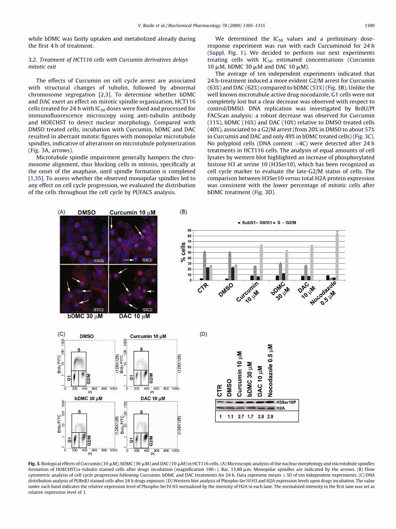
The effects of Curcumin on cell cycle arrest are associatedwith structural changes of tubulin, followed by abnormalchromosome segregation [2,3]. To determine whether bDMCand DAC exert an effect on mitotic spindle organization, HCT116cells treated for 24 h with IC50 doses were fixed and processed forimmunofluorescence microscopy using anti-tubulin antibodyand HOECHST to detect nuclear morphology. Compared withDMSO treated cells, incubation with Curcumin, bDMC and DACresulted in aberrant mitotic figures with monopolar microtubulespindles, indicative of alterations on microtubule polymerization(Fig. 3A, arrows).
Microtubule spindle impairment generally hampers the chro-mosome alignment, thus blocking cells in mitosis, specifically atthe onset of the anaphase, until spindle formation is completed[1,35]. To assess whether the observed monopolar spindles led toany effect on cell cycle progression, we evaluated the distributionof the cells throughout the cell cycle by PI/FACS analysis.
Fig. 3. Biological effects of Curcumin (10 mM), bDMC (30 mM) and DAC (10 mM) in HCT11
formation of HOECHST/a-tubulin stained cells after drugs incubation (magnification
cytometric analysis of cell cycle progression following Curcumin, bDMC and DAC treatm
distribution analysis of PI/BrdU stained cells after 24 h drugs exposure. (D) Western blot ana
under each band indicates the relative expression level of Phospho-Ser10 H3 normalized by
relative expression level of 1.
We determined the IC50 values and a preliminary dose-response experiment was run with each Curcuminoid for 24 h(Suppl. Fig. 1). We decided to perform our next experimentstreating cells with IC50 estimated concentrations (Curcumin10 mM, bDMC 30 mM and DAC 10 mM).
The average of ten independent experiments indicated that24 h-treatment induced a more evident G2/M arrest for Curcumin(63%) and DAC (62%) compared to bDMC (51%) (Fig. 3B). Unlike thewell known microtubule active drug nocodazole, G1 cells were notcompletely lost but a clear decrease was observed with respect tocontrol/DMSO. DNA replication was investigated by BrdU/PIFACScan analysis: a robust decrease was observed for Curcumin(11%), bDMC (16%) and DAC (10%) relative to DMSO treated cells(40%), associated to a G2/M arrest (from 20% in DMSO to about 57%in Curcumin and DAC and only 49% in bDMC treated cells) (Fig. 3C).No polyploid cells (DNA content >4C) were detected after 24 htreatments in HCT116 cells. The analysis of equal amounts of celllysates by western blot highlighted an increase of phosphorylatedhistone H3 at serine 10 (H3Ser10), which has been recognized ascell cycle marker to evaluate the late-G2/M status of cells. Thecomparison between H3Ser10 versus total H2A protein expressionwas consistent with the lower percentage of mitotic cells afterbDMC treatment (Fig. 3D).
6 cells. (A) Microscopic analysis of the nuclear morphology and microtubule spindles
100�). Bar, 15.80 mm. Monopolar spindles are indicated by the arrows. (B) Flow
ents for 24 h. Data represent means � SD of ten independent experiments. (C) DNA
lysis of Phospho-Ser10 H3 and H2A expression levels upon drugs incubation. The value
the intensity of H2A in each lane. The normalized intensity in the first lane was set as

Fig. 4. bDMC induces a concomitant G2/M and G1/S arrest. (A) Distribution of BrdU positive and negative cells in G1 phase after drugs treatment. Results are mean � SD of
three independent experiments. (B) Western blot and RT-PCR analysis of Cyclin B1 expression after drugs treatment. The intensity of immunoreactive and RT-PCR bands have been
quantitated to actin and GAPDH, respectively. The normalized intensity in the first lane was set as relative expression level of 1. Values, mean of four independent experiments,
*P < 0.05, **P < 0.01 versus DMSO. (C) Distribution of cells in G0/G1 (left panel) and G2/M (right panel) phases after the release (4–8–10–12 h) from 24 h drugs exposure. (D) DNA
distribution analysis of PI/BrdU stained cells after G0/G1 synchronization and release in drugs-added media.
V. Basile et al. / Biochemical Pharmacology 78 (2009) 1305–13151310
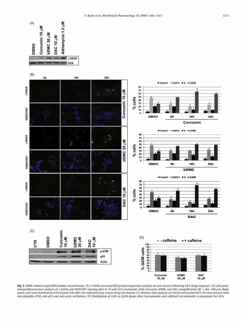
Fig. 5. bDMC induces rapid DNA double-strand breaks. (A) g-H2AX and total H2A protein expression analysis of acid extracts following 24 h drugs exposure. (B) Left panel:
immunofluorescence analysis of g-H2AX and HOECHST staining after 8, 16 and 24 h treatments with Curcumin, bDMC and DAC (magnification 40�). Bar, 100 mm. Right
panel: cell cycle distribution of PI stained cells after the indicated time-course drugs incubation. (C) Western blot analysis of control and treated HCT116 total extracts with
anti-phospho-ATM, anti-p53 and anti-actin antibodies. (D) Distribution of cells in G2/M phase after Curcuminoids and caffeine/Curcuminoids co-treatment for 24 h.
V. Basile et al. / Biochemical Pharmacology 78 (2009) 1305–1315 1311
V. Basile et al. / Biochemical Pharmacology 78 (2009) 1305–13151312
3.3. Bis-DemethoxyCurcumin triggers a concurrent G1/S and mitotic
arrest
Prolonged mitotic arrest can lead cells (i) to apoptosis or (ii) toescape mitosis via mitotic slippage through the ubiquitination andproteolysis of Cyclin B [36]. Mitotic slippage leads to theaccumulation of post-mitotic G1 cells, which are subsequentlyarrested in their cell cycle progression by activating a p53-dependent G1 checkpoint [37–39].
To determine whether G1 cells resulted from escaped progres-sion through mitosis and cytokinesis rather than a direct G1 arrest,we pulsed HCT116 with BrdU for 1 h before drug treatments(Fig. 4A). After Curcumin and DAC incubation about 45% of the totalG1 cells was found to be BrdU labeled, hinting that about one-halfof G1 cells were originated by cells that failed to arrest in G2/M.Conversely, by treating cells with bDMC, G1 cells were almostfound to be BrdU unlabeled (77%), suggesting that the thispopulation has originated from cells that were already in G1 beforethe incubation with BrdU. These data pointed out a more stablemitotic block after bDMC with respect to Curcumin and DAC (onlythe 23% of BrdU labeled cells escaped from the G2/M blockcompared to 55–60% after Curcumin and DAC incubation). Tofurther confirm that Curcumin and DAC treated cells exit mitosisvia mitotic slippage, we evaluated Cyclin B1 expression levels.Western blot analysis demonstrated that Cyclin B1 levelsdecreased upon Curcumin, bDMC and DAC incubation comparedto DMSO (Fig. 4B, upper panel). Differently from bDMC treatedcells, in which protein reduction was associated to its transcrip-tional inhibition, the decreased Cyclin B1 staining after Curcuminand DAC exposure could be ascribed to its proteolysis, as RT-PCRanalysis did not show changes in gene transcription (Fig. 4B, lowerpanel).
We next determined whether bDMC could induce a long-lastingblock in cell cycle progression by drug-release experiments. Therelease from the mitotic block induced by bDMC was slowed downup to 12 h and was associated to a modest increase in G0/G1 eventscompared to Curcumin and DAC (Fig. 4C).
To characterize the G1/S arrest induced by bDMC, we performedsynchronization-release experiments and we analyzed S phaseentry. The experimental design is outlined schematically in Fig. 4D.Serum depletion for 24 h (0.5% FCS) resulted in G0/G1 arrest ofHCT116 (between 80% and 85%); S phase fractions were determinedby quantifying BrdU incorporation after release into 10% FCS freshmedia added with each drug. Upon 10 up to 14 h from the release inDMSO, Curcumin and DAC, about 18–30% of cells clearly shifted toearly S phase. A delay from S to G2/M was then observed forCurcumin and DAC compared to control cells: DMSO-release shiftedabout 35% of cells to G2/M after 18 h, while the same percentage wasreached after 22 h with Curcumin (31%) and DAC (30%), explainingthe lack of the G2/M block usually observed after 24 h-treatment.With regards to bDMC, the kinetic of the release from G0/G1 to earlyS was comparable to Curcumin and DAC; conversely, BrdU positivecells were clearly inhibited from early to late S. After 22 h, about 60%of cells were still retained in synthesis. The release in nocodazole-medium demonstrated that cells took 14 h to accumulate insynthesis (36%), similarly to DMSO (35%), and after 22 h werecompletely shifted in G2/M (96.9%).
3.4. Bis-DemethoxyCurcumin induces DNA damage concomitantly to
the cell cycle block
Many human cells exposed to MITs acquire DNA damagecausing a significant delay in mitotic exit upon removal of theblock [40,41].
Compared to Curcumin and DAC, bDMC showed a slowerkinetic in re-entering the cell cycle after the release, hinting the
possibility that it might induce DNA damage during the cell cycleblock.
To explore this possibility, we examined by western blot theactivation of g-H2AX, which is known to be phosphorylatedrapidly at Ser 139 in response to DNA DSBs. All the Curcuminoidsinduced g-H2AX after 24 h-treatment (Fig. 5A). The normalizationwith respect to the total amount of H2A indicated that thestrongest H2AX phosphorylation was triggered by bDMC, similarlyto the effects of Adriamycin, a potent antitumor drug that inducesDNA DSBs.
To investigate the possible interaction between the cell cycleimpairment and DNA damage we performed a time courseexperiment. PI cell cycle analysis and g-H2AX immunofluores-cence were carried out on 8, 16 and 24 h-treated cells. 16 hincubation accumulated cells in G2/M as efficiently as 24 h (Fig. 5B,right panel). Interestingly, only bDMC induced g-H2AX alreadyupon 16 h treatment (Fig. 5B, left panel). These data suggest thatbDMC-induced DNA damage is concurrent to the cell cycle arrest,while DSBs observed after Curcumin and DAC incubation can bethe effect of a prolonged mitotic block.
DSBs are most closely associated to H2AX phosphorylation byATM activation, which acts as a regulator of cell cycle checkpointand DNA repair [42]. The ATM/ATR-dependent pathway triggerscell cycle arrest until DNA integrity is restored by repair processes[43,44]. ATM activation after Curcuminoids treatment wasexplored by the analysis of Ser1981 phosphorylation. Phospho-ATM was detected after Curcumin, bDMC and DAC incubation, butnot after DMSO addition (Fig. 5C). Co-incubation of cells with anon-toxic dose of caffeine (1 mM), which inhibits ATM/ATR, didnot result in the release of cells from the G2/M arrest, suggestingthat ATM activation did not participate in maintaining the mitoticblock induced by Curcuminoids (Fig. 5D).
3.5. Inactivation of p53 and p21 abrogates the post-mitotic G1 arrest
As cellular insults (e.g. DNA damage and microtubule disrup-tion) induce signaling pathways which mediate p53 activation, weinvestigated p53 expression levels after Curcuminoids treatment.Western blot shown in Fig. 5C clearly detected an increase of p53,particularly after bDMC exposure.
Which is the role of p53 in cell cycle arrest induced by Curcuminand its derivatives has not been yet elucidated. We thereforeanalyzed cell cycle progression in HCT116/E6 cells, in which p53 isinhibited by the overexpression of the viral protein E6. Cell cyclephase distributions showed that p53 did not participate to the G2/M block induced by Curcumin, bDMC and DAC (Fig. 6A). Thecomparison between HCT116 (Fig. 3B) and HCT116/E6 (Fig. 6A)clearly highlighted that p53 inhibition even induces a more robustG2/M arrest. Compared to HCT116, G0/G1 cells were reduced ofabout 50%, while S cells increased of about 50–60% upon Curcuminand DAC treatments. No effect on DNA replication (S phase) waseven observed after bDMC incubation compared to DMSO.Nocodazole treatment of HCT116/E6 did not show any differencein cell cycle phase distribution compared to HCT116. These datasuggest that one of the main determinant in the subsequent fateafter prolonged mitotic arrest is whether the cells can functionallyactivate p53 and its pathway. Cells entering mitosis in the presenceof microtubule drugs can escape mitosis and subsequently arrest inthe next G1 by p53 activation. Differently, cells lacking p53 surviveand reproduce, as suggested by the increase of S phase. The role ofp53 in controlling G1 to S passage is more evident for bDMC, as theG1/S block is a direct effect of the drug.
The CDK-inhibitor p21CIP1/WAF1 is induced after DNA damageand plays a role in the G1 arrest, but not in preventing G2progression [37]. p21 was overexpressed after Curcuminoidstreatment compared to control cells (Fig. 6B, upper panel).

Fig. 6. Role of p53 and p21CIP1/WAF1 in the antiproliferative effects of Curcumin and its derivatives. (A) Flow cytometric analysis of HCT116/E6 cell cycle progression following
Curcumin, bDMC, DAC and nocodazole treatments. Data represent means � SD of three independent experiments. (B) p21 expression analysis of protein and mRNA levels of
HCT116 and HCT116/E6 by western blot (left panel) and RT-PCR (right panel). The actin protein and GAPDH mRNA were used as internal controls. (C) Cell cycle distribution of PI
stained HCT116 p21 �/� cells after 24 h drugs exposure. (D) ChIP analysis of p53 binding to p21CIP1/WAF1 promoter after DMSO, Curcumin, bDMC and DAC exposure.
V. Basile et al. / Biochemical Pharmacology 78 (2009) 1305–1315 1313
V. Basile et al. / Biochemical Pharmacology 78 (2009) 1305–13151314
Nocodazole treatment, which accumulated a higher percentage ofmitotic cells, did not show higher p21 levels, suggesting that itsactivation is not triggered by the inability to exit mitosis. p21transcriptional activation has been shown to be both p53-dependent and -independent. To elucidate the role of p53 inmediating p21 overexpression we performed RT-PCR and westernblot analysis in HCT116 cells compared to HCT116/E6 cells. Fig. 6B(lower panel) shows that p21 was activated also in the absence ofp53, and a higher expression was still detected upon bDMCtreatment. There is evidence suggesting that Curcumin antipro-liferative effect is mediated by p21-induced cell cycle arrest, even ifit has been recently demonstrated that in HCT116 cells Curcumininduces apoptosis in a p21-independent manner [45]. To under-stand whether p21 was necessary to inhibit cell cycle progressionafter Curcuminoids treatments, we performed PI cell cycle analysisof HCT116 p21�/� treated cells (Fig. 6C). With respect to HCT116/E6 cells, a further decrease of G0/G1 and an increase of G2/Mevents were detected, while no differences in S phase wereobserved following Curcumin and DAC incubation. bDMC showedeven an increase of replicative events with respect to DMSO (28%versus 19%).
To elucidate whether transcriptional activation of p21 wasmediated by p53, we performed a ChIP analysis with chromatinfrom HCT116 treated cells immunoprecipitated with anti-p53 andanti-Flag as unrelated antibody. Fig. 6D shows that after Curcuminand DAC treatment p53 was recruited to the p21 promoter. On theother hand, only a weak binding of p53 was observed upon bDMCincubation, thus suggesting that bDMC induces a p53-independentactivation of the p21 gene.
Thus, we concluded that both p53 and p21 are not involved inmediating the G2 arrest induced by Curcuminoids, but they arelikely required to maintain G1 arrested cells, which originate as aconsequence of mitotic slippage for Curcumin and DAC, rather thana direct G1/S block for bDMC.
4. Discussion
Although phase I clinical trials demonstrated that Curcumin iswell-tolerated orally and no dose-limiting toxicity was observed[5,46], it has poor bioavailability and may undergo intestinalmetabolism, which can be to a great extent the consequence of itsdeficient pharmaceutical profile [21,47,48]. Currently, Curcuminderivatives represent a well-established strategy to enhancemetabolic stability and therefore antiproliferative activity againsthuman cancer cells. In this report, we have investigated theanalogs bDMC and DAC through biochemical and molecularstudies with the aim to identify the connections between structureand mode of action.
It has been recently demonstrated that the deletion of b-diketone moiety enhances in vitro the stability of Curcumin in pH7.4 condition [49]. We show that bDMC and DAC, althoughpreserving the b-diketone, are more stable than Curcumin inculture medium, hinting that the degradation mechanisms ofCurcuminoids are probably connected to the presence of a strongintra-molecular hydrogen bond between the vicinal hydroxy andmethoxy groups. DAC exhibits a limited decomposition both inculture medium and inside the cell. Differently, increased stabilityof bDMC in culture medium is coupled with lower cellularconcentration, suggesting that it is easily metabolized orconjugated to other molecules into the cells, leading to a quickbiological response (Fig. 1).
Even if both DAC and bDMC show the same effect onmicrotubule spindle organization (Fig. 3A), the analysis of cellcycle progression in HCT116 cells corroborates the hypothesisthat the two analogs may have different properties and activities.While DAC treatment induces a delayed mitotic exit comparable
to Curcumin, bDMC exposure results in a concomitant mitoticand G1/S block. Additionally, the BrdU pulse/chase experiment(Fig. 4A) indicates that, although less robust, the mitotic arrestinduced by bDMC is more stable with respect to Curcumin andDAC. A higher percentage of BrdU labeled G1 cells can bedetected upon Curcumin and DAC prolonged treatmentscompared to bDMC, hinting that cells exiting mitosis subse-quently accumulate in G1 and are prevented from S phase entry.bDMC long-lasting block is further supported by drug-releaseexperiments.
We demonstrate that p53 is not required for the mitotic arrestinduced by microtubule poisons, but the experiments performedon HCT116/E6 and HCT116 p21 �/� cells clearly indicate that p53and p21CIP1/WAF1 are necessary for both Curcumin and DAC post-mitotic G1 arrest and for the direct G1/S block activated by bDMC(Fig. 6). We believe that a concurrent p53-dependent and -independent p21 transactivation occurs following Curcuminoidstreatment. As in HCT116 p21 �/� compared to HCT116/E6 cells,only bDMC shows an increase of the replicative events, it ispossible that a p53-independent activation of p21CIP1/WAF1 isrequired to maintain active the primary G1/S checkpoint.Conversely, the G1 post-mitotic arrest triggered via mitoticslippage by Curcumin, DAC and, to a lesser extent, by bDMC,induces the p21CIP1/WAF1 gene through the transcription factor p53(Graphical abstract). This hypothesis is supported by ChIP analysisshown in Fig. 6D: Curcumin and DAC exposure induces the bindingof p53 on the p21CIP1/WAF1 promoter, whereas upon bDMCtreatment only a faint recruitment is observed.
Western blot and immunofluorescence assays detect theactivation of a DNA damage response elicited upon mitoticimpairment for Curcumin and DAC (Fig. 5). It is tempting tospeculate that DNA damage is activated as an effect of a severemitotic block induced by Curcumin and DAC, but it is not clear ifDNA damage occurs in mitotic or post-mitotic G1 arrested cells.Compared with the DNA damage responses during G1 and G2,considerably less is known about the DNA damage activationduring mitosis. As bDMC is concerned, DNA damage is observedconcurrently to the cell cycle impairment, thus pointing thismolecule as a both spindle-disrupting and DNA-damaging drug.There is strong evidence highlighting that DNA damage occurredin mitotic cells inhibits mitotic exit after the block is removedand that this delay is not an ATM-dependent mechanism[50,51]. This is coherent with our data showing that bDMCinduces a long-lasting G2/M block, which is not reverted bycaffeine pretreatment. We cannot rule out that DNA damage istriggered in G1 arrested cells (Graphical abstract). This hypoth-esis need further investigation to elucidate the events andregulatory mechanisms of bDMC effects on cell cycle progres-sion, which ensure its possible development in anti-cancerclinical applications.
Acknowledgments
This work was supported by AIRC-MFAG (My First AIRC Grant).Special thanks to Prof. R. Mantovani (University of Milan) and Prof.Monica Saladini (University of Modena and Reggio Emilia) forscientific suggestions and helpful discussion. We are thankful to‘‘Centro Interdipartimentale Grandi Strumenti (CIGS)’’ of theUniversity of Modena and Reggio Emilia and to the ‘‘FondazioneCassa di Risparmi di Modena’’ which supplied NMR Spectrometer.
Appendix A. Supplementary data
Supplementary data associated with this article can be found, inthe online version, at doi:10.1016/j.bcp.2009.06.105.
V. Basile et al. / Biochemical Pharmacology 78 (2009) 1305–1315 1315
References
[1] Jordan MA, Wilson L. Microtubules as a target for anticancer drugs. Nat RevCancer 2004;4:253–65.
[2] Gupta KK, Bharne SS, Rathinasamy K, Naik NR, Panda D. Dietary antioxidantcurcumin inhibits microtubule assembly through tubulin binding. FEBS J2006;273:5320–32.
[3] Holy JM. Curcumin disrupts mitotic spindle structure and induces micronu-cleation in MCF-7 breast cancer cells. Mutat Res 2002;518:71–84.
[4] Hanif R, Qiao L, Shiff SJ, Rigas B. Curcumin, a natural plant phenolic foodadditive, inhibits cell proliferation and induces cell cycle changes in colonadenocarcinoma cell lines by a prostaglandin-independent pathway. J Lab ClinMed 1997;130:576–84.
[5] Cheng AL, Hsu CH, Lin JK, Hsu MM, Ho YF, Shen TS, et al. Phase I clinical trial ofcurcumin, a chemopreventive agent, in patients with high-risk or pre-malig-nant lesions. Anticancer Res 2001;21:2895–900.
[6] Sharma RA, Euden SA, Platton SL, Cooke DN, Shafayat A, Hewitt HR, et al. PhaseI clinical trial of oral curcumin: biomarkers of systemic activity and compli-ance. Clin Cancer Res 2004;10:6847–54.
[7] Sahu RP, Batra S, Srivastava SK. Activation of ATM/Chk1 by curcumin causescell cycle arrest and apoptosis in human pancreatic cancer cells. Br J Cancer2009;100:1425–33.
[8] Aggarwal BB, Kumar A, Bharti AC. Anticancer potential of curcumin: preclinicaland clinical studies. Anticancer Res 2003;23:363–98.
[9] Jee SH, Shen SC, Tseng CR, Chiu HC, Kuo ML. Curcumin induces a p53-dependent apoptosis in human basal cell carcinoma cells. J Invest Dermatol1998;111:656–61.
[10] Kawamori T, Lubet R, Steele VE, Kelloff GJ, Kaskey RB, Rao CV, et al. Chemo-preventive effect of curcumin, a naturally occurring anti-inflammatory agent,during the promotion/progression stages of colon cancer. Cancer Res 1999;59:597–601.
[11] Mehta K, Pantazis P, McQueen T, Aggarwal BB. Antiproliferative effect ofcurcumin (diferuloylmethane) against human breast tumor cell lines. Antic-ancer Drugs 1997;8:470–81.
[12] Singletary K, MacDonald C, Iovinelli M, Fisher C, Wallig M. Effect of the beta-diketones diferuloylmethane (curcumin) and dibenzoylmethane on rat mam-mary DNA adducts and tumors induced by 7,12-dimethylbenz[a]anthracene.Carcinogenesis 1998;19:1039–43.
[13] Hayun R, Okun E, Berrebi A, Shvidel L, Bassous L, Sredni B, et al. Rapamycin andcurcumin induce apoptosis in primary resting B chronic lymphocytic leukemiacells. Leuk Lymphoma 2009;50:625–32.
[14] Purkayastha S, Berliner A, Fernando SS, Ranasinghe B, Ray I, Tariq H, et al.Curcumin blocks brain tumor formation. Brain Res 2009.
[15] Li ZX, Ouyang KQ, Jiang X, Wang D, Hu Y. Curcumin induces apoptosis andinhibits growth of human Burkitt’s lymphoma in xenograft mouse model. MolCells 2009;27:283–9.
[16] Lin YT, Wang LF, Hsu YC. Curcuminoids suppress the growth of pharynx andnasopharyngeal carcinoma cells through induced apoptosis. J Agric Food Chem2009.
[17] Mann CD, Neal CP, Garcea G, Manson MM, Dennison AR, Berry DP. Phyto-chemicals as potential chemopreventive and chemotherapeutic agents inhepatocarcinogenesis. Eur J Cancer Prev 2009;18:13–25.
[18] Srivastava RK, Chen Q, Siddiqui I, Sarva K, Shankar S. Linkage of curcumin-induced cell cycle arrest and apoptosis by cyclin-dependent kinase inhibitorp21(/WAF1/CIP1). Cell Cycle 2007;6:2953–61.
[19] Shishodia S, Amin HM, Lai R, Aggarwal BB. Curcumin (diferuloylmethane)inhibits constitutive NF-kappaB activation, induces G1/S arrest, suppressesproliferation, and induces apoptosis in mantle cell lymphoma. Biochem Phar-macol 2005;70:700–13.
[20] Sa G, Das T. Anti cancer effects of curcumin: cycle of life and death. Cell Div2008;3:14.
[21] Sharma RA, Gescher AJ, Steward WP. Curcumin: the story so far. Eur J Cancer2005;41:1955–68.
[22] Shishodia S, Sethi G, Aggarwal BB. Curcumin: getting back to the roots. Ann N YAcad Sci 2005;1056:206–17.
[23] Wang YJ, Pan MH, Cheng AL, Lin LI, Ho YS, Hsieh CY, et al. Stability of curcuminin buffer solutions and characterization of its degradation products. J PharmBiomed Anal 1997;15:1867–76.
[24] Blasius R, Duvoix A, Morceau F, Schnekenburger M, Delhalle S, Henry E, et al.Curcumin stability and its effect on glutathione S-transferase P1-1 mRNAexpression in K562 cells. Ann N Y Acad Sci 2004;1030:442–8.
[25] Anand P, Thomas SG, Kunnumakkara AB, Sundaram C, Harikumar KB, Sung B,et al. Biological activities of curcumin and its analogues (Congeners) made byman and mother nature. Biochem Pharmacol 2008;76:1590–611.
[26] Yodkeeree S, Chaiwangyen W, Garbisa S, Limtrakul P. Curcumin, demethox-ycurcumin and bisdimethoxycurcumin differentially inhibit cancer cell
invasion through the down-regulation of MMPs and uPA. J Nutr Biochem2009;20:87–95.
[27] Mishra S, Narain U, Mishra R, Misra K. Design, development and synthesis ofmixed bioconjugates of piperic acid–glycine, curcumin–glycine/alanine andcurcumin–glycine–piperic acid and their antibacterial and antifungal proper-ties. Bioorg Med Chem 2005;13:1477–86.
[28] Sumanont Y, Murakami Y, Tohda M, Vajragupta O, Matsumoto K, Watanabe H.Evaluation of the nitric oxide radical scavenging activity of manganese com-plexes of curcumin and its derivative. Biol Pharm Bull 2004;27:170–3.
[29] Vajragupta O, Boonchoong P, Berliner LJ. Manganese complexes of curcuminanalogues: evaluation of hydroxyl radical scavenging ability, superoxidedismutase activity and stability towards hydrolysis. Free Radic Res 2004;38:303–14.
[30] Mishra S, Kapoor N, Mubarak Ali A, Pardhasaradhi BV, Kumari AL, Khar A, et al.Differential apoptotic and redox regulatory activities of curcumin and itsderivatives. Free Radic Biol Med 2005;38:1353–60.
[31] Basile V, Mantovani R, Imbriano C. DNA damage promotes histone deacetylase4 nuclear localization and repression of G2/M promoters, via p53 C-terminallysines. J Biol Chem 2006;281:2347–57.
[32] Benatti P, Basile V, Merico D, Fantoni LI, Tagliafico E, Imbriano C. A balancebetween NF-Y and p53 governs the pro- and anti-apoptotic transcriptionalresponse. Nucleic Acids Res 2008;36:1415–28.
[33] Hsu YC, Weng HC, Lin S, Chien YW. Curcuminoids-cellular uptake by humanprimary colon cancer cells as quantitated by a sensitive HPLC assay and itsrelation with the inhibition of proliferation and apoptosis. J Agric Food Chem2007;55:8213–22.
[34] Kunwar A, Barik A, Mishra B, Rathinasamy K, Pandey R, Priyadarsini KI.Quantitative cellular uptake, localization and cytotoxicity of curcumin innormal and tumor cells. Biochim Biophys Acta 2008;1780:673–9.
[35] Weaver BA, Cleveland DW. Decoding the links between mitosis, cancer, andchemotherapy: The mitotic checkpoint, adaptation, and cell death. Cancer Cell2005;8:7–12.
[36] Brito DA, Rieder CL. Mitotic checkpoint slippage in humans occurs via cyclin Bdestruction in the presence of an active checkpoint. Curr Biol 2006;16:1194–200.
[37] Andreassen PR, Lacroix FB, Lohez OD, Margolis RL. Neither p21WAF1 nor 14-3-3sigma prevents G2 progression to mitotic catastrophe in human coloncarcinoma cells after DNA damage, but p21WAF1 induces stable G1 arrestin resulting tetraploid cells. Cancer Res 2001;61:7660–8.
[38] Vogel C, Kienitz A, Hofmann I, Muller R, Bastians H. Crosstalk of the mitoticspindle assembly checkpoint with p53 to prevent polyploidy. Oncogene2004;23:6845–53.
[39] Meek DW. The role of p53 in the response to mitotic spindle damage. PatholBiol (Paris) 2000;48:246–54.
[40] Dalton WB, Nandan MO, Moore RT, Yang VW. Human cancer cells commonlyacquire DNA damage during mitotic arrest. Cancer Res 2007;67:11487–92.
[41] Rieder CL, Maiato H. Stuck in division or passing through: what happenswhen cells cannot satisfy the spindle assembly checkpoint. Dev Cell 2004;7:637–51.
[42] Shiloh Y. The ATM-mediated DNA-damage response: taking shape. TrendsBiochem Sci 2006;31:402–10.
[43] Zhou BB, Elledge SJ. The DNA damage response: putting checkpoints inperspective. Nature 2000;408:433–9.
[44] Ahn J, Urist M, Prives C. The Chk2 protein kinase. DNA Repair (Amst)2004;3:1039–47.
[45] Watson JL, Hill R, Lee PW, Giacomantonio CA, Hoskin DW. Curcumin inducesapoptosis in HCT-116 human colon cancer cells in a p21-independent manner.Exp Mol Pathol 2008;84:230–3.
[46] Sharma RA, McLelland HR, Hill KA, Ireson CR, Euden SA, Manson MM, et al.Pharmacodynamic and pharmacokinetic study of oral Curcuma extract inpatients with colorectal cancer. Clin Cancer Res 2001;7:1894–900.
[47] Sharma RA, Steward WP, Gescher AJ. Pharmacokinetics and pharmacody-namics of curcumin. Adv Exp Med Biol 2007;595:453–70.
[48] Ireson C, Orr S, Jones DJ, Verschoyle R, Lim CK, Luo JL, et al. Characterization ofmetabolites of the chemopreventive agent curcumin in human and rathepatocytes and in the rat in vivo, and evaluation of their ability to inhibitphorbol ester-induced prostaglandin E2 production. Cancer Res 2001;61:1058–64.
[49] Liang G, Shao L, Wang Y, Zhao C, Chu Y, Xiao J, et al. Exploration and synthesisof curcumin analogues with improved structural stability both in vitro and invivo as cytotoxic agents. Bioorg Med Chem 2009;17:2623–31.
[50] Mikhailov A, Cole RW, Rieder CL. DNA damage during mitosis in human cellsdelays the metaphase/anaphase transition via the spindle-assembly check-point. Curr Biol 2002;12:1797–806.
[51] Smits VA, Klompmaker R, Arnaud L, Rijksen G, Nigg EA, Medema RH. Polo-likekinase-1 is a target of the DNA damage checkpoint. Nat Cell Biol 2000;2:672–6.